Equine cervical ultrasonography - ansagerhestehospital.dk · Equine cervical ultrasonography A...
25
Equine cervical ultrasonography A final thesis Royal Veterinary and Agricultural University of Copenhagen Authors Lise C. Berg, DVM Jon V. Nielsen, DVM, MRCVS Tutor Preben D. Thomsen, Dr. Med. Vet Assistant tutor Martin B. Thøfner, PhD Copenhagen
-
Upload
nguyenxuyen -
Category
Documents
-
view
220 -
download
0
Transcript of Equine cervical ultrasonography - ansagerhestehospital.dk · Equine cervical ultrasonography A...
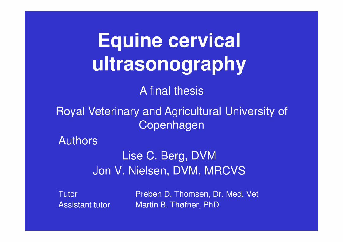
Equine cervical ultrasonography
A final thesis
Royal Veterinary and Agricultural University of
Copenhagen
Authors
Lise C. Berg, DVM
Jon V. Nielsen, DVM, MRCVS
Tutor Preben D. Thomsen, Dr. Med. Vet
Assistant tutor Martin B. Thøfner, PhD
Copenhagen
Equine cervical ultrasonography
Part 1: Ultrasonographic description of the equine cervical anatomy
Part 2: High degree of accuracy of Part 2: High degree of accuracy of ultrasound-guided intraarticular injection of cervical facet joints in horses – A
cadaveric study
Equine cervical ultrasonography
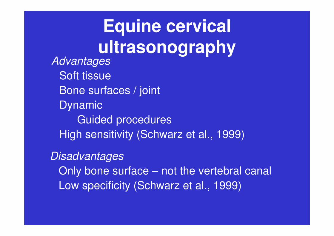
Advantages
Soft tissue
Bone surfaces / joint
Dynamic
Guided proceduresGuided procedures
High sensitivity (Schwarz et al., 1999)
Disadvantages
Only bone surface – not the vertebral canal
Low specificity (Schwarz et al., 1999)
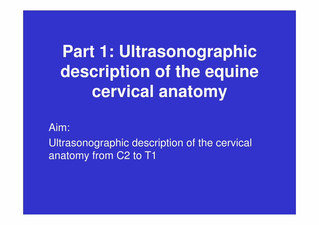
Part 1: Ultrasonographic description of the equine
cervical anatomy
Aim:
Ultrasonographic description of the cervical
anatomy from C2 to T1
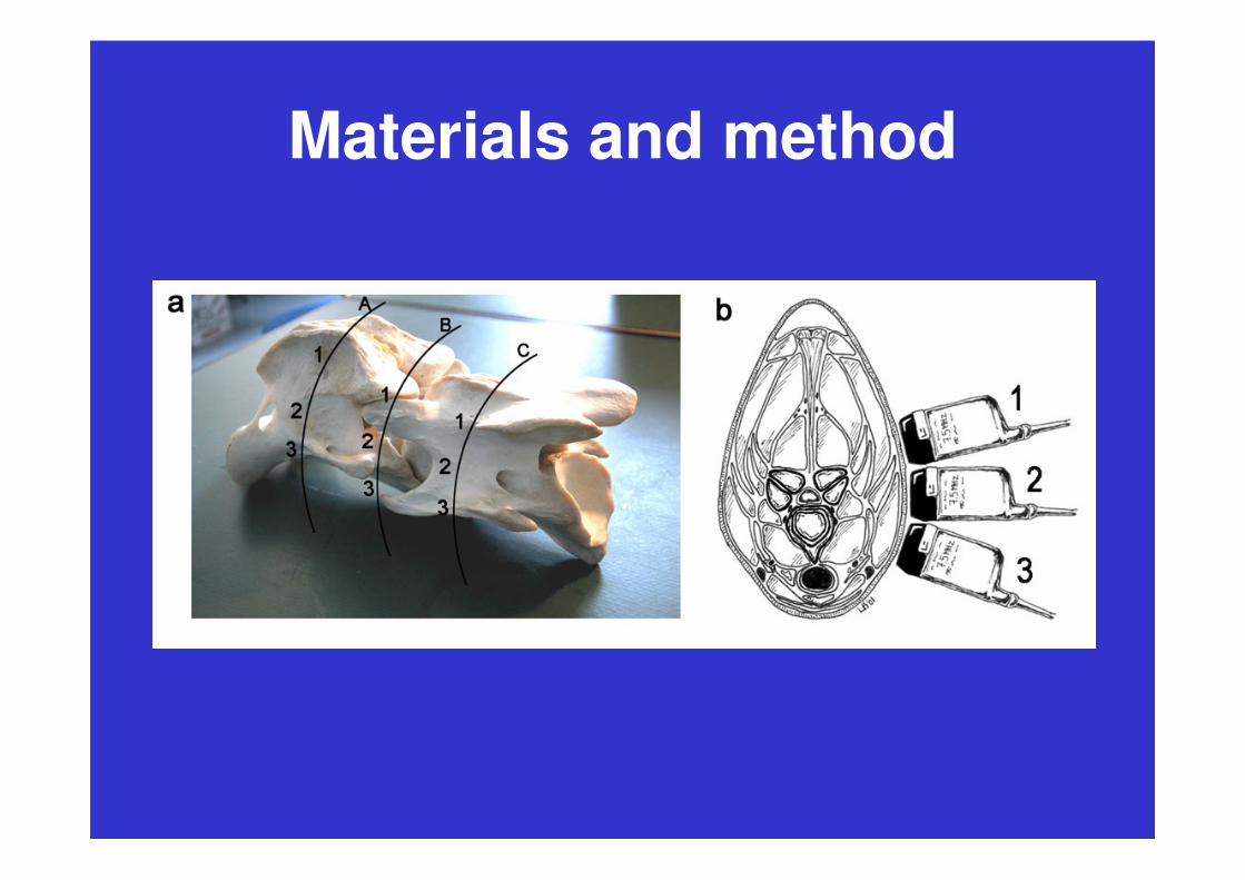
Materials and method
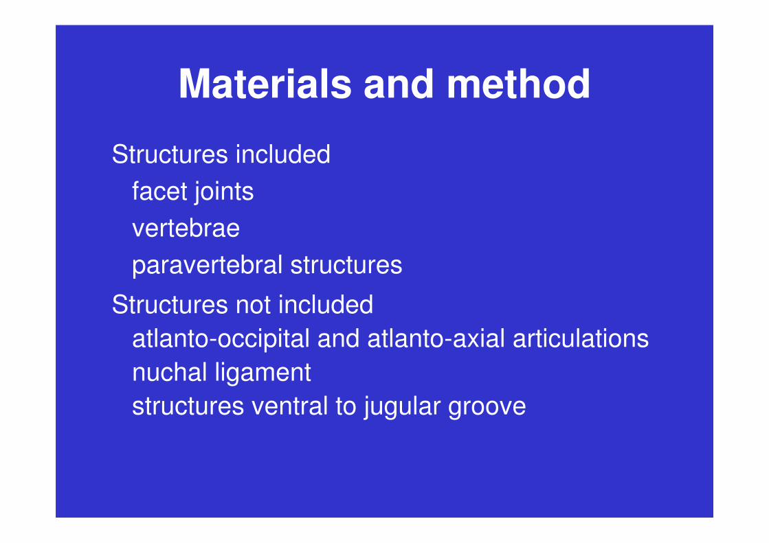
Structures included
facet joints
vertebrae
paravertebral structuresparavertebral structures
Structures not included
atlanto-occipital and atlanto-axial articulations
nuchal ligament
structures ventral to jugular groove

Materials and method
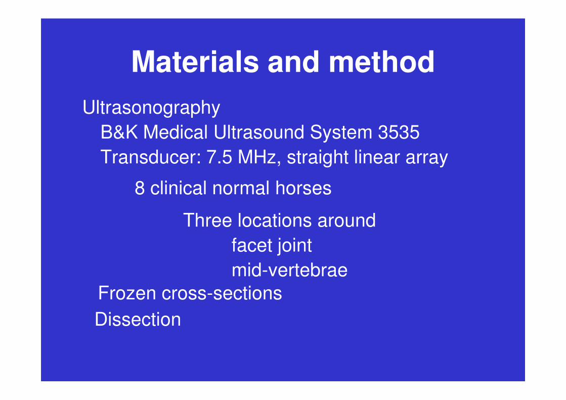
Ultrasonography
B&K Medical Ultrasound System 3535
Transducer: 7.5 MHz, straight linear array
8 clinical normal horses
Three locations around
facet joint
mid-vertebrae
Materials and method
Materials and method
Ultrasonography
B&K Medical Ultrasound System 3535
Transducer: 7.5 MHz, straight linear array
8 clinical normal horses
Frozen cross-sections
Three locations around
facet joint
mid-vertebrae
Dissection

Results
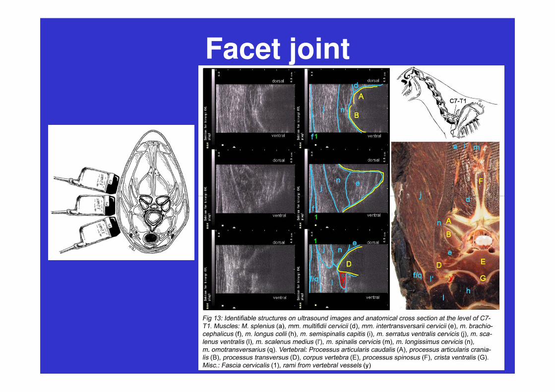
Facet joint

Mid-vertebrae
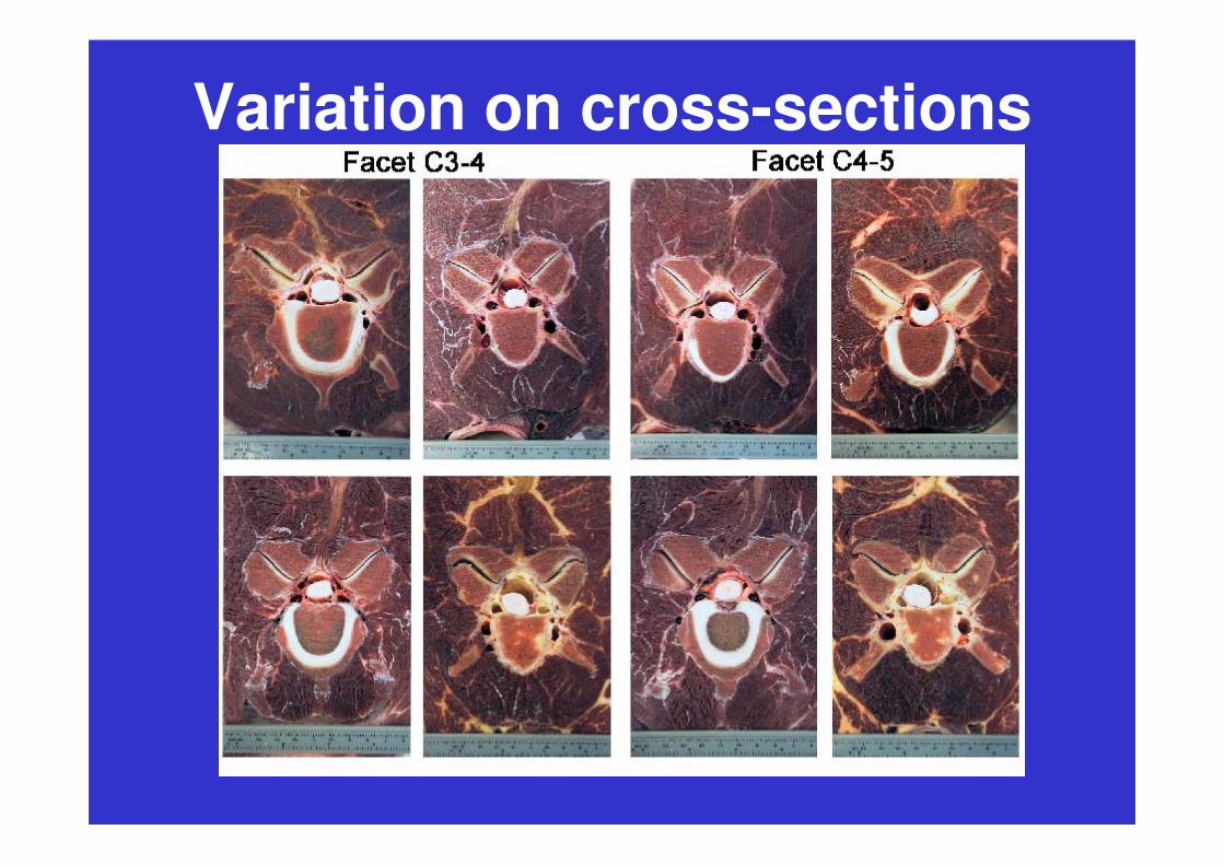
Variation on cross-sections

Ultrasonographic variation
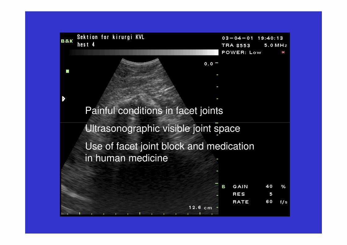
Painful conditions in facet joints
Ultrasonographic visible joint space
Use of facet joint block and medication
in human medicine

Part 2: High degree of accuracy of
ultrasound-guided intraarticular
injection of cervical facet joints in
horses – A cadaveric study
Aim:
Investigate the possibility and accuracy of ultrasound-guided cervical intraarticular facet joint injection
Investigate different factors influence on the result
Materials and method
Insertion of the needle
lateral to the transducer without needle-guide attachment
dorsal to the transducer with needle-guide attachment
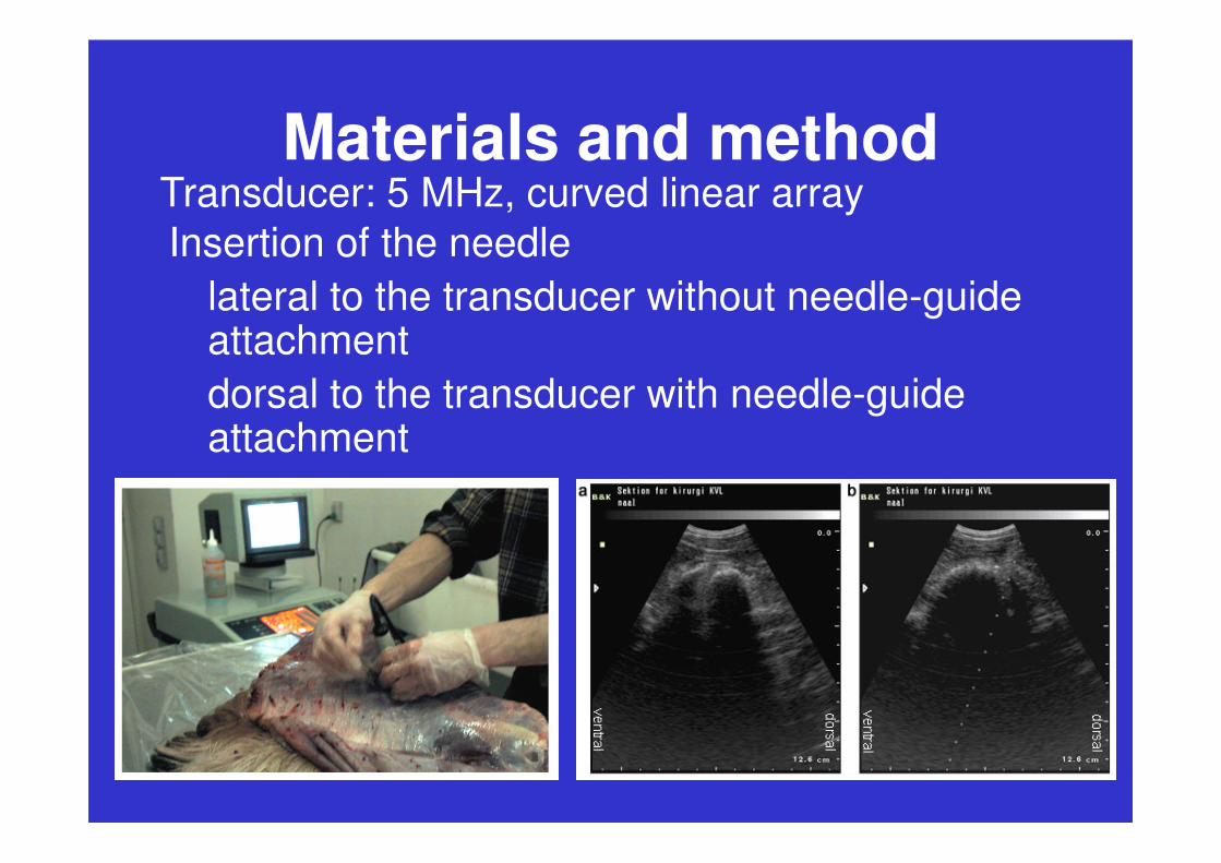
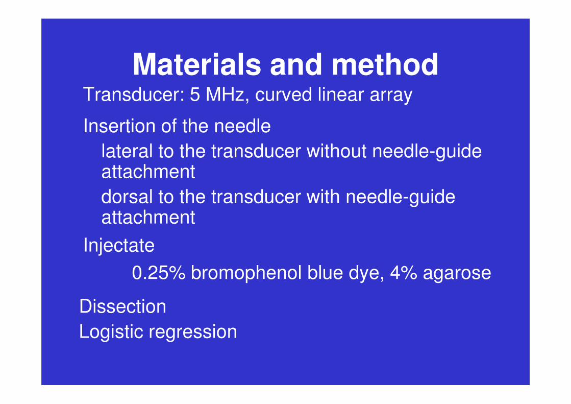
Transducer: 5 MHz, curved linear array
attachment
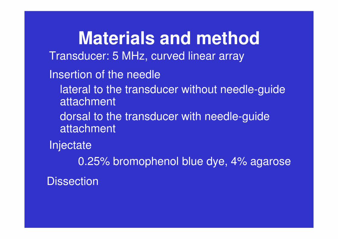
Materials and methodTransducer: 5 MHz, curved linear array
Insertion of the needle
lateral to the transducer without needle-guide attachment
dorsal to the transducer with needle-guide
Injectate
0.25% bromophenol blue dye, 4% agarose
Dissection
dorsal to the transducer with needle-guide attachment
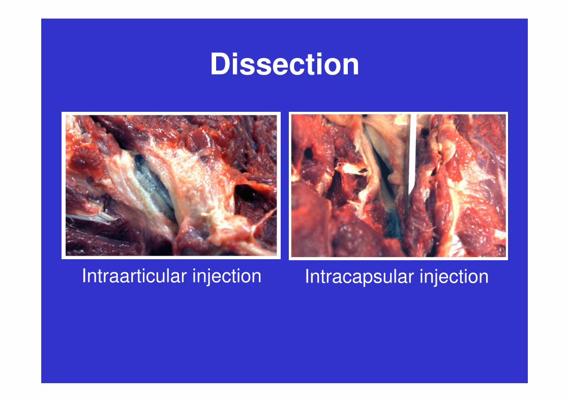
Dissection
Intraarticular injection Intracapsular injection
Materials and methodTransducer: 5 MHz, curved linear array
Insertion of the needle
lateral to the transducer without needle-guide attachment
dorsal to the transducer with needle-guide
Injectate
0.25% bromophenol blue dye, 4% agarose
Dissection
dorsal to the transducer with needle-guide attachment
Logistic regression
Results
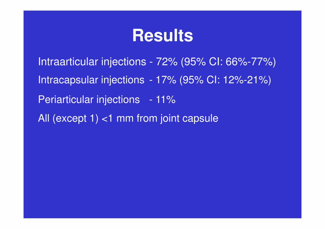
Intraarticular injections - 72% (95% CI: 66%-77%)
Periarticular injections - 11%
All (except 1) <1 mm from joint capsule
Intracapsular injections - 17% (95% CI: 12%-21%)
All (except 1) <1 mm from joint capsule

Results
60%
70%
80%
90%
100%
Percent
Periarticular >5 mm (Score 4)
Periarticular <5 mm (Score 3)
0%
10%
20%
30%
40%
50%
60%
1 2 3 4 5 6 7 8
Number of neck injected
Periarticular <5 mm (Score 3)
Intracapsular (Score 2)
Intraarticular (Score 1)
Results
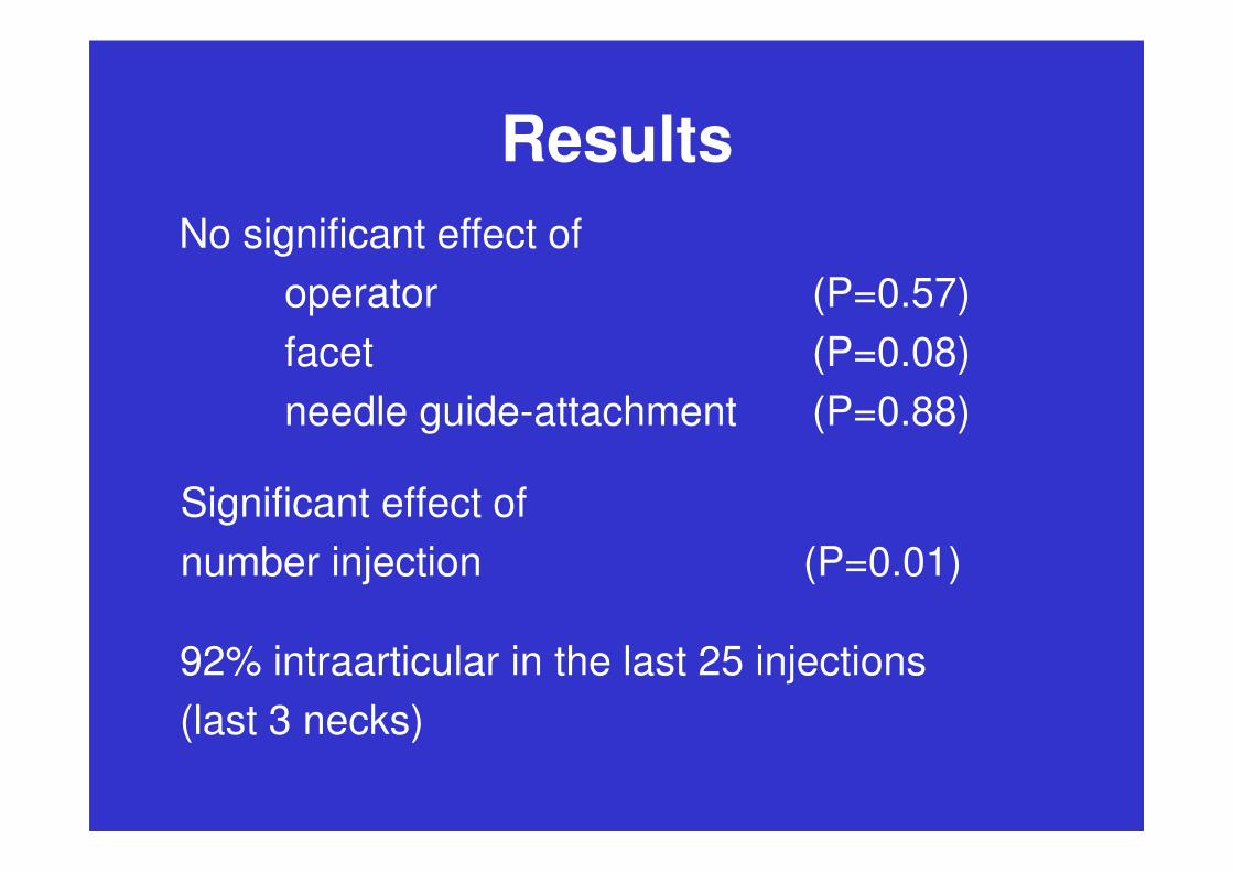
No significant effect of
operator (P=0.57)
facet (P=0.08)
needle guide-attachment (P=0.88)
Significant effect of
number injection (P=0.01)
92% intraarticular in the last 25 injections
(last 3 necks)
Conclusion
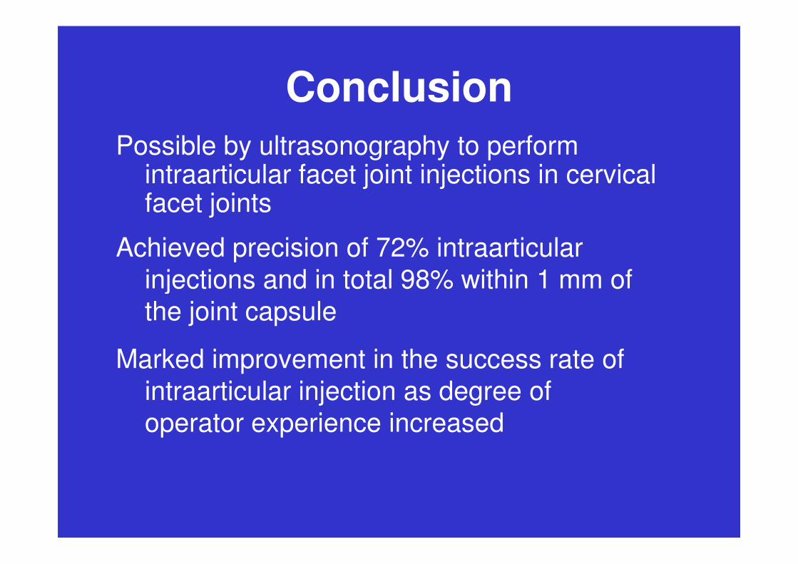
Possible by ultrasonography to perform intraarticular facet joint injections in cervical facet joints
Achieved precision of 72% intraarticular
injections and in total 98% within 1 mm of injections and in total 98% within 1 mm of
the joint capsule
Marked improvement in the success rate of
intraarticular injection as degree of
operator experience increased
Perspectives
Adjustment of method
Projection parallel with joint space
Diffusion (sensitivity, specificity)
Effect of intracapsular injections Effect of intracapsular injections
Effect of periarticular injections
Medial branch block
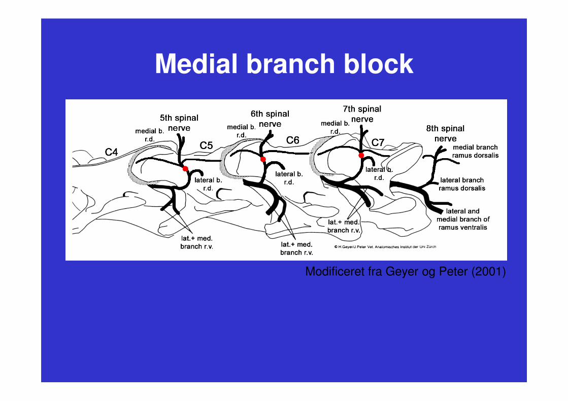
Medial branch block
Modificeret fra Geyer og Peter (2001)