Epithelial Cell Surface Modifications - prime.edu.pk 1st Year/Lect. Epi. cell... · Epithelial...
53
Transcript of Epithelial Cell Surface Modifications - prime.edu.pk 1st Year/Lect. Epi. cell... · Epithelial...


Epithelial Cell Surface Modifications
Surface modifications of epithelial cells reflect functions of cells at that surface
Apical surface: microvilli, cilia, stereocilia
Basal surface: Basement membrane,
Hemi-desmosomes,
Plasma membrane interdigitations
Lateral surface: A-Junctional complex
i. Zonula occludens
ii. Zonula Adherence
iii. Macula adherence
iv. Fascia Adherence
B- Communicating Junction
C- Plasma membrane interdigitations

Cell-1 Cell-2 Cell-3z z z c z v
Apical surfacesL
ate
ral
surf
ace
Basal surfaces
Cilia
Microvilli

MICROVILLI
Extension of cytoplasm -
1mm long
Actin-containing
microfilaments
FUNCTION - increase
the surface area
LIGHT MICROSCOPE -
- Brush border &
Striated border

Microvilli
TW
*
**
*
*
*

Small intestine, plastic, toluidine blue
Apical surface with striated border made of microvilli
Basal
surface
Lumen
Simple columnar epithelium with striated border

Microvilli are anchored to an electron-dense structure, the terminal web.

microvilli
(duodenum) glycocalyx lumen
Attachment site
of the glycocalyx
on the plasma
membrane
Plasma
membrane
The glycocalyx is exceptionally well developed
on the surface of the intestinal epithelium.
The glycocalyx (cell surface coat)

Specializations of the cell surface • Microvilli
Scanning
EM.
Sea urchin
embryo
Microvilli are fine finger-like projection of the plasma membrane,
with limited capability of motion. Ø : 50 - 100 nm, length ~ 1 - 2 µm

Variation in morphology of microvilli
Uterine glands Placenta Intestine

MICROVILLI
SEM OR TEM ??????

The glycocalyx is a carbohydrate-rich zone on the
cell surface, made up of proteins and
oligosaccharides bound to the outer surface of the
plasma membrane.
The composition and arrangement of the glycocalyx is cell-
specific.

Microvillus and terminal web
Freeze-fracture preparation
Actin filaments(microfilaments)
TW*
TW*=Terminal web
Actin filaments(microfilaments)
Microvillus

• Stereocilia
Sensory
epithelium
✓are long microvilli
( 3 - 25 µm),
✓Stereocilia are capable of
moving passively.
✓e.g. epididymis and
the sensory cells of the
internal ear.

Stereocilia
Epididymis


• Pseudopodia
macrophage
B- Lymphocytes
Pseudopodia are foot-like, long cell projections.
Their inner structure resembles that of the microvilli,
but they are less strictly ordered.

• Pseudopodia are formed and withdrawn in several
minutes.
• Pseudopodia are capable of active, independent
movement. Their biological role is to ensure the
active movement of the cells.
Several cell-types use pseudopodia for locomotion:
For exampleplasma cells
granulocytes
macrophages (phagocytic cells!).
lymphocytes

Cilia • Long cytoplasmic extensions that have a core
of organized microtubules within them
• Length = 5-10µm ,Width= 0.2µm
• Have complex arrangement of microtubules (9+2) called “axoneme”
• Extend from basal body within cytoplasm
• Cilia are of two types, motile and nonmotile (primary) cilia
• Motile (beat synchronously) and thus facilitate flow of fluid over an epithelium (tubular organ like trachea, oviduct)
• A single non motile cilium (primary cilium ) is present on nearly every human cell that is in the G0 stage of the cell cycle.
• Similar in structure to flagella (sperm)

SEM LM
Cilia
Trachea, H & E

Cilia ( Kinocilia )
kinocilia
and
microvilli
trachea
Cilia are parallelly oriented, motile, finger-like protrusions of cell
membrane (Ø 300 nm, length 7 - 10 µm), connected to an
electron dense basal body.
basal body

Cilia and flagella are built up by highly ordered
microtubular system
Axoneme

Relationship of cilia and basal bodies
Plasmamembrane
Cytoplasm
Basal body
Cilium
TEM
Basal body
Basal body

Cilia
Tubulin subunit
ATPase
Ross, Fig. 5.5

Immotile Cilia Syndrome (Kartagener’s syndrome)
•Autosomal recessive diseases that are more prevalent in men•Several different syndromes that are due to varying modifications in ciliary structure
•Patients frequently have chronic respiratory problems resulting in abnormal mucociliary transport in the tracheobronchial tree
•Men are usually sterile
•Diagnostic EMs reveal abnormal cilia (absent/defective dynein
arms)
Dynein

Lateral surface specializations

Zonula Occludens
Zonula Adherenens
Macula Adherenens
Communicating Junction
Hemidesmosome

Lateral surface specializationsIntercellular junctions
Infoldings of plasma membranes
Terminal Bar (LM)
Junctionalcomplex
Junctional complex (TEM)

Epithelial cells are held together by intercellular junctions
Epithelia consist of closely apposed cells that are tightly linked both mechanically and functionally.
Several classes of morphologically distinct intercellular junctions contribute to these functions:
1. Occluding junctions
form seals and are impermeable
2. Adhering junctions
glue cells together and are associated with specific cytoskeletal elements
3. Communicating junctions
permit x-talk ( metabolic & electrical Communications

Junctional complex
Terminal bar is LM term which refers to a series of
intercellular junctions at the lateral surfaces of cells. In TEM this
region is called a Junctional complex, which is made of a
few different types of intercellular junctions
Zonula: zone or belt-like
Macula: spot
Zonula occludens
Zonula adherens
Macula adherens

Junctional complex
Lumen
ZA
ZO
Cell A
MA
Cell B
**

TIGHT JUNCTION -
ZONULA OCCLUDENS

Zonula occludens (Tight junction)
Inhibition of paracellular
diffusion
Cell A
Cell Blumen
Cell B
Z. occludens
Cell A

•Integral membrane proteins of two cells link across the extracellular space and occlude the space between the two cells. These proteins form “sealing strands”.
•Occludin is one of the integral
membrane proteins involved.
Tight Junctions
(“sealing strands”)
Interacting plasma membranesof 2 cells
Cell A
Cell B

Lateral cell membrane junction:
interdigitation
Cell 1
Cell 2
Cell 1
Cell 2

Basal striation and basal lamina

Infoldings of basal portion of kidney tubule cell showing infoldings of the plasma membrane and alignment of mitochondria
Drawing of kidney
tubule cell

Adhering (anchoring) junctions
• Actin filament attachment sites
-cell-cell adhering junctions (Zonula adherens)
• Intermediate filament attachment sites
-cell-cell (Macula adherens = Desmosomes)
-cell-matrix (Hemidesmosomes)
• Associated with a “glue” that helps hold cells together
ZO
ZA
MA
Hemidesmosome


Fine structure
Intercellular anchoring proteins:
Cadherins (Ca 2+ -dependent cell
adhesion proteins)
Actin
cytoskeleton
Ca2+
Cadherin
Cell 1
Cell 2

Zonula adherens
• Belt-like junction which
circumscribes the cell, usually near
the apical surface.
• Mechanically links cells to one
another (using *Cadherins as the
“glue”).
• Cadherins are integral membrane
glycoproteins that form homodimers
and require Ca++.
• Anchors the thin (actin) filaments of
the cell to the plasma membrane
via attachment proteins within the
cell.
ZA
Actin filaments
ZO
Cell A
Cell B
*
*

EM
Plaques
Cadherin
Intermediary
filaments
Ca2+
Fine structure
1. cell
2. cell

Desmosomes: “spot welds” are abundant in epithelia that are exposed to much abrasion
Examples: stratified squamous epithelium of the gingiva, tongue,
skin, oral mucosa

Cytoplasmic
plaque has
attachment
proteins
Cadherins
Keratin
(intermediate)
filamentsPlasma membranes
Intercellular
space
Intercellularspace
Desmosome (Macula adherens)

Basal surface specializations
• Basement membrane
• Junctional specialization:
hemidesmosome (integrins)
• Plasma membrane interdigitations

HEMIDESMOSOME
LOCATION - contact zone
between epithelium and basal
lamina
DISK-SHAPED
MORPHOLOGY - half a
desmosome
FUNCTION - binds the
epithelial cell to basal lamina

Found at base of stratified epithelia and connect the plasma membrane to the extracellular matrix via integrins
Glue is NOT cadherins but integrins
Plasma membrane Hemidesmosome
Basal lamina
Hemidesmosomes
Basallamina
Plasmamembrane

Hemidesmosomes and basal lamina
Basal lamina
HD HD

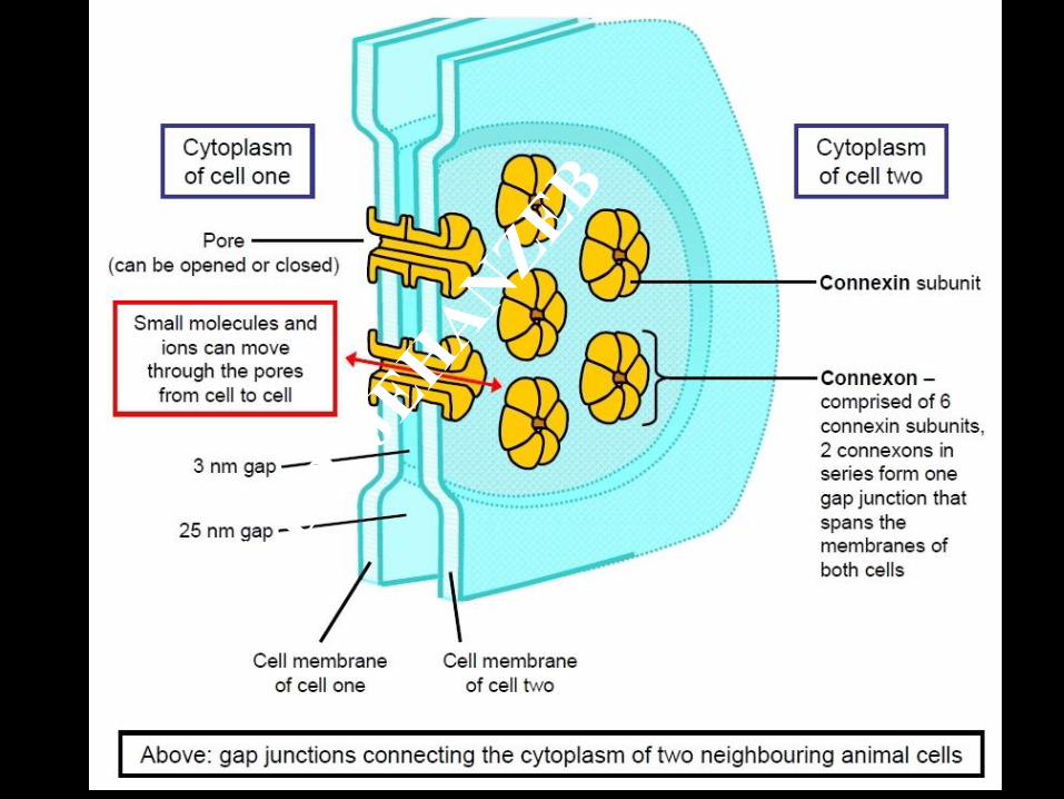
GAP JUNCTION -
NEXUS

Fine structure
Plasma membrane proteins (connexins) form a
channel (connexon).
Connexons of two adjacent cell membranes join
together in the intercellular space to constitute a
continuous hidrophilic channel, the nexus.
Small molecules ( < 1000 Da):
ions, second messengers,
metabolites etc. can freely pass.
The two cells are metabolically
and electrically coupled ( in
excitable tissues: electrical
synapse).
Enhanced intracellular Ca2+
concentration (cell damage!)
results in closing of the nexus.