Environmental Issues in Sports Medicine
64
Environmental Issues in Sports Medicine Jeremiah Penn, MD Sanford Orthopedics and Sports Medicine Bismarck, ND
Transcript of Environmental Issues in Sports Medicine

Environmental Issues in Sports MedicineJeremiah Penn, MDSanford Orthopedics and Sports MedicineBismarck, ND

Lecture Objectives
� Identify common environmental illnesses
� Describe prevention of environmental illness
� Describe treatment for life-threatening and non-emergent environmental illness

Mt Everest 29,029 ft above sea level
First climbed by Edmund Hillary and Tenzing Norgay on May 29, 1953
Number of summits in 1975: 15
Number of summits in 1995: 83
Number of summits in 2004: 330
Number of summits in 2010: 513
Introduction
� Outdoor sports are increasing in popularity
� Participants are becoming more “extreme”
� Family physicians need to be able to recognize and treat these problems in their patient population
Environmental Illness
� Heat related Illness
� Cold injury
� Altitude
� UV Light
� Lightning

Heat related Illness
� Heat edema
� Heat rash
� Heat syncope
� Heat cramps
� Heat exhaustion
� Heat stroke

Human Heat Loss
� Convection
� Conduction
� Evaporation
� Radiation

Chicago Marathon 2007

Wet Bulb Globe Temperature
� Developed by USMC in 1956 at Parris Island, SC
� Takes into account temperature, humidity, wind speed, and solar radiation
� WBGT = 0.7Tw + 0.2Tg + 0.1Td

Wet Bulb Globe Temperature
Category Temperature (°F) Flag
1 <79.9 None
2 80 – 84.9 Green
3 85 – 87.9 Yellow
4 88 – 89.9 Red
5 ≥90 Black

Heat Index Chart
� Developed by RG Steadman in 1979
� Takes into account temperature and relative humidity
� Much easier to calculate, don’t need special equipment

Heat Edema
� Transient venodilation to facilitate core heat loss
� Normal body temperature
� Dependant edema
� Treat with hydration, elevation of lower extremities, and cooling

Heat Rash
� Also called prickly heat, miliaria rubra
� Profuse sweating saturates skin and clogs sweat ducts
� Pruritic rash, normal body temperature
� Treat with cooling, reduced clothing, antihistamines, lotions

Heat Syncope
� Occurs at end of activity in elevated temperatures
� Decrease in muscle contractions combined with peripheral vasodilation
� Normal body temperature
� Present with orthostasis, syncope, rapid mental status recovery when supine
� Treat with cool environment, supine position, elevated legs, fluid replacement, untie shoes

Heat Cramps
� Generally not acclimated to conditions
� Excess heat exposure with profuse sweating
� Generally inadequate fluid and electrolyte intake
� Temp <104 °F
� Painful muscle spasms, usually calves, quads, abdominal muscles
� Treat with stretching, cooling, fluid and electrolyte replacement
� Pickle juice, Gatorade with extra salt


Korey Stringer
May 8, 1974 – August 1, 2001

Heat Exhaustion
� May be initial presentation of heat illness
� Body temperature between 98.6 °F and 104 °F
� Malaise, fatigue, dizziness
� May have profuse sweating, nausea/vomiting, headache, fainting, weakness, cold/clammy skin, tachycardia
� Normal mental status
� Stable neurologic status
� May progress to heat stroke if not recognized and treated

Heat Stroke
� Symptoms of heat exhaustion along with the following
� Core temp >104 °F
� Hot skin with or without sweating
� CNS disturbance (Confusion, ataxia, irritability, coma)
� May have hypotension, seizure, hyperventilation
� Classic vs. Exertional

Risk Factors
� Age <15 or >65
� EtOH, medications
� Dehydration**
� Prev. heat illness
� Poor acclimatization
� Overmotivation
� Sickle cell trait
� Activity level
� Obesity
� Excessive clothing
� Lack of water/shade
� Temperature
� Humidity
� WBGT

Medications
� Alpha adrenergic agents
� Amphetamines
� Anticholinergics
� Antihistamines
� Antihypertensives
� Benzodiazepines
� Illicit drugs
� Laxatives
� MAOIs
� Thyroid agonists
� TCAs
� Typical antipsychotics
� Dietary supplements
� EtOH

Prevention
� Education (athletes, coaches, trainers)
� Acclimatization (NCAA, ACSM guidelines)
� Daily weights to monitor for dehydration
� Proper uniforms
� Condition monitoring
� Adjusting practice times

Treatment
� Medical Emergency!
� Rectal Temp
� ABCs
� Cool first, then transport
� Ice bath
� Cool mist and fan
� Ice at groin/axilla

Complications
� Seizures
� benzodiazepines
� Hypotension
� IV fluids, may need pressors
� Rhabdomyolysis
� IV fluids, diuretics, alkalinize urine (pH > 7)
� Liver damage
� Avoid acetaminophen
� Arrythmias
� Avoid cardioversion until myocardium has cooled

Return to Play
� Mild illness – 24 hours post event with proper rest and rehydration
� Heat stroke – at least one week
� Monitor daily weights
� Normalization of lab values
� Graduated return to activity
� Address all risk factors

Cold Injury
� Hypothermia
� Frostbite
� Trenchfoot

Hypothermia
� Normal core temperature 99.6 °F(+/- 4 °F)
� Thermoregulation through hypothalamus
� Voluntary muscular activity
� Involuntary shivering
� Increased metabolic rate (higher epi and norepi)
� Peripheral vasoconstriction

Hypothermia
� Risk factors
� CNS depressants
� Phenothiazines
� Hypoglycemia, peripheral neuropathy, hypothyroidism, adrenal insufficiency
� Ethanol
� Age
� Exhaustion
� Malnutrition

Hypothermia
� Core temp <95 °F
� 1.8% increase in mortality rate with each 1.8 °F drop in core temperature
� Need a true core temperature (rectal temp with appropriate thermometer)
Mild Hypothermia
� Core temp 90° -95° F
� Cool, pale, core
� Uncontrolled shivering
� Dysarthria
� Ataxia
� Confusion
� Tachycardia
� Maximal peripheral vasoconstrictions
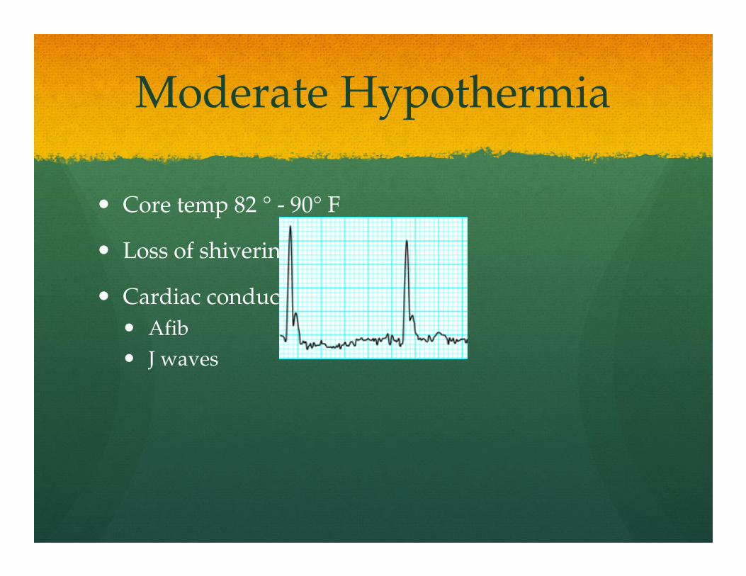
Moderate Hypothermia
� Core temp 82 ° - 90° F
� Loss of shivering
� Cardiac conduction irregularities
� Afib
� J waves

Severe Hypothermia
� Looks dead, core temp <82 °F
� No pulse or blood pressure
� Agonal or absent respirations
� Dilated pupils, areflexic
� Ventricular arrhythmia on EKG
Hypothermia
� Prevent further heat loss
� Passive external rewarming
� Active external rewarming
� Warm blankets
� hot water bottles
� Warmed forced air
� Active Core rewarming
� Warmed IV fluids
� Warmed oxygen
� NG, colonic, bladder irrigations
� Peritoneal dialysis
� Cardiopulmonary bypass
� Hemodialysis
Hypothermia
� ABCs
� Limit movement
� Avoid chest compressions if any cardiac or respiratory activity
� A patient isn’t dead until he’s warm and dead


Frostbite
� Direct freezing of tissues
� Ambient temp < 32° F
� Exposed areas and distal extremities most at risk
� Risk factors include
� Raynauds, PAD, constrictive clothing, nicotine

Frostbite
� Tissue cooling, vasoconstriction, hyperviscosity
� Extracellular ice formation
� Intracellular dehydration and hyperosmolality
� Cell membrane damage
� Microcirculatory stasis, sludging, thrombosis, leads to hypoxia
� Thawing leads to capillary leakage and tissue edema, causing more ischemia
� Usually several freeze thaw cycles in severe injuries

1st Degree Frostbite
� Partial skin freezing
� Erythema, edema, hyperemia, no blisters
� No necrosis
� Skin may peel a week or two later
� Stings, throbs, aches, burns, hyperhidrosis

2nd Degree Frostbite
� Full thickness injury
� Erythema, edema
� Vesicles with clear fluid
� May form blackened eschar
� Numbness, vasomotor dysfunction if severe

3rd Degree Frostbite
� Full thickness skin and subQ freezing
� Hemorrhagic blisters
� Skin necrosis
� Blue-gray discolorations

4th Degree Frostbite
� Full thickness, skin, subQ, muscle, tendon, and bone freezing
� Little edema
� Mottled, deep red, or cyanotic
� Becomes dry, black, and mummified

Frostbite
� Rewarm in 104 -108 F water bath
� Ensure no refreezing
� Very painful process (narcotics and NSAIDs)
� Tetanus immunization
� After rewarming, separate digits and splint

Frostbite
� Debride clear blisters but not hemorrhagic ones
� Early surgery for compartment syndrome or escharotomy
� Amputation after tissue injury demarcates unless infected (may take several weeks)
� Physeal injury may develop in children with frostbite

Trenchfoot
� Lengthy exposure to cold water (32 °-50° F)
� Prolonged vasoconstriction leads to ischemia
� Appears hyperemic, then cyanotic mottling and swelling
� May have persistant paresthesias
� Prevent with frequent sock changes and keeping shoes dry

Altitude Ilness
White Butte, ND Elevation 3,506 ftMt. Whitney, CA Elevation 14,505 ft
Altitude Illness
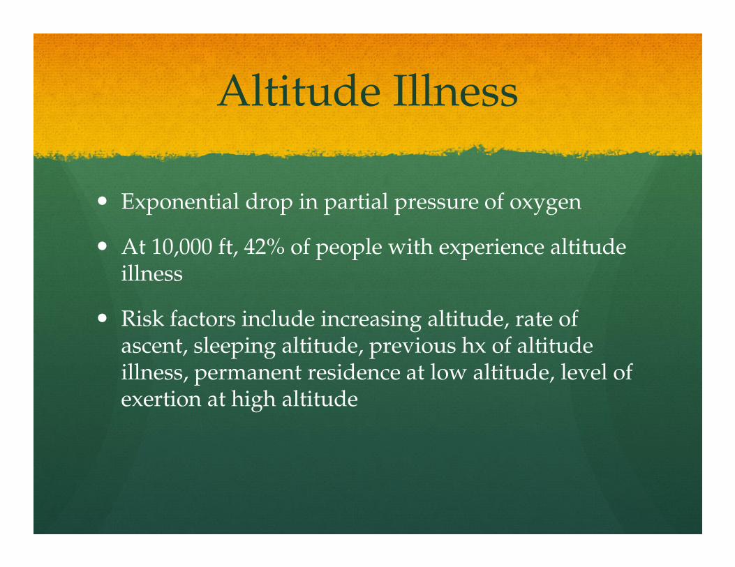
� Exponential drop in partial pressure of oxygen
� At 10,000 ft, 42% of people with experience altitude illness
� Risk factors include increasing altitude, rate of ascent, sleeping altitude, previous hx of altitude illness, permanent residence at low altitude, level of exertion at high altitude
Altitude Illness
� High Altitude Headache
� Acute Mountain Sickness
� High Altitude Cerebral Edema
� High Altitude Pulmonary Edema

HAH/AMS
� High altitude headache – usually attributed to lack of sleep, poor nutrition, or dehydration
� Treat with NSAIDs or acetominophen
� Acute mountain sickness – HAH plus 1 of the following: GI irritation, dizziness, fatigue, or sleep disturbance
� Treat with stopping ascent (rare), descent, oxygen, dexamethasone, acetazolamide

High Altitude Cerebral Edema
� Defined as altered conciousness or ataxia in someone with AMS or HAPE
� Drowsiness, poor decision making, pyschomotorslowing, stupor
� Exam may reveal papilledema, retinal hemorrhages, global encephalopathy
� Untreated, leads to death via cerebral herniation
High Altitude Pulmonary Edema
� Most common cause of death from altitude related illness
� Dry cough and decreased exercise tolerance
� Progresses to tachycardia, resting tachypnea, hemoptysis, respiratory distress and fever
� Treat with descent, supplemental oxygen, nifedipine, nitric oxide
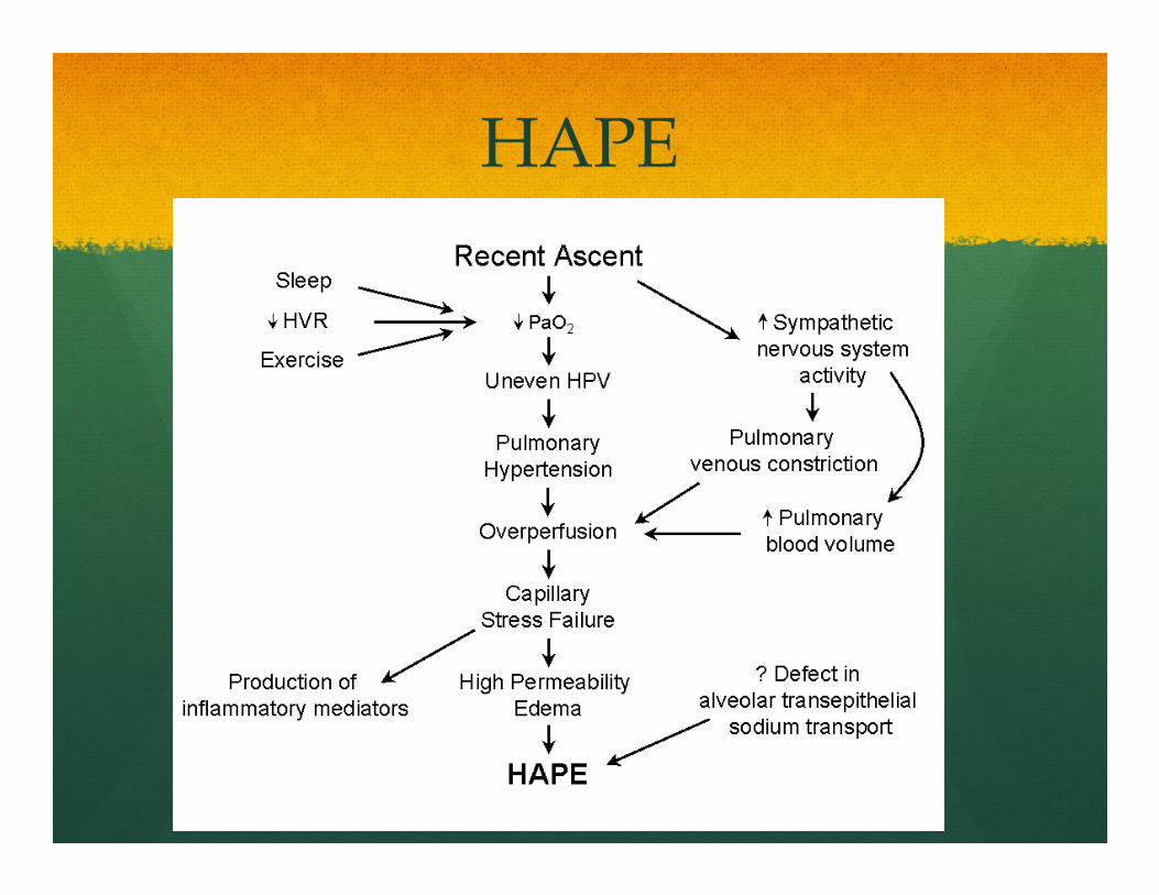
HAPE

Altitude Illness


UV Light
� Outdoor competition increases UV exposure
� Short term consequences of sunburn
� Long term consequences of melanoma, basal cell cancer and squamous cell cancer

UV Light
� UVA - makes up 90% of UV light, wavelength of 320-400 nm, penetrates to deep cutaneous tissue, damages DNA through free radical formation
� UVB – wavelength of 290 to 320 nm, primary cause of sunburn
� UVC – wavelength of 200-290 nm, blocked by ozone

Sunburn
� Direct injury from UV radiation
� Vasodilation leading to erythema, edema, vesicles, and bullae
� Initial symptoms at 3-5 hours post exposure, peaking at 24 hours

Skin Cancer
� Frequent severe sunburns as a youth increase risk of melanoma and basal cell cancer
� Prolonged exposure at lower levels increases risk of squamous cell CA

UV Light
� Treat sunburns with moisturizers and pain medications
� Options include topical and systemic steroids, NSAIDS, antihistamines, antioxidants, emollients
� Prevent sun damage with UVA/UVB sunscreen with SPF between 15-30
� SPF 15 filters 92% of UVB exposure
� Participate at low sun times and wear protective clothing

Lightning
� About 300 injuries per year in the US
� 70-90 % of victims survive but 75% have permanent injuries
� Highest incidence of injury in areas of highest lightning flashes (Central Florida in US)

Lightning
� 3 sources of injury
� Electrical current
� Heat production
� Concussive force
� 3 forms of human contact
� Direct Strike (3-5%)
� Contact (1-2%)
� Side flash/splash (35%)
Lightning
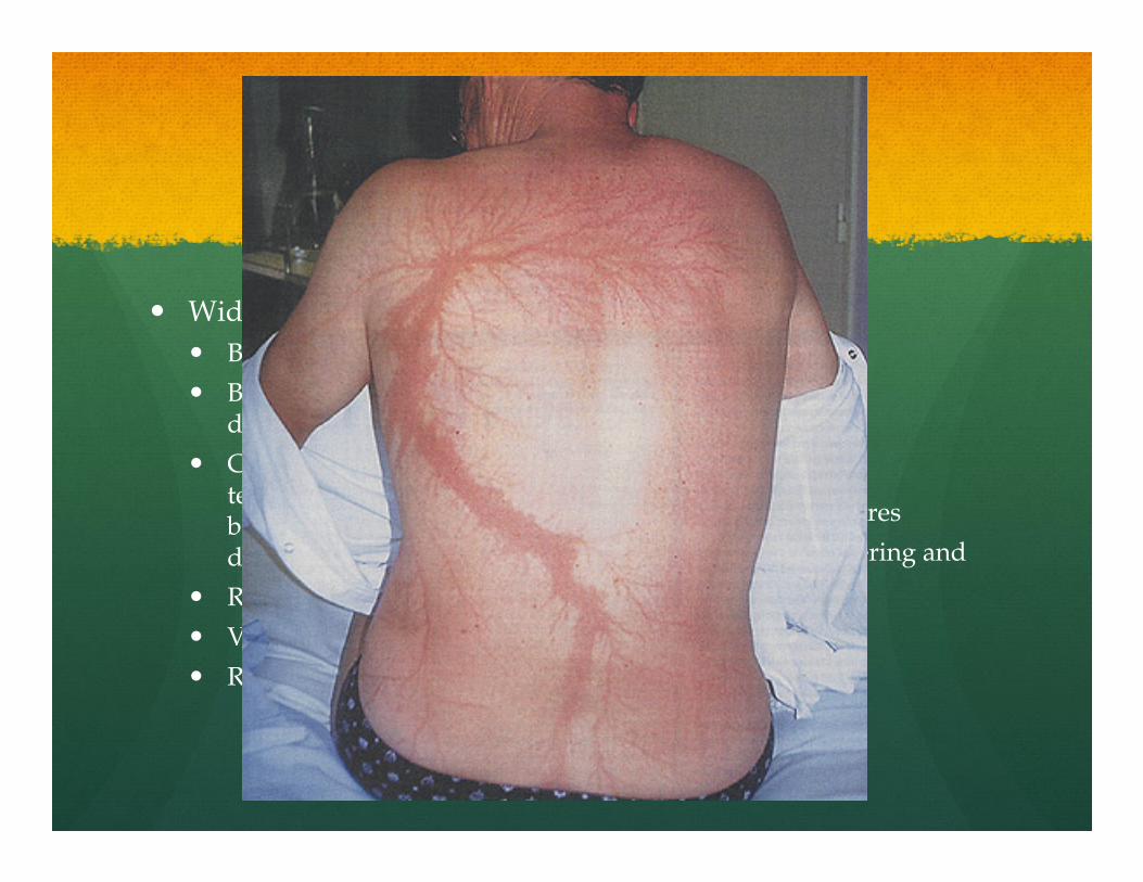
� Wide spectrum of injuries
� Blunt force trauma
� Bilateral shoulder dislocation and cervical fx
� Confusion, amnesia, temporary deafness or blindness, LOC, dysesthesia or paralysis
� Respiratory arrest
� Vfib/cardiac arrest
� Rhabdomyolysis (rare)
� Burns
� Contact
� Flash
� Linear
� Punctate
� Lichtenberg figures
� Superficial blistering and erythema

Lightning
� ATLS protocol
� EKG, C-spine films
� UA, chem 14, cardiac profile
� Burn treatment/referral
� Post-injury support network
Lightning
� 30 seconds - 30 minutes rule
� Shelter in substantial building or metal roofed automobile
� Increased storm activity on summer afternoons, also peak time for sporting events

Thank You