Engineering mouse models with myelodysplastic - Haematologica
13
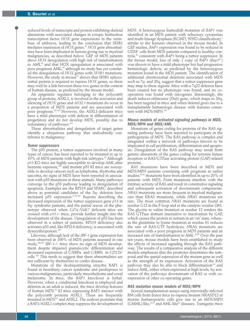
10 REVIEW ARTICLE haematologica | 2013; 98(1) Introduction Myelodysplastic syndromes (MDS) are thought to arise from a malignant hematopoietic stem cell (HSC) that has sus- tained irreversible genetic or epigenetic events resulting in dominance of the abnormal clone. Approximately 30% of MDS cases progress to acute myelogenous leukemia (AML). Dysmyelopoiesis and increased apoptosis are the main fea- tures of MDS. Bone marrow (BM) isolated from MDS patients exhibits dysplasia of at least one lineage (according to the World Health Organization (WHO) classification), 1,2 hypercellularity, and increased apoptosis correlating with peripheral blood cytopenia. 3,4 Because MDS patients present with a range of different characteristics, classification has become important, and prognostic scoring techniques have been developed to address patient management and treat- ment. 2,5,6 Recently, these classification and scoring techniques have also taken into account the presence of cytogenetic abnormalities. Cytogenetic abnormalities are relatively fre- quent in MDS; deletions and rare chromosomal reciprocal translocations have been identified, while DNA sequencing and micro-arrays (RNA, CGH) have confirmed additional mutations and deletions. 7,8 Thus, genetic and epigenetic abnormalities are linked to MDS pathogenesis. 9 Efforts have been made to develop accurate animal models of MDS that would help us understand the pathogenesis of the disease and the mechanisms of its transformation to AML. Various strate- gies have been used to express MDS-associated candidate genes in animal models, either by transduction of candidate genes under the control of retroviral promoters and subse- quent transplantation of transduced mouse BM cells into syn- genic mice, or by the establishment of transgenic mice expressing the candidate genes under the control of myeloid- specific promoters (Figure 1). Indications of function have also been obtained by analysis of knock-in (KI) and knock-out (KO) models of various genes. Recently, analyses of xenografts of human MDS cells have yielded promising results. Hematopoietic analysis of these animal models has revealed a great variety of hematologic abnormalities, some of which are not necessarily reminiscent of either the human MDS or MDS/myeloproliferative neoplasms (MDS/MPN). This review focuses on the animal models expressing the most frequent genetic abnormalities associated with MDS and MDS/MPN, including chronic myelomonocytic leukemia (CMML) and juvenile myelomonocytic leukemia (JMML), as well as those models that resemble the human MDS disease regardless of the expressed or deleted genes (Table 1). The WHO classification of human MDS and MDS/MPN is shown in the Online Supplementary Tables S1 and S2. 1,2 Our working model of disease progression is illustrated in Figure 2. Starting with an initiation event targeting the stem cell, an abnormal clone arises which has a selective growth advantage and expands. This is the phase that we recognize as pre-leukemia or MDS. Several events then follow until a leukemic clone arises and accelerates the disease to give rise to acute leukemia. The genes that drive this progression are those that code for differentiation, proliferation and apoptosis. A block in differentiation can occur in any compartment; although in MDS it is thought to occur at a very immature stage, giving rise to leukemic stem cells (LSC), so-called because they are cells that have acquired self-renewal capacity. Early MDS is often characterized as pro-apoptotic, and as disease progress- es, the cells become more anti-apoptotic. There is abnormal proliferation throughout the disease process. Novel signaling pathways and cells involved in MDS/MPN Recent insights into the pathogenesis of MDS have high- lighted the importance of genomic instability (GI), epigenetic Engineering mouse models with myelodysplastic syndrome human candidate genes; how relevant are they? Stephanie Beurlet, Christine Chomienne, and Rose Ann Padua INSERM U940, Paris, France; Université Paris-Diderot, Sorbonne Paris Cité, Institut Universitaire d’Hématologie, UMRS 940, Paris, France; Hôpital St-Louis, Paris, France Manuscript received on May 12, 2012. Revised version arrived on September 7, 2012. Manuscript accepted on October 1, 2012. Correspondence: Rose Ann Padua. E-mail: [email protected] Myelodysplastic syndromes represent particularly challenging hematologic malignancies that arise from a large spectrum of genetic events resulting in a disease characterized by a range of different presentations and out- comes. Despite efforts to classify and identify the key genetic events, little improvement has been made in ther- apies that will increase patient survival. Animal models represent powerful tools to model and study human dis- eases and are useful pre-clinical platforms. In addition to enforced expression of candidate oncogenes, gene inac- tivation has allowed the consequences of the genetic effects of human myelodysplastic syndrome to be studied in mice. This review aims to examine the animal models expressing myelodysplastic syndrome-associated genes that are currently available and to highlight the most appropriate model to phenocopy myelodysplastic syndrome disease and its risk of transformation to acute myelogenous leukemia. ©2013 Ferrata Storti Foundation. This is an open-access paper. doi:10.3324/haematol.2012.069385 ABSTRACT
Transcript of Engineering mouse models with myelodysplastic - Haematologica

10
REVIEW ARTICLE
haematologica | 2013; 98(1)
Introduction
Myelodysplastic syndromes (MDS) are thought to arisefrom a malignant hematopoietic stem cell (HSC) that has sus-tained irreversible genetic or epigenetic events resulting indominance of the abnormal clone. Approximately 30% ofMDS cases progress to acute myelogenous leukemia (AML).Dysmyelopoiesis and increased apoptosis are the main fea-tures of MDS. Bone marrow (BM) isolated from MDSpatients exhibits dysplasia of at least one lineage (accordingto the World Health Organization (WHO) classification),1,2
hypercellularity, and increased apoptosis correlating withperipheral blood cytopenia.3,4 Because MDS patients presentwith a range of different characteristics, classification hasbecome important, and prognostic scoring techniques havebeen developed to address patient management and treat-ment.2,5,6 Recently, these classification and scoring techniqueshave also taken into account the presence of cytogeneticabnormalities. Cytogenetic abnormalities are relatively fre-quent in MDS; deletions and rare chromosomal reciprocaltranslocations have been identified, while DNA sequencingand micro-arrays (RNA, CGH) have confirmed additionalmutations and deletions.7,8 Thus, genetic and epigeneticabnormalities are linked to MDS pathogenesis.9 Efforts havebeen made to develop accurate animal models of MDS thatwould help us understand the pathogenesis of the disease andthe mechanisms of its transformation to AML. Various strate-gies have been used to express MDS-associated candidategenes in animal models, either by transduction of candidategenes under the control of retroviral promoters and subse-quent transplantation of transduced mouse BM cells into syn-genic mice, or by the establishment of transgenic miceexpressing the candidate genes under the control of myeloid-specific promoters (Figure 1). Indications of function have
also been obtained by analysis of knock-in (KI) and knock-out(KO) models of various genes. Recently, analyses ofxenografts of human MDS cells have yielded promisingresults. Hematopoietic analysis of these animal models hasrevealed a great variety of hematologic abnormalities, someof which are not necessarily reminiscent of either the humanMDS or MDS/myeloproliferative neoplasms (MDS/MPN).This review focuses on the animal models expressing themost frequent genetic abnormalities associated with MDSand MDS/MPN, including chronic myelomonocytic leukemia(CMML) and juvenile myelomonocytic leukemia (JMML), aswell as those models that resemble the human MDS diseaseregardless of the expressed or deleted genes (Table 1). TheWHO classification of human MDS and MDS/MPN is shownin the Online Supplementary Tables S1 and S2.1,2 Our workingmodel of disease progression is illustrated in Figure 2. Startingwith an initiation event targeting the stem cell, an abnormalclone arises which has a selective growth advantage andexpands. This is the phase that we recognize as pre-leukemiaor MDS. Several events then follow until a leukemic clonearises and accelerates the disease to give rise to acuteleukemia. The genes that drive this progression are those thatcode for differentiation, proliferation and apoptosis. A blockin differentiation can occur in any compartment; although inMDS it is thought to occur at a very immature stage, givingrise to leukemic stem cells (LSC), so-called because they arecells that have acquired self-renewal capacity. Early MDS isoften characterized as pro-apoptotic, and as disease progress-es, the cells become more anti-apoptotic. There is abnormalproliferation throughout the disease process.
Novel signaling pathways and cells involved in MDS/MPNRecent insights into the pathogenesis of MDS have high-
lighted the importance of genomic instability (GI), epigenetic
Engineering mouse models with myelodysplastic syndrome human candidate genes; how relevant are they?Stephanie Beurlet, Christine Chomienne, and Rose Ann Padua
INSERM U940, Paris, France; Université Paris-Diderot, Sorbonne Paris Cité, Institut Universitaire d’Hématologie, UMRS 940, Paris,France; Hôpital St-Louis, Paris, France
Manuscript received on May 12, 2012. Revised version arrived on September 7, 2012. Manuscript accepted on October 1, 2012. Correspondence: Rose Ann Padua. E-mail: [email protected]
Myelodysplastic syndromes represent particularly challenging hematologic malignancies that arise from a largespectrum of genetic events resulting in a disease characterized by a range of different presentations and out-comes. Despite efforts to classify and identify the key genetic events, little improvement has been made in ther-apies that will increase patient survival. Animal models represent powerful tools to model and study human dis-eases and are useful pre-clinical platforms. In addition to enforced expression of candidate oncogenes, gene inac-tivation has allowed the consequences of the genetic effects of human myelodysplastic syndrome to be studiedin mice. This review aims to examine the animal models expressing myelodysplastic syndrome-associated genesthat are currently available and to highlight the most appropriate model to phenocopy myelodysplastic syndromedisease and its risk of transformation to acute myelogenous leukemia.
©2013 Ferrata Storti Foundation. This is an open-access paper. doi:10.3324/haematol.2012.069385
ABSTRACT
Mouse models myelodysplastic syndrome human candidate genes
haematologica | 2013; 98(1) 11
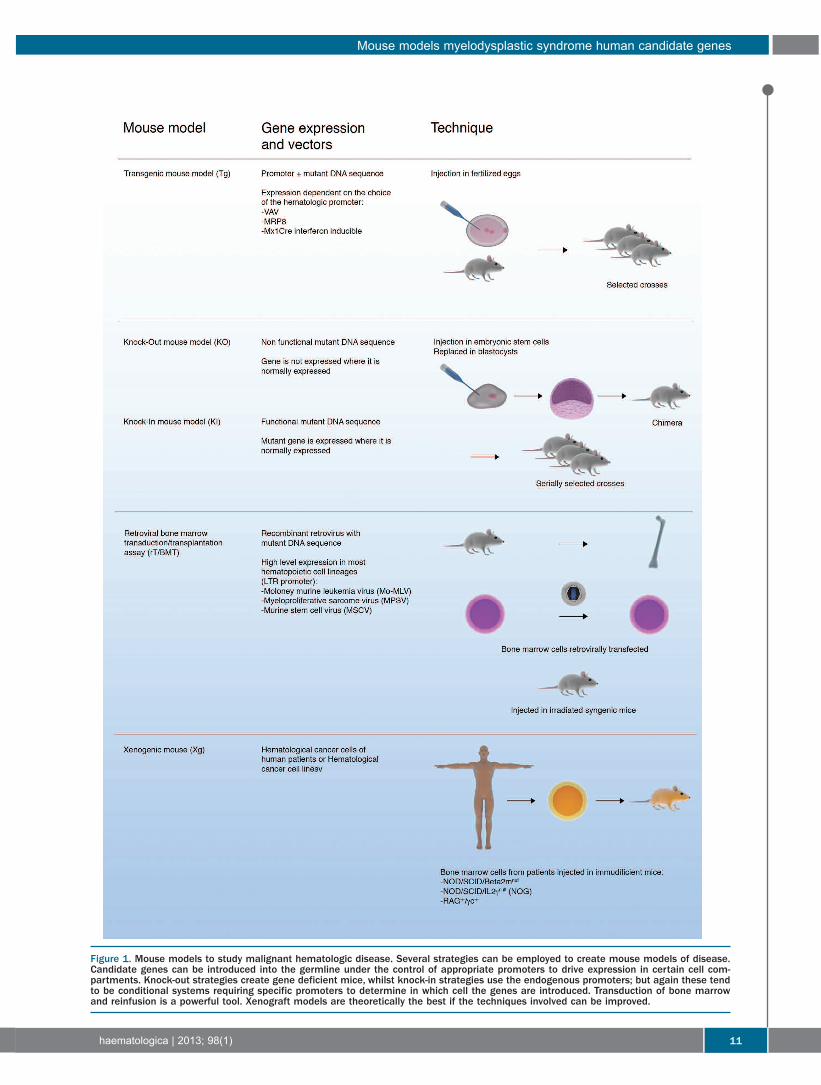
Figure 1. Mouse models to study malignant hematologic disease. Several strategies can be employed to create mouse models of disease.Candidate genes can be introduced into the germline under the control of appropriate promoters to drive expression in certain cell com-partments. Knock-out strategies create gene deficient mice, whilst knock-in strategies use the endogenous promoters; but again these tendto be conditional systems requiring specific promoters to determine in which cell the genes are introduced. Transduction of bone marrowand reinfusion is a powerful tool. Xenograft models are theoretically the best if the techniques involved can be improved.

alterations and abnormalities of the spliceosome machin-ery. The MDS stem cell and its niche have also proved tobe of significance (Figure 2).
Genomic instability (GI)Karyotypic changes are indicative of increased GI.
Increased GI can result in increased misrepair. Non-homologous end joining (NHEJ), the main pathway fordouble-strand break repair, gives rise to chromosomaldeletions and translocations. It has been hypothesizedthat increased GI can drive initiation and progression ofmalignant disease.68 Aging mice display increased chro-mosome instability mediated by MDM2, a regulator ofthe p53 tumor suppressor and DNA repair.69 The expres-sion of many oncogenes, such as mutant RAS, results inincreased DNA damage-inducing reactive oxygen species(ROS). As MDS is essentially a disease associated withaging, it is not surprising that increased chromosomeinstability has been observed in a subgroup of MDS thatis associated with a poor prognosis (20%), thereby sug-gesting that GI may be a driver of disease progression.70GI in mouse models of MDS has yet to be investigated;although a mouse model of AML with MDS-relatedchanges mediated by RAS and BCL-233 displayedincreased ROS, DNA damage and misrepair which can berescued by treatment with either RAC1 inhibitors or anti-oxidants.71 Furthermore, BM cells isolated from NPM1+/-MPN-like mice display multiple centrosomes, thus indi-cating that centrosome amplification is associated withNPM1 loss and may participate in leukemia pathogenesisin this mouse model.14 DNA damage and chromosomalinstability is observed in Fanconi anemia (FA) patients,which is an important cause of childhood MDS.72Unfortunately, mouse models harboring a disruptedmouse homolog of FANCC fail to develop MDS.73However, the double mutant Fancc–/– , Fancg–/–mice devel-op spontaneous hematologic abnormalities including BMfailure, AML, MDS and complex random chromosomal
abnormalities that the single mutant mice do not dis-play.74
Altered gene expression through epigenetic modifications and gene silencingDysregulation of the epigenetic machinery can lead to
oncogenic transformation. Approximately 30% of MDSpatients show inactivation, by hypermethylation, of thegene encoding p15INK4b.75 This strongly suggests that theINK4 family member plays a role in the regulation of cellcycle arrest at the early G1 phase as a myeloid tumor sup-pressor.76 Ink4b KO mice (Ink4b-/-) show defects in the dif-ferentiation of common myeloid progenitors, resulting inan imbalance between erythroid and myeloid potential,although follow-up analysis of these mice did not revealany signs of MDS.77 Other genes modified by epigeneticmechanisms have been identified, such as DAPK1, CDH1(E-Cadherins), and TSP1 (thrombospondin).78,79Hypermethylation of DNA is known to contribute to pro-gression of MDS to AML, thereby inhibiting differenttumor suppressor genes. Mouse models designed toaddress this epigenetic progression have yet to be charac-terized. An exception is the EVI1 mouse model, in whichEVI1 expression in the BM gives rise to an MDS pheno-type and causes silencing of the miRNA124 by methyla-tion. This epigenetic event results in perturbation of celldivision and self-renewal in this mouse model.80The TET2 gene (Ten-Eleven Translocation-2) was origi-
nally identified from an MDS patient with a rearrange-ment of 4q2424 and numerous groups have identifiedsomatic inactivating mutations (frameshifts or misense) inMDS, MPD and CMML patients.23-25 As one of the mostfrequent mutations found in myeloid malignancies, TET2has been proposed to be a tumor suppressor as loss offunction tends to lead to disruption of progenitor myeloiddifferentiation leading to disease progression.81 Its functionis to convert 5-methlycytosine to 5-hdroxymethyl cyto-sine (5-hmc) and it is, therefore, involved in the epigenetic
s. Beurlet et al.
12 haematologica | 2013; 98(1)
Figure 2. Multi-step model of leukemo-genesis. Starting with a stem cell, an ini-tiating event occurs which gives rise toan abnormal clone with a growth advan-tage, which expands. Further geneticevents drive the progression of the dis-ease towards overt leukemia. Genomicinstability and misrepair increase as dis-ease progresses. Some of the eventsthat may drive progression includeincreased methylation to down-regulategenes and mutations of the ribosomemachinery to further deregulate genes.

control of gene regulation; 5-hmc has been found to bedecreased in granulocyte DNA from MPD patients withTET2 mutations.81 Animal models of TET2 using condi-tional knock-out systems give rise to CMML-like diseases:Tet2LacZ mice, Tet2Lox mice26 and Tet2–/–VavCre+ mice27develop a CMML-like phenotype with splenomegaly,leukocytosis with abnormal myelomonocytic differentia-tion, and expansion of the hematopoietic stem cell com-partment.26,27
Altered stem cell and niche in MDS mouse modelsBlood and BM samples from most MDS patients are
technically difficult to engraft into NOD/SCID-b2mnullmice. When xenografts are obtained, they regenerateprogeny that display phenotypic and genotypic abnor-malities of the original neoplastic clone, such as trisomy8 and del(5q). Interestingly, injecting BM cells isolatedfrom MDS patients with human stroma-derived cells, viaintramedullary injection, into NOD/SCID-b2mnullmice82 showed the engraftment of early precursors con-taining del(5q) with a loss of more mature cells that hadadditionally acquired trisomy 8. Therefore, the 5q dele-tion may be an earlier event than trisomy 8.82 In anotherstudy, the results of MDS sample engraftment suggestedthe presence of a relatively late type of ‘multilineage butmyeloid-restricted’ neoplastic ‘stem’ cell with repopulat-ing activity, limited self-renewal ability, and skewed dif-ferentiation potential.83 These findings also show thatthe repopulating cells present in MDS patients includeresidual normal or pre-neoplastic repopulating cells withnormal features that outcompete the neoplastic clone ifadequately stimulated. Recently, a transgenic approachhas provided evidence that disturbance of the endostealniche can result in MDS.22 Transgenic mice lackingDicer1 expression in mesenchymal cells of the osteolin-eage were generated, OsxCre+/Dicerfl/fl.22 TheOsxCre+/Dicerfl/fl mutant mice develop fatal neutrope-nia with hyperplastic BM and dysmyelopoiesis which ishighly suggestive of MDS. Increased apoptosis of Lin-/c-KIT+ progenitor cells has also been observed in thesemice. However, the MDS phenotype was not trans-plantable. Interestingly, when OsxCre+/Dicerfl/fl micewere transplanted with BM cells from wild-type micethey developed the same MDS phenotype. Althoughmutations of Dicer have yet to be discovered in MDSpatients, gene expression analysis of Dicer-/- osteoprog-enitors showed a downregulation of SDB, the genefound mutated in Schwachman-Bodian-Diamond syn-drome, which induces BM failure and MDS in children.Recently, downregulation of Dicer has been found asso-ciated with poor prognosis in chronic lymphocyticleukemia (CLL) patients.84 Recent studies have highlight-ed the role of innate immune signaling molecules in thephysiopathology of BM failure: in 5q- patients, deletionof chromosome 5 correlates with a loss of two miRs,miR145 and miR146a,85 which are abundant inhematopoietic progenitors and target two moleculesimplicated in innate immune signaling,86 Toll-interleukin-1 receptor domain-containing adaptor protein (TIRAP)and tumor necrosis factor receptor-associated factor-6(TRAF6), respectively. In KO mouse models of miR145and miR146a targeted specifically in hematopoietic stemand progenitor cells, animals develop thrombocytosis,mild neutropenia, and megakaryocytic dysplasia, whichrepresent several clinical features of 5q-MDS.87
MDS mouse models of chromosomal deletions: candidate genes for 5q- syndromeDeletion of chromosome 5 (5q-) is the most common
cytogenetic abnormality found in MDS (10-15% ofpatients). It is associated with refractory anemia, throm-bocytosis, hypolobulated megakaryocytes and low risk ofevolving into AML.16 Because deletion of 5q represents themajor cytogenetic abnormality associated with MDS, ithas been hypothesized that this region may contain animportant MDS suppressor gene.16,18 One such candidate isthe NPM (Nucleophosmin) gene, located in the 5q region.Mutations of the NPM1 gene have also been found in4.4% of MDS patients.12 NPM is a nuclear phosphopro-tein, which has a putative function in ribosome assemblyand DNA repair. While NPM1-/- mice lack definitivehematopoiesis, NPM1+/- mice develop features similar tothe human 5q-syndrome, such as dyserythropoiesis anddysmegakaryopoiesis that is sometimes associated withthrombocytosis,13 albeit after a long latency period of 10-24 months.14 The commonly deleted region (CDR) associ-ated with the 5q-syndrome has been narrowed down to aregion of approximately 1.5 Mb located between 5q31 and5q32.The CDR is divided into two regions of synteny on
mouse chromosomes 11 and 18. Partial deletion of thisregion in mice results in macrocytic anemia, prominenterythroid dysplasia, and monolobulated megakaryocytes,and is associated with moderate thrombocytopenia andneutropenia.17 The candidate genes included in this regionare: Cd74, RPS14, NDST1, SYNPO, MYOZ3, RBM22,DCTN4 and NID67. In humans, the candidate genes of theCDR include FAT2 (FAT tumor suppressor homolog 2),SPARC (secreted protein, acidic and rich in cysteine), andRPS14 (40S ribosomal protein S14). The FAT2 candidategene was evaluated in vivo but deficient mice failed todevelop hematologic disease.17 RPS14 is a strong candidateas demonstrated in vitro, whereby silencing RPS14 expres-sion by RNA interference in human BM cells reproduced a5q-cell phenotype.15 Interestingly, Diamond-Blackfan ane-mia is also associated with expression of the ribosomalprotein RPS1988 and transgenic mice expressing aninducible transgene of a mutant form of RPS19 (loss offunction) develop anemia with dyserythropoiesis, reducederythroid progenitors and a block in terminal erythroidmaturation.89 RPS14 is thought to be a tumor suppressor.16SPARC is a component protein of the extracellular
matrix. SPARC-null mice develop mild thrombocytopeniaand exhibit an impaired ability to form BFU-E colonies.18SPARC is also a candidate tumor suppressor as methyla-tion of the SPARC promoter has been observed in bothlung and pancreatic cancers90,91 and is often associated withbreast cancer cell invasion.92The human APC (adenomatous polyposis coli) gene is
located on chromosome 5q22, in close proximity to theregion that is deleted in 5q-syndrome. APC is a well-char-acterized tumor suppressor associated with colon cancer.APC/min mice (carrying a truncated, non-functional alleleof APC) develop an MDS/MPN hematologic disease withmacrocytic anemia, leukocytosis, monocytosis andsplenomegaly.19 The mice display altered stem cell func-tion demonstrated by an impaired repopulating potentialin secondary recipients.19 However, APC haploinsufficien-cy is observed in 95% of MDS patients with 5q-syndromeand APC+/- mice develop a disease that recapitulates sev-eral characteristic features of human MDS.20 Thus, hap-
Mouse models myelodysplastic syndrome human candidate genes
haematologica | 2013; 98(1) 13

s. Beurlet et al.
14 haematologica | 2013; 98(1)
Table 1. Gene alterations found in MDS patients and their corresponding mouse models with corresponding hematologic disease entities accord-ing to WHO classification 5.Gene Alterations Alterations Type Mouse models disease entities Hematologic
found in MDS patients studied in mouse of according to WHO Classification 2008 featuresMDS disease entities model mouse model described
(Frequency)
Gene silencing and altered tumor suppressor genesNPM1 t (3,5) translocation NPM/MLF1 NPM1 NPM1+/- Some features of MDS13 Dyserythropoiesis ,
MDS, AML with MDS MDS/MPN like and Myeloid leukemia14 Dysmegakariopoiesis13
<1%10,11 Leukocytosis with organs infiltration14
NPM1 MutationsMDS/AML12
(4.4%)RPS14 Haplo insufficiency LSCE** KO MDS with isolated del(5q) Macrocytic Anemia ,
5q- syndrome15,16 Cd74-Nid67region 5q- syndrome like17 Mild thrombocytopenia and mild neutropenia,Dyserythropoiesis
Dysmegakariopoiesis,Pro-apoptotic profile
SPARC Haplo insufficiency SPARC KO MDS with isolated del(5q) Thrombocytopenia, No Dysplasia5q- syndrom16 Some features of 5q- syndrome18 Impaired ability to form BFU-E
APC Haplo insufficiency APC Heterozygote KO MDS/MPN Macrocytic anemia, Leukocytosis, 5q- syndrome16 CMML like19,20 Monocytosis, Splenomegaly
(95%)BAP1 Heterozygous frameshift mutation BAP-1 Conditional KO MDS/MPN Anemia, thrombocytopenia,
RMCD21 CreERT2 CMML like21 leukocytosis with monocytosis, splenomegaly, dyserythropoiesis,
dysmyelopoiesisAltered stem cell and nicheDicer 1 No Dicer Tg MDS Neutropenia +/-Anemia
Inducible Osterix RCMD like22 +/- Thromcytopenia Dysgranulopoiesis Dysmegariopoiesis
Pro-apoptotic profile, 2% AML transformation
Tet2 Mutations (loss of function) Tet2 KO MDS/MPN Leukocytosis MonocytosisCMML 20%,23 22%24 Tet2LacZ/LacZ CMML like26 Anemia ThrombocytopeniaMDS 6%,23 19%24 Conditional KO (Incomplete penetrance) Splenomegaly
MPN 23%25 Mix1CreMPD 12%24 Conditional KO MDS/MPN Leukocytosis Monocytosis
VavCre CMML like27 Myeloid dysplasia, SplenomegalyRAS signaling moleculesNRAS Point mutation NRASG12D rT/BMT AML with maturation Leukocytosis Monocytosis
MDS, CMML LTR- LK * +/- high count of myeloblasts20 to 40%28-30 or MDS/MPN Pro-apoptotic profile
Atypical CML like31 of lymphocytes andmyelomonocytic cells
NRASG12D rT/BMT AML (monocytic/monoblastic)32 High BM blasts with LTR- MCSV* Or MDS/MPN monocytic/monoblastic
CMML like differentiationLeukocytosis with
Monocytosis HepatosplenomegalyNRASG12D Tg MDS33 Thrombocytopenia
MRP8 promoter RAEB-1 like or Dysmyelopoeisis,Cooperation with BCL-2 AML post-MDS33 dysplasia, propapoptotic profile
BM blasts about 15%Thrombocytopenia Neutropenia, some dysplasia, anti-apoptotic
profile, BM blasts >60%LSL-NRASG12D Conditional KI MPD34 Leukocytosis, splenomegaly
Mix1CreLSL-NRASG12D Conditional KI AML-like34 Myeloblasts M4/M5 subtype
Mix1Cre+MOL4070LTR
continued on the next page

loinsufficiency is a mechanism of activating the oncogenicpotential of a gene. However, other candidate genes locat-ed in the 5q-region either fail to confer hematopoietic dis-ease or have not yet been studied.
Spliceosomal mutationsRecurrent mutations in a number of genes encoding core
spliceosomal proteins have been identified in all sub-groups of MDS. Heterozygous missense mutations in the
U2AF1 and SF3B1 genes that encode spliceosome subunitshave been described. SF3B1 mutation is prevalent in bothlow-risk MDS with ringed sideroblasts and chronic lym-phocytic leukemia (CLL) and is associated with a goodprognosis. U2AF1 and SRSF2 are frequently mutated inmyeloid hematopoietic malignancies, especially in CMMLand advanced forms of MDS, and are associated withshorter survival.93-96 SF3B1 null homozygous mice are notviable and heterozygous mutant mice are healthy with
Mouse models myelodysplastic syndrome human candidate genes
haematologica | 2013; 98(1) 15
continued from the previous page
KRAS Point mutation LSL-KRASG12D Conditional KI MDS/MPN Anemia DyserythropoiesisMDS,CMML, JMML Mix1Cre JMML or CMML like35,36 Leukocytosis Monocytosis20 to 25%(62;70;71)
KRASG12D rT/BMT MDS/MPN Leukocytosis with LTR-MCSV* CMML like37 Monocytosis Hepatosplenomegaly
FLT3 FLT3-ITD FLT3-ITD KI MDS/MPNMDS, CMML Endogenous CMML like38 Leukocytosis Monocytosis5%(40) Splenomegaly
PDGFRβ Translocation t(5;12) TEL-PDGFRb BMT MDS/MPN Leukocytosis with CMML with eosinophilia LTR-MCSV* CMML like40 Neutrophilia and
Rare(95) MonocytosisHepatosplenomegaly
NF1 Loss and mutation NF1 Inducible KO MDS/MPN Leukocytosis MonocytosisPediatric MDS/MPN Mx1CreXNF1flox JMML like42 Splenomegaly
14%(84)PTPN11 Mutations Ptpn11D61Y KI MDS/MPN Anemia, Leukocytosis
JMML Endogenous JMML like44 Monocytosis, Splenomegaly 34%(86) Hypersensivity to GM-CSF
Nuclear and Transcription FactorsEVI1 Inv(3), t(3,12), t(3;21) Evi1 rT/BMT MDS Pancytopenia, Dyserythropoiesis Evi1-MDS1 MDS, MDS/AML, t-MDS/AML LTR-MSCV* RCMD like49 Dysmegakariopoiesis, Pro-apoptotic profile
(Rare)45-47
Overexpression(Common)48 rT/BMT
RUNX1/AML1 Mutations Mutant AML1 D171N LTR-Retrovirus* AML with MDS related changes51 Multilineage, Dysplasia,MDS subgroup MDS/AML (unspecified) Leukocytosis, Hepatosplenomegaly,
(5 to 20%)(7) High blasts count in BMt-MDS/AML(40%)50 Mutant AML1 S971fs MDS RAEB-251 Multilineage, Dysplasia, Pancytopenia
BM blasts<20%NUP98 Translocations NUP98-HOX Tg MDS Thrombocytopenia, Anemia,
MDS, MDS/AML, t-MDS/AML VAV promoter RCMD like sometimes AML53,54 Neutropenia, Multilineage dysplasia1 - 2 %52 Pro-apoptotic profile
CBFb Translocation Inv 16 CBFb-MYH11 Conditional KI AML56 AML with monocytic differentiationt-MDS/AML Mx1Cre promoter Multilineage differentiation impairment
1,5%55
MLL Translocations MLL-AF9 KI AML61 No MDS featurest-MDS, t-MDS/AML Endogenous promoter AML with Mac-1+/c-KIT+ phenotype
12%57-59 MLL-AF9 rT/BMT AML62 No MDS featuresAmplification LTR-MSCV* AML with Mac-1+/c-KIT+ phenotype
MDS Mostly RAEB, t-MDS, MLL-ENL Conditional KI MPD63 Accelerated to AML with caffeine, AML with MDSrc endogenous promoter an inhibitor of DNA damage response
<1%60 kinases ATM and ATR withincrease in Mac-1/c-KIT/GR-1 phenotype
C/EBPa� Mutations C/EBPa� mutant p30 KI AML66 No MDS featuresMDS, CMML Endogenous Promoter AML with Mac-1+/c-KIT+ phenotypeRare64,65
AML5 to 14%64,65
SALL4 Overexpression SALL4B isoform Tg MDS Neutropenia, Multilineage dysplasiaAML and MDS CMV promoter RCMD like67 50% AML transformation
60% (4B isoform)67 Pro-apoptotic profile in young mice
reduced levels of transcripts and protein exhibiting skeletalalterations with associated changes in ectopic homeoboxtranscription factor HOX gene expressions in the verte-brae of embryos, leading to the conclusion that SF3B1mediates repression of HOX genes.97 HOX gene abnormal-ities have been implicated in fusions giving rise to myeloidmalignancies, as described below. GEP of MDS patientsshow HOX deregulation with high risk of transformationto AML98 and that HOX upregulation is associated withpoor prognosis AML,99 although no study has yet correlat-ed the deregulation of HOX genes with SF3B1 mutations.However, the study in mouse97 shows that SF3B1 spliceo-somal protein is required to repress HOX genes, so theremay well be a link between these two genes in the contextof human disease, as predicted by the mouse model.An epigenetic regulator belonging to the polycomb
group of proteins, ASXL1, is involved in the activation andsilencing of HOX genes and ASXL1 mutations do occur ina proportion of MDS patients and are associated withpoor prognosis.100-102 However, the ASXL1-deficient micehave a mild phenotype with defects in differentiation ofprogenitors and do not develop MDS, possibly due toredundancy of pathways.103These abnormalities and deregulation of target genes
identify a ubiquitous pathway that undoubtedly con-tributes to malignancy.
Tumor suppressorsThe p53 protein, a tumor suppressor involved in many
types of cancer, has been reported to be mutated in up to10% of MDS patients with high-risk subtypes.28 Althoughp53 KO mice are highly susceptible to develop AML afterbenzene exposure,104 and mutant p53 KI mice are suscep-tible to develop cancers such as lymphoma, thymoma andsarcoma, no signs of MDS have been reported in associa-tion with p53 mutations in these animals. Many pathwaysconverge to the p53 pathway leading to deregulation ofapoptosis. Examples are the RPS14 and SPARC describedabove as potential candidates of the 5q- deletion haveincreased p53 expression.17,91,105 Reports describing theincreased expression of the tumor suppressor gene p53 in5q- syndrome patients, and the partial rescue of the phe-notype observed when Cd74-Nid67 deficient mice arecrossed with p53-/- mice, provide further insight into thedevelopment of the disease. Upregulation of p53 has beenobserved in a subset of patients. RPS19 deficiency alsoactivates p53 and, like RPS14 deficiency, is associated withdyserythropoiesis.105Likewise, although lack of the IRF-1 gene expression has
been observed in 100% of MDS patients assessed in onestudy,106,107 IRF-1-/- mice show no sign of MDS develop-ment despite impaired granulocytic differentiation anddecreased expression of C/EBPa� and C/EBPe� in CD11b+cells.108 This tends to suggest that these abnormalities arenot sufficient by themselves to confer disease.Mutations of the deubiquitinating enzyme BAP1 is
found in hereditary cancer syndrome and predisposes tovarious malignancies, particularly mesothelioma and uvealmelanoma. In mice, the BAP1 knockout is lethal.However, when a conditional knockout is employed anddeletion as an adult is induced, the mice develop featuresof human MDS.21 KI mice expressing BAP1 interact withthe polycomb proteins ASXL1. These are frequentlymutated in MDS101 and ASXL2. The authors postulate thata BAP1/ASXL1 complex may suppress the development of
MDS. A heterozygous frameshift mutation of BAP1 wasidentified in an MDS patient with refractory cytopeniasand multi-lineage dysplasia (RCMD, WHO classification),2similar to the features observed in the mouse model. InGEP studies, BAP1 expression was found to be reduced inCD34+ cells from MDS patients compared to healthy con-trols,96 consistent with BAP1 being a tumor suppressor. Inthe mouse model, loss of only 1 copy of BAP1 (Bap1fl/+)was shown to have a mild phenotype but had progressivehematologic defects, as predicted by the heterozygousmutation found in the MDS patient. The identification ofadditional chromosomal deletions associated with MDSsuch as 7q- and 20q, suggest that a tumor suppressor genemay map to these regions. Mice with a 7q22 deletion havebeen created but no phenotype was found, and no co-operation was found with oncogenes. Interestingly, thedeath inducer-obliterator (Dido) gene, which maps to 20q,has been targeted in mice and when deleted gives rise to atransplantable hematologic disease with features consis-tent with MDS/MPN.109
Mouse models of activated signaling pathways in MDS,MDS/MPN and MDS/AMLMutations of genes coding for proteins of the RAS sig-
naling pathway have been reported to participate in thepathogenesis of MDS. The RAS pathway is complex andintegrated within a network of pathways known to beimplicated in cell proliferation, differentiation and apopto-sis. Deregulation of the RAS pathway may result fromgenetic alterations of the genes coding for tyrosine kinasereceptors or RAS/GTPase activating protein (GAP)-relatedproteins.
RAS mutations have been described in MDS andMDS/MPN patients correlating with prognosis in earlierstudies;28-30 mutations have been identified in up to 20% ofpatients with MDS. Point mutations interfere with theintrinsic activity of RAS and result in constitutive signalingand subsequent activation of downstream components.NRAS mutations are more frequent in myeloid malignan-cies than KRAS mutations, while HRAS mutations arerare. The most common NRAS mutations are found atresidue G12 in the P-loop and at the catalytic residue Q61.The glycine to valine mutation at residue 12 renders theRAS GTPase domain insensitive to inactivation by GAP,which causes the protein to remain in an ‘on’ state, where-as the glutamine to lysine mutation at residue 61 reducesthe rate of RAS-GTP hydrolysis. NRAS mutations areassociated with a poor prognosis in MDS patients and anincreased rate of transformation to AML.28,30 Over the pastten years, mouse models have been established to studythe effects of increased signaling through the RAS path-way. The results of a comparative analysis of the differentmodels emphasize that the promoter determines the tem-poral and the spatial expression of the mutant gene as wellas the strength of its expression. Activation of the RASpathway may also be able to block differentiation110 andinduce AML, either when expressed at high levels, by acti-vation of the pathways downstream of RAS or with co-expression of other co-operating genes.
RAS mutation mouse models of MDS/MPNSeveral transplantation assays using retrovirally-infected
BM cells from donor mice to express mutant NRASD12 inmurine hematopoietic cells give rise to an MDS/MPN(CMML)-like,32,37 and AML-like32 diseases. Transgenic mice
s. Beurlet et al.
16 haematologica | 2013; 98(1)

expressing KRASG12D from its endogenous locus usingan Mx1Cre system also develop a rapid and lethalMDS/MPN with leukocytosis, monocytosis, severe ane-mia and BM features of dyserythropoiesis resemblingJMML and CMML.35,36,111,112 Furthermore, mice expressingNRASG12D from its endogenous locus (the Mx1Cre Lox-STP-Lox (LSL)-NRASG12D mice) develop an indolentMPD with a long latency, and eventually die of MPD,MDS-like lymphoproliferation found concomitant withmyeloid disease and histiocytic sarcoma. Together withthe MOL4070LTR retrovirus, these mice develop AML,34stressing once again the requirement of genetic eventsworking together for the onset of disease. Overexpressionof EVI1 as a co-operating event for AML transformationwas identified. A transgenic model expressing mutantNRASG12V via the VAV promoter113 and rt/BMT modelsexpressing either MRASQ71L (a recently identified RASisoform, also known as RRAS) or HRASG12V developmast cell MPN-like diseases including mastocytosis, mastcell leukemia and malignant histiocytosis.114
Mutated RAS mouse models of MDS and MDS/AMLKogan et al. have described a transgenic mouse model
expressing mutant NRASD12 under the control of themyeloid promoter MRP8 with a phenotype associatedwith dysplastic features in the myeloid BM.115 CrossingNRASD12 and hBCL-2 transgenic mice yields two distinctmodels of MDS and AML with MDS-related changesdepending on the promoter driving BCL-2 expression:when the expression of the MRP8-driven NRASD12 trans-gene is associated with the expression of the hBCL-2 genedriven by the MMTV-LTR promoter, mice develop a dis-ease resembling human MDS with excess BM blasts orMDS-like and increased apoptosis. However, when bothtransgenes, NRAS and hBCL2, are driven by the sameMRP8 promoter, the mice develop a disease with charac-teristics of human AML, high BM blast counts, and persist-ence of MDS dysplastic features in myeloid cells, and anapoptosis resistance profile. Expanded LSK (Lin-/Sca-1+/c-Kit+) populations and increased hBCL-2 expression in theRAS-GTP complex within the expanded Sca-1+ compart-ment are observed in both disease models. The diseasesare transplantable, which underscores the view that thesegenetic alterations are functional at the stem cell level.33In the MDS-like mouse model, most RAS and BCL-2
double-staining localizes to the plasma membrane whereactive pro-apoptotic RAS is normally located, whereas inthe AML post-MDS disease, RAS and BCL-2 co-localize atthe mitochondria, where anti-apoptotic BCL-2 is normallyfound. The co-localization of NRAS and BCL2 was alsofound in MDS patients.33 The apoptotic profile of theseNRASD12/hBCL-2 mice has been characterized by termi-nal deoxynucleotidyl transferase (TdT)-mediated dUTPnick end-labeling (TUNEL) of liver sections showingincreased TUNEL in the single mutant NRASD12 miceand the MMTVtTA/TBCL-2/NRASD12 mice.33 Analysisof the AML post-MDS model shows a stepwise increasein double-strand breaks, increased misrepair and increasedROS production.71
Other RAS pathway mouse models of MPN/MDSApproximately 30% of MDS/MPN JMML patients har-
bor an NF1 gene mutation, and approximately 14% ofJMML patients are also diagnosed with neurofibromatosis1.41 NF1 encodes neurofibromin, a tumor suppressor gene
product that has a GAP function. Transgenic mice createdusing the inducible Mix1-cre system to express an inac-tive, mutant NF1 protein develop a progressive and fatalMDS/MPN with granulocytosis, monocytosis andsplenomegaly, thus representing an appropriate model forJMML.42 Likewise, PTPN11 encodes the tyrosine phos-phatase SHP2, which is frequently mutated in JMMLpatients (34%).43 Chan et al. have developed an inducibleKI mouse model that expresses the inactive PTPN11D61Ymutation, these mice die of a fatal myeloproliferative dis-ease (MPD). The selective hypersensitivity of BM cells toGM-CSF, the hallmark of JMML, confirms the JMMLmodel.44Abnormalities of the RAS signaling pathways tend to
suggest that these mouse models are master regulatorsand in some cases, depending on the context, can alonegive rise to rapid disease.
Tyrosine kinasesAmongst the genes coding for tyrosine kinase receptors,
the FLT-3 Internal Tandem Duplication (ITD) mutation isfound in 5% of MDS patients including CMML,116 whilemutation incidence is 33% for patients who progress toAML.117 RAS mutations and FLT3 ITD have rarely beenfound together, suggesting that their pathways are conver-gent or that only one of these pathways needs to be alteredto give rise to disease. Various methods have been used toexpress FLT3-ITD in hematopoietic cells in animal models,which mainly develop an MPN phenotype.118-120 In particu-lar, two KI models closely resemble human CMML,exhibiting leukocytosis, monocytosis and splenomegaly.38,121 Translocations involving TEL, a member of the ETS
transcription factor family, have been found to be associ-ated with MDS and MDS/MPN. The fusion partners ofTEL identified in association with MDS include SYK,MDS2, CDX2 and MN1, while the expression of the TEL-PDGFR� fusion protein has been shown to be associatedwith CMML. The translocation t(5;12) (TEL-PGFRb) rarelyoccurs in MPN and MDS.39 Murine transplantation of BMcells transfected with TEL-PDGFRb� results in the devel-opment of an MDS/MPN disease resembling CMML.40
MDS mouse models of altered transcription factor andnuclear factor expression in MDS/MPNUnlike in AML, alterations in transcriptional factor
activity resulting from chromosomal translocations areless frequent in MDS (13-16%).122,123 Nevertheless, severaltranscription factor genes have been reported to be mutat-ed or truncated in MDS, including RUNX1 (AML1), CBFb,C/EBPa, TEL, MLL, EVI1, RARa, P53, IRF-1, and SALL4.7Both point mutations in the RUNT domain and truncationof the C-terminus domain of the RUNX1 (AML1) gene arefound in 15-40% of MDS patients with excess blasts.7Mice expressing the RUNX1 (AML1) S291fs mutationdevelop MDS with excess blasts and erythroid dysplasia.51TEL has been discussed above.Abnormalities of EVI1 (ectopic viral integration 1), such
as Inv3, t(3;3), t(3;21), are found in up to 2% of MDSpatients.45-47 EVI1 is a nuclear transcription factor that isrequired for normal hematopoiesis; EVI1 deficient micedie as embryos and it has been shown to be essential forthe maintenance of HSCs.124-126 GATA-2, essential for pro-liferation of definitive HSCs, is one of the targets of EVI1and is reduced in HSCs of EVI1 deficient mouse embryos.Expression of EVI1 is frequently increased in MDS48 and
Mouse models myelodysplastic syndrome human candidate genes
haematologica | 2013; 98(1) 17
associated with poor prognosis in AML patients.127Transplantation of lineage-negative BM cells infected withthe MSCV retrovirus containing the EVI1 gene inducesfatal MDS with pancytopenia, hypercellular BM, dysery-thropoiesis and dysmegakaryopoiesis through molecularinhibition of GATA-1 by EVI1.49,128 Transgenic mice withEVI1 driven by the Sca1 promoter had impaired erythro-poiesis129 and increased susceptibility to development ofmyeloid leukemia upon retroviral infection of newbornmice with the Cas-Br-M MuLV. Translocations involving NUP98 have been identified in
both MDS and therapy-related MDS (t-MDS)patients.52,130,131 Nucleoporins are molecules involved in thenuclear import and export of both proteins and RNA.NUP98 has been identified as a fusion partner of 19 vari-ous proteins classified into two groups: the homeoboxgenes (HOXA9, HOXD13 and PMX1) and non-homeoboxgenes (DDX10, RNA helicase; and TOP1, human DNAtopoisomerase 1).52,132 NUP98-HOXD13 fusions under thecontrol of the VAV promoter develop fatal MDS within 14months.52-54,130,131 CBFb-MYH11-mediated translocation(inversion 16) is not frequently found in MDS but is asso-ciated with t-MDS with eosinophilia.55 CBFb-MYH11transgenic mice expressing the fusion protein under thecontrol of the MRP8 promoter have been reported todevelop dysgranulopoiesis115 whereas the CBF-MYH11 KImouse model develops AML.56 Chromosomal transloca-tions involving MLL, such as t(11;16)(MLL-CBP)58,59 andt(11;19),57 as well as MLL gene tandem duplications (MLL-PTD),7 have been described in MDS and t-MDS, althoughfrequencies are low. However, none of the MLL-AF9mouse models, whether it be KI61 or retroviral BM trans-duction/transplantation assay (rt/BMT) models, appear todevelop MDS, and overt AML rapidly develops.62,133 Theconditional MLL-ENL mice develop MPD early (within 7months) and later develop AML. This can be acceleratedby treating with caffeine, an inhibitor of ATM and ATRkinases which are part of the fail-safe DNA damageresponse machinery,63 thus, demonstrating the importanceof GI in the development of disease. Likewise, KI micewith CEBPa� mutations develop AML.66
SALL4 overexpression has also been detected inMDS.67,134,135 Interestingly, transgenic mice expressing theSALL4B isoform under the control of the CMV promoterdevelop MDS-like features within 6-8 months, with dys-plastic neutrophils, erythrocytes and megakaryocytes, aswell as increased blast counts leading to AML transforma-tion in 50% of mice.67
Growth factorsMyeloid differentiation-associated cytokine pathways
have been implicated in MDS. Approximately 40% ofpatients with severe congenital neutropenia express trun-cated granulocyte colony-stimulating factor receptor (G-CSFR) protein, and those who develop secondary MDStypically harbor an acquired G-CSFR mutation.136Alterations in erythropoietin (EPO), granulocyte-macrophage colony-stimulating factor (GM-CSF) and G-CSF signaling have been recapitulated in mice. Alone theydo not induce MDS but instead confer maturation distur-bances, proliferative advantage of an abnormal clone, andconsequent effects on apoptosis.137-139 The macrophagecolony-stimulating factor receptor (M-CSFR) encoded byFMS is mutated in approximately 12% of MDS patientsand 20% of CMML patients and is associated in some
cases with a poor outcome.140 CSF-1 deficient osteopetrot-ic Op/Op mice display a macrophage deficiency andmonocytopenia as well as defective bone formation, butdo not develop MDS.141-143
ConclusionsTo engineer relevant animal models of MDS, we must
ask what genetic alterations are important, how manygenetic events are required, and in which stem cells shouldthey be expressed for the development of a given MDSdisease in patients. However, this review emphasizes thatthe expression or inactivation of one such candidate genein mouse models can generate various disease profiles,MDS, MDS/MPN and AML, depending on the vector, thepromoter or the mouse strain with a predominant descrip-tion of the MDS/MPN or AML phenotypes. This suggeststhat, very often, the driver of the malignant clone is prolif-eration and/or that induction of dysmyelopoiesis withincreased apoptosis is either rare or difficult to analyze.The Knudson 2-hit hypothesis of loss of one allele andmutation of the other in the case of p53 may not be appro-priate for MDS as time is needed for disease to develop;the age of onset in the majority of cases is 65-75 years.This suggests that multiple steps may be required. It is alsofeasible that mutations in a gene that controls chromatinstructure/methylation may hit several genes at once, andagain such a mutation could initiate the disease but willstill need other events for the disease to develop if it is toresemble the human condition. This has hampered theidentification and establishment of true MDS mouse mod-els. Nevertheless, this review shows that there are now anumber of mouse models that may qualify as bona fideMDS models based on their pro-apoptotic features, andpossibly more may qualify once their apoptotic status hasbeen investigated. It is clear from the use of the emergingtechnologies of gene expression profiling, sequencing andepigenomics that the expression of one gene or transgenein human or mouse stem cells results from and in thederegulation of a number of networks and pathways.Increased genomic instability as a result of increasedinflammation, irradiation, genotoxic stress, and mutagens,is one way of generating mutations, deletions, and translo-cations, and these alterations in turn enhance genomicinstability. Thus mouse models may serve as tools that canbe used to investigate the integrity of the genome and dis-sect the various altered pathways. The identification ofthe NRAS:BCL2 complex in MDS and MDS-AML patientsfrom data observed in MDS and AML-post MDS mousemodels is one such example.Of the animal models for which apoptosis has been
identified, the genes used include NRASD12 in co-opera-tion with BCL-2,33 transcription factors of the Class II typeEVI1,49 Sall4B,67 genes important in ribosome biogenesisNPM,13 RPS14,17 NUP98-HOXD13,53,54 and the microenvi-ronment Dicer 1,22 which appear to confer MDS-like dis-eases. However, despite the power of mouse model-based
biology, there is no getting away from the criticism thatmice are not men. The ongoing development of immun-odeficient mice that can be xenografted with human MDSBM hematopoietic and/or stromal cells offers a furtherlevel of understanding of MDS-genesis and a patient-tai-lored study that is not limited to one or few transgenes.Finally, the potential of these mice as pre-clinical models
is unique as they allow us to test the drugs required to tar-
s. Beurlet et al.
18 haematologica | 2013; 98(1)

get a specific pathway (apoptosis, proliferation, differenti-ation), a disease status (MDS, MDS/MPD or AML), andhopefully in the near future a diseased stem cell (MDS,AML or mesenchymal). Furthermore, mouse models thatresemble MDS transformation to AML further highlightthe possibility of discovering drugs that may either preventtransformation or allow us to reverse an AML to an MDSphenotype. Recent clinical data have stressed the survivaladvantage obtained with such therapeutic approaches.
AcknowledgmentsSB is a recipient of an INSERM fellowship. RAP and CC are
members of the COST action BM0801 and the EuropeanLeukemia Network.
Authorship and DisclosuresInformation on authorship, contributions, and financial & other
disclosures was provided by the authors and is available with theonline version of this article at www.haematologica.org.
Mouse models myelodysplastic syndrome human candidate genes
haematologica | 2013; 98(1) 19
References
1. Hebeda KM, Fend F. Changed concepts anddefinitions of myeloproliferative neoplasms(MPN), myelodysplastic syndromes (MDS)and myelodysplastic/myeloproliferativeneoplasms (MDS/MPN) in the updated 2008WHO classification. J Hematop. 2009;2(4):205-10.
2. Vardiman JW, Thiele J, Arber DA, BrunningRD, Borowitz MJ, Porwit A, et al. The 2008revision of the World Health Organization(WHO) classification of myeloid neoplasmsand acute leukemia: rationale and importantchanges. Blood. 2009;114(5):937-51.
3. Parker JE, Mufti GJ. Ineffectivehaemopoiesis and apoptosis in myelodys-plastic syndromes. Br J Haematol. 1998;101(2):220-30.
4. Rajapaksa R, Ginzton N, Rott LS, GreenbergPL. Altered oncoprotein expression andapoptosis in myelodysplastic syndromemarrow cells. Blood. 1996;88(11):4275-87.
5. Bennett JM. World Health Organizationclassification of the acute leukemias andmyelodysplastic syndrome. Int J Hematol.2000;72(2):131-3.
6. Harris NL, Jaffe ES, Diebold J, Flandrin G,Muller-Hermelink HK, Vardiman J, et al.World Health Organization classification ofneoplastic diseases of the hematopoietic andlymphoid tissues: report of the ClinicalAdvisory Committee meeting-Airlie House,Virginia, November 1997. J Clin Oncol.1999;17(12):3835-49.
7. Dicker F, Haferlach C, Sundermann J,Wendland N, Weiss T, Kern W, et al.Mutation analysis for RUNX1, MLL-PTD,FLT3-ITD, NPM1 and NRAS in 269 patientswith MDS or secondary AML. Leukemia.2010;24(8):1528-32.
8. Akagi T, Ogawa S, Dugas M, Kawamata N,Yamamoto G, Nannya Y, et al. Frequentgenomic abnormalities in acute myeloidleukemia/myelodysplastic syndrome withnormal karyotype. Haematologica. 2009;94(2):213-23.
9. Tormo M, Marugan I, Calabuig M.Myelodysplastic syndromes: an update onmolecular pathology. Clin Transl Oncol.2010;12(10):652-61.
10. Arber DA, Chang KL, Lyda MH, Bedell V,Spielberger R, Slovak ML. Detection ofNPM/MLF1 fusion in t(3;5)-positive acutemyeloid leukemia and myelodysplasia.Hum Pathol. 2003;34(8):809-13.
11. Yoneda-Kato N, Look AT, Kirstein MN,Valentine MB, Raimondi SC, Cohen KJ, et al.The t(3;5)(q25.1;q34) of myelodysplasticsyndrome and acute myeloid leukemia pro-duces a novel fusion gene, NPM-MLF1.
Oncogene. 1996;12(2):265-75.12. Bains A, Luthra R, Medeiros LJ, Zuo Z. FLT3
and NPM1 mutations in myelodysplasticsyndromes: Frequency and potential valuefor predicting progression to acute myeloidleukemia. Am J Clin Pathol. 2011;135(1):62-9.
13. Grisendi S, Bernardi R, Rossi M, Cheng K,Khandker L, Manova K, et al. Role of nucle-ophosmin in embryonic development andtumorigenesis. Nature. 2005;437(7055):147-53.
14. Sportoletti P, Grisendi S, Majid SM, ChengK, Clohessy JG, Viale A, et al. Npm1 is ahaploinsufficient suppressor of myeloid andlymphoid malignancies in the mouse. Blood.2008;111(7):3859-62.
15. Ebert BL, Pretz J, Bosco J, Chang CY,Tamayo P, Galili N, et al. Identification ofRPS14 as a 5q- syndrome gene by RNAinterference screen. Nature. 2008;451(7176):335-9.
16. Eisenmann KM, Dykema KJ, Matheson SF,Kent NF, Deward AD, West RA, et al. 5q-myelodysplastic syndromes: chromosome5q genes direct a tumor-suppression net-work sensing actin dynamics. Oncogene.2009;28(39):3429-41.
17. Barlow JL, Drynan LF, Hewett DR, HolmesLR, Lorenzo-Abalde S, Lane AL, et al. A p53-dependent mechanism underlies macrocyticanemia in a mouse model of human 5q- syn-drome. Nat Med. 2010;16(1):59-66.
18. Lehmann S, O'Kelly J, Raynaud S, Funk SE,Sage EH, Koeffler HP. Common deletedgenes in the 5q- syndrome: thrombocytope-nia and reduced erythroid colony formationin SPARC null mice. Leukemia. 2007;21(9):1931-6.
19. Lane SW, Sykes SM, Al-Shahrour F,Shterental S, Paktinat M, Lo CC, et al. TheApc(min) mouse has altered hematopoieticstem cell function and provides a model forMPD/MDS. Blood. 2010;115(17):3489-97.
20. Wang J, Fernald AA, Anastasi J, Le BeauMM, Qian Z. Haploinsufficiency of Apcleads to ineffective hematopoiesis. Blood.2010;115(17):3481-8.
21. Dey A, Seshasayee D, Noubade R, FrenchDM, Liu J, Chaurushiya MS, et al. Loss ofthe tumor suppressor BAP1 causes myeloidtransformation. Science. 2012;337(6101):1541-6.
22. Raaijmakers MH, Mukherjee S, Guo S,Zhang S, Kobayashi T, Schoonmaker JA, etal. Bone progenitor dysfunction inducesmyelodysplasia and secondary leukaemia.Nature. 2010;464(7290):852-7.
23. Tefferi A, Lim KH, Abdel-Wahab O, LashoTL, Patel J, Patnaik MM, et al. Detection ofmutant TET2 in myeloid malignancies otherthan myeloproliferative neoplasms: CMML,
MDS, MDS/MPN and AML. Leukemia.2009;23(7):1343-5.
24. Delhommeau F, Dupont S, Della V, V, JamesC, Trannoy S, Masse A, et al. Mutation inTET2 in myeloid cancers. N Engl J Med.2009;360(22):2289-301.
25. Tefferi A, Pardanani A, Lim KH, Abdel-Wahab O, Lasho TL, Patel J, et al. TET2mutations and their clinical correlates inpolycythemia vera, essential thrombo-cythemia and myelofibrosis. Leukemia.2009;23(5):905-11.
26. Quivoron C, Couronné L, Della Valle V,Lopez CK, Plo I, Wagner-Ballon O, et al.TET2 inactivation results in pleiotropichematopoietic abnormalities in mouse andis a recurrent event during human lym-phomagenesis. Cancer Cell. 2011;20(1):25-38.
27. Moran-Crusio K, Reavie L, Shih A, Abdel-Wahab O, Ndiaye-Lobry D, Lobry C, et al.Tet2 loss leads to increased hematopoieticstem cell self-renewal and myeloid transfor-mation. Cancer Cell. 2011;20(1):11-24.
28. Padua RA, Guinn BA, Al-Sabah AI, SmithM, Taylor C, Pettersson T, et al. RAS, FMSand p53 mutations and poor clinical out-come in myelodysplasias: a 10-year follow-up. Leukemia. 1998;12(6):887-92.
29. Bowen DT, Frew ME, Hills R, Gale RE,Wheatley K, Groves MJ, et al. RAS mutationin acute myeloid leukemia is associated withdistinct cytogenetic subgroups but does notinfluence outcome in patients younger than60 years. Blood. 2005;106(6):2113-9.
30. Paquette RL, Landaw EM, Pierre RV, KahanJ, Lubbert M, Lazcano O, et al. N-ras muta-tions are associated with poor prognosis andincreased risk of leukemia in myelodysplas-tic syndrome. Blood. 1993;82(2):590-9.
31. MacKenzie KL, Dolnikov A, Millington M,Shounan Y, Symonds G. Mutant N-rasinduces myeloproliferative disorders andapoptosis in bone marrow repopulatedmice. Blood. 1999;93(6):2043-56.
32. Parikh C, Subrahmanyam R, Ren R.Oncogenic NRAS rapidly and efficientlyinduces CMML- and AML-like diseases inmice. Blood. 2006;108(7):2349-57.
33. Omidvar N, Kogan S, Beurlet S, le Pogam C,Janin A, West R, et al. BCL-2 and mutantNRAS interact physically and functionally ina mouse model of progressive myelodyspla-sia. Cancer Res. 2007;67(24):11657-67.
34. Li Q, Haigis KM, McDaniel A, Harding-Theobald E, Kogan SC, Akagi K, et al.Hematopoiesis and leukemogenesis in miceexpressing oncogenic NrasG12D from theendogenous locus. Blood. 2011;117(6):2022-32.
35. Braun BS, Tuveson DA, Kong N, Le DT,Kogan SC, Rozmus J, et al. Somatic activa-

tion of oncogenic Kras in hematopoietic cellsinitiates a rapidly fatal myeloproliferativedisorder. Proc Natl Acad Sci USA. 2004;101(2):597-602.
36. Van Meter ME, Diaz-Flores E, Archard JA,Passegue E, Irish JM, Kotecha N, et al. K-RasG12D expression induces hyperprolifer-ation and aberrant signaling in primaryhematopoietic stem/progenitor cells. Blood.2007;109(9):3945-52.
37. Parikh C, Subrahmanyam R, Ren R.Oncogenic NRAS, KRAS, and HRAS exhibitdifferent leukemogenic potentials in mice.Cancer Res. 2007;67(15):7139-46.
38. Lee BH, Tothova Z, Levine RL, Anderson K,Buza-Vidas N, Cullen DE, et al. FLT3 muta-tions confer enhanced proliferation and sur-vival properties to multipotent progenitors ina murine model of chronic myelomonocyticleukemia. Cancer Cell. 2007;12(4):367-80.
39. Greipp PT, Dewald GW, Tefferi A.Prevalence, breakpoint distribution, and clin-ical correlates of t(5;12). Cancer GenetCytogenet. 2004;153(2):170-2.
40. Stover EH, Chen J, Lee BH, Cools J,McDowell E, Adelsperger J, et al. The smallmolecule tyrosine kinase inhibitor AMN107inhibits TEL-PDGFRbeta and FIP1L1-PDGFRalpha in vitro and in vivo. Blood.2005;106(9):3206-13.
41. Side L, Taylor B, Cayouette M, Conner E,Thompson P, Luce M, et al. Homozygousinactivation of the NF1 gene in bone mar-row cells from children with neurofibro-matosis type 1 and malignant myeloid disor-ders. N Engl J Med. 1997;336(24):1713-20.
42. Le DT, Kong N, Zhu Y, Lauchle JO, AiyigariA, Braun BS, et al. Somatic inactivation ofNf1 in hematopoietic cells results in a pro-gressive myeloproliferative disorder. Blood.2004;103(11):4243-50.
43. Tartaglia M, Niemeyer CM, Fragale A, SongX, Buechner J, Jung A, et al. Somatic muta-tions in PTPN11 in juvenile myelomonocyt-ic leukemia, myelodysplastic syndromesand acute myeloid leukemia. Nat Genet.2003;34(2):148-50.
44. Chan G, Kalaitzidis D, Usenko T, Kutok JL,Yang W, Mohi MG, et al. LeukemogenicPtpn11 causes fatal myeloproliferative disor-der via cell-autonomous effects on multiplestages of hematopoiesis. Blood.2009;113(18):4414-24.
45. Mitani K, Ogawa S, Tanaka T, Miyoshi H,Kurokawa M, Mano H, et al. Generation ofthe AML1-EVI-1 fusion gene in thet(3;21)(q26;q22) causes blastic crisis inchronic myelocytic leukemia. EMBO J.1994;13(3):504-10.
46. Mochizuki N, Shimizu S, Nagasawa T,Tanaka H, Taniwaki M, Yokota J, et al. Anovel gene, MEL1, mapped to 1p36.3 ishighly homologous to the MDS1/EVI1 geneand is transcriptionally activated int(1;3)(p36;q21)-positive leukemia cells.Blood. 2000;96(9):3209-14.
47. Suzukawa K, Parganas E, Gajjar A, Abe T,Takahashi S, Tani K, et al. Identification of abreakpoint cluster region 3' of theribophorin I gene at 3q21 associated withthe transcriptional activation of the EVI1gene in acute myelogenous leukemias withinv(3)(q21q26). Blood. 1994;84(8):2681-8.
48. Russell M, List A, Greenberg P, WoodwardS, Glinsmann B, Parganas E, et al. Expressionof EVI1 in myelodysplastic syndromes andother hematologic malignancies without3q26 translocations. Blood. 1994;84(4):1243-8.
49. Laricchia-Robbio L, Fazzina R, Li D, RinaldiCR, Sinha KK, Chakraborty S, et al. Point
mutations in two EVI1 Zn fingers abolishEVI1-GATA1 interaction and allow ery-throid differentiation of murine bone mar-row cells. Mol Cell Biol. 2006;26(20):7658-66.
50. Harada Y, Harada H. Molecular pathwaysmediating MDS/AML with focus onAML1/RUNX1 point mutations. J CellPhysiol. 2009;220(1):16-20.
51. Watanabe-Okochi N, Kitaura J, Ono R,Harada H, Harada Y, Komeno Y, et al. AML1mutations induced MDS and MDS/AML ina mouse BMT model. Blood. 2008;111(8):4297-308.
52. Lam DH, Aplan PD. NUP98 gene fusions inhematologic malignancies. Leukemia2001;15(11):1689-95.
53. Choi CW, Chung YJ, Slape C, Aplan PD.Impaired differentiation and apoptosis ofhematopoietic precursors in a mouse modelof myelodysplastic syndrome.Haematologica. 2008;93(9):1394-7.
54. Slape C, Lin YW, Hartung H, Zhang Z, WolffL, Aplan PD. NUP98-HOX translocationslead to myelodysplastic syndrome in miceand men. J Natl Cancer Inst Monogr.2008;(39):64-8.
55. Leroy H, Roumier C, Grardel-Duflos N,Macintyre E, Lepelley P, Fenaux P, et al.Unlike AML1, CBFbeta gene is not deregu-lated by point mutations in acute myeloidleukemia and in myelodysplastic syn-dromes. Blood. 2002;99(10):3848-50.
56. Kuo YH, Gerstein RM, Castilla LH. Cbfb-SMMHC impairs differentiation of commonlymphoid progenitors and reveals an essen-tial role for RUNX in early B-cell develop-ment. Blood. 2008;111(3):1543-51.
57. Block AW, Carroll AJ, Hagemeijer A,Michaux L, van LK, Olney HJ, et al. Rarerecurring balanced chromosome abnormali-ties in therapy-related myelodysplastic syn-dromes and acute leukemia: report from aninternational workshop. GenesChromosomes Cancer. 2002;33(4):401-12.
58. Rowley JD, Reshmi S, Sobulo O, Musvee T,Anastasi J, Raimondi S, et al. All patientswith the T(11;16)(q23;p13.3) that involvesMLL and CBP have treatment-related hema-tologic disorders. Blood. 1997;90(2):535-41.
59. Taki T, Sako M, Tsuchida M, Hayashi Y. Thet(11;16)(q23;p13) translocation in myelodys-plastic syndrome fuses the MLL gene to theCBP gene. Blood. 1997;89(11):3945-50.
60. Andersen MK, Christiansen DH, KirchhoffM, Pedersen-Bjergaard J. Duplication oramplification of chromosome band 11q23,including the unrearranged MLL gene, is arecurrent abnormality in therapy-relatedMDS and AML, and is closely related tomutation of the TP53 gene and to previoustherapy with alkylating agents. GenesChromosomes Cancer. 2001;31(1):33-41.
61. Johnson JJ, Chen W, Hudson W, Yao Q,Taylor M, Rabbitts TH, et al. Prenatal andpostnatal myeloid cells demonstrate step-wise progression in the pathogenesis of MLLfusion gene leukemia. Blood. 2003;101(8):3229-35.
62. Chen W, Kumar AR, Hudson WA, Li Q, WuB, Staggs RA, et al. Malignant transforma-tion initiated by Mll-AF9: gene dosage andcritical target cells. Cancer Cell.2008;13(5):432-40.
63. Takacova S, Slany R, Bartkova J, StraneckyV, Dolezel P, Luzna P, et al. DNA DamageResponse and Inflammatory Signaling Limitthe MLL-ENL-Induced Leukemogenesis InVivo. Cancer Cell. 2012;21(4):517-31.
64. Fuchs O, Provaznikova D, Kocova M,Kostecka A, Cvekova P, Neuwirtova R, et al.
CEBPA polymorphisms and mutations inpatients with acute myeloid leukemia,myelodysplastic syndrome, multiple myelo-ma and non-Hodgkin's lymphoma. BloodCells Mol Dis. 2008;40(3):401-5.
65. Gombart AF, Hofmann WK, Kawano S,Takeuchi S, Krug U, Kwok SH, et al.Mutations in the gene encoding the tran-scription factor CCAAT/enhancer bindingprotein alpha in myelodysplastic syndromesand acute myeloid leukemias. Blood.2002;99(4):1332-40.
66. Kirstetter P, Schuster MB, Bereshchenko O,Moore S, Dvinge H, Kurz E, et al. Modelingof C/EBPalpha mutant acute myeloidleukemia reveals a common expression sig-nature of committed myeloid leukemia-ini-tiating cells. Cancer Cell. 2008;13(4):299-310.
67. Ma Y, Cui W, Yang J, Qu J, Di C, Amin HM,et al. SALL4, a novel oncogene, is constitu-tively expressed in human acute myeloidleukemia (AML) and induces AML in trans-genic mice. Blood. 2006;108(8):2726-35.
68. Sallmyr A, Fan J, Rassool FV. Genomic insta-bility in myeloid malignancies: increasedreactive oxygen species (ROS), DNA doublestrand breaks (DSBs) and error-prone repair.Cancer Lett. 2008;270(1):1-9.
69. Lushnikova T, Bouska A, Odvody J, DupontWD, Eischen CM. Aging mice haveincreased chromosome instability that isexacerbated by elevated Mdm2 expression.Oncogene. 2011;30(46):4622-31.
70. Heilig CE, Loffler H, Mahlknecht U, JanssenJW, Ho AD, Jauch A, et al. Chromosomalinstability correlates with poor outcome inpatients with myelodysplastic syndromesirrespectively of the cytogenetic risk group. JCell Mol Med. 2010;14(4):895-902.
71. Rassool FV, Gaymes TJ, Omidvar N, BradyN, Beurlet S, Pla M, et al. Reactive oxygenspecies, DNA damage, and error-pronerepair: a model for genomic instability withprogression in myeloid leukemia? CancerRes. 2007;67(18):8762-71.
72. Soulier J. Fanconi anemia. Hematology AmSoc Hematol Educ Program. 2011;2011492-7.
73. Aube M, Lafrance M, Charbonneau C,Goulet I, Carreau M. Hematopoietic stemcells from fancc(-/-) mice have lower growthand differentiation potential in response togrowth factors. Stem Cells. 2002;20(5):438-47.
74. Pulliam-Leath AC, Ciccone SL, Nalepa G, LiX, Si Y, Miravalle L, et al. Genetic disruptionof both Fancc and Fancg in mice recapitulatesthe hematopoietic manifestations of Fanconianemia. Blood. 2010;116(16):2915-20.
75. Tien HF, Tang JH, Tsay W, Liu MC, Lee FY,Wang CH, et al. Methylation of thep15(INK4B) gene in myelodysplastic syn-drome: it can be detected early at diagnosisor during disease progression and is highlyassociated with leukaemic transformation.Br J Haematol. 2001;112(1):148-54.
76. Drexler HG. Review of alterations of thecyclin-dependent kinase inhibitor INK4 fam-ily genes p15, p16, p18 and p19 in humanleukemia-lymphoma cells. Leukemia.1998;12(6):845-59.
77. Rosu-Myles M, Taylor BJ, Wolff L. Loss ofthe tumor suppressor p15Ink4b enhancesmyeloid progenitor formation from com-mon myeloid progenitors. Exp Hematol.2007;35(3):394-406.
78. Greco M, D'Alo F, Scardocci A, Criscuolo M,Fabiani E, Guidi F, et al. Promoter methyla-tion of DAPK1, E-cadherin and throm-bospondin-1 in de novo and therapy-related
s. Beurlet et al.
20 haematologica | 2013; 98(1)

myeloid neoplasms. Blood Cells Mol Dis.2010;45(3):181-5.
79. Issa JP. Epigenetic changes in the myelodys-plastic syndrome. Hematol Oncol ClinNorth Am. 2010;24(2):317-30.
80. Dickstein J, Senyuk V, Premanand K,Laricchia-Robbio L, Xu P, Cattaneo F, et al.Methylation and silencing of miRNA-124 byEVI1 and self-renewal exhaustion ofhematopoietic stem cells in murinemyelodysplastic syndrome. Proc Natl AcadSci USA. 2010;107(21):9783-8.
81. Pronier E, Almire C, Mokrani H,Vasanthakumar A, Simon A, da Costa ReisMonte Mor, et al. Inhibition of TET2-medi-ated conversion of 5-methylcytosine to 5-hydroxymethylcytosine disturbs erythroidand granulomonocytic differentiation ofhuman hematopoietic progenitors. Blood.2011;118(9):2551-5.
82. Kerbauy DM, Lesnikov V, Torok-Storb B,Bryant E, Deeg HJ. Engraftment of distinctclonal MDS-derived hematopoietic precur-sors in NOD/SCID-beta2-microglobulin-deficient mice after intramedullary trans-plantation of hematopoietic and stromalcells. Blood. 2004;104(7):2202-3.
83. Thanopoulou E, Cashman J, Kakagianne T,Eaves A, Zoumbos N, Eaves C. Engraftmentof NOD/SCID-beta2 microglobulin nullmice with multilineage neoplastic cells frompatients with myelodysplastic syndrome.Blood. 2004;103(11):4285-93.
84. Zhu DX, Fan L, Lu RN, Fang C, Shen WY,Zou ZJ, et al. Downregulated Dicer expres-sion predicts poor prognosis in chronic lym-phocytic leukemia. Cancer Sci.2012;103(5):875-81.
85. Ebert BL. Genetic deletions in AML andMDS. Best Pract Res Clin Haematol.2010;23(4):457-61.
86. Starczynowski DT, Kuchenbauer F,Argiropoulos B, Sung S, Morin R, MuranyiA, et al. Identification of miR-145 and miR-146a as mediators of the 5q- syndrome phe-notype. Nat Med. 2010;16(1):49-58.
87. Wegrzyn J, Lam JC, Karsan A. Mouse mod-els of myelodysplastic syndromes. Leuk Res.2011;35(7):853-62.
88. Draptchinskaia N, Gustavsson P, AnderssonB, Pettersson M, Willig TN, Dianzani I, et al.The gene encoding ribosomal protein S19 ismutated in Diamond-Blackfan anaemia. NatGenet. 1999;21(2):169-75.
89. Devlin EE, Dacosta L, Mohandas N, ElliottG, Bodine DM. A transgenic mouse modeldemonstrates a dominant negative effect ofa point mutation in the RPS19 gene associat-ed with Diamond-Blackfan anemia. Blood.2010;116(15):2826-35.
90. Gao J, Song J, Huang H, Li Z, Du Y, Cao J, etal. Methylation of the SPARC gene promot-er and its clinical implication in pancreaticcancer. J Exp Clin Cancer Res. 2010;2928.
91. Suzuki M, Hao C, Takahashi T, ShigematsuH, Shivapurkar N, Sathyanarayana UG, et al.Aberrant methylation of SPARC in humanlung cancers. Br J Cancer. 2005;92(5):942-8.
92. Helleman J, Jansen MP, Ruigrok-Ritstier K,van Staveren IL, Look MP, Meijer-van GelderME, et al. Association of an extracellularmatrix gene cluster with breast cancer prog-nosis and endocrine therapy response.Clin.Cancer Res. 2008;14(17):5555-64.
93. Yoshida K, Sanada M, Shiraishi Y, Nowak D,Nagata Y, Yamamoto R, et al. Frequent path-way mutations of splicing machinery inmyelodysplasia. Nature. 2011;478(7367):64-9.
94. Papaemmanuil E, Cazzola M, Boultwood J,Malcovati L, Vyas P, Bowen D, et al. Somatic
SF3B1 mutation in myelodysplasia with ringsideroblasts. N Engl J Med. 2011;365(15):1384-95.
95. Makishima H, Visconte V, Sakaguchi H,Jankowska AM, Abu KS, Jerez A, et al.Mutations in the spliceosome machinery, anovel and ubiquitous pathway in leukemo-genesis. Blood. 2012;119(14):3203-10.
96. Graubert TA, Shen D, Ding L, Okeyo-Owuor T, Lunn CL, Shao J, et al. Recurrentmutations in the U2AF1 splicing factor inmyelodysplastic syndromes. Nat Genet.2012;44(1):53-7.
97. Isono K, Mizutani-Koseki Y, Komori T,Schmidt-Zachmann MS, Koseki H.Mammalian polycomb-mediated repressionof Hox genes requires the essential spliceo-somal protein Sf3b1. Genes Dev. 2005;19(5):536-41.
98. Mills KI, Kohlmann A, Williams PM,Wieczorek L, Liu WM, Li R, et al.Microarray-based classifiers and prognosismodels identify subgroups with distinct clin-ical outcomes and high risk of AML transfor-mation of myelodysplastic syndrome.Blood. 2009;114(5):1063-72.
99. Eklund E. The role of Hox proteins in leuke-mogenesis: insights into key regulatoryevents in hematopoiesis. Crit Rev Oncog.2011;16(1-2):65-76.
100.Devillier R, Gelsi-Boyer V, Brecqueville M,Carbuccia N, Murati A, Vey N, et al. Acutemyeloid leukemia with myelodysplasia-related changes are characterized by a spe-cific molecular pattern with high frequencyof ASXL1 mutations. Am J Hematol.2012;87(7):659-62.
101.Gelsi-Boyer V, Brecqueville M, Devillier R,Murati A, Mozziconacci MJ, Birnbaum D.Mutations in ASXL1 are associated withpoor prognosis across the spectrum ofmalignant myeloid diseases. J HematolOncol. 2012;512.
102.Bejar R, Stevenson KE, Caughey BA, Abdel-Wahab O, Steensma DP, Galili N, et al.Validation of a Prognostic Model and theImpact of Mutations in Patients WithLower-Risk Myelodysplastic Syndromes. JClin Oncol. 2012;30(27):3376-82.
103.Fisher CL, Pineault N, Brookes C, HelgasonCD, Ohta H, Bodner C, et al. Loss-of-function Additional sex combs like 1 muta-tions disrupt hematopoiesis but do not causesevere myelodysplasia or leukemia. Blood.2010;115(1):38-46.
104.Hirabayashi Y, Yoon BI, Li GX, Kanno J,Inoue T. Mechanism of benzene-inducedhematotoxicity and leukemogenicity: cur-rent review with implication of microarrayanalyses. Toxicol Pathol. 2004;32(Suppl):212-6.
105.Dutt S, Narla A, Lin K, Mullally A,Abayasekara N, Megerdichian C, et al.Haploinsufficiency for ribosomal proteingenes causes selective activation of p53 inhuman erythroid progenitor cells. Blood.2011;117(9):2567-76.
106.Harada H, Kondo T, Ogawa S, Tamura T,Kitagawa M, Tanaka N, et al. Acceleratedexon skipping of IRF-1 mRNA in humanmyelodysplasia/leukemia; a possible mecha-nism of tumor suppressor inactivation.Oncogene. 1994;9(11):3313-20.
107.Maratheftis CI, Bolaraki PE, Giannouli S,Kapsogeorgou EK, Moutsopoulos HM,Voulgarelis M. Aberrant alternative splicingof interferon regulatory factor-1 (IRF-1) inmyelodysplastic hematopoietic progenitorcells. Leuk Res. 2006;30(9):1177-86.
108.Testa U, Stellacci E, Pelosi E, Sestili P,Venditti M, Orsatti R, et al. Impaired
myelopoiesis in mice devoid of interferonregulatory factor 1. Leukemia. 2004;18(11):1864-71.
109.Futterer A, Campanero MR, Leonardo E,Criado LM, Flores JM, Hernandez JM, et al.Dido gene expression alterations are impli-cated in the induction of hematologicalmyeloid neoplasms. J Clin Invest. 2005;115(9):2351-62.
110.Darley RL, Hoy TG, Baines P, Padua RA,Burnett AK. Mutant N-RAS induces ery-throid lineage dysplasia in human CD34+cells. J Exp Med. 1997;185(7):1337-47.
111.Braun BS, Archard JA, Van Ziffle JA,Tuveson DA, Jacks TE, Shannon K. Somaticactivation of a conditional KrasG12D allelecauses ineffective erythropoiesis in vivo.Blood. 2006;108(6):2041-4.
112.Chan IT, Kutok JL, Williams IR, Cohen S,Kelly L, Shigematsu H, et al. Conditionalexpression of oncogenic K-ras from itsendogenous promoter induces a myelopro-liferative disease. J Clin Invest. 2004;113(4):528-38.
113.Wiesner SM, Jones JM, Hasz DE,Largaespada DA. Repressible transgenicmodel of NRAS oncogene-driven mast celldisease in the mouse. Blood.2005;106(3):1054-62.
114.Guo X, Schrader KA, Xu Y, Schrader JW.Expression of a constitutively active mutantof M-Ras in normal bone marrow is suffi-cient for induction of a malignant mastocy-tosis/mast cell leukemia, distinct from thehistiocytosis/monocytic leukemia inducedby expression of activated H-Ras.Oncogene. 2005;24(14):2330-42.
115.Kogan SC, Lagasse E, Atwater S, Bae SC,Weissman I, Ito Y, et al. ThePEBP2betaMYH11 fusion created byInv(16)(p13;q22) in myeloid leukemiaimpairs neutrophil maturation and con-tributes to granulocytic dysplasia. Proc NatlAcad Sci USA. 1998;95(20):11863-8.
116.Pinheiro RF, de Sa ME, Silva MR, Alberto FL,Chauffaille ML. FLT3 internal tandem dupli-cation during myelodysplastic syndromefollow-up: a marker of transformation toacute myeloid leukemia. Cancer GenetCytogenet. 2008;183(2):89-93.
117.Shih LY, Huang CF, Wang PN, Wu JH, LinTL, Dunn P, et al. Acquisition of FLT3 or N-ras mutations is frequently associated withprogression of myelodysplastic syndrome toacute myeloid leukemia. Leukemia.2004;18(3):466-75.
118.Grundler R, Miething C, Thiede C, PeschelC, Duyster J. FLT3-ITD and tyrosine kinasedomain mutants induce 2 distinct pheno-types in a murine bone marrow transplanta-tion model. Blood. 2005;105(12):4792-9.
119.Kelly LM, Liu Q, Kutok JL, Williams IR,Boulton CL, Gilliland DG. FLT3 internal tan-dem duplication mutations associated withhuman acute myeloid leukemias inducemyeloproliferative disease in a murine bonemarrow transplant model. Blood. 2002;99(1):310-8.
120.Lee BH, Williams IR, Anastasiadou E,Boulton CL, Joseph SW, Amaral SM, et al.FLT3 internal tandem duplication mutationsinduce myeloproliferative or lymphoid dis-ease in a transgenic mouse model.Oncogene. 2005;24(53):7882-92.
121.Li L, Piloto O, Nguyen HB, Greenberg K,Takamiya K, Racke F, et al. Knock-in of aninternal tandem duplication mutation intomurine FLT3 confers myeloproliferative dis-ease in a mouse model. Blood. 2008;111(7):3849-58.
122.Breccia M, Mengarelli A, Mancini M, Biondo
Mouse models myelodysplastic syndrome human candidate genes
haematologica | 2013; 98(1) 21

F, Gentilini F, Latagliata R, et al.Myelodysplastic syndromes in patientsunder 50 years old: a single institution expe-rience. Leuk Res. 2005;29(7):749-54.
123.Li L, Liu XP, Nie L, Yu MH, Zhang Y, Qin TJ,et al. Unique cytogenetic features of primarymyelodysplastic syndromes in Chinesepatients. Leuk Res. 2009;33(9):1194-8.
124.Kataoka K, Sato T, Yoshimi A, Goyama S,Tsuruta T, Kobayashi H, et al. Evi1 is essen-tial for hematopoietic stem cell self-renewal,and its expression marks hematopoietic cellswith long-term multilineage repopulatingactivity. J Exp Med. 2011;208(12):2403-16.
125.Yuasa H, Oike Y, Iwama A, Nishikata I,Sugiyama D, Perkins A, et al. Oncogenictranscription factor Evi1 regulateshematopoietic stem cell proliferationthrough GATA-2 expression. EMBO J.2005;24(11):1976-87.
126.Goyama S, Yamamoto G, Shimabe M, SatoT, Ichikawa M, Ogawa S, et al. Evi-1 is a crit-ical regulator for hematopoietic stem cellsand transformed leukemic cells. Cell StemCell. 2008;3(2):207-20.
127.Barjesteh van Waalwijk van Doorn-Khosrovani, Erpelinck C, van Putten WL,Valk PJ, van der Poel-van de Luytgaarde,Hack R, et al. High EVI1 expression predictspoor survival in acute myeloid leukemia: astudy of 319 de novo AML patients. Blood.2003;101(3):837-45.
128.Buonamici S, Li D, Chi Y, Zhao R, Wang X,Brace L, et al. EVI1 induces myelodysplasticsyndrome in mice. J Clin Invest.2004;114(5):713-9.
129.Louz D, van den Broek M, Verbakel S,Vankan Y, van Lom K, Joosten M, et al.
Erythroid defects and increased retrovirally-induced tumor formation in Evi1 transgenicmice. Leukemia. 2000;14(11):1876-84.
130.Beachy SH, Aplan PD. Mouse models ofmyelodysplastic syndromes. Hematol OncolClin North Am. 2010;24(2):361-75.
131.Lin YW, Slape C, Zhang Z, Aplan PD.NUP98-HOXD13 transgenic mice develop ahighly penetrant, severe myelodysplasticsyndrome that progresses to acute leukemia.Blood. 2005;106(1):287-95.
132.Romana SP, Radford-Weiss I, Ben AR,Schluth C, Petit A, Dastugue N, et al. NUP98rearrangements in hematopoietic malignan-cies: a study of the Groupe Francophone deCytogenetique Hematologique. Leukemia.2006;20(4):696-706.
133.Somervaille TC, Cleary ML. Identificationand characterization of leukemia stem cellsin murine MLL-AF9 acute myeloidleukemia. Cancer Cell. 2006;10(4):257-68.
134.Shuai X, Zhou D, Shen T, Wu Y, Zhang J,Wang X, et al. Overexpression of the noveloncogene SALL4 and activation of theWnt/beta-catenin pathway in myelodys-plastic syndromes. Cancer GenetCytogenet. 2009;194(2):119-24.
135.Yang J, Chai L, Gao C, Fowles TC, Alipio Z,Dang H, et al. SALL4 is a key regulator ofsurvival and apoptosis in human leukemiccells. Blood. 2008;112(3):805-13.
136.Tidow N, Kasper B, Welte K. Clinical impli-cations of G-CSF receptor mutations. CritRev Oncol Hematol. 1998;28(1):1-6.
137.Hermans MH, Antonissen C, Ward AC,Mayen AE, Ploemacher RE, Touw IP.Sustained receptor activation and hyperpro-liferation in response to granulocyte colony-
stimulating factor (G-CSF) in mice with asevere congenital neutropenia/acutemyeloid leukemia-derived mutation in theG-CSF receptor gene. J Exp Med. 1999;189(4):683-92.
138.Liu F, Kunter G, Krem MM, Eades WC, CainJA, Tomasson MH, et al. Csf3r mutations inmice confer a strong clonal HSC advantagevia activation of Stat5. J Clin Invest.2008;118(3):946-55.
139.Socolovsky M, Nam H, Fleming MD, HaaseVH, Brugnara C, Lodish HF. Ineffective ery-thropoiesis in Stat5a(-/-)5b(-/-) mice due todecreased survival of early erythroblasts.Blood. 2001;98(12):3261-73.
140.Ridge SA, Worwood M, Oscier D, Jacobs A,Padua RA. FMS mutations in myelodysplas-tic, leukemic, and normal subjects. Proc NatlAcad Sci.USA. 1990;87(4):1377-80.
141.Lieschke GJ, Grail D, Hodgson G, MetcalfD, Stanley E, Cheers C, et al. Mice lackinggranulocyte colony-stimulating factor havechronic neutropenia, granulocyte andmacrophage progenitor cell deficiency, andimpaired neutrophil mobilization. Blood.1994;84(6):1737-46.
142.Wiktor-Jedrzejczak W, Bartocci A, FerranteAW Jr, Ahmed-Ansari A, Sell KW, PollardJW, et al. Total absence of colony-stimulat-ing factor 1 in the macrophage-deficientosteopetrotic (op/op) mouse. Proc Natl AcadSci USA. 1990;87(12):4828-32.
143.Yoshida H, Hayashi S, Kunisada T, OgawaM, Nishikawa S, Okamura H, et al. Themurine mutation osteopetrosis is in the cod-ing region of the macrophage colony stimu-lating factor gene. Nature. 1990;345(6274):442-4.
s. Beurlet et al.
22 haematologica | 2013; 98(1)