
Myelodysplastic Syndromes -...
26
136 American Society of Hematology Myelodysplastic Syndromes Peter L. Greenberg, Neal S. Young, and Norbert Gattermann The myelodysplastic syndromes (MDS) are charac- terized by hemopoietic insufficiency associated with cytopenias leading to serious morbidity plus the additional risk of leukemic transformation. Therapeutic dilemmas exist in MDS because of the disease’s multifactorial pathogenetic features, heterogeneous stages, and the patients’ generally elderly ages. Underlying the cytopenias and evolu- tionary potential in MDS are innate stem cell lesions, cellular/cytokine-mediated stromal defects, and immunologic derangements. This article reviews the developing understanding of biologic and molecular lesions in MDS and recently avail- able biospecific drugs that are potentially capable of abrogating these abnormalities. Dr. Peter Greenberg’s discussion centers on decision-making approaches for these therapeutic options, considering the patient’s clinical factors and risk-based prognostic category. One mechanism underlying the marrow failure present in a portion of MDS patients is immuno- logic attack on the hemopoietic stem cells. Consid- erable overlap exists between aplastic anemia, paroxysmal nocturnal hemoglobinuria, and subsets of MDS. Common or intersecting pathophysiologic mechanisms appear to underlie hemopoietic cell destruction and genetic instability, which are characteristic of these diseases. Treatment results and new therapeutic strategies using immune modulation, as well as the role of the immune system in possible mechanisms responsible for genetic instability in MDS, will be the subject of discussion by Dr. Neal Young. A common morphological change found within MDS marrow cells, most sensitively demonstrated by electron microscopy, is the presence of ringed sideroblasts. Such assessment shows that this abnormal mitochondrial iron accumulation is not confined to the refractory anemia with ring sideroblast (RARS) subtype of MDS and may also contribute to numerous underlying MDS patho- physiological processes. Generation of abnormal sideroblast formation appears to be due to mal- function of the mitochondrial respiratory chain, attributable to mutations of mitochondrial DNA, to which aged individuals are most vulnerable. Such dysfunction leads to accumulation of toxic ferric iron in the mitochondrial matrix. Understanding the broad biologic consequences of these derange- ments is the focus of the discussion by Dr. Norbert Gattermann. * Professor of Medicine, Hematology Division, Room S161, Stanford University Medical Center, Stanford, CA 94305 and Head, Hematology Section, Palo Alto VA Health Care System, Palo Alto CA 94304. Dr. Greenberg has received research support from Amgen, Johnson & Johnson, Celgene, and Genentech. I. CONTROVERSIES AND THERAPEUTIC OPTIONS IN MYELODYSPLASTIC SYNDROME: BIOLOGICALLY TARGETED APPROACHES Peter L. Greenberg, MD* Therapeutic dilemmas abound in myelodysplastic syn- drome (MDS) because of the disease’s multifactorial pathogenetic features and heterogeneous stages, and the patients’ generally elderly ages. Underlying the cyto- penias and evolutionary potential in MDS are innate stem cell lesions, cellular/cytokine-mediated stromal defects, and immunologic derangements. Given the developing understanding of biologic and molecular lesions in MDS and recently available biospecific drugs that are poten- tially capable of abrogating these abnormalities, specific targets are being evaluated for possible therapeutic in- tervention. Goals of therapy range from symptom management/ hematologic improvement (using low-intensity treatment with biologically targeted agents) to attempts at chang- ing the natural history of the disease (generally using high-intensity treatment, including chemotherapy and hemopoietic stem cell transplantation). This review will
-
Upload
truongdung -
Category
Documents
-
view
235 -
download
0
Transcript of Myelodysplastic Syndromes -...
136 American Society of Hematology
Myelodysplastic Syndromes
Peter L. Greenberg, Neal S. Young, and Norbert Gattermann
The myelodysplastic syndromes (MDS) are charac-terized by hemopoietic insufficiency associatedwith cytopenias leading to serious morbidity plusthe additional risk of leukemic transformation.Therapeutic dilemmas exist in MDS because of thedisease’s multifactorial pathogenetic features,heterogeneous stages, and the patients’ generallyelderly ages. Underlying the cytopenias and evolu-tionary potential in MDS are innate stem celllesions, cellular/cytokine-mediated stromal defects,and immunologic derangements. This articlereviews the developing understanding of biologicand molecular lesions in MDS and recently avail-able biospecific drugs that are potentially capableof abrogating these abnormalities.
Dr. Peter Greenberg’s discussion centers ondecision-making approaches for these therapeuticoptions, considering the patient’s clinical factorsand risk-based prognostic category.
One mechanism underlying the marrow failurepresent in a portion of MDS patients is immuno-logic attack on the hemopoietic stem cells. Consid-erable overlap exists between aplastic anemia,paroxysmal nocturnal hemoglobinuria, and subsetsof MDS. Common or intersecting pathophysiologicmechanisms appear to underlie hemopoietic cell
destruction and genetic instability, which arecharacteristic of these diseases. Treatment resultsand new therapeutic strategies using immunemodulation, as well as the role of the immunesystem in possible mechanisms responsible forgenetic instability in MDS, will be the subject ofdiscussion by Dr. Neal Young.
A common morphological change found withinMDS marrow cells, most sensitively demonstratedby electron microscopy, is the presence of ringedsideroblasts. Such assessment shows that thisabnormal mitochondrial iron accumulation is notconfined to the refractory anemia with ringsideroblast (RARS) subtype of MDS and may alsocontribute to numerous underlying MDS patho-physiological processes. Generation of abnormalsideroblast formation appears to be due to mal-function of the mitochondrial respiratory chain,attributable to mutations of mitochondrial DNA, towhich aged individuals are most vulnerable. Suchdysfunction leads to accumulation of toxic ferriciron in the mitochondrial matrix. Understanding thebroad biologic consequences of these derange-ments is the focus of the discussion by Dr. NorbertGattermann.
* Professor of Medicine, Hematology Division, Room S161,Stanford University Medical Center, Stanford, CA 94305 andHead, Hematology Section, Palo Alto VA Health Care System,Palo Alto CA 94304.
Dr. Greenberg has received research support from Amgen,Johnson & Johnson, Celgene, and Genentech.
I. CONTROVERSIES AND THERAPEUTIC OPTIONS
IN MYELODYSPLASTIC SYNDROME:BIOLOGICALLY TARGETED APPROACHES
Peter L. Greenberg, MD*
Therapeutic dilemmas abound in myelodysplastic syn-drome (MDS) because of the disease’s multifactorialpathogenetic features and heterogeneous stages, and thepatients’ generally elderly ages. Underlying the cyto-penias and evolutionary potential in MDS are innate stemcell lesions, cellular/cytokine-mediated stromal defects,and immunologic derangements. Given the developingunderstanding of biologic and molecular lesions in MDSand recently available biospecific drugs that are poten-tially capable of abrogating these abnormalities, specific
targets are being evaluated for possible therapeutic in-tervention.
Goals of therapy range from symptom management/hematologic improvement (using low-intensity treatmentwith biologically targeted agents) to attempts at chang-ing the natural history of the disease (generally usinghigh-intensity treatment, including chemotherapy andhemopoietic stem cell transplantation). This review will
Hematology 2002 137
center on decision-making approaches for these thera-peutic options, considering clinical factors such as thepatient’s age, performance status, and risk-based prog-nostic category. The format for this review will be toattempt to respond to questions generally posed by pa-tients to their physicians regarding this problematic dis-ease, about which a great deal of uncertainty and con-troversy exist:
• What is my disease?
• How long will I live with my disease? What prob-lems should I anticipate experiencing? What is mychance of developing leukemia?
• What treatments are available for my disease? Whichtreatment(s) should I receive? When should I receivethem?
• How can I learn more about my illness? Are thereclinical trials with which I can and should becomeinvolved? How do I find out about them?
What is my disease?
A. Diagnostic ClassificationMDS is characterized by hemopoietic insufficiency as-sociated with cytopenias leading to potentially seriousmorbidity (transfusion-dependent anemia, bleedingmanifestations) and mortality (death from infection inthe setting of neutropenia), plus the additional risk ofleukemic transformation. The disease may arise de novoor may develop following treatment with mutagenizingagents after the patient has been treated with chemo-therapy or chemoradiotherapy for other diseases (usu-ally other malignancies). The latter variant is termedsecondary or treatment-related MDS. MDS is generallyrelatively indolent, often with a pace of disease com-prising at least several months and with a rate of pro-gression related to a number of defined clinical features.
The French-American-British (FAB) classificationinitially categorized patients morphologically for the di-agnostic evaluation of MDS.1 Of importance for diag-nosis is the morphologic finding of dysplastic changesin at least 2 of the 3 hemopoietic cell lines. These in-clude megaloblastoid erythropoiesis, nucleocytoplasmicasynchrony in the early myeloid and erythroid precur-sors, and dysmorphic megakaryocytes.2 MDS patientshave been classified by FAB as having 1 of 5 subtypesof disease:
• Refractory anemia (RA): < 5% marrow blasts;
• RA with ringed sideroblasts (RARS): < 5% blastsplus ≥ 15% ringed sideroblasts;
• RA with excess of blasts (RAEB): 5-20% marrowblasts;
• RAEB in transformation (RAEB-T): 21-30% mar-row blasts; and
• Chronic myelomonocytic leukemia (CMML): ≤ 20%marrow blasts plus monocytosis > 1000/mm3.
CMML has been categorized as MDS, although it oftenhas characteristics of a myeloproliferative disorder(MPD). Some groups have separated these patients intoproliferative and nonproliferative/dysplastic subtypes,with prognosis being most dependent on the proportionof marrow blasts. Patients with the dysplastic form havebeen classified within the FAB subtypes based on theirpercentage of marrow blasts.
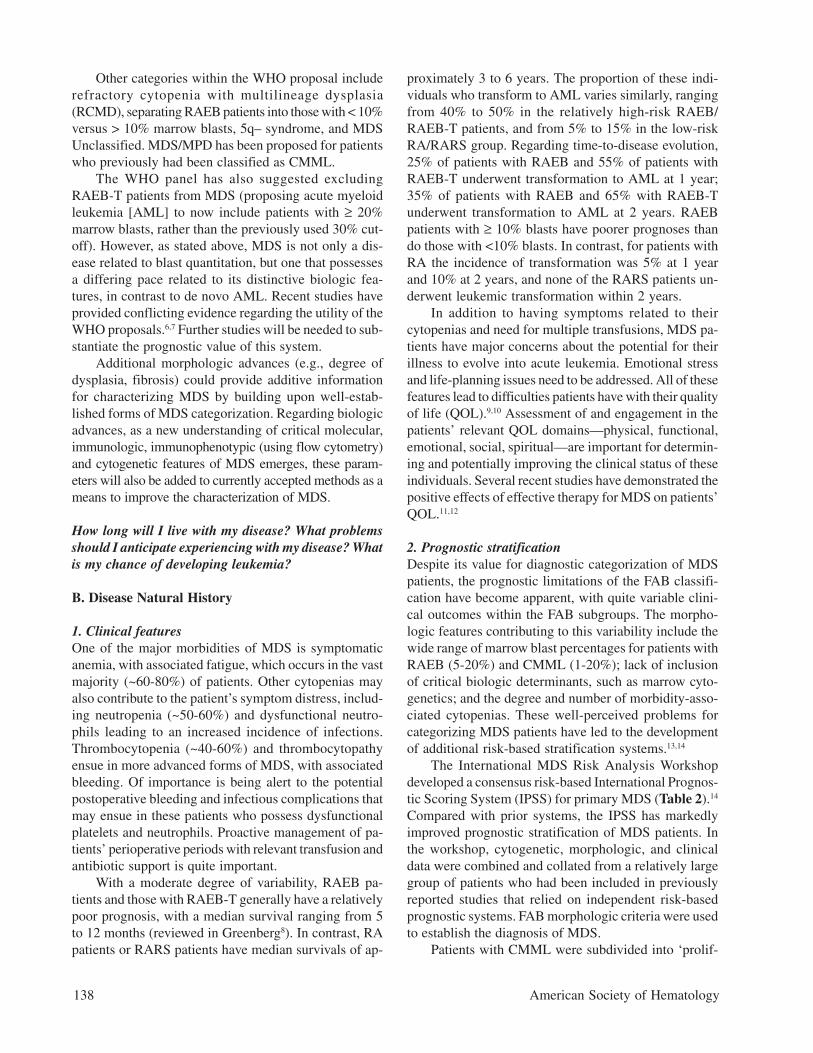
Methods are needed to enhance our ability to stratifypatients by their morphologic and biologic features. Suchapproaches could improve prognostication and treatmentfor these individuals. Regarding morphologic ap-proaches, a World Health Organization (WHO) panelhas recently issued a report with proposals for reclassi-fying MDS,3,4 although it has not yet been universallyaccepted because of certain controversial issues.5 In thisreport, suggestions have been made to modify the FABdefinitions of MDS. Although most prior data require atleast 2-line dysplasia to diagnose MDS, the WHO guide-lines accept unilineage dysplasia for the diagnosis of RAand RARS, so long as other causes of the dysplasia areabsent and the dysplasia persists for at least 6 months.Table 1 provides a comparison of the FAB and WHOclassifications.
Table 1. Classifications of myelodysplastic syndrome (MDS).
FAB1 WHO3,4
RA RA (unilineage)†5q– syndrome‡RCMD
RARS RARS† (unilineage)RCMD (with RS)
RAEB RAEB-IRAEB-II
RAEB-T AML
CMML MDS/MPD§
— Unclassified
Abbreviations: MDS, myelodysplastic syndrome; FAB, French-American-British; WHO, World Health Organization; RA, refractoryanemia; RCMD, refractory cytopenia with multilineage dysplasia;RARS, RA with ringed sideroblasts; RS, ringed sideroblasts; RAEB,RA with excess of blasts; RAEB-T, RAEB in transformation; AML,acute myeloid leukemia; CMML, chronic myelomonocytic leukemia;MPD, myeloproliferative disorder.
†Requires ≥ 6 months of anemia unrelated to other causes.
‡< 5% marrow blasts, micromegakaryocytes, thrombocytosis.
§MDS: WBC ≤13,000/mm3; MPD: WBC >13,000/mm3.
138 American Society of Hematology
Other categories within the WHO proposal includerefractory cytopenia with multilineage dysplasia(RCMD), separating RAEB patients into those with < 10%versus > 10% marrow blasts, 5q– syndrome, and MDSUnclassified. MDS/MPD has been proposed for patientswho previously had been classified as CMML.
The WHO panel has also suggested excludingRAEB-T patients from MDS (proposing acute myeloidleukemia [AML] to now include patients with ≥ 20%marrow blasts, rather than the previously used 30% cut-off). However, as stated above, MDS is not only a dis-ease related to blast quantitation, but one that possessesa differing pace related to its distinctive biologic fea-tures, in contrast to de novo AML. Recent studies haveprovided conflicting evidence regarding the utility of theWHO proposals.6,7 Further studies will be needed to sub-stantiate the prognostic value of this system.
Additional morphologic advances (e.g., degree ofdysplasia, fibrosis) could provide additive informationfor characterizing MDS by building upon well-estab-lished forms of MDS categorization. Regarding biologicadvances, as a new understanding of critical molecular,immunologic, immunophenotypic (using flow cytometry)and cytogenetic features of MDS emerges, these param-eters will also be added to currently accepted methods as ameans to improve the characterization of MDS.
How long will I live with my disease? What problemsshould I anticipate experiencing with my disease? Whatis my chance of developing leukemia?
B. Disease Natural History
1. Clinical featuresOne of the major morbidities of MDS is symptomaticanemia, with associated fatigue, which occurs in the vastmajority (~60-80%) of patients. Other cytopenias mayalso contribute to the patient’s symptom distress, includ-ing neutropenia (~50-60%) and dysfunctional neutro-phils leading to an increased incidence of infections.Thrombocytopenia (~40-60%) and thrombocytopathyensue in more advanced forms of MDS, with associatedbleeding. Of importance is being alert to the potentialpostoperative bleeding and infectious complications thatmay ensue in these patients who possess dysfunctionalplatelets and neutrophils. Proactive management of pa-tients’ perioperative periods with relevant transfusion andantibiotic support is quite important.
With a moderate degree of variability, RAEB pa-tients and those with RAEB-T generally have a relativelypoor prognosis, with a median survival ranging from 5to 12 months (reviewed in Greenberg8). In contrast, RApatients or RARS patients have median survivals of ap-
proximately 3 to 6 years. The proportion of these indi-viduals who transform to AML varies similarly, rangingfrom 40% to 50% in the relatively high-risk RAEB/RAEB-T patients, and from 5% to 15% in the low-riskRA/RARS group. Regarding time-to-disease evolution,25% of patients with RAEB and 55% of patients withRAEB-T underwent transformation to AML at 1 year;35% of patients with RAEB and 65% with RAEB-Tunderwent transformation to AML at 2 years. RAEBpatients with ≥ 10% blasts have poorer prognoses thando those with <10% blasts. In contrast, for patients withRA the incidence of transformation was 5% at 1 yearand 10% at 2 years, and none of the RARS patients un-derwent leukemic transformation within 2 years.
In addition to having symptoms related to theircytopenias and need for multiple transfusions, MDS pa-tients have major concerns about the potential for theirillness to evolve into acute leukemia. Emotional stressand life-planning issues need to be addressed. All of thesefeatures lead to difficulties patients have with their qualityof life (QOL).9,10 Assessment of and engagement in thepatients’ relevant QOL domains—physical, functional,emotional, social, spiritual—are important for determin-ing and potentially improving the clinical status of theseindividuals. Several recent studies have demonstrated thepositive effects of effective therapy for MDS on patients’QOL.11,12
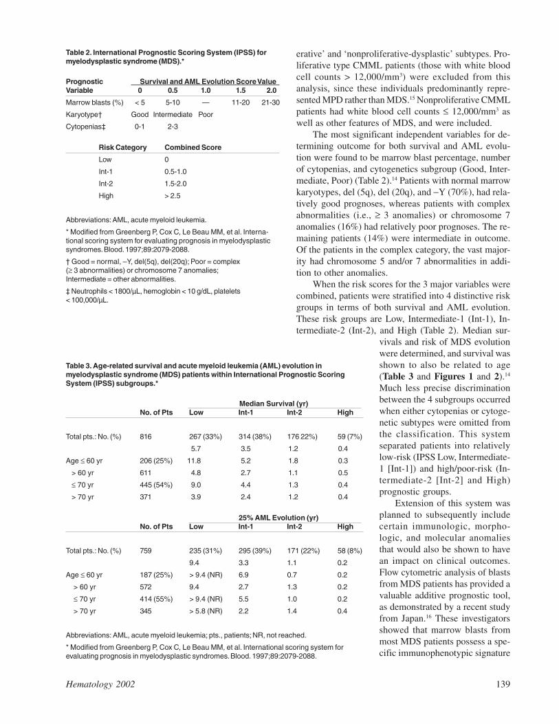
2. Prognostic stratificationDespite its value for diagnostic categorization of MDSpatients, the prognostic limitations of the FAB classifi-cation have become apparent, with quite variable clini-cal outcomes within the FAB subgroups. The morpho-logic features contributing to this variability include thewide range of marrow blast percentages for patients withRAEB (5-20%) and CMML (1-20%); lack of inclusionof critical biologic determinants, such as marrow cyto-genetics; and the degree and number of morbidity-asso-ciated cytopenias. These well-perceived problems forcategorizing MDS patients have led to the developmentof additional risk-based stratification systems.13,14
The International MDS Risk Analysis Workshopdeveloped a consensus risk-based International Prognos-tic Scoring System (IPSS) for primary MDS (Table 2).14
Compared with prior systems, the IPSS has markedlyimproved prognostic stratification of MDS patients. Inthe workshop, cytogenetic, morphologic, and clinicaldata were combined and collated from a relatively largegroup of patients who had been included in previouslyreported studies that relied on independent risk-basedprognostic systems. FAB morphologic criteria were usedto establish the diagnosis of MDS.
Patients with CMML were subdivided into ‘prolif-
Hematology 2002 139
erative’ and ‘nonproliferative-dysplastic’ subtypes. Pro-liferative type CMML patients (those with white bloodcell counts > 12,000/mm3) were excluded from thisanalysis, since these individuals predominantly repre-sented MPD rather than MDS.15 Nonproliferative CMMLpatients had white blood cell counts ≤ 12,000/mm3 aswell as other features of MDS, and were included.
The most significant independent variables for de-termining outcome for both survival and AML evolu-tion were found to be marrow blast percentage, numberof cytopenias, and cytogenetics subgroup (Good, Inter-mediate, Poor) (Table 2).14 Patients with normal marrowkaryotypes, del (5q), del (20q), and −Y (70%), had rela-tively good prognoses, whereas patients with complexabnormalities (i.e., ≥ 3 anomalies) or chromosome 7anomalies (16%) had relatively poor prognoses. The re-maining patients (14%) were intermediate in outcome.Of the patients in the complex category, the vast major-ity had chromosome 5 and/or 7 abnormalities in addi-tion to other anomalies.
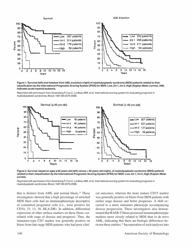
When the risk scores for the 3 major variables werecombined, patients were stratified into 4 distinctive riskgroups in terms of both survival and AML evolution.These risk groups are Low, Intermediate-1 (Int-1), In-termediate-2 (Int-2), and High (Table 2). Median sur-
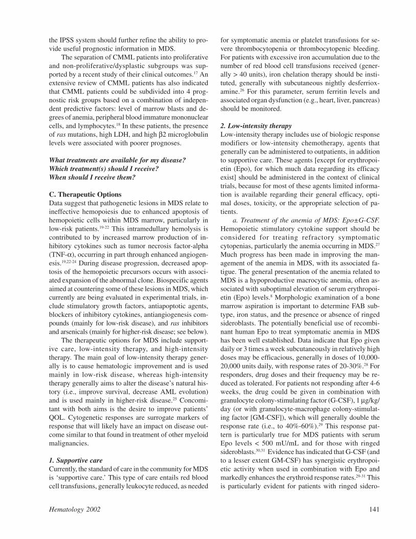
vivals and risk of MDS evolutionwere determined, and survival wasshown to also be related to age(Table 3 and Figures 1 and 2).14
Much less precise discriminationbetween the 4 subgroups occurredwhen either cytopenias or cytoge-netic subtypes were omitted fromthe classification. This systemseparated patients into relativelylow-risk (IPSS Low, Intermediate-1 [Int-1]) and high/poor-risk (In-termediate-2 [Int-2] and High)prognostic groups.
Extension of this system wasplanned to subsequently includecertain immunologic, morpho-logic, and molecular anomaliesthat would also be shown to havean impact on clinical outcomes.Flow cytometric analysis of blastsfrom MDS patients has provided avaluable additive prognostic tool,as demonstrated by a recent studyfrom Japan.16 These investigatorsshowed that marrow blasts frommost MDS patients possess a spe-cific immunophenotypic signature
Table 3. Age-related survival and acute myeloid leukemia (AML) evolution inmyelodysplastic syndrome (MDS) patients within International Prognostic ScoringSystem (IPSS) subgroups.*
Median Survival (yr)No. of Pts Low Int-1 Int-2 High
Total pts.: No. (%) 816 267 (33%) 314 (38%) 176 22%) 59 (7%)
5.7 3.5 1.2 0.4
Age ≤ 60 yr 206 (25%) 11.8 5.2 1.8 0.3
> 60 yr 611 4.8 2.7 1.1 0.5
≤ 70 yr 445 (54%) 9.0 4.4 1.3 0.4
> 70 yr 371 3.9 2.4 1.2 0.4
25% AML Evolution (yr)No. of Pts Low Int-1 Int-2 High
Total pts.: No. (%) 759 235 (31%) 295 (39%) 171 (22%) 58 (8%)
9.4 3.3 1.1 0.2
Age ≤ 60 yr 187 (25%) > 9.4 (NR) 6.9 0.7 0.2
> 60 yr 572 9.4 2.7 1.3 0.2
≤ 70 yr 414 (55%) > 9.4 (NR) 5.5 1.0 0.2
> 70 yr 345 > 5.8 (NR) 2.2 1.4 0.4
Abbreviations: AML, acute myeloid leukemia; pts., patients; NR, not reached.
* Modified from Greenberg P, Cox C, Le Beau MM, et al. International scoring system forevaluating prognosis in myelodysplastic syndromes. Blood. 1997;89:2079-2088.
Table 2. International Prognostic Scoring System (IPSS) formyelodysplastic syndrome (MDS).*
Prognostic Survival and AML Evolution Score ValueVariable 0 0.5 1.0 1.5 2.0
Marrow blasts (%) < 5 5-10 — 11-20 21-30
Karyotype† Good Intermediate Poor
Cytopenias‡ 0-1 2-3
Risk Category Combined Score
Low 0
Int-1 0.5-1.0
Int-2 1.5-2.0
High > 2.5
Abbreviations: AML, acute myeloid leukemia.
* Modified from Greenberg P, Cox C, Le Beau MM, et al. Interna-tional scoring system for evaluating prognosis in myelodysplasticsyndromes. Blood. 1997;89:2079-2088.
† Good = normal, –Y, del(5q), del(20q); Poor = complex(≥ 3 abnormalities) or chromosome 7 anomalies;Intermediate = other abnormalities.
‡ Neutrophils < 1800/µL, hemoglobin < 10 g/dL, platelets< 100,000/µL.
140 American Society of Hematology
that is distinct from AML and normal blasts.16 Theseinvestigators showed that a high percentage of enrichedMDS blast cells had an immunophenotype descriptiveof committed progenitor cells (i.e., were positive forCD34, 33, 13, 38, HLA-DR). In addition, differentialexpression of other surface markers on these blasts cor-related with stage of disease and prognosis. Thus, theimmature-type CD7 marker was generally positive onblasts from late-stage MDS patients who had poor clini-
cal outcomes, whereas the more mature CD15 markerwas generally positive on blasts from MDS patients withearlier stage disease and better prognoses. A shift oc-curred to a more immature phenotype accompanyingdisease progression. These investigators also demon-strated that RAEB-T blasts possessed immunophenotypicmarkers more closely related to MDS than to de novoAML, indicating that there are biologic differences be-tween these entities.16 Incorporation of such analyses into
Figure 1. Survival (left) and freedom from AML evolution (right) of myelodysplastic syndrome (MDS) patients related to theirclassification by the International Prognostic Scoring System (IPSS) for MDS: Low, Int-1, Int-2, High (Kaplan-Meier curves). AMLindicates acute myeloid leukemia.
Reprinted with permission from Greenberg P, Cox C, Le Beau MM, et al. International scoring system for evaluating prognosis inmyelodysplastic syndromes. Blood. 1997;89:2079-2088.
Figure 2. Survival, based on ages ≤≤≤≤≤ 60 years old (left) versus > 60 years old (right), of myelodysplastic syndrome (MDS) patientsrelated to their classification by the International Prognostic Scoring System (IPSS) for MDS: Low, Int-1, Int-2, High (Kaplan-Meiercurves).
Reprinted with permission from Greenberg P, Cox C, Le Beau MM, et al. International scoring system for evaluating prognosis inmyelodysplastic syndromes. Blood. 1997;89:2079-2088.
Hematology 2002 141
the IPSS system should further refine the ability to pro-vide useful prognostic information in MDS.
The separation of CMML patients into proliferativeand non-proliferative/dysplastic subgroups was sup-ported by a recent study of their clinical outcomes.17 Anextensive review of CMML patients has also indicatedthat CMML patients could be subdivided into 4 prog-nostic risk groups based on a combination of indepen-dent predictive factors: level of marrow blasts and de-grees of anemia, peripheral blood immature mononuclearcells, and lymphocytes.18 In these patients, the presenceof ras mutations, high LDH, and high β2 microglobulinlevels were associated with poorer prognoses.
What treatments are available for my disease?Which treatment(s) should I receive?When should I receive them?
C. Therapeutic OptionsData suggest that pathogenetic lesions in MDS relate toineffective hemopoiesis due to enhanced apoptosis ofhemopoietic cells within MDS marrow, particularly inlow-risk patients.19-22 This intramedullary hemolysis iscontributed to by increased marrow production of in-hibitory cytokines such as tumor necrosis factor-alpha(TNF-α), occurring in part through enhanced angiogen-esis.19,22-24 During disease progression, decreased apop-tosis of the hemopoietic precursors occurs with associ-ated expansion of the abnormal clone. Biospecific agentsaimed at countering some of these lesions in MDS, whichcurrently are being evaluated in experimental trials, in-clude stimulatory growth factors, antiapoptotic agents,blockers of inhibitory cytokines, antiangiogenesis com-pounds (mainly for low-risk disease), and ras inhibitorsand arsenicals (mainly for higher-risk disease; see below).
The therapeutic options for MDS include support-ive care, low-intensity therapy, and high-intensitytherapy. The main goal of low-intensity therapy gener-ally is to cause hematologic improvement and is usedmainly in low-risk disease, whereas high-intensitytherapy generally aims to alter the disease’s natural his-tory (i.e., improve survival, decrease AML evolution)and is used mainly in higher-risk disease.25 Concomi-tant with both aims is the desire to improve patients’QOL. Cytogenetic responses are surrogate markers ofresponse that will likely have an impact on disease out-come similar to that found in treatment of other myeloidmalignancies.
1. Supportive careCurrently, the standard of care in the community for MDSis ‘supportive care.’ This type of care entails red bloodcell transfusions, generally leukocyte reduced, as needed
for symptomatic anemia or platelet transfusions for se-vere thrombocytopenia or thrombocytopenic bleeding.For patients with excessive iron accumulation due to thenumber of red blood cell transfusions received (gener-ally > 40 units), iron chelation therapy should be insti-tuted, generally with subcutaneous nightly desferriox-amine.26 For this parameter, serum ferritin levels andassociated organ dysfunction (e.g., heart, liver, pancreas)should be monitored.
2. Low-intensity therapyLow-intensity therapy includes use of biologic responsemodifiers or low-intensity chemotherapy, agents thatgenerally can be administered to outpatients, in additionto supportive care. These agents [except for erythropoi-etin (Epo), for which much data regarding its efficacyexist] should be administered in the context of clinicaltrials, because for most of these agents limited informa-tion is available regarding their general efficacy, opti-mal doses, toxicity, or the appropriate selection of pa-tients.
a. Treatment of the anemia of MDS: Epo±G-CSF.Hemopoietic stimulatory cytokine support should beconsidered for treating refractory symptomaticcytopenias, particularly the anemia occurring in MDS.27
Much progress has been made in improving the man-agement of the anemia in MDS, with its associated fa-tigue. The general presentation of the anemia related toMDS is a hypoproductive macrocytic anemia, often as-sociated with suboptimal elevation of serum erythropoi-etin (Epo) levels.8 Morphologic examination of a bonemarrow aspiration is important to determine FAB sub-type, iron status, and the presence or absence of ringedsideroblasts. The potentially beneficial use of recombi-nant human Epo to treat symptomatic anemia in MDShas been well established. Data indicate that Epo givendaily or 3 times a week subcutaneously in relatively highdoses may be efficacious, generally in doses of 10,000-20,000 units daily, with response rates of 20-30%.28 Forresponders, drug doses and their frequency may be re-duced as tolerated. For patients not responding after 4-6weeks, the drug could be given in combination withgranulocyte colony-stimulating factor (G-CSF), 1 µg/kg/day (or with granulocyte-macrophage colony-stimulat-ing factor [GM-CSF]), which will generally double theresponse rate (i.e., to 40%-60%).29 This response pat-tern is particularly true for MDS patients with serumEpo levels < 500 mU/mL and for those with ringedsideroblasts.30,31 Evidence has indicated that G-CSF (andto a lesser extent GM-CSF) has synergistic erythropoi-etic activity when used in combination with Epo andmarkedly enhances the erythroid response rates.29-31 Thisis particularly evident for patients with ringed sidero-
142 American Society of Hematology
blasts or with serum Epo levels < 500 mU/mL.Iron repletion needs to be verified prior to institut-
ing Epo therapy. Often, despite adequate iron stores, poorutilization of storage iron in many of these patients maymake oral iron supplementation helpful. Erythroid re-sponse generally occurs within 2 to 3 months of treat-ment. If no response occurs by that time, this treatmentshould be considered a failure and discontinued and al-ternative therapies considered (generally within a clini-cal trial). These therapies to consider (indicated below)include specific attempts at altering biologic lesions inMDS: anti-angiogenic compounds (anti-vascular endot-helial growth factor [VEGF] drugs), anti-tumor necro-sis factor (TNF) agents (e.g., Enbrel), thalidomide, 5-azacytidine (AzaC), immunosuppressive type therapies(e.g., antithymocyte globuline [ATG], cyclosporin), sin-gly or in combination. As multiple biologic abnormali-ties contribute to the cytopenias of MDS, it is likely thatcombination therapy will be needed to sustain hemato-logic responses in these patients.
A recent report indicated the presence of neutraliz-ing anti-erythropoietin antibodies associated with purered cell aplasia and Epo resistance in a small number ofpatients with renal failure who had received chronic Epotreatment.32 Virtually all such cases have been reportedfrom Europe and may relate to differing Epo prepara-tions or manufacturing processes which differ from thoseemployed elsewhere. Morphologic marrow examinationand Epo antibody assays will aid in diagnosis of thisiatrogenic problem.
For attempting to enhance erythropoiesis in MDSby using less frequent dosing, a modified form of eryth-ropoietin (darbepoetin) with a 3-fold longer biologic half-life than standard Epo has shown good efficacy with lessfrequent dosing for patients with anemias induced byrenal failure or chemotherapy.33 Trials are ongoing toassess the role of this agent in MDS.
b. Treatment of neutropenia. Although low neutro-phil levels have been demonstrated to be improved witheither recombinant human G-CSF or GM-CSF, data havenot shown decreased infections or improved infectionmanagement with these agents in MDS. A Phase III ran-domized trial in high-risk MDS that compared chronicadministration of G-CSF to observation and demon-strated no difference in the incidence or pace of devel-opment of AML or of infections in the 2 study arms.34
For those patients with RAEB, a shortened survival wasnoted in the G-CSF arm. Thus, this agent is not recom-mended for chronic prophylactic use alone in MDS.Rather, a suggested approach is to employ these cyto-kines in neutropenic MDS patients with recurrent orantibiotic-refractory infections.
c. Treatment of thrombocytopenia. For treating
thrombocytopenia in MDS, relatively low doses (10 µg/kg/d) of IL-11 have recently been shown in a Phase I/IItrial to be more effective than higher doses of the drug.35
Further studies are ongoing with this agent. Occasionalefficacy of danazol has also been reported in such pa-tients. Studies are warranted to determine the potentialefficacy of thrombopoietin for the thrombocytopeniapresent in MDS.
d. 5-azacytidine. As a form of low-intensity chemo-therapy, recent encouraging data in MDS have been re-ported with the hypomethylating agent AzaC.36 In a ran-domized Phase III trial, AzaC decreased the risk of leu-kemic transformation; it also improved the survival of aportion of the patients.36 This trial evaluated 171 MDSpatients, comparing AzaC (75 mg/m2/d subcutaneouslyfor 7 days every 28 days, predominantly as an outpa-tient) with supportive care. Patients in the supportive carearm whose disease worsened were permitted to crossover to AzaC. Responses occurred in 60% of patientson the AzaC arm (7% complete response, 16% partialresponse, 37% improved) compared with 5% (improved)receiving supportive care. Median time to leukemic trans-formation or death was 21 months for AzaC versus 13months for supportive care. Transformation to AMLoccurred in 15% of patients on the AzaC arm and in38% receiving supportive care. Eliminating the con-founding effect of early crossover to AzaC, a landmarkanalysis after 6 months showed median survival of anadditional 18 months for AzaC and 11 months for sup-portive care. QOL assessment found significant majoradvantages in physical function, symptoms, and psycho-logical state for patients initially randomized to AzaC.12
Thus, AzaC treatment resulted in significantly higherresponse rates, improved QOL, reduced risk of leuke-mic transformation, and improved survival comparedwith supportive care. This agent appears to provide anew treatment option that is superior to supportive care forpatients with MDS. Further trials with this agent are ongo-ing to attempt to confirm and extend these findings.
e. Immunosuppressive therapy. The biologic re-sponse modifiers potentially capable of altering immunemedicated subtypes of MDS which are now availableinclude ATG and cyclosporine37-40 and will be discussedin detail by Dr. Neal Young in Section II. However, thesedata, though encouraging for specific subtypes of MDS(mainly hypoplastic MDS patients with normal cytoge-netics and evidence of a paroxysmal nocturnal hemo-globinuria [PNH] clone and HLA-DR2 subtype), requirefurther evaluation in more extended clinical trials.
f. Anti-TNF, anti-angiogenesis agents.NumerousPhase I/II clinical trials evaluating the roles of specificbiologic response modifiers have been used to treat MDS.Relatively small pilot trials with anti-TNF fusion pro-

Hematology 2002 143
tein (etanercept, Enbrel) indicated erythroid responsesin about 30% of patients in one trial, and lower levels ofresponses in another.41,42
Clinical trials assessing the role of antiangiogenesiscompounds [e.g., thalidomide, anti-VEGF agents] areongoing. Although thalidomide has multiple mechanismsof action, its role as an antiangiogenic or anti-TNF agentmay be important in its therapeutic effects. Results oftreatment with thalidomide have been reported in 2 sepa-rate trials, with a 19% response rate in one and 56% inthe other, including erythroid and trilineage responses.43,44
In the latter trial, relatively high doses of thalidomide(i.e. > 300 mg/day) were tolerated, an unusual findingfor this elderly population. Additional studies, includ-ing those with less toxic metabolites of thalidomide, areongoing to better define the role of these agents in MDS.
Use of a number of biologic response modifiers(amifostine, pentoxyphylline, interferon, low-dose AraC,retinoids, vitamin D analogues, butyrates) has previouslybeen shown to have quite limited efficacy in Phase I-IItrials with MDS patients.8,45 Other ongoing clinical tri-als include the use of agents capable of inhibiting neo-plastic growth, such as anti-ras type compounds [farnesyltransferase inhibitors (FTIs)] and arsenic trioxide. Re-cent preliminary studies have shown efficacy of FTIs intreating poor-risk AML, CMML, and MPDs.46,47
3. High-intensity therapyHigh-intensity therapy includes intensive induction che-motherapy or hemopoietic (marrow or peripheral blood)stem cell transplantation (HSCT).8,48 Although these ap-proaches have a greater chance of changing the naturalhistory of the disease, they also have an attendant in-creased risk of regimen-related morbidity and mortal-ity. The management of patients with relatively high-risk (i.e., IPSS Int-2, High) MDS or MDS-AML is par-ticularly problematic and, as indicated above, must in-volve consideration of their age and performance sta-tus. These patients are generally elderly, often with ma-jor co-morbidities, and have a higher proportion of bio-logic abnormalities of their HSCs. The marrow precur-sors of high-risk MDS and MDS-AML patients are of-ten associated with multilineage dysplasia and possessconcomitant features of primitive marrow stem cells,poor-risk marrow cytogenetic abnormalities, and a higherdegree of cellular MDR than do those from de novoAML.49,50
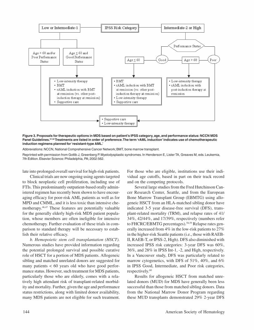
For individuals < 60 years old with relatively high-risk disease and good performance status, preferenceshould be given to high-intensity therapy (Figure 3).51,52
In this clinical setting, using an HLA-matched donor forallogeneic HSCT is the preferred high-intensity therapyoption. For those lacking a matched donor, induction
chemotherapy (intensive or with experimental biologicagents), AzaC, or experimental trials are preferred. Forhigh-risk patients > 60 years old with good performancestatus, AzaC or high-intensity experimental trials arepreferred (Figure 3).
a. Chemotherapy. Responses to standard chemo-therapy in MDS or MDS-related AML are lower than inde novo AML.8,53 This disparity is due to both the gener-ally elderly age of MDS patients and the differing biol-ogy of the MDS stem cell and marrow stroma, includingpoor-risk cytogenetics, earlier stem cell phenotype, andincreased expression of multidrug resistance (MDR)markers. Recent comparative studies have not demon-strated benefit of several different intensive chemo-therapy regimens in MDS.54 Trials of intensive chemo-therapy in MDS have established that advanced age (>45-50 years) is an unfavorable prognostic factor. Fur-thermore, MDS patients with karyotypic abnormalitieshave lower complete remission and survival rates com-pared to patients with normal cytogenetics.
A high degree of MDR-1 (p170 glycoprotein) oc-curs in advanced MDS precursors with associated de-creased responses and response durations with manystandard treatment regimens of induction chemo-therapy.50 Numerous compounds, including quinine,cyclosporin A, and the cyclosporin A analog PSC-833(Valspodar), have been tested for their ability to reversethe MDR phenotype. The French Cooperative Leuke-mia Group reported the results of a Phase III random-ized study that showed that quinine plus chemotherapysignificantly increased the complete remission rate andsurvival in MDR-1-positive MDS patients compared tochemotherapy alone.55 This positive experience has gen-erally not been reproduced with other MDR-1 modulat-ing agents.56 Additional MDR modulating agents pluschemotherapy are being evaluated for treating patientswith advanced MDS. Given the multiple possible resis-tance mechanisms extant in MDS/leukemic cells, aftersuch lesions are identified, it may be useful to try com-binations of MDR blockers.
Another hypomethylating agent with activity andmechanism of action similar to AzaC is decitabine (5-aza-2′-deoxycytidine). With doses requiring hospitaliza-tion of patients, encouraging results have been showntreating high-risk MDS patients, some showing cytoge-netic conversion.57 The overall response rate was 49%,with 64% responses in patients with a high-risk IPSS.The median response duration was 31 weeks, and themedian survival from the start of therapy was 15 months(14 months for the high-risk IPSS patients). In severalpatients, normalization of clonal cytogenetic abnormali-ties (often high-risk) was seen. An ongoing phase IIIstudy aims to establish whether these responses trans-
144 American Society of Hematology
late into prolonged overall survival for high-risk patients.Clinical trials are now ongoing using agents targeted
to block neoplastic cell proliferation, including use ofFTIs. This predominantly outpatient-based orally admin-istered regimen has recently been shown to have encour-aging efficacy for poor-risk AML patients as well as forMPD and CMML, and it is less toxic than intensive che-motherapy.46,47 These features are potentially valuablefor the generally elderly high-risk MDS patient popula-tion, whose members are often ineligible for intensivechemotherapy. Further evaluation of these trials in com-parison to standard therapy will be necessary to estab-lish their relative efficacy.
b. Hemopoietic stem cell transplantation (HSCT).Numerous studies have provided information regardingthe potential prolonged survival and possible curativerole of HSCT for a portion of MDS patients. Allogeneicsibling and matched unrelated donors are suggested formany patients < 60 years old who have good perfor-mance status. However, such treatment for MDS patients,particularly those who are elderly, comes with a rela-tively high attendant risk of transplant-related morbid-ity and mortality. Further, given the age and performancestatus restrictions, along with limited donor availability,many MDS patients are not eligible for such treatment.
For those who are eligible, institutions use their indi-vidual age cutoffs, based in part on their track recordand on the competing protocols.
Several large studies from the Fred Hutchinson Can-cer Research Center, Seattle, and from the EuropeanBone Marrow Transplant Group (EBMTG) using allo-geneic HSCT from an HLA-matched sibling donor haveindicated 3-5 year disease-free survival (DFS), trans-plant-related mortality (TRM), and relapse rates of 41/34%, 42/44%, and 17/39%, respectively (numbers referto FHCRC/EBMTG percentages).58,59 Relapse rates gen-erally increased from 4% in the low-risk patients to 27%in the higher-risk Seattle patients (i.e., those with RAEB-II, RAEB-T, or IPSS-2, High). DFS also diminished withincreased IPSS risk categories: 3-year DFS was 60%,36%, and 28% in IPSS Int-1, -2, and High, respectively.In a Vancouver study, DFS was particularly related tomarrow cytogenetics, with DFS of 51%, 40%, and 6%in IPSS Good, Intermediate, and Poor risk categories,respectively.60
Results for allogeneic HSCT from matched unre-lated donors (MUD) for MDS have generally been lesssuccessful than those from matched sibling donors. Datafrom the National Marrow Donor Program regardingthese MUD transplants demonstrated 29% 2-year DFS
Figure 3. Proposals for therapeutic options in MDS based on patient’s IPSS category, age, and performance status: NCCN MDSPanel Guidelines.51,52 Treatments are listed in order of preference. The term ‘rAML induction’ indicates use of chemotherapeuticinduction regimens planned for ‘resistant-type AML.’
Abbreviations: NCCN, National Comprehensive Cancer Network; BMT, bone marrow transplant.
Reprinted with permission from Gotlib J, Greenberg P. Myelodysplastic syndromes. In Henderson E, Lister TA, Greaves M, eds. Leukemia,7th Edition. Elsevier Science: Philadelphia, PA; 2002:562.
Hematology 2002 145
and 54% TRM but relapse rates of only 14%.61 Graftfailure and GVHD were particular problems in thesetransplants. Improved DFS was independently associ-ated with less advanced MDS subtype, higher cell dose,recipient cytomegalovirus (CMV) seronegativity, shorterinterval from diagnosis to transplantation, and transplan-tation in recent years. Higher TRM was independentlyassociated with older recipient and donor age, HLA mis-match, and recipient CMV seropositivity. However, re-cent studies from Seattle have demonstrated improvedresults for these patients using targeted busulfan levelsplus cytoxan in preparative regimens.62 Allogeneic HSCTwith related/unrelated matched donors in 109 patientshad 3 year DFS, TRM, and relapse rates of 56/59%, 28/30%, and 16/11%, respectively. In addition, relapse-freesurvival was improved in those receiving peripheralblood compared to marrow stem cells.
Non-myeloablative HSCT regimens63 with improvedpatient tolerance, particularly for the elderly MDS pa-tients, are important experimental options currently be-ing evaluated. In certain research settings, autologousHSCT are performed after chemotherapy-induced remis-sion.64 Comparative clinical trials are needed to estab-lish whether such transplants should be performed be-fore or after achievement of remission following induc-tion chemotherapy. Major progress is being made intransplant biology and clinical approaches, with specificfocus for MDS. Such advances should provide improvedtherapeutic options for treating these patients within theforeseeable future.
Autologous HSCT has been used for treating somepatients in complete remission after induction chemo-therapy. Although the transplant-related mortality is lowwith this approach, it is attended by a relatively highrelapse rate.
4. Factors determining treatment approachesBased on the above biologic and clinical considerations,deliberations by the U.S. National Comprehensive Can-cer Network (NCCN) Panel for MDS Practice Guide-lines have suggested51 that management approaches uti-lizing an algorithm composed of the patient’s: (a) IPSSprognostic subgroup categorization; (b) Age; and (c)Performance status.
This approach provides a useful risk-based methodto stratify therapeutic options and permits individual-ized prioritization of low- versus high-intensity therapyfor patients in differing risk categories. Careful moni-toring for disease progression and the patient’s desiresplay major roles in the timing and decision to embark onspecific treatment. It is useful to stratify patients intoage groups > 60 or < 60 years old, as this is often the ageof eligibility for certain intensive therapeutic options,
particularly for HSCT. Medical centers use differing agecutoff points, and thus biologic rather than chronologicage may be used to establish patients’ protocol eligibility.
Low-intensity treatments (e.g., with biologic re-sponse modifiers) are generally preferred for MDS pa-tients in the lower-risk categories (IPSS Low and Int-1),whereas high intensity (e.g., intensive chemotherapy orHSCT) is used for the higher-risk patient group (IPSSInt-2, High), depending on the patient’s age, performancestatus, and personal preferences51 (Figure 3 and ref. 52).
A finite period of observation (at least 4-6 weeks) isneeded to determine the degree of clinical stability forMDS patients. Individuals with unstable disease (i.e.,those with evidence of clinical progression, including adecrease in blood counts or increasing transfusions) needfollow-up evaluation, which should include a repeatmarrow examination. If the patient remains in the Int-1or Low-risk group, the treatment options indicated forthose patients (Table 3) should be continued. If, how-ever, the patient progresses to the Int-2 or High-riskgroups (or the patient had initially been in one of theserisk groups), the treatment path for these higher-riskcategories should be used.
For patients with poor performance status in eitherrisk category, only supportive care or low-intensity treat-ment is suggested. This proposal relates to their concur-rent co-morbidities, rendering them unlikely to toleratehigh-intensity treatments.
How can I learn more about my illness? Are thereclinical trials with which I can and should becomeinvolved? How do I find out about them?
D. ResourcesMany institutions have health libraries with medicalbooks, some focused intended for the lay public, thatreview the hematologic malignancies. Stocking suchbooks in these libraries could be useful for MDS pa-tients and their families, particularly if the information/jargon is “translated” in concert with health care profes-sionals. Both the MDS Foundation (www.mds-foundation.org) and the Aplastic Anemia-MDS Foun-dation (www.aamds.org) have developed websites andpublished booklets providing information for patients,physicians, and other health care professionals regard-ing MDS. These websites describe ongoing clinical tri-als and indicate national and international institutionsthat are involved in these trials and with physicians whopossess special expertise in evaluating and treating MDS.Patient and physician contact with physicians at theseinstitutions and their involvement in these trials will becentral for progress to occur in developing appropriatetherapeutic directions for this disease.

146 American Society of Hematology
II. ROLE OF THE IMMUNE SYSTEM
IN THE PANCYTOPENIA OF MDS AND
IMMUNOSUPPRESSIVE THERAPIES
Neal S. Young, MD*
The concept of MDS originated in the merger of two ill-defined but clearly different hematologic processes—“preleukemia” and “refractory anemia”—and the FABclassification attempted to collect several disparate di-agnostic categories by shared histologic features, mainlythe appearance of dysmorphic marrow cells. This orga-nizational scheme is now fracturing: on one side, themore obviously malignant categories—RA with excessblasts and chronic myelomonocytic leukemia—havebeen ceded to classifications of the acute leukemias andmyeloproliferative processes; on the other, the indistinctboundary between hypocellular MDS and aplastic ane-mia (AA), largely ignored by the FAB classification, hasbeen enlarged with the recognition of similar pathophysi-ologic mechanisms and common responses to therapiesextending to some pancytopenias associated with franklycellular bone marrows.
Indeed, pancytopenia, not leukemia, is the proxi-mate cause of death in patients who die as a result ofMDS. Why hematopoiesis fails, and especially the ex-planation for the development of bone marrowhypocellularity that accompanies this failure, is notclearly understood. Nevertheless, recent successful thera-peutic pilot studies and novel results of laboratory stud-ies, reviewed here and elsewhere,1,2 suggest that MDS isclosely related to diseases in which the pathophysiol-ogy of bone marrow failure is mediated at least in partby the immune system.
A. MDS and AAFor the clinician, MDS and AA can be difficult to dis-tinguish. AA has been defined historically by extremelylow bone marrow cellularity on biopsy. A significantproportion of MDS also shows marrow hypocellularity,with large series averaging about 20% of cases; hypo-cellular MDS has not been reported to be markedly dif-ferent in clinical characteristics like age, FAB subtype,or prognosis from the more common cellular disease.3,4
However, between AA and MDS the differential diag-nosis often relies on “soft” histologic criteria, such asmyeloid cells lacking granules (an appearance that canalso arise from technical staining artifact), dyserythro-
poiesis (with megaloblastoid changes frequently seen inaplasia), or dysmorphic megakaryocytes (but these cellsare usually scanty in a hypocellular aspirate smear). Bonemarrow cell cytogenetic abnormalities are more objec-tive evidence of MDS, but some authorities regard find-ings such as trisomy 6 or 8 as simply delineating a sub-set of otherwise typical AA. Both AA and MDS are as-sociated with expansion of clones of PIG-A mutated stemcells, lacking cell surface expression of the glycosyl-phosphoinositol-linked proteins diagnostic of paroxys-mal nocturnal hemoglobinuria (PNH), leading to diag-noses of AA/PNH or MDS/PNH.
The distinction between MDS and AA becomes fur-ther blurred because some AA patients, usually after im-munosuppressive therapy, appear to evolve to MDS, withrecurrent pancytopenia associated with bone marrowdysplasia or chromosomal aberrations. In our series of122 patients with severe AA treated at NIH with anti-thymocyte globulin (ATG) and cyclosporine, 13 hadevolved at 5 years; in the European Group for BoneMarrow Transplantation experience, the risk for evolu-tion to MDS was estimated at about 12% at 12 yearsposttreatment.5,6 There are stereotypical patterns of chro-mosome changes:7 monosomy 7 is most frequent, asso-ciated with refractory pancytopenia and leukemic con-version in patients who have not responded to immuno-suppressive treatment; in contrast, trisomy 8 typically isobserved in patients whose adequate blood counts oftenrequire continued cyclosporine therapy.
AA is a more uniform clinical entity than is MDS:blood count findings are usually striking, the bone mar-row morphology is unambiguous, and the response totherapy is relatively predictable. The essential patho-physiology of acquired AA also has been delineated, atleast in outline,8 as an efficient and specific immune sys-tem attack on hematopoietic stem and progenitor celltargets, leading to the absence of these cells on morpho-logic, phenotypic, and functional assays. T cells of Th1/Tc1 cytokine profile, producing interferon-γ and tumornecrosis factor (TNF), induce apoptosis in their marrowtargets through activation of the Fas receptor, leading todestruction of the hematopoietic cell compartment. Atthe time of presentation with severe pancytopenia thisprocess is advanced; surrogate stem cell assays suggestthat the stem cell pool is reduced to a few percentagepoints or less of normal. Most patients do respond withhematologic improvement to immunosuppressive thera-pies, but both relapse of pancytopenia and a requirementfor continued cyclosporine administration to maintainadequate blood counts are common.
* Chief, Hematology Branch, Building 10, Room 7C103,National Institutes of Health, 9000 Rockville Pike, Bethesda,MD 20892-1652
Hematology 2002 147
B. The Origin of Hematopoietic Failure in MDS
1. MDS hematopoiesis in tissue cultureThe highly varied pattern of hematopoiesis in tissue cul-ture in MDS should be contrasted with a monotonouspicture in AA. Cell proliferation rates are often elevated,as measured by in vitro nucleotide incorporation,9 au-tonomous colony formation,10 or expression of specificcell markers like MIB-1,11 reflective of the cellular char-acter of most MDS bone marrows. Hematopoietic pro-genitor-derived colony formation is variable: early andlate erythroid progenitors (CFU-E and BFU-E, respec-tively) are usually diminished, consistent with the preva-lence of anemia, while myelopoiesis from granulocyte-macrophage colony forming cells (CFU-GM) may benear normal in the majority of the same cases.12 Moreprimitive multipotential progenitors (CFU-GEMM) gen-erally have been reduced,13 as have long-term culture-initiating cells (LTC-IC) (secondary colony formationwas usually low, but a quarter of patients neverthelessretained normal numbers;14 others found blast colony-forming normal in number but deficient in secondaryerythroid colonies).15 Entirely normal colony formation,as occurs in a significant proportion of patients, is foundin good prognostic subtypes, such as 5q– and ringedsideroblast anemia. Conversely, patients with low CFU-GM numbers, aberrant myeloid maturation in vitro, pre-dominant cluster over colony formation, and abnormalimmunophenotypes of progenitors (all findings seen inacute myelogenous leukemia) have an expectedly poorprognosis.12 The response of progenitors to hemato-poietins in vitro is poor, even with high concentrationsof purified or recombinant factors, alone or in combina-tion. Tissue culture studies have not been especially pre-dictive of therapeutic outcomes in clinical interventionaltrials, but pharmacologic concentrations of erythropoi-etin, G-CSF, or GM-CSF do increase lineage-specificcolony growth, at least in a subset of patients.16 PurifiedCD34 cells from MDS patients have generally normal,if variable, response profiles to early acting growth fac-tors such as stem cell factor, interleukin (IL)-3, and IL-6,17,18 but primitive colony formation, when low, has beenunaffected by addition of exogenous factors.19
2. Cell death in the dysplastic marrowThe occurrence in MDS of pancytopenia despite nor-mal to increased numbers of marrow precursor cells ledto the inference that cell death in the hematopoietic cellcompartment is dominant over cell proliferation (for re-views, see refs. 20-22). That the histologic abnormalitiesof MDS might reflect not only a process of intramedul-lary cellular destruction but specifically apoptosis or pro-grammed cell death was first proposed by Clark and
Lampert,23 and more recent, quantitative studies havesupported this conclusion: for example, an elevatedapoptotic index of 3% in MDS compared to normalmarrow values of 1%, as based on morphological andultrastructural changes on thin section biopsies.24 Poorgrowth of colonies and dominance of clusters may re-flect a high initial rate of growth followed by failure ofboth proliferation and differentiation.25 However, cul-tured MDS cells also manifest much more striking de-grees of apoptosis than may be seen in fresh cells, a po-tential source of artifact.26 In biopsy specimens, singlecells can be examined for evidence of apoptosis, but theirexact lineage usually cannot be confidently determined,and not only hematopoietic precursors but also stromalcells, endothelium, and fat may score positive.27
Other more specific measurements of apoptosis havebeen abnormally elevated in MDS in comparison withnormal and also leukemic marrow: in situ end labeling(ISEL) detection of DNA strand breaks; terminal deoxy-nucleotide transferase incorporation of nucleotides on3′ ends of DNA (TUNEL); detection of subgenomicDNA in histograms of cell populations subjected tofluorescence-activated flow cytometry; binding ofannexin-V to exposed phosphatidylserine of cell mem-branes, also assessed by flow cytometry; and the detec-tion of apoptosis-related proteins like bcl-2 in individualpermeabilized cells. Mechanistically, low bcl-2 expres-sion has correlated with high apoptotic rates,10,28,29 and en-hanced bcl-2 expression has been linked to leukemictransformation; sequential caspase activation consistentwith appropriate death signal transduction also has beenreported.30 Improvements in blood counts with growthfactor therapy31 and anti-inflammatory agents likepentoxifylline32 have correlated with lowered rates ofapoptosis. However, the MDS patient populations ex-amined have been very heterogeneous, and the range ofresults broad; for example, in one large study of 175patients by ISEL, 71 showed high and 43 low levels ofapoptosis, and 61 were normal.33
More detailed conclusions have proven controver-sial. First, one appealing hypothesis proposed that pro-grammed cell death dominated early in the pancytopenicphase of MDS, and that leukemic transformation repre-sented escape from the apoptotic (intracellular) processor (extracellular) environment. Higher rates of apoptosishave been observed in RA and RAEB compared withtransforming MDS or acute leukemia,28,34-36 while oth-ers have found increased rates in late stages of disease.37
Second, a question remains as to the extent of apoptosisin MDS, with quantitative inferences ranging from mini-mally or modestly increased26 to massive destruction,38
arguments confounded by significant levels of apoptosisin normal marrow specimens, and often relatively small
148 American Society of Hematology
differences between MDS and normal. Third, is apoptosisrestricted to certain hematopoietic cells in MDS? Evi-dence from flow cytometric analysis of individual cellssupports apoptosis in the CD34 cell compartment.34,36,39
Others have suggested that cell death is far more fre-quent among mature cells (of high compared with lowdensity)38 and rare by TUNEL measurements in CD34cells within biopsies.40
3. Pro-inflammatory cytokines andimmune triggering of apoptosis in MDSSimilar mechanisms of hematopoietic cell destructionhave been hypothesized to operate in MDS as for AA.Early observations included measurements of overex-pression by cultured MDS patients’ blood41 and marrowcells42 of lymphocyte type I cytokines, TNF, and inter-feron-γ. TNF levels have been elevated in MDS marrowbiopsies30,43 and in plasma and sera,44,45 as has circulat-ing soluble TNF receptor.46 Levels have declined on treat-ment with anticytokine therapies.47,48 Multiplex poly-merase chain reaction has shown elevated marrow mes-senger RNA (mRNA) for TNF among other cytokinesin a subset of MDS patients.49 MDS bone marrows haveevidence of downstream effects of TNF activity such ascaspase activity,30 induction of nitric oxide synthase,50
and intracellular redox changes leading to DNA dam-age.51 As with levels of apoptosis, different groups havereported variable correlations: TNF expression has beenelevated with RA45 or disease transforming to leukemia,52
heralded a poor prognosis,53 or been found entirely un-related to MDS subtype.54
The source of TNF in MDS marrow has been as-sumed to be the macrophage;52 in one large study ofmarrow biopsies, TNF, transforming growth factor(TGF), and monocyte/macrophage numbers were highlycorrelated.55 Stromal elements56-58 also have been impli-cated. Our group identified T lymphocytes functionallyinhibitory of autologous marrow progenitor cells, withappropriate class I histocompatibility restriction and withdiminished activity after successful immunosuppressivetherapy (see below).59 Additionally, MDS patients mayhave increased cytotoxic T cells60 and a skewed T cellrepertoire, as assessed by T cell receptor Vβ type.59
TNF expression has been studied in relationship toinduction on hematopoietic target cells of Fas, a cell sur-face protein and member of the tumor necrosis factorreceptor family; Fas triggering initiates programmed celldeath. Flow cytometry has been used to measure in-creased Fas and Fas-ligand surface expression on MDSCD34 cells; in functional assays, monoclonal antibody-mediated blockade of Fas, Fas ligand, or TNF enhancedMDS hematopoiesis in long-term bone marrow colonyculture and progenitor assays.45 Addition of anti-Fas
ligand antibody in other experiments also decreasedapoptosis in MDS marrow samples.61 TNF-relatedapoptosis inducing ligand (TRAIL) also may be involvedin MDS: multiple members of this family and its recep-tors were overexpressed in patient marrows, and TRAILaddition has modulated hematopoietic colony formationby MDS but not normal marrow, with its effects varyingwith disease stage.62,63 Other in vitro experiments havelinked Fas to ineffective erythropoiesis of MDS: in dif-ferentiating erythroid cells the rate of apoptosis in MDSCD34-cell derived culture was high, Fas was over-expressed in fresh and cultured cells, and the peak ofapoptosis correlated with Fas ligand expression.64 Flowcytometric results for Fas and Fas-ligand expression havebeen confirmed by amplification of complementary DNA(cDNA) from mRNA45,65 and immunohistochemistry.35,65
Fas expression usually has not correlated to the degreeof apoptosis in clinical specimens.66,67 Fas expression hasbeen more striking in RA than in other MDS subtypes45
and may decrease in the MDS marrow during transfor-mation to leukemia.35 An important clue as to the iden-tity of a target of Fas-mediated cytotoxicity has beensuggested by experiments using marrows from patientswith different cytogenetic abnormalities: trisomy 8 cellsexpress Fas and markers of apoptosis and are suscep-tible to Fas triggering in vitro, in contrast to cells frompatients with monosomy 7 or other chromosomechanges.68
C. Clinical Immune Dysfunction in MDSGlobal peculiarities of immune system function haveincluded decreased NK cell activity, antibody dependentcell killing, mitogenic response, and diminished CD4cell numbers, and for B cell function altered immuno-globulin levels, monoclonal gammopathies, and variousautoantibodies (reviewed in ref. 69). In a large Japanesesingle institution collection of 153 patients, 63% hadsome abnormal immunological test: hypergamma-globulinemia was most common followed by hypo-globulinemia, and the presence of antinuclear antibod-ies, rheumatoid factor, anti-DNA antibodies, and posi-tive Coombs test.70 Frank autoimmune diseases may beincreased in patients with MDS.69 In the series fromHyogo, Japan, described above, 12% of 153 patientsdeveloped some autoimmune disorder.70 Of 30 patientsfrom the University of Minnesota, 18 showed systemicacute autoimmune phenomena: various combinations ofvasculitis, arthritis, pleuritis, pericarditis, myositis, andneurologic symptoms.71 Many cases of lymphoid andplasma cell neoplasms occurring with MDS have beencollected.72 Of special importance for pathophysiologyas well as treatment, expanded populations of histologi-cally recognizable large granular lymphocytes (LGL)
Hematology 2002 149
in the marrow also can coexist with MDS. In a survey of83 recent referrals we found 9 patients with MDS whoalso had a clonal expansion of circulating CD8+, CD57+,CD56+ LGL cells consistent with the diagnosis of LGLleukemia.73 Individual patients may show evidence ofMDS with AA, PNH, and LGL.73,74
D. Immunosuppressive Therapies in MDS
1. PrednisoneAn early study showed that prednisone alone improvedlow blood counts in a minority of patients with MDSand that the response could be predicted in vitro by en-hancement of CFU-GM growth.75 Subsequent to this re-port, some investigators used prednisone to treat MDS,but low response rates, transient responses, and increasedrisk of infection make corticosteroids unattractive agentsin MDS, and their mode of action remains unclear.
2. ATGBecause of its similarity to aplastic anemia, hypoplasticMDS with severe cytopenia was occasionally treatedwith ATG: a summary of individual cases and small se-ries reveals hematological responses in 8 of 13 hypo-plastic MDS patients.76,77 Based on these reports, otherunpublished observations, and the hypothesis that a Tcell-mediated process may cause pancytopenia, weevaluated ATG as immunosuppressive treatment to im-prove marrow function in MDS.78 The completed study79
involved 61 patients who were red cell- or platelet-trans-fusion dependent; 37 had RA, 14 RAEB, and 10 RARS(23 had hypocellular marrow biopsies). Most had failedprevious treatment with single or multiple agents. Pa-tients received ATG at 40 mg/kg/day for 4 days. Twenty-one (34%) patients became red cell transfusion-indepen-dent within 8 months of treatment (median, 75 days).Transfusion-independence was maintained in 76% of re-sponding patients for a median of 32 months (range, 20-58). Twenty-three of 41 (56%) severely thrombocy-topenic patients had sustained platelet count increasesbetween 25,000 and 290,000/µL and 18/41 severely neu-tropenic patients achieved sustained neutrophil counts> 1000/µL. At last follow-up 39 patients were alive withan actuarial survival of 64% at a median of 34 months.Of the 21 responders, 20 survive and 1 has died follow-ing leukemic progression. In the others, no significantalteration in the bone marrow appearance or cellularitywas observed and cytogenetic abnormalities, present in4, persist. Three relapsed to transfusion-dependence but1 regained red cell transfusion-independence after a sec-ond course of ATG. Of the 40 non-responders, 21 died,15 from cytopenia and 7 from progression to leukemia.Response conferred significant survival benefit (at 4
years, 95% versus 38%). In the subset of 41/61 patientswith an Intermediate-1 prognostic score, responders had100% survival at 3 years and no disease progressionversus 45% survival and 51% probability of disease pro-gression in non-responders. In a multivariate analysis of82 patients treated with either ATG or cyclosporine,younger age, shorter duration of red cell transfusion de-pendence, and the presence of HLA DRB1 15 predicteda response to immunosuppression. The presence of anexpanded PNH clone also is a favorable marker.80 (Ab-normalities in the T cell receptor Vβ chain repertoirewere common, occurring both in responders and non-responders, and were thus of no prognostic value.)
Subsequent to the NIH trial, ATG responses in MDShave been reported from other centers in Japan81 and, ina preliminary report of a British multicenter study, 35%of 30 patients (mainly RA) have improved.82
3. CyclosporineCyclosporine also had been occasionally used in MDS.83
Jonasova and co-workers systematically treated 16 pa-tients with RA and 1 with RAEB with standard doses ofcyclosporine for 5-31 months: substantial hematologi-cal responses were observed in 14, mostly occurringaround 3 months.84 Transfusion-independence wasachieved in all 12 patients requiring red cells beforecyclosporine administration, and significant increases inleukocyte and platelet counts also occurred. Responseswere unrelated to marrow cellularity or the presence ofblasts; among 6 patients with abnormal karyotype, 3 re-sponded (all were 5q–). Four of 8 Japanese MDS pa-tients improved with cyclosporine81 and another 4 cases,whose MDS included erythroid hypoplasia and evidenceof T cell clonal expansion, became transfusion-indepen-dent with cyclosporine treatment;85 in another patient,however, cyclosporine treatment was associated withseeming leukemic progression, which was reversed withdrug discontinuation.86
4. Soluble TNF receptorExcessive amounts of soluble receptor offer a direct andspecific mechanism to inhibit TNF’s putative negativeeffects on hematopoiesis in MDS. In small pilot trialsfrom multiple institutions, the soluble receptor, Enbrel,has been well tolerated, but treatment has produced verymodest clinical responses in a minority of patients.87-89
In one study from Seattle and Stanford, among 12evaluable patients, 4 showed increased hemoglobin lev-els, and 2 each also had somewhat higher platelet orneutrophil numbers.87 At NIH, only 1 patient of 15evaluable became temporarily red cell transfusion-in-dependent (as did a patient in an Italian case report);89
progression to leukemia was seen in 3 cases.88 In both
150 American Society of Hematology
trials, TNF had inconsistent effects on marrow hemato-poietic progenitor cell growth pre- and post-treatment,nor was there correlation with TNF plasma levels.
5. Other drugsAmifostine can protect cells from oxidative stress afterexposure to cytokines including TNF and suppress in-flammatory cytokine release. Pentoxifylline interfereswith the lipid signaling pathway used by the pro-inflammatory cytokines TNF, TGF, and IL-1. Thus boththese drugs may have immunosuppressive activity.Pentoxifylline and ciprofloxacin appeared to lower TNFlevels in 14 patients with MDS, without any hemato-logical responses.90 Among thalidomide’s diverse actionsis modulation of T helper cells from a Th1 to a Th2cytokine profile. However, in vitro correlations with clini-cal responses were stronger for angiogenesis parametersthan with cytokine markers.91 Clinical results with theseagents in MDS are discussed in detail above.
ConclusionsWe know very little about the interaction of the immunesystem, the marrow microenvironment, and the MDSstem cell. Similarities between MDS and AA, includingthe response to immunosuppressive treatment, raise ques-tions about the relationship between these two diseases.The origins of MDS remains unclear; as in AA, a viraletiology is possible—feline leukemia virus infection isassociated with MDS in cats and simian retroviruses withdysmorphic changes in monkey marrow! Neither theantigens evoking the T cell response nor the precisemechanism of T cell-mediated myelosuppression is de-fined; more precise genomic annotation of cytogeneticabnormalities92,93 and chip and microarray gene expres-sion approaches94-97 should provide clues and even an-swers. The abnormal cytokine milieu conceivably couldbe the initiator of genetic instability leading to clonalevolution in AA and leukemia in MDS; if true, immunemodulation at an early stage of clonal evolution mighthelp to maintain disease stability.
III. RING SIDEROBLAST FORMATION AND
ITS ROLE IN MDS PATHOPHYSIOLOGY
Norbert Gattermann, MD, PhD*
Marrow erythroblasts appear as ringed sideroblasts onPrussian blue staining if their mitochondria contain densedeposits of iron. For authentication on light microscopy,three criteria must be met: (1) the Prussian blue-positiveiron granules must be abnormally large, (2) they mustexceed five or six in number, and (3) they must form anarc extending around at least 30% of the nucleus. If bone
marrow ringed sideroblasts exceed 15 percent of eryth-roblasts in a patient with MDS, the case is usually clas-sified as RARS. The presence of ringed sideroblasts is acommon morphological change in MDS, which may beclosely connected with a basic pathomechanism ofpreleukemia. Elucidating the cause of mitochondrial ironoverload may therefore shed some light on other riddlesof MDS pathology.
A. Prevalence of MitochondrialIron Overload in MDS
Ringed sideroblasts are not confined to the sideroblastictype of MDS (RARS according to the FAB classifica-tion). They are also found in RA, RAEB, and even insome cases of RAEB-T. Jacobs and Bowen1 stated thatin RA, “the number of erythroblasts with ring sideroticgranules may vary from 1% to 90% and there is no cleardemarcation between ‘sideroblastic’ and ‘non-sideroblastic’ cases.” Juneja et al2 found an astonishingprevalence of ringed sideroblasts when they examinedby light microscopy the bone marrow of 133 patientswith primary MDS. Ringed sideroblasts ranging from1% to 86% of cells were found in 57% of cases. Among46 patients with < 5% blasts in the bone marrow (corre-sponding to RA and RARS), a great majority (87%) hadmore than 20% ringed sideroblasts. Among 65 patientswith RAEB, ringed sideroblasts were present in 40%.One fifth of the patients with RAEB had more than 20%ringed sideroblasts. Among 22 patients with RAEB-T, 7had ringed sideroblasts. Only 2 patients with RAEB-Thad more than 20% ringed sideroblasts. Juneja et al2
defined ringed sideroblasts as erythroblasts containingfive or more Prussian blue-positive granules coveringone third or more of the circumference of the nucleus.Still, the criteria may have been softer than usual, sinceabnormal size of the granules was apparently not re-quired. From the large database of the Düsseldorf MDSRegistry, the following data were retrieved: among pa-tients with < 5% blasts in the bone marrow (correspond-ing to RA and RARS), 35% had more than 20% ringedsideroblasts (41% had more than 15% RS). Among pa-tients with RAEB, 12% had more than 20% ringedsideroblasts (15% had more than 15% RS) (U. Germing,personal communication).
Electron microscopy is more sensitive to detect mi-tochondrial iron overload. When Maldonado et al3 stud-ied the erythrocytic line in RA (preleukemia) andmyelomonocytic leukemia, they found iron overload,
* Klinik fur Haematologie, Onkologie und KlinischeImmunologie, Universitatsklinikum Dusseldorf, Moorenstr. 5,D-40225 Dusseldorf, Germany
Hematology 2002 151
including the presence of large numbers of definitelypathologic sideroblasts, in all their patients withpreleukemia. There were iron deposits in a significantnumber of the mitochondria and in a large number ofthe normoblasts. Often, but not always, the presence ofiron in the mitochondria was accompanied by degen-erative changes consisting of swelling, vacuolization, andother signs of internal disarray such as rupture or sepa-ration of the cristae and formation of myelin figures.Sakura et al4 studied the ultrastructural abnormalities oferythroblasts in 30 patients with RA. Iron-laden mito-chondria were found in erythroblasts of intermediatestage maturation in 30% of the patients, and the inci-dence of erythroblasts with this abnormality in each pa-tient ranged from 0% to 28.6% (mean ± SD = 4.3 ± 8%).Cohen et al5 used transmission electron microscopy toexamine bone marrow aspirates from 26 patients withMDS, including 3 patients with RARS. Iron deposits inthe mitochondria were often seen, accompanied bymarked alterations of the mitochondrial structure. Re-cently, van de Loosdrecht et al6 also investigated the ul-trastructural characteristics of erythroblasts in MDS.Among 22 patients, only 2 had been diagnosed as hav-ing sideroblastic anemia (RARS). Nevertheless, 16 of22 patients (73%) showed iron-laden mitochondria. In55% of the cases, the mitochondria were enlarged withor without disruption of internal cristae and/or mitochon-drial membranes, which was significantly associated withaccumulation of iron.
B. Previous Misconception of the PathomechanismIt has been speculated that an enzyme defect of the hemesynthetic pathway (Figure 4, see Color Figures, page516) leads to a shortage of heme precursors insideroblastic anemia. Iron, which is imported into mito-chondria for heme synthesis, would then lack its reac-tion partner (protoporphyrin IX) and would thereforeaccumulate in the mitochondrial matrix. However, suchan enzyme defect of protoporphyrin synthesis has beenfound only in hereditary X-linked sideroblastic anemia,where delta-aminolaevulinic acid synthase, the first en-zyme of heme synthesis, is mutated.7 A primary enzymedefect has not been identified in acquired idiopathicsideroblastic anemia. This is not surprising, sinceKushner et al reported that in these patients, protopor-phyrin IX is elevated rather than reduced.8 This findingvirtually excludes an enzyme defect upstream in the path-way. Instead, it suggests that the last step of heme syn-thesis, namely incorporation of iron into protoporphyrinIX, is disturbed. This step is catalyzed by ferrochelatase,an enzyme of the inner mitochondrial membrane.
However, ferrochelatase is not the culprit in sidero-blastic anemia, since it has been demonstrated that in-
creased red cell protoporphyrin concentrations are notcorrelated with low ferrochelatase activities.9 One istherefore left with the paradox of marked deficiency ofheme synthesis despite (a) abundant protoporphyrin IX,(b) more than ample iron in the mitochondrial matrix,and (c) normal ferrochelatase. Why is iron not properlyinserted into protoporphyrin IX to make heme?
C. New Pathogenetic Model ofMitochondrial Iron Accumulation
The answer is that iron is probably not in the right chemi-cal form. It has been shown by energy-dispersive x-rayanalysis that iron accumulates in sideroblastic anemiain the ferric form (Fe3+), mainly as ferric phosphate.10
Ferrochelatase accepts only ferrous iron (Fe2+) for hemesynthesis. If iron is not provided in the right chemicalform, it cannot be utilized and will thus accumulate inthe mitochondrial matrix. A new pathogenetic model istherefore proposed11 that no longer postulates an elusiveenzyme defect of heme synthesis but concentrates onderanged mitochondrial iron metabolism.
1. Evolutionary considerationSelectivity for Fe2+ is a feature of ferrochelatase that hasbeen conserved during evolution. Ferrochelatase wasalready involved in the synthesis of heme molecules inphotosynthetic bacteria more than 3 billion years ago,when the atmosphere of the earth was virtually free ofmolecular dioxygen, and all the iron that was availablewas Fe2+.12 Initially, iron metabolism was anaerobic andexclusively based on Fe2+. An important change occurredwhen the ancestors of cyanobacteria appeared. Thesebacteria developed a sophisticated photosynthesis thatused water and liberated dioxygen into the atmosphere,thus making aerobic existence possible. Over the nextbillion years, atmospheric concentration of dioxygenincreased, and abundant and soluble Fe2+ was convertedinto Fe3+. When ferrous iron disappeared, organisms hadto find ways to utilize ferric iron instead. Evolution isconservative, taking parts of the old and building uponthem to create something new. Accordingly, the prin-ciple of Fe2+ uptake through cell membranes was con-served, together with the enzyme ferrochelatase, whichuses Fe2+ for heme synthesis. The problem was solvedby developing membrane-bound ferrireductases, whichbring Fe3+ into the ferrous form. Ferrochelatase wastherefore not forced to change its habit of selectivelyusing Fe2+. In eukaryotic cells, ferrochelatase is locatedin the inner mitochondrial membrane. More than a bil-lion years ago, a eukaryotic cell engulfed a smaller aero-bic prokaryotic cell (a particular type of purple bacte-rium). This was followed by the establishment of a sym-biotic relation. Evolution of the prokaryotic endosym-
152 American Society of Hematology
biont, which apparently possessed ferrochelatase, led tothe development of mitochondria.
2. Iron uptake into mitochondriaIt is still unclear how iron is guided through the cyto-plasm toward the mitochondria, but it most likely trav-els as a ferric iron compound because ferrous iron isextremely unstable in the presence of oxygen. Upon ar-rival at the mitochondrion, Fe3+ must be converted intoFe2+ because, similar to the uptake of iron through cyto-plasmic membranes, iron must be in the ferrous form tocross the inner mitochondrial membrane.13 Mitochon-dria are capable of catalyzing this conversion, and thereis experimental evidence that the electrons necessary forthe reaction are provided by the mitochondrial respira-tory chain.14,15 It is not known where exactly the reduc-tion of iron occurs, but circumstantial evidence pointsto respiratory chain complex IV (cytochrome c oxidase).Mitochondrial iron uptake is an energy-dependent pro-cess. It does not require adenosine triphosphate (ATP),but it requires a membrane potential over the inner mi-tochondrial membrane.13 This membrane potential is cre-ated by the respiratory chain, which pumps protons outof the mitochondrial matrix and thereby creates a strongelectrochemical proton gradient (Figure 5A, see ColorFigures, page 516). This proton gradient is also neededfor mitochondrial ATP production.
3. Maintenance of iron in the ferrous stateAfter traversing the inner mitochondrial membrane, ironmust be maintained in its ferrous form. If reoxidized, itwill become unavailable to ferrochelatase. How isreoxidation of iron prevented? In a recent paper, Orkin’sgroup 16 identified genes that are upregulated duringerythroid differentiation. Among them was the gene foruncoupling protein-2 (Ucp2). This protein may be in-volved in preventing reoxidation of iron. Uncouplingproteins allow protons to slip back into the mitochon-drial matrix, thereby diminishing the proton gradient(Figure 5A, page 516), which stimulates the respiratorychain to run faster to restore the gradient. The faster itruns, the more heat it produces and the more oxygen itconsumes. Uncoupling proteins physiologically induceheat production for such things as reviving hibernatinganimals or protecting newborn babies. In erythropoieticcells, heat production is probably irrelevant; however,increased consumption of oxygen is likely to be impor-tant, because it may create a low-oxygen environment,which is needed to protect ferrous iron from reoxidation.Something like this was already suspected 40 years agoby Porra and Jones,17 who investigated ferrochelatase.They found that the enzyme did not work in the pres-ence of oxygen.17 They also noted that “the total inhibi-
tion by air of ferrochelatase activity in soluble extractsis interesting since there are many examples of aerobicheme synthesis in avian erythrocytes, crudehemolysates, and liver homogenates. It is possible thatsecondary oxidative processes in these crude prepa-rations may maintain anaerobic conditions at the siteof iron insertion.” This requirement of near-anaerobicconditions (resembling those for iron metabolism 3 bil-lion years ago) explains why Ucp2 is needed in the mi-tochondria of erythroid cells.
4. The role of mitochondrial respiratory chain defectsThe mitochondrial respiratory chain thus plays an im-portant role in mitochondrial iron metabolism: (a) It pro-vides the electrons needed to convert Fe3+ into Fe2+. (b)It provides the membrane potential needed to carry ironthrough the inner mitochondrial membrane. (c) In col-laboration with Ucp2 it removes oxygen from the mito-chondrial matrix, thereby providing a low-oxygen envi-ronment which protects the delivery of ferrous iron toferrochelatase.
First evidence of respiratory chain dysfunction inthe bone marrow of patients with sideroblastic anemiawas provided by Aoki,18 who examined several mitochon-drial enzymes. He found that cytochrome c oxidase andoligomycin-sensitive ATPase, both components of therespiratory chain, had reduced activity, whereas citratesynthase, an enzyme of the mitochondrial matrix, wasnot impaired. The reduced activity of cytochrome c oxi-dase and oligomycin-sensitive ATPase was not second-ary to excess mitochondrial iron, because enzyme mea-surements were performed using the patients’ granulo-cytes, which do not show mitochondrial iron loading.
The function of the respiratory chain (RC) can becompromised by mitochondrial DNA (mtDNA) muta-tions. This is because several RC subunits have theirgenes on the mtDNA. Altogether, the large multienzymecomplexes of the RC are built of about 80 subunits. Mostof them are encoded by nuclear DNA, but 13 subunitsare encoded by mtDNA. The human mtDNA is a double-stranded ring molecule with a length of about 16,500bp. The entire sequence has been known since 1981. Itcontains only 13 protein genes, all of them coding forsubunits of the respiratory chain and ATP synthase. Incontrast to chromosomal DNA, the mitochondrial ge-nome is extraordinarily compact: it has no introns, mak-ing it very likely that mutations will strike a coding DNAsequence. mtDNA has no protective histones and alsolacks an effective DNA repair system. Because it is lo-cated near the inner mitochondrial membrane, mtDNAis highly exposed to oxygen free radicals generated bythe respiratory chain. Altogether, this results in a muta-tion rate more than 10 times that of chromosomal DNA.
Hematology 2002 153
Mutations of mtDNA can cause human disease. Heredi-tary mtDNA diseases are usually characterized by pro-gressive neuromuscular and cognitive dysfunction be-cause neurons and muscle cells are particularly depen-dent on ATP regeneration by an intact respiratory chain.However, mtDNA mutations can have deleterious effectson almost any organ system, including the bone mar-row, as illustrated by Pearson’s syndrome.
Pearson’s syndrome is a rare congenital disordercharacterized mainly by permanent lactic acidosis, pan-creatic insufficiency, other metabolic derangements, andsevere RA, with moderate numbers of ringed sideroblastsin the bone marrow. The anemia is usually accompaniedby neutropenia and thrombocytopenia. The bone mar-row shows dysplastic changes, including prominent vacu-olization of precursor cells. Pearson’s syndrome is causedby large deletions of mtDNA.19 The link between mito-chondrial DNA damage and the sideroblastic phenotypein Pearson’s syndrome must involve malfunction of therespiratory chain.
Gradual effects on iron metabolism and energy pro-duction can be envisaged, depending on the degree ofRC dysfunction (Figure 5B, see Color Figures, page516). mtDNA mutations causing a mild defect, whichonly moderately disturbs the flux of electrons throughthe RC, will slow down oxygen consumption and willthus leave more oxygen than usual in the mitochondrialmatrix. Bowen and Peddie20 showed that bone marrowmononuclear cells (MNCs) from patients with MDSconsume less oxygen than MNCs from normal controls.Since a mild RC defect will not cause a marked decreasein the mitochondrial membrane potential, ferrous ironwill continue to be imported. However, some of it willbecome reoxidized (Fe2+ → Fe3+) because increased oxy-gen is available. Fe3+ will be rejected by ferrochelataseand will thus accumulate in the mitochondrial matrix. Inorder to accumulate large amounts of iron, mitochon-dria must still be fit enough to convert ferric iron intoferrous iron, which is a requirement for transmembranepassage, and to provide an adequate membrane poten-tial for iron import. Therefore, this scenario may corre-spond to pure sideroblastic anemia (PSA). The diseaseis characterized by massive mitochondrial iron overload,but does not show significant dysplasia in nonerythroidcells. The absence of multilineage dysplasia may reflecta mild degree of mitochondrial dysfunction.21
MtDNA mutations with a stronger impact on RCactivity will decrease proton pumping and will there-fore diminish the mitochondrial membrane potential(∆Ψm). Decreased ∆Ψm was found in bone marrowcells, particularly in erythroblasts, of patients withsideroblastic anemia by Matthes et al.22 The membranepotential may still be strong enough to support mitochon-
drial iron import, but ATP regeneration may now be com-promised. Since ATP deficiency can be detrimental todiverse cell functions, such a scenario may correspondto sideroblastic anemia with multilineage dysplasia.
mtDNA mutations producing a severe RC defect canbe expected to destroy much of the mitochondrial mem-brane potential. If, therefore, iron import is no longerguaranteed, the amount of iron entering the mitochon-drial matrix will not be sufficient to create a sideroblasticphenotype. Paradoxical though it may appear, severe RCdefects may cause less mitochondrial iron overload. Still,heme synthesis will be severely compromised. Bystrongly interfering with cellular differentiation, severeRC defects may also contribute to the phenotype of ad-vanced MDS, which is characterized by increased blastcells in the bone marrow.
In summary, respiratory chain defects can easilyimpair mitochondrial iron metabolism and heme syn-thesis. Since heme synthesis, together with globin chainsynthesis, is the main biosynthetic task of red cell pre-cursors, these bone marrow cells are particularly vul-nerable to respiratory chain dysfunction. Mitochondrialdefects may therefore provide an answer to the questionwhy anemia is an early and usually the predominant fea-ture of myelodysplastic syndromes.
D. Mitochondrial DNA Mutations inPatients with MDS
Since there are no specific chromosomal abnormalitiesassociated with the sideroblastic phenotype in MDS,Pearson’s syndrome provided a welcome hint that mu-tations of the mitochondrial genome might be involved.Another hint comes from the phenomenon of intraclonalheterogeneity of the sideroblastic phenotype. Not allerythroblasts in sideroblastic anemia are ringed sidero-blasts, and even the ringed sideroblasts differ in theirdegree of mitochondrial iron overload. This has beendemonstrated even within single erythroid colonies cul-tured in vitro.23-25 In those colonies, cells of the samematurational stage show different degrees of mitochon-drial iron overload. This finding is hard to explain interms of a nuclear gene defect, which should affect allcells of the clone in the same way. An explanation iseasier to find if mitochondrial genetics are considered.Because cells contain hundreds of mitochondria, theycan simultaneously harbor normal mitochondria andmitochondria with mutant mtDNA. Coexistence of nor-mal and mutant mtDNA is called heteroplasmy.Heteroplasmy is typical of mtDNA diseases. When aheteroplasmic cell divides, the mutant and normal mito-chondria are randomly segregated to the daughter cells.This results in a heterogeneous population consisting ofnearly normal cells, grossly abnormal cells, and inter-
154 American Society of Hematology
mediate cells. Applied to sideroblastic anemia, randomsegregation of mitochondrial DNA could explain thevariable expression of the sideroblastic phenotype.
We have not been able to find large deletions ofmtDNA in patients with acquired idiopathic sideroblasticanemia or any other type of MDS.26 Because large dele-tions represent very severe defects of mtDNA, strongselective pressure during incessant clonal proliferationmay eliminate hematopoietic progenitor cells carryingdetectable amounts of deleted mtDNA. Instead, we haveidentified numerous point mutations of mtDNA.27-30 Sincedisease-related mtDNA mutations are usually charac-terized by coexistence of mutant and wild-type mtDNA,we are specifically searching for heteroplasmy in thepatients’ bone marrow cells. Initially, we employed high-density RFLP (restriction fragment length polymor-phism) analysis and, later, TGGE (temperate gradientgel electrophoresis). Recently, we have been using tem-perature-dependent heteroduplex analysis with auto-mated denaturing HPLC (the WAVE system, Trans-genomic, Crewe, UK). To cover the whole mitochon-drial genome, 67 overlapping segments are amplified byPCR and subjected to heteroduplex analysis. Pathologi-cal findings are confirmed by automated fluorescentDNA sequencing. We identified mtDNA mutations thatare heteroplasmic, are not present in controls, and affectconserved nucleotides/amino acids. On the basis of 30patients whose mitochondrial genome has now beenscanned completely, we estimate that such clonal mtDNAmutations are detectable in at least one third of MDSpatients. In those patients who were available for addi-tional investigations, the clonal mtDNA mutation wasfound to be present in blood and bone marrow cells, butnot in buccal mucosa cells or cultured skin fibroblasts.Most likely, the mutations occurred in multipotent he-matopoietic stem cells. Mutations identified to date in-volve protein genes (cytochrome c oxidase subunits Iand II, cytochrome b, NADH dehydrogenase subunit 5,ATPase subunit 8), mitochondrial transfer RNAs (tRNAsfor Ala, Leu, Ser, Gly, Lys, Thr, Pro, Ile, Trp), and mito-chondrial ribosomal RNAs (16S-rRNA and 12S-rRNA).Mitochondrial tRNA mutations are well established as acause of mtDNA disease. Through impairment of mito-chondrial protein synthesis, they can affect all respira-tory chain complexes containing subunits encoded bymtDNA. A similar effect can be postulated for muta-tions in mitochondrial ribosomal RNAs.
E. Broader Implications of Mitochondrial DysfunctionBesides impaired heme synthesis and the formation ofringed sideroblasts, there may be other consequences ofmitochondrial dysfunction in MDS bone marrow cells.
1. ATP deficiencyMitochondrial DNA diseases are largely explained byimpaired mitochondrial energy production. The perfor-mance of differentiated cells, involving pumps and chan-nels and other ATP-dependent “machines,” strongly re-lies on mitochondrial ATP regeneration. More basic cel-lular functions may also be affected. In mammalian cells,there is a hierarchy of ATP-consuming processes.31 Ifenergy supply becomes inadequate, cells shut down theirmetabolic activities in a certain order. Pathways of mac-romolecule synthesis, i.e., protein and RNA/DNA syn-thesis, are most sensitive to energy supply.31 This maybe pertinent to the proliferation and function of bonemarrow cells in MDS. In a study of ineffective erythro-poiesis in sideroblastic anemia, Wickramasinghe et al32
showed an accumulation of early polychromatic cells inG
2 and the presence of cells that were apparently ar-
rested after a period in DNA synthesis. DNA synthesiswas rarely seen in cells with pronounced siderotic de-posits. A later study with electron microscope autorad-iography corroborated the concept that cells with largequantities of mitochondrial deposits become arrested intheir progress through the cell cycle.33 The data also re-vealed that the accumulation of increasing quantities ofiron-containing material within the mitochondria of anerythroblast was frequently (but not invariably) associ-ated with and was possibly responsible for (a) a depres-sion of RNA synthesis and (b) a more marked depres-sion of protein synthesis.
Cell proliferation appears to be particularly com-promised in advanced stages of MDS. Jensen andHokland34 analyzed by multiparameter flow cytometrythe proliferative activity of myelopoiesis in myelodys-plasia. There was a clear decrease in the immature my-eloid cell proliferative activity upon progression withinthe FAB groups. The authors concluded that the blockin differentiation in MDS patients might be coupled to asimultaneous block in proliferation, especially in ad-vanced stages.
ATP deficiency may also contribute to chromosomalinstability. Since the mitotic spindle apparatus dependson ATP-consuming motor proteins, cells with inadequateATP supply may have difficulty in correctly segregatingtheir chromosomes during mitosis. Notably, karyotypeanomalies in MDS are characterized by losses or gainsof whole chromosomes, or large deletions, rather thanspecific translocations. Chromosomal instability seemsto be virtually absent in patients with pure sideroblasticanemia, for whom only a mild RC defect is proposed.These patients have the lowest frequency of karyotypeanomalies, the lowest incidence of leukemic transfor-mation, and the best survival among all MDS patients.35,36
In the case of more severe RC defects, impairment of
Hematology 2002 155
ATP-dependent DNA repair mechanisms may be anotherpathway contributing to genomic instability.
2. ApoptosisIncreased apoptosis of bone marrow cells is a majormechanism of ineffective hemopoiesis in MDS. How-ever, it is not clear what triggers apoptosis. Intrinsic de-fects within the clonal cells may be at least as importantas extrinsic apoptosis-inducing signals. In recent years,it has become clear that mitochondria play a central roleas regulators and effectors of apoptosis. Mitochondrialdysfunction can lead to necrotic and apoptotic celldeath.37 It is thus conceivable that mitochondrial defectspredispose bone marrow cells to apoptosis. Hyper-activation of the mitochondria-caspase-9 pathway wasseen in low-risk MDS and appears to be more importantfor the induction of apoptosis than the death receptor-caspase-8 pathway.38 Apoptosis can be induced by spe-cific mitochondrial respiratory chain inhibitors.39 mtDNAmutations may have a similar consequence in MDS bonemarrow cells.
While apoptosis is significantly increased in the“early” MDS subtypes RA, RARS, and RAEB (<10%blasts), it is comparable to normal controls in advancedMDS.40,41 This does not contradict the proposal that mi-tochondrial defects are more severe in advanced MDS.With more severe mitochondrial dysfunction, genomicinstability may come into play, providing ample oppor-tunity to acquire chromosomal lesions that help to es-cape apoptotic control.
3. Pyrimidine nucleotide synthesisAn enzyme necessary for de novo pyrimidine nucleotidesynthesis, dihydroorotate dehydrogenase (DHODH), islocated in the inner mitochondrial membrane, with itsfunction depending on interaction with the respiratorychain. We have shown that RC inhibition impairs pyri-midine synthesis and thereby affects DNA precursor poolconcentrations (Gattermann N, Dadak M, Wulfert M,Hofhaus G, Simmonds A, unpublished data). If cellulardeoxyribonucleoside triphosphate levels are perturbed,a wide range of genetic events may follow as a result ofaberrant DNA replication or repair.42 In that way mito-chondrial dysfunction may again contribute to genomicinstability. In addition, impaired pyrimidine nucleotidesynthesis may help to explain the megaloblastic changesthat are often found in MDS bone marrow cells.
F. SynopsisFigure 6 (see Color Figures, page 517) shows howmtDNA mutations fit into the picture of MDS pathogen-esis. With advancing age, hematopoietic stem cells ap-parently accumulate mtDNA mutations. Age-dependent
accumulation of mtDNA mutations has been demon-strated in brain, skeletal muscle, heart, liver, skin, andbuccal mucosa cells (see Nekhaeva et al43 and referencestherein). We have shown that it also occurs in the bonemarrow.26 Some mtDNA mutations will be irrelevant;44
some may be severe. Some of the affected stem cellswill not be able to cope with their mitochondrial dam-age and will die, probably by apoptosis. This may con-tribute to age-related depletion of the bone marrow re-serve. Other stem cells harboring mutant mtDNA willcontinue to proliferate, and their daughter cells may showdysfunction and morphological changes, but this is notMDS, since the bone marrow is not clonal. To avoid anymisunderstandings it should be emphasized that mtDNAmutations per se do not cause MDS because they cannotprovide the cellular growth advantage that is necessaryfor clonal expansion. Only if a stem cell carrying mu-tant mtDNA becomes transformed by additional muta-tions in the cell nucleus can a clonal bone marrow beestablished and the clinical picture of MDS arise. How-ever, mtDNA mutations may play a major role in shap-ing the phenotype of the MDS clone, as exemplified bythe formation of ringed sideroblasts. In addition, bycontributing to genomic instability, mtDNA mutations mayfacilitate the transforming event that initiates an MDS cloneand may also promote further chromosomal changes thatdrive the clonal evolution toward leukemia.
REFERENCES
I. Controversies and Therapeutic Optionsin Myelodysplastic Syndrome:Biologically Targeted Approaches
1. Bennett JM, Catovsky D, Daniel MT, et al. Proposals for theclassification of the myelodysplastic syndromes. Br J Haematol.1982;51:189-199.
2. Kouides P, Bennett J. Morphology and classification of themyelodysplastic syndromes and their pathologic variants.Semin Hematol. 1996;33:95-110.
3. Harris N, Jaffe E, Diebold J, et al. WHO Classification ofneoplastic diseases of the hematopoietic and lymphoid tissues:report of the clinical advisory committee meeting. J Clin Oncol.1999;17:3835-3849.
4. Brunning R, Bennett J, Flandrin G, et al. Myelodysplasticsyndromes. In: Jaffe E, Harris N, Stein H, et al, eds. WHOClassification of Tumours: Pathology and Genetics ofHaematopoietic and Lymphoid Tissues. Lyon: IARC Press;2001:61-73.
5. Greenberg P, Anderson J, De Witte T, et al. Problematic WHOre-classification of myelodysplastic syndromes. J Clin Oncol.2000;18:3447-3449.
6. Germing U, Gattermann N, Strupp C, Aivado M, Aul C.Validation of the WHO proposals for a new classification ofprimary myelodysplastic syndromes: a retrospective analysis of1600 patients. Leuk Res. 2000;24:983-992.
7. Nosslinger T, Reisner R, Koller E, et al. Myelodysplasticsyndromes, from French-American-British to World Health
156 American Society of Hematology
Organization: comparison of classifications on 431 unselectedpatients from a single institution. Blood. 2001;98:2935-2941.
8. Greenberg PL. The myelodysplastic syndromes. In: Hoffman R,Benz E, Shattil S, et al, eds. Hematology: Basic Principles andPractice. 3rd ed. New York, NY: Churchill Livingstone;2000:1106-1129.
9. Thomas ML. Quality of life and psychosocial adjustment inpatients with myelodysplastic syndromes. Leuk Res.1998;22(suppl 1):S41-S47.
10. Thomas ML, Zhang J, Greenberg P. Quality of life in individu-als with myelodysplastic syndromes: a descriptive study.[abstract] Blood. 1999;94(suppl 1):662a.
11. Casadevall N, Durieux P, Dubois S, et al. Erythropoietin plusGCSF in the treatment of anemia in MDS: results of a random-ized trial with impact on quality of life. [abstract] Blood.2001;98(suppl 1):848a.
12. Kornblith AB, Herndon JE, Silverman LR, et al. Impact ofazacytidine on the quality of life of patients withmyelodysplastic syndrome treated in a randomized phase IIItrial: a CALGB study. J Clin Oncol. 2002;20:2441-2452.
13. Sanz G, Sanz M, Greenberg P. Prognostic factors and scoringsystems in myelodysplastic syndromes. Haematologica.1998;83:358-368.
14. Greenberg P, Cox C, Le Beau MM, et al. International scoringsystem for evaluating prognosis in myelodysplastic syndromes.Blood. 1997;89:2079-2088.
15. Bennett JM, Catovsky D, Daniel MT, et al. The chronic myeloidleukemias: guidelines for distinguishing chronic granulocytic,atypical chronic myeloid, and chronic myelomonocyticleukemia. Br J Haematol. 1994;87:746-750.
16. Ogata K, Nakamura K, Yokose N, et al. Clinical significance ofphenotypic features of blasts in patients with myelodysplasticsyndromes. Blood. In press.
17. Nosslinger T, Reisner R, Gruner H, et al. Dysplastic versusproliferative CMML: a retrospective analysis of 91 patientsfrom a single institution. Leuk Res. 2001;25:741-747.
18. Onida F, Kantarjian H, Smith T, et al. Prognostic factors andscoring systems in chronic myelomonocytic leukemia. Blood.2002;99:840-849.
19. Shetty V, Mundle S, Alvi S, et al. Measurement of apoptosis,proliferation and three cytokines in 46 patients withmyelodysplastic syndromes. Leuk Res. 1996;20:891-900.
20. Rajapaksa R, Ginzton N, Rott LS, Greenberg PL. Alteredoncoprotein expression and apoptosis in myelodysplasticsyndrome marrow cells. Blood. 1996;88:4275-4287.
21. Parker J, Mufti G, Rasool F, Mijovic A, Devereux S, PagliucaA. The role of apoptosis, proliferation and the Bcl2-relatedproteins in the myelodysplastic syndromes and acute myeloidleukemia secondary to MDS. Blood. 2000;96:3932-3938.
22. Greenberg PL. Apoptosis and its role in the myelodysplasticsyndromes: implications for disease natural history andtreatment. Leuk Res. 1998;22:1123-1136.
23. Gersuk GM, Beckham C, Loken MR, et al. A role for tumournecrosis factor-alpha, Fas and Fas-Ligand in marrow failureassociated with myelodysplastic syndrome. Br J Haematol.1998;103:176-188.
24. Bellamy WT, Richter L, Sirjani D, et al. Vascular endothelialcell growth factor is an autocrine promoter of abnormallocalized immature myeloid precursors and leukemia progeni-tor formation in myelodysplastic syndromes. Blood.2001;97:1427-1434.
25. Cheson B, Bennett J, Kantarjian H, et al. Report of aninternational working group to standardize response criteria formyelodysplastic syndromes. Blood. 2000;96:3671-3674.
26. Jensen PD, Heickendorff L, Pedersen B, et al. The effect of iron
chelation on haemopoiesis in MDS patients with transfusionaliron overload. Br J Haematol. 1996;94:288-299.
27. Greenberg PL. The role of hemopoietic growth factors in thetreatment of myelodysplastic syndromes. Int J Pediatr HematolOncol. 1997;4:231-238.
28. Hellstrom-Lindberg E. Efficacy of erythropoietin in themyelodysplastic syndromes: a meta-analysis of 205 patientsfrom 17 studies. Br J Haematol. 1995;89:67-71.
29. Negrin RS, Stein R, Doherty K, et al. Maintenance treatment ofthe anemia of myelodysplastic syndromes with recombinanthuman G-CSF plus erythropoietin: evidence for in vivosynergy. Blood. 1996;87:4076-4081.
30. Hellstrom-Lindberg E, Negrin R, Stein R, et al. Erythroidresponse to treatment with G-CSF plus erythropoietin for theanemia of patients with myelodysplastic syndromes: proposalfor a predictive model. Br J Haematol. 1997;99:344-351.
31. Hellstrom-Lindberg E, Ahlgren T, Begguin Y, et al. Treatmentof the anemia of myelodysplastic syndromes with G-CSF pluserythropoietin: results from a randomized phase II study andlong term follow-up on 71 patients. Blood. 1998;92:68-75.
32. Casadevall N, Nataf J, Viron B, et al. Pure red-cell aplasia andantierythropoietin antibodies in patients treated with recombi-nant erythropoietin. N Engl J Med. 2002;346:469-475.
33. Smith R, Jaiyesimi I, Meza L, et al. Novel erythropoiesisstimulating protein (NESP) for the treatment of anaemia ofchronic disease associated with cancer. Br J Cancer.2001;84(suppl 1):24-30.
34. Greenberg P, Taylor K, Larson R, et al. Phase III randomizedmulticenter trial of G-CSF vs. observation for myelodysplasticsyndromes. [abstract] Blood. 1993;82(suppl 1):196a.
35. Kurzrock R, Cortes J, Thomas DA, Jeha S, Pilat S, Talpaz M.Pilot study of low-dose interleukin-11 in patients with bonemarrow failure. J Clin Oncol. 2001;19:4165-4172.
36. Silverman LR, Demakos EP, Peterson BL, et al. Randomizedcontrolled trial of azacitidine in patients with themyelodysplastic syndrome: a study of the CALGB. J ClinOncol. 2002;20:2429-2440.
37. Molldrem J, Caples M, Mavroudis D, et al. Antithymocyteglobulin for patients with myelodysplastic syndromes. Br JHaematol. 1997;99:699-705.
38. Dunn DE, Tanawattanacharoen P, Boccuni P, et al. Paroxysmalnocturnal hemoglobinuria cells in patients with bone marrowfailure syndromes. Ann Intern Med. 1999;131:401-408.
39. Maciejewski JP, Follmann D, Nakamura R, et al. Increasedfrequency of HLA-DR2 in patients with paroxysmal nocturnalhemoglobinuria and the PNH/aplastic anemia syndrome. Blood.2001;98:3513-3519.
40. Jonasova A, Neuwirtova R, Cermak J, et al. Promisingcyclosporin A therapy for myelodysplastic syndrome. LeukRes. 1997;21:842.
41. Deeg HJ, Gotlib J, Beckham C, et al. Soluble TNF receptorfusion protein (etanercept) for the treatment of myelodysplasticsyndrome. Leukemia. 2002;16:162-164.
42. Maciejewski JP, Ristiano AM, Sloand EM, et al. A pilot studyof the recombinant soluble human tumour necrosis factorreceptor (p75)-Fc fusion protein in patients withmyelodysplastic syndromes. Br J Haematol. 2002;117:119-126.
43. Raza A, Meyer P, Dutt D, et al. Thalidomide producestransfusion independence in long-standing refractory anemiasof patients with myelodysplastic syndromes. Blood.2001;98:958-965.
44. Strupp C, Germing U, Aivado M, et al. Thalidomide for thetreatment of patients with myelodysplastic syndromes.Leukemia. 2002;6:1-6.
45. List AF, Holmes H, Greenberg PL, Bennett JM, Oster W. Phase
Hematology 2002 157
II study of amifostine in patients with myelodysplasticsyndromes. [abstract] Blood. 1999;94(suppl 1):305a.
46. Karp JE, Lancet JE, Kaufmann SH, et al. Clinical and biologicactivity of the farnesyltransferase inhibitor R115777 in adultswith refractory and relapsed acute leukemias. Blood.2001;97:3361-3369.
47. Gotlib J, Dugan K, Katamneni U, et al. I/II study offarnesyltransferase inhibitor R115777 (Zarnestra) in patientswith myeloproliferative disorders (MPDs). Proc Am Soc ClinOncol. 2002;21:4a.
48. Anderson JE, Thomas ED. The Seattle experience with bonemarrow transplantation for MDS. Leuk Res. 1997;21(suppl1):S51.
49. Sonneveld P, Van Dongen J, Hagemeijet A, et al. Highexpression of MDR-1 in marrow cells from patients withmyelodysplastic syndrome. Leukemia. 1993;7:963-969.
50. List A, Spier C, Cline A, et al. Expression of the multi-drugresistance gene product (P-glycoprotein) in myelodysplasia isassociated with a stem cell phenotype. Br J Haematol.1991;78:28.
51. Greenberg PL, Bishop M, Deeg J, et al. NCCN PracticeGuidelines for Myelodysplastic Syndromes. Oncology.1998;12(11A):53-80.
52. Gotlib J, Greenberg P. Myelodysplastic syndromes. In:Henderson E, Lister TA, Greaves M, eds. Leukemia. 7th ed.Philadelphia, PA: Elsevier Science; 2002:545-582.
53. Tricot G, Boogaerts MA. The role of aggressive chemotherapyin the treatment of myelodysplastic syndromes. Br J Haematol.1986;63:477-483.
54. Estey EH, Thall PF, Cortes JE, et al. Comparison of idarubicin +ara-C-, fludarabine + ara-C-, and topotecan + ara-C-basedregimens in treatment of newly diagnosed acute myeloidleukemia, refractory anemia with excess blasts in transforma-tion, or refractory anemia with excess blasts. Blood.2001;98:3575-3583.
55. Wattel E, Solary E, Hecquet B, et al. Quinine improves theresults of intensive chemotherapy in myelodysplastic syn-dromes expressing P glycoprotein: results of a randomizedstudy. Br J Haematol. 1998;102:1015-1024.
56. Greenberg P, Advani R, Tallman M, et al. Treatment ofrefractory/relapsed AML with PSC833 plus mitoxantrone,etoposide, cytarabine (PSC-MEC) vs MEC: randomized phaseIII trial (ECOG 2995). [abstract] Blood. 1999;94(suppl 1):383a.
57. Lubbert M, Wijermans P, Kunzmann R, et al. Cytogeneticresponses in high-risk myelodysplastic syndrome followinglow-dose treatment with the DNA methylation inhibitor 5-aza-2'-deoxycytidine. Br J Haematol. 2001;14:349-357.
58. Jurado M, Deeg HJ, Storer B, et al. Hematopoietic stem celltransplantation for advanced myelodysplastic syndrome afterconditioning with busulfan and fractionated total bodyirradiation is associated with low relapse rate but considerablenonrelapse mortality. Biol Blood Marrow Transplant.2002;8:161-169.
59. Demuynck H, Verhoef G, Zachee P, et al. Treatment of patientswith MDS with allogeneic bone marrow transplantation fromgenotypically similar HLA-identical sibling and alternativedonors. Bone Marrow Transplant. 1996;17:745.
60. Nevill TJ, Fung HC, Shepherd JD, et al. Cytogenetic abnormali-ties in primary myelodysplastic syndrome are highly predictiveof outcome after allogeneic bone marrow transplantation.Blood. 1998;92:1910-1917.
61. Castro-Malaspina H, Harris RE, Gajewski J, et al. Unrelateddonor marrow transplantation for myelodysplastic syndromes:outcome analysis in 510 transplants facilitated by the NationalMarrow Donor Program. Blood. 2002;99:1943-1951.
62. Deeg HJ, Storer B, Slattery J, et al. Conditioning with targetedbusulfan and cyclophosphamide for hemopoietic stem celltransplantation from related and unrelated donors in patientswith myelodysplastic syndrome. Blood. In press.
63. Giralt S, Estey E, Albitar M, et al. Engraftment of allogeneichematopoietic progenitor cells with purine analog-containingchemotherapy: harnessing graft-versus leukemia withoutmyeloablative therapy. Blood. 1997;89:4531-4536.
64. de Witte T, Suciu S, Verhoef G, et al. Intensive chemotherapyfollowed by allogeneic or autologous stem cell transplantationfor patients with myelodysplastic syndromes (MDSs) and acutemyeloid leukemia following MDS. Blood. 2001;98:2326-2331.
II. Role of the Immune System in the Pancytopeniaof MDS and Immunosuppressive Therapies
1. Greenberg PL. Apoptosis and its role in the myelodysplasticsyndromes: implications for disease natural history andtreatment. Leuk Res. 1998;22:1123-1136.
2. Yoshida Y. The aplasia-myelodysplasia enigma: a re-emergingquestion. Int J Hematol. 1999;70:65-67.
3. Tomonaga M, Nagai K. Hypocellular myelodysplasticsyndromes and hypocellular acute myeloid leukemia: relation-ship to aplastic anemia. In: Bennett JM, ed. TheMyelodysplastic Syndromes: Pathobiology and ClinicalManagement. New York, NY: Marcel Dekker; 2002:121-138.
4. Barrett AJ, Saunthararajah Y, Molldrem J. Myelodysplasticsyndrome and aplastic anemia: distinct entities or diseaseslinked by a common pathophysiology? Semin Hematol.2000;37:15-29.
5. Socié G, Henry-Amar M, Bacigalupo A, et al. Malignant tumorsoccurring after treatment of aplastic anemia. N Engl J Med.1993;329:1152-1157.
6. Socie G, Rosenfeld S, Frickhofen N, Gluckman E, Tichelli A.Late clonal diseases of treated aplastic anemia. Semin Hematol.2000;37:91-101.
7. Maciejewski JP, Risitano AM, Nunez O, Young NS. Distinctclinical outcomes for cytogenetic abnormalities evolving fromaplastic anemia. Blood. 2002;99:3129-3135.
8. Young NS, Maciejewski J. The pathophysiology of acquiredaplastic anemia. N Engl J Med. 1997;336:1365-1372.
9. Raza A, Alvi S, Borok RZ, et al. Excessive proliferationmatched by excessive apoptosis in myelodysplastic syndromes:the cause-effect relationship. Leuk Lymphoma. 1997;27:111-118.
10. Bincoletto C, Saad ST, Soares da Silva E, Queiroz ML.Autonomous proliferation and bcl-2 expression involvinghaematopoietic cells in patients with myelodysplastic syn-drome. Br J Cancer. 1998;78:621-624.
11. Shimazaki K, Ohshima K, Suzumiya J, Kawasaki C, Kikuchi M.Evaluation of apoptosis as a prognostic factor inmyelodysplastic syndromes. Br J Haematol. 2000;110:584-590.
12. Juvonen E, Aimolahati A, Volin L, Ruutu T. The prognosticvalue of in vitro cultures of erythroid and megakaryocyticprogenitors in myelodysplastic syndromes. Leuk Res.1999;23:889-894.
13. Carlo-Stella C, Cazzola M, Bernasconi P, et al. In vitro growthof bone marrow-derived multipotent and lineage-restrictedhematopoietic progenitor cells in myelodysplastic syndromes.Haematologica. 1989;74:181-186.
14. Sato T, Kim S, Selleri C, Young NS, Maciejewski JP. Measure-ment of secondary colony formation after 5 weeks in long-termcultures in patients with myelodysplastic syndrome. Leukemia.1998;12:1187-1194.
15. Backx B, Broeders L, Touw I, Lowenberg B. Blast colony-forming cells in myelodysplastic syndrome: decreased potential
158 American Society of Hematology
to generate erythroid precursors. Leukemia. 1993;7:75-79.16. Nagler A, MacKichan ML, Negrin RS, Donlon T, Greenberg
PL. Effects of granulocyte colony-stimulating factor therapy onin vitro hemopoiesis in myelodysplastic syndromes. Leukemia.1995;9:30-39.
17. Soligo D, Servida F, Cortelezzi A, et al. Effects of recombinanthuman stem cell factor (rb-SCF) on colony formation and long-term bone marrow cultures (LTBMC) in patients withmyelodysplastic syndromes. Eur J Haematol. 1994;52:53-60.
18. Asano H, Hotta T, Ichihara M, Murate T, Kobayashi M, Saito H.Growth analysis of marrow CD34-positive hematopoieticprogenitor cells in patients with myelodysplastic syndromes.Leukemia. 1994;8:833-838.
19. Carlo-Stella C, Cazzola M, Bergamaschi G, et al. Growth ofhuman hematopoietic colonies from patients withmyelodysplastic syndromes to recombinant human granulocyte-macrophage colony-stimulating factor. Leukemia. 1989;3:363-366.
20. Parker JE, Mufti GJ. The role of apoptosis in the pathogenesisof the myelodysplastic syndromes. Int J Hematol. 2001;73:416-428.
21. Yoshida Y, Mufti GJ. Apoptosis and its significance in MDS:controversies revisited. Leuk Res. 1999;23:777-785.
22. Shimazaki K, Ohshima K, Suzumiya J, Kawasaki C, Kikuchi M.Apoptosis and prognostic factors in myelodysplastic syn-dromes. Leuk Lymphoma. 2002;43:257-260.
23. Clark DM, Lampert I. Apoptosis is a common histopathologicalfinding in myelodysplasia, the correlate of ineffective hemato-poiesis. Leuk Lymphoma. 1990;2:415-418.
24. Bogdanovic Ad, Trpinac DP, Jankovic GM, Bumbasirevic VZ,Obradovic M, Colovic MD. Incidence and role of apoptosis inmyelodysplastic syndrome: morphological and ultrastructuralassessment. Leukemia. 2001;11:656-659.
25. Ohmori M, Ohmori S, Ueda Y, Yoshida Y, Okuda M. Ineffectivehematopoiesis in the myelodysplastic syndromes (MDS) asstudied by daily in situ observation of colony-cluster formation.Int J Cell Cloning. 1990;9:521.
26. Lepelley P, Campergue L, Grardel N, Preudhomme C, CossonA, Fenaux P. Is apoptosis a massive process in myelodysplasticsyndromes? Br J Haematol. 1996;95:368-371.
27. Parcharidou A, Raza A, Economopoulos T, et al. Extensiveapoptosis of bone marrow cells as evaluated by the in situ end-labelling (ISEL) technique may be the basis for ineffectivehaematopoiesis in patients with myelodysplastic syndromes.Eur J Haematol. 1999;62:19-26.
28. Kurotaki H, Tsushima Y, Nagai K, Yagihashi S. Apoptosis, bcl-2 expression and p53 accumulation in myelodysplasticsyndrome, myelodysplastic-syndrome-derived acute myelog-enous leukemia and de novo acute myelogenous leukemia. ActaHaematol. 2000;102:115-123.
29. Davis RL, Greenberg PL. Bcl-2 expression by myeloidprecursors in myelodsyplastic syndromes: relation to diseaseprogression. Leuk Res. 1998;22:767-777.
30. Mundle SD, Reza S, Ali A, et al. Correlation of tumor necrosisfactor α (TNFα ) with high caspase 3-like activity inmyelodysplastic syndromes. Cancer Lett. 1999;140:201-207.
31. Hellström-Lindberg E, Kanter-Lewensohn L, Öst Å. Morpho-logical changes and apoptosis in bone marrow from patientswith myelodysplastic syndromes treated with granulocyte-CSFand erythropoietin. Leuk Res. 1997;21:415-425.
32. Novitzky N, Mohamed R, Finlayson J, du Toit C. Increasedapoptosis of bone marrow cells and preserved proliferativecapacity of selected progenitors predict for clinical response toanti-inflammatory therapy in myelodysplastic syndromes. ExpHematol. 2000;28:941-949.
33. Dar S, Mundle S, Andric T, et al. Biological characteristics of
myelodysplastic syndrome patients who demonstrated highversus no intramedullary apoptosis. Eur J Haematol.1999;62:90-94.
34. Rajapaksa R, Ginzton N, Rott LS, Greenberg PL. Alteredoncoprotein expression and apoptosis in myelodysplasticsyndrome marrow cells. Blood. 1996;88:4275-4287.
35. Bouscary D, De Vos J, Guesnu M, et al. Fas/Apo-1 (CD95)expression and apoptosis in patients with myelodysplasticsyndromes. Leukemia. 1997;11:839-845.
36. Parker JE, Mufti GJ, Rasool F, Mijovic A, Devereux S, PagliucaA. The role of apoptosis, proliferation, and the Bcl-2-relatedproteins in the myelodysplastic syndromes and acute myeloidleukemia secondary to MDS. Blood. 2000;96:3932-3928.
37. Raza A, Mundle S, Shetty V, et al. Novel insights into thebiology of myelodysplastic syndromes: excessive apoptosis andthe role of cytokines. Int J Hematol. 1996;63:265-278.
38. Shetty V, Hussaini S, Broady-Robinson L, et al. Intramedullaryapoptosis of hematopoietic cells in myelodysplastic syndromepatients can be massive: apoptotic cells recovered from high-density fraction of bone marrow aspirates. Blood.2000;96:1388-1392.
39. Tsoplou P, Kouraklis-Symeonidis A, Thanopoulou E, Zikos P,Orphanos V, Zoumbos NC. Apoptosis in patients withmyelodysplastic syndromes: differential involvement of marrowcells in “good” versus “poor” prognosis patients and correlationwith apoptosis-related genes. Leukemia. 1999;13:1554-1563.
40. Mundle S, Venugopal P, Shetty V, et al. The relative extent andpropensity of CD34+ vs. CD34- cells to undergo apoptosis inmyelodysplastic marrows. Int J Hematol. 1999;69:152-159.
41. Koike M, Ishiyama T, Tomoyasu S, Tsuruoka N. Spontaneouscytokine overproduction by peripheral blood mononuclear cellsfrom patients with myelodysplastic syndromes and aplasticanemia. Leuk Res. 1995;19:639-644.
42. Kitagawa M, Saito I, Kuwata T, et al. Overexpression of tumornecrosis factor (TNF)-alpha and interferon (INF)-gamma bybone marrow cells from patients with myelodysplasticsyndromes. Leukemia. 1997;11:2049-2054.
43. Molinar L, Berki T, Hussain A, Nemeth P, Losonczy H.Detection of TNF alpha expression in the bone marrow anddetermination of TNF production of peripheral blood mono-nuclear cells in myelodysplastic syndrome. Pathol Oncol Res.2000;6:18-23.
44. Verhoef GE, Schouwer P, Ceuppens JL, Van Damme JO,Goossens W, Boogaerts MA. Measurement of serum cytokinelevels in patients with myelodysplastic syndrome. Leukemia.1992;6:1268-1272.
45. Gersuk GM, Beckham C, Loken MR, et al. A role for tumournecrosis factor-alpha: Fas and Fas-ligand in marrow failureassociated with myelodysplastic syndrome. Br J Haematol.1998;103:176-188
46. Shinohara K, Muraki K, Ota I, Nawata R, Oeda E, TakahayashiT. Increased levels of soluble tumor necrosis factor receptors.Am J Hematol. 2000;65:326-327.
47. Hofmann WK, Seipelt G, Ottmann OG, et al. Effect oftreatment with amifostine used as a single agent in patients withrefractory anemia on clinical outcome and serum tumornecrosis factor alpha levels. Ann Hematol. 2000;79:255-258.
48. Reza S, Shetty V, Dar S, Qawi H, Raza A. Tumor necrosisfactor-alpha levels decrease with anticytokine therapy inpatients with myelodysplastic syndromes. J Int Cytokine Res.1998;18:871-877.
49. Allampallam K, Shetty V, Hussaini S, et al. Measurement ofmRNA expression for a variety of cytokines and its receptors inbone marrows of patients with myelodysplastic syndromes.Anticancer Res. 1999;19:5323-5328.
50. Kitagawa M, Takahashi M, Yamaguchi S, et al. Expression of
Hematology 2002 159
inducible nitric oxide synthase (NOS) in bone marrow cells ofmyelodysplastic syndromes. Leukemia. 1999;13:699-703.
51. Peddie CM, Wolf CR, McLellan LI, Collins AR, Bowen DT.Oxidative DNA damage in CD34+ myelodysplastic cells isassociated with intracellular redox changes and elevated plasmatumour necrosis factor-alpha concentration. Br J Haematol.1997;99:625-631.
52. Reza S, Dar S, Andric T, et al. Biologic characteristics of 164patients with myelodysplastic syndromes. Leuk Lymphoma.1999;33:3-4.
53. Alexandrakis M, Coulocheri S, Xylouri I, et al. Elevated serumTNF-alpha concentrations are predictive of shortened survivalin patients with high-risk myelodysplastic syndromes.Haematologia (Budap). 1998;29:13-24.
54. Deeg HJ, Beckham C, Loken MR, et al. Negative regulators ofhemopoiesis and stroma function in patients withmyelodysplastic syndrome. Leuk Lymphoma. 2000;37:405-414.
55. Allampallam K, Shetty V, Mundle S, et al. Biological signifi-cance of proliferation, apoptosis, cytokines, and monocyte/macrophage cells in bone marrow biopsies of 145 patients withmyelodysplastic syndrome. Int J Hematol. 2002;75:289-97.
56. Aizawa S, Hiramoto M, Hoshi H, Toyama K, Shima D, HandaH. Establishment of stromal cell line from an MDS RA patientwhich induced an apoptotic change in hematopoietic andleukemic cells in vitro. Exp Hematol. 2000;28:148-155.
57. Flores-Figueroa E, Gutierrez-Espindola G, Montesinos JJ,Arana-Trejo RM, Mayani H. In vitro characterization ofhematopoietic microenvironment cells from patients withmyelodysplastic syndrome. Leuk Res. 2002;26:677-686.
58. Tauro S, Hepburn MD, Peddie CM, Bowen DT, Pippard MJ.Functional disturbance of marrow stromal microenvironment inthe myelodysplastic syndromes. Leukemia. 2002;16:785-790.
59. Molldrem J, Jiang YZ, Stetler-Stevenson M, Mavroudis D,Hensel N, Barrett AJ. Haematological response of patients withmyelodysplastic syndrome to antithymocyte globulin isassociated with a loss of lymphocyte-mediated inhibition ofCFU-GM and alterations in T-cell receptor Vbeta profiles. Br JHaematol. 2000;28:148-155.
60. Kook H, Zeng W, Guibin C, Kirby M, Young NS, MaciejewskiJP. Increased cytotoxic T cells with effector phenotype inaplastic anemia and myelodysplasia. Exp Hematol.2001;29:1270-1277.
61. Mundle SD, Mativi BY, Bagai K, et al. Spontaneous down-regulation of Fas-associated phosphatase-1 may contribute toexcessive apoptosis in myelodysplastic marrows. Int J Hematol.1999;70:83-90.
62. Zang DY, Goodwin RG, Loken MR, Bryant E, Deeg HJ.Expression of tumor necrosis factor-related apoptosis-inducingligand, Apo2L, and its receptors in myelodysplastic syndrome:effects on in vitro hemopoiesis. Blood. 2001;98:3058-3065.
63. Plasilova M, Zivny J, Jelinek J, et al. TRAIL (Apo2L) sup-presses growth of primary human leukemia and myelodysplasiaprogenitors. Leukemia. 2002;16:67-73.
64. Plasilova M, Zivny J, Jelinek J, et al. TRAIL (Apo2L) sup-presses growth of primary human leukemia and myelodysplasiaprogenitors. Leukemia. 2002;16:67-73.
65. Kitagawa M, Yamaguchi S, Takahashi M, Tanizawa T,Hirokawa K, Kamiyama R. Localization of Fas and Fas ligandin bone marrow cells demonstrating myelodysplasia. Leukemia.1998;12:486-492.
66. Lepelley P, Grardel N, Emy O, Iaru T, Obein V, Cosson A,Fenaux P: Fas/APO-1 (CD95) expression in myelodysplasticsyndromes. Leuk Lymph. 1998;30:307-312.
67. Won JH, Yu BW, Hong DS, et al. The expression of the Fasantigen and apoptosis in patients with myelodysplastic
syndromes and its clinical relevance [abstract]. Blood.1996;88:95a.
68. Sloand EM, Kim S, Fuhrer M, et al. Fas-mediated apoptosis isimportant in regulating cell replication and death in trisomy 8hematopoietic cells but not in cells with other cytogeneticabnormalities. Blood. 2002; In press.
69. Hamblin TJ. Immunology of the myelodysplastic syndromes.In: Bennett JM, ed. The Myelodysplastic Syndromes:Pathobiology and Clinical Management. New York, NY: MarcelDekker; 2002:65-87.
70. Okamoto T, Okada M, Mori A, et al. Correlation betweenimmunological abnormalities and prognosis in myelodysplasticsyndrome patients. Int J Hematol. 1997;66:345-351.
71. Enright H, Jacob HS, Vercellotti G, Howe R, Belzer M, MillerW. Paraneoplastic autoimmune phenomena in patients withmyelodysplastic syndromes: response to immunosuppressivetherapy. Br J Haematol. 1995;91:403-408.
72. Copplestone JA, Mufti GJ, Hamblin TJ, Oseier DG. Immuno-logical abnormatlities in myelodysplastic syndromes. II.Coexistent lymphoid or plasma cell neoplasms: a report of 20cases unrelated to chemotherapy. Br J Haematol. 1986;63:149-159.
73. Saunthararajah Y, Molldrem JL, Rivera M, et al. Coincidence ofmyelodysplastic syndrome with large granular lymphocyticleukemia. Br J Haematol. 2001;112:195-200.
74. Karadimitris A, Li K, Notaro R, et al. Association of clonal T-cell large granular lymphocyte disease and paroxysmalnocturnal haemoglobinuria (PNH): further evidence for apathogenetic link between T cells, aplastic anaemia and PNH.Br J Haematol. 2001;115:1010-1014.
75. Bagby GC, Gabourel JD, Linman JW. Gluticocorticoid therapyin the preleukemic syndrome (hemopoietic dysplasia). AnnIntern Med. 1980;92:55-58.
76. Tichelli A, Gratwohl A, Wuersch A, Nissen C, Speck B.Antilymphocyte globulin for myelodysplastic syndrome? Br JHaematol. 1988;68:139-140.
77. Biesma DH, van den Tweel JG, Verdonck LF. Immunosuppres-sive therapy for hypoplastic myelodysplastic syndrome. Cancer.1997;79:1548-1551.
78. Molldrem J, Caples M, Mavroudis D, Plante M, Young NS,Barrett AJ. Antithymocyte globulin (ATG) abrogates cytopeniasin patients with myelodysplastic syndrome. Br J Haematol.1997;99:699-705.
79. Molldrem J, Rivera M, Bahceci E, et al. Treatment of bonemarrow failure of myelodysplastic syndrome withantithymocyte globulin. Ann Intern Med. 2002;137:156-63.
80. Dunn DE, Tannawattanacharoen P, Boccuni P, et al. Paroxysmalnocturnal hemoglobinuria cells in patients with bone marrowfailure syndromes. Ann Intern Med. 1999;131:401-408.
81. Asano Y, Maeda M, Uchida N, et al. Immunosuppressivetherapy for patients with refractory anemia. Ann Hematol.2001;80:634-638.
82. Milojkovic D, Mufti GJ. Therapeutic strategies in the care ofpatients with myelodysplastic syndromes. In: Bennett JM, ed.The Myelodysplastic Syndromes: Pathobiology and ClinicalManagement. New York, NY: Marcel Dekker; 2002:465-515.
83. Miescher PA, Favre H, Beris P. Autoimmune myelodysplasias.Semin Hematol. 1991;28:322-329.
84. Jonasova A, Neuwirtova R, Cermak J, et al. Cyclosporin Atherapy in hypoplastic MDS patients and certain refractoryanaemias without hypoplastic bone marrow. Br J Haematol.1998;200:304-309.
85. Shimamoto T, Iguchi T, Ando K, et al. Successful treatmentwith cyclosporin A for myelodysplastic syndrome witherythroid hypoplasia associated with T-cell receptor gene
160 American Society of Hematology
rearrangements. Br J Haematol. 2001;114:358-361.86. Itoh M, Yago K, Shimada H, Tohyama K. Reversible accelera-
tion of disease progression following cyclosporin A treatmentin a patient with myelodysplastic syndrome. Int J Hematol.2002;75:302-304.
87. Deeg HJ, Gotlib J, Beckham C, et al. Soluble TNF receptorfusion protein (etanercept) for the treatment of myelodysplasticsyndrome: a pilot study. Leukemia. 2002;16:162-164.
88. Maciejewski JP, Risitano AM, Wisch L, Geller N, Barrett J,Young NS. A pilot study of the recombinant soluble humantumour necrosis factor receptor (p75)-Fc fusion protein inpatients with myelodysplastic syndrome. Br J Haematol.2002;116:1-8.
89. Stasi R, Amadori S. Infliximab chimaeric anti-tumour necrosisfactor alpha monoclonal antibody treatment for patients withmyelodysplastic syndromes. Br J Haematol. 2002;116:334-337.
90. Nemunaitis J, Rosenfeld C, Getty L, et al. Pentoxifylline andciprofloxacin in patients with myelodysplastic syndrome. Am JClin Oncol. 1995;18:189-193.
91. Raza A, Meyer P, Lisak L, et al. Thalidomide producestransfusion independence in long-standing refractory anemiasof patients with myelodysplastic syndromes. Blood.2001;98:958-965.
92. Boultwood J, Fidler C, Strickson AJ, et al. Narrowing andgenomic annotation of the commonly deleted region of the 51-syndrome. Blood. 2002;99:4638-4641.
93. Kratz CP, Emerling BM, Bonifas J, et al. Genomic structure ofthe PIK3CG gene on chromosome band 7q22 and evaluation asa candidate myeloid tumor suppressor. Blood. 2002:99;372-374.
94. Miyazato A, Ueno S, Ohmine K, et al. Identification ofmyelodysplastic syndrome-specific genes by DNA microarrayanalysis with purified hematopoietic stem cell fraction. Blood.2001;98:422-427.
95. Gu J, Zhang QH, Huang QH, et al. Gene expression in CD24(+)cells from normal bone marrow and leukemic origins. HematolJ. 2000;1:206-217.
96. Chen G, Miyazato A, Zeng W, Maciejewski J, Young NS.Distinct gene expression profile in CD34 cells from patientswith specific karyotype defects in myelodysplasia [abstract].Blood. 2002;98(suppl):728a.
97. Hofmann WK, Vos S, Hoelzer D, Wachsman W, Koeffer HP.Myelodysplastic syndromes: defects in expression of genesinvolved in differentiation and cytoprotection of CD34+ cells asdetected by oligonucleotide microarrays [abstract]. Blood.2002;98:729a
III. Ring Sideroblast Formation and Its Role inMDS Pathophysiology
1. Jacobs A, Bowen DT. Pathogenesis and evolution of refractoryanaemia. In: Mufti GJ, Galton DAG, eds. The MyelodysplasticSyndromes. Edinburgh: Churchill Livingstone; 1992:33-53.
2. Juneja SK, Imbert M, Sigaux S, Jouault H, Sultan C. Prevalenceand distribution of ringed sideroblasts in primarymyelodysplastic syndromes. J Clin Pathol. 1983;36:566-569.
3. Maldonado JE, Maigne J, Lecoq D. Comparative electron-microscopic study of the erythrocytic line in refractory anemia(preleukemia) and myelomonocytic leukemia. Blood Cells.1976;2:167-185.
4. Sakura T, Murakami H, Saitoh T, Naruse T, Tsuchiya J.Ultrastructural abnormalities of bone marrow erythroblasts inrefractory anemia. Ultrastructural Pathol. 1998;22:173-180.
5. Cohen AM, Alexandrova S, Bessler H, Mittelman M, CycowitzZ, Djaldetti M. Ultrastructural observations on bone marrowcells of 26 patients with myelodysplastic syndromes. Leuk
Lymph. 1997;27:165-172.6. van de Loosdrecht AA, Brada SJL, Blom NR, et al. Mitochon-
drial disruption and limited apoptosis of erythroblasts areassociated with high risk myelodysplasia. An ultrastructuralanalysis. Leukemia Res. 2001;25:385-393.
7. Bottomley SS. Sideroblastic anemias. In: Lee GR, Foerster J,Lukens JN, Paraskevas F, Greer JP, Rodgers G, eds. Wintrobe’sClinical Hematology. 10th ed. Philadelphia, PA: LippincottWilliams & Wilkins; 1998:1022-1045.
8. Kushner JP, Lee GR, Wintrobe MM, Cartwright GE. Idiopathicrefractory sideroblastic anemia. Clinical and laboratoryinvestigation of 17 patients and review of the literature.Medicine. 1971;50:139-159.
9. Pasanen AVO, Vuopio P, Borgstrôm GH, Tenhunen R. Haembiosynthesis in refractory sideroblastic anaemia associated withthe preleukaemic syndrome. Scand J Haematol. 1981;27:35-44.
10. Grasso JA, Myers TJ, Hines JD, Sullivan AL. Energy-dispersiveX-ray analysis of the mitochondria of sideroblastic anaemia. BrJ Haematol. 1980;46:57-72.
11. Gattermann N, Aul C, Schneider W. Is acquired idiopathicsideroblastic anemia (AISA) a disorder of mitochondrial DNA?Leukemia. 1993;7:2069-2076.
12. Aisen P. Iron metabolism: an evolutionary perspective. In:Brock JH, Halliday JW, Pippard MJ, Powell LW, eds. IronMetabolism in Health and Disease. London: W. B. Saunders;1994:1-30.
13. Lange H, Kispal G, Lill R. Mechanism of iron transport to thesite of heme synthesis inside yeast mitochondria. J Biol Chem.1999;274:19898-19996.
14. Flatmark T, Romslo I. Energy-dependent accumulation of ironby isolated rat liver mitochondria. Requirement for reducingequivalents and evidence for a unidirectional flux of Fe(II)across the inner membrane. J Biol Chem. 1975;250:6433-6438.
15. Williams DM, Loukopoulos D, Lee GR, Cartwright GE. Role ofcopper in mitochondrial iron metabolism. Blood. 1976;48:77-85.
16. Shirihai OS, Gregory T, Yu C, Orkin SH, Weiss MJ. ABC-me: anovel mitochondrial transporter induced by GATA-1 duringerythroid differentiation. EMBO J. 2000;19:2492-2502.
17. Porra RJ, Jones OTG. Studies on ferrochelatase. Biochem J.1963;87:181-192.
18. Aoki Y. Multiple enzymatic defects in mitochondria inhematological cells of patients with primary sideroblasticanemia. J Clin Invest. 1980;66:43-49.
19. Rôtig A, Cormier V, Koll F, et al. Site-specific deletions of themitochondrial genome in the Pearson marrow-pancreassyndrome. Genomics. 1991;10:502-504.
20. Bowen D, Peddie C. Mitochondrial oxygen consumption andineffective haematopoiesis in patients with myelodysplasticsyndromes. Br J Haematol. 2002;118:345-346.
21. Bröker S, Meunier B, Rich P, Gattermann N, Hofhaus G.MtDNA mutations associated with sideroblastic anaemia causea defect of mitochondrial cytochrome c oxidase. Eur JBiochem. 1998;258:132-138.
22. Matthes TW, Meyer G, Samii K, Beris P. Increased apoptosis inacquired sideroblastic anaemia. Br J Haematol. 2000;111:843-852.
23. Hutcheson JR, Ogawa M, Eguchi M, Spicer SS. Idiopathicsideroblastic anemia: presence of sideroblastic changes in theerythropoietic precursors cultured from peripheral blood. ExpHematol. 1979;7:328-333.
24. Mizoguchi H, Kubota K, Suda T, Takaku F, Miura Y. Erythroidand granulocyte/macrophage progenitor cells in primary acquiredsideroblastic anemia. Int J Cell Cloning. 1983;1:15-23.
25. Amenomori T, Tomonaga M, Jinnai I, et al. Cytogenetic and
Hematology 2002 161
cytochemical studies on progenitor cells of primary acquiredsideroblastic anemia (PASA): involvement of multipotentmyeloid stem cells in PASA clone and mosaicism with normalclone. Blood. 1987;70:1367-1372.
26. Gattermann N, Berneburg M, Heinisch J, Aul C, Schneider W.Detection of the ageing-associated 5-Kb deletion of mitochon-drial DNA in blood and bone marrow of hematologicallynormal adults. Absence of the deletion in clonal bone marrowdisorders. Leukemia. 1995;9:1704-1710.
27. Gattermann N, Retzlaff S, Wang Y-L, et al. A heteroplasmicpoint mutation of mitochondrial tRNALeu(CUN) in non-lymphoid cell lineages from a patient with acquired idiopathicsideroblastic anaemia. Br J Haematol. 1996;93:845-855.
28. Gattermann N, Retzlaff S, Wang Y-L, et al. Heteroplasmic pointmutations of mitochondrial DNA affecting subunit I ofcytochrome c oxidase in two patients with acquired idiopathicsideroblastic anemia. Blood. 1997;90:4961-4972.
29. Wang Y-L, Choi H-K, Heinisch J, Aul C, Gattermann N. TheMERRF mutation of mitochondrial DNA in the bone marrow ofa patient with acquired idiopathic sideroblastic anemia. Am JHematol. 1999;60:83-84.
30. Gattermann N. From sideroblastic anemia to the role ofmitochondrial DNA mutations in myelodysplastic syndromes.Leukemia Res. 2000;24:141-151.
31. Buttgereit F, Brand MD. A hierarchy of ATP-consumingprocesses in mammalian cells. Biochem J. 1995;312:163-167.
32. Wickramasinghe SN, Chalmers DG, Cooper EH. A study ofineffective erythropoiesis in sideroblastic anaemia anderythraemic myelosis. Cell Tissue Kinetics. 1968;1:43-50.
33. Wickramasinghe SN, Hughes M. Capacity of ringedsideroblasts to synthesize nucleic acids and protein in patientswith primary acquired sideroblastic anaemia. Br J Haematol.1978;38:345-352.
34. Jensen IM, Hokland P. The proliferative activity of myelopoie-sis in myelodysplasia evaluated by multiparameter flowcytometry. Br J Haematol. 1994;87:477-482.
35. Gattermann N, Aul C, Schneider W. Two types of acquiredidiopathic sideroblastic anaemia (AISA). Br J Haematol.1990;74:45-52.
36. Germing U, Gattermann N, Aivado M, Hildebrandt B, Aul C.Two types of acquired idiopathic sideroblastic anaemia (AISA):a time-tested distinction. Br J Haematol. 2000;108:724-728.
37. Kroemer G, Reed JC. Mitochondrial control of cell death.Nature Med. 2000;6:513-519.
38. Tehranchi R, Fadeel B, Forsblom A-M, Christensson B,Zhivotovsky B, Hellström-Lindberg E. Evidence for a role ofmitochondrial signaling in apoptosis in low-riskmyelodysplastic syndromes. [abstract] Blood. 2001;98:730a.
39. Wolvetang EJ, Johnson KL, Krauer K, Ralph S, Linnane AW.Mitochondrial respiratory chain inhibitors induce apoptosis.FEBS L ett. 1994;339:40-44.
40. Parker JE, Fishlock KL, Mijovic J, Czepulkowski B, PagliucaA, Mufti GJ. ‘Low-risk’ myelodysplastic syndrome is associ-ated with excessive apoptosis and an increased ratio of pro-versus anti-apoptotic bcl-2-related proteins. Br J Haematol.1998;103:1075-1082.
41. Berger G, Hunault-Berger M, Rachieru P, et al. Increasedapoptosis in mononucleated cells but not CD34+ cells in blasticforms of myelodysplastic syndromes. Hematology J.2001;2:87-96.
42. Kunz BA, Kohalmi SE, Kunkel TA, Mathews CK, McIntoshEM, Reidy JA. Deoxyribonucleoside triphosphate levels: Acritical factor in the maintenance of genetic stability. MutationRes. 1994;318:1-64.
43. Nekhaeva E, Bodyak ND, Kraytsberg Y, et al. Clonallyexpanded mtDNA point mutations are abundant in individualcells of human tissues. Proc Natl Acad Sci U S A.2002;99:5521-5526.
44. Coller HA, Khrapko K, Bodyak ND, Nekhaeva E, Herrero-Jimenez P, Thilly WG. High frequency of homoplasmicmitochondrial DNA mutations in human tumors can beexplained without selection. Nat Genet. 2001;28:147-150.