Engineered protein coatings to improve the ... · PDF fileEngineered protein coatings to...
14
Engineered protein coatings to improve the osseointegration of dental and orthopaedic implants Jordan Raphel a, 1 , Johan Karlsson b, 1 , Silvia Galli c, 1 , Ann Wennerberg c , Christopher Lindsay a , Matthew G. Haugh a , Jukka Pajarinen d , Stuart B. Goodman d , Ryo Jimbo c, e , Martin Andersson b , Sarah C. Heilshorn a, * a Department of Materials Science and Engineering, Stanford University, Stanford, CA, USA b Department of Chemistry and Chemical Engineering, Chalmers University of Technology, Gothenburg, Sweden c Department of Prosthodontics, Faculty of Odontology, Malm€ o University, Malm€ o, Sweden d Department of Orthopaedic Surgery, Stanford University, Stanford, CA, USA e Department of Oral and Maxillofacial Surgery and Oral Medicine, Faculty of Odontology, Malm€ o University, Malm€ o, Sweden article info Article history: Received 15 September 2015 Received in revised form 19 December 2015 Accepted 29 December 2015 Available online 6 January 2016 Keywords: Engineered proteins Biomedical applications Functional coatings Hydrogels Tissue engineering abstract Here we present the design of an engineered, elastin-like protein (ELP) that is chemically modified to enable stable coatings on the surfaces of titanium-based dental and orthopaedic implants by novel photocrosslinking and solution processing steps. The ELP includes an extended RGD sequence to confer bio-signaling and an elastin-like sequence for mechanical stability. ELP thin films were fabricated on cp- Ti and Ti6Al4V surfaces using scalable spin and dip coating processes with photoactive covalent cross- linking through a carbene insertion mechanism. The coatings withstood procedures mimicking dental screw and hip replacement stem implantations, a key metric for clinical translation. They promoted rapid adhesion of MG63 osteoblast-like cells, with over 80% adhesion after 24 h, compared to 38% adhesion on uncoated Ti6Al4V. MG63 cells produced significantly more mineralization on ELP coatings compared to uncoated Ti6Al4V. Human bone marrow mesenchymal stem cells (hMSCs) had an earlier increase in alkaline phosphatase activity, indicating more rapid osteogenic differentiation and mineral deposition on adhesive ELP coatings. Rat tibia and femur in vivo studies demonstrated that cell-adhesive ELP-coated implants increased bone-implant contact area and interfacial strength after one week. These results suggest that ELP coatings withstand surgical implantation and promote rapid osseointegration, enabling earlier implant loading and potentially preventing micromotion that leads to aseptic loosening and premature implant failure. © 2015 Elsevier Ltd. All rights reserved. 1. Introduction Orthopaedic and dental implants have been widely employed for decades, with steep rises in use projected over the next 10 þ years as the population ages and life expectancy increases [1,2]. However, up to 10% of these implants fail prematurely [3e9]. The leading cause of failure for orthopaedic and dental implants is aseptic loosening of the implant from the surrounding bone tissue [10e13]. A common hypothesis for aseptic loosening is that poor osseointegration at early times allows for micromotion of the implant relative to the bone [14e18]. In the case of hips, knees, and teeth, these areas experience and transfer stresses during activities such as walking and chewing, which may lead to worsening of micromotion and ultimately result in the need for a revision surgery. Dental and or- thopaedic implants are commonly made from titanium and titanium alloys, particularly Ti6Al4V, due to their strong passivating oxide layer leading to good biocompatibility, resistance to corrosion, and well-suited mechanical properties [19e22]. Many in the field have hypothesized that altering the implant surface or adding a coating to improve the osseointegration of the implant could improve stability in the short-term and ultimately reduce failures and revision sur- geries in the long-term [23e25]. Patients often begin load-bearing activities on their orthopaedic and dental implants immediately post-operation [26e29], * Corresponding author. 476 Lomita Mall, McCullough Rm 246, Stanford, CA, 94305, USA. E-mail address: [email protected] (S.C. Heilshorn). 1 Authors contributed equally to this work. Contents lists available at ScienceDirect Biomaterials journal homepage: www.elsevier.com/locate/biomaterials http://dx.doi.org/10.1016/j.biomaterials.2015.12.030 0142-9612/© 2015 Elsevier Ltd. All rights reserved. Biomaterials 83 (2016) 269e282
Transcript of Engineered protein coatings to improve the ... · PDF fileEngineered protein coatings to...

lable at ScienceDirect
Biomaterials 83 (2016) 269e282
Contents lists avai
Biomaterials
journal homepage: www.elsevier .com/locate/biomater ia ls
Engineered protein coatings to improve the osseointegration of dentaland orthopaedic implants
Jordan Raphel a, 1, Johan Karlsson b, 1, Silvia Galli c, 1, Ann Wennerberg c,Christopher Lindsay a, Matthew G. Haugh a, Jukka Pajarinen d, Stuart B. Goodman d,Ryo Jimbo c, e, Martin Andersson b, Sarah C. Heilshorn a, *
a Department of Materials Science and Engineering, Stanford University, Stanford, CA, USAb Department of Chemistry and Chemical Engineering, Chalmers University of Technology, Gothenburg, Swedenc Department of Prosthodontics, Faculty of Odontology, Malm€o University, Malm€o, Swedend Department of Orthopaedic Surgery, Stanford University, Stanford, CA, USAe Department of Oral and Maxillofacial Surgery and Oral Medicine, Faculty of Odontology, Malm€o University, Malm€o, Sweden
a r t i c l e i n f o
Article history:Received 15 September 2015Received in revised form19 December 2015Accepted 29 December 2015Available online 6 January 2016
Keywords:Engineered proteinsBiomedical applicationsFunctional coatingsHydrogelsTissue engineering
* Corresponding author. 476 Lomita Mall, McCull94305, USA.
E-mail address: [email protected] (S.C. Heils1 Authors contributed equally to this work.
http://dx.doi.org/10.1016/j.biomaterials.2015.12.0300142-9612/© 2015 Elsevier Ltd. All rights reserved.
a b s t r a c t
Here we present the design of an engineered, elastin-like protein (ELP) that is chemically modified toenable stable coatings on the surfaces of titanium-based dental and orthopaedic implants by novelphotocrosslinking and solution processing steps. The ELP includes an extended RGD sequence to conferbio-signaling and an elastin-like sequence for mechanical stability. ELP thin films were fabricated on cp-Ti and Ti6Al4V surfaces using scalable spin and dip coating processes with photoactive covalent cross-linking through a carbene insertion mechanism. The coatings withstood procedures mimicking dentalscrew and hip replacement stem implantations, a key metric for clinical translation. They promoted rapidadhesion of MG63 osteoblast-like cells, with over 80% adhesion after 24 h, compared to 38% adhesion onuncoated Ti6Al4V. MG63 cells produced significantly more mineralization on ELP coatings compared touncoated Ti6Al4V. Human bone marrow mesenchymal stem cells (hMSCs) had an earlier increase inalkaline phosphatase activity, indicating more rapid osteogenic differentiation and mineral deposition onadhesive ELP coatings. Rat tibia and femur in vivo studies demonstrated that cell-adhesive ELP-coatedimplants increased bone-implant contact area and interfacial strength after one week. These resultssuggest that ELP coatings withstand surgical implantation and promote rapid osseointegration, enablingearlier implant loading and potentially preventing micromotion that leads to aseptic loosening andpremature implant failure.
© 2015 Elsevier Ltd. All rights reserved.
1. Introduction
Orthopaedic and dental implants have beenwidely employed fordecades,with steep rises in use projected over the next 10þ years asthe population ages and life expectancy increases [1,2]. However, upto 10% of these implants fail prematurely [3e9]. The leading cause offailure for orthopaedic and dental implants is aseptic loosening ofthe implant from the surrounding bone tissue [10e13]. A commonhypothesis for aseptic loosening is that poor osseointegration at
ough Rm 246, Stanford, CA,
horn).
early times allows for micromotion of the implant relative to thebone [14e18]. In the case of hips, knees, and teeth, these areasexperience and transfer stresses during activities such as walkingand chewing, which may lead to worsening of micromotion andultimately result in the need for a revision surgery. Dental and or-thopaedic implants are commonlymade fromtitaniumand titaniumalloys, particularly Ti6Al4V, due to their strong passivating oxidelayer leading to good biocompatibility, resistance to corrosion, andwell-suited mechanical properties [19e22]. Many in the field havehypothesized that altering the implant surface or adding a coating toimprove the osseointegration of the implant could improve stabilityin the short-term and ultimately reduce failures and revision sur-geries in the long-term [23e25].
Patients often begin load-bearing activities on their orthopaedicand dental implants immediately post-operation [26e29],

J. Raphel et al. / Biomaterials 83 (2016) 269e282270
indicating that an ideal coating would initiate the process ofosseointegration as early as possible. An ideal coating must fulfillthree main requirements. First, it must be easily processable to beapplied to implants of any shape and size. Second, it must bestrongly adhered to and stable on the implant surface in order tosurvive the harsh implantation conditions and daily wear. Third,the coating should facilitate early osseointegration by encouragingbone deposition on the implant surface and stabilizing the bone-implant interface. With these criteria in mind, researchers havesought to produce coatings using both extracellular matrix (ECM)proteins and small cell-binding peptides [30,31]. However, issuesregarding batch-to-batch reproducibility constrain the potential ofnaturally derived materials [31,32]. Furthermore, directly tetheringcell-binding peptides to the implant surface limits the methodsthat can be employed to modify other coating properties. Alterna-tively, recombinant protein based coatings are attractive as theyretain the bioactivity of naturally derived materials, can be manu-factured reproducibly, and are highly customizable [32,33].
Here, we introduce a coating for titanium implants made froman engineered, photocrosslinkable, elastin-like protein (ELP) thatprovides each of these functionalities. ELPs are polypeptides basedon the repetitive VPGXG amino acid sequence, where X is anyresidue other than proline [34e36]. We chose to employ ELPsbecause they have been shown to be biocompatible and resilientpolymers that can be produced and purified at scale [37e46]. WhileELP biomaterials have been developed for a broad range of poten-tial drug delivery and soft tissue engineering applications [47e58],they have only recently been explored for bone regeneration. Wanget al. utilized an ELP modified with octaglutamic acid motifs inorder to bind hydroxyapatite (HAp), the main mineral componentof bone, and to improve the mechanical properties of bone cement[59]. Prieto et al. used ELP block copolymers to nucleate and growHAp in simulated body fluid, achieving spherical, 1e3 nm HApnanoparticles after 1 week [60]. Both studies created novel ELP-based systems for bone composites in cell-free environments. Sal-vagni et al. previously demonstrated that ELP coatings can improvethe in vitro alkaline phosphatase activity of human bone marrowmesenchymal stem cells (hMSCs) when tethered to titanium byorganosilane chemistry [61]. As polymer surface tethering mayresult in subsequent polymer “degrafting” upon exposure toexternal forces [62], we sought to design an alternative coatingmechanism that would be commercially scalable, reproducible, andable to withstand the large forces experienced during clinical im-plantation and wear. Specifically, we hypothesized that a photo-reactive carbene insertion mechanism could be used to form stablecovalent bonds between the cell-instructive ELP thin films and theimplant surface, thereby enhancing mineralization in vitro andin vivo.
Our ELP is a block copolymer interspersing elastin-like domainswith bioactive RGD domains derived from an extended fibronectinsequence to enable biomimetic ligand presentation [63]. This ma-terial is termed RGD ELP (Fig. S1). As a control material, weemployed a version of the ELP where the glycine and aspartic acidresidues within the RGD sequence are scrambled [63]; this materialis termed scrambled ELP (Fig. S1). The ELP is modified with aphotoreactive diazirine moiety to allow for UV-mediated cross-linking to form stable films [33]. Previous analysis of our photo-crosslinkable ELP thin films focused on in vitro cytocompatibilitytests with human adipose-derived stem cells for general use inregenerative medicine therapies [33]. In this work, we identify thecarbene insertion mechanism as a novel strategy to create cova-lently crosslinked, mechanically stable protein thin films onmetallic implants for orthopaedic and dental applications. Inparticular, we demonstrate the versatility of ELP thin films by usingboth spin and dip coating to form stable coatings on common
implant materials (cp-Ti and Ti6Al4V) in a variety of geometries(discs, screws, and rods). Using a novel conjugation chemistry thatenabled photocrosslinking of the ELP to itself and to the implantsurface, these ELP coatings were able to withstand implantationprocedures mimicking dental and orthopaedic implant surgeries.In vitro, the ELP coatings improved the rate of osteogenic differ-entiation and bone mineral deposition of hMSCs. In vivo, our ELPcoating with an extended RGD sequence increased the bone-implant contact area and interfacial strength, key indicators ofosseointegration, at early time points. Taken together, the obtaineddata suggest the clinical potential of these coatings for improveddental and orthopaedic implant performance.
2. Materials and methods
2.1. Recombinant synthesis and purification of ELP
RGD ELP and scrambled ELP (Fig. S1) were recombinantlyexpressed and purified as previously reported [63]. Briefly, aplasmid encoding the RGD ELP or scrambled ELP protein sequencewas transformed into an Escherichia coli host (BL21(DE3), NewEngland Biolabs) and expression was induced by activating the T7-lac promoter with isopropyl b-D-1 thiogalactopyranoside (Sigma).After growth, the cells were lysed, and the target protein was pu-rified by repeated centrifugation at alternating temperatures (4 �Cand 37 �C), utilizing the lower critical solution temperature of theELP. ELP was dialyzed, lyophilized, and stored at 4 �C.
2.2. Diazirine conjugation to ELP
A heterobifunctional (N-hydroxy succinimide) ester diazrinecrosslinker, (NHS-diazirine, succinimidyl 4,4’-azipentanoate, PierceBiotechnology) was conjugated to the primary amines contained inELP chains as previously reported [33]. Briefly, the NHS-diazirinewas dissolved in dimethyl sulfoxide (0.5e1 g/mL) and mixed witha solution of ELP (50mg/mL in phosphate buffered saline (PBS)) to afinal stoichiometric ratio of 1:1 functional groups. After reaction,the solution was dialyzed, lyophilized, and stored at 4 �C. All sub-sequent references to ELP refer to this diazirine-modified photo-crosslinkable form.
2.3. Substrate preparation
Glass coverslips (d ¼ 12-mm, Azer Scientific) were rinsed in 70%ethanol and then dried with N2 gas. The coverslips were stored at4 �C for 1 h prior to ELP deposition. A Ti6Al4V sheet(1000 mm � 600 mm � 1.60 mm, Titanium Industries, Inc.) wasfabricated into 12-mm diameter discs. The discs were wet grit-polished with successively finer levels of sandpaper (400, 600,1000 grit) prior to sonication in a series of 1% Triton X-100 in DIH2O, acetone, 70% ethanol, and DI H2O. The cleaned discs weredried with N2 gas and stored at 4 �C. Ti6Al4V wire with diameter of0.889 mm (Fort Wayne Metals) was cut to 10-mm lengths prior toundergoing the same cleaning and storage process outlined abovefor the Ti6Al4V discs. cp-Ti dental screws (Neodent, Brazil) weresubmerged in 70% ethanol, dried with N2 gas, and stored at 4 �C.
2.4. Processing of ELP coatings
Spin coated ELP films were prepared from 50 mg/mL (5 wt.%)solutions of ELP dissolved in 1x PBS at 4 �C. Substrates were placedon the stage of a spin coater (WS-400-6NPP, Laurell Technologies),then the protein solution was applied to the substrate surface andspread with the pipette tip to gain complete surface coverage. Thesubstrate was then spun at 4000 rpm for 90 s. For glass coverslips,

J. Raphel et al. / Biomaterials 83 (2016) 269e282 271
14 mL of ELP solution was added per substrate. For Ti6Al4V discs,7 mL of ELP solution was added per substrate. After spin coating,samples were crosslinked with ultraviolet (UV) light using a365 nm, 8 Watt light source (3UV-38, UVP) at a distance ofapproximately 5 cm for 1 h. For Ti6Al4V discs, a second spin coatinglayer of 7 mL of ELP solution was applied, followed by a second UVcrosslinking step. Cp-Ti dental screws were spin coated under thesame conditions using 40 mL of ELP solution.
Dip coated ELP films were also prepared from 50mg/mL (5wt.%)solutions of ELP dissolved in 1x PBS at 4 �C. The ELP solution wasthen placed into a trough custom fabricated using 3D printing(Fig. S2). 12-mm glass coverslips, 12-mm Ti6Al4V discs, or 10-mmTi6Al4V rods were loaded into a custom, 3D-printed holderattached to a dip coater (Fig. S2). Samples were lowered at 1 mm/sinto the ELP solution, held for 30 s, and then withdrawn at removalspeeds between 0.25 mm/s and 10 mm/s. Samples were then UVcrosslinked under the same conditions described above. After UVcrosslinking, both spin and dip coated samples were rinsed in 1xPBS for 1 h, rocking, at room temperature in order to remove anyunbound ELP prior to further experiments.
2.5. Coating thickness measurements
ELP film heights were measured using profilometry. Sampleswere scratched prior to being placed on the stage of the profil-ometer (Dektak 150, Bruker). Sample scans were between 1 and5 mm in length for between 10 and 60 s and with a tip force be-tween 5 and 15 mg. Film height was determined by finding theaverage step height between a selected region of the coating and aselected region of the scratched area. A minimum of 2 sampleswere tested with a minimum of 3 scans per sample.
2.6. Film stability in solution
Photocrosslinked ELP samples were rinsed for given periods oftime in 1x PBS in a 37 �C incubator. Non-UV treated ELP films wereused to measure the initial amount of protein deposition. Sampleswere then removed from the wash solution and placed in freshwells. The mass of retained protein was determined by a bicin-choninic acid (BCA) assay (QuantiPro, Sigma Aldrich). The sampleswere submerged in 500 mL of 1x PBS, followed by the addition of500 mL of BCA reagent (25:25:1 QA buffer:QB buffer:copper II sul-fate solution). The reaction was incubated at 60 �C for 1 h, equili-brated at room temperature for 20 min, and quantified byabsorbance at 562 nm for comparison to a standard curve of ELP insolution.
2.7. Contact angle measurement
Contact angle of DI H2O on Ti6Al4V discs was measured using agoniometer (Rame-Hart 290) using 15 mL of DI H2O. The discs wereeither untreated or had been spin coated with 14 mL 1x PBS andexposed to UV light for 1 h.10% benzoyl chloride (Sigma) solution indimethylformamide (DMF) was added to the surface of the discs for1 h. The samples were rinsed 3 times with DMF prior to drying withN2 gas.
2.8. Film stability on implants
Spin coated cp-Ti dental screws were screwed into a syntheticbone mimic (10 PCF polyurethane foam block, Sawbones) and thenextracted via unscrewing. The screws were imaged via scanningelectron microscopy (SEM) before implantation and afterextraction.
The stability of ELP coatings dip coated onto Ti6Al4V rods was
determined as previously reported [64]. Briefly, Ti6Al4V rods weredip coated in a 5 wt.% ELP solution containing 0.15 wt.% ELP fluo-rescently labeled with rhodamine B isothiocyanate (Sigma). Theintercondylar notch of cadaver mouse femurs was opened withsuccessively larger needles ranging from 25 to 21 gauge to create adrill-hole reaching the medullary cavity. Rods were then manuallypress-fit into the medullary cavity. The rod was removed bycreating a superficial, longitudinal incision in the femur and liftingthe rod out. Images of the coated rods pre- and post-implantationwere taken on a transilluminator (ChemiDoc MP, Bio-Rad) andconfocal microscope (Leica SPE) using a rhodamine filter. Thefluorescence intensity was quantified by measuring average pixelintensity in ImageJ (National Institutes of Health, Bethesda, MD,USA). 8 rods were implanted and imaged, with three images takenper rod.
2.9. Scanning electron microscopy (SEM) characterization
SEM samples were prepared using the above protocols. Sampleswere then crosslinked in 4% paraformaldehyde for 15e60 min priorto undergoing graded ethanol dehydration (30%, 50%, 70%, 90%, andtwo 100% ethanol rinses for 10 min each). The samples then un-derwent critical point drying (Autosamdri-815, Tousimis) in CO2
prior to gold-palladium sputter coating. Dental screw micrographswere obtained on a variable-pressure SEM (Hitachi Se3400N Var-iable Pressure SEM, operated at 15 kV, pressure 50e60 Pa, using aDeben Coolstage for temperature control). All other micrographswere obtained on a field emission SEM (Zeiss Sigma FESEM, oper-ated at 2e3 kV).
2.10. MG63 cell culture
MG63 human osteoblast-like cells (CRL-1427, ATCC) were ob-tained, thawed, and passaged according to supplier recommenda-tions. Briefly, cells were grown in EMEM medium (Fisher)supplemented with 10% heat-inactivated fetal bovine serum (FBS),1% penicillin/streptomycin at 37 �C with 5% atmospheric CO2. Cellswere passaged using 0.25% trypsin-EDTA (1x). For mineralizationstudies, the MG63 medium was supplemented with 8 mM CaCl2(Sigma). Cells were seeded at 2.375 � 104 cells/disc onto 12-mmsubstrates in 24 well plates. Media was changed every other day.
2.11. hMSC cell culture
Human, bone marrow-derived mesenchymal stem cells (PT-2501, Lonza) were obtained, thawed, and passaged according tosupplier recommendations. Briefly, cells were grown in DMEMmedium supplemented with 10% FBS, 2% penicillin/streptomycin,and 1% Glutamax at 37 �C with 5% atmospheric CO2. Osteogenicstudies used additional supplements of 100 nM dexamethasone,50 mg/mL ascorbic acid, and 10 mM b-glycerophosphate. Cells werepassaged using 0.25% trypsin-EDTA (1x). Cells were seeded at1 � 104 cells/coverslip onto 12-mm glass coverslips in 24 wellplates. Media was changed every 3e4 days.
2.12. Cell spreading analysis
Spreading of cells seeded onto transparent glass coverslips wasquantified by phase contrast microscopy on an invertedmicroscope(Axiovert, Zeiss). Coverslips were pre-marked with a grid template.Adhesion counts were taken at four pre-defined locations on thegrid per sample (n ¼ 3). Spread cells were defined as those whichlacked a refractory halo and were non-spherical in appearance [33].Cultures were fixed overnight in 4% paraformaldehyde and blockedwith 10% normal goat serum or FBS containing 0.1% v/v Triton X-

J. Raphel et al. / Biomaterials 83 (2016) 269e282272
100 in 1x PBS for 1 h at room temperature. After rinsing, sampleswere stained with 6-diamidino-2-phenylindole (DAPI, 2 mg/mL,Roche) to visualize cell nuclei and with rhodamine-conjugatedphalloidin (1:200 dilution, Invitrogen) to visualize F-actin. Fluo-rescent images were obtained with a confocal microscope.
2.13. Cell adhesion analysis
Adhesion onto Ti6Al4V substrates was quantified by assayingthe amount of DNA per sample. Briefly, the samples were rinsed in1x PBS to remove non-adhered cells, transferring the substrates tofresh wells, and lysing in a buffer containing 10 mM Tris, 1 mMMgCl2, 20 mM ZnCl2, and 0.02% Triton X-100 in DI H2O. The sampleswere then frozen at�20 �C, thawed, and processed according to theinstructions of the PicoGreen assay kit (Quant-iT PicoGreen dsDNAassay kit, Life Technologies). The quantity of dsDNA was correlatedto a cell number by creating a cell number standard curve, relatingPicoGreen fluorescent signal to known numbers of cells. The per-centage of cells adhered was then calculated by dividing theamount of dsDNA detected in the samples by the total amount ofdsDNA for the seeded cells.
2.14. Mineralization assay
Seeded substrates were moved to fresh wells and rinsed twicewith 1x PBS. 1 mL of 0.5 N HCl was added to each well, and plateswere put on a rocker overnight at room temperature. Calciumcontent was quantified using the Calcium (CPC) Liquicolor Test(0150e250, Stanbio). Briefly, aliquots of the HCl wash were addedto a working reagent and allowed to react for 5 min at room tem-perature prior to measuring the absorbance at 550 nm. A standardcurve of calcium in HCl diluted with the same working reagent wascreated to correlate absorbance values with calcium content re-ported in total calcium deposited per sample. For mineralizationassays, n ¼ 4 per condition, and each sample was assayed intriplicate.
2.15. Alkaline phosphatase assay
Intracellular alkaline phosphatase (ALP) was assayed using theSIGMAFAST p-nitrophenyl phosphate tablets kit (p-NPP kit, Sigma).Briefly, substrates were placed in 1 mL of the same lysis buffer usedto quantify cell DNA content. The cell lysate was then mixed with5 mM p-NPP. This same 5 mM p-NPP solution mixed with an ALPenzyme solution and serially diluted to make a standard curve. Allsamples were incubated for 1 h in the dark at room temperature. A3 M NaOH stop solution was added to each well before the 405 nmabsorbance was read. The data are reported as ALP activitynormalized to DNA concentration to provide an indication of dif-ferentiation on a per cell basis.
2.16. In vivo study
2.16.1. Endotoxin purificationELP underwent endotoxin purification prior to use in live rodent
studies to remove residual lipopolysaccharide from proteinexpression. Briefly, ELP was dissolved at 10 wt. % in a 9% formic acidsolution and heated to 100 �C for 30 min. The solution was thendiluted 5-fold in deionized water prior to freezing at �80 �C andlyophilization. The ELP was then resolubilized at 5 wt. % in deion-ized water and centrifuged at 4 �C for 1 h at 10,000 rpm. The su-pernatant was transferred to a fresh tube, and sodium chloride wasadded to a final concentration of 0.3 M. The solution was thencentrifuged at 37 �C for 1 h at 10,000 rpm. The pellet was resus-pended in deionized water, dialyzed, frozen, and lyophilized prior
to resuspension for spin coating onto cp-Ti screws. Endotoxin levelswere quantified using the PyroGene rFC assay kit (Lonza) followingmanufacturer's instructions. Final endotoxin levels of RGD ELP andScrambled ELP were 0.1 EU/mL, below the FDA limit of 0.5 EU/mL(Fig. S3) [65].
2.16.2. Animal studyThe endotoxin purified ELP coatings were evaluated in an in vivo
rodent study. Three sample groups were included in the study; RGDELP, scrambled ELP and uncoated Ti screws. The RGD ELP and thescrambled ELP were deposited onto the Ti screws according to theprocedure described above in the sections of Substrate Preparationand Processing of ELP Coatings. Twenty-four female Spra-gueeDawley rats weighing in average 250 g (age 12 weeks) wereused as an animal model. The implants were inserted bilaterally inproximal and distal sites of each tibial metaphysis and in thefemoral distal metaphysis. Prior to surgery, the animals wereinduced anesthesia with a mixture of ketamine (Imalg�ene 1000®,Merial, Sanofi, France) and medetomidine (Domitor®, Zoetis,France) in a dose of 0.2 mL/100 g administrated intra-peritoneum.Potentiation of anesthesia was obtained with sub-cutaneousadministration of 0.1 ml/100 g of buprenorphine (Bupr�ecare, Ani-malcare, UK). During the whole procedure the rats were perfusedwith NaCl 0.9%, and allowed to breath spontaneously with supportof 100% O2 and their oxygen saturation in blood and cardiac fre-quency were constantly monitored. Screw installation sites wereprepared in sequence with ∅1.4 mm, 1.8 mm and 2 mm roundburrs, followed by final tapping of the osteotomies, under constantperfusion of 0.9% NaCl. The four-stage drilling procedure was per-formed to avoid excess heat formation and to lower the risk ofunforeseen fractures or other means of compromising bone quality.The implants were screwed into the pre-drilled holes in the tibiaand femur and the subcutaneous wound was closed with resorb-able sutures (5-0, vicryl, Ethicon, Johnson & Johnson, Brussels,Belgium) and skin with non-resorbable sutures (4-0, Ethilon,Johnson & Johnsson). Immediately after surgery the rats weretreated with analgesic therapy intramuscularly injected at 0.1 mL/100 g (Meloxicam, Metacam, Boehringer Ingelheim Vetmedica Inc.,US), then supported for the next 3 days. Antibiotics were admin-istrated orally for 5 days. The animals were group housed (three percage) and allowed free postoperative movements with food andwater ad libitum. Prior to explantation, animals were anesthetizedand subsequently given an overdose of pentobarbital/saline (1:1) at10 mL/kg. The samples were harvested at three different timepoints; one, four and eight weeks. Non-decalcified histology andscrew removal torque biomechanical testing were performed forthe in vivo evaluation of the different samples. The animal studywas approved by the ethical committee for animal experiments atthe Ecole Nationale Veterinaire D'Alfort, Maisons-Alfort, France,and it was conducted taking the highest care to minimize animalpain and discomfort.
2.16.3. HistomorphometryThe implants were retrieved with surrounding bone for histo-
morphmetric evaluation. Bone-implant contact (BIC) measure-ments were performed to compare the groups of RGD ELP,scrambled ELP and uncoated Ti screws after one, four and eightweeks of healing (n ¼ 8 samples per group and per healing time).The retrieved bone blocs were fixated and dehydrated in ascendingethanol concentrations. Thereafter, the samples were infiltratedwith 2-hydroxyethyl methacrylate light-curing resin (Technovit7200 VLC, Heraeus Kulzer, Wehrheim, Germany) and finallyembedded in the same plastic resin. The samples were then cutalong the longitudinal axis of the implants utilizing an Exakt saw(EXACT® Apparatebau, Norderstedt, Germany) and ground to

J. Raphel et al. / Biomaterials 83 (2016) 269e282 273
obtain sections with a thickness of 20e30 mm. The sections weresubsequently stained with toluidine blue prior to analysis. Imagesof the specimens were captured with a light microscope (EclipseME600; Nikon, Tokyo, Japan). Quantitative measurements of theBIC in the captured images were performed using ImageJ. Theamount of bone-matrix in direct contact with the implant surfacewas quantified in each slide and it was then calculated as per-centage over the total surface of the implant, as visible on eachslide. Direct contact was considered when between the bone ma-trix and the implant surface there was no visible space and thematrix was adherent to the surface. All the measurements whereperformed on images at 100X magnification. The quantificationwas done by one expert operator, who measured all the implantsand was blinded to the different sample groups.
2.16.4. Biomechanical analysisThe implants in the femur were analyzed with removal torque
(RTQ) quantification. Immediately after animal euthanasia, the skinand the muscles were dissected and the torque needed to loosenthe screws from the surrounding bone was recorded with a digitaltorque gauge (Tohnichi, Tokio, Japan). The values were recorded asN*cm and were used to describe the strength of the interface be-tween the implant surfaces and the bone.
2.17. Statistics
Analysis was done using one-way ANOVA (GraphPad Prism)followed by Tukey's multiple comparison test when appropriate. Ap-value of <0.05 was considered significant.
3. Results and discussion
3.1. Stable ELP coatings are chemically conjugated to titanium-based implant surfaces
Orthopaedic and dental implants come in a variety of sizes andgeometries, from cylindrical screws to asymmetric hip and kneeprostheses. Our first goal was to create easily processable and stablecoatings that could be fabricated directly on the surface of any ofthese implants. This required a versatile material that could beprocessed by multiple deposition techniques to accommodate arange of implant geometries. While spin coat processing is feasiblefor smaller, symmetric implants such as dental screws, dip coatprocessing is useful for larger and asymmetric implants such as hipand knee prostheses. Therefore, we sought to demonstrate thatboth spin and dip coating of photocrosslinkable ELP could be suc-cessfully used to coat Ti6Al4V, a common dental and orthopaedicimplant material.
We synthesized and purified photocrosslinkable ELP as previ-ously described. As previously reported, using an E. coli expressionhost, wewere able to produce large quantities (1 g purified ELP/12 Lexpression) of our recombinant ELP in a scalable manner. Thephotocrosslinkable ELP was solubilized in a buffered saline,aqueous solution and deposited at 4 �C via either spin or dip coating(Fig. S4). As expected, the thickness of the ELP coating was tunableby altering the processing conditions. For spin coated films, thethickness was linearly increased by spin coating successive ELPlayers after photocrosslinking. The base coating layer was approx-imately 80 nm in height, with each additional layer increasing totalthickness by an average of 65 nm per layer. For dip coated films,thicker films were obtained by increasing the removal speed fromthe ELP bath, as expected [66]. Removing the samples at 1 mm/s,the speed used for the remainder of our experiments, resulted in athickness of 110 nm. For both processing methods, no significantdifference in thickness was found between scrambled and RGD ELP
coatings. The ability to tune the thickness of the ELP coatings couldallow for specific or customized coatings to be applied to implants,with thicker coatings being employed for gap-filling applicationsand thinner coatings maintaining underlying surface nano-topography, a physical characteristic found to be important inosseointegration [67].
The next coating requirement is to form a strong and stableinterface with the implant surface, as many coating technologieslead to weakly adhered coatings that can delaminate [19,68e71].Elastin is a tough and resilient protein [72], and we hypothesizedthat covalent conjugation of the ELP coating to the metallic surfacewould withstand the clinical stresses experienced during implan-tation. Based on the literature, we proposed a covalent conjugationmechanism whereby pendant diazirines on ELP form activatedcarbenes upon UV exposure that are able to interpose into the OeHbonds present at titanium surfaces (Fig. 1A) [73,74]. Furthermore,titanium surface hydroxyls have been shown to increase withexposure to UV light, thereby presentingmore sites for covalent ELPconjugation [75]. To explore this hypothesis, surface contact anglemeasurements were performed on Ti6Al4V substrates irradiatedwith UV light in the presence of phosphate buffered saline (PBS).This UV exposure resulted in a significant increase in surface hy-drophilicity, with the contact angle decreasing from 50.83� to26.33�, consistent with an increase in surface hydroxyls (Fig. 1B,top). Samples were then exposed to a 10% benzoyl chloride solu-tion, a hydrophobic reagent that covalently reacts with hydroxylgroups. Ti6Al4V discs that had not been previously exposed to UVlight showed no significant change in contact angle, implying thebenzoyl chloride was unable to sufficiently couple to the surfacedue to a low concentration of surface hydroxyls. However, sampleswith UV exposure became significantly more hydrophobic uponreaction with benzoyl chloride, confirming that UV exposure in-creases surface hydroxyl concentration (Fig. 1B, bottom). This resultsuggests that UV exposure can increase the surface hydroxyl con-tent of titanium-based substrates.
Our photocrosslinking reaction involves the generation of highlyreactive carbene intermediates from diazirine moities activated byexposure to UV light [76]. These carbenes are then able to interposethemselves into heteroatom-H or CeH bonds [77], allowing forsite-independent crosslinking between one photoreactive ELPchain and any neighboring amino acid side chain [78]. To demon-strate the ability of photocrosslinking to create stable networks, ELPsolutions were spin or dip coated onto Ti6Al4V substrates to createthin films prior to UV light exposure. Over three weeks of contin-uous washing at physiological conditions (37 �C, PBS), the amountof protein in the processed films remained constant (Fig. 1C). Thesedata confirm that the photo-initiated reaction was successful atcrosslinking the ELP into a complete network. As expected, the cell-adhesive (RGD) and non-adhesive (scrambled) variants of our ELPresulted in stable films with similar amounts of protein and had nostatistical differences in protein retention over time.
Taken together, these data demonstrate that ELP can be pro-cessed in a straightforward manner to fabricate stable coatings oncommon implant materials. As opposed to many of the harsh sol-vents and processing conditions used for other types of orthopaediccoating deposition, which can alter the microstructure and integ-rity of the underlying implant or weaken and inactivate the coatingitself [22,79e81], our ELP coatings were deposited and crosslinkedbetween 4 �C and room temperature. The facile and versatile pro-cessing of our ELP coatings using benign conditions suggests astraightforward pathway for clinical translation of the coatingtechnology. Furthermore, the highly active nature of the carbeneinsertion mechanism suggests that this coating chemistry may bebroadly applicable to a wide range of other coating/implant mate-rial combinations.

Fig. 1. ELP processing to form a covalently crosslinked, thin film on titanium. A) Schematic of proposed ELP conjugation to titanium substrates upon exposure to UV light. B) Contactangle measurements of H2O on Ti6Al4V discs before and after PBS spin coating and UV exposure (top) and after exposure to 10% benzoyl chloride solution (bottom). C) Long-termpassive stability of spin or dip coated ELP films of both scrambled and RGD variants.
J. Raphel et al. / Biomaterials 83 (2016) 269e282274
3.2. ELP coatings can withstand procedures mimicking clinicallyrelevant implantation protocols
Next we tested the ability of the ELP coatings to endure thetypes of clinical procedures used during implantation of dentalscrews and orthopaedic prostheses. Dental screwswere spin coatedwith ELP and photocrosslinked prior to being screwed into a syn-thetic bone mimic, a polyurethane foam with a similar tensilemodulus and compressive strength of the humanmandible [82,83].The screws were then extracted, subjecting them to an additionalshear stress. Screws were imaged via SEM before implantation andafter extraction to characterize the presence and intactness of theELP film. The thin, conformal ELP coating can be visualized alongthe threads of the screw, with a buildup of ELP on the inside of athread (Fig. 2A, arrowed). After screw removal, the ELP coatingremained intact along the vast majority of the screw surface, whichcan be seen by comparing the pre- and post-implantation images ofthe surface (Fig. 2A, top right and bottom left). An image of a raredefect in the coating is shown to contrast the continuous coatingseen in other regions as well as to highlight the coating failuremode of peeling rather than flaking or cracking (Fig. 2A, bottomright). These results suggest that photocrosslinked ELP coatings canremain intact during implantation, a critical prerequisite for laterbioactive functionality at the implant site.
To demonstrate film stability during another common mode ofdevice implantation, rods mimicking the stem portion of hip andknee prostheses were dip coated with ELP and press fit into themedullary cavity of mouse cadaver femurs. This model replicateskey aspects of clinical press fitting of cementless implants, a
common implantation method for hip replacements [84]. To eval-uate the integrity of the ELP coating post-implantation, the ELP filmwas labeled with a fluorogenic probe. The majority (73%) of the ELPfilm survived the insertion and extraction procedures (Fig. 2B).Importantly, low and high magnification images of the implantedrods after extraction showed that the remaining coating was pre-sent along the entire length of the rod (Fig. 2C). These resultssuggest that the ELP coatings have the potential to withstand im-plantation procedures similar to those used clinically for the pressfitting of orthopaedic implants and support future studies in largeranimal models.
3.3. ELP coatings improve osteoblast adhesion and mineralization
After demonstrating the processability and strong adhesion ofthe coating to implant surfaces, we assessed the ability of thecoating to facilitate key components of osseointegration in vitro. Inparticular, we looked at the efficacy of the coatings in promotingrapid adhesion, spreading and mineralization from MG63osteoblast-like cells. Rapid bone mineral deposition on the implantsurface is hypothesized to result in earlier stabilization of implants[85]. MG63 cells spread significantly more rapidly on RGD ELP filmsthat contained the cell-adhesive ligand compared to the negative-control, scrambled-sequence ELP coatings presented on glass cov-erslips (Fig. 3A). Moreover, MG63s on RGD ELP coatings demon-strated more lamellipodia protrusions than cells on scrambled ELPfilms, which appeared round and poorly adherent to the substrate(Fig. 3B).
We next quantified MG63 adhesion to spin and dip coated ELP

Fig. 2. Active stability of ELP coatings on implants after clinically relevant implantation procedures. A) SEM of spin coated cp-Ti dental screw pre-implantation and post-extractionfrom a mandible mimetic polyurethane foam block. The presence of a buildup of ELP is highlighted with a white arrow (top left). A small defect in the ELP film is shown post-implantation (bottom right). B) Mean fluorescence measurements of press fit Ti6Al4V rods dip coated in fluorescently-labeled ELP solution, *p < 0.0001, n ¼ 9. C) Trans-illuminator (top) and fluorescence confocal microscopy (bottom) images of press fit Ti6Al4V rods dip coated with fluorescently-labeled ELP solution.
J. Raphel et al. / Biomaterials 83 (2016) 269e282 275
films on Ti6Al4V discs. Cell adhesion was statistically improvedafter 24 h for both spin and dip coated RGD ELP coatings (83% and89%, respectively) compared to uncoated Ti6Al4V (only 38%)(Fig. 3CeD). Interestingly, elevated MG63 adhesion was seen after24 h on the scrambled ELP coatings relative to the bare Ti6Al4V.Presumably this increase is due to non-specific absorption of serumproteins onto the ELP coatings, consistent with previous studies ofcell adhesion on ELP coatings at longer time points [61,86e88]. Theobserved quantitative and morphological differences in MG63 atearly time points suggests the potential for osteoblasts to begindepositing minerals rapidly on our RGD ELP coatings.
Calcium phosphate mineralization is an early-stage phenotypicmarker of new bone formation. Calcium phosphates, particularlyhydroxyapatite, are the main mineral component of bone tissue. Toassess the potential for bone deposition on implant surfaces, cal-cium concentration was assayed in MG63 cultures seeded ontouncoated and coated Ti6Al4V. By day 3 for both spin and dip coatedsubstrates, RGD ELP had significantly higher calcium concentrationthan uncoated Ti6Al4V, a trend which continued through the twoweek study timecourse. Also at day 3, the RGD ELP coating enabledsignificantly higher calcium deposition than the negative control,scrambled ELP variant. Elevated calcium deposition on the scram-bled ELP coatings at later time points was again believed to be dueto non-specific serum protein absorption, facilitating increasedadhesion of MG63 cells and subsequent mineral deposition inosteogenic medium. At one week post-seeding, RGD ELP coateddiscs had more than two-fold higher calcium content than un-coated discs (Fig. 3EeF). Notably, therewas no significant differencein calcium content at any time point between the spin coated anddip coated RGD ELP samples, showing that both coating methodsare equivalently well suited for inducing calcium mineraldeposition.
Hydroxyapatite crystals in bone adopt an elongated, plate-likemorphology [89e91]. The dimensions of the plate-like mineralshave reported lengths of 20e50 nm and thicknesses of a fewnanometers [90,92]. Therefore, we were interested not only in theamount of calcium deposited on our ELP coatings, but also in themorphology of the mineral crystals. Spin coated RGD ELP filmsseeded with MG63s were observed by SEM over two weeks. Oneday post-seeding, the MG63s on the RGD ELP appeared more
aligned, elongated, and abundant than cells on the scrambled ELPand uncoated Ti6Al4V, matching the adhesion data previously re-ported (Fig. 4). By day 7, the mineralization on the uncoatedTi6Al4V appeared to consist of small, nanometer-scale, spherespacked together into larger aggregates. The mineralization on theELP coated samples was a combination of nanometer-scale spheresand larger, irregularly shaped particles on the order of tens ofnanometers in size. By day 14, the morphology of the mineraliza-tion on the scrambled and RGD ELP coatings clearly showed a moreplate-like, blossoming, morphology. While the largest plate-likecrystals were on the order of 100 nm in length and 10 nm inthickness, the majority of these crystals had length scales ofapproximately 20 nm and thickness of approximately 2 nm,matching the size order found in physiological hydroxyapatitecrystals. These findings, combined with the increased amount ofmineralization on ELP coated Ti6Al4V, suggest that these coatingsmay promote successful bone deposition for improvedosseointegration.
3.4. ELP coatings enable rapid differentiation of human bonemarrow mesenchymal stem cells and enhance mineralization
Implantation locations for dental and orthopaedic prostheses,such as the medullary cavity of the femur, are rich in the bonemarrow subpopulation of mesenchymal stem cells [93e96].Mesenchymal stem cells are multipotent progenitor cells whichhave the capacity to differentiate down multiple cell lineages,including adipogenic, chondrogenic, and osteogenic lineages[97e101]. They serve as the progenitor population for osteogeniccells during bone formation, remodeling, and fracture repair [102].As such, they have been studied extensively for use in bone tissueengineering applications [61,103e106]. We assessed the ability ofthe ELP coatings to influence spreading, adhesion, osteogenic dif-ferentiation, and mineralization potential of human bone marrow-derived mesenchymal stem cells (hMSC) as a progenitor cell pop-ulation that commonly interacts with dental and orthopaedic im-plants. hMSCs spread significantly faster and more completely toRGD ELP-coated glass, with 45% of cells exhibiting spreading after2 h and 90% at 24 h compared to only 15% and 77% of cells onscrambled ELP-coated glass at those times, respectively (Fig. 5A). As

Fig. 3. Spreading, adhesion and mineralization of MG63 cells seeded onto ELP coatings on glass and Ti6Al4V. A) Spreading of MG63 cells onto scrambled ELP- and RGD ELP-coatedglass substrates. B) Cell morphology and spreading of MG63 cells on scrambled ELP- and RGD ELP-coated glass. C&D) Adhesion of MG63 cells on spin (C) and dip (D) coated Ti6Al4Vover 24 h. E&F) Total MG63 calcium mineralization deposition on spin (E) and dip (F) coated Ti6Al4V over 14 days. *p < 0.05, **p < 0.01, ***p < 0.001, ****p < 0.0001.
J. Raphel et al. / Biomaterials 83 (2016) 269e282276
with MG63 cells, hMSCs on RGD ELP coated glass exhibitedlamellipodia at 4 h, while hMSCs on scrambled ELP-coated glassremained spherical (Fig. 5B). This rapid spreading demonstratedthe ability of the RGD ligand to induce binding of the clinically-relevant hMSCs to our coatings.
Interestingly, similar to the MG63 data (Fig. 3C, D), adhesion ofhMSCs onto uncoated titanium surfaces and ELP-coated titaniumsurfaces had no statistical differences at 6 h (Fig. 5C). Furthermore,at 24 h, hMSC adhesion was significantly higher on uncoated tita-nium surfaces, demonstrating that non-specific adsorption ofserum proteins is sufficient to induce cell adhesion. These data areconsistent with previous reports of titanium fouling leading tohMSC adhesion [61]. Nonetheless, statistical differences betweenthe RGD ELP and scrambled ELP coatings at 2 h and 24 h demon-strate that the RGD domain is still available and active to cells, evenin the presence of serum proteins. Thus, while not leading toenhanced hMSC adhesion in this context, the RGD ligand may stillpromote downstream signaling. Consistent with this notion, at day14, the amount of DNA per sample is significantly enhanced for RGDELP-coated titanium relative to uncoated titanium or scrambled ELPcoatings (Fig. 5D). The RGD ligand is well known to promote pro-liferation of MSCs; thus, this enhancement in DNA is thought toreflect a faster rate of proliferation [107,108]. The higher DNA
content on scrambled ELP-coated titanium relative to uncoated ti-tanium at time points between 10 and 28 days is presumably due tothe non-specific adsorption of serum proteins. These data areconsistent with previous reports that the number of hMSCs on ELPcoated-titanium between days 7 and 22were statistically similar oncoatings with and without the RGD ligand [61].
The ability of hMSCs to deposit bone mineralization is depen-dent on them first differentiating down the osteogenic lineage[102]. While many different biomarkers have been used to assessthis differentiation at various stages, intracellular alkaline phos-phatase (ALP) activity is one of the most widely employed metricsfor mid-stage osteogenic differentiation [109]. ALP activity wasquantified relative to DNA concentration as an indicator of differ-entiation on a per cell basis. Thus, at day 3, when DNA concentra-tion was at a low ~5 ng, the normalized ALP activity wascorrespondingly high for all substrates tested (Fig. 5D, E). Whencomparing the remaining time points, we find that ALP activityspikes for both ELP-coated titanium surfaces at day 7. In contrast,hMSCs seeded on bare titanium surfaces did not experience a spikein ALP activity until day 14. Interestingly, no consistent differencesin ALP activity were noted between the RGD ELP and scrambled ELPcoatings, suggesting that signaling from fouling proteins mayobscure the activity of the engineered RGD ligand. Previously, ALP

Fig. 4. MG63 and mineralization morphology on uncoated, scrambled ELP, and RGD ELP spin coated Ti6Al4V. SEM images were taken at 1, 7, and 14 days post-seeding of MG63s inmineralization medium (standard growth medium þ 8 mM CaCl2).
J. Raphel et al. / Biomaterials 83 (2016) 269e282 277
levels have shown a characteristic spike between days 5 and 14post-seeding during osteogenic differentiation, matching our ob-servations [98,110]. The presence of an early characteristic ALPspike is a strong indication of earlier osteogenic differentiation ofhMSCs on our ELP coatings.
From day 10 onward, there was significantly more calcium onELP coatings than the bare titanium control, culminating in a 45%increase in calcium content at 28 days (Fig. 5F). This increase inmineralization correlated with a drop in alkaline phosphataseproduction, a trend that has been widely established in the litera-ture [111,112]. As with ALP activity, no consistent differences inmineralization were noted between the RGD ELP and scrambledELP coatings at the time points chosen. These data are similar to theresults observed for MG63 mineralization at days 7 and 14, whenboth ELP coatings performed significantly better than bare tita-nium, but were not statistically different from each other (Fig. 3E,F). Since the hMSCs must undergo differentiation down an osteo-genic lineage prior to mineralization [111], the delay in minerali-zation time compared with MG63s, which began depositingmineral almost immediately, was expected. For the MG63s, thelargest differences in mineralization occurred at an early day 3 timepoint, when cells on RGD ELP coatings deposited four times as
much calcium as cells on bare titanium while significantly out-performing cells on scrambled ELP coatings (Fig. 3E, F). Thus, it ispossible that additional time points for the hMSC assays may revealmore complex kinetic relationships between hMSC adhesion, pro-liferation, differentiation, andmineral deposition in response to theengineered RGD ligand, which may become fouled at later timepoints. Nevertheless, the ability of hMSCs and MG63s to producesignificantly more early mineralization on ELP surfaces indicatesthese coatings may be beneficial for improved rapid osseointegra-tion of orthopaedic and dental implants.
3.5. ELP coatings improve osseointegration and bone formationin vivo
To assess the potential of the ELP coatings to improve osseoin-tegration in vivo, an animal study using ELP-coated cp-Ti dentalscrews implanted into rat tibia and femur was performed. Threesample groups were included, RGD ELP, scrambled ELP, and un-coated Ti implants. All screws healed uneventfully and on the his-tological slides newly formed bone was observed around allimplants. The bone formed at the different implant surfaces wasmeasured via bone-implant contact (BIC). After one week, mainly
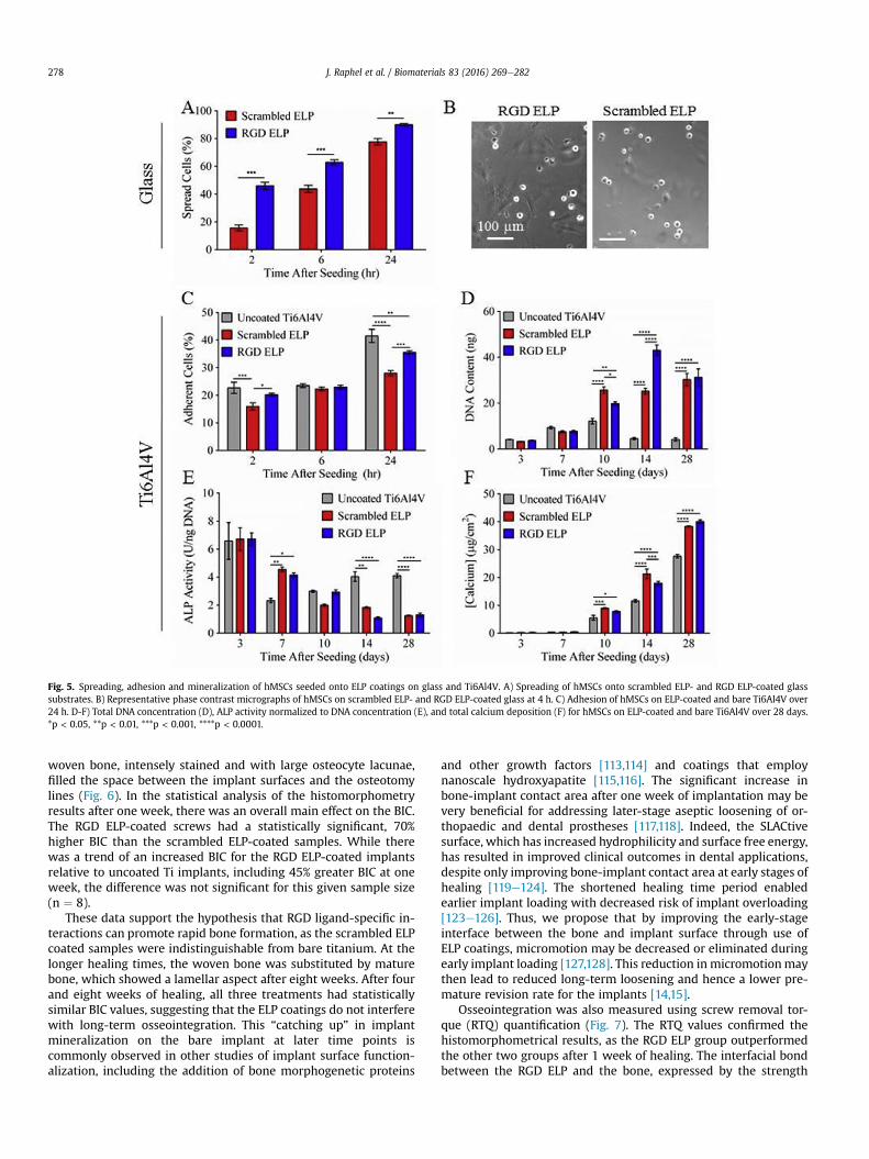
Fig. 5. Spreading, adhesion and mineralization of hMSCs seeded onto ELP coatings on glass and Ti6Al4V. A) Spreading of hMSCs onto scrambled ELP- and RGD ELP-coated glasssubstrates. B) Representative phase contrast micrographs of hMSCs on scrambled ELP- and RGD ELP-coated glass at 4 h. C) Adhesion of hMSCs on ELP-coated and bare Ti6Al4V over24 h. D-F) Total DNA concentration (D), ALP activity normalized to DNA concentration (E), and total calcium deposition (F) for hMSCs on ELP-coated and bare Ti6Al4V over 28 days.*p < 0.05, **p < 0.01, ***p < 0.001, ****p < 0.0001.
J. Raphel et al. / Biomaterials 83 (2016) 269e282278
woven bone, intensely stained and with large osteocyte lacunae,filled the space between the implant surfaces and the osteotomylines (Fig. 6). In the statistical analysis of the histomorphometryresults after one week, there was an overall main effect on the BIC.The RGD ELP-coated screws had a statistically significant, 70%higher BIC than the scrambled ELP-coated samples. While therewas a trend of an increased BIC for the RGD ELP-coated implantsrelative to uncoated Ti implants, including 45% greater BIC at oneweek, the difference was not significant for this given sample size(n ¼ 8).
These data support the hypothesis that RGD ligand-specific in-teractions can promote rapid bone formation, as the scrambled ELPcoated samples were indistinguishable from bare titanium. At thelonger healing times, the woven bone was substituted by maturebone, which showed a lamellar aspect after eight weeks. After fourand eight weeks of healing, all three treatments had statisticallysimilar BIC values, suggesting that the ELP coatings do not interferewith long-term osseointegration. This “catching up” in implantmineralization on the bare implant at later time points iscommonly observed in other studies of implant surface function-alization, including the addition of bone morphogenetic proteins
and other growth factors [113,114] and coatings that employnanoscale hydroxyapatite [115,116]. The significant increase inbone-implant contact area after one week of implantation may bevery beneficial for addressing later-stage aseptic loosening of or-thopaedic and dental prostheses [117,118]. Indeed, the SLACtivesurface, which has increased hydrophilicity and surface free energy,has resulted in improved clinical outcomes in dental applications,despite only improving bone-implant contact area at early stages ofhealing [119e124]. The shortened healing time period enabledearlier implant loading with decreased risk of implant overloading[123e126]. Thus, we propose that by improving the early-stageinterface between the bone and implant surface through use ofELP coatings, micromotion may be decreased or eliminated duringearly implant loading [127,128]. This reduction inmicromotionmaythen lead to reduced long-term loosening and hence a lower pre-mature revision rate for the implants [14,15].
Osseointegration was also measured using screw removal tor-que (RTQ) quantification (Fig. 7). The RTQ values confirmed thehistomorphometrical results, as the RGD ELP group outperformedthe other two groups after 1 week of healing. The interfacial bondbetween the RGD ELP and the bone, expressed by the strength

Fig. 6. In vivo response to spin coated ELP films on cp-Ti dental screws implanted into rat femurs. A) Bone-implant contact (BIC) area quantification for ELP coated and uncoateddental screws over 8 weeks post-surgery. n ¼ 8, *p < 0.05. B) Histological sections of inserted screws at 1 week (top). Expanded histological images of bone contact with screwthreads for BIC area quantification (bottom). Section thickness ¼ 20e30 mm. Toluidine blue staining.
J. Raphel et al. / Biomaterials 83 (2016) 269e282 279
necessary to break this bond, was 84% stronger than that of theuncoated Ti implants at 1 week, although the difference was notstatistically significant for this sample size (n ¼ 6, p ¼ 0.075). Theaverage RTQ for the RGD ELP group was significantly higher thanthat of the scrambled ELP surfaces (p ¼ 0.033), suggesting thatrapid osseointegration was due to the specific presence of the RGDligand, which is consistent with the in vitro data. At 4 weeks, theRTQ values increased for all three test groups, testifying to thefurther development of osseointegration between the implantsurfaces and the bone. However, at 8 weeks, only the ELP-coatedsamples continued to increase in RTQ, while the osseointegrationof the Ti group remained stable between these two time points.This suggests the possibility of further improved osseointegrationfor the coated screws at longer time points due to amechanism thatdoes not require the RGD ligand.
Though the in vivo studies do not show a statistically significantimprovement in BIC or RTQ using the RGD ELP coating relative touncoated Ti implants, there is a trend of increased osseointegrationmetrics at one week. The ability of the coatings to withstand theimplantation procedures and produce potential benefits early inthe healing process make these coatings an interesting candidatefor continued studies. Future efforts to iterate and improve the ELP
coatings, in conjunction with larger in vivo studies, may lead tosignificant improvements.
4. Conclusion
We have designed an engineered, photocrosslinkable, stable ELPcoating to address the issue of aseptic loosening of dental and or-thopaedic implants by increasing the speed of osseointegration oftitanium implants. In order to achieve this goal, three primary re-quirements had to be met. First, the coating needed to be easilyprocessable to be applied to a variety of implants.We demonstratedthe ability to use aqueous spin and dip coat processing to make thinfilm coatings on cp-Ti and Ti6Al4V substrates, including discs,screws, and rods. Second, the coating needed to be stable towithstand implantation and daily wear. We confirmed a mecha-nism for direct conjugation of photocrosslinkable ELP to cp-Ti andTi6Al4V surfaces mediated by UV light, resulting in a covalentlycrosslinked ELP network attached directly to the substrate surfacevia a carbene insertion mechanism. The ELP coatings remainedintact in physiological conditions for three weeks and were able towithstand clinically related implantation procedures, a key step forpotential clinical use that has not previously been demonstrated for

Fig. 7. In vivo response to spin coated ELP films on cp-Ti dental screws implanted intorat femurs and tibias was tested using removal torque (RTQ) to measure the interfacialstrength between the implant surfaces and peri-implant bone. Quantificationextended over 8 weeks post-surgery; n ¼ 6 at 1 week and n ¼ 8 at weeks 4 and 8;*p < 0.05.
J. Raphel et al. / Biomaterials 83 (2016) 269e282280
these types of coatings. Finally, the coating needed to improve therate of mineral deposition. Our ELP coatings enabled rapid adhesionof MG63 cells and hMSCs, induced differentiation of hMSCs downan osteogenic lineage, and increased mineral deposition from bothcell types. Furthermore, the performed in vivo studies showed thatimplants coated with our RGD ELP coating significantly increasedthe bone-implant contact and interfacial strength compared to ascrambled ELP coating after one week of healing. The obtainedresult of enhanced bone formation at early time points is crucial toavoid the risk of aseptic loosening of bone-anchoring implants andit would enable earlier loading of the implant post-implantation.Combined, these results suggest that photocrosslinkable ELP coat-ings improve early-stage osseointegration of implants, which maylead to increased durability and reduction in the number of revisionsurgeries for aseptic loosening.
Acknowledgments
JR, JK, and SG contributed equally to this work. We thank FortWayne Metals for the Ti6Al4V rods; Dr. Rebecca DiMarco for herassistance with SEM on dental screws; Dr. Jeffrey Tok and theStanford Soft & Hybrid Materials Facility (SMF) for experimentalconsultation and use of the profilometer and goniometer; Dr.Robert Beardsley Zeller for assistance in the design, fabrication, andprogramming of the dip coater; Dr. Jerome Geronimo and theStanford 3-Dimensional Printing Facility for 3D printing of dipcoater accessories; Dr. Lydia-Marie Joubert and the Beckman CellSciences Imaging Facility (CSIF) for assistance with and use of theSEM; and Dr. Patrick Benitez for assistance with endotoxin purifi-cation. We acknowledge funding from the Irish Research CouncilELEVATE fellowship (MGH), the Jane and Aatos Erkko Foundation(JP), the Wallenberg Foundation through the Wallenberg AcademyFellow Program (MA) and Materials Science an Area of Advance atChalmers University of Technology, the National Science Founda-tion (DMR 0846363 to SCH), and the National Institutes of Health(2R01 AR055650, R01 063717 to SBG, U19 AI116484-01, R21EB018407-01, R21 AR062359-01 to SCH).
Appendix A. Supplementary data
Supplementary data related to this article can be found at http://
dx.doi.org/10.1016/j.biomaterials.2015.12.030.
References
[1] S. Kurtz, K. Ong, E. Lau, F. Mowat, M. Halpern, Projections of primary andrevision hip and knee arthroplasty in the United States from 2005 to 2030,J. Bone Jt. Surg. Am. 89 (2007) 780.
[2] Milleniumresearchgroup U.S. markets for dental implants, Available from:http://www.mrg.net/Products-and-Services/Syndicated-Report.aspx?r¼RPUS22DE13 accessed: May, 2015.
[3] D. Schwartz-Arad, N. Kidron, E. Dolev, A long-term study of implants sup-porting overdentures as a model for implant success, J. Periodontol. 76(2005) 1431.
[4] I.K. Karoussis, U. Bragger, G.E. Salvi, W. Burgin, N.P. Lang, Effect of implantdesign on survival and success rates of titanium oral implants: a 10-yearprospective cohort study of the ITI Dental Implant System, Clin. Oral Im-plants Res. 15 (2004) 8.
[5] L.W. Lindquist, G.E. Carlsson, T. Jemt, A prospective 15-year follow-up studyof mandibular fixed prostheses supported by osseointegrated implants.Clinical results and marginal bone loss, Clin. Oral Implants Res. 7 (1996) 329.
[6] B.R. Chrcanovic, T. Albrektsson, A. Wennerberg, Reasons for failures of oralimplants, J. Oral Rehabil. 41 (2014) 443.
[7] B. Pommer, S. Frantal, J. Willer, M. Posch, G. Watzek, G. Tepper, Impact ofdental implant length on early failure rates: a meta-analysis of observationalstudies, J. Clin. Periodontol. 38 (2011) 856.
[8] K.T. Makela, M. Matilainen, P. Pulkkinen, A.M. Fenstad, L. Havelin,L. Engesaeter, et al., Failure rate of cemented and uncemented total hip re-placements: register study of combined Nordic database of four nations, BMJ348 (2014) f7592.
[9] S. Kurtz, F. Mowat, K. Ong, N. Chan, E. Lau, M. Halpern, Prevalence of primaryand revision total hip and knee arthroplasty in the United States from 1990through 2002, J. Bone Jt. Surg. Am. 87 (2005) 1487.
[10] W.C. Schroer, K.R. Berend, A.V. Lombardi, C.L. Barnes, M.P. Bolognesi,M.E. Berend, et al., Why are total knees failing today? Etiology of total kneerevision in 2010 and 2011, J. Arthroplast. 28 (2013) 116.
[11] D.F. Dalury, D.L. Pomeroy, R.S. Gorab, M.J. Adams, Why are total kneearthroplasties being revised? J. Arthroplast. 28 (2013) 120.
[12] J.C. Clohisy, G. Calvert, F. Tull, D. Mcdonald, W.J. Maloney, Reasons for revi-sion hip surgery: a retrospective review, Clin. Orthop. Relat. Res. (2004) 188.
[13] S. Sakka, K. Baroudi, M.Z. Nassani, Factors associated with early and latefailure of dental implants, J. Investig. Clin. Dent. 3 (2012) 258.
[14] M. Sundfeldt, L.V. Carlsson, C.B. Johansson, P. Thomsen, C. Gretzer, Asepticloosening, not only a question of wear: a review of different theories, ActaOrthop. Scand. 77 (2006) 177.
[15] S.B. Goodman, The effects of micromotion and particulate materials on tissuedifferentiation. Bone chamber studies in rabbits, Acta Orthop. Scand. Suppl.258 (1994) 1.
[16] X. Liu, G.L. Niebur, Bone ingrowth into a porous coated implant predicted bya mechano-regulatory tissue differentiation algorithm, Biomech. Model.Mechanobiol. 7 (2008) 335.
[17] L. Ryd, B.E. Albrektsson, L. Carlsson, F. Dansgard, P. Herberts, A. Lindstrand, etal., Roentgen stereophotogrammetric analysis as a predictor of mechanicalloosening of knee prostheses, J. Bone Jt. Surg. Br. 77 (1995) 377.
[18] S. Szmukler-Moncler, H. Salama, Y. Reingewirtz, J.H. Dubruille, Timing ofloading and effect of micromotion on bone-dental implant interface: reviewof experimental literature, J. Biomed. Mater. Res. 43 (1998) 192.
[19] X. Liu, P.K. Chu, C. Ding, Surface modification of titanium, titanium alloys, andrelated materials for biomedical applications, Mater. Sci. Eng. R. 47 (2004) 49.
[20] M. Long, H.J. Rack, Titanium alloys in total joint replacementea materialsscience perspective, Biomaterials 19 (1998) 1621.
[21] M. Niinomi, Mechanical properties of biomedical titanium alloys, Mater. Sci.Eng. A 243 (1998) 231.
[22] M. Geetha, A.K. Singh, R. Asokamani, A.K. Gogia, Ti based biomaterials, theultimate choice for orthopaedic implants e a review, Prog. Mater. Sci. 54(2009) 397.
[23] L. Le Guehennec, A. Soueidan, P. Layrolle, Y. Amouriq, Surface treatments oftitanium dental implants for rapid osseointegration, Dent. Mater. 23 (2007)844.
[24] H. Schliephake, D. Schamweber, Chemical and biological functionalization oftitanium for dental implants, J. Mater. Chem. 18 (2008) 2404.
[25] S.B. Goodman, Z. Yao, M. Keeney, F. Yang, The future of biologic coatings fororthopaedic implants, Biomaterials 34 (2013) 3174.
[26] P.I. Branemark, P. Engstrand, L.O. Ohrnell, K. Grondahl, P. Nilsson, K. Hagberg,et al., Branemark Novum: a new treatment concept for rehabilitation of theedentulous mandible. Preliminary results from a prospective clinical follow-up study, Clin. Implant Dent. Relat. Res. 1 (1999) 2.
[27] M. Esposito, M.G. Grusovin, H. Maghaireh, H.V. Worthington, Interventionsfor replacing missing teeth: different times for loading dental implants,Cochrane Database Syst. Rev. 3 (2013) CD003878.
[28] R.A. Berger, J.J. Jacobs, R.M. Meneghini, C. Della Valle, W. Paprosky,A.G. Rosenberg, Rapid rehabilitation and recovery with minimally invasivetotal hip arthroplasty, Clin. Orthop. Relat. Res. (2004) 239.
[29] K.R. Berend, A.V. Lombardi Jr., T.H. Mallory, Rapid recovery protocol for peri-operative care of total hip and total knee arthroplasty patients, Surg. Tech.

J. Raphel et al. / Biomaterials 83 (2016) 269e282 281
Int. 13 (2004) 239.[30] D.M. Ferris, G.D. Moodie, P.M. Dimond, C.W. Gioranni, M.G. Ehrlich,
R.F. Valentini, RGD-coated titanium implants stimulate increased bone for-mation in vivo, Biomaterials 20 (1999) 2323.
[31] H. Schliephake, D. Scharnweber, M. Dard, S. Rossler, A. Sewing, J. Meyer, etal., Effect of RGD peptide coating of titanium implants on periimplant boneformation in the alveolar crest. An experimental pilot study in dogs, Clin.Oral Implants Res. 13 (2002) 312.
[32] R. Agarwal, C. Gonzalez-Garcia, B. Torstrick, R.E. Guldberg, M. Salmeron-Sanchez, A.J. Garcia, Simple coating with fibronectin fragment enhancesstainless steel screw osseointegration in healthy and osteoporotic rats, Bio-materials 63 (2015) 137.
[33] J. Raphel, A. Parisi-Amon, S. Heilshorn, Photoreactive elastin-like proteins foruse as versatile bioactive materials and surface coatings, J. Mater. Chem. 22(2012) 19429.
[34] S.R. Macewan, A. Chilkoti, Elastin-like polypeptides: biomedical applicationsof tunable biopolymers, Biopolymers 94 (2010) 60.
[35] D.W. Urry, Physical chemistry of biological free energy transduction asdemonstrated by elastic protein-based polymers, J. Phys. Chem. B 101 (1997)11007.
[36] D.W. Urry, Molecular machines: how motion and other functions of livingorganisms can result from reversible chemical changes, Angew. Chem. Int.Ed. Engl. 32 (1993) 819.
[37] A. Chilkoti, T. Christensen, J.A. Mackay, Stimulus responsive elastin bio-polymers: applications in medicine and biotechnology, Curr. Opin. Chem.Biol. 10 (2006) 652.
[38] D. Chow, M.L. Nunalee, D.W. Lim, A.J. Simnick, A. Chilkoti, Peptide-basedbiopolymers in biomedicine and biotechnology, Mater. Sci. Eng. R Rep. 62(2008) 125.
[39] A.C. Rincon, I.T. Molina-Martinez, B. De Las Heras, M. Alonso, C. Bailez,J.C. Rodriguez-Cabello, et al., Biocompatibility of elastin-like polymer poly(-VPAVG) microparticles: in vitro and in vivo studies, J. Biomed. Mater. Res. A78 (2006) 343.
[40] R.E. Sallach, W. Cui, J. Wen, A. Martinez, V.P. Conticello, E.L. Chaikof, Elastin-mimetic protein polymers capable of physical and chemical crosslinking,Biomaterials 30 (2009) 409.
[41] S.C. Heilshorn, J.C. Liu, D.A. Tirrell, Cell-binding domain context affects cellbehavior on engineered proteins, Biomacromolecules 6 (2005) 318.
[42] D.E. Meyer, A. Chilkoti, Purification of recombinant proteins by fusion withthermally-responsive polypeptides, Nat. Biotechnol. 17 (1999) 1112.
[43] C.M. Bellingham, M.A. Lillie, J.M. Gosline, G.M. Wright, B.C. Starcher,A.J. Bailey, et al., Recombinant human elastin polypeptides self-assemble intobiomaterials with elastin-like properties, Biopolymers 70 (2003) 445.
[44] D.W. Urry, T. Hugel, M. Seitz, H.E. Gaub, L. Sheiba, J. Dea, et al., Elastin: arepresentative ideal protein elastomer, Philos. Trans. R. Soc. B 357 (2002)169.
[45] K. Trabbic-Carlson, L.A. Setton, A. Chilkoti, Swelling and mechanical behav-iors of chemically cross-linked hydrogels of elastin-like polypeptides, Bio-macromolecules 4 (2003) 572.
[46] D.C. Chow, M.R. Dreher, K. Trabbic-Carlson, A. Chilkoti, Ultra-high expressionof a thermally responsive recombinant fusion protein in E. coli, Biotechnol.Prog. 22 (2006) 638.
[47] I. Massodi, G.L. Bidwell 3rd, D. Raucher, Evaluation of cell penetrating pep-tides fused to elastin-like polypeptide for drug delivery, J. Control. Release108 (2005) 396.
[48] J.R. Mcdaniel, D.J. Callahan, A. Chilkoti, Drug delivery to solid tumors byelastin-like polypeptides, Adv. Drug Deliv. Rev. 62 (2010) 1456.
[49] A. Girotti, J. Reguera, J.C. Rodriguez-Cabello, F.J. Arias, M. Alonso,A. Matestera, Design and bioproduction of a recombinant multi(bio)func-tional elastin-like protein polymer containing cell adhesion sequences fortissue engineering purposes, J. Mater. Sci. Mater. Med. 15 (2004) 479.
[50] P.C. Bessa, R. Machado, S. Nurnberger, D. Dopler, A. Banerjee, A.M. Cunha, etal., Thermoresponsive self-assembled elastin-based nanoparticles for de-livery of BMPs, J. Control. Release 142 (2010) 312.
[51] Z. Megeed, J. Cappello, H. Ghandehari, Genetically engineered silk-elastinlikeprotein polymers for controlled drug delivery, Adv. Drug Deliv. Rev. 54(2002) 1075.
[52] H. Betre, L.A. Setton, D.E. Meyer, A. Chilkoti, Characterization of a geneticallyengineered elastin-like polypeptide for cartilaginous tissue repair, Bio-macromolecules 3 (2002) 910.
[53] M.K. Mchale, L.A. Setton, A. Chilkoti, Synthesis and in vitro evaluation ofenzymatically cross-linked elastin-like polypeptide gels for cartilaginoustissue repair, Tissue Eng. 11 (2005) 1768.
[54] D.L. Nettles, A. Chilkoti, L.A. Setton, Applications of elastin-like polypeptidesin tissue engineering, Adv. Drug Deliv. Rev. 62 (2010) 1479.
[55] D.L. Nettles, K. Kitaoka, N.A. Hanson, C.M. Flahiff, B.A. Mata, E.W. Hsu, et al., Insitu crosslinking elastin-like polypeptide gels for application to articularcartilage repair in a goat osteochondral defect model, Tissue Eng. Part A 14(2008) 1133.
[56] S.C. Heilshorn, K.A. Dizio, E.R. Welsh, D.A. Tirrell, Endothelial cell adhesion tothe fibronectin CS5 domain in artificial extracellular matrix proteins, Bio-materials 24 (2003) 4245.
[57] H. Martinez-Osorio, M. Juarez-Campo, Y. Diebold, A. Girotti, M. Alonso,F.J. Arias, et al., Genetically engineered elastin-like polymer as a substratumto culture cells from the ocular surface, Curr. Eye Res. 34 (2009) 48.
[58] A.V. Janorkar, P. Rajagopalan, M.L. Yarmush, Z. Megeed, The use of elastin-like polypeptide-polyelectrolyte complexes to control hepatocytemorphology and function in vitro, Biomaterials 29 (2008) 625.
[59] E. Wang, S.H. Lee, S.W. Lee, Elastin-like polypeptide based hydroxyapatitebionanocomposites, Biomacromolecules 12 (2011) 672.
[60] S. Prieto, A. Shkilnyy, C. Rumplasch, A. Ribeiro, F.J. Arias, J.C. Rodriguez-Cabello, et al., Biomimetic calcium phosphate mineralization with multi-functional elastin-like recombinamers, Biomacromolecules 12 (2011) 1480.
[61] E. Salvagni, G. Berguig, E. Engel, J.C. Rodriguez-Cabello, G. Coullerez,M. Textor, et al., A bioactive elastin-like recombinamer reduces unspecificprotein adsorption and enhances cell response on titanium surfaces, ColloidsSurf. B 114 (2014) 225.
[62] H.A. Klock, J. Genzer, Expanding the polymer mechanochemistry toolboxthrough surface-initiated polymerization, ACS Macro Lett. 4 (2015) 636.
[63] K.S. Straley, S.C. Heilshorn, Independent tuning of multiple biomaterialproperties using protein engineering, Soft Matter 5 (2009) 114.
[64] M. Keeney, H. Waters, K. Barcay, X. Jiang, Z. Yao, J. Pajarinen, et al., MutantMCP-1 protein delivery from layer-by-layer coatings on orthopedic implantsto modulate inflammatory response, Biomaterials 34 (2013) 10287.
[65] Fda. Guidance for Industry: Pyrogen and Endotoxins Testing, Available from:http://www.fda.gov/drugs/guidancecomplianceregulatoryinformation/guidances/ucm314718.htm#_ftn27 accessed: June, 2015.
[66] L.E. Scriven, Physics and applications of dip coating and spin coating, MRSProc. 121 (1988) 717.
[67] G. Mendonca, D.B. Mendonca, F.J. Aragao, L.F. Cooper, Advancing dentalimplant surface technologyefrom micron- to nanotopography, Biomaterials29 (2008) 3822.
[68] S.W. Kweh, K.A. Khor, P. Cheang, An in vitro investigation of plasma sprayedhydroxyapatite (HA) coatings produced with flame-spheroidized feedstock,Biomaterials 23 (2002) 775.
[69] T.W. Bauer, R.C. Geesink, R. Zimmerman, J.T. Mcmahon, Hydroxyapatite-coated femoral stems. Histological analysis of components retrieved at au-topsy, J. Bone Jt. Surg. Am. 73 (1991) 1439.
[70] T.W. Bauer, Hydroxyapatite: coating controversies, Orthopedics 18 (1995)885.
[71] R.D. Bloebaum, D. Beeks, L.D. Dorr, C.G. Savory, J.A. Dupont, A.A. Hofmann,Complications with hydroxyapatite particulate separation in total hiparthroplasty, Clin. Orthop. Relat. Res. (1994) 19.
[72] J. Gosline, M. Lillie, E. Carrington, P. Guerette, C. Ortlepp, K. Savage, Elasticproteins: biological roles and mechanical properties, Philos. Trans. R. Soc. B357 (2002) 121.
[73] H. Sigrist, A. Collioud, J. Cl�emence, H. Gao, R. Luginbuhl, M. Sanger, et al.,Surface immobilization of biomolecules by light, Opt. Eng. 34 (1995) 2339.
[74] H. Gao, M. Sanger, R. Luginbuhl, H. Sigrist, Immunosensing with photo-immobilized immunoreagents on planar optical wave guides, Biosens. Bio-electron. 10 (1995) 317.
[75] N. Sakai, A. Fujishima, T. Watanabe, K. Hashimoto, Quantitative evaluation ofthe photoinduced hydrophilic conversion properties of TiO2 thin film sur-faces by the reciprocal of contact angle, J. Phys. Chem. B 107 (2003) 1028.
[76] H. Bayley, J.R. Knowles, Photogenerated reagents for membrane labeling. 1.Phenylnitrene formed within the lipid bilayer, Biochemistry 17 (1978) 2414.
[77] A. Sinz, Chemical cross-linking and mass spectrometry to map three-dimensional protein structures and protein-protein interactions, MassSpectrom. Rev. 25 (2006) 663.
[78] H. Sigrist, M. Mühlemann, M. Dolder, Philicty of amino acid side-chains forphotogenerated carbenes, J. Photochem. Photobiol. B 7 (1990) 277.
[79] B.G. Zhang, D.E. Myers, G.G. Wallace, M. Brandt, P.F. Choong, Bioactivecoatings for orthopaedic implants-recent trends in development of implantcoatings, Int. J. Mol. Sci. 15 (2014) 11878.
[80] J.E. Hulshoff, T. Hayakawa, K. Van Dijk, A.F. Leijdekkers-Govers, J.P. Van DerWaerden, J.A. Jansen, Mechanical and histologic evaluation of Ca-P plasma-spray and magnetron sputter-coated implants in trabecular bone of the goat,J. Biomed. Mater. Res. 36 (1997) 75.
[81] H. Wang, N. Eliaz, Z. Xiang, H.P. Hsu, M. Spector, L.W. Hobbs, Early boneapposition in vivo on plasma-sprayed and electrochemically deposited hy-droxyapatite coatings on titanium alloy, Biomaterials 27 (2006) 4192.
[82] C.E. Misch, Z. Qu, M.W. Bidez, Mechanical properties of trabecular bone inthe human mandible: implications for dental implant treatment planningand surgical placement, J. Oral Maxillofac. Surg. 57 (1999) 700.
[83] Sawbones. Biomechanical Test Materials Properties, Available from: http://www.sawbones.com/UserFiles/Docs/biomechanical_catalog.pdf accessed:June, 2015.
[84] M. Pennington, R. Grieve, J.S. Sekhon, P. Gregg, N. Black, J.H. Van Der Meulen,Cemented, cementless, and hybrid prostheses for total hip replacement: costeffectiveness analysis, BMJ 346 (2013) f1026.
[85] A.J. Garcia, C.D. Reyes, Bio-adhesive surfaces to promote osteoblast differ-entiation and bone formation, J. Dent. Res. 84 (2005) 407.
[86] J.C. Liu, S.C. Heilshorn, D.A. Tirrell, Comparative cell response to artificialextracellular matrix proteins containing the RGD and CS5 cell-binding do-mains, Biomacromolecules 5 (2004) 497.
[87] J.C. Liu, D.A. Tirrell, Cell response to RGD density in cross-linked artificialextracellular matrix protein films, Biomacromolecules 9 (2008) 2984.
[88] S. Ravi, J.M. Caves, A.W. Martinez, C.A. Haller, E.L. Chaikof, Incorporation offibronectin to enhance cytocompatibility in multilayer elastin-like proteinscaffolds for tissue engineering, J. Biomed. Mater. Res. A 101 (2013) 1915.

J. Raphel et al. / Biomaterials 83 (2016) 269e282282
[89] S. Weiner, W. Traub, Organization of hydroxyapatite crystals within collagenfibrils, FEBS Lett. 206 (1986) 262.
[90] S. Weiner, W. Traub, Bone structure: from angstroms to microns, FASEB J. 6(1992) 879.
[91] J.Y. Rho, L. Kuhn-Spearing, P. Zioupos, Mechanical properties and the hier-archical structure of bone, Med. Eng. Phys. 20 (1998) 92.
[92] B. Clarke, Normal bone anatomy and physiology, Clin. J. Am. Soc. Nephrol. 3(Suppl. 3) (2008) S131.
[93] J.E. Davies, Mechanisms of endosseous integration, Int. J. Prosthodont. 11(1998) 391.
[94] R.W. Bucholz, Nonallograft osteoconductive bone graft substitutes, Clin.Orthop. Relat. Res. (2002) 44.
[95] G.T. Huang, S. Gronthos, S. Shi, Mesenchymal stem cells derived from dentaltissues vs. those from other sources: their biology and role in regenerativemedicine, J. Dent. Res. 88 (2009) 792.
[96] J.I. Ahn, S. Terry Canale, S.D. Butler, K.A. Hasty, Stem cell repair of physealcartilage, J. Orthop. Res. 22 (2004) 1215.
[97] A.I. Caplan, Mesenchymal stem cells, J. Orthop. Res. 9 (1991) 641.[98] M.F. Pittenger, A.M. Mackay, S.C. Beck, R.K. Jaiswal, R. Douglas, J.D. Mosca, et
al., Multilineage potential of adult human mesenchymal stem cells, Science284 (1999) 143.
[99] D.J. Prockop, Marrow stromal cells as stem cells for nonhematopoietic tis-sues, Science 276 (1997) 71.
[100] J.N. Beresford, J.H. Bennett, C. Devlin, P.S. Leboy, M.E. Owen, Evidence for aninverse relationship between the differentiation of adipocytic and osteogeniccells in rat marrow stromal cell cultures, J. Cell Sci. 102 (Pt 2) (1992) 341.
[101] J.U. Yoo, T.S. Barthel, K. Nishimura, L. Solchaga, A.I. Caplan, V.M. Goldberg, etal., The chondrogenic potential of human bone-marrow-derived mesen-chymal progenitor cells, J. Bone Jt. Surg. Am. 80 (1998) 1745.
[102] S.P. Bruder, D.J. Fink, A.I. Caplan, Mesenchymal stem cells in bone develop-ment, bone repair, and skeletal regeneration therapy, J. Cell. Biochem. 56(1994) 283.
[103] J.R. Mauney, V. Volloch, D.L. Kaplan, Role of adult mesenchymal stem cells inbone tissue engineering applications: current status and future prospects,Tissue Eng. 11 (2005) 787.
[104] A.I. Caplan, Review: mesenchymal stem cells: cell-based reconstructivetherapy in orthopedics, Tissue Eng. 11 (2005) 1198.
[105] R.S. Tuan, G. Boland, R. Tuli, Adult mesenchymal stem cells and cell-basedtissue engineering, Arthritis. Res. Ther. 5 (2003) 32.
[106] R.S. Tuan, Role of adult stem/progenitor cells in osseointegration andimplant loosening, Int. J. Oral Maxillofac. Implants 26 (Suppl.) (2011) 50.
[107] J.J. Schmidt, J. Jeong, H. Kong, The interplay between cell adhesion cues andcurvature of cell adherent alginate microgels in multipotent stem cell cul-ture, Tissue Eng. Part A 17 (2011) 2687.
[108] I.L. Kim, S. Khetan, B.M. Baker, C.S. Chen, J.A. Burdick, Fibrous hyaluronic acidhydrogels that direct MSC chondrogenesis through mechanical and adhesivecues, Biomaterials 34 (2013) 5571.
[109] M.C. Vemuri, L.G. Chase, M.S. Rao, Mesenchymal stem cell assays and ap-plications, Methods Mol. Biol. 698 (2011) 3.
[110] J.E. Aubin, Regulation of osteoblast formation and function, Rev. Endocr.Metab. Disord. 2 (2001) 81.
[111] N. Jaiswal, S.E. Haynesworth, A.I. Caplan, S.P. Bruder, Osteogenic differenti-ation of purified, culture-expanded human mesenchymal stem cells in vitro,J. Cell. Biochem. 64 (1997) 295.
[112] B. Genge, G. Sauer, L. Wu, F. Mclean, R. Wuthier, Correlation between loss ofalkaline phosphatase activity and accumulation of calcium during matrixvesicle-mediated mineralization, J. Biol. Chem. 263 (1988) 18513.
[113] J.J. Yoo, Y.J. Park, S.H. Rhee, H.J. Chun, H.J. Kim, Synthetic peptide-conjugatedtitanium alloy for enhanced bone formation in vivo, Connect. Tissue Res. 53(2012) 359.
[114] M. Ramazanoglu, R. Lutz, C. Ergun, C. Von Wilmowsky, E. Nkenke,K.A. Schlegel, The effect of combined delivery of recombinant human bonemorphogenetic protein-2 and recombinant human vascular endothelialgrowth factor 165 from biomimetic calcium-phosphate-coated implants onosseointegration, Clin. Oral Implants Res. 22 (2011) 1433.
[115] M.S. Bryington, M. Hayashi, Y. Kozai, S. Vandeweghe, M. Andersson,A. Wennerberg, et al., The influence of nano hydroxyapatite coating onosseointegration after extended healing periods, Dent. Mater. 29 (2013) 514.
[116] L. Melin Svanborg, L. Meirelles, V. Franke Stenport, P. Kjellin, F. Currie,M. Andersson, et al., Evaluation of bone healing on sandblasted and Acidetched implants coated with nanocrystalline hydroxyapatite: an in vivostudy in rabbit femur, Int. J. Dent. (2014) 2014.
[117] F. Marco, F. Milena, G. Gianluca, O. Vittoria, Peri-implant osteogenesis inhealth and osteoporosis, Micron 36 (2005) 630.
[118] M.A. Freeman, P. Plante-Bordeneuve, Early migration and late aseptic failureof proximal femoral prostheses, J. Bone Jt. Surg. Br. 76 (1994) 432.
[119] S. Sista, C. Wen, P.D. Hodgson, G. Pande, The influence of surface energy oftitanium-zirconium alloy on osteoblast cell functions in vitro, J. Biomed.Mater. Res. A 97 (2011) 27.
[120] B.S. Kopf, S. Ruch, S. Berner, N.D. Spencer, K. Maniura-Weber, The role ofnanostructures and hydrophilicity in osseointegration: In-vitro protein-adsorption and blood-interaction studies, J. Biomed. Mater. Res. A 103 (2015)2661.
[121] D. Buser, N. Broggini, M. Wieland, R.K. Schenk, A.J. Denzer, D.L. Cochran, etal., Enhanced bone apposition to a chemically modified SLA titanium surface,J. Dent. Res. 83 (2004) 529.
[122] M.M. Bornstein, P. Valderrama, A.A. Jones, T.G. Wilson, R. Seibl, D.L. Cochran,Bone apposition around two different sandblasted and acid-etched titaniumimplant surfaces: a histomorphometric study in canine mandibles, Clin. OralImplants Res. 19 (2008) 233.
[123] T.W. Oates, P. Valderrama, M. Bischof, R. Nedir, A. Jones, J. Simpson, et al.,Enhanced implant stability with a chemically modified SLA surface: a ran-domized pilot study, Int. J. Oral Maxillofac. Implants 22 (2007) 755.
[124] N.P. Lang, G.E. Salvi, G. Huynh-Ba, S. Ivanovski, N. Donos, D.D. Bosshardt,Early osseointegration to hydrophilic and hydrophobic implant surfaces inhumans, Clin. Oral Implants Res. 22 (2011) 349.
[125] M. Schatzle, R. Mannchen, U. Balbach, C.H. Hammerle, H. Toutenburg,R.E. Jung, Stability change of chemically modified sandblasted/acid-etchedtitanium palatal implants. A randomized-controlled clinical trial, Clin. OralImplants Res. 20 (2009) 489.
[126] P. Valderrama, T.W. Oates, A.A. Jones, J. Simpson, J.D. Schoolfield,D.L. Cochran, Evaluation of two different resonance frequency devices todetect implant stability: a clinical trial, J. Periodontol. 78 (2007) 262.
[127] H. Kienapfel, C. Sprey, A. Wilke, P. Griss, Implant fixation by bone ingrowth,J. Arthroplast. 14 (1999) 355.
[128] Y. Abu-Amer, I. Darwech, J.C. Clohisy, Aseptic loosening of total joint re-placements: mechanisms underlying osteolysis and potential therapies,Arthritis. Res. Ther. 9 (Suppl. 1) (2007) S6.