Endometrial Cancer - Repositório Comum: Página … · with endometrial hyperplasia (Kurman et al....
30
Med Radiol Diagn Imaging (2016) DOI 10.1007/174_2016_84, © Springer International Publishing Switzerland Endometrial Cancer Mariana Horta and Teresa Margarida Cunha Abstract Endometrial cancer is the most common gyne- cological malignancy in well-developed coun- tries. Biologically and clinicopathologically, endometrial carcinomas are divided into two types: type 1 or estrogen-dependent carcinomas and type 2 or estrogen-independent carcinomas. Type 1 cancers correspond mainly to endome- trioid carcinomas and account for approxi- mately 90 % of endometrial cancers, whereas type 2 cancers correspond to the majority of the other histopathological subtypes. The vast majority of endometrial cancers present as abnormal vaginal bleedings in postmenopausal women. Therefore, 75 % of cancers are diagnosed at an early stage, which makes the overall prognosis favorable. The first diagnostic step to evaluate women with an abnormal vaginal bleeding is the mea- surement of the endometrial thickness with transvaginal ultrasound. If endometrial thick- ening or heterogeneity is confirmed, a biopsy should be performed to establish a definite histopathological diagnosis. Magnetic resonance imaging is not considered in the International Federation of Gynaecology and Obstetrics staging system. Nonetheless it plays a relevant role in the preoperative staging of endometrial carcinoma, helping to define the best therapeutic management. Moreover, it is impor- tant in the diagnosis of treatment complications, in the surveillance of therapy response, and in the assessment of recurrent disease. M. Horta (*) Centro Hospitalar Lisboa Ocidental, Estrada do Forte do Alto do Duque, 1495-005 Lisbon, Portugal e-mail: [email protected] T.M. Cunha Rua Professor Lima Basto, 1099-023 Lisbon, Portugal e-mail: [email protected] Contents 1 Endometrial Cancer: Background .............. 000 1.1 Epidemiology .................................................. 000 1.2 Pathology and Risk Factors............................. 000 1.3 Symptoms and Diagnosis................................ 000 2 Endometrial Cancer Staging ........................ 000 2.1 MR Protocol for Staging Endometrial Carcinoma ....................................................... 000 2.2 MR Findings According to the Stage of Endometrial Carcinoma .............................. 000 3 Recent Advances in Functional MR Imaging in Assessing Endometrial Carcinoma ............................... 000 4 Therapeutic Approaches............................... 000 4.1 Surgery ............................................................ 000 4.2 Adjuvant Treatment ........................................ 000 4.3 Fertility-Sparing Treatment............................. 000 5 Follow-Up and Recurrent Endometrial Carcinoma...................................................... 000 5.1 Treatment of Recurrence ................................. 000 6 Prognosis ........................................................ 000 Conclusion .............................................................. 000 References ............................................................... 000
Transcript of Endometrial Cancer - Repositório Comum: Página … · with endometrial hyperplasia (Kurman et al....

Med Radiol Diagn Imaging (2016)DOI 10.1007/174_2016_84, © Springer International Publishing Switzerland
Endometrial Cancer
Mariana Horta and Teresa Margarida Cunha
Abstract
Endometrial cancer is the most common gyne-cological malignancy in well-developed coun-tries. Biologically and clinicopathologically, endometrial carcinomas are divided into two types: type 1 or estrogen- dependent carcinomas and type 2 or estrogen-independent carcinomas. Type 1 cancers correspond mainly to endome-trioid carcinomas and account for approxi-mately 90 % of endometrial cancers, whereas type 2 cancers correspond to the majority of the other histopathological subtypes.
The vast majority of endometrial cancers present as abnormal vaginal bleedings in postmenopausal women. Therefore, 75 % of cancers are diagnosed at an early stage, which makes the overall prognosis favorable.
The first diagnostic step to evaluate women with an abnormal vaginal bleeding is the mea-surement of the endometrial thickness with transvaginal ultrasound. If endometrial thick-ening or heterogeneity is confirmed, a biopsy should be performed to establish a definite histopathological diagnosis.
Magnetic resonance imaging is not considered in the International Federation of Gynaecology and Obstetrics staging system. Nonetheless it plays a relevant role in the preoperative staging of endometrial carcinoma, helping to define the best therapeutic management. Moreover, it is impor-tant in the diagnosis of treatment complications, in the surveillance of therapy response, and in the assessment of recurrent disease.
M. Horta (*) Centro Hospitalar Lisboa Ocidental, Estrada do Forte do Alto do Duque, 1495-005 Lisbon, Portugale-mail: [email protected]
T.M. Cunha Rua Professor Lima Basto, 1099-023 Lisbon, Portugale-mail: [email protected]
Contents
1 Endometrial Cancer: Background .............. 0001.1 Epidemiology .................................................. 0001.2 Pathology and Risk Factors ............................. 0001.3 Symptoms and Diagnosis ................................ 000
2 Endometrial Cancer Staging ........................ 0002.1 MR Protocol for Staging Endometrial
Carcinoma ....................................................... 0002.2 MR Findings According to the Stage
of Endometrial Carcinoma .............................. 000
3 Recent Advances in Functional MR Imaging in Assessing Endometrial Carcinoma ............................... 000
4 Therapeutic Approaches ............................... 0004.1 Surgery ............................................................ 0004.2 Adjuvant Treatment ........................................ 0004.3 Fertility-Sparing Treatment ............................. 000
5 Follow-Up and Recurrent Endometrial Carcinoma ...................................................... 000
5.1 Treatment of Recurrence ................................. 000
6 Prognosis ........................................................ 000
Conclusion .............................................................. 000
References ............................................................... 000

1 Endometrial Cancer: Background
1.1 Epidemiology
Endometrial cancer is the fifth most common malig-nancy in females worldwide, after breast, colorec-tal, cervical, and lung cancers, accounting for 4.8 % of all cancers in women (Ferlay et al. 2013).
In contrast to cervical cancer, endometrioid can-cer peak incidence rates occur in well- developed countries (Europe and North America), where it is the most common gynecological malignancy (Ferlay et al. 2013). This is probably due to life-style factors, in particular obesity, which has been linked to about 50 % endometrial cancers in these countries (Epstein and Blomqvist 2014; Calle and Kaaks 2004). The increase in life expectancy is also responsible for its rising (Koh et al. 2014).
Endometrial cancer is more likely to occur in postmenopausal women in their sixth and seventh decades, with cases only rarely reported under the age of 35 in the United Kingdom (Patel et al. 2010; Cancer Incidence Statistics 2015).
Despite its relatively high prevalence, approx-imately 82 % of endometrial cancers are diag-nosed at an early stage, which makes its overall prognosis favorable (Cancer Incidence Statistics 2015). Its worldwide mortality rate is low and it has been estimated at 2.2 % (Ferlay et al. 2013).
1.2 Pathology and Risk Factors
The World Health Organization (WHO) classifi-cation of uterine corpus tumors was revised in 2014 (Kurman et al. 2014). Endometrial carcino-mas (also known as adenocarcinoma of the endo-metrium) are classified under the “Epithelial Tumours and Precursors” group and clinicopath-ologically divided into the following types: endo-metrioid carcinoma; mucinous carcinoma; serous endometrial intraepithelial carcinoma; serous carcinoma; clear-cell carcinoma; neuroendocrine tumors; mixed cell adenocarcinoma; undifferen-tiated carcinoma; and dedifferentiated carcinoma (Table 1) (Kurman et al. 2014).
Although carcinosarcoma belongs to the “Mixed Epithelial and Mesenchymal Tumours”
group of the uterine corpus, it should be staged as an endometrial carcinoma (Kurman et al. 2014).
Biologically and clinicopathologically, endo-metrial carcinomas are divided into two types: type 1 or estrogen-dependent carcinomas and type 2 or estrogen-independent carcinomas (Table 2).
Type 1 correspond mainly to endometrioid carcinomas and accounts for approximately 90 % of endometrial cancers, whereas type 2 cancers correspond to the majority of the other histopath-ological subtypes (Epstein and Blomqvist 2014).
Type 1 endometrial cancers are associated with prolonged unopposed estrogen exposure, therefore with the following risk factors: estrogen replacement therapy; tamoxifen therapy for breast cancer; polycystic ovarian syndrome; estrogen producing ovarian tumors; obesity (con-version of androgen to estrone in adipose tissue); diabetes; early menarche and late menopause and nulliparity (Shapiro et al. 1985; Fisher et al. 1994; Renehan et al. 2008; Soliman et al. 2006; McPherson et al. 1996).
Endometrioid cancers are endometrial glandu-lar neoplasms that can be preceded or associated
Table 1 World Health Organization classification of epi-thelial tumors and precursors of the uterine corpus
Precursors
Hyperplasia without atypia
Atypical hyperplasia/endometrioid intraepithelial neoplasia
Endometrial cancer
Endometrioid carcinoma
Squamous differentiation
Villoglandular
Secretory
Mucinous carcinoma
Serous endometrial intraepithelial carcinoma
Serous carcinoma
Clear-cell carcinoma
Neuroendocrine tumors
Low-grade neuroendocrine tumor (carcinoid tumor)
High-grade neuroendocrine carcinoma (small cell neuroendocrine carcinoma; large cell neuroendocrine carcinoma)
Mixed cell adenocarcinoma
Undifferentiated carcinoma
Dedifferentiated carcinoma
Adapted from Kurman et al. (2014)
M. Horta and T.M. Cunha

with endometrial hyperplasia (Kurman et al. 2014; Colombo et al. 2013). They are subdivided into three grades of differentiation (well to poorly dif-ferentiated) according to the amount of solid com-ponents (Colombo et al. 2013; Tirumani et al. 2013). Low-grade tumors are usually diagnosed at an early stage and generally have a good outcome, whereas high-grade endometrioid cancers are asso-ciated with a poor prognosis (Tirumani et al. 2013).
Endometrioid cancers are characteristically associated with PTEN, K-RAS, and CTNNB gene mutations (Tirumani et al. 2013).
Mucinous carcinomas are a rare form of type 1 endometrial cancer, where more than 50 % of glandular cells mucin can be found. They are in the majority of times well differentiated and associated with good prognosis (Kurman et al. 2014; Jalloul et al. 2012).
Type 2 tumors are constituted by the other his-tological types, namely, serous carcinoma and
clear-cell carcinoma (Tirumani et al. 2013; Jalloul et al. 2012). They have a more aggressive clinical behavior, a poorer prognosis, and are usually diagnosed at higher stage (Tirumani et al. 2013; Jalloul et al. 2012).
Genetics changes such as p53 and p16 inactiva-tion genes and gene overexpression of HER-2/neu are frequently present in these types of carcinomas (Tirumani et al. 2013; Hecht and Mutter 2006).
Although classically considered estrogen- independent cancers, Setiawan and her col-leagues found an overlap between risk factors of type 1 and type 2 endometrial cancers (Setiawan et al. 2013). Parity, oral contraceptives use, age at menarche, diabetes and smoking were associated with both type 2 (mainly serosal and mixed carci-nomas) and type 1 cancers (Setiawan et al. 2013). The risk factors of type 2 endometrial cancer and high-grade endometrioid tumors were similar, whereas body mass index was more associated with type 1 cancer (Setiawan et al. 2013).
Serous carcinomas make up the majority of type 2 endometrial cancers. They are character-ized by the presence of complex papillary archi-tecture associated with remarkable nuclear pleomorphism (Kurman et al. 2014). They are not associated with endometrial hyperplasia, but may be preceded by serous endometrial intraepi-thelial carcinoma, a noninvasive form of carci-noma confined to the epithelium that generally develops in a polyp or in an atrophic endome-trium (Kurman et al. 2014).
Clear-cell carcinoma is a rare form of type 2 cancer that resembles its ovarian counterpart (Kurman et al. 2014).
Undifferentiated and dedifferentiated carcino-mas of the endometrium are also rare types. The former is characterized by the presence of cells with no differentiation, whereas the latter is com-posed partially by undifferentiated type carci-noma and by International Federation of Gynaecology Obstetrics (FIGO) grade 1 or 2 endometrioid carcinoma (Kurman et al. 2014).
Mixed carcinomas are made of two or more types of endometrial carcinomas, with at least one being a type 2 carcinoma (Kurman et al. 2014).
Neuroendocrine tumors of the endometrium are extremely rare tumors.
Table 2 Types of endometrial cancer
Type 1 endometrial cancer
Endometrioid carcinoma (90 %)
Mucinous carcinoma (rarely)
Commonly associated with endometrial hyperplasia
Estrogen-dependent carcinomas
Risk factors:
Estrogen replacement therapy
Tamoxifen therapy for breast cancer
Polycystic ovarian syndrome
Estrogen producing ovarian tumors
Obesity (conversion of androgen to estrone in adipose tissue)
Diabetes
Early menarche and late menopause
Nulliparity
Usually diagnosed at an early stage
Good prognosis
Type 2 endometrial cancer
Other histological types, namely, serous carcinoma and clear-cell carcinoma
Generally develops in a polyp or in an atrophic endometrium (serous carcinoma)
May present with distant disease even without myometrial invasion (serous carcinoma and clear-cell carcinoma)
Usually diagnosed at a high stage
Aggressive clinical behavior
Poor prognosis
Endometrial Cancer

Although they present different pathological and epidemiological features, in the latest WHO classification they have been grouped under endometrial carcinomas (Kurman et al. 2014).
Endometrial neuroendocrine tumors may be of low grade (carcinoid tumors) or of high grade (small cell neuroendocrine carcinomas and large cell neuroendocrine tumors).
The majority of cases reported in the literature are of small cell neuroendocrine carcinomas, which present as their lung counterpart (Lopes Dias et al. 2015).
They tend to be aggressive tumors with a poor prognosis. Women are generally in the post-menopausal age and tend to present with vaginal bleeding, but sometimes due to their aggressive-ness, metastatic pain can be present (Lopes Dias et al. 2015; Eichhorn and Young 2001).
Carcinosarcomas are an admixture of carcino-matous and mesenchymal sarcomatous compo-nents (Kurman et al. 2014). Carcinomatous elements are generally high-grade endometrioid, serous, clear-cell, or undifferentiated carcinomas. Carcinosarcomas are classified under the “Mixed epithelial and mesenchymal” group (Kurman et al. 2014). However, due to their carcinomatous component and to the fact that their risk factors present symptoms and behavior similar to those of high-grade endometrial carcinomas, they are staged as such.
The radiological and pathological appear-ances of these tumors tend to differ from the other histopathological endometrial cancer sub-types, as they tend to present as large hemor-rhagic and necrotic masses that distend the endometrial cavity, compressing and frequently invading the myometrium and the cervical stroma. Their prognosis is poor since they tend to present lymphatic and sometimes hematogenous spread at the time of diagnosis. Recurrence is also frequent.
The majority of endometrial cancers are spo-radic. Nonetheless, about 5 % of endometrial cancers are caused by genetic mutations (e.g., Lynch syndrome, Cowden syndrome). These cancers tend to occur 10–20 years before the spo-radic type (Koh et al. 2014; Resnick et al. 2009).
1.3 Symptoms and Diagnosis
About 90 % of patients with endometrial carci-noma present with abnormal vaginal bleeding (Koh et al. 2014; Colombo et al. 2013). Therefore, endometrial carcinoma is often diagnosed at an early and treatable stage, at which the prognosis is good.
Some women also seek care owing to pelvic pain or pressure (usually patients with advanced stage disease). Moreover, patients may present with atypical glandular cells on cervical cytol-ogy, which requires further evaluation for prema-lignant or malignant diseases of the endocervix and of the endometrium.
The first diagnostic step to evaluate women with pre- and postmenopausal abnormal vaginal bleeding should be the measurement of endome-trial thickness with transvaginal ultrasound (TVUS) (Fig. 1) (Bennet et al. 2011). Transabdominal ultrasound may also be used as an adjunct modality, particularly in large leiomy-omatous uterus or when women cannot tolerate TVUS (Bennet et al. 2011).
In postmenopausal women with vaginal bleed-ing an upper threshold of 5 mm or of 4 mm for normal endometrial thickness should be consid-ered (Bennet et al. 2011; Gull et al. 2003; Karlsson et al. 1995; Smith-Bindman et al. 1998). In this age group, an endometrial thickness measurement
Fig. 1 Serous endometrial carcinoma in a 59-year-old woman presenting with vaginal bleeding. Transvaginal ultrasonography detected thickening of the endometrium (arrow), which was posteriorly submitted to biopsy that diagnosed a serous endometrial carcinoma
M. Horta and T.M. Cunha

≤5 mm is associated with the absence of endome-trial cancer (Gupta et al. 2002). This cut-off value should also be used in women under hormone replacement therapy and under tamoxifen (Bennet et al. 2011).
In symptomatic patients in premenopausal age, an upper threshold of normal endometrial thick-ness is more difficult to establish due to its varia-tions during the menstrual cycle. In this group of women, the assessment of the endometrium should be performed during the early first half of the men-strual cycle (Bennet et al. 2011). In these women, an endometrial thickness >16 mm has shown to have a sensitivity of 67 % and a specificity of 75 % for detecting endometrial abnormalities (Bennet et al. 2011; Hulka et al. 1994). A heterogeneous endometrium or an area of focal thickness should be also further investigated (Bennet et al. 2011; Goldstein et al. 2001).
If the endometrium is thickened or heteroge-neous, a sample of the endometrium must be undertaken to establish a definite histopathologi-cal diagnosis. In these cases a hysteroscopy with biopsy or resection should be performed.
Before treatment, the initial evaluation should include a medical history, a physical and gyneco-logical examination, a complete blood count, and a chest radiograph (Koh et al. 2014).
If the age of the onset of the disease is <50 years and if there is history of familiar colorectal or endometrial cancer, screening for genetic mutations should be performed (Koh et al. 2014).
Women with Lynch syndrome, without endo-metrial cancer, should be screened annually with an endometrial biopsy (Koh et al. 2014; Järvinen et al. 2009; Meyer et al. 2009). Prophylactic hys-terectomy with bilateral salpingoophorectomy may be considered in these patients (Koh et al. 2014; Schmeler et al. 2006).
2 Endometrial Cancer Staging
Endometrial carcinoma is staged with the International Federation of Gynaecology Obstetrics (FIGO) system, which was last revised
in 2009 (Table 3) (Creasman 2009; Pecorelli 2009). This classification defines that endome-trial carcinoma is staged on the basis of surgico- pathological findings.
The complete surgical staging procedure implies hysterectomy with bilateral salpingo- oophorectomy, assessment of the abdominal cav-ity with biopsies of suspicious peritoneal lesions, cytology of peritoneal washings, and pelvic and retroperitoneal lymphadenectomy (Koh et al. 2014). In patients with serous carcinoma, clear- cell carcinoma, and carcinosarcoma, the surgical staging procedure should be the same as for ovar-ian cancer (Koh et al. 2014).
The FIGO 2009 staging classification is as fol-lows (Table 3) (Pecorelli 2009):
In stage I, the tumor is confined to the uterus. This stage is subdivided in two substages: stage IA (the tumor invades <50 % of the myo-metrium) and stage IB (the tumor invades ≥50 % of the myometrium).
Table 3 Endometrial Cancer Staging: FIGOa 2009
Stage I—The tumor is confined to the uterine corpus
IA—Absence or invasion of <50 % of the myometrium
IB—Invasion of ≥50 % of the myometrium
Stage II—The tumor invades the cervical stroma, but not beyond the uterus
Stage III—There is local or regional involvement
IIIA—The tumor invades the serosa and/or the adnexa
IIIB—Presence of vaginal and/or parametrial involvement
IIIC—Presence of pelvic or para-aortic lymphadenopathies
IIIC1—Presence of pelvic lymphadenopathies
IIIC2—Presence of para-aortic lymphadenopathies, with or without pelvic lymphadenopathies
Stage IV—The tumor invades the bladder mucosa and/or the intestinal mucosa, and/or there are distant metastases
IVA—The tumor invades the bladder mucosa and/or the intestinal mucosa
IVB—Presence of distant metastases, including abdominal metastases and/or inguinal lymphadenopathies.
Adapted from Pecorelli (2009)aFIGO International Federation of Gynaecology and Obstetrics
Endometrial Cancer

In stage II, the tumor invades the cervical stroma, but not beyond the uterus.
Stage III is subdivided into three substages. In stage IIIA, the tumor invades the serosa or/and the adnexa (direct extension or metastasis). In stage IIIB, there is involvement of the parametria and/or the vagina (direct extension or metasta-sis). In stage IIIC, there is lymph node involve-ment (IIIC1 if there are positive pelvic lymph nodes and IIIC2 if there are positive para-aortic lymph nodes).
Stage IV is also subdivided into two sub-stages. The tumor is in stage IVA if there is mucosal invasion of the bladder or/and the bowel and it is in stage IVB if there are distant metasta-sis including abdominal metastasis and/or the presence of positive inguinal lymph nodes.
MR imaging is not considered in the FIGO staging of endometrial carcinoma, but due to its high contrast resolution and reproducibility, it has an important role in the preoperative staging of the disease. Thus, it is crucial for tailoring the surgical approach to these patients.
There is still no consensus on whether com-plete surgical staging with primary pelvic and para-aortic lymphadenopathy should be performed at stage I, namely, in patients with low recurrence risk (Bonatti et al. 2015; Todo et al. 2010; May et al. 2010). However, it is known that the tumor histological type and grade, the presence of myo-metrial invasion ≥50 %, and the presence of lym-phovascular space invasion correlate with the presence of lymph node metastasis and with over-all survival (Rechichi et al. 2010; Sala et al. 2013; Boronow 1990; Larson et al. 1996). From these features, only the histological type and grade can be assessed preoperatively without imaging. Nonetheless, discrepancies of up to 15 % between the pre- and postoperative tumoral histopathologi-cal results have been described (Sala et al. 2013; Frei et al. 2000).
MR can accurately determine the depth of myometrial invasion. Therefore, in conjunction with the histopathological grade it may be used to select the patients that might be candidates for pelvic and para-aortic lymphadenectomy, pre-cluding surgery in low-risk patients, thus avoid-ing the morbidities associated with this procedure.
Presence of lymph node metastases has been reported in more than 30 % of the cases of endo-metrial cancer with ≥50 % of myometrial inva-sion and in only 5 % of cases when the tumor invades <50 % of the myometrium (Larson et al. 1996; Gallego et al. 2014).
Lymphovascular invasion is generally assessed postoperatively. It is related not only to the likeli-hood of lymph node involvement, but with tumor relapse and poor survival (Sala et al. 2013; Fujii et al. 2015; Briët et al. 2005). Not many studies have addressed the role of MR in diagnosing lymphovascular space involvement. However, a study by Nougaret et al. showed that whole tumor volume and ADC could be useful in its prediction (Nougaret et al. 2015).
Cervical stromal invasion is also associated with lymph node metastasis and poor survival. MR can accurately diagnose cervical stromal invasion and parametrial invasion. This is partic-ularly important so surgeons can avoid cutting through the tumor and thus perform an extensive resection (Sala et al. 2013).
Moreover, MR is helpful is diagnosing advanced disease involving the adnexa and the peritoneum, which generally are contraindica-tions to laparoscopic and robotic surgery (Sala et al. 2013; Venkat et al. 2012; Amant et al. 2007). Other extra-uterine coexistent pathologies can also be diagnosed that may help determining the surgical approach.
Therefore, MR is useful not only in planning surgical treatment but also in selecting more advanced and difficult surgical cases that should be guided to specialized oncologic centers.
MR is also useful in determining the origin (cervical or endometrial) of a biopsy proven adeno-carcinoma (Fig. 2). Vargas and colleagues showed that by assessing the epicenter of the tumor, the endometrial versus the cervical origin of an adeno-carcinoma could be determined with an accuracy of 85–88 % (Vargas et al. 2011). Furthermore, endo-metrial thickening, expansion of the endometrial cavity by a mass, and the presence of a tumor invad-ing the myometrium may aid the discrimination between these two types of tumors (Haider et al. 2006). This distinction is particularly important, because early stage cervical adenocarcinomas are
M. Horta and T.M. Cunha
treated with surgery, advanced stage cervical adeno-carcinomas are treated with chemotherapy and radiotherapy and endometrial carcinomas are treated primarily with surgery.
Although fertility-sparing progesterone ther-apy is not the standard of care for endometrial cancers, it might be considered according to patients request in treatment of low-grade endo-metrial cancers that do not invade the myome-trium. In these cases, MR is essential in defining which patients are candidates to this approach.
2.1 MR Protocol for Staging Endometrial Carcinoma
In our institution the MR protocol for staging endometrial carcinoma is as follows (Table 4):
Patients are advised to fast 4–6 h before the procedure. A fast acting laxative enema to clean
the bowel is administered the day before the exam and during the morning of the exam. Patients are encouraged to void before the exami-nation since a full bladder degrades the quality of T2-weighted images.
In order to reduce bowel peristalsis and hence improve image quality, 1 mg of glucagon intrave-nous is administered before the exam. Alternatively, 20 mg or 40 mg of hyoscine butylbromide intra-muscular/intravenous can be administered.
Patients are advised to avoid the use of vaginal tampon.
MR imaging can either be performed in a 1.5 T or a 3 T MR machine. 3 T MR has been not shown to offer significant benefit over 1.5 T MR in the evaluation of endometrial cancer, since 3 T MR is more prone to susceptibility and chemical- shift artifacts that impair the image quality (Wakefield et al. 2013; Haldorsen and Salvesen 2012; Hori et al. 2013).
Fig. 2 Endometrial carcinoma that invades the cervical stroma in a 53-year-old woman. Sagittal T2-weighted image. Distinction between endometrial and cervical ori-gin of an adenocarcinoma may be challenging. Radiologists should look for the epicenter of the tumor (>50 %) (circle), and other signs that may help defining endometrial origin, such as endometrial thickness (arrow), expansion of the uterine cavity by a mass (squared brackets), and invasion of the myometrium (dashed arrow)
Table 4 MR protocol scheme for endometrial cancer staging
MRI protocol for endometrial cancer staging
Abdomen
T2 FSE axial (6 mm/1 mm)—from the diaphragm to the iliac crests
Axial DWI and the respective ADC maps (6 mm)
Pelvis
T1 FSE axial (5 mm/0.5 mm)
T2 FSE axial (5 mm/0.5 mm); T2 FSE sagittal (4 mm/0.4 mm); T2 FSE axial oblique of the uterine corpus (perpendicular to the uterine cavity; 4 mm/0.4 mm)
Dynamic contrast-enhanced study in the axial oblique plane of the uterine corpus: 3D T1, FS pre- and postcontrast administration, five acquisition phases (until 150 s; 2 mm)
Axial DWI and the respective ADC maps (5 mm/0.5 mm)
Protocol variants
If there is suspicion of cervix invasion:
T2 FSE axial oblique of the uterine cervix (perpendicular to the cervical canal; 4 mm/0.4 mm)
Dynamic contrast-enhanced study in the sagittal plane: 3D T1, FS pre- and postcontrast administration, five acquisition phases (until 150 s; 2 mm)
Axial oblique plane of the uterine cervix postcontrast injection 3D T1, FS in late phase (4 min; 2 mm)
Endometrial Cancer

A pelvic phase array is used along with ante-rior and superior saturation bands.
The patient should be placed in a supine posi-tion for the whole exam. However, prone position may be used in uncooperative patients.
Axial imaging of the abdomen (from diaphragm to iliac crests) should be per-formed for the evaluation of advanced disease, with fast recovery, fast spin-echo T2- weighted images (6 mm/1 mm, breath-hold). Abdo-minal axial diffusion- weighted images (DWI)
(6 mm, b- values—0, 500, 1000 s/mm2) and the respective ADC maps should also be obtained.
Evaluation of the pelvis should be performed with axial fast spin-echo T1-weighted images (5 mm/0.5 mm), axial fast spin-echo T2-weighted images (5 mm/0.5 mm), and sagittal fast spin- echo T2-weighted images (4 mm/0.4 mm).
Fast spin-echo T2-weighted axial oblique images (perpendicular to the uterine cavity, 4 mm/0.4 mm) are helpful in assessing myome-trial invasion (Fig. 3).
a c
b d
Fig. 3 (a, b) Axial oblique plane of the uterine corpus (perpendicular to the uterine cavity); (c, d) axial oblique plane of the uterine cervix (perpendicular to the cervical canal)
M. Horta and T.M. Cunha

The dynamic contrast-enhanced MR images are obtained after the injection of 0.1 mmol/kg of gadolinium at a rate of 2 mL/s. In our institution, images are acquired using a 3D-recalled echo fat-suppressed T1-weighted sequence, pre- and post-contrast injection during five acquisition phases in the axial-oblique plane (perpendicular to the uterine cavity), until 150 s (2 mm). The dynamic study tends to be avoided in patients with renal impairment.
DWI study (b-values—0, 600, 1000 s/mm2, 5 mm/0.5 mm) and the respective ADC maps are performed in the axial plane.
If there is suspicion of cervix invasion, axial oblique of the cervix fast spin-echo T2-weighted images (perpendicular to the cervical canal, 4 mm/0.4 mm) are obtained to evaluate parame-trial invasion (Fig. 3). In these cases, the dynamic study should be performed in the sagittal plane using a 3D-reccaled echo fat-suppressed T1-weighted sequence, pre- and postcontrast injection during five acquisition phases until 150 s (2 mm). Furthermore, axial oblique of the cervix (perpendicular to cervical canal) postcon-trast injection 3D-reccaled echo fat-suppressed T1-weighted images are obtained in a late phase (4 min, 2 mm), to better assess cervical stromal invasion (Fig. 3).
2.2 MR Findings According to the Stage of Endometrial Carcinoma
On conventional imaging, the normal uterus anatomy is better depicted on T2-weighted images. The endometrium tends to be hyperin-tense, the junctional zone is hypointense and the outer myometrium shows intermediate intensity (Fig. 4).
Endometrial cancer is isointense to the endo-metrium on T1-weighted images and it most fre-quently shows heterogeneous moderate to high-signal intensity on T2-weighted images. However, it can also show low-signal intensity on T2-weighted images (Fig. 5).
On DCE-MR, in the arterial phase (≈30 s) endometrial tumors enhance earlier than does the
normal endometrium, as this is the most adequate sequence to depict small endometrial tumors confined to the endometrium. At the equilibrium phase (≈120–180 s), tumors tend to be hypoin-tense relative to the myometrium. However, they can remain isointense and a minority can even be hyperintense (Figs. 5 and 6).
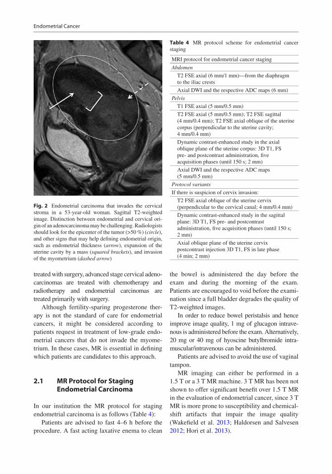
On DWI-MR the tumors usually show restricted diffusion (hyperintensity on high b-value sequences and hypointensity on the respective ADC map) (Fig. 6). ADC values have shown to be significantly lower in endometrial carcinoma than in normal endometrium and benign conditions such as endometrial polyps and submucosal leiomyomas (Fujii et al. 2008; Tamai et al. 2007; Takeuchi et al. 2009; Rechichi et al. 2011) (Fig. 7).Therefore, this functional sequence can be helpful in diagnosing an endo-metrial tumor when biopsy cannot be easily per-formed (i.e., in the case of cervical stenosis) or when the histopathological diagnosis is not con-clusive. Cross-reference with conventional sequences is, however, mandatory to avoid pit-falls, since restricted diffusion may be present also in retained secretions in the endometrial cavity.
Several studies have shown that there is no relation between the tumor grade, aggressive-ness, and ADC values (Rechichi et al. 2011;
Fig. 4 Normal uterine T2-weighted anatomy. Sagittal T2-weighted image. Hyperintense endometrium (arrow); hypointense junctional zone (dashed arrow); outer myo-metrium showing intermediate intensity (dotted line); hypointense cervical stroma (arrowhead)
Endometrial Cancer

Bharwani et al. 2011). However, in one study higher grade tumors were associated with lower ADC values but there was a considerable over-lap in estimation of histological grade based on ADC values (Tamai et al. 2007). Moreover, high ADC values may be found in high-grade tumors with a large necrotic component (Sala et al. 2010).
2.2.1 Stage I DiseaseStage I endometrial carcinomas account for approximately 74 % of endometrial cancers (Cancer Incidence Statistics 2015). The most important role of the radiologist, when facing a stage I endometrial cancer is to determine the extent of myometrial invasion, since stage I dis-ease is subdivided into stage IA disease, if the
a b
c d
Fig. 5 MR features of endometrial cancer. (a) endometri-oid cancer showing heterogeneous high-signal intensity on sagittal T2-weighted image (arrow); (b) endometrioid cancer showing hyposignal intensity on axial oblique T2-weighted image, a less common appearance (dashed arrow); (c, d) endometrioid cancer distending the uterine
cavity and compressing the myometrium showing a remarkable heterogeneous high-signal intensity on axial oblique T2 weighted image (dotted arrows) and mixed hypointensity and isointensity relative to outer myome-trium after administration of gadolinium in the equilib-rium phase (dotted arrows)
M. Horta and T.M. Cunha

tumor only invades <50 % of the myometrium, and stage IB disease, if the tumor invades ≥50 % of the myometrium (Figs. 8 and 9) (Pecorelli 2009). If the patient is considering fertility- sparing treatment, it is also important to clearly define if the tumor is confined to the endometrium or if it invades the superficial myometrium, which pre-cludes this approach (Fig. 10) (Jafari Shobeiri et al. 2013; Kesterson and Fanning 2012).
T2-weighted images are useful in determining myometrial invasion. A focal or diffuse thicken-ing of the endometrium with a clearly visualized intact junctional zone is a sign that the tumor is confined to the endometrium (Fig. 8). Breaching and irregularity of the hypointense signal of the junctional zone implies superficial myometrial invasion (Manfredi et al. 2005).
The depth of myometrial invasion should be based on the thickness of remaining myometrium
where the tumor is at its deepest point into the myometrium (Haldorsen and Salvesen 2012; Koyama et al. 2007).
However, there are situations where the evalua-tion of myometrial invasion by T2-weighted images may be difficult or even impossible, such as the presence of: a tumor in the cornua (Fig. 11); a fibromatous uterus (Fig. 11); a less conspicuous junctional zone in postmenopausal women (Fig. 12); a thinned myometrium in postmenopausal women (Fig. 13); an adenomyotic uterus (Fig. 14); a polypoid uterus; a congenital abnormality (Fig. 15); and the presence of poor tumor signal differ-ence/isointensity between the tumor and the myo-metrium (Figs. 6 and 16) (Kinkel 2006; Scoutt et al. 1995; Yamashita et al. 1993a; Saez et al. 2000; Fanning et al. 1990). In these cases, DCE-MR and DWI-MR play an important role, when combined with T2-weighted images.
a b
c
Fig. 6 (a) Endometrioid carcinoma in a 79-year-old woman, isointense to the myometrium after administra-tion of gadolinium in the equilibrium phase, making it dif-ficult to access the depth of myometrial invasion (arrow);
(b, c) diffusion-weighted image (b = 1000 mm/s2) and the respective ADC map show typical tumoral restricted dif-fusion, enabling the radiologist to diagnose invasion of ≥50 % the myometrium (stage IB) (arrow)
Endometrial Cancer
On DCE-MR imaging, in the arterial phase (≈ 30 s), the junctional zone typically enhances avidly when compared to the endometrial tumor (Manfredi et al. 2005). Disruption of this suben-dometrial enhancement usually indicates myo-metrial invasion (Fujii et al. 2015; Manfredi et al. 2005; Nakao et al. 2006; Yamashita et al. 1993b; Kaneda et al. 2011; Frei and Kinker 2001). This is particularly important when the patient is being considered for fertility-sparing treatment since the FIGO 2009 division of stage I disease is only based on the presence of invasion of the inner or
outer portions of the myometrium (Fig. 10) (Jafari Shobeiri et al. 2013; Kesterson and Fanning 2012).
On the other hand, it is in the equilibrium phase (≈120 s) that is better diagnosed deep myo-metrial invasion. In this phase there is more pro-nounced contrast-to-noise ratio between the outer myometrium, which is markedly hyperintense, and the endometrial tumor, which is usually hypointense (Figs. 9, 13 and 15) (Manfredi et al. 2005). Attention must be paid to the presence of peritumoral inflammatory enhancement, which
a b
c d
Fig. 7 Benign polyp in a 60-year-old woman. (a) Axial oblique T2-weighted image shows a heterogeneous hyperintense filling of the endometrial cavity; (b) axial oblique 3D fat-suppressed T1-weighted sequence after the administration of gadolinium in the arterial phase (30 s) shows lesional heterogeneous enhancement (arrow). Note the avid enhancement of the junctional zone in this phase when compared to the outer myometrium (dashed
arrow). Both T2 and DCE-MR characteristics of this benign polyp mimic those of endometrial carcinoma; (c) axial diffusion-weighted image (b = 1000 s/mm2) shows high-signal intensity of the tumor; (d) the apparent diffu-sion coefficient map also shows high-signal intensity. In contrast to what would be expected in an endometrial car-cinoma, this benign polyp does not show restricted diffusion
M. Horta and T.M. Cunha

can exaggerate the appearance of myometrial invasion (Sala et al. 2013).
DWI-MR has shown to be very useful in assessing myometrial invasion, especially in patients who cannot receive intravenous contrast, in patients with isointense or hyperintense tumors
relative to the myometrium following contrast administration, and in patients with adenomyosis (Figs. 16, 14 and 16) (Takeuchi et al. 2009; Sala et al. 2010).
Moreover, DWI-MR is very accurate in assessing myometrial invasion.
a b
Fig. 8 Stage IA endometrioid carcinoma. (a) Axial oblique T2-weighted image; (b) axial oblique 3D fat- suppressed T1-weighted sequence after the administration of gadolinium in the arterial phase (30 s). There is preser-vation of the regular hypointense signal on T2-weighted
image and of the regular enhancement of the junctional zone after administration of gadolinium in the arterial phase (dashed arrow), thus the tumor is confined to the endometrial cavity (arrow). Note the intramural leiomy-oma in the posterior corpus (dotted arrow)
a b
Fig. 9 Stage IB endometrioid carcinoma in an 85-year- old woman. (a) Axial oblique T2-weighted image; (b) axial oblique 3D fat-suppressed T1-weighted sequence after the administration of gadolinium in the equilibrium phase (120 s). There is extension of the tumor to the outer
portion of the left posterior myometrium (arrow), which is clearly depicted after the administration of gadolinium in the equilibrium phase, where there is more pronounced contrast-to-noise ratio between the hypointense tumor and the hyperintense outer myometrium
Endometrial Cancer

a b
Fig. 10 Endometrioid carcinoma in a woman considering fertility-sparing therapy. (a) Axial oblique T2-weighted image; (b) axial oblique 3D fat-suppressed T1-weighted sequence after the administration of gadolinium in the arterial phase (30 s). Axial oblique T2-weighted image is
inconclusive regarding myometrial invasion, whereas DCE-MR in the arterial phase clearly shows that there is no disruption of the junctional zone enhancement and therefore the tumor does not invade the myometrium
a
f g h i
j k
b c d e
M. Horta and T.M. Cunha

a b c
Fig. 12 Stage IA mucinous carcinoma of the endometrium in a 72-year-old woman with a thinned junctional zone. (a) Sagittal T2-weighted image; (b) axial oblique T2-weighted image; (c) axial oblique 3D fat-suppressed T1-weighted sequence after the administration of gadolinium in the arte-rial phase (30 s). The junctional zone of this postmeno-pausal woman cannot be identified in T2-weighted images,
making it difficult to assess the depth of tumoral myome-trial invasion. The use of T2-weighted images alone might suggest that the tumor invades the outer portion of the myo-metrium (arrow). However contrast-enhanced image in the arterial phase clearly shows an enhancing regular junctional zone (dashed arrow) and the tumor confined to the endo-metrial cavity (dotted arrow)
a b c
Fig. 13 Stage IA endometrioid carcinoma in a 72-year- old woman with myometrial thinning secondary to stretching due to a large tumor. (a) Sagittal T2-weighted image; (b) axial T2-weighted image; (c) axial oblique 3D fat-sup-pressed T1-weighted sequence after the administration of gadolinium in the equilibrium phase (120 s). Distortion at the base of this polypoid tumor is difficult to distinguish
from outer myometrial tumoral invasion on T2 weighted images (arrow). Contrast-enhanced image in the equilib-rium phase clearly shows a hypointense tumor (dashed arrow) that does not invade the outer hyperintense myome-trium (dotted arrow). Note the T2 hypointense intramural leiomyoma (asterisk)
Fig. 11 Endometrial adenocarcinoma in a 71-year-old patient, previously submitted to radiation therapy due to cervical cancer. (a–e) Sagittal T2-weighted images fail to clearly show a small isointense tumor in the right uterine cornua (yellow arrow), due to the coexistence of submu-cosal leiomyomas (dotted arrow); (f–i) axial oblique T2-weighted images better depict this isointense lesion
and clearly differentiate it from the hypointense leiomyo-mas; (j) axial diffusion-weighted image (b = 1000 s/mm2) shows high-signal intensity of the tumor; (k) the apparent diffusion coefficient map shows hyposignal intensity, therefore tumoral restricted diffusion. In contrast, the leio-myoma present in the left cornua, does not show restricted diffusion
Endometrial Cancer

a b
Fig. 14 Endometrioid carcinoma in a 71-year-old woman with an adenomyotic uterus. (a) Axial oblique T2-weighted image. (b) Axial oblique 3D fat-sup-pressed T1-weighted sequence after the administration of gadolinium in the equilibrium phase (120 s). The
presence of posterior adenomyosis (dashed arrow) impedes the assessment of tumoral (arrow) myometrial invasion on both T2-weighted image and contrast-enhanced image. Diffusion-weighted images could be useful in this case
a b
c d
Fig. 15 Stage IB endometrioid carcinoma in a 76-year- old woman with a bicornuate bicollis uterus. (a) Axial T2-weighted image; (b) axial oblique 3D fat-suppressed T1-weighted sequence after the administration of gado-linium in the equilibrium phase (120 s); (c) axial diffusion- weighted image (b = 1000 s/mm2). (d) ADC map. In this
bicornuate bicollis uterus, the tumoral invasion of the anterior outer myometrium is difficult to depict on both T2-weighted image and after administration of contrast. Diffusion-weighted image and the respective ADC map clearly identify a focal area of restricted diffusion in the anterior outer myometrium, staging the tumor as IB
M. Horta and T.M. Cunha

Recent studies have shown that DWI-MR is significantly superior in the assessment of myo-metrial invasion when compared to DCE-MR (Bonatti et al. 2015; Rechichi et al. 2010; Takeuchi et al. 2009; Beddy et al. 2012).
A prospective study conducted by Rechichi et al. has concluded that DWI-MR is highly accurate in assessing the depth of myometrial invasion and per-haps it could replace DCE-MR as a complement to T2-weighted images in the preoperative evaluation of endometrial carcinoma (Rechichi et al. 2010). These findings were also supported by the study of Bonatti et al. that also stated that T2-weighted images and DWI could replace the combination of T2-weighted images and contrast-enhanced T1-weighted images in the preoperative staging of endometrial carcinoma (Bonatti et al. 2015).
Takeuchi and coworkers also reported an accuracy of 94 % for DWI and of 88 % for DCE-MR in the assessment of myometrial inva-sion (Takeuchi et al. 2009).
ADC maps have also shown to provide accu-rate measurement of the depth of myometrial invasion (Gallego et al. 2014).
Nonetheless, a recent meta-analysis assessing only nine studies showed that although DWI-MR has a slightly higher specificity in the diagnosis of myometrial invasion, DCE-MR and DWI-MR do not differ significantly in sensitivity and speci-ficity in presurgical diagnosis of myometrial invasion (Andreano et al. 2014).
2.2.2 Stage II DiseaseStage II disease is characterized by cervical stro-mal invasion (Pecorelli 2009).
In the majority of cases, T2-weighted images are helpful in diagnosing cervical stromal inva-sion by depicting a heterogeneous hyperintense tumor disrupting the hypointense cervical stroma (Manfredi et al. 2005).
Axial oblique plane of the cervix (per-pendicular to the cervical canal) postcontrast injection 3D-reccaled echo fat-suppressed T1-weighted images in a late phase (≈80–240 s) are also very useful in assessing cervical stromal invasion, as they show the disruption of cervical enhancement by the tumor (Fig. 17) (Tirumani et al. 2013; Haldorsen and Salvesen
a b
c d
e
Fig. 16 Stage IB polypoid endometrioid carcinoma that distends the uterine cavity and causes myometrial thinning in a 68 year-old woman. (a) Axial T2-weighted image; (b) axial oblique 3D fat-suppressed T1-weighted sequence after the administration of gadolinium in the equilibrium phase (120 s); (c) axial diffusion-weighted image (b = 1000 s/mm2); (d) ADC map; (e) sagittal T2-weighted image. The
endometrial tumor is heterogeneous and shows both areas of hypointense and hyperintense signal relatively to the outer myometrium after administration of gadolinium in the equi-librium phase. This makes it difficult to assess myometrial invasion. Outer tumoral myometrial invasion in this thinned myometrium is better depicted in the diffusion-weighted image and in the respective ADC map (arrows)
Endometrial Cancer

2012; Seki et al. 2000). Normal enhance-ment of the cervical epithelium is present in tumors that project into the endocervical canal
but do not invade the cervical stroma (Fig. 18) (Tirumani et al. 2013; Haldorsen and Salvesen 2012).
a b
Fig. 17 Stage II endometrioid carcinoma in an 83-year- old woman (a) sagittal T2-weighted image; (b) axial oblique plane of the cervix (perpendicular to the cervical canal) postcontrast injection 3D-reccaled echo fat- suppressed T1-weighted images in a late phase (4 min).
Sagittal T2-weighted image shows a voluminous endome-trial tumor that prolapses in the cervical canal. The axial oblique plane of the cervix is very useful in detecting stro-mal invasion, especially after the administration of gado-linium in a late phase (4 min) (arrow)
a b c
Fig. 18 Endometrial cancer prolapsed into the endocervi-cal canal (a) Axial oblique plane of the cervix (perpen-dicular to the cervical canal) T2-weighted image: (b) sagittal T2-weighted image; (c) sagittal postcontrast injection 3D-reccaled echo fat-suppressed T1-weighted
images in the equilibrium phase (120 s). Both axial oblique and sagittal T2-weighted images raise the suspi-cion of cervical invasion. However, there is normal enhancement of the cervical epithelium without any dis-ruption, which excludes stage II disease
M. Horta and T.M. Cunha

2.2.3 Stage III DiseaseStage III disease is defined by local or regional spread of the tumor and it is subdivided into three substages (Pecorelli 2009).
In stage IIIA endometrial cancer, the tumor invades the serosa or/and the adnexa (Figs. 19 and 20).
Disruption of the serosal hypointense signal on T2-weighted images or loss of the enhancing myometrial outer margin on DCE-MR indicates serosal invasion (Fig. 19) (Tirumani et al. 2013; Manfredi et al. 2005).
The invasion of the ovaries may be due to direct extension via transmyometrial invasion or due to metastasis. Nodular foci in the adnexa are particularly frequent in type II endometrial can-
cer and in high-grade endometrioid carcinoma (Tirumani et al. 2013). DWI-MR is very useful in depicting these cases as well as pelvic peritoneal disease (Fig. 20) (Tirumani et al. 2013; Sala et al. 2010; Shen et al. 2008).
In stage IIIB disease, there is tumoral involve-ment of the parametria and/or the vagina by direct extension or by skip metastasis (Figs. 21, 22 and 23) (Pecorelli 2009). Images of the axial oblique plane of the cervix (perpendicular to the cervical canal) are very helpful in detecting parametrial invasion (Fig. 21). Detection of parametrial inva-sion is particularly important so surgeons can avoid perform a simple hysterectomy and thus perform a more extensive resection (Sala et al. 2013).
In stage IIIC endometrial cancer there is lymph node involvement (IIIC1 if there are positive pelvic lymph nodes and IIIC2 if there are positive para-aortic lymph nodes) (Fig. 24) (Pecorelli 2009).
MR sensitivity for detecting tumoral lymph node involvement is mostly low (Haldorsen and Salvesen 2012). The assessment of lymphade-nopathies on MR imaging is still based on mor-phologic criteria (shortest axis diameter >10 mm) (Haldorsen and Salvesen 2012; Manfredi et al. 2004). DWI-MR helps detect lymph nodes, but there is a significant overlap with the ADC values of benign and malignant nodes (Nougaret et al. 2013; Nakai et al. 2008; Roy et al. 2010).
2.2.4 Stage IV DiseaseStage IV endometrial cancers are subdivided in two substages: stage IVA if there is invasion of
Fig. 19 Stage IIIA endometrioid carcinoma. (a) Axial oblique T2-weighted image shows disruption of the T2 hypointense serosal signal (dashed arrow) and tumoral invasion of the left ovary (arrow)
a b c
Fig. 20 Stage IIIA endometrioid carcinoma with subtle peritoneal nodules (a) axial T2-weighted image; (b) axial diffusion-weighted image (b = 1000 s/mm2); (c) ADC
map. Subtle peritoneal implants can be better depicted with diffusion-weighted images (arrow)
Endometrial Cancer

the bladder or/and the bowel (Figs. 25 and 26); stage IVB if there are distant metastasis includ-ing abdominal metastasis and/or the presence of
positive inguinal lymph nodes or/and para-aortic nodes above the renal hila (Fig. 27) (Pecorelli 2009).
For an endometrial tumor to be considered as stage IVA, a lesion transgressing the hypoin-tense T2-signal of the muscularis propria of the bladder and/or of the bowel must be clearly seen, as well as disrupting the hyperintense sig-nal of the mucosa (Figs. 25 and 26) (Tirumani et al. 2013). Moreover, tumoral enhancement within the lumen should also be present (Figs. 25 and 26). This is particularly important in the differential diagnosis of bladder bullous edema (a sign of tumoral involvement of the bladder serosa and muscular layer, but not of the mucosa), since it is not considered stage IVA disease (Sala et al. 2013).
Although rare at presentation, distant hema-togenous disease most commonly affects the liver, lungs, and bone (Fig. 27) (Numazaki et al. 2009).
In type 2 endometrial cancer, and in high-grade endometrioid endometrial cancer, stage IVB disease may present as in ovarian cancers with peritoneal carcinomatosis (Tirumani et al. 2013; Sala et al. 2013).
a b
Fig. 21 Stage IIIB endometrioid carcinoma (a) sagittal T2-weighted image; (b) axial oblique plane of the cervix (perpendicular to the cervical canal) T2-weighted image. Parametrial invasion can be better depicted in the axial
oblique plane of the cervix, where disruption of the hypointense stromal signal can be seen as well as tumor extending to both parametria (arrows)
Fig. 22 Stage IIIB endometrioid carcinoma. (a) Sagittal T2-weighted image shows disruption of the T2 hypoin-tense vaginal signal with invasion of the inferior third of the vagina (arrows)
M. Horta and T.M. Cunha
3 Recent Advances in Functional MR Imaging in Assessing Endometrial Carcinoma
In perfusion MR, repeated acquisitions of sequential images during the passage of gadolin-ium through the tumor are performed. Tumoral signal intensity versus time graphs and quantita-tive perfusion maps may be obtained, making it possible to evaluate microvascularity parameters
(Haldorsen and Salvesen 2012). The value of per-fusion MR in assessing endometrial cancer is not yet established and validation studies are neces-sary to study its potential benefit.
Haldorsen et al. suggested that DCE-MRI may offer new information about the histological subtype and clinical course of endometrial carci-noma (Haldorsen et al. 2013). On the other hand, other studies regarding DCE-perfusion MR and tumor grading have shown contradictory results (Haldorsen et al. 2013; Ippolito et al. 2014a, b).
a b
c
d
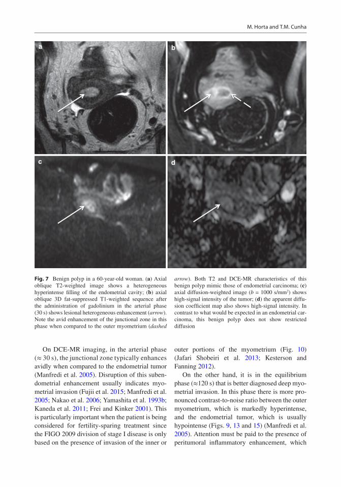
Fig. 23 Stage IIIB endometrioid carcinoma in a 62-year- old woman with skip vaginal metastasis. (a) Sagittal T2-weighted image; (b) axial T2-weighted image; (c) axial diffusion-weighted image (b = 1000 s/mm2); (d)
ADC map. In the anterior lower third of the vagina a small lesion showing restricted diffusion can be seen (arrow). It was diagnosed as a vaginal metastasis
Endometrial Cancer

a b
Fig. 24 Stage IIIC endometrial cancer (a) axial T2-weighted image shows pelvic lymphadenopathies (arrows) (stage IIIC1 disease); (b) axial T2-weighted
image shows para-aortic lymphadenopathies (dashed arrows) (Stage IIIC2 disease)
a
d e
b c
Fig. 25 Stage IVA endometrioid carcinoma that invades the sigmoid colon in a 75-year-old woman. (a) Sagittal T2-weighted image; (b) sagittal 3D fat-suppressed T1-weighted sequence after the administration of gado-linium in the equilibrium phase (120–180 s); (c) axial T2-weighted image; (d) axial diffusion-weighted image
(b = 1000 s/mm2); (e) ADC map. T2-weighted images show disruption of the hypointense signal of the sigmoid colon wall and mucosal invasion. There is tumoral enhancement within the lumen. The tumor shows also marked restricted diffusion, which can also be seen in the sigmoid lumen
M. Horta and T.M. Cunha

4 Therapeutic Approaches
4.1 Surgery
All medical operable patients with endometrial cancer should be considered for surgery (Koh et al. 2014).
The standard surgical approach of endome-trial cancer consists of total hysterectomy with bilateral salpingo-oophorectomy, assessment of the abdominal cavity with biopsies of suspicious peritoneal lesions, cytology of peritoneal wash-ing, and at least excision of enlarged lymph nodes (Koh et al. 2014).
Although, systematic pelvic and para-aortic lymphadenopathy is considered part of FIGO surgico-pathological staging, there is still no consensus if it should be performed in cases of treatment of stage I endometrial cancer (Koh et al. 2014; Colombo et al. 2013; Soliman et al. 2010).
A randomized study has shown that in patients where systematic lymphadenectomy was per-formed, a higher rate of postoperative complica-tions was detected with no improvement of disease-free and overall survival rates (Benedetti Panici et al. 2008). The ASTEC surgical trial also concluded that pelvic lymphadenectomy in patients with early disease did not improve over-all or recurrence-free survival (ASTEC study group et al. 2009). These two studies had impor-tant limitations and caution must be taken to avoid overinterpretation.
On the other hand, a large retrospective study, the SEPAL study, showed survival benefits in patients with intermediate (FIGO IA and IB, grade 3 endometrioid carcinoma; FIGO IB, grade 1 and 2 endometrioid carcinoma with lympho-vascular space invasion; FIGO IC and II endome-trioid carcinoma; any type of type 2 endometrial cancer) and high risk (FIGO III and IV endome-trioid carcinoma) endometrial carcinoma when
a
d e
b c
Fig. 26 Stage IVA endometrioid carcinoma that invades the bladder in a 76-year-old woman. (a) Sagittal T2-weighted image; (b) axial T2-weighted image; (c) axial oblique 3D fat-suppressed T1-weighted sequence after the administration of gadolinium in the equilibrium phase (120 s); (d) axial diffusion-weighted image
(b = 1000 s/mm2); (e) ADC map. T2-weighted images show disruption of the hypointense signal of the bladder wall and mucosal invasion. There is tumoral enhancement within the lumen. The tumor shows also marked restricted diffusion, which can also be seen in the bladder lumen
Endometrial Cancer

submitted to combined pelvic and para-aortic lymphadenectomy (Todo et al. 2010).
Women with stage II should be considered for radical hysterectomy with bilateral salpingo- oophorectomy and pelvic and para-aortic lymph-adenectomy (Colombo et al. 2013).
In patients with a good performance status with advanced stage disease (FIGO stage III and IV), maximum debulking surgery of resettable tumors should be performed. Anterior and posterior pel-vic exenteration can be considered for stage IVA disease as well as palliative surgery in patients with metastatic disease (Colombo et al. 2013).
Women with serous, clear-cell carcinomas and carcinosarcomas should be submitted to the same surgical-pathological staging of ovarian cancer (Koh et al. 2014). Serous and clear-cell carcinomas can occur in atrophic endometrium and may present with distant disease even without myometrial invasion; therefore, omentectomy,
diaphragmatic cytology or biopsy, and random peritoneal biopsies should be performed (Koh et al. 2014; Colombo et al. 2013). Pelvic and para-aortic lymphadenectomy is mandatory (Colombo et al. 2013).
4.2 Adjuvant Treatment
Radiation therapy is usually used in the adjuvant setting, in the form of external beam radiation therapy (EBRT) and vaginal brachytherapy (VBT).
The purpose of adjuvant radiation therapy is to eliminate microscopic disease in the lymph nodes and in the central pelvic region, including the vagi-nal cuff; thus reducing the risk of recurrence.
According to the 2014 guidelines of the American Society for Radiation Oncology (ASTRO), patients with endometrioid carcinoma
a b
c
Fig. 27 Stage IVB disease (a) axial T2-weighted image showing a pulmonary metastasis (arrow); (b) axial T2-weighted image showing an umbilical metastasis
(Sister Mary Joseph node) (dashed arrow); (c) axial T2-weighted image showing hepatic metastasis (dotted arrows) and a left adrenal metastasis (asterisk)
M. Horta and T.M. Cunha

that do not require adjuvant radiotherapy (RT) are women with no residual disease in the hys-terectomy specimen and patients with grade 1 or 2 endometrial cancer with no myometrial inva-sion or with <50 % of myometrial invasion, especially when no high risk features are pres-ent (Klopp et al. 2014). Patients with grade 3 endometrioid carcinoma without myometrial invasion and patients with grade 1 or 2 cancer with <50 % of myometrial invasion and higher risk factors, such as being age >60 years and/or lymphovascular space invasion, can be ade-quately treated with or without VBT (Klopp et al. 2014).
These guidelines also identify the patients with endometrioid cancer for whom vaginal cuff brachytherapy is as effective as pelvic radiation therapy and consequently should be preferred. These patients are women with grade 1 or 2 can-cers with ≥50 % of myometrial invasion and women with grade 3 tumors with <50 % of myo-metrial invasion (Klopp et al. 2014).
In order to decrease pelvic recurrence, the fol-lowing women with early phase endometrial can-cer should receive adjuvant EBRT: patients with stage I, grade 3 cancer with ≥50 % of myometrial invasion or patients with cervical stromal inva-sion; patients with stage I, grade 1 or 2 cancer, with ≥50 % of myometrial invasion and other risk factors such as being of age >60 years and/or lymphovascular space invasion (Klopp et al. 2014).
If there is a negative prognostic factor, pelvic RT and/or chemotherapy should be performed for stage I and stage II disease (Colombo et al. 2013).
In patients with stage III and IVA disease, external EBRT and adjuvant chemotherapy are justifiable options. These patients can be con-sidered for concurrent chemoradiation followed by adjuvant chemotherapy (Klopp et al. 2014).
If metastatic disease is present, chemotherapy- radiotherapy can be performed for palliative treatment (Colombo et al. 2013).
Patients with serous carcinomas, clear-cell carcinomas, and carcinosarcomas should be considered for platinum-based adjuvant chemo-therapy with or without radiation therapy (Koh et al. 2014).
4.3 Fertility-Sparing Treatment
Fertility-sparing treatment with progestin hor-monal therapy may be considered for women who express the desire to preserve fertility and have atypical endometrial hyperplasia or grade 1 endometrioid carcinoma without myometrial involvement in MR (Jafari Shobeiri et al. 2013; Kesterson and Fanning 2012; Colombo et al. 2016). However, this is not the standard of care and the decision to proceed with conservative management is associated with several hazards. Risks include an inadequately staged and treated advanced endometrial cancer; the presence of a synchronous or meta-synchronous cancer; the presence of an inherited genetic predisposition; and the absence of standard medical management and surveillance (Kesterson and Fanning 2012).
5 Follow-Up and Recurrent Endometrial Carcinoma
The majority of recurrences will occur within the first 3 years (80 %) (Tirumani et al. 2013). However, only 15 % of patients will develop recurrent disease (Sala et al. 2013; Magrina et al. 2011).
The recommended follow-up is a clinical and gynecological exam at 3–4 months intervals dur-ing the first 2 years and then every 6 months until 5 years (Colombo et al. 2013).
During follow-up, radiological exams should only be performed if clinically indicated (Colombo et al. 2013).
Most of endometrial cancer recurrences occur in the lymph nodes (46 %) and in the vaginal vault (42 %) (Sohaib et al. 2007). The perito-neum and lungs are also reported sites of recur-rence, although less frequent (Sohaib et al. 2007).
MR imaging is very useful in diagnosing vagi-nal vault recurrences, which characteristically display the same signal characteristics as of those of the primary tumor (Tirumani et al. 2013). They tend to appear as a mass disrupting the low signal intensity of the vaginal vault (Sala et al. 2013). MR in these cases plays a role in defining surgi-cal resectability of pelvic recurrences.
Endometrial Cancer

PET-CT has shown high sensitivity (93 %), specificity (93 %), and accuracy (93 %) in detect-ing recurrent disease (Kitajima et al. 2008). However, more prospective larger studies must determine its role for routine screening (Salani et al. 2011).
5.1 Treatment of Recurrence
Treatment of recurrence varies according to the site, to previous treatments, and to the initial stage of the disease.
Vaginal recurrence is the most common and the standard of treatment is EBRT plus VBP (Colombo et al. 2013).
Central pelvic recurrence is usually treated with surgery and radiation therapy, whereas regional pelvic recurrence is treated with radiation therapy plus chemotherapy if possible (Colombo et al. 2013).
Recurrent serous and clear-cell carcinomas should be treated with the same chemotherapeu-tic regimens of ovarian cancer (Colombo et al. 2013).
6 Prognosis
As stated previously, endometrial carcinoma gen-erally has a good outcome. Prognostic factors include histological subtype; histological grade; tumor stage; depth of invasion; and lymphovas-cular space invasion (Colombo et al. 2013).
Reported 5-year survival rates for stage IA and IB are of 88 % and of 75 %, respectively. The prognosis of the different types of stage III sub-stages differs significantly from one another, with estimated 5-year survival rates of 58 % for stage IIIC1 and of 47 % for stage IIIC2. Stage IV disease 5-year survival is estimated at 15–17 % (American Cancer Society 2016).
The presence of lymph node metastasis, namely para-aortic, has shown to have a strong effect on prognosis (Todo et al. 2010). Tumor grade, lymphovascular space involvement, and the depth of myometrial invasion have been
reported as high risk factors for the tumoral involvement of lymph nodes (Patel et al. 2010).
Invasion of the outer half of the myometrium is also strongly associated with a diminished 5-year survival rate (Patel et al. 2010; Amant et al. 2005). Furthermore, it was demonstrated that patients with lymphovascular space involve-ment had an overall 5-year survival rate of 64 %, and on the other hand women without lympho-vascular space involvement had an overall 5-year survival rate of 88 % (Colombo et al. 2013).
Conclusion
Magnetic resonance imaging is not considered in the International Federation of Gynaecology and Obstetrics staging system. However, it plays a relevant role in the preoperative staging of endometrial carcinomas, helping to define the best therapeutic approaches. Moreover, it is important in the diagnosis of treatment compli-cations, in the surveillance of therapy response, and in the assessment of recurrent disease.
Functional MR techniques and specific MR sequence planes play an important role in preventing errors in endometrial cancer staging.
To help prevent diagnostic errors and in order to guide appropriate therapeutic man-agement, radiologists should be aware of common magnetic resonance imaging endo-metrial appearances for all stages and the fre-quent mistakes in the assessment of endometrial cancer.
References
Amant F, Moerman P, Neven P, Timmerman D, Van Limbergen E, Vergote I (2005) Endometrial cancer. Lancet 366(9484):491–505
Amant F, Moerman P, Neven P, Timmerman D, Van Limbergen E, Vergote I (2007) Treatment modalities in endometrial cancer. Curr Opin Oncol 19(5):479–485
American Cancer Society (2016) http://www.cancer.org/cancer/endometrialcancer/detailedguide/endometrial- uterine- cancer-survival-rates. Accessed 25 May 2016
Andreano A, Rechichi G, Rebora P, Sironi S, Valsecchi MG, Galimberti S (2014) MR diffusion imaging for preoperative staging of myometrial invasion in patients with endometrial cancer: a systematic review and
M. Horta and T.M. Cunha

meta-analysis. Eur Radiol 24(6):1327–1338. doi:10.1007/s00330-014-3139-4
ASTEC Study Group, Kitchener H, Swart AM, Qian Q, Amos C, Parmar MK (2009) Efficacy of sys-tematic pelvic lymphadenectomy in endome-trial cancer (MRC ASTEC trial): a randomised study. Lancet 373(9658):125–136. doi:10.1016/S0140-6736(08)61766-3
Beddy P, Moyle P, Kataoka M, Yamamoto AK, Joubert I, Lomas D et al (2012) Evaluation of depth of myome-trial invasion and overall staging in endometrial can-cer: comparison of diffusion-weighted and dynamic contrast-enhanced MR imaging. Radiology 262(2): 530–537. doi:10.1148/radiol.11110984
Benedetti Panici P, Basile S, Maneschi F, Alberto Lissoni A, Signorelli M, Scambia G et al (2008) Systematic pelvic lymphadenectomy vs no lymphadenectomy in early-stage endometrial carcinoma: randomized clini-cal trial. J Natl Cancer Inst 100(23):1707–1716. doi:10.1093/jnci/djn397
Bennet GL, Andreotti RF, Lee SI, Dejesus Allison SO, Brown DL, Dubinsky T et al (2011 Jul) ACR Appropriateness Criteria® on Abnormal Vaginal Bleeding. J Am Coll Radiol 8(7):460–468. doi:10.1016/j.jacr.2011.03.011
Bharwani N, Miquel ME, Sahdev A, Narayanan P, Malietzis G, Reznek RH et al (2011) Diffusion- weighted imaging in the assessment of tumour grade in endometrial cancer. Br J Radiol 84(1007):997–1004. doi:10.1259/bjr/14980811
Bonatti M, Stuefer J, Oberhofer N, Negri G, Tagliaferri T, Schifferle G et al (2015) MRI for local staging of endometrial carcinoma: is endovenous contrast medium administration still needed? Eur J Radiol 84(2):208–214. doi:10.1016/j.ejrad.2014.11.010
Boronow RC (1990) Advances in diagnosis, staging, and management of cervical and endometrial cancer Stages I and II. Cancer 65(3 Suppl):648–659
Briët JM, Hollema H, Reesink N, Aalders JG, Mourits MJE, Hoor ten KA et al (2005) Lymphvascular space involvement: an independent prognostic factor in endometrial cancer. Gynecol Oncol 96(3):799–804
Calle EE, Kaaks R (2004) Overweight, obesity and can-cer: epidemiological evidence and proposed mecha-nisms. Nat Rev Cancer 4(8):579–591
Cancer Incidence Statistics (2015) http://www.cancerre-searchuk.org/health-professional/cancer-statistics/statistics- by-cancer-type/uterine-cancer. Accessed on 22 May 2015
Colombo N, Preti E, Landoni F, Carinelli S, Colombo A, Marini C et al (2013) Endometrial cancer: ESMO Clinical Practice Guidelines for diagnosis, treatment and follow-up. Ann Oncol 24(Suppl 6):vi33–vi38. doi:10.1093/annonc/mdt353
Colombo N, Creutzberg C, Amant F, Bosse T, González- Martín A, Ledermann J et al (2016) ESMO-ESGO- ESTRO consensus conference on endometrial cancer: diagnosis, treatment and follow-up. Int J Gynecol Cancer 26(1):2–30. doi:10.1097/IGC.0000000000000609
Creasman W (2009) Revised FIGO staging for carcinoma of the endometrium. Int J Gynaecol Obstet 105(2):109. doi:10.1016/j.ijgo.2009.02.010
Eichhorn JH, Young RH (2001) Neuroendocrine tumors of the genital tract. Am J Clin Pathol 115(Suppl):S94–112
Epstein E, Blomqvist L (2014) Imaging in endometrial cancer. Best Pract Res Clin Obstet Gynaecol 28(5):721–739. doi:10.1016/j.bpobgyn.2014.04.007
Fanning J, Tsukada Y, Piver MS (1990) Intraoperative frozen section diagnosis of depth of myometrial invasion inen-dometrial adenocarcinoma. Gynecol Oncol 37(1):47–50
Ferlay J, Soerjomataram I, Ervik M, Dikshit R, Eser S, Mathers C, Rebelo M et al (2013) Cancer Incidence and Mortality Worldwide: IARC CancerBase No. 11 [Internet]. Lyon: International Agency for Research on Cancer. Available from: http://globocan.iarc.fr. Accessed 23 May 2015
Fisher B, Costantino JP, Redmond CK, Fisher ER, Wickerham DL, Cronin WM (1994) Endometrial can-cer in tamoxifen-treated breast cancer patients: find-ings from the National Surgical Adjuvant Breast and Bowel Project (NSABP) B-14. J Natl Cancer Inst 86(7):527–537
Frei KA, Kinker K (2001) Staging endometrial cancer: role of magnetic resonance imaging. J Magn Reson Imaging 13(6):850–855
Frei KA, Kinkel K, Bonél HM, Lu Y, Zaloudek C, Hricak H (2000) Prediction of deep myometrial invasion in patients with endometrial cancer: clinical utility of contrast-enhanced MR imaging–a meta-analysis and bayesian analysis. Radiology 216(2):444–449
Fujii S, Matsusue E, Kigawa J, Sato S, Kanasaki Y, Nakanishi J et al (2008) Diagnostic accuracy of the apparent diffusion coefficient in differentiating benign from malignant uterine endometrial cavity lesions: ini-tial results. Eur Radiol 18(2):384–389
Fujii S, Kido A, Baba T, Fujimoto K, Daido S, Matsumura N (2015) Subendometrial enhancement and peri-tumoral enhancement for assessing endometrial cancer on dynamic contrast enhanced MR imag-ing. Eur J Radiol 84(4):581–589. doi:10.1016/j.ejrad.2015.01.004
Gallego JC, Porta A, Pardo MC, Fernández C (2014) Evaluation of myometrial invasion in endometrial cancer: comparison of diffusion-weighted mag-netic resonance and intraoperative frozen sections. Abdom Imaging 39(5):1021–1026. doi:10.1007/s00261-014-0134-9
Goldstein RB, Bree RL, Benson CB, Benacerraf BR, Carlos R et al (2001) Evaluation of the woman with postmenopausal bleeding: society of radiologists in ultrasound-sponsored consensus conference state-ment. J Ultrasound Med 20(10):1025–1036
Gull B, Karlsson B, Milsom I, Granber S (2003) Can ultrasound replace dilation and curettage? A longitudi-nal evaluation of postmenopausal bleeding and trans-vaginal sonographic measurement of the endometrium as predictors of endometrial cancer. Am J Obstet Gynecol 188(2):401–408
Endometrial Cancer

Gupta JK, Chien PF, Voit D, Clark TJ, Khan KS (2002) Ultrasonographic endometrial thickness for diagnos-ing endometrial pathology in women with postmenopausal bleeding: a meta-analysis. Acta Obstet Gynecol Scand 81(9):799–816
Haider MA, Patlas M, Jhaveri K, Chapman W, Fyles A, Rosen B (2006) Adenocarcinoma involving the uter-ine cervix: magnetic resonance imaging findings in tumours of endometrial, compared with cervical, ori-gin. Can Assoc Radiol J 57(1):43–48
Haldorsen IS, Salvesen HB (2012) Staging of endometrial carcinomas with MRI using traditional and novel MRI techniques. Clin Radiol 67(1):2–12. doi:10.1016/j.crad.2011.02.018
Haldorsen IS, Grüner R, Husby JA, Magnussen IJ, Werner HMJ, Salvesen ØO et al (2013) Dynamic contrast- enhanced MRI in endometrial carcinoma identifies patients at increased risk of recurrence. Eur Radiol 23(10):2916–2925. doi:10.1007/s00330-013-2901-3
Hecht JL, Mutter GL (2006) Molecular and pathologic aspects of endometrial carcinogenesis. J Clin Oncol 24(29):4783–4791
Hori M, Kim T, Onishi H, Imaoka I, Kagawa Y, Murakami T et al (2013) Endometrial cancer: preoperative stag-ing using three-dimensional T2-weighted turbo spin- echo and diffusion-weighted MR imaging at 3.0 T: a prospective comparative study. Eur Radiol 23(8):2296–2305. doi:10.1007/s00330-013-2815-0
Hulka CA, Hall DA, McCarthy K, Simeone JF (1994) Endometrial polyps, hyperplasia, and carcinoma in postmenopausal women: differentiation with endo-vaginal sonography. Radiology 191(3):755–758
Ippolito D, Minutolo O, Cadonici A, Talei Franzesi C, Bonaffini P, Perego P et al (2014a) Endometrial can-cer: diagnostic value of quantitative measurements of microvascular changes with DCE-MR imaging. MAGMA 27(6):531–538. doi:10.1007/s10334-014-0435-6
Ippolito D, Cadonici A, Bonaffini PA, Minutolo O, Casiraghi A, Perego P et al (2014b) Semiquantitative perfusion combined with diffusion-weighted MR imaging in pre-operative evaluation of endometrial carcinoma: Results in a group of 57 patients. Magn Reson Imaging 32(5):464–472. doi:10.1016/j.mri.2014.01.009
Jafari Shobeiri M, Mostafa Gharabaghi P, Esmaeili H, Ouladsahebmadarek E, Mehrzad-Sadagiani M (2013) Fertility sparing treatment in young patients with early endometrial adenocarcinoma: case series. Pak J Med Sci 29(2):651–655
Jalloul RJ, Elshaikh MA, Ali-Fehmi R, Haley MM, Yoon J, Mahan M et al (2012) Mucinous adenocarcinoma of the endometrium. Int J Gynecol Cancer 22(5):812–818. doi:10.1097/IGC.0b013e31824a
Järvinen HJ, Renkonen-Sinisalo L, Aktán-Collán K, Peltomäki P, Aaltonen LA, Mecklin J-P (2009) Ten years after mutation testing for Lynch syndrome: can-cer incidence and outcome in mutation-positive and mutation-negative family members. J Clin Oncol 27(28):4793–4797. doi:10.1200/JCO.2009.23.7784
Kaneda S, Fujii S, Fukunaga T, Kakite S, Kaminou T, Kigawa J et al (2011) Myometrial invasion by endo-metrial carcinoma: evaluation with 3.0 T MR imaging. Abdom Imaging 36(5):612–618. doi:10.1007/s00261-011-9719-8
Karlsson B, Granberg S, Wikland M, Ylostalo P, Torvid K, Marsal K et al (1995) Transvaginal ultrasonogra-phy of the endometrium in women withpostmeno-pausal bleeding—a Nordic multicenter study. Am J Obstet Gynecol 172(5):1488–1494
Kesterson JP, Fanning J (2012) Fertility-sparing treatment of endometrial cancer: options, outcomes and pitfalls. J Gynecol Oncol 23(2):120–124. doi:10.3802/jgo.2012.23.2.120
Kinkel K (2006) Pitfalls in staging uterine neoplasm with imaging: a review. Abdom Imaging 31(2):164–173
Kitajima K, Murakami K, Yamasaki E, Domeki Y, Kaji Y, Morita S et al (2008) Performance of integrated FDG- PET/contrast-enhanced CT in the diagnosis of recurrent uterine cancer: comparison with PET and enhanced CT. Eur J Nucl Med Mol Imaging 36(3):362–372. doi:10.1007/s00259-008-0956-1
Klopp A, Smith BD, Alektiar K, Cabrera A, Damato AL, Erikson B et al (2014) The role of postopera-tive radiation therapy for endometrial cancer: Executive Summary of an American Society for Radiation Oncology evidence-based guideline. Pract Radiat Oncol 4(3):137–144. doi:10.1016/j.prro.2014.01.003
Koh WJ, Greer BE, Abu-Rustum NR, Apte SM, Campos SM, Chan J et al (2014) Uterine Neoplasms, version 1.2014. J Natl Compr Canc Netw 12(2):248–280
Koyama T, Tamai K, Togashi K (2007) Staging of carci-noma of the uterine cervix and endometrium. Eur Radiol 17(8):2009–2019
Kurman RJ, Carcangiu ML, Herrington CS, Young RH (2014) Tumours of the Uterine Corpus. In: WHO clas-sification of tumours of female reproductive organs, vol 6, 4th edn. International Agency for Research on Cancer, Lyon, pp. 121–155
Larson DM, Connor GP, Broste SK, Krawisz BR, Johnson KK (1996) Prognostic significance of gross myome-trial invasion with endometrial cancer. Obstet Gynecol 88(3):394–398
Lopes Dias J, Cunha TM, Gomes FV, Callé C, Félix A (2015) Neuroendocrine tumours of the female genital tract: a case-based imaging review with pathological correlation. Insights Imaging 6(1):43–52. doi:10.1007/s13244-014-0378-5
Magrina JF, Zanagnolo V, Giles D, Noble BN, Kho RM, Magtibay PM (2011) Robotic surgery for endome-trial cancer: comparison of perioperative outcomes and recurrence with laparoscopy, vaginal/laparos-copy and laparotomy. Eur J Gynaecol Oncol 32(5): 476–480
Manfredi R, Mirk P, Maresca G, Margariti PA, Testa A, Zannoni GF et al (2004) Local-regional staging of endometrial carcinoma: role of MR imaging in surgi-cal planning. Radiology 231(2):372–378
M. Horta and T.M. Cunha
Manfredi R, Gui B, Maresca G, Fanfani F, Bonomo L (2005) Endometrial cancer: magnetic resonance imag-ing. Abdom Imaging 30(5):626–636
May K, Bryant A, Dickinson HO, Kehoe S, Morrison J (2010) Lymphadenectomy for the management of endometrial cancer. Cochrane Database Syst Rev 20(1):CD007585. doi:10.1002/14651858.CD007585.pub2
McPherson CP, Sellers TA, Potter JD, Bostick RM, Folsom AR (1996) Reproductive factors and risk of endometrial cancer. The Iowa Women's Health Study. Am J Epidemiol 143(12):1195–1202
Meyer LA, Broaddus RR, Lu KH (2009) Endometrial cancer and Lynch syndrome: clinical and pathologic considerations. Cancer Control 16(1):14–22
Nakai G, Matsuki M, Inada Y, Tatsugami F, Tanikake M, Narabayashi I et al (2008) Detection and evaluation of pelvic lymph nodes in patients with gynecologic malignancies using body diffusion-weighted mag-netic resonance imaging. J Comput Assist Tomogr 32(5):764–768. doi:10.1097/RCT.0b013e318153fd43
Nakao Y, Yokoyama M, Hara K, Koyamatsu Y, Yasunaga M, Araki Y et al (2006) MR imaging in endometrial carcinoma as a diagnostic tool for the absence of myometrial invasion. Gynecol Oncol 102(2):343–347
Nougaret S, Tirumani SH, Addley H, Pandey H, Sala E, Reinhold C (2013) Pearls and pitfalls in MRI of gyne-cologic malignancy with diffusion-weighted tech-nique. AJR Am J Roentgenol 200(2):261–276. doi:10.2214/AJR.12.9713
Nougaret S, Reinhold C, Alsharif SS, Addley H, Arceneau J, Molinari N et al (2015) Endometrial cancer: com-bined MR volumetry and diffusion-weighted imaging for assessment of myometrial and lymphovascular invasion and tumor grade. Radiology 276(3):797–808. doi:10.1148/radiol.15141212
Numazaki R, Miyagi E, Konnai K, Ikeda M, Yamamoto A, Onose R et al (2009) Analysis of stage IVB endo-metrial carcinoma patients with distant metastasis: a review of prognoses in 55 patients. Int J Clin Oncol 14(4):344–350. doi:10.1007/s10147-009-0878-3
Patel S, Liyanage SH, Sahdev A, Rockall AG, Reznek RH (2010) Imaging of endometrial and cervical cancer. Insights Imaging 1(5–6):309–328
Pecorelli S (2009) Revised FIGO staging for carcinoma of the vulva, cervix, and endometrium. Int J Gynaecol Obstet 105(2):103–104
Rechichi G, Galimberti S, Signorelli M, Perego P, Valsecchi MG, Sironi S (2010) Myometrial invasion in endometrial cancer: diagnostic performance of diffusion- weighted MR imaging at 1.5-T. Eur Radiol 20(3):754–762. doi:10.1007/s00330-009-1597-x
Rechichi G, Galimberti S, Signorelli M, Franzesi CT, Perego P, Valsecchi MG et al (2011) Endometrial cancer: correlation of apparent diffusion coeffi-cient with tumor grade, depth of myometrial inva-sion, and presence of lymph node metastases. AJR Am J Roentgenol 197(1):256–262. doi:10.2214/AJR.10.5584
Renehan AG, Tyson M, Egger M, Heller RF, Zwahlen M (2008) Body-mass index and incidence of cancer: a systematic review and meta-analysis of prospective observational studies. Lancet 371(9612):569–578. doi:10.1016/S0140-6736(08)60269-X
Resnick KE, Hampel H, Fishel R, Cohn DE (2009) Current and emerging trends in Lynch syndrome iden-tification in women with endometrial cancer. Gynecol Oncol 114(1):128–134. doi:10.1016/j.ygyno.2009.03.003 Epub 2009 Apr 17
Roy C, Bierry G, Matau A, Bazille G, Pasquali R (2010) Value of diffusion-weighted imaging to detect small malignant pelvic lymph nodes at 3 T. Eur Radiol 20(8):1803–1811. doi:10.1007/s00330-010-1736-4
Saez F, Urresola A, Larena JA, Martín JI, Pijuán JI, Schneider J et al (2000) Endometrial carcinoma: assessment of myometrial invasion with plain and gadolinium-enhanced MR imaging. J Magn Reson Imaging 12(3):460–466
Sala E, Rockall A, Rangarajan D, Kubik-Huch RA (2010) The role of dynamic contrast-enhanced and diffusion weighted magnetic resonance imaging in the female pelvis. Eur J Radiol 76(3):367–385. doi:10.1016/j.ejrad.2010.01.026
Sala E, Rockall AG, Freeman SJ, Mitchell DG, Reinhold C (2013) The added role of MR imaging in treatment stratification of patients with gynecologic malignan-cies: what the radiologist needs to know. Radiology 266(3):717–740. doi:10.1148/radiol.12120315
Salani R, Backes FJ, Fung MF, Holschneider CH, Parker LP, Bristow RE et al (2011 Jun) Posttreatment surveil-lance and diagnosis of recurrence in women with gynecologic malignancies: society of Gynecologic Oncologists recommendations. Am J Obstet Gynecol 204(6):466–478. doi:10.1016/j.ajog.2011.03.008
Schmeler KM, Lynch HT, Chen LM, Munsell MF, Soliman PT, Clark MB et al (2006) Prophylactic sur-gery to reduce the risk of gynecologic cancers in the lynch syndrome. N Engl J Med 354(3):261–269
Scoutt LM, McCarthy SM, Flynn SD, Lange RC, Long F, Smith RC et al (1995) Clinical stage I endometrial car-cinoma: pitfalls in preoperative assessment with MR imaging Work in progress. Radiology 194(2): 567–572
Seki H, Takano T, Sakai K (2000) Value of dynamic MR imaging in assessing endometrial carcinoma involve-ment of the cervix. AJR Am J Roentgenol 175(1):171–176
Setiawan VW, Yang HP, Pike MC, McCann SE, Yu H, Xiang Y-B et al (2013) Type I and II endometrial can-cers: have they different risk factors? J Clin Oncol 31(20):2607–2618. doi:10.1200/JCO.2012.48.2596
Shapiro S, Kelly JP, Rosenberg L, Kaufman DW, Helmrich SP, Rosenshein NB et al (1985) Risk of localized and widespread endometrial cancer in relation to recent and discontinued use of conjugated estrogens. N Engl J Med 313(16):969–972
Shen S-H, Chiou Y-Y, Wang J-H, Yen M-S, Lee R-C, Lai C-R et al (2008) Diffusion-weighted single-shot echo- planar imaging with parallel technique in assessment
Endometrial Cancer

of endometrial cancer. Am J Roentgenol 190(2):481–488. doi:10.2214/AJR.07.2155
Smith-Bindman R, Kerlikowske K, Feldstein VA, Subak L, Scheidler J, Segal M et al (1998) Endovaginal ultra-sound to exclude endometrial cancer and other endo-metrial abnormalities. JAMA 280(17):1510–1517
Sohaib SA, Houghton SL, Meroni R, Rockall AG, Blake P, Reznek RH (2007) Recurrent endometrial cancer: patterns of recurrent disease and assessment of prog-nosis. Clin Radiol 62(1):28–34
Soliman PT, Wu D, Tortolero-Luna G, Schmeler KM, Slomovitz BM, Bray MS et al (2006) Association between adiponectin, insulin resistance, and endome-trial cancer. Cancer 106(11):2376–2381
Soliman PT, Frumovitz M, Spannuth W, Greer MJ, Sharma S, Schmeler KM et al (2010 Nov) Lymphadenectomy during endometrial cancer staging: Practice patterns among gynecologic oncologists. Gynecol Oncol 119(2):291–294. doi:10.1016/j.ygyno.2010.07.011
Takeuchi M, Matsuzaki K, Nishitani H (2009) Diffusion- weighted magnetic resonance imaging of endometrial cancer: differentiation from benign endometrial lesions and preoperative assessment of myometrial invasion. Acta Radiol 50(8):947–953. doi:10.1080/02841850903099981
Tamai K, Koyama T, Saga T, Umeoka S, Mikami Y, Fujii S et al (2007) Diffusion-weighted MR imaging of uterine endometrial cancer. J Magn Reson Imaging 26(3):682–687
Tirumani SH, Shanbhogue AK, Prasad SR (2013) Current concepts in the diagnosis and management of
endometrial and cervical carcinomas. Radiol Clin North Am 51(6):1087–1110. doi:10.1016/j.rcl.2013.07.003
Todo Y, Kato H, Kaneuchi M, Watari H, Takeda M, Sakuragi N (2010) Survival effect of para-aortic lymph-adenectomy in endometrial cancer (SEPAL study): a retrospective cohort analysis. Lancet 375(9721):1165–1172. doi:10.1016/S0140-6736(09)62002-X
Vargas HA, Akin O, Zheng J, Moskowitz C, Soslow R, Abu-Rustum N et al (2011) The value of MR imaging when the site of uterine cancer origin is uncertain. Radiology 258(3):785–792. doi:10.1148/radiol.10101147
Venkat P, Chen LM, Young-Lin N, Kiet TK, Young G, Amatori D et al (2012) An economic analysis of robotic versus laparoscopic surgery for endometrial cancer: costs, charges and reimbursements to hospitals and professionals. Gynecol Oncol 125(1):237–240. doi:10.1016/j.ygyno.2011.11.036
Wakefield JC, Downey K, Kyriazi S, deSouza NM (2013) New MR techniques in gynecologic cancer. AJR Am J Roentgenol 200(2):249–260. doi:10.2214/AJR.12.8932
Yamashita Y, Mizutani H, Torashima M, Takahashi M, Miyazaki K, Okamura H et al (1993a) Assessment of myometrial invasion by endometrial carcinoma: trans-vaginal sonography vs contrast-enhanced MR imag-ing. AJR Am J Roentgenol 161(3):595–599
Yamashita Y, Harada M, Sawada T, Takahashi M, Miyazaki K, Okamura H (1993b) Normal uterus and FIGO stage I endometrial carcinoma: dynamic gadolinium- enhanced MR imaging. Radiology 186(2):495–501
M. Horta and T.M. Cunha