Prevention and treatment of Encephalitozoon cuniculi infection in rabbits with fenbendazole
ENCEPHALITOZOON CUNICULI:
DIAGNOSTIC TEST AND METHODS OF INACTIVATION
Carly N. Jordan
Thesis submitted to the Faculty of the Virginia Polytechnic Institute and
State University in partial fulfillment of the requirements for the degree of
Master of Science
in
Biomedical and Veterinary Sciences
Approved by:
David S. Lindsay, Chair
Anne M. Zajac
Nammalwar Sriranganathan
Yasuhiro Suzuki
July 14, 2005
Blacksburg, Virginia
Keywords: Encephalitozoon cuniculi, microsporidia,
diagnosis, high pressure processing, disinfectants
ENCEPHALITOZOON CUNICULI:
DIAGNOSTIC TEST AND METHODS OF INACTIVATION
Carly N. Jordan
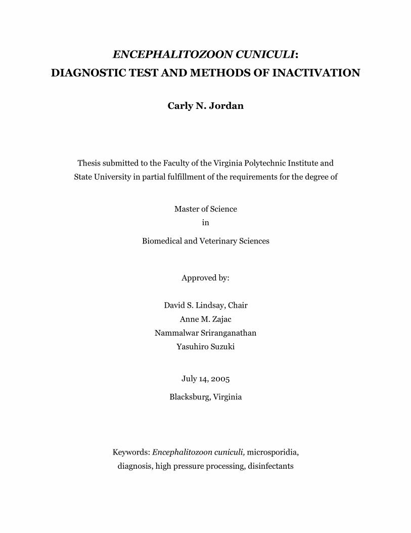
(ABSTRACT)
Encephalitozoon cuniculi is a zoonotic protozoan parasite in the phylum Microspora that has been shown to naturally infect several host species, including humans, rabbits and dogs. Currently, serological diagnosis of infection is made using the immunofluorescense assay (IFA) or enzyme-linked immunosorbent assay (ELISA). Although these methods are sensitive and reliable, there are several drawbacks to both tests. Cross-reactivity between other Encephalitozoon species is common, and specialized equipment is required for IFA and ELISA. Most wildlife species are unable to be tested using these methods, because species-specific antibodies are required. One goal of this work was to develop a new serological test for diagnosing E. cuniculi infection that would be more practical for use in small veterinary and medical clinics. The effectiveness of the agglutination test was examined in CD-1 and C3H/He mice infected with E. cuniculi or one of 2 other Encephalitozoon species. The results indicate that the agglutination test is 86% sensitive and 98% specific for E. cuniculi, with limited cross-reactivity to E. intestinalis. The test is fast and easy to conduct, and requires no specialized equipment or species-specific antibodies. Recent reports of microsporidial DNA in crop irrigation waters suggest that unpasteurized juice products may be contaminated with E. cuniculi. High pressure processing (HPP) is an effective means of eliminating bacteria and extending the shelf life of products while maintaining the sensory features of food and beverages. The effect of HPP on the in vitro infectivity of E. cuniculi spores was examined. Spores were exposed to between 140 and 550 MPa for 1 min, and then spores were loaded onto cell culture flasks or were kept for examination by transmission electron microscopy (TEM). Spores treated with between 200 and 275 MPa showed reduction in infectivity. Following treatment of 345 MPa or more, spores were unable to infect host cells. No morphologic changes were observed in pressure-treated spores using TEM.
The effect of disinfectants on in vitro infectivity of E. cuniculi spores was also examined. Spores of E. cuniculi were exposed to several dilutions of commercial bleach, HiTor and Roccal, and 70% ethanol for 10 minutes and then loaded onto Hs68 cells. The results of this study showed that all concentrations of disinfectants tested were lethal to E. cuniculi spores. Encephalitozoon cuniculi spores are more sensitive to disinfectants than are coccidian oocysts and other parasite cysts.

iii
Dedication
I dedicate this thesis to my father, Monty Wetch, for inspiring me to study disease
and for showing me that miracles happen every day. I love you.

iv
Acknowledgements
First I would like to thank Dr. David Lindsay and my graduate committee
members for their patience and guidance throughout my graduate education. I thank
Laura Douglas, Dan Holliman, Virginia Viers, Kathy Lowe and Terry Lawrence for their
technical assistance and expertise. A special thanks to Kay Carlson for teaching me
most every laboratory technique I used for this work. Thanks also to Nancy Tenpenny,
Heather Norton, Dave Goodwin, Julie Tucker and Jenn DiCristina for serving as
wonderful distractions from the daily grind. I would especially like to thank my fellow
parasitology graduate students, Alexa Rosypal and Sheila Mitchell, for their advice and
friendship and for making everyday so much fun. Thanks Mom and Dad for nurturing
my curiosity and always encouraging me to follow my dreams. Thanks to my siblings,
Ashley and Jeremy, for listening and feigning interest while I rambled on about
parasites. Finally, thanks to my husband, Jason, for supporting me during all these
years of education and for believing that someday I will do something great.
v
TABLE OF CONTENTS
Page
Abstract i
Dedication ii
Acknowledgements iii
List of Tables vi
List of Figures vii
I. INTRODUCTION 1 II. REVIEW OF RELATED LITERATURE 1. Title page 4 2. Introduction 5 3. Pathology of microsporidiosis in humans 5 4. Microsporidia infection in domestic animals 6 5. Zoonotic potential of Microsporidia 10 6. Diagnosis of microsporidia infection 11 7. High pressure processing 15 8. Literature Cited 17 9. Tables and Figures 23 III. DIRECT AGGLUTINATION TEST FOR ENCEPHALITOZOON CUNICULI 1. Title page 29 2. Abstract 30 3. Introduction 31 4. Materials and Methods 33 5. Results 36 6. Discussion 38 7. Acknowledgements 39 8. References 40 9. Tables and Figures 42
vi
IV. EFFECTS OF HIGH PRESSURE PROCESSING ON IN VITRO INFECTIVITY OF ENCEPHALITOZOON CUNICULI SPORES 1. Title page 44 2. Abstract 45 3. Introduction 46 4. Materials and Methods 48 5. Results 50 6. Discussion 51 7. Literature Cited 52 8. Tables and Figures 53 V. ACTIVITY OF DISINFECTANTS AGAINST SPORES OF ENCEPHALITOZOON CUNICULI 1. Title page 55 2. Abstract 56 3. Introduction 57 4. Materials and Methods 58 5. Results and Discussion 60 6. References 61 7. Tables and Figures 62 VI. GENERAL CONCLUSIONS 64
VII. APPENDIX 67 PREVALENCE OF AGGLUTINATING ANTIBODIES TO TOXOPLASMA GONDII AND SARCOCYSTIS NEURONA IN BEAVERS (CASTOR CANADENSIS) FROM MASSACHUSETTS (Formatted for publication in the Journal of Parasitology)
VIII. VITA 76
vii
LIST OF TABLES
Chapter 2 Table 1: Prevalence of E. cuniculi in pet rabbits as determined by serologic surveys 25 Table 2: Prevalence of microsporidia in cats and dogs as determined by serologic surveys 26 Table 3: Prevalence of microsporidia in livestock as determined by identification of parasites in fecal samples or by IFA 27 Table 4: Morphological characteristics of microsporidia 28 Chapter 3 Table 1: Results of the agglutination test for E. cuniculi 42 Chapter 4 Table 1: Infectivity of E. cuniculi spores in cell culture following high pressure processing 53 Chapter 5 Table 1: Concentrations of bleach stock and hypochlorite tested 62 Table 2: Concentrations of active compounds in disinfectants 63
viii
LIST OF FIGURES
Chapter 2 Figure 1: General life cycle of Microsporidia 23 Figure 2: Transmission electron micrographs showing spores of E. cuniculi and E. intestinalis developing within CV-1 cells 24 Chapter 4 Figure 1: Transmission electron micrographs showing pressure-treated
(410 MPa) and control spores of E. cuniculi 54
1
CHAPTER 1
INTRODUCTION
Encephalitozoon cuniculi is an important zoonotic parasite infecting a wide range
of host species. In humans, symptoms are typically observed only in
immunocompromised individuals, such as AIDS patients, organ transplant recipients
and cancer patients undergoing chemotherapy. Encephalitozoon cuniculi primarily
infects the brain, eyes and urinary tract, causing encephalitis, cataracts, kidney failure
and death. Symptomatic human infections with E. cuniculi are rare, but infection with
other microsporidia species is more common. It is estimated that between 8-10% of the
AIDS population in the United States is clinically infected with E. intestinalis or
Enterocytozoon bieneusi (Deplazes et al., 2000). These microsporidia species primarily
infect intestinal enterocytes, causing fatty diarrhea, nausea and weight loss that may last
several months. Drugs are available to treat microsporidia infections in humans, but
these typically only relieve symptoms temporarily, with frequent relapses following
cessation of treatment. One goal of this study was to examine the activity of a newly
approved anti-parasitic drug on E. cuniculi spores in vitro.
Encephalitozoon cuniculi is also an important parasite of domestic animals,
including dogs, cats, rabbits, cows, sheep and horses. This parasite was first identified
in a colony of laboratory rabbits in 1922 as a cause of vestibular disease (Wright and
Craighead, 1922), and it is still a common pathogen of purpose-bred and pet rabbits
(Harcourt-Brown and Holloway, 2003). Rabbits are the only host in which clinical signs
2
are observed in otherwise healthy adult animals. Encephalitozoon cuniculi infects the
eyes, kidneys and brain of rabbits, causing clinical signs such as cataracts, head tilt,
paralysis and death. In dogs, disease is only seen in puppies. Clinical signs of E.
cuniculi infection in pups are almost entirely neurological, and include blindness, ataxia
and seizures. Another goal of this research was to examine several commercially
available disinfectants for their effects on E. cuniculi spores in order to identify useful
methods of sterilizing animal facilities and housing.
Encephalitozoon cuniculi is the only microsporidian parasite that is considered to
be zoonotic, though some evidence suggests that other species may also be zoonotic.
Three strains of E. cuniculi have been characterized: Type 1 from dogs, type 2 from
rodents, type 3 from rabbits (Didier et al., 1995). In areas where cohabitation with dogs
is common, humans are primarily infected with type 1, and the same is true for type 3
from rabbits.
The most common methods of serological diagnosis are the immunofluorescense
assay (IFA) or enzyme-linked immunosorbent assay (ELISA). Although these methods
are sensitive and reliable for detecting antibodies to E. cuniculi, there are several
drawbacks to both tests. Cross-reactivity between other Encephalitozoon species is
common, and specialized equipment is required for IFA and ELISA. Most wildlife
species are unable to be tested using these methods, because species-specific antibodies
are required. One goal of this study was to develop a simple test for detecting antibodies
to E. cuniculi.
Aside from zoonotic transmission, it is likely that humans are ingesting
microsporidia spores from contaminated food and water. A recent report by Thurston-
Enriquez et al. (2002) found DNA from human microsporidia species in crop irrigation
3
waters from throughout North and Central America. Microsporidia spores are
extremely resistant to environmental factors and could remain infective long after crops
were harvested. A final goal of this research was to examine the effects of high pressure
processing on spores of E. cuniculi as a method for sterilizing juice products.
Literature Cited
Deplazes, P., Mathis, A., and Weber, R. 2000. Epidemiology and zoonotic aspects of
microsporidia of mammals and birds. Contributions to Microbiology 6:236-260.
Harcourt-Brown, F. M., Holloway, H. K. R. 2003. Encephalitozoon cuniculi in pet
rabbits. Veterinary Record 152: 427-431.
Thurston-Enriquez, J.A., P. Watt, S.E. Dowd, R. Enriquez, I.L. Pepper, and C.P. Gerba.
2002. Detection of protozoan parasites and microsporidia in irrigation waters used for
crop production. Journal of Food Protection 65: 378-382.
Wright, J.H., and Craighead, E.M. 1922. Infectious motor paralysis in young rabbits.
Journal of Experimental Medicine 36:135-141.
4
CHAPTER 2
REVIEW OF RELATED LITERATURE
Carly N. Jordan, Anne M. Zajac and David S. Lindsay.
Center for Molecular Medicine and Infectious Diseases, Department of Biomedical
Sciences and Pathobiology, Virginia-Maryland Regional College of Veterinary Medicine,
Virginia Tech, 1410 Prices Fork Road, Blacksburg, Virginia 24061-0342.
Email:[email protected]
5
Introduction
Microsporidia are obligate intracellular parasites of the phylum Microspora.
There are over 1200 species of microsporidia, and natural infections have been
documented in arthropods, reptiles, amphibians, birds, mammals, and other protozoa
(Weiss, 2001). The infective spore stage is characterized by the presence of a polar tube
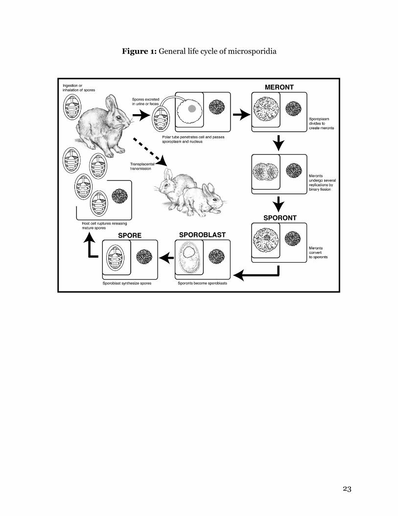
that is used to infect host cells (Weiss, 2001). Hosts are infected by ingestion or
inhalation of spores passed in the urine or feces (Figure 1) (Bigliardi and Luciano,
2001). Transplacental transmission has been shown to occur in dogs, rabbits, horses
and cows, but not in humans (McInnes and Stewart, 1991; Harcourt-Brown, 2003; Van
Rensburg, 1991; Reetz, 1995). Infection with microsporidia is usually asymptomatic,
except in young or immunocompromised hosts (Wasson and Peper, 2000). Human and
animal infections with microsporidia were recognized prior to the AIDS pandemic, but
overall awareness of the Phylum as important parasites of warm-blooded animals came
about only after the advent of a large immunocompromised human population
associated with HIV infection, transplantation or chemotherapy.
Pathology of microsporidiosis in humans
There are four species of microsporidia of interest in human and veterinary
medicine: Encephalitozoon cuniculi, E. intestinalis, E. hellem, and Enterocytozoon
bieneusi. Among human cases of microsporidia infection, E. intestinalis and Ent.
bieneusi are most frequent (Franzen and Muller, 2001). Studies conducted among AIDS
patients across the United States have shown that approximately 10% of patients
sampled were infected with Ent. bieneusi, and about 8% were infected with E.
6
intestinalis (Deplazes et al., 2000). Encephalitozoon hellem is the third most common
human microsporidian, and E. cuniculi has been reported in a small number of human
cases (Franzen and Muller, 2001).
Intestinal pathology is the most common symptom of human microsporidia
infection, and is usually caused by Ent. bieneusi or E. intestinalis (Snowden, 2004).
Intestinal enterocytes are the primary cells infected, and fatty diarrhea due to
malabsorption is the most common symptom. Patients may experience diarrhea and
nausea intermittently for several months, accompanied by progressive weight loss
(Franzen and Muller, 2001).
Encephalitozoon species are more widespread in their pathology, with all three
species able to cause severe disseminated infection. Keratoconjunctivitis is common
among Encephalitozoon species, and is usually caused by E. hellem (Franzen and
Muller, 2001). This condition may be asymptomatic, but conjunctival inflammation and
loss of visual acuity are common, with corneal ulcers developing in a few cases (Franzen
and Muller, 2001). Sinusitis is another common manifestation of E. hellem, and rhinitis
and nasal polyps occur in some cases (Franzen and Muller, 2001)(Wasson and Peper,
2000). Urinary tract infections are rarely seen in patients infected with E. cuniculi or E.
intestinalis. Some urinary tract infections are asymptomatic, but severe disease can
occur. Hematuria and cystitis are common symptoms, and severe infection can lead to
renal failure (Franzen and Muller, 2001; Wasson and Peper, 2000).
Microsporidia infection in domestic animals
Encephalitozoon cuniculi was first identified in 1922 in a colony of research
rabbits, and today it is a common pathogen infecting domestic and purpose-bred rabbits
7
(Snowden, 2004; Wasson and Peper, 2000; Wright and Craighead, 1922). There have
not been any large-scale studies of encephalitozoonosis in pet rabbits in the U.S., but
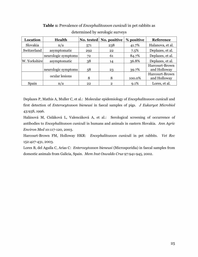
surveys conducted in Europe have shown high rates of infection in asymptomatic pet
rabbits (7-42%) and in those with neurological symptoms (40-85%)(Table 1).
Many rabbits infected with E. cuniculi are asymptomatic, or they demonstrate
only minor clinical signs, such as slower reactions to stimuli (Harcourt-Brown and
Holloway, 2003). Clinical signs of E. cuniculi infection in rabbits are almost entirely
neurological, with some cases developing ocular lesions and cataracts (Wasson and
Peper, 2000; Harcourt-Brown and Holloway, 2003; Snowden and Shadduck, 1999).
Lethargy and head tilt are usually the first signs, and ataxia, paresis and hind limb
paralysis follow (Wasson and Peper, 2000; Harcourt-Brown and Holloway, 2003). This
presentation has often been confused with a diagnosis of vestibular disease caused by
bacterial infection. Full paralysis and death may occur in severe cases. Infection of the
kidneys leads to polydypsia and polyuria, and gross examination reveals a characteristic
pitted appearance of the shrunken fibrotic kidneys (Harcourt-Brown and Holloway,
2003). Although gross lesions are not usually visible, meningoencephalitis can be
observed upon histological examination (Wasson and Peper, 2000; Harcourt-Brown
and Holloway, 2003).
Dogs may be naturally infected by a few species of microsporidia, but infection
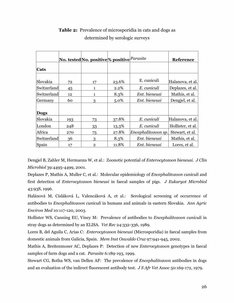
with Encephalitozoon cuniculi is most common. Serologic studies for E. cuniculi have
been conducted worldwide, and the prevalence of dogs with specific antibodies ranges
from 8% to 38% (Table 2).
Encephalitozoon cuniculi is usually asymptomatic in adult dogs but can be fatal
to pups born to infected dams (Szabo and Shadduck, 1987a; McInnes and Stewart, 1991;
8
Stewart et al., 1979). The most common signs of E. cuniculi infection in dogs are
neurological, and they include depression, incoordination, ataxia, blindness and
seizures (McInnes and Stewart, 1991; Stewart et al., 1979; Didier et al., 1998).
Histological examination of tissues typically shows multifocal or disseminated
intracellular parasites in the brain with varying amounts of inflammation and vasculitis.
Nephritis and hepatitis are also common, with parasites primarily localized in renal
cortical tubular epithelium and sometimes in the glomerular epithelium (Szabo and
Shadduck, 1987b; Botha, et al., 1986; Shadduck et al., 1978).
Encephalitozoon intestinalis and Enterocytozoon bieneusi are primarily human
parasites, but infections have been documented to occur naturally in dogs (Bornay-
Llinares et al., 1998; Mathis et al., 1999a; Lores et al., 2002). Limited serologic studies
have been conducted in parts of Europe that show the prevalence of antibodies to Ent.
bieneusi may be as high as 11% (Table 2). No clinical data is available about the
pathology of these infections in dogs.
Encephalitozoon cuniculi and Ent. bieneusi have been shown to infect domestic
cats, though these infections are rare and poorly studied (Mathis et al., 1999a; Pang and
Shadduck, 1985). Limited serologic surveys in Europe have been conducted to detect
antibodies against E. cuniculi and Ent. bieneusi. Up to 8% of cats tested positive for
Ent. bieneusi and up to 23% of cats exhibited antibodies to E. cuniculi (Table 2).
One case of Encephalitozoonosis in a naturally infected Siamese kitten has been
documented (Rensburg and Plessis, 1971). Clinical signs included severe muscle spasms
and depression (Rensburg and Plessis, 1971). Upon histopathological examination of
tissues, meningoencephalitis and interstitial nephritis were evident (Rensburg and
Plessis, 1971). A single attempt at experimental infection has been reported in young
9
cats (Pang and Shadduck, 1985). The animals were generally asymptomatic, although
they were thin and had rough coats (Pang and Shadduck, 1985). Upon examination of
tissues, interstitial nephritis was present in all cats, and a few exhibited
meningoencephalitis (Pang and Shadduck, 1985).
In a limited number of reports, microsporidia have been shown to naturally infect
horses, sheep, goats, pigs and cows, but very little is known about the true prevalence or
the pathogenicity of infection in these hosts. Serological surveys have been conducted
reporting infection of livestock species with E. cuniculi and Ent. bieneusi (Table 3).
Encephalitozoon intestinalis and Ent. bieneusi have been shown to occur
naturally in pigs (Bornay-Llinares et al., 1998; Deplazes et al., 1996a; Dengjel et al.,
2001; Buckholt et al., 2002). A recent survey of pigs at various slaughterhouses
throughout the New England states found that 31% of over 200 pigs were positive for
Ent. bieneusi by PCR (Buckholt et al., 2002). No clinical signs were reported, but
parasites were observed in the liver of one of the pigs (Buckholt et al., 2002).
Natural infections by E. cuniculi, E. intestinalis and Ent. bieneusi have been
documented to occur in cattle (Halanova et al., 1999; Reetz, 1995). Clinical signs of
infected cows have not been described, but abortion has resulted in a few cases.
Examination of aborted fetuses has demonstrated E. cuniculi in the placenta, brain,
liver, kidneys, myocardia, and lungs (Reetz, 1995).
A group of researchers in Israel discovered that horses are frequently infected
with E. cuniculi. The study examined 102 horses from 3 private riding farms across the
country. The horses were examined for clinical manifestations of E. cuniculi infection,
and serum was drawn from each animal. Among the 73 asymptomatic horses, 43 (60%)
were seropositive, and 25 of 30 (80%) symptomatic horses were seropositive by
10
immunofluorescent assay (IFA)(Levkutova, et al., 2004). Symptomatic horses had a
variety of clinical signs, mainly cholic (11) and neurological signs (6)(Levkutova, et al.,
2004). A few animals exhibited cholic, reproductive disturbances, lameness, sinusitis or
pruritis (Levkutova, et al., 2004).
Two cases of abortion resulting from infection of asymptomatic mares with E.
cuniculi have been documented (Van Rensburg et al., 1991; Patterson-Kane et al.,
2003). In one case, E. cuniculi was found only in the placenta, causing necrosis of
chorionic villi (Patterson-Kane et al., 2003). The second case found E. cuniculi in the
kidneys of the aborted fetus (Van Rensburg et al., 1991). Severe lymphoplasmacytic
interstitial nephritis was noted, but no other organs were affected, and no parasites were
seen in the placenta (Van Rensburg et al., 1991).
Zoonotic potential of microsporidia
Microsporidia are widespread throughout the animal kingdom, but only recently
have they begun appearing in humans. Although there is still no direct evidence of
animal-human transmission, it is likely that some microsporidia are zoonotic. Recent
scientific information supports the concept that E. intestinalis is a parasite primarily
found in humans while E. cuniculi and E. hellem are primarily found in animal hosts.
Currently, only E. cuniculi is considered a zoonotic parasite. Three strains of E. cuniculi
have been described (Didier et al., 1995a). Strain I has been isolated from rabbits
(Desplazes et al., 1996b; Mathis et al., 1997), strain II is generally associated with
rodents, and strain III from dogs (Snowden et al., 1999; Didier et al., 1996). Strain I is
found in humans in Europe (Mathis et al., 1997; Rossi et al., 1998), while strain III is the
human isolate found in the Americas (Didier et al., 1995a). This is certainly interesting,
11
considering that rabbits are more commonly kept as pets or for food in Europe, and
dogs are the most popular pet in the Western hemisphere.
Encephalitozoon hellem also has three genotypes, with type I being the most
common strain found in humans (Mathis et al., 1999b). Ribosomal RNA gene
sequencing of human and avian isolates has been conducted to examine its zoonotic
potential. The results showed 100% homology between the avian and human isolates
(Snowden et al., 2000).
A recent report described the first discovery of E. intestinalis in dogs, pigs and
cows using molecular analysis of fecal samples (Bornay-Llinares et al., 1998). Another
study sequenced the SSUrRNA gene of E. intestinalis found in fecal samples obtained
from gorillas and the humans who share their habitat and found them to be identical
(Graczyk et al., 2002).
Diagnosis of microsporidia infection
Staining methods There are several staining techniques available for identification of microsporidia
spores. However, staining methods can, at best, identify microsporidia, but cannot
distinguish between species. Current staining methods are also hindered by the fact
that microsporidia are often mistaken for bacteria or yeasts.
Calcoflour white M2R is a chemofluorescent stain that is commonly used for
identifying microsporidia in fecal smears, urine sediments, and respiratory lavages
(Didier et al., 1995b). This is the most sensitive method for staining microsporidia, and
the procedure can be completed in only 15 minutes (Didier et al., 1995b). However, this
12
stain is not specific for microsporidia, and spores may be confused with yeasts or other
particulate matter in the sample (Garcia, 2002). A compound microscope with a UV
light source is needed for this staining method, so it is used primarily in diagnostic
laboratories rather than in clinical settings.
Chromotrope-based (modified trichrome) staining methods are also used for
identifying microsporidia in feces, urine sediment or respiratory secretion samples.
Chromotrope stains give spores a pinkish red color, and quick-hot Gram-chromotrope
stains color spores a dark violet (del Aguila et al., 1998). Characteristic morphological
features of microsporidia that are visible using these stains are a belt-like stripe, a
vacuole and Gram-positive granules. Again, spores may be confused for bacteria or
yeasts.
Giemsa and Gram stains are not recommended for fecal smears because they do
not differentiate between microsporidia and bacteria or yeast that may be present in the
sample. However, Giemsa stains may be useful in urine or tissue samples, and spores
stain light blue (Garcia, 2002).
Molecular methods Polymerase chain reaction (PCR) is a much more reliable alternative to staining
for diagnosing microsporidia infection. PCR can be used to positively identify species of
microsporidia from feces, urine sediment or tissue samples. Specific primers and assays
are available for E. cuniculi, E. intestinalis and E. hellem on an experimental basis or
through medical diagnostic laboratories on a limited basis (del Aguila et al., 1998).
Based on expense of equipment and availability of primers, this is not a practical
solution for identifying microsporidia infection in medical or veterinary laboratories.
13
Morphological methods
Transmission electron microscopy is another way to identify and differentiate
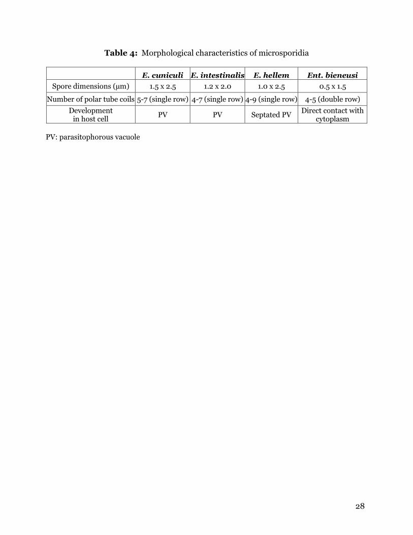
between species of microsporidia. Enterocytozoon bieneusi is slightly smaller than
Encephalitozoon species; Ent. bieneusi is 0.5 x 1.5 µm in size, while the Encephalitozoon
species range from 1.0-1.5 x 2.0-2.5 µm (Weiss, 2001). The number of polar tube coils is
useful in differentiating between species of microsporidia (Table 4). Encephalitozoon
intestinalis can be distinguished from all other Encephalitozoon species by examining
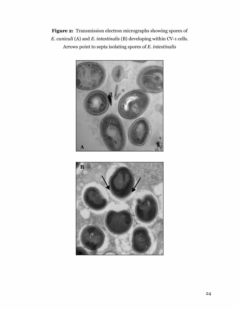
the parasitophorous vacuole (PV) within an infected host cell (Figure 2). This is the only
species of microsporidia known to exist in a septated PV that isolates each developing
spore (Chu and West, 1996). Enterocytozoon bieneusi does not create a PV; instead it
develops in direct contact with the host cell cytoplasm (Wasson and Peper, 2000).
Although TEM is a reliable way of diagnosis, it is obviously the least practical and most
expensive method for use in medical and veterinary practices.
Serological methods
Monoclonal and polyclonal antibodies have been developed for E. cuniculi, E.
intestinalis, E. hellem and Ent. bieneusi for use in IFA tests on an experimental basis
and in some diagnostic laboratories (Alfa Cisse et al., 2002; Aldras et al., 1994; Enriquez
et al., 1997). IFA is the gold standard for serological diagnosis of microsporidia
infection. IFA titers greater than 1:20 are considered positive (Didier et al., 1998).
Western blotting is another technique that can identify E. cuniculi antibodies in
serum. Recently, a group of researchers used Western blotting to examine proteins of
the polar tube and exospore of E. cuniculi (van Gool et al., 2004). Serum from an HIV-
negative laboratory worker who accidentally infected himself with E. cuniculi was
14
collected at 1 and 20 months post-infection and tested against spores of E. cuniculi and
E. intestinalis. When testing the serum samples against E. cuniculi spores, they found
that at 1 month (PI) serum exhibited only one strong band at 28 kDa (van Gool et al.,
2004). By 20 months PI, strong bands were formed at 17, 20, 28, 30, 32, 34-38 and 47
kDa (van Gool et al., 2004). Weak bands were present at 27, 44, 55, 70 and 150 kDa
(van Gool et al., 2004). When the serum was tested against E. intestinalis spores, only
one band was present at 26 kDa (van Gool et al., 2004).
Another group experimentally infected rabbits with a human isolate of E. cuniculi
for testing by Western blot. When sera from infected rabbits was tested against E.
cuniculi spores, seven bands were observed. Five bands were found between 18 and 45
kDa, with approximate weights of 19, 23, 28, 35 and 36 kDa (Croppo et al., 1997). Dark
bands were seen at 90 and 190 kDa, and a dark doublet was observed immediately
below the 45 kDa marker (Croppo et al., 1997).
The enzyme-linked immunosorbent assay (ELISA) is another method for
diagnosing microsporidia infection serologically. Two groups of researchers have
evaluated the ELISA for diagnosis of E. cuniculi by comparing the results with IFA.
Boot et al. (2000) examined 135 purpose-bred rabbits diagnosed with infection by
histology and 75 rabbits without a history of illness. A few samples were positive by only
one method, but no statistically significant differences were calculated between the
sensitivities and specificities of the two assays (Boot et al., 2000).
Another study examined sera from rabbits, dogs, mice and squirrel monkeys by
the dot-ELISA test and compared the results to the IFA. A few samples were positive by
ELISA and negative by IFA, but none were positive by IFA and not ELISA (Beckwith et
al., 1988). The agreement between the two tests was calculated using a Kappa value.
15
The Kappa values were 1.0 for dogs, 0.77 for rabbits and monkeys and 0.74 for mice
(Beckwith et al., 1988). The researchers found the dot-ELISA to be more sensitive than
IFA to E. cuniculi antibodies (Beckwith et al., 1988).
Although IFAs and ELISAs are sensitive methods for diagnosing infection with E.
cuniculi, neither is practical for all laboratories. IFA tests require a compound
microscope with a fluorescent light source, and ELISA tests require a plate reader to
interpret results. Also, serological screening of many wildlife species is not yet possible
due to the requirement for species-specific antibodies. One main goal of this research is
to develop a method of serological diagnosis that will address each of these problems.
High pressure processing
High pressure processing (HPP) is used commercially as a non-thermal means to
extend the shelf life of foods and beverages by eliminating pathogens and denaturing
destructive enzymes (Tewari et al., 1999). HPP acts primarily by disrupting hydrogen
bonds and three-dimensional configuration of protein molecules. It has a number of
advantages over traditional thermal processing, including shorter processing time,
minimal heat damage problems, no adverse changes due to ice-phase forms during
pressure-shift freezing, minimal modifications to functionality, and preservation of
flavor, texture, color and vitamin C (Tewari et al., 1999).
Recent reports have proposed the possibility that microsporidia may be present
in juice products (Slifko et al., 2000a). A recent study examined water sources in North
and Central America for the presence of protozoan parasites (Thurston-Enriquez et al.,
2002). The water sources sampled were used for irrigation of various fruit and
vegetable crops. Thurston-Enriquez et al. (2002) discovered that DNA from human
16
pathogenic microsporidia was present in all water sources examined. Microsporidia in
irrigation water could adhere to the surface of fruits and vegetables. Since spores are
extremely resistant to damage, they may remain viable on the produce for weeks or
months. These facts also suggest that microsporidia may be present in juice products.
Studies have shown that HPP is useful in eliminating bacteria and protozoa from
juices without altering taste or appearance. Slifko et al. (2000b) examined the effects of
HPP on Cryptosporidium parvum oocysts in apple and orange juice. It was determined
that 550 MPa for 60 sec decreases the in vitro infectivity of C. parvum oocysts. Lindsay
et al. (in press) show that exposure of Toxoplasma gondii oocysts to 340 MPa for 60 sec
reduces viability of oocysts in vivo. A major goal of this research is to determine
whether HPP will be useful for inactivating spores of E. cuniculi.
17
Literature Cited
Alfa Cisse O, Ouattara A, Thellier M, et al.: Evaluation of an immunofluorescent-antibody
test using monoclonal antibodies directed against Enterocytozoon bieneusi and
Encephalitozoon intestinalis for diagnosis of intestinal microsporidiosis in Bamako
(Mali). J Clin Microbiol 40:1715-1718, 2002.
Aldras AM, Orenstein JM, Kotler DP, et al.: Detection of microsporidia by indirect
immunofluorescence antibody test using polyclonal and monoclonal antibodies. J Clin
Microbiol 32:608-612, 1994.
Beckwith C, Peterson N, Liu JJ, et al.: Dot enzyme-linked immunosorbent assay (dot
ELISA) for antibodies to Encephalitozoon cuniculi. Lab Anim Sci 38:573-576, 1988.
Bigliardi E, Luciano S: Cell biology and invasion of the microsporidia. Microbe Infect
3:373-379, 2001.
Boot R, Hansen AK, Hansen CK, et al.: Comparison of assays for antibodies to
Encephalitozoon cuniculi in rabbits. Lab Anim 34:281-289, 2000.
Bornay-Llinares FJ, da Silva AJ, Moura H, et al.: Immunologic, microscopic, and
molecular evidence of Encephalitozoon intestinalis (Septata intestinalis) infection in
mammals other than humans. J Infect Dis 178:820-826, 1998.
Botha WS, Stewart CG, Van Dellen AF: Observations of the pathology of experimental
encephalitozoonosis in dogs. J S Afr Vet Med Assoc 57:17-24, 1986.
Buckholt MA, Lee JH, Tzipori S: Prevalence of Enterocytozoon bieneusi in swine: an 18-
month survey at a slaughterhouse in Massachusetts. Appl Environ Microbiol 68:2595-9,
2002.

18
Chu P, West AB: Encephalitozoon (Septata) intestinalis: Cytologic, histologic, and
electron microscopic features of a systemic intestinal pathogen. Am J Clin Pathol
106:606-614, 1996.
Croppo GP, Visvesvara GS, Leitch GJ, et al.: Western blot and immunofluorescence
analysis of a human isolate of Encephalitozoon cuniculi established in culture from the
urine of a patient with AIDS. J Parasitol 83:66-69, 1997.
del Aguila C, Croppo GP, Moura H, et al.: Ultrastructure, immunofluorescence, Western
blot, and PCR analysis of eight isolates of Encephalitozoon (Septata) intestinalis
established in culture from sputum and urine samples and duodenal aspirates of five
patients with AIDS. J Clin Microbiol 36:1201-1208, 1998.
Dengjel B, Zahler M, Hermanns W, et al.: Zoonotic potential of Enterocytozoon bieneusi.
J Clin Microbiol 39:4495-4499, 2001.
Deplazes P, Mathis A, Muller C, et al.: Molecular epidemiology of Encephalitozoon
cuniculi and first detection of Enterocytozoon bieneusi in faecal samples of pigs. J
Eukaryot Microbiol 43:93S, 1996a.
Deplazes P, Mathis A, Baumgartner R, et al.: Immunologic and molecular characteristics
of Encephalitozoon-like microsporidia isolated from humans and rabbits indicate that
Encephalitozoon cuniculi is a zoonotic parasite. Clin Infect Dis 22:557-9, 1996b.
Deplazes P, Mathis A, Weber R: Epidemiology and zoonotic aspects of microsporidia of
mammals and birds. Contrib Microbiol 6:236-260, 2000.
Didier ES, Vossbrinck CR, Baker MD, et al.: Identification and characterization of three
Encephalitozoon cuniculi strains. Parasitology 111:411-421, 1995a.
Didier ES, Orenstein JM, Aldras A, et al.: Comparison of three staining methods for
detecting microsporidia in fluids. J Clin Microbiol 33:3138-3145, 1995b.
19
Didier ES, Visvesvara GS, Baker MD, et al.: A microsporidian isolated from an AIDS
patient corresponds to Encephalitozoon cuniculi III, originally isolated from domestic
dogs. J Clin Microbiol 34:2835-2837, 1996.
Didier PJ, Didier ES, Snowden KS, et al.: Encephalitozoonosis. Infectious Diseases of the
Dog and Cat 465-470, 1998.
Enriquez FJ, Ditrich O, Palting JD, et al.: Simple diagnosis of Encephalitozoon species
microsporidial infections by using a panspecific antiexospore monoclonal antibody. J Clin
Microbiol 35:724-729, 1997.
Franzen C, Muller A: Microsporidiosis: human diseases and diagnosis. Microbes Infect
3:389-400, 2001.
Garcia LS: Laboratory identification of the microsporidia. J Clin Microbiol 40:1892-
1902, 2002.
Graczyk TK, Bosco-Nizeyi J, da Silva AJ, et al.: A single genotype of Encephalitozoon
intestinalis infects free-ranging gorillas and people sharing their habitats in Uganda.
Parasitol Res 88:926-931, 2002.
Halánová M, Letková V, Macák V, et al.: The first finding of antibodies to
Encephalitozoon cuniculi in cows in Slovakia. Vet Parasitol 82:167-171, 1999.
Harcourt-Brown FM, Holloway HKR: Encephalitozoon cuniculi in pet rabbits. Vet Rec
152:427-431, 2003.
Lindsay, D.S., M.V. Collins, C.N. Jordan, G.J. Flick, and J.P. Dubey. Effects of high
pressure processing on infectivity of Toxoplasma gondii oocysts for mice. Journal of
Parasitology. In press.
20
Lores B, del Aguila C, Arias C: Enterocytozoon bieneusi (Microsporidia) in faecal samples
from domestic animals from Galicia, Spain. Mem Inst Oswaldo Cruz 97:941-945, 2002.
Mathis A, Michel M, Kuster H, et al.: Two Encephalitozoon cuniculi strains of human
origin are infectious to rabbits. Parasitology 114:29-35, 1997.
Mathis A, Breitenmoser AC, Deplazes P: Detection of new Enterocytozoon genotypes in
faecal samples of farm dogs and a cat. Parasite 6:189-193, 1999a.
Mathis A, Tanner I, Weber R, et al.: Genetic and phenotypic intraspecific variation in the
microsporidian Encephalitozoon hellem. Int J Parasit 29:767-770, 1999b.
McInnes EF, Stewart CG: The pathology of subclinical infection of Encephalitozoon
cuniculi in canine dams producing pups with overt encephalitozoonosis. J S Afr Vet Med
Assoc 62:51-54, 1991.
Pang VF, Shadduck JA: Susceptibility of cats, sheep, and swine to a rabbit isolate of
Encephalitozoon cuniculi. Am J Vet Res 46:1071-1077, 1985.
Patterson-Kane JC, Caplazi P, Rurangirwa F, et al.: Encephalitozoon cuniculi placentitis
and abortion in a Quarterhorse mare. J Vet Diagn Invest 15:57-59, 2003.
Reetz J: Microsporidien als ursache von aborten beim rind. Tierärztl Umschau 50:550-
554, 1995.
Rensburg IBJ, Plessis JL: Nosematosis in a cat: a case report. J S Afr Vet Med Assoc
42:327-331, 1971.
Rossi P, La Rosa G, Ludovisi A, et al.: Identification of a human isolate of
Encephalitozoon cuniculi type I from Italy. Int J Parasit 28:1361-1366, 1998.
21
Shadduck JA, Bendele R, Robinson GT: Isolation of the causative organism of canine
encephalitozoonosis. Vet Pathol 15:449-460, 1978.
Slifko, T.R., H.V. Smith, and J.B. Rose: Emerging parasite zoonoses associated with water
and food. Int J Parasit 30: 1379-1393, 2000a.
Slifko, T.R., E. Raghubeer, and J.B. Rose: Effect of high hydrostatic pressure on
Cryptosporidium parvum infectivity. J Food Prot 63: 1262-1267, 2000b.
Snowden K, Logan K, Didier ES: Encephalitozoon cuniculi strain III is a cause of
encephalitozoonosis in both humans and dogs. J Infect Dis 180:2086-2088, 1999.
Snowden KS, Shadduck, JA: Microsporidia in higher vertebrates. The Microsporidia and
Microsporidiosis 393-417, 1999.
Snowden KF, Logan K, Phalen DN: Isolation and characterization of an avian isolate of
Encephalitozoon hellem. Parasitology 121:9-14, 2000.
Snowden KF: Zoonotic microsporidia from animals and arthropods with a discussion of
human infections. World Class Parasites 9:123-134, 2004.
Stewart CG, Van Dellen AF, Botha WS: Canine encephalitozoonosis in kennels and the
isolation of Encephalitozoon in tissue culture. J S Afr Vet Med Assoc 50:165-168, 1979.
Szabo JR, Shadduck JA: Immunologic and clinicopathologic evaluation of adult dogs
inoculated with Encephalitozoon cuniculi. J Clin Microbiol 26:557-563. 1987a.
Szabo JR, Shadduck JA: Experimental encephalitozoonosis in neonatal dogs. Vet Pathol
24:99-109, 1987b.
Tewari, G., D.S. Jayas, and R.A. Holley: High pressure processing of foods: An overview.
Science Des Aliments 19: 619-661, 1999.
22
Thurston-Enriquez, J.A., P. Watt, S.E. Dowd, R. Enriquez, I.L. Pepper, and C.P. Gerba:
Detection of protozoan parasites and microsporidia in irrigation waters used for crop
production. J Food Prot 65: 378-382, 2002.
van Gool T, Biderre C, Delbac F, et al.: Serodiagnostic studies in an immunocompetent
individual infected with Encephalitozoon cuniculi. J Infect Dis 189:2243-2249, 2004.
van Rensburg IBJ, Volmann DH, Soley JT, et al.: Encephalitozoon infection in a still-born
foal. J S Afr Vet Assoc 62:130-132, 1991.
Wasson K, Peper RL: Mammalian microsporidiosis. Vet Pathol 37:113-128, 2000.
Weiss LM: Microsporidia: emerging pathogenic protists. Acta Tropica 78:89-102, 2001.
Wright JH, Craighead EM: Infectious motor paralysis in young rabbits. J Exp Med
36:135-141, 1922.
23
Figure 1: General life cycle of microsporidia
24
Figure 2: Transmission electron micrographs showing spores of
E. cuniculi (A) and E. intestinalis (B) developing within CV-1 cells.
Arrows point to septa isolating spores of E. intestinalis
A
B
25
Table 1: Prevalence of Encephalitozoon cuniculi in pet rabbits as
determined by serologic surveys
Location Health No. tested No. positive % positive Reference
Slovakia n/a 571 238 41.7% Halanova, et al.
Switzerland asymptomatic 292 22 7.5% Deplazes, et al.
neurologic symptoms 72 61 84.7% Deplazes, et al.
W. Yorkshire asymptomatic 38 14 36.8% Deplazes, et al.
neurologic symptoms 58 23 39.7% Harcourt-Brown
and Holloway
ocular lesions
8 8 100.0% Harcourt-Brown
and Holloway
Spain n/a 22 2 9.1% Lores, et al.
Deplazes P, Mathis A, Muller C, et al.: Molecular epidemiology of Encephalitozoon cuniculi and
first detection of Enterocytozoon bieneusi in faecal samples of pigs. J Eukaryot Microbiol
43:93S, 1996.
Halánová M, Cisláková L, Valencáková A, et al.: Serological screening of occurrence of
antibodies to Encephalitozoon cuniculi in humans and animals in eastern Slovakia. Ann Agric
Environ Med 10:117-120, 2003.
Harcourt-Brown FM, Holloway HKR: Encephalitozoon cuniculi in pet rabbits. Vet Rec
152:427-431, 2003.
Lores B, del Aguila C, Arias C: Enterocytozoon bieneusi (Microsporidia) in faecal samples from
domestic animals from Galicia, Spain. Mem Inst Oswaldo Cruz 97:941-945, 2002.
26
Table 2: Prevalence of microsporidia in cats and dogs as
determined by serologic surveys
No. tested No. positive% positive Parasite Reference
Cats
Slovakia 72 17 23.6% E. cuniculi Halanova, et al.
Switzerland 45 1 2.2% E. cuniculi Deplazes, et al.
Switzerland 12 1 8.3% Ent. bieneusi Mathis, et al.
Germany 60 3 5.0% Ent. bieneusi Dengjel, et al.
Dogs
Slovakia 193 73 37.8% E. cuniculi Halanova, et al.
London 248 33 13.3% E. cuniculi Hollister, et al.
Africa 270 75 27.8% Encephalitozoon sp. Stewart, et al.
Switzerland 36 3 8.3% Ent. bieneusi Mathis, et al.
Spain 17 2 11.8% Ent. bieneusi Lores, et al.
Dengjel B, Zahler M, Hermanns W, et al.: Zoonotic potential of Enterocytozoon bieneusi. J Clin
Microbiol 39:4495-4499, 2001.
Deplazes P, Mathis A, Muller C, et al.: Molecular epidemiology of Encephalitozoon cuniculi and
first detection of Enterocytozoon bieneusi in faecal samples of pigs. J Eukaryot Microbiol
43:93S, 1996.
Halánová M, Cisláková L, Valencáková A, et al.: Serological screening of occurrence of
antibodies to Encephalitozoon cuniculi in humans and animals in eastern Slovakia. Ann Agric
Environ Med 10:117-120, 2003.
Hollister WS, Canning EU, Viney M: Prevalence of antibodies to Encephalitozoon cuniculi in
stray dogs as determined by an ELISA. Vet Rec 24:332-336, 1989.
Lores B, del Aguila C, Arias C: Enterocytozoon bieneusi (Microsporidia) in faecal samples from
domestic animals from Galicia, Spain. Mem Inst Oswaldo Cruz 97:941-945, 2002.
Mathis A, Breitenmoser AC, Deplazes P: Detection of new Enterocytozoon genotypes in faecal
samples of farm dogs and a cat. Parasite 6:189-193, 1999.
Stewart CG, Botha WS, van Dellen AF: The prevalence of Encephalitozoon antibodies in dogs
and an evaluation of the indirect fluorescent antibody test. J S Afr Vet Assoc 50:169-172, 1979.
27
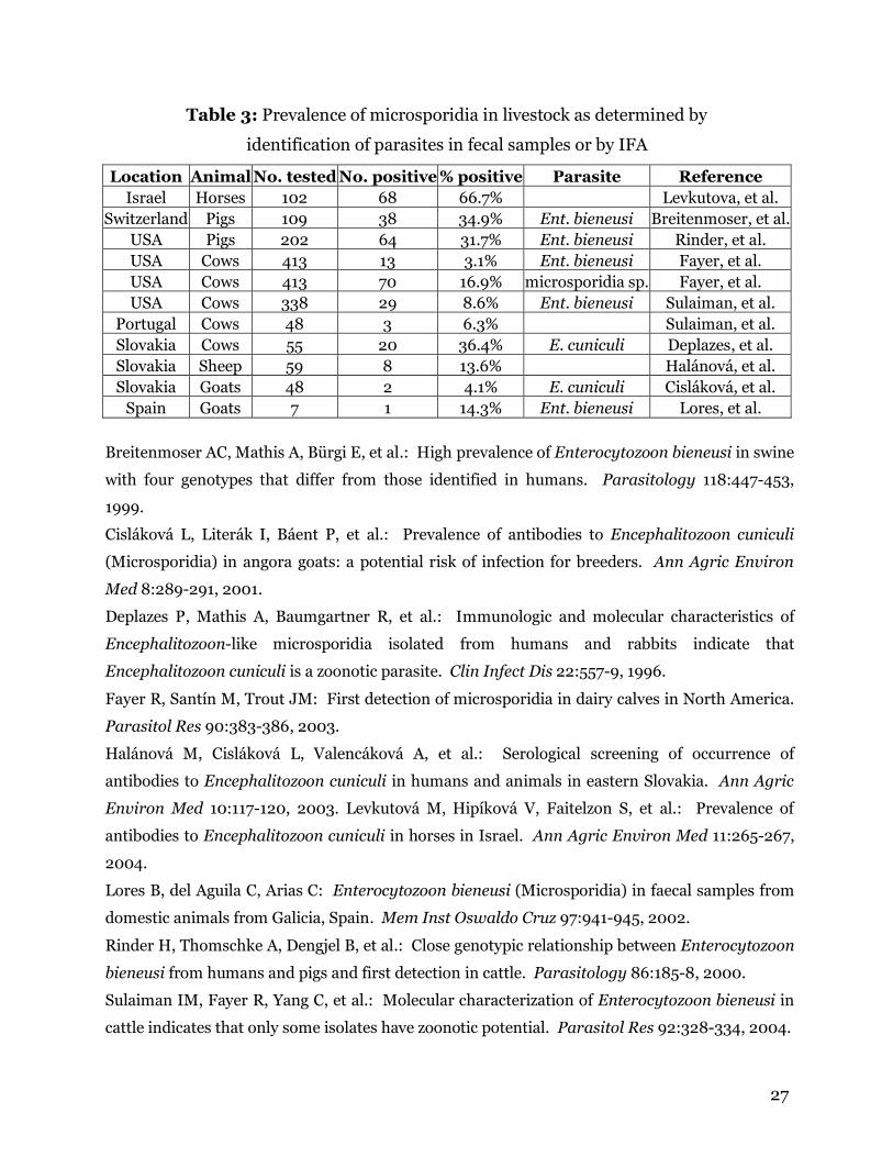
Table 3: Prevalence of microsporidia in livestock as determined by
identification of parasites in fecal samples or by IFA
Location Animal No. tested No. positive % positive Parasite Reference Israel Horses 102 68 66.7% Levkutova, et al.
Switzerland Pigs 109 38 34.9% Ent. bieneusi Breitenmoser, et al.USA Pigs 202 64 31.7% Ent. bieneusi Rinder, et al. USA Cows 413 13 3.1% Ent. bieneusi Fayer, et al. USA Cows 413 70 16.9% microsporidia sp. Fayer, et al. USA Cows 338 29 8.6% Ent. bieneusi Sulaiman, et al.
Portugal Cows 48 3 6.3% Sulaiman, et al. Slovakia Cows 55 20 36.4% E. cuniculi Deplazes, et al. Slovakia Sheep 59 8 13.6% Halánová, et al. Slovakia Goats 48 2 4.1% E. cuniculi Cisláková, et al.
Spain Goats 7 1 14.3% Ent. bieneusi Lores, et al.
Breitenmoser AC, Mathis A, Bürgi E, et al.: High prevalence of Enterocytozoon bieneusi in swine
with four genotypes that differ from those identified in humans. Parasitology 118:447-453,
1999.
Cisláková L, Literák I, Báent P, et al.: Prevalence of antibodies to Encephalitozoon cuniculi
(Microsporidia) in angora goats: a potential risk of infection for breeders. Ann Agric Environ
Med 8:289-291, 2001.
Deplazes P, Mathis A, Baumgartner R, et al.: Immunologic and molecular characteristics of
Encephalitozoon-like microsporidia isolated from humans and rabbits indicate that
Encephalitozoon cuniculi is a zoonotic parasite. Clin Infect Dis 22:557-9, 1996.
Fayer R, Santín M, Trout JM: First detection of microsporidia in dairy calves in North America.
Parasitol Res 90:383-386, 2003.
Halánová M, Cisláková L, Valencáková A, et al.: Serological screening of occurrence of
antibodies to Encephalitozoon cuniculi in humans and animals in eastern Slovakia. Ann Agric
Environ Med 10:117-120, 2003. Levkutová M, Hipíková V, Faitelzon S, et al.: Prevalence of
antibodies to Encephalitozoon cuniculi in horses in Israel. Ann Agric Environ Med 11:265-267,
2004.
Lores B, del Aguila C, Arias C: Enterocytozoon bieneusi (Microsporidia) in faecal samples from
domestic animals from Galicia, Spain. Mem Inst Oswaldo Cruz 97:941-945, 2002.
Rinder H, Thomschke A, Dengjel B, et al.: Close genotypic relationship between Enterocytozoon
bieneusi from humans and pigs and first detection in cattle. Parasitology 86:185-8, 2000.
Sulaiman IM, Fayer R, Yang C, et al.: Molecular characterization of Enterocytozoon bieneusi in
cattle indicates that only some isolates have zoonotic potential. Parasitol Res 92:328-334, 2004.
28
Table 4: Morphological characteristics of microsporidia
E. cuniculi E. intestinalis E. hellem Ent. bieneusi
Spore dimensions (µm) 1.5 x 2.5 1.2 x 2.0 1.0 x 2.5 0.5 x 1.5
Number of polar tube coils 5-7 (single row) 4-7 (single row) 4-9 (single row) 4-5 (double row)
Development in host cell
PV PV Septated PV Direct contact with
cytoplasm
PV: parasitophorous vacuole
29
CHAPTER 3
DIRECT AGGLUTINATION TEST FOR
ENCEPHALITOZOON CUNICULI
Carly N. Jordan1,2, Anne M. Zajac1, Karen S. Snowden3 and David S. Lindsay1.
1 Center for Molecular Medicine and Infectious Diseases, Department of Biomedical
Sciences and Pathobiology, Virginia-Maryland Regional College of Veterinary
Medicine, Virginia Tech, 1410 Prices Fork Road, Blacksburg, Virginia 24061-0342.
2 Present address: Department of Cellular Biology, University of Georgia, 724
Biological Sciences Building, Athens, Georgia, 30602-2607.
3 Department of Pathobiology, College of Veterinary Medicine, Texas A&M University,
College Station, Texas.
* Corresponding author. Tel: +1 540 231 6302; fax: +1 540 231 3426
Email address: [email protected] (D.S. Lindsay)
Formatted for publication in Veterinary Parasitology (in review)
30
ABSTRACT
Encephalitozoon cuniculi is a small protozoan parasite in the phylum
Microspora. It has been shown to naturally infect several host species, including
humans. Infection with microsporidia is usually asymptomatic, except in young or
immunocompromised hosts. Currently, serological diagnosis of infection is made using
the indirect immuno fluorescent antibody assay (IFA) or enzyme-linked immunosorbent
assay (ELISA). Although these methods are sensitive and reliable, there are several
drawbacks to the IFA and ELISA tests. Cross-reactivity between other Encephalitozoon
species is common, and specialized equipment is required to conduct these tests. This
paper reports the development of a direct agglutination test for detecting IgG antibodies
to E. cuniculi. The utility of the agglutination test was examined in CD-1 and C3H/He
mice infected with E. cuniculi or one of 2 other Encephalitozoon species. The results
indicate that the agglutination test is 86% sensitive and 98% specific for E. cuniculi,
with limited cross-reactivity to E. intestinalis. No cross reactivity to E. hellem was
observed. The test is fast and easy to conduct, and species-specific antibodies are not
required.
Key words: Encephalitozoon cuniculi, Encephalitozoon intestinalis, Encephalitozoon
hellem, agglutination test, immunofluorescent antibody assay, western blot
31
1. INTRODUCTION
Microsporidia are small protozoan parasites characterized by the use of a polar
tube for host cell invasion (Weiss, 2001). There are several species of microsporidia that
infect humans and domestic animals. Encephalitozoon cuniculi is a common
microsporidian that has an extremely diverse host range. It was first identified as a
cause of vestibular disease in laboratory rabbits, and remains a common pathogen of
purpose-bred and pet rabbits today (Snowden, 2004; Wasson and Peper, 2000; Wright
and Craighead, 1922). Rabbits are the only host species in which clinical signs are
commonly observed in immunocompetent adult animals (Snowden, 2004). Head tilt is
usually observed first, and disease may progress to full or partial paralysis and death
(Harcourt-Brown and Holloway, 2003; Wasson and Peper, 2000). In other host
species, such as dogs, clinical signs are limited to young or immunocompromised
animals, and include depression, blindness and seizures (Didier et al., 1998; McInnes
and Stewart 1991; Pang and Shadduck, 1985; Rensburg and Plessis, 1971).
In humans, symptoms are observed primarily in immunocompromised
populations, such as AIDS patients and individuals receiving immunosuppresent drugs.
Although this disease is rare in humans, it is often fatal. Encephalitozoon cuniculi can
cause severe disseminated infection in humans, but it primarily infects the eyes, sinuses,
urinary tract and brain (Franzen and Muller, 2001). Infection of the brain and
meninges, leading to edema, encephalitis and lesions in the brain, is usually fatal
(Franzen and Muller, 2001).
Several species of microsporidia are suspected zoonoses, but E. cuniculi is the
only species that is currently widely accepted to be a zoonotic parasite (Didier et al.,
1995). For this reason, it is important that veterinarians and human physicians have
32
access to reliable methods for detecting antibodies to microsporidia infection.
Currently, infections are most often identified by enzyme-linked immunosorbent assays
(ELISA) or indirect immunofluorescent antibody assay (IFA), the gold standard for
serological diagnosis of microsporidia infection (Enriquez et al., 1997; Boot et al., 2000;
Beckwith et al., 1988). IFA titers greater than 1:20 and ELISA titers greater than 1:800
are considered to be indicative of exposure (Didier et al., 1998). The purpose of the
present study was to develop a direct agglutination test that is based on agglutination of
E. cuniculi spores by antibodies present in serum.
33
2. MATERIALS AND METHODS
Spores of E. cuniculi and E. intestinalis were obtained from the American Type
Culture Collection, Manassas, VA [ATCC# 50502 (E. cuniculi) and 50506 (E.
intestinalis)]. Spores of E. hellem were obtained from Dr. Ron Fayer, USDA/ARS,
Environmental Microbial Safety Laboratory, Beltsville, MD. Human foreskin fibroblasts
(Hs68) (ATCC #CRL-1635) were cultured in 75 cm2 cell culture flasks in a CO2 incubator
at 37ºC in RPMI 1640 media containing 10% fetal bovine serum and antibiotics.
Microsporidia spores were inoculated onto confluent Hs68 cells and allowed to grow for
several weeks. Encephalitozoon cuniculi spores were harvested from Hs68 cell cultures
by removing the supernatant from infected flasks and passing it through a 3.0 µm filter.
CD-1 or C3H/He mice were inoculated either orally or subcutaneously with 10 x 106
spores of E. cuniculi, E. intestinalis or E. hellem. Serum was collected weekly from at
least 2 mice per group via retro-orbital sinus. Two interferon-γ (IFN-γ) gene knock-out
BALB/c mice were also infected with E. cuniculi, and serum was collected at 4 weeks
post-infection (PI).
Spores of E. cuniculi were collected from infected cell cultures and fixed in 2 ml
of 37% formaldehyde solution for 10 to 15 sec in a 15 ml conical centrifuge tube and then
diluted with phosphate buffered saline (PBS, pH 7.4) up to 15 ml and stored at 4°C.
Prior to use in the agglutination test, the spores were washed twice in PBS and
resuspended in alkaline buffer-eosin solution (7.02 g NaCl; 3.09 g H3BO3; 24 ml 1N
NaOH; 4 g horse serum albumin factor V; 50 mg eosin Y; 1.0 g sodium aside; distilled
H2O to make 1 L; pH 8.7). The eosin aids in the visualization of the agglutination
reaction. Next, 0.5 ml of 0.2 M 2-mercaptoethanol was added to each 1 ml of the spore
34
buffer solution to destroy IgM antibodies that may be present in the test serum, and to
prevent non-specific agglutination caused by IgM molecules.
The direct agglutination test was conducted in 96 well round bottom plates. Test
sera were diluted with PBS and 25 µl of serial test dilutions were combined with 75 µl of
antigen solution and mixed thoroughly by pipetting up and down several times. The
plates were covered with parafilm and incubated overnight at 37 °C in a CO2 incubator.
Positive and negative control mouse sera were separately examined on each plate. The
agglutination reactions were read the next morning. Diffuse opacity across the entire
diameter of the well was considered a positive agglutination reaction. A central discrete
opaque dot or button was considered a negative reaction. Several concentrations of
spores (13, 67, 100, 133 and 200 x 106 spores/ml) were tested to determine which gave
the most definitive results. Serum harvested from E. cuniculi, E. hellem and E.
intestinalis-infected mice was tested using this method, along with samples from mice
infected with Toxoplasma gondii or Sarcocystis neurona. Serum from 2 dogs naturally
infected with E. cuniculi was also examined (NEED REF from Dr. Snowden). Serum
samples from beavers and raccoons were also screened using the agglutination test.
The sensitivity and specificity of the antibody agglutination test was calculated
using standard formulas. Any samples that were falsely positive or negative by the
agglutination test were compared to normal samples using Western blot to identify any
differences in serum proteins that could cause false results. IFA testing was also
performed on 6 positive samples from 7 weeks PI to confirm the results of the
agglutination test.
35
Cross-reactivity between E. cuniculi and E. intestinalis was examined using
Western blotting and IFA. For Western blotting, serum from E. cuniculi-infected CD-1
mice was tested against both E. cuniculi and E. intestinalis spores. 5 x 10⁶ spores were
loaded per well of a 10% polyacrylamide gel, and the serum was diluted to 1:100 in TBS.
Peroxidase-labeled goat anti-mouse antibodies diluted to 1:500 in tris-buffered saline
(TBS)(pH 7.4) served as secondary antibodies. The reaction was visualized by exposing
peroxidase-labeled nitrocellulose strips to the developing solution (60 mg 4-chloro-1-
naphthol, 10 ml cold methanol, 100 ml TBS, 0.6 ml H2O2) until bands appeared. IFA
testing was conducted using serum from E. cuniculi-infected mice and spores of E.
cuniculi and E. intestinalis. Serum dilutions of 1:20 and 1:40 were tested using
flourescein-labeled goat anti-mouse antibodies diluted to 1:50 in phosphate-buffered
saline (PBS) (pH 8.4)
36
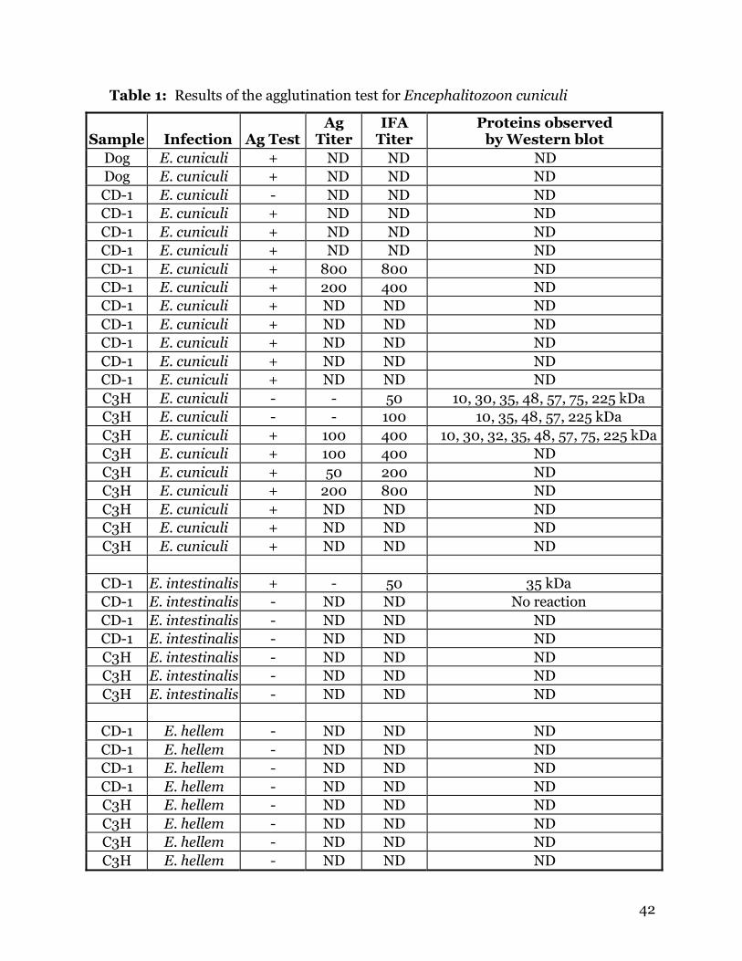
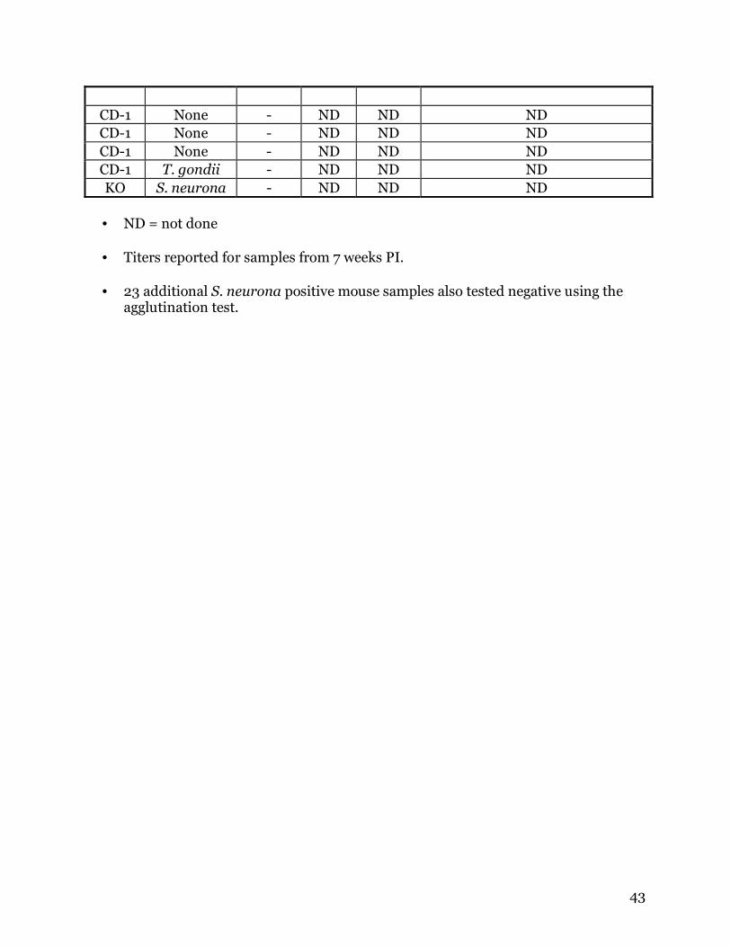
3. RESULTS
The results of this study indicate that the agglutination test is effective for
detecting antibodies of Encephalitozoon cuniculi in mice and dogs (Table 1). No cross
reactivity occurred to E. hellem and only minor cross reactivity to E. intestinalis was
present (Table 1). Of 20 samples from E. cuniculi-infected mice, only 3 were falsely
negative. However, 1 of the 3 mice was only sampled up to 4 weeks PI, and 5 weeks was
required for some mice to seroconvert in the agglutination test. Both samples from
naturally infected dogs were positive by the agglutination test. Of 44 samples from mice
not infected with E. cuniculi, only 1 was positive by the agglutination test, an E.
intestinalis-infected mouse. The sensitivity of the agglutination test was 86% and the
specificity to be 98%. The positive predictive value for this test is 95%.
Of the three falsely negative samples, 2 came from C3H/He mice, and one came
from a BALB/c INF-g knock-out mouse. There was not sufficient serum remaining for
Western blot analysis of the BALB/c sample, but the test was conducted for the C3H/He
mice. The samples from these 2 mice were compared against another mouse from the
same experimental group. The results of Western blotting showed that both falsely
negative samples were lacking a 32 kDa protein that was present in the truly positive
sample. One falsely positive sample was also missing proteins of 30 and 75 kDa. All 3
samples were examined using IFA, and all samples were positive, although the falsely
negative samples had lower titers.
The falsely positive sample was from a mouse infected with E. intestinalis,
indicating that there is still a possibility of cross-reactivity with other Encephalitozoon
species using this test. Western blot analysis was performed on the falsely positive
sample and another sample from the same group of E. intestinalis-infected mice. The
37
results showed the presence of a single band approximately 35 kDa in size in the falsely
positive sample. No bands were observed in the truly negative sample.
Western blot analysis was performed using both E. cuniculi and E. intestinalis
spores against serum from E. cuniculi-infected mice. Spores of E. cuniculi reacted with
proteins at 120, 75, 42, 32, 30, 19 and 15 kDa. A large band was observed from 45 to 58
kDa. Western blot analysis of spores from E. intestinalis against serum from E.
cuniculi-infected mice revealed several cross-reactive bands. Thin bands were observed
at 120, 75, 58, 40, 38 and 30 kDa; heavy bands were present at 35 and 50 kDa.
Cross-reactivity with the IFA test was observed when serum from E. cuniculi-
infected mice was tested against E. intestinalis spores. A much weaker reaction was
observed against E. intestinalis than E. cuniculi spores.
Serum from 42 raccoons (Hancock et al., 2005) and 62 beavers (Jordan et al.,
2005) were also examined using the agglutination test. Antibodies to E. cuniculi were
present in 1 of 42 raccoons and 0 of 62 beavers using the agglutination test.

38
4. DISCUSSION
Encephalitozoon cuniculi is an important parasite in human and veterinary
medicine. Rabbits and dogs are the most commonly infected domestic animals, and
research suggests that the parasite is transmitted from these animals to their human
owners. Currently, ELISA and IFA testing are the 2 serologic tests commonly used in
research and hospital laboratories. While these methods are reliable for diagnosing
infection, they have distinct disadvantages being that each method requires the use of
specialized equipment. ELISA requires a special plate reader, and IFA requires a
compound microscope with a fluorescent light source. Also, much cross-reactivity
occurs between Encephalitozoon species using IFA, and genetic analysis is often
required to confirm the infective species.
One advantage of the direct agglutination test is that no species-specific reagents
are needed. This is helpful in examining exotic and wildlife species for serological
evidence of exposure. For example, we examined sera from raccoons and beavers using
our agglutination test. Commercial anti-sera is available only for raccoon, therefore
without our agglutination test we would not have been able to test the beavers.
Our study describes the development of a new serologic test based on
agglutination of E. cuniculi spores with serum antibodies. This test is highly sensitive
and specific for antibodies to E. cuniculi, and cross-reactivity is reduced as compared to
IFA. No specialized equipment is required in order to conduct this test, and results are
available within 24 hours. Since species-specific antibodies are not required, this assay
can be used to screen many animal species.
39
ACKNOWLEDGMENTS
This study was supported by funds from a Clinical Research grant from the
College of Veterinary Medicine, Virginia-Maryland Regional College of Veterinary
Medicine to DSL.
40
REFERENCES
Boot, R., Hansen, A.K., Hansen, C.K., Nozari, N., Thuis, H. C., 2000. Comparison of
assays for antibodies to Encephalitozoon cuniculi in rabbits. Lab. Anim. 34, 281-289.
Beckwith, C., Peterson, N., Liu, J.J., Shadduck, J. A., 1988. Dot enzyme-linked
immunosorbent assay (dot ELISA) for antibodies to Encephalitozoon cuniculi. Lab. Anim.
Sci. 38, 573-576.
Didier, E.S., Vossbrinck, C.R., Baker, M.D., Rogers, L.B., Bertucci, D.C., Shadduck, J. A.,
1995. Identification and characterization of three Encephalitozoon cuniculi strains.
Parasitology 111, 411-421.
Didier, P. J., Didier, E. S., Snowden, K. S., Shadduck, J. A., 1998. Encephalitozoonosis, In.
Infectious Diseases of the Dog and Cat, Greene, C. E. (ed.), Saunders, Philadelphia pp.
465-470.
Enriquez, F. J., Ditrich, O., Palting, J. D., Smith, K., 1997. Simple diagnosis of
Encephalitozoon species microsporidial infections by using a panspecific antiexospore
monoclonal antibody. J. Clin. Microbiol. 35, 724-729.
Franzen, C., Muller, A., 2001. Microsporidiosis: human diseases and diagnosis. Microb.
Infect. 3, 389-400.
Hancock, K., Zajac, A. M., Pung, O. J., Elvinger, F., Rosypal A. C., Lindsay, D. S. 2005.
Prevalence of antibodies to Trypanosoma cruzi in raccoons (Procyon lotor) from an
urban area of northern Virginia. J Parasitol. 91, 470-472.
Harcourt-Brown, F. M., Holloway, H. K. R., 2003. Encephalitozoon cuniculi in pet
rabbits. Vet. Rec. 152, 427-431.
41
Jordan, C. N., Kaur, T., Koenen, K., Destefano, S., Zajac, A. M., Lindsay, D. S. 2005.
Prevalence of agglutinating antibodies to Toxoplasma gondii and Sarcocystis neurona
in beavers (Castos canadensis) from Massachusetts. J. Parasitol. 91, in press.
McInnes, E. F., Stewart, C. G., 1991. The pathology of subclinical infection of
Encephalitozoon cuniculi in canine dams producing pups with overt encephalitozoonosis.
J. S. Afric. Vet. Med. Assoc. 62, 51-54.
Pang V. F., Shadduck, J. A., 1985. Susceptibility of cats, sheep, and swine to a rabbit
isolate of Encephalitozoon cuniculi. Am. J. Vet. Res. 46, 1071-1077.
Rensburg, I. B. J., Plessis, J. L. 1971. Nosematosis in a cat: a case report. J. S. Afric. Vet.
Med. Assoc. 42, 327-331.
Snowden, K. F. 2004. Zoonotic microsporidia from animals and arthropods with a
discussion of human infections. In World Class Parasites. Volume 9. "Opportunistic
Infections: Toxoplasma, Sarcocystis, and Microsporidia". Lindsay, D.S., Weiss, L. M.
(eds.) Kluwer Academic Press Boston, MA. pp.123-134.
Wasson, K., Peper, P. L., 2000. Mammalian microsporidiosis. Vet. Pathol. 37, 113-128.
Weiss, L. M. 2001. Microsporidia: emerging pathogenic protists. Acta Trop. 78, 89-102.
Wright, J. H., Craighead, E. M., 1922. Infectious motor paralysis in young rabbits. J.
Exp. Med. 36, 135-141.
42
Table 1: Results of the agglutination test for Encephalitozoon cuniculi
Sample Infection Ag TestAg
TiterIFA
Titer Proteins observed
by Western blot Dog E. cuniculi + ND ND ND . Dog E. cuniculi + ND ND ND CD-1 E. cuniculi - ND ND ND CD-1 E. cuniculi + ND ND ND CD-1 E. cuniculi + ND ND ND CD-1 E. cuniculi + ND ND ND CD-1 E. cuniculi + 800 800 ND CD-1 E. cuniculi + 200 400 ND CD-1 E. cuniculi + ND ND ND CD-1 E. cuniculi + ND ND ND CD-1 E. cuniculi + ND ND ND CD-1 E. cuniculi + ND ND ND CD-1 E. cuniculi + ND ND ND C3H E. cuniculi - - 50 10, 30, 35, 48, 57, 75, 225 kDa C3H E. cuniculi - - 100 10, 35, 48, 57, 225 kDa C3H E. cuniculi + 100 400 10, 30, 32, 35, 48, 57, 75, 225 kDaC3H E. cuniculi + 100 400 ND C3H E. cuniculi + 50 200 ND C3H E. cuniculi + 200 800 ND C3H E. cuniculi + ND ND ND C3H E. cuniculi + ND ND ND C3H E. cuniculi + ND ND ND
CD-1 E. intestinalis + - 50 35 kDa CD-1 E. intestinalis - ND ND No reaction CD-1 E. intestinalis - ND ND ND CD-1 E. intestinalis - ND ND ND C3H E. intestinalis - ND ND ND C3H E. intestinalis - ND ND ND C3H E. intestinalis - ND ND ND
CD-1 E. hellem - ND ND ND CD-1 E. hellem - ND ND ND CD-1 E. hellem - ND ND ND CD-1 E. hellem - ND ND ND C3H E. hellem - ND ND ND C3H E. hellem - ND ND ND C3H E. hellem - ND ND ND C3H E. hellem - ND ND ND
43
CD-1 None - ND ND ND CD-1 None - ND ND ND CD-1 None - ND ND ND CD-1 T. gondii - ND ND ND KO S. neurona - ND ND ND • ND = not done
• Titers reported for samples from 7 weeks PI.
• 23 additional S. neurona positive mouse samples also tested negative using the
agglutination test.
44
CHAPTER 4
EFFECTS OF HIGH PRESSURE PROCESSING
ON IN VITRO INFECTIVITY OF
ENCEPHALITOZOON CUNICULI
Carly N. Jordan, Anne M. Zajac, Dan R. Holliman*, George J. Flick*
and David S. Lindsay.
Center for Molecular Medicine and Infectious Diseases, Department of Biomedical
Sciences and Pathobiology, Virginia-Maryland Regional College of Veterinary Medicine,
Virginia Tech, 1410 Prices Fork Road, Blacksburg, Virginia 24061-0342.
*Department of Food Science and Technology, Virginia Tech, Duck Pond Drive,
Blacksburg, Virginia 24061 Email: [email protected]
Formatted for publication in the Journal of Parasitology (in press)
45
ABSTRACT
High pressure processing (HPP) has been shown to be an effective means of
eliminating bacteria and destructive enzymes from a variety of food products. HPP
extends the shelf life of products while maintaining the sensory features of food and
beverages. The present study examined the effects of HPP on the infectivity of
Encephalitozoon cuniculi spores in vitro. Spores were exposed to between 140 and 550
MPa for 1 min in a commercial HPP unit. Following treatment the spores were loaded
onto cell culture flasks or were kept for examination by transmission electron
microscopy. No effect was observed on the infectivity of spores treated with 140 MPa.
Spores treated with between 200 and 275 MPa showed reduction in infectivity.
Following treatment of 345 MPa or more, spores were unable to infect host cells. No
morphologic changes were observed in pressure-treated spores using transmission
electron microscopy.
46
INTRODUCTION
Encephalitozoon cuniculi is a small protist parasite in the phylum Microspora.
There are over 1200 species of microsporidia, and the phylum is characterized by a
unique organelle used to infect host cells, the polar tube. Hosts are infected by ingestion
or inhalation of spores passed in the urine or feces, or by transplacental transmission in
some animals. Infection with E. cuniculi is usually asymptomatic, except in young or
immunocompromised hosts. Diarrhea is the most common symptom of infection in
immunocompromised individuals, but disseminated infections can occur, causing
conjunctivitis, sinusitis, nephritis and encephalitis (Franzen and Muller, 2001).
Microsporidia infection can be fatal in immunocompromised patients. Human and
animal infections with E. cuniculi were recognized prior to the AIDS pandemic, but
overall awareness of the Phylum as important parasites of warm-blooded animals came
about only after the advent of a large immunocompromised population.
High pressure processing (HPP) is used commercially as a non-thermal means to
extend the shelf life of foods and beverages by eliminating pathogens and denaturing
destructive enzymes (Tewari et al., 1999). It has a number of advantages over
traditional thermal processing, including shorter processing time, minimal heat damage
problems, no adverse changes due to ice-phase forms during pressure-shift freezing,
minimal modifications to functionality, and preservation of flavor, texture, color and
vitamin C (Tewari et al., 1999).
The present study was conducted in response to recent reports discussing the
possibility that microsporidia may be present in juice products (Slifko et al., 2000a). A
recent study examined water sources in North and Central America for the presence of
protozoan parasites (Thurston-Enriquez et al., 2002). The water sources sampled were
47
used for irrigation of various fruit and vegetable crops. Thurston-Enriquez et al. (2002)
discovered that DNA from human pathogenic microsporidia was present in all water
sources examined. Microsporidia in irrigation water could adhere to the surface of
fruits and vegetables. Since spores are extremely resistant to damage, they may remain
viable on the produce for weeks or months. These facts also suggest that microsporidia
may be present in juice products.
Studies have shown that HPP is useful in eliminating bacteria and protozoa from
juices without altering taste or appearance. Slifko et al. (2000b) examined the effects of
HPP on Cryptosporidium parvum oocysts in apple and orange juice. It was determined
that 550 MPa for 60 sec decreases the in vitro infectivity of C. parvum oocysts. Lindsay
et al. (2004) show that exposure of Toxoplasma gondii oocysts to 340 MPa for 60 sec
reduces viability of oocysts in vivo. Another study found that subjecting orange juice to
pressures of 400 MPa for 60 sec significantly reduced the viability of E. coli O157:H7,
but some organisms were able to survive the treatment (Linton et al., 1999). The
present study was conducted to determine the effect of HPP on the infectivity of E.
cuniculi spores in cell culture.
48
MATERIALS AND METHODS
Encephalitozoon cuniculi (American Type Culture Collection, #50502) spores
were harvested from HS68 cell cultures (see below) by removing the supernatant from
infected flasks and passing it through a 2 µm filter. In trials 1 and 2, spores were
suspended in Hank’s balanced salt solution (HBSS). In trial 3, spores were suspended
in pasteurized apple cider. Spores were sealed in leak proof bags at a concentration of
20 x 106 spores/ml. These bags were placed in additional bags containing a 10% bleach
solution to protect against contamination of the processor. The spores were exposed to
pressures ranging from 140 to 550 MPa and held at the desired pressure for one minute.
Three trials were conducted for in vitro infectivity studies, and each measurement was
made in triplicate.
Human foreskin fibroblast cells (American Type Culture Collection, Hs68, no.
CRL-1635) were grown to confluence in 25 cm2 cell culture flasks. They were grown in
RPMI 1640 media with 10% fetal bovine serum, 1% penicillin/streptomycin and 1%
sodium pyruvate added. One milliliter of the spore suspension was removed from each
bag and loaded into individual flasks of Hs68 cell cultures. The cultures were examined
at 30 days, and each flask was evaluated for percent of host cells infected.
One additional study was conducted for examination by transmission electron
microscopy (TEM). Spores were treated with 410 MPa or 275 MPa and compared to
untreated control spores for changes in morphology. After pressure treatment, 1 ml
from each bag was removed and centrifuged to pellet the spores. The spores were fixed
in 3% (v/v) glutaraldehyde in PBS (pH 7.4). Spore pellets were post-fixed in 1% (w/v)
osmium tetroxide in 0.1 M phosphate buffer, dehydrated in a series of ethanols, passed
through 2 changes of propylene oxide, and embedded in Poly/Bed 812 resin
49
(Polysciences Inc., Warrington, Pennsylvania). Spores were examined with a Zeiss 10CA
transmission electron microscope operating at 60 kV, and digital photos were taken
using an ATM camera system (Advanced Microscopy Techniques Corp., Danvers,
Massachusetts). Eleven TEM pictures were taken of spores treated with 410 MPa, and 3
were taken of spores treated with 275 MPa. These photos were compared to 13 TEM
photos of untreated E. cuniculi spores.
50
RESULTS
No effect was observed on the infectivity of spores treated with 140 MPa. Spores
treated with pressures between 275 and 310 MPa showed reduction in infectivity, but
this result was variable. Following treatment of 345 MPa or more, spores were unable to
infect host cells (Table 1). Examination by TEM revealed no morphologic changes in the
pressure treated spores as compared to controls (Figure 1). This is not unexpected,
since HPP acts primarily by disrupting hydrogen bonds and three-dimensional
configuration of protein molecules.
51
DISCUSSION
This study was conducted to examine the possibility of using high pressure
processing to sterilize juice products that may contain microsporidia. There is as least
one report that DNA from human pathogenic microsporidia has been found in water
sources used for irrigation of produce crops (Thurston-Enriquez et al., 2002). Since
microsporidia are resistant to environmental conditions, spores that adhere to produce
may still be viable when crops are processed for juice. Some companies are hesitant to
pasteurize juice products for fear of weakening the flavor. It is unknown whether
pasteurization kills microsporidia, but non-pasteurized products could certainly contain
viable spores. High pressure processing is a viable alternative for producers who do not
wish to pasteurize juices. Pathogenic bacteria and protozoa can be eliminated, and the
processing time required for HPP is shorter than for pasteurization.
52
LITERATURE CITED
Franzen, C., and A. Muller. 2001. Microsporidiosis: human diseases and diagnosis.
Microbes and Infection 3: 389-400.
Lindsay, D.S., M.V. Collins, C.N. Jordan, G.J. Flick, and J.P. Dubey. Effects of high
pressure processing on infectivity of Toxoplasma gondii oocysts for mice. Journal of
Parasitology. In press.
Linton, M., J.M. McClements, and M.F. Patterson. Inactivation of Escherichia coli
O157:H7 in orange juice using a combination of high pressure and mild heat. Journal of
Food Protection 62: 277-279.
Slifko, T.R., H.V. Smith, and J.B. Rose. 2000a. Emerging parasite zoonoses associated
with water and food. International Journal for Parasitology 30: 1379-1393.
Slifko, T.R., E. Raghubeer, and J.B. Rose. 2000b. Effect of high hydrostatic pressure on
Cryptosporidium parvum infectivity. Journal of Food Protection 63: 1262-1267.
Soave, R. 2001. Prophylaxis strategies for solid-organ transplantation. Clinical
Infectious Diseases 33: S26-S31.
Tewari, G., D.S. Jayas, and R.A. Holley. 1999. High pressure processing of foods: An
overview. Science Des Aliments 19: 619-661.
Thurston-Enriquez, J.A., P. Watt, S.E. Dowd, R. Enriquez, I.L. Pepper, and C.P. Gerba.
2002. Detection of protozoan parasites and microsporidia in irrigation waters used for
crop production. Journal of Food Protection 65: 378-382.
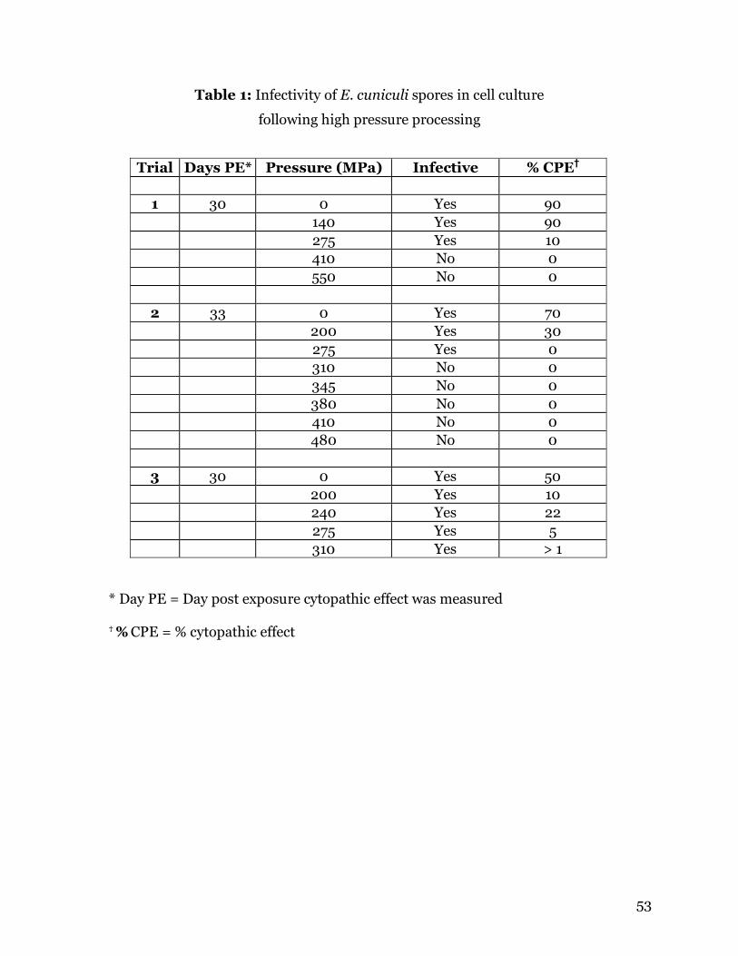
53
Table 1: Infectivity of E. cuniculi spores in cell culture
following high pressure processing
Trial Days PE* Pressure (MPa) Infective % CPE† 1 30 0 Yes 90 140 Yes 90 275 Yes 10 410 No 0 550 No 0
2 33 0 Yes 70 200 Yes 30 275 Yes 0 310 No 0 345 No 0 380 No 0 410 No 0 480 No 0
3 30 0 Yes 50 200 Yes 10 240 Yes 22 275 Yes 5 310 Yes > 1
* Day PE = Day post exposure cytopathic effect was measured
† % CPE = % cytopathic effect
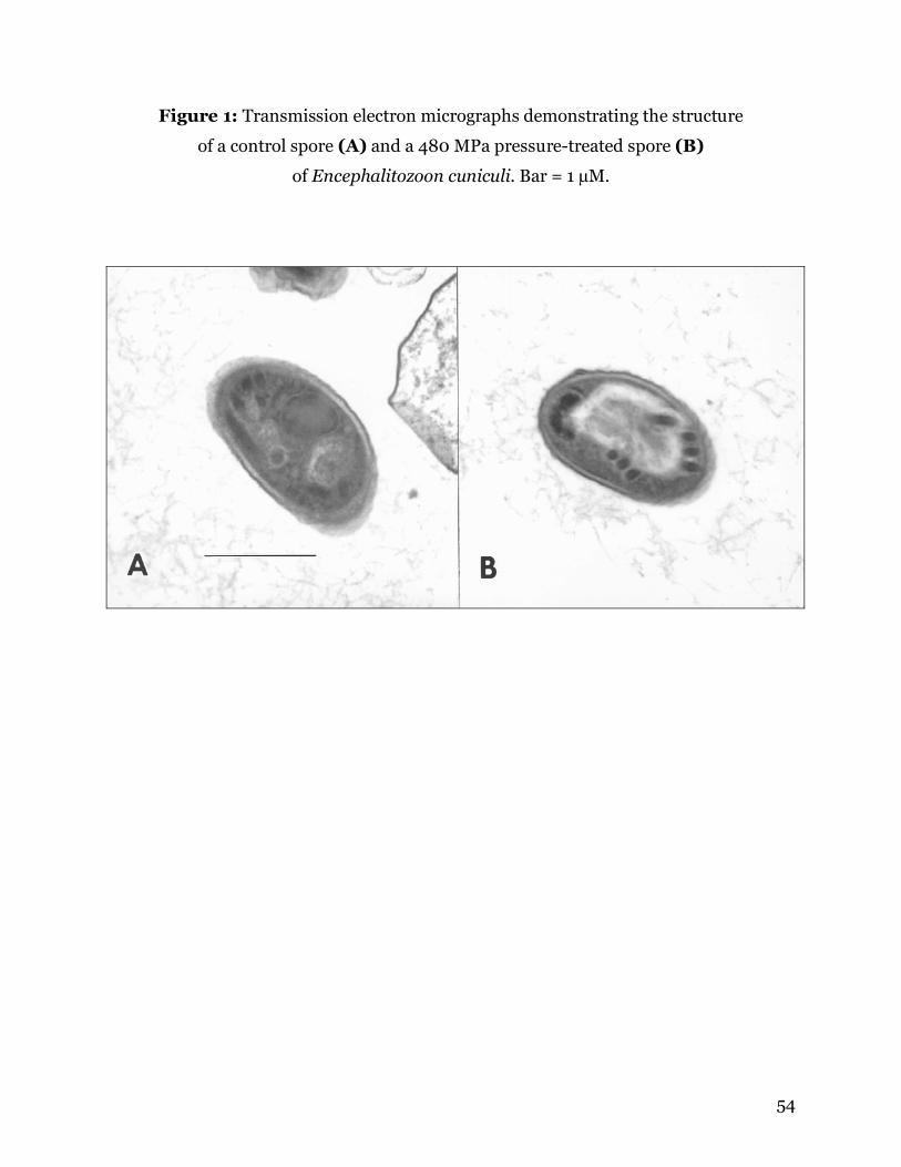
54
Figure 1: Transmission electron micrographs demonstrating the structure
of a control spore (A) and a 480 MPa pressure-treated spore (B)
of Encephalitozoon cuniculi. Bar = 1 µM.
55
CHAPTER 5
ACTIVITY OF DISINFECTANTS AGAINST SPORES OF
ENCEPHALITOZOON CUNICULI
Carly N. Jordana,b and David S. Lindsaya*.
a Center for Molecular Medicine and Infectious Diseases, Department of Biomedical
Sciences and Pathobiology, Virginia-Maryland Regional College of Veterinary
Medicine, Virginia Tech, 1410 Prices Fork Road, Blacksburg, Virginia 24061-0342.
b Present address: Department of Cellular Biology, University of Georgia, 724
Biological Sciences Building, Athens, Georgia, 30602-2607.
* Corresponding author: Tel: +1 540 231 6302; fax: +1 540 231 3426
Email address: [email protected] (D.S. Lindsay)
Formatted for publication in Veterinary Parasitology (in review)
56
ABSTRACT
Encephalitozoon cuniculi is a small protist parasite in the phylum Microspora.
Hosts are infected by ingestion or inhalation of spores passed in the urine or feces.
Infection with E. cuniculi is usually asymptomatic, except in young or
immunocompromised hosts. This study examined the effects of various disinfectants on
in vitro infectivity of E. cuniculi spores. Fifty million spores of E. cuniculi were exposed
to several dilutions of commercial bleach, HiTor and Roccal, and 70% ethanol for 10
minutes and then loaded onto Hs68 cells. The results of this study found that all
concentrations of disinfectants tested were lethal to E. cuniculi spores. Spores of E.
cuniculi are more sensitive to disinfectants than are coccidial oocysts and other parasite
cysts.
Keywords: Encephalitozoon cuniculi, microsporidia, disinfectants, sterilization
57
1. INTRODUCTION
Microsporidia are important parasites of humans and a wide variety of animal
species. Encephalitozoon cuniculi is a common pathogen of rabbits and other domestic
animals, and occasionally causes disease in immunocompromised humans. Serologic
surveys conducted throughout Europe report rates of infection as high as 42% in
asymptomatic rabbits (Halanova et al., 2003) and 85% in rabbits exhibiting
neurological symptoms (Deplazes et al., 1996). Clinical signs of infection in rabbits are
almost entirely neurological, with some cases developing ocular lesions and cataracts.
Lethargy and head tilt are usually the first signs, and ataxia, paresis and hind limb
paralysis follow (Harcourt-Brown and Holloway, 2003). In humans, E. cuniculi
primarily infects the eyes, sinuses, urinary tract and brain, but severe disseminated
infections have been observed in severely immunosuppressed individuals (Franzen and
Muller, 2001).
The present study examined the effectiveness of various disinfectants for
inactivating spores of E. cuniculi. Few reports are available on the effects of chemical
disinfectants on the viability of microsporidia. Waller (1979) found that 30 minutes of
exposure to 70% ethanol kills E. cuniculi spores. A more recent study examined several
chemicals for their effect on E. intestinalis spores. It was reported that exposure to 70%
ethanol for 30 seconds resulted in 100% reduction in infectivity as compared to controls
(Santillana-Hayat et al., 2002). Exposure to bleach (37.8 g/L NaOCl) for 5 minutes
resulted in complete destruction of the spores (Santillana-Hayat et al., 2002). The
present study examines the effect of ethanol, bleach, and 2 commercial products on the
in vitro infectivity of spores of E. cuniculi in order to identify products that may be
useful for sterilizing rabbit cages.
58
2. MATERIALS AND METHODS
2.1. Encephalitozoon cuniculi and cell culture
Encephalitozoon cuniculi spores (American Type Culture Collection, Manassas,
Virginia, ATCC # 50502) were grown and harvested from human fibroblast (Hs68,
American Type Culture Collection, ATCC CRL-1635) cells as previously described
(Jordan et al., 2005). The Hs68 cells were grown in RPMI 1640 medium containing
10% fetal bovine serum and antibiotics as previously described and grown to confluence
in 75 cm2 flasks. Spores were collected from the supernatant, filtered through a 3 µM
filter to remove host cell debris, and counted in a hemocytometer prior to use in the
present study. Monolayers of HS68 cell grown in 25 cm2 flasks were used to determine
the efficacy of disinfection treatments on E. cuniculi spores.
2.2. Efficacy of disinfectants
Several dilutions of commercial bleach (The Clorox Company, Oakland,
California) (Table 1) and 70% ethanol were prepared in distilled water. Commercially-
available disinfectants Roccal (Pharmacia & Upjohn Company, Kalamazoo, Michigan)
and HiTor (Huntington Professional Products, St. Paul, Minnesota) were diluted
according to manufacturer’s directions (0.39%) and at a concentration of 0.1% in
distilled water. Active components of all disinfectants are listed in Table 2. All test
solutions were chilled at 4ºC to prevent any temperature increase that could result due
to exothermic reactions. Tubes containing 50 x 106 spores of E. cuniculi were washed in
Hank’s Balanced Salt Solution (HBSS) and centrifuged to pellet spores. Spores were
resuspended in 10 ml of the desired solution, with one tube suspended in HBSS to serve
59
as a no treatment control. Tubes were held at 4ºC for 10 minutes, and then centrifuged
at 1500 rpm for 10 minutes to pellet spores. Disinfectant solutions were discarded, and
spores were washed twice in HBSS by centrifugation. Spores were resuspended in 10%
cell culture growth media and added to Hs68 cell cultures. A small amount of each
suspension was examined using a compound microscope. Cell cultures were examined
daily for evidence of E. cuniculi infection.
60
3. RESULTS & DISCUSSION
All dilutions of bleach, HiTor and Roccal, and 70% ethanol were lethal to
spores of E. cuniculi after 10 minutes exposure at 4ºC. The HBSS treated control spores
were infectious for Hs68 cells. Examination of the spore suspensions found that E.
cuniculi spores were completely destroyed by all concentrations of bleach. No spores
were visible under the microscope; only cellular debris was observed. HiTor is sold as a
germicidal detergent designed to disinfect, clean and deodorize in one step. Roccal is
labeled as a veterinary and animal care disinfectant that is safe for use in animal
breeding and housing facilities. Both dilutions of HiTor and Roccal rendered E.
cuniculi non-infective for cell culture, but spores appeared normal when viewed using
light microscopy. Seventy percent ethanol was also lethal to spores, rendering them
non-infective. Spores treated with ethanol were normal when examined using light
microscopy. All disinfectants examined should be effective for the killing of E. cuniculi
spores in rabbit cages and in other areas of facilities that house rabbits.
61
REFERENCES
Deplazes, P., Mathis, A., Muller, C., Weber, R., 1996. Molecular epidemiology of
Encephalitozoon cuniculi and first detection of Enterocytozoon bieneusi in faecal
samples of pigs. J. Eukaryot. Microbiol. 43, 93S.
Franzen, C., Muller, A., 2001. Microsporidiosis: human diseases and diagnosis.
Microbes Infect. 3, 389-400.
Halanova, M., Cislakova, L., Valencakova, A., Balent, P., Adam, J., Travnicek, M., 2003.
Serological screening of occurrence of antibodies to Encephalitozoon cuniculi in
humans and animals in Eastern Slovakia. Ann. Agric. Environ. Med. 10, 117-120.
Harcourt-Brown, F.M., Holloway, H.K.R., 2003. Encephalitozoon cuniculi in pet rabbits.
Vet. Rec. 152,427-431.
Jordan, C.N., Zajac, A.M., Holliman, D., Flick, G.J., Lindsay, D.S., 2005. Effects of high
pressure processing on in vitro infectivity of Encephalitozoon cuniculi. J. Parasitol. In
press.
Santillana-Hayat, M., Sarfati, C., Fournier, S., Chau, F., Porcher, R., Molina, J., Derouin,
F., 2002. Effects of chemical and physical agents on viability and infectivity of
Encephalitozoon intestinalis determined by cell culture and flow cytometry.
Antimicrobial Agents Chemother. 46, 2049-2051.
Waller, T., 1979. Sensitivity of Encephalitozoon cuniculi to various temperatures,
disinfectants and drugs. Lab. Anim. 13, 227-230.
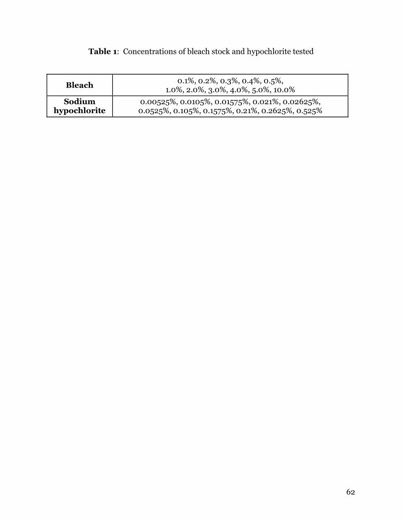
62
Table 1: Concentrations of bleach stock and hypochlorite tested
Bleach 0.1%, 0.2%, 0.3%, 0.4%, 0.5%,
1.0%, 2.0%, 3.0%, 4.0%, 5.0%, 10.0%
Sodium hypochlorite
0.00525%, 0.0105%, 0.01575%, 0.021%, 0.02625%, 0.0525%, 0.105%, 0.1575%, 0.21%, 0.2625%, 0.525%
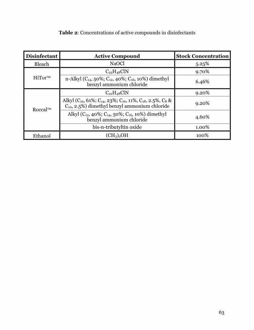
63
Table 2: Concentrations of active compounds in disinfectants
Disinfectant Active Compound Stock Concentration
Bleach NaOCl 5.25%
C22H48ClN 9.70% HiTor n-Alkyl (C14, 50%; C12, 40%; C16, 10%) dimethyl
benzyl ammonium chloride 6.46%
C22H48ClN 9.20%
Alkyl (C12, 61%; C14, 23%; C16, 11%, C18, 2.5%, C8 & C10, 2.5%) dimethyl benzyl ammonium chloride
9.20%
Alkyl (C12, 40%; C14, 50%; C16, 10%) dimethyl benzyl ammonium chloride
4.60%
Roccal
bis-n-tributyltin oxide 1.00%
Ethanol (CH3)2OH 100%
64
CHAPTER 6
GENERAL CONCLUSIONS
Encephalitozoon cuniculi is an important parasite in human and veterinary
medicine. Rabbits and dogs are the most commonly infected domestic animals, and
research suggests that the parasite may be transmitted from these animals to their
human owners. Because E. cuniculi is a zoonotic pathogen, it is important that
veterinarians are able to accurately diagnose infection. Currently, ELISA and IFA
testing are the 2 serologic tests commonly used in research and hospital laboratories,
however, these methods each require the use of specialized equipment that may not be
present in private veterinary practices.
Chapter 3 describes the development of a new serologic diagnostic test for the
presence of agglutinating antibodies to E. cuniculi in serum. The results indicate that
this test is highly sensitive and specific to E. cuniculi with limited cross-reactivity to
other Encephalitozoon species. Of 20 samples from E. cuniculi- infected mice, only 3
were falsely negative. However, 1 of the 3 mice was only sampled up to 4 weeks PI, and
5 weeks was required for some mice to give positive results. Western blot analysis
performed on 2 of the 3 false negative samples showed no bands in these samples, but a
truly positive sample exhibited bands approximately 62 and 69 kDa in size. All 3
samples were examined using IFA, and all samples were positive, although the falsely
negative samples had significantly lower titers. Samples from 2 naturally infected dogs
were positive by the agglutination test.
65
Of 44 samples from mice not infected with E. cuniculi, only 1 was positive by the
agglutination test, an E. intestinalis-infected mouse. The falsely positive sample was
from a mouse infected with E. intestinalis, indicating that there is still a possibility of
cross-reactivity with other Encephalitozoon species using this test. Western blot
analysis was performed on this sample and another from the same group of mice. The
results showed the presence of a single band approximately 35 kDa in the falsely
positive sample. No bands were observed in the truly negative sample.
Cross-reactivity between E. cuniculi and E. intestinalis was examined using
Western blot analysis. When serum from E. cuniculi-infected mice used as the primary
antibody, spores of E. cuniculi reacted with proteins at 120, 75, 42, 32, 30, 19 and 15
kDa. A large smear was observed from 45 to 58 kDa. Western blot analysis of spores
from E. intestinalis against serum from E. cuniculi-infected mice revealed several cross-
reactive bands. Thin bands were observed at 120, 75, 58, 40, 38 and 30 kDa; heavy
bands reacted at 35 and 50 kDa. Cross-reactivity with the IFA test was observed when
serum from E. cuniculi-infected mice was tested against E. intestinalis spores, however
a much weaker reaction was observed against E. intestinalis than E. cuniculi spores.
Statistical analysis of the agglutination test found the sensitivity to be 86% and
the specificity to be 98%. The agglutination test would be more practical than the IFA or
ELISA for use in a small veterinary clinic. The test is easy to conduct and requires only
basic equipment that is likely already present in most clinics. The test can be completed
in under 10 minutes, and results are available within 24 hours. Because no species-
specific antibodies are required, this test would be useful for serological screening of
exotic and wildlife species.
66
Another study was conducted to examine the possibility of using high pressure
processing to sterilize juice products that may contain microsporidia. There is as least
one report that DNA from human pathogenic microsporidia has been found in water
sources used for irrigation of produce crops. Since microsporidia are resistant to
environmental conditions, spores that adhere to produce may still be viable when crops
are processed for juice. Chapter 4 describes the results of this study, showing that
pressures of 345 MPa or more render spores of E. cuniculi non-infective in cell culture.
Examination of pressure-treated spores by TEM microscopy did not reveal any
morphological changes. This is not unexpected, since HPP acts primarily by disrupting
hydrogen bonds and three-dimensional configuration of protein molecules. High
pressure processing is a viable alternative for producers who do not wish to pasteurize
juices. Pathogenic bacteria and protozoa can be eliminated, and the processing time
required for HPP is shorter.
In Chapter 5, the effects of several commercially available disinfectants were
examined to identify means of inactivating microsporidia spores in animal facilities and
cages. All dilutions of bleach (0.1, 0.2, 0.3, 0.4, 0.5, 1, 2, 3, 4, 5, 10%), HiTor and
Roccal (0.1, 0.39%) and 70% ethanol were lethal to E. cuniculi spores after 10 minutes
exposure at 4ºC. Examination of the spore suspensions showed that E. cuniculi spores
were completely destroyed by all concentrations of bleach. No spores were visible under
the microscope, and only cellular debris was observed. Spores treated with HiTor,
Roccal or ethanol appeared normal using light microscopy. All disinfectants examined
should be effective for the killing of E. cuniculi spores in animal cages and facilities.
67
CHAPTER 7
APPENDIX
PREVALENCE OF AGGLUTINATING ANTIBODIES TO
TOXOPLASMA GONDII AND SARCOCYSTIS NEURONA
IN BEAVERS (CASTOR CANADENSIS) FROM
MASSACHUSETTS.
Carly N. Jordan, Taranjit Kaur, Kiana Koenen*, Steven Destefano*,
Anne M. Zajac and David S. Lindsay.
Department of Biomedical Sciences and Pathobiology, Center for Molecular Medicine
and Infectious Diseases, Virginia-Maryland Regional College of Veterinary Medicine,
Virginia Tech, 1410 Prices Fork Road, Blacksburg, Virginia 24061-0342.
* USGS, Massachusetts Cooperative Fish and Wildlife Research Unit, University of
Massachusetts, Amherst, Massachusetts. Email: [email protected]
Formatted for publication in the Journal of Parasitology (in press)
68
ABSTRACT
This study examined the seroprevalence of Toxoplasma gondii and
Sarcocystis neurona in a population of beavers (Castor canadensis) from
Massachusetts. Blood samples were taken on filter paper and shipped to
Virginia Tech for testing. The samples were tested at various dilutions against
each parasite antigen by modified agglutination tests to determine whether
antibodies to either parasite were present in the blood. Six (9.7%) of 62 samples
were positive for T. gondii, with 2 samples having titers of 1:25 and 4 having
titers of 1:50. Four (6.5%) of 62 samples were positive for S. neurona, with 2
samples having titers of 1:25 and 2 having titers of 1:50.
69
INTRODUCTION
Toxoplasma gondii is an important protozoal pathogen of humans and
other warm-blooded vertebrates. Domestic and wild felids are the definitive
host, and they excrete oocysts into the environment. Intermediate hosts can
become infected by ingestion of sporulated oocysts in contaminated feed or
water or by ingesting tissue cysts in prey.
Sarcocystis neurona is an obligate heteroxenous parasite with the
opossum as its definitive host. Intermediate hosts are infected by ingestion of
sporocysts passed in opossum feces. Sarcocystis neurona is important because it
is the causative agent of equine protozoal myeloencephalitis, a severe
neurological disease that can occur when a horse accidentally ingests S. neurona
sporocysts. Horses may become infected by drinking water contaminated with
sporocysts.
This study examines the prevalence of T. gondii and S. neurona in beavers
as an indication of contamination in local water sources. Little is known about
the prevalence of apicomplexan parasites in beavers, Castor canadensis. The
present study was done to determine the seroprevalence of T. gondii and S.
neurona in beavers from Massachusetts.
70
MATERIALS AND METHODS
Beavers sampled in this study originated in various locations in
Massachusetts. Beavers were live-trapped as part of a larger study on rabies.
Given the difficulty of venipuncture in beavers, an alternative method for
collecting samples was employed. Drops of blood were blotted onto filter paper
following a cutaneous tail stick using a hypodermic needle. This method of
collecting blood has been used in toxoplasmosis testing of congenitally infected
infants (Hsu et al., 1992; Patel and Holliman, 1994; Paul et al., 2001), sheep
(Uggla and Nilsson, 1987) and cats (Nogami et al., 1992). The filter paper blots
were kept on ice packs and mailed to the Center for Molecular Medicine and
Infectious Diseases, Department of Biomedical Sciences and Pathobiology,
Virginia-Maryland Regional College of Veterinary Medicine, Virginia Tech for
testing. One square centimeter sections containing the dried blood were cut
from the filter paper blots and submerged in 200 µl phosphate-buffered saline
(PBS). The paper soaked overnight at 4ºC to allow blood proteins to dissolve
into the PBS. The eluate was removed and placed into clean tubes for use in
agglutination testing.
The modified direct agglutination test (MAT) was used to examine beaver
eluates for agglutinating IgG antibodies to T. gondii (Dubey and Desmonts,
1987) and S. neurona (Lindsay and Dubey, 2001). A dilution of 1:25 was used to
screen sera. We examined serum samples from 31 beavers in 2002, 17 in 2003,
and 14 beavers in 2004. Positive beaver sera were further examined at dilutions
of 1:50 and 1:100.
71
RESULTS
Positive T. gondii MAT results were found in 6 of 62 (9.7%) beavers from
2002. Four samples were positive at 1:50, and 2 samples were positive only at
1:25. Four of 62 (6.4%) beavers from 2002 were found positive for S. neurona
by the MAT. Two were positive at 1:50, and 2 were positive only at 1:25. No
samples from 2003 or 2004 were positive for T. gondii or S. neurona.
72
DISCUSSION
Toxoplasma gondii has been isolated from a beaver in Kansas (Smith and
Frenkel, 1995), and prevalence data is abundant for infection in wildlife. The
prevalence of T. gondii in beavers reported here is low compared to data from
other surveys of aquatic wildlife in the United States. One study in North
Carolina found that 45% of river otters sampled were seropositive for T. gondii
using a latex agglutination test (Tocidlowski et al., 1997).
The infected beavers in this study may be ingesting T. gondii oocysts
excreted by wild or domestic cats from contaminated water or foliage. Bobcats
are the only wild cats that live in Massachusetts, where they are most common in
the central and western regions. Although no prevalence data exists for T.
gondii in bobcats from Massachusetts, studies have been conducted in other
parts of the United States. Serum from 52 bobcats from various states was
tested for the presence of antibodies to T. gondii, and 50% of the animals were
seropositive (Kikuchi et al., 2004).
We are unaware of any previous reports of S. neurona infection in
beavers. However, a recent study examined other wildlife species from
Connecticut for the presence of agglutinating antibodies to S. neurona. Eleven
of 24 (46%) skunks and 12 of 12 (100%) raccoons had positive titers to S.
neurona (Mitchell et al., 2002). Another study found that 24 of 25 (96%)
raccoons from Massachusetts were seropositive for S. neurona (Lindsay et al.,
2001). Compared to these and other studies, the prevalence of S. neurona
infection in the beavers tested in this study is low.
73
Opossums are the definitive host for S. neurona, and they are abundant
throughout Massachusetts. Prevalence data is not available for S. neurona
infection in opossums from Massachusetts, but studies have been conducted in
Maryland and Michigan. These studies found that 6 of 11 (54%) opossums from
Maryland (Dubey, 2000), and 31 of 206 (15%) opossums from Michigan
(Elsheikha et al., 2004) were infected with S. neurona.
The prevalence of T. gondii and S. neurona in beavers from Massachusetts
is lower than what has been observed in other wildlife species. However, even a
small number of infected animals indicates that the parasites are present in the
area, and infective stages are likely to be found in water sources.
74
LITERATURE CITED
Dubey, J.P., and G. Desmonts. 1987. Serological responses of equids fed
Toxoplasma gondii oocysts. Equine Veterinary Journal 19: 337-339.
Dubey, J.P. 2000. Prevalence of Sarcocystis species sporocysts in wild-caught
opossums (Didelphis virginiana). Journal of Parasitology 86: 705-710.
Elsheikha, H.M., Murphy, A.J., and L.S. Mansfield. 2004. Prevalence of
Sarcocystis species sporocysts in Northern Virginia opossums (Didelphis
virginiana). Parasitology Research 93: 427-431.
Hsu, H., G.F. Grady, J.H. Maguire, B.J. Weiblen, and R. Hoff. Newborn
screening for congenital Toxoplasma infection: Five years experience in
Massachusetts, USA. Scandinavian Journal of Infectious Disease Supp 84: 59-
64.
Kikuchi, Y., Chomel, B.B., Kasten, R.W., Martenson, J.S., Swift, P.K., and S.J.
O’Brien. 2004. Seroprevalence of Toxoplasma gondii in free-ranging or captive
pumas (Felis concolor) and bobcats (Lynx rufus). Veterinary Parasitology 120:
1-9.
Lindsay, D.S., and J.P. Dubey. 2001. Direct agglutination test for the detection
of antibodies to Sarcocystis neurona in experimentally infected animals.
Veterinary Parasitology 95: 179-186.
Lindsay, D.S., Rosypal, A.C., Spencer, J.A., Cheadle, M.A., Zajac, A.M.,
Rupprecht, C., Dubey, J.P., and B.L. Blagburn. 2001. Prevalence of
agglutinating antibodies to Sarcocystis neurona in raccoons, Procyon lotor,
from the United States. Veterinary Parasitology 100: 131-134.
75
Mitchell, S.M., Richardson, D.J., Cheadle, M.A., Zajac, A.M., and D.S. Lindsay.
2002. Prevalence of agglutinating antibodies to Sarcocystis neurona in skunks
(Mephitis mephitis), raccoons (Procyon lotor), and opossums (Didelphis
virginiana) from Connecticut. Journal of Parasitology 88: 1027-1029.
Nogami, S., Kamata, H., Maruyama, S., Furuya, H., and I. Inoue. 1992.
Preservation of feline anti-Toxoplasma gondii antibody activity using blood
absorbed on filter paper stored under different conditions. Research in
Veterinary Science 52: 387-388.
Patel, B., and R.E. Holliman. 1994. Antibodies to Toxoplasma gondii in eluates
from filter paper blood specimens. British Journal of Biomedical Science 51:
104-108.
Paul, M., Petersen, E., and J. Szczapa. 2001. Prevalence of congenital
Toxoplasma gondii infection among newborns from the Poznan region of
Poland: validation of a new combined enzyme immunoassay for Toxoplasma
gondii-specific immunoglobulin A and immunoglobulin M antibodies. Journal of
Clinical Microbiology 39: 1912-1916.
Smith, D.D., and J.K. Frenkel. 1995. Prevalence of antibodies to Toxoplasma
gondii in wild mammals of Missouri and east central Kansas: biologic and
ecologic considerations of transmission. Journal of Wildlife Diseases 31: 15-21.
Tocidlowski, M.E., Lappin, M.R., Summer, P.W., and M.K. Stoskopf. 1997.
Serologic survey for toxoplasmosis in river otters. Journal of Wildlife Diseases
33: 649-652.
Uggla, A., and L.A. Nilsson. 1987. Evaluation of a solid-phase immunoassay
(DIG-ELISA) for the serodiagnosis of ovine toxoplasmosis. Veterinary
Immunology and Immunopathology 14: 309-318.
76
CHAPTER 8
VITA
Carly Nicole Jordan was born October 12, 1981 in Houston, Texas to Janet and
Monty Wetch. She moved to Pennsylvania in 1995 and graduated from Carlisle High
School in 1999. In July 2003, she earned a Bachelors degree in biology from Virginia
Tech. Following graduation she enrolled as a Masters student in the department of
Biomedical Sciences and Pathobiology through the Virginia-Maryland Regional College
of Veterinary Medicine at Virginia Tech. There she studied parasitology with Dr. David
Lindsay, focusing her work on microsporidia. During this time she was an active
member of several professional societies, including the Southeastern Society of
Parasitologists, American Society of Parasitologists, and American Society of Tropical
Medicine and Hygiene. In June 2004, Carly was married to her high school love, Jason
Jordan. Following completion of this degree, she will attend the University of Georgia
where she will continue studying parasitology while pursuing a PhD in the department
of Cellular Biology.