
EMI Mk1 GE Lightspeed 16
24
Transcript of EMI Mk1 GE Lightspeed 16
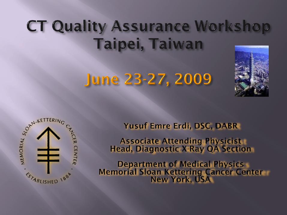
GE Lightspeed 162007
EMI Mk11972

History of CT


• Many components now in gantry or under the desk
Size reduction
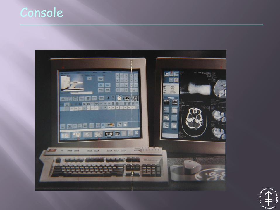
Console

• X-ray CT is a cross-sectional imaging modality that derives a two dimensional distribution of some quantity (x-ray attenuation) from one dimensional projections
• In x-ray CT, the primary quantity is attenuation, derived from transmission measurements
• Using a stack of relatively thin slices, the three dimensional problem is reduced to a two dimensional one
What is CT?
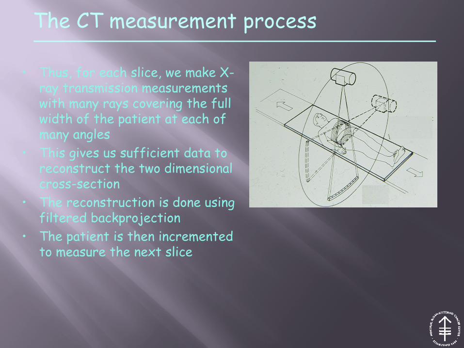
• Thus, for each slice, we make X-ray transmission measurements with many rays covering the full width of the patient at each of many angles
• This gives us sufficient data to reconstruct the two dimensional cross-section
• The reconstruction is done using filtered backprojection
• The patient is then incremented to measure the next slice
The CT measurement process

A CT SCANNER IS BASICALLY A DENSITY MEASURING DEVICE !

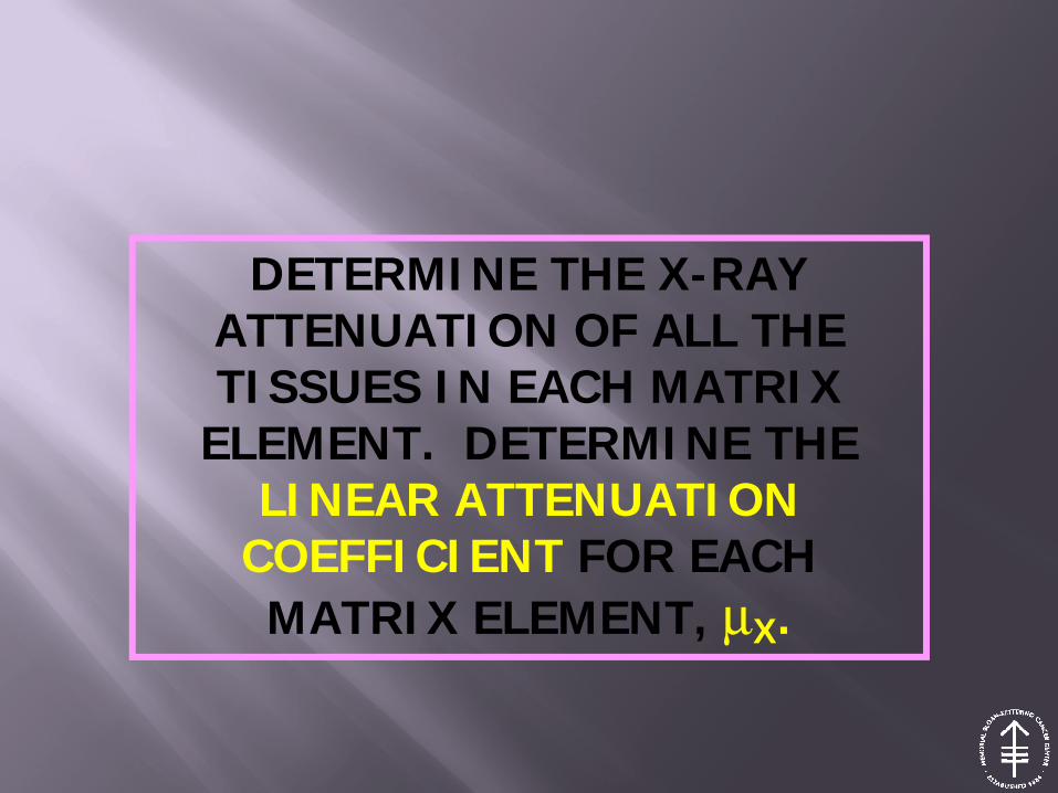
DETERMINE THE X-RAY ATTENUATION OF ALL THE TISSUES IN EACH MATRIX
ELEMENT. DETERMINE THELINEAR ATTENUATION
COEFFICIENT FOR EACHMATRIX ELEMENT, μX.
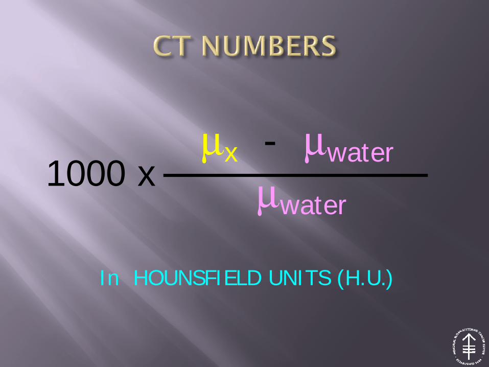
μx - μwater
μwater1000 x
In HOUNSFIELD UNITS (H.U.)

MATERIAL
LINEARATTENUATION
COEF.(cm-1)
CT NUMBER(H.U.)
AIR 0.000208 -1000
WATER 0.190 0.0
BONE 0.414 +1000
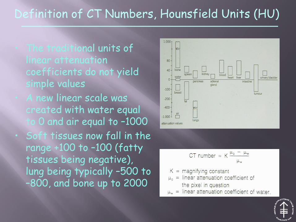
• The traditional units of linear attenuation coefficients do not yield simple values
• A new linear scale was created with water equal to 0 and air equal to –1000
• Soft tissues now fall in the range +100 to –100 (fatty tissues being negative), lung being typically –500 to –800, and bone up to 2000
Definition of CT Numbers, Hounsfield Units (HU)
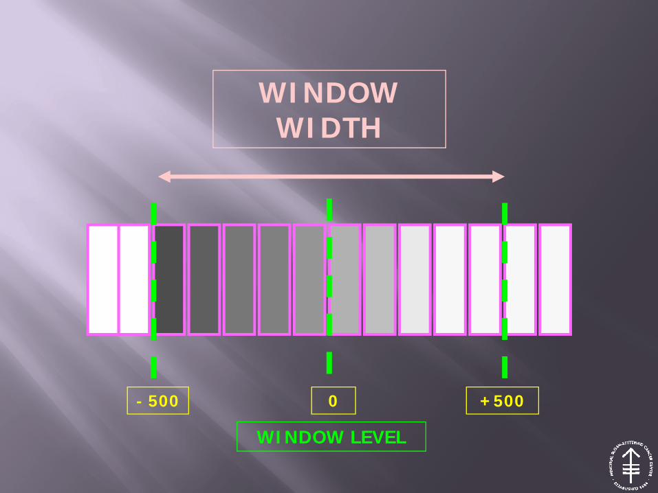
WINDOW WIDTH
- 500 0 +500
WINDOW LEVEL
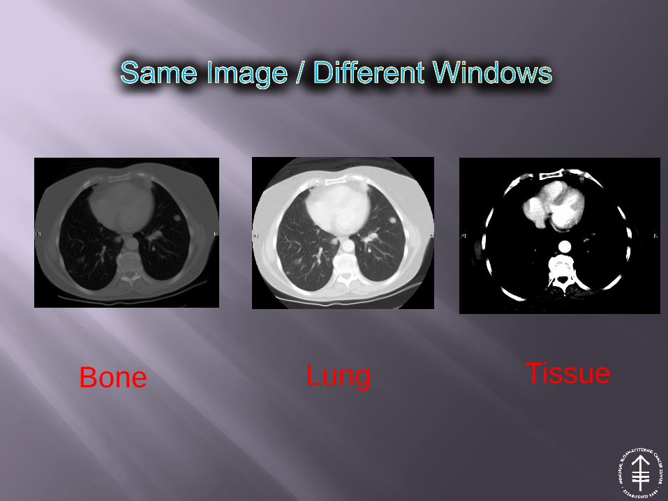
Bone Lung Tissue
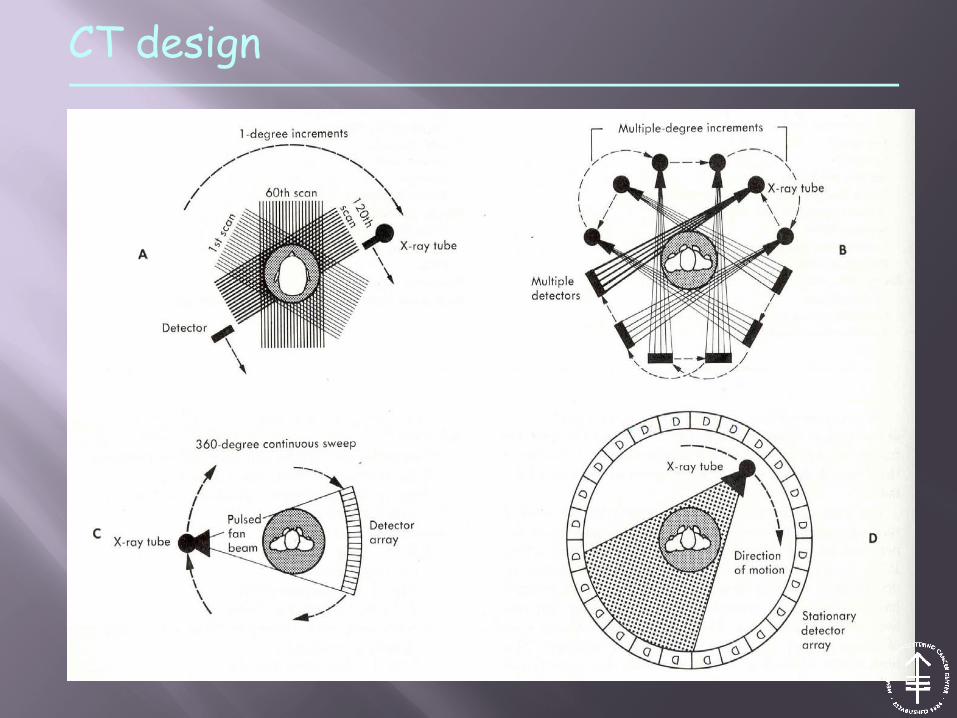
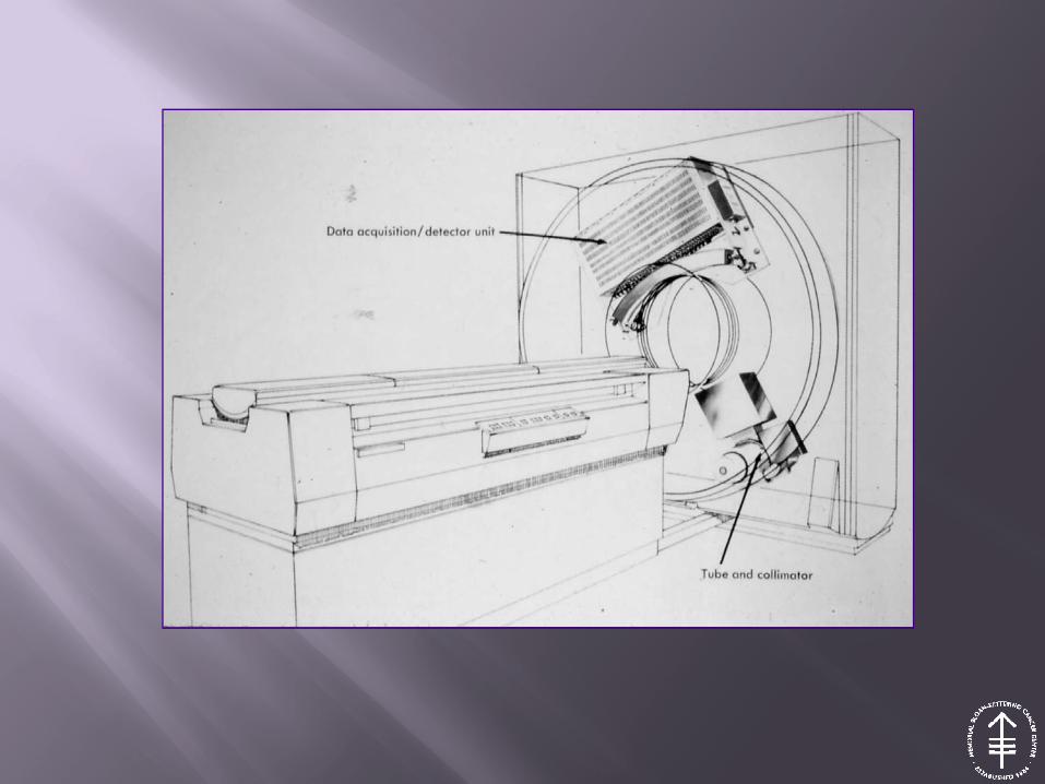
CT design
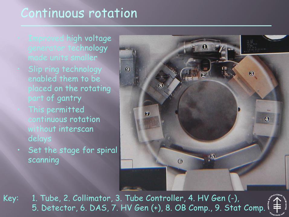
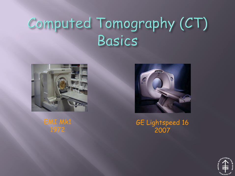
• Improved high voltage generator technology made units smaller
• Slip ring technology enabled them to be placed on the rotating part of gantry
• This permitted continuous rotation without interscan delays
• Set the stage for spiral scanning
Continuous rotation
Key: 1. Tube, 2. Collimator, 3. Tube Controller, 4. HV Gen (-), 5. Detector, 6. DAS, 7. HV Gen (+), 8. OB Comp., 9. Stat Comp.
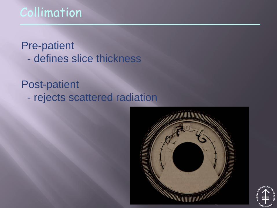
Collimation
Pre-patient- defines slice thickness
Post-patient- rejects scattered radiation

Detectors

Detectors
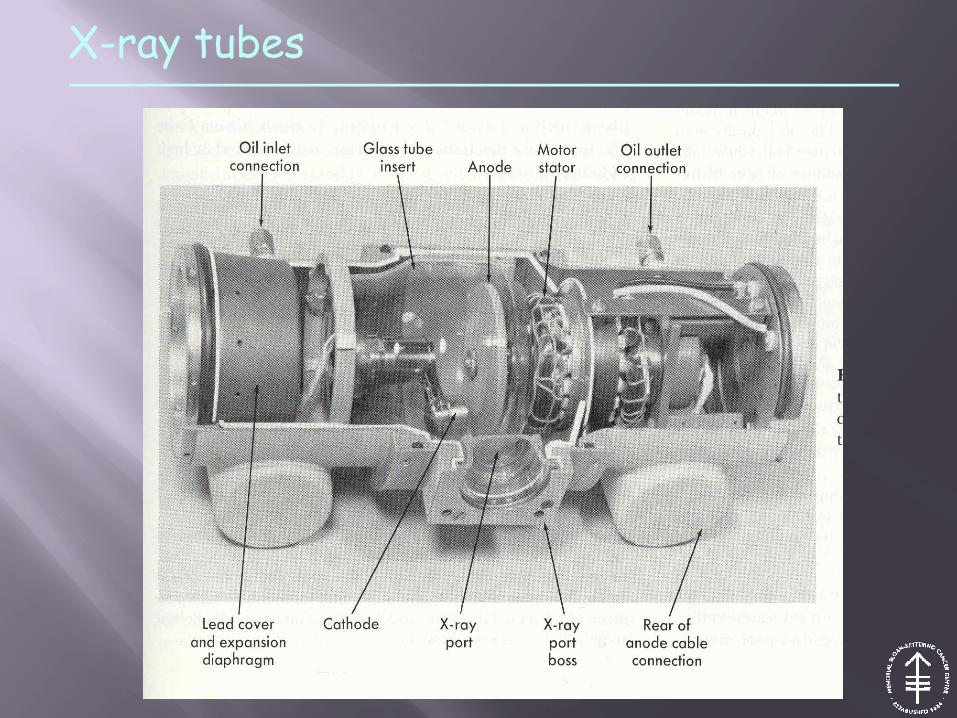
X-ray tubes