Insulin Pump What to tell your patient!! Prakash Abraham Isla Fairley.
Authors’ correctionE. A. L. Fairley, J. Kendrick-Jones and J. A. Ellis (1999). The Emery-Dreifuss muscular dystrophy phenotype arisesfrom aberrant targeting and binding of emerin at the inner nuclear membrane. J. Cell Science 112 (15), 2571-2582.
All references to S54P or Ser54Pro are incorrect and should read as S54F or Ser54Phe, respectively.
Authors’ correctionD. Gabriel, U. Hacker, J. Köhler, A. Müller-Taubenberger, J.-M. Schwartz, M. Westphal and G. Gerisch (1999).The contractile vacuole network of Dictyostelium as a distinct organelle: its dynamics visualized by a GFP marker protein.J. Cell Science 112 (22), 3995-4005.
In Fig. 7 of this paper the numbers indicating seconds should be exchanged between panels C and E, as shown below.
Fig. 7
Authors’ correctionK. P. Williams, P. Rayhorn, G. Chi-Rosso, E. A. Garber, K. L. Strauch, G. S. B. Horan, J. O. Reilly, D. P. Baker, F. R.Taylor, V. Koteliansky and R. B. Pepinsky (1999). Functional antagonists of sonic hedgehog reveal the importance of theN terminus for activity. J. Cell Science 112 (23), 4405-4414.
In the discussion on page 4412, paragraph 3 line 21, the digit duplication assay was incorrectly quoted as mouse. The correctassay is chick embryo digit duplication. In addition, line 23 should state (S. Pagan, D. P. Baker, K. P. Williams and C. J.Tabin, unpublished data).

INTRODUCTION
There are three major forms of muscular dystrophy, referred toas Duchenne, Becker and Emery-Dreifuss types, alldistinguishable by progressive skeletal muscle wasting andcardiac abnormalities to varying degrees (reviewed by Emery1989, 1996). The first two types are due to genetic defects inthe cytoskeletal/plasma membrane-associated protein,dystrophin, which is part of the glycoprotein complex linkingactin to the extracellular matrix, whereas the third form arisesfrom genetic defects in nuclear proteins (Manilal et al., 1996;Nagano et al., 1996; Bione et al., 1994, Bonne et al., 1999). X-linked EDMD is due to the absence of or defects in emerin,which is localized to the inner nuclear membrane (Yorifuji etal., 1997) in all tissues examined. It has been reported to beadditionally present in the intercalated discs of heart (Cartegniet al., 1997), although this may be due to an antibody artifact(Manilal et al., 1999). Recently, the gene product of theautosomal dominant form of EDMD has been identified asnuclear lamin A, an intermediate filament protein of the
nucleoplasm (Bonne et al., 1999). The unexpected finding thatthe EDMD arises due to defects in nuclear proteins suggeststhat the pathophysiology of EDMD may be very different fromthe other types of muscular dystrophy.
Human emerin mRNA and protein show ubiquitous tissuedistribution, with the highest expression in skeletal and cardiacmuscle (Bione et al., 1994; Manilal et al., 1996; Nagano et al.,1996). Human emerin is a serine-rich protein of 254 aminoacids (Bione et al., 1994) with an Mr of 28,993. Structuralanalysis predicts emerin to be a type II membrane protein, witha transmembrane region 11 amino acids from the carboxylterminus and a large hydrophilic N-terminal amino domainorientated towards the nucleoplasm, containing 22 potentialphosphorylation sites for a range of kinases. Mutations occurthroughout the gene encoding emerin and there are nomutational ‘hot spots’. The majority of the mutations so farstudied produce no detectable emerin either byimmunoblotting or immunohistochemistry in any tissueexamined (Manilal et al., 1996, 1997, 1998a; Nagano et al.,1996; Mora et al., 1997; Ellis et al., 1998, 1999; Yates et al.,
2571Journal of Cell Science 112, 2571-2582 (1999)Printed in Great Britain © The Company of Biologists Limited 1999JCS0436
The product of the X-linked Emery-Dreifuss musculardystrophy gene is a single-membrane-spanning proteincalled emerin, which is localized to the inner nuclearmembrane of all tissues studied. To examine whether anumber of the mutant forms of emerin expressed inpatients are mislocalized, we transfected GFP-emerincDNA constructs reflecting these mutations intoundifferentiated C2C12 myoblasts and showed thatboth wild type and all the mutant emerins are targetedto the nuclear membrane, but the mutants to a lesserextent. Mutant Del236-241 (deletion in transmembraneregion) was mainly expressed as cytoplasmicaggregates, with only trace amounts at the nuclearenvelope. Complete removal of the transmembraneregion and C-terminal tail relocated emerin to thenucleoplasm. Mutations in emerin’s N-terminal domainhad a less severe effect on disrupting nuclear envelopetargeting. This data suggests that emerin contains
multiple non-overlapping nuclear-membrane-targetingdeterminants.
Analysis of material immunoisolated using emerinantibodies, from either undifferentiated C2C12 myoblastsor purified hepatocyte nuclei, demonstrated that both A-and B-type lamins and nuclear actin interact with emerin.This is the first report of proteins interacting with emerin.The EDMD phenotype can thus arise by either the absenceor a reduction in emerin at the nuclear envelope, and bothof these disrupt its interactions with that of structuralcomponents of the nucleus. We propose that an emerin-nuclear protein complex exists at the nuclear envelope andthat one of its primary roles is to stabilize the nuclearmembrane against the mechanical stresses that aregenerated in muscle cells during contraction.
Key words: emerin, nuclear targeting, lamin binding, Emery-Dreifussmuscular dystrophy.
SUMMARY
The Emery-Dreifuss muscular dystrophy phenotype arises from aberrant
targeting and binding of emerin at the inner nuclear membrane
Elizabeth A. L. Fairley1,2, John Kendrick-Jones1 and Juliet A. Ellis2,*1MRC Laboratory of Molecular Biology, Hills Road, Cambridge CB2 2QH, UK2Department of Medical Genetics, Cambridge Institute of Medical Research, Wellcome Trust/MRC building, Addenbrooke’sHospital, Hills Road, Cambridge CB2 2XY, UK*Author for correspondence (e-mail: [email protected])
Accepted 20 May; published on WWW 7 July 1999
2572
1999). However, a small number of mutations have beenreported to produce modified forms of emerin (Mora et al.,1997; Manilal et al., 1998a; Ellis et al., 1998, 1999; Yates etal., 1999). Interestingly, despite the different mutations inEDMD, producing varying effects on emerin expression, theclinical phenotype of all these patients is similar.
The functions of the integral membrane proteins in the innernuclear membrane are not completely known, although theavailable evidence suggests that they may have a structural rolein maintaining nuclear architecture (Gerace and Foisner, 1994).Interactions between nuclear membrane components, nuclearlamina and chromatin are crucially important for maintainingthe structure of the nuclear membrane-chromatin organizationduring interphase and for the disassembly and reformation ofthe nuclear membrane during mitosis. Emerin possesses twoshort regions of homology to rat lamina-associated polypeptide2 (LAP2; Furukawa et al., 1995; Harris et al., 1995), anotherinner nuclear membrane protein, suggesting that emerin is amember of the nuclear lamina-associated protein family. Thisfamily includes lamina-associated polypeptide 1 (LAP1;Martin et al., 1995) and the lamin-B receptor (LBR; Soullamand Worman, 1993). LAP2 has been shown to interact in a cell-cycle dependent manner with both lamin B1 and chromatin andthe binding sites for both have been identified. At the onset ofmitosis, LAP 2 is phosphorylated, causing it to dissociate fromthe lamina network and chromatin and disperse throughout theER (Yang et al., 1997a). Both the nuclear lamins and LAP2reassociate with the chromatin at late anaphase. This suggeststhat reassembly of the nuclear envelope at the end of mitosisinvolves sorting of integral membrane proteins to chromosomesurfaces by binding interactions with lamins and chromatin(Yang et al., 1997a). The cell cycle-dependent binding of LAP2to lamin B1 also controls the increase in nuclear volume seenduring interphase in cycling cells (Yang et al., 1997b), whichallows them to enter S phase.
Recent data suggests that emerin has a role in cell cycle-dependent events, since it is also localized at intranuclear sites,where it colocalizes with the nuclear lamins (Squarzoni et al.,1998; Manilal et al., 1998b) and binds tightly to unidentifiedinsoluble matrix components (Ellis et al., 1998; Squarzoni etal., 1998). In addition, emerin can occur in four differentlyphosphorylated forms, three of which appear to be associatedwith the cell cycle (Ellis et al., 1998). During mitosis emerinbecomes dispersed throughout the cell, no longer colocalizingwith the lamins, and then participates in the reconstitution ofmembranes around the daughter nuclei at telophase (Manilal
et al., 1998a). Phosphorylation of emerin may be involved incontrolling these events.
In the present paper, we show that mutant forms of emerinexpressed in a number of EDMD patients are targeted to theinner nuclear membrane, but in a less efficient mannercompared to wild type. These results suggest that emerincontains multiple nuclear membrane localization signals, someof which are involved directly in nuclear targeting and othersin retention at the nuclear membrane. We demonstrate thatwild-type emerin binds to both A- and B-type lamins and tonuclear actin, suggesting that interactions between nuclearcomponents are essential for skeletal and cardiac musclefunction and that loss of integrity of the nuclear membrane maydirectly produce the muscular dystrophy phenotype.
MATERIALS AND METHODS
Cell lines and cell cultureCOS-7, green monkey fibroblasts and C2C12 cells, a subclone of theC2 mouse myoblast cell line (Yaffe and Saxel, 1977), were obtainedfrom the European Collection of Cell Lines (ECACC) and cultured inDulbecco’s Minimal Essential Medium (DMEM; Gibco BRL)supplemented with 10% fetal bovine serum (FBS; Sigma) and 2 mMglutamine. To avoid spontaneous differentiation of the C2C12 cells,they were passaged at 75% confluence.
Antibodies The following antibodies against emerin were used: affinity-purifiedrabbit polyclonal antibodies AP2, AP5, AP8 and AP9 raised againsthuman emerin, as shown in Fig. 1 and as described previously (Elliset al., 1998). An affinity-purified rabbit polyclonal antibody, AP1, wasraised against a bacterial fusion protein expressing residues 114-183of rat recombinant emerin. This antibody is to the region of leasthomology between rat and human emerin, and recognizes rat emerinwith a greater affinity than the AP8 antibody. These affinity-purifiedantibodies were used at 1:3000 dilution on immunoblots and 1:100 inboth immunoprecipitation and immunofluorescence experiments.Antibodies used to label intracellular compartments in the emerinlocalization experiments included: a rat polyclonal antibodyMAC256, which recognizes resident endoplasmic reticulum (ER)proteins (specific for KDEL motif) obtained from Dr Moreman(University of Georgia, USA) and used at 1:100; a rat monoclonalantibody against the lysosomal membrane glycoprotein, LAMP-1(Developmental Studies Hybridoma Bank, University of Iowa, USA)used at 1:400; a mouse monoclonal antibody β-actin (clone AC-74;Sigma No. A 5316) used at 1:50; a mouse monoclonal antibody laminA (clone 133A2; McKeon et al., 1986) used at 1:300 and a rabbitpolyclonal antibody to lamin B (Moir et al., 1994) used at 1:50.
E. A. L. Fairley and others
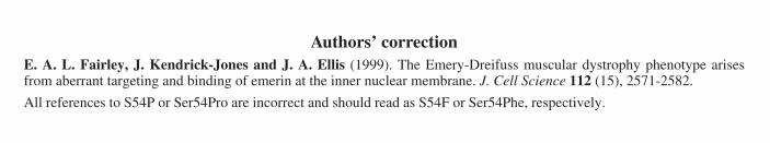
Fig. 1. Secondary structural features of emerin,regions used to raise polyclonal antisera and theposition of mutations used in our study. The boxedsection represents the full-length emerin protein.Polyclonal antisera were raised against the regionsshown by horizontal arrows labelled with thenumbers given to the resulting affinity-purifiedantisera (AP2, AP5, AP8, AP9). The positions ofthe mutations found in EDMD patients used in thisstudy are shown by the short vertical arrowslabelled S54P, Del 95-99, P183T/H, Del236-241.Other numbers show the positions of amino acids inthe human emerin sequence. TM, transmembraneregion; NLS, bipartite nuclear localization signal;LAP2, regions in emerin 41% identical to LAP2.
AP8
LAP2254221
LAP26 44
NLS35 46
TM
S54P(CM1)
Del 95-99 (Y1)
P183T (Y15)P183H (WG)
Del 236-241 (Y3)
1 70 220244
254140
AP2
AP9
AP5

2573Emerin and Emery-Dreifuss muscular dystrophy
Immunoblots from the emerin immunoprecipitation experiments wereprobed with rabbit polyclonal antibody to actin (against C11 peptide;Sigma No. A 2066) used at 1:1000; rat polyclonal antibody to laminA/C (Moir et al., 1994) and the aforementioned rabbit polyclonalantibody to lamin B, were both used at 1:1000.
Mutant human emerin cDNA constructsA full-length human emerin cDNA clone in plasmid pBluescript KS−
(Ellis et al., 1998) was used as the starting template to make themutant cDNA plasmid constructs. All the constructs chosen wereknown to occur in EDMD patients and to express modified forms ofemerin and are shown in Table 1. The position of the mutations in theemerin gene are shown in Fig. 1. Site-directed mutagenesis wasperformed on the wild-type cDNA emerin clone by PCR,incorporating restriction sites HindIII and NdeI at the 5′ end andBamHI at the 3′ end, using the primers listed in Table 2.
PCR was performed in a total reaction volume of 50 µl with 0.4 ngwild-type cDNA plasmid DNA as template. Final reagentconcentrations in the PCR reaction mixture were 20 mM Tris-HCl,pH 8.4, 50 mM KCl, 1.5 mM MgCl2, 400 µM dNTPs and 50 pmol/µlof each primer (Table 2); 5 units Pfu polymerase (Clontech). Thesamples were denatured at 95°C for 3 minutes and then for 1 minuteat 94°C, 1 minute at 43°C and 1.5 minutes at 74°C for 5 cycles. AllDNA samples were annealed and amplified for 25 cycles (1 minuteat 94°C, 1 minute at 55°C, 1 minute at 74°C), except for the primersthat were used to synthesize the P183H mutant, which required anannealing temperature of 50°C. The application was finished with afinal extension at 74°C for 7 minutes.
The PCR fragments generated were cloned into the HindIII andBamHI sites of mammalian expression vector pEGFP-C2 (Clontech),which contains the Green Fluorescent Protein (GFP). Another emerincDNA clone was constructed in pEGFP-C1 by subcloning residues 1-220 from the fusion protein FP9 construct in vector pET-29b (seebelow) to provide a mutant form of emerin, which lacks thetransmembrane and C-terminal tail region.
All constructs made were sequenced in both directions using aSequence Version 2.0 DNA Sequencing Kit (Amersham Life Science)and Thermo Sequence dye terminator cycle sequencing pre-mix kit(Amersham Life Science).
Transient transfections Undifferentiated C2C12 myoblasts were transfected according tothe SuperFect™ Transfection Protocol of Qiagen™, using aSuperfect:DNA ratio of 1:6. For immunofluorescence experiments,subconfluent cells were plated on coverslips (22 mm2) in 6-wellplates, 1 day prior to being transfected with 2 µg of DNA (purified
on Qiagen columns according to the QIAprep Spin Miniprep kitprotocol). A time course of emerin expression was conducted toidentify the optimum time for the correct localization of wild-typeemerin. Cells were then fixed and immunolabelled as described below.Expression levels of the GFP-emerin constructs were examined byimmunoblotting the transfected cells for emerin expression withaffinity-purified antibody AP8.
Immunofluorescence microscopyFor microscopic visualization, transfected cells were washed 3 timeswith phosphate buffered saline (PBS; 145 mM NaCl, 7.5 mMNa2HPO4, 2.5 mM NaH2PO4, pH 7.4), prior to being fixed in either4% (w/v) paraformaldehyde for 20 minutes (for Rhodamine-Phalloidin and endogenous emerin staining), methanol for 10 minutes(for immunolabelling for lysosomal marker, LAMP 1) or 2% (w/v)paraformaldehyde/0.1% (w/v) glutaraldehyde mix for 25 minutes (forimmunolabelling for lamins A and B and the ER marker MAC256).
Table 1. Modified forms of emerin expressed in EDMD patients used in our studyNucleotide Nucleotide Protein expression
Patient change position2 Mutation type Effect and size References
Y1 (Del95-99) Del. 15 bp1 878 In-frame deletion Del. YEESY Reduced levels Ellis et al. (1998); Yates et al. (1999)(95-99) 31 kDa
Y3 (Del236-241) Del. 18 bp 1763 In-frame deletion Del. VIVLFF Very reduced levels Yates et al. (1999); Ellis et al. (1999)(236-241) 34 kDa
Y15 (P183T) C→A 1605 Missense Pro 183→Thr Normal levels Ellis et al. (1999); Yates et al. (1999)34 kDa
WG (P183H) C→A 1606 Missense Pro 183→His Normal levels Ellis et al. (1999)34 kDa
CM1 (S54P) C→T 394 Missense Ser 54→Phe Normal levels C. Muller, UP34 kDa
1Deletion.2Genomic numbering.UP, unpublished data.All mutations are listed in the EDMD database (Yates and Wehnert, 1999).
Table 2. Primers used to generate modified forms ofemerin
Wild-type emerin 5′-TTTTA AGC TTA CAT ATG GAC AAC TAC (1-254) GCA GAT-
3′-TTTTGGA TCC TGG CTC CCT CTA GAA GGG GTT-
Del236-241 5′-TTTTA AGC TTA CAT ATG GAC AAC TAC GCA GAT-
3′-TTTTGGA TCC TGG CTC CCT CTA GAA GGG GTT GCC TTC TTC AGC CTG CAT GAA GTG GTA AAT AAA GAC CAG GAA AAG CAG CAG CTG-
Del95-99 5′-AGA AAG GGC TAC AAT GAC GAC TAC TTC ACC ACC AGG ACT TAT-
3′-ATA AGT CCT GGT GGT GAA GTA GTC GTC ATT GTA GCC CTT GCT-
Serine 54 Proline 5′-CCC AGC TCG TTC GCC GCC TCC-3′-GGA GGC GGC GAA CGA GCT GGG-
Proline 183 Histidine 5′-TCC TAT TAT CAT ACT TCC TCC-3′-GGA GGA AGT ATG ATA ATA GGA–
Proline 183 Threonine 5′-TCC TAT TAT ACT ACT TCC TCC-3′-GGAGGA AGT AGT ATA ATA GGA-
Restriction enzyme sites are underlined. Mutated DNA bases are shown in bold.

2574
The cells fixed in 4% (w/v) paraformaldehyde were washed 3×10minutes in PBS, permeabilised in 0.2% (w/v) PBS/Triton X-100 for5 minutes, washed 3×10 minutes in PBS and blocked with PBS/0.2%(w/v) fish gelatin (Sigma) 4×10 minutes, prior to being incubated withthe primary antibody for 1 hour in a humidified chamber. Thosecells fixed with methanol were washed 3×10 minutes in PBS, thenblocked with PBS/0.2% (w/v) fish gelatin 4×10 minutes prior toprimary antibody incubation. Those cells fixed with 2% (w/v)paraformaldehyde/0.1% (w/v) glutaraldehyde were washed 3×10minutes in PBS, permeabilised with PBS/Triton X-100 for 5 minutes,washed 3×10 minutes in PBS, quenched in 1 mg/ml NaBH4 in PBSfor 10 minutes, washed 3×10 minutes in PBS, blocked with PBS/0.2%(w/v) fish gelatin 4×10 minutes prior to primary antibody incubation.After antibody incubation all cells were washed thoroughly 4×10minutes with PBS/fish gelatin for 5 minutes and incubated with theappropriate secondary antibody for 30 minutes in a humidifiedchamber. The secondary antibodies included a goat anti-rabbit IgGconjugated with Texas Red (Amersham Life Sciences) at 1:400, anda sheep anti-mouse IgG conjugated with FITC (Amersham LifeSciences) at 1:150. Thereafter the coverslips were washed thoroughly3×10 minutes in PBS/fish gelatin and 3×PBS for 5 minutes, rinsed indeionised water and placed on slides washed in 70% methanol and airdried. 20 µl of Mowiol mounting medium (87% (v/v) glycerol, 0.2MTris-HCl, pH 8.5, 12% (w/v) Mowiol) were added. The samples werestored in the dark at room temperature prior to examination.
Immunofluorescence microscopy was performed on an MRC-1024Laser Scanning Confocal Imaging System (Bio-Rad). Scanning wasdone with a Nikon PlanApo 60× lens having a numerical aperture of1.4 using Texas Red emission filter 605 DF32 and FITC emissionfilter 522 DF32. Computerized images were processed by mrc2M(Michio Ono; e-mail [email protected]:), Adobe Photoshop4.0 and Adobe illustrator 7.0 software on a Power Macintosh4400/200. The Lasersharp Processing Screen software was usedto quantify the fluorescence intensity of GFP-emerin present atthe nuclear envelope. These fluorescence intensities were measuredper unit area (100 pixels) of nuclear envelope, to allowcomparisons between the transfectants expressing different forms ofGFP-emerin. The average fluorescence intensity was quantified froma 25-cell unbiased selection, for each construct expressed. Statistical
analysis was performed using the GraphPad Prism version 2.0asoftware.
Expression and purification of fusion proteinsFour bacterial fusion proteins expressing fragments of humanrecombinant emerin spanning residues 1-70 (FP8), 70-140 (FP2), 1-174 (FP3) and 1-220 (FP9) were expressed and purified in theexpression plasmid pET-29b (C-terminal his6 tag; Novagen, Madison,WI) and a fifth fusion protein (residues 244-254) was expressed andpurified as a glutathione S-transferase (GST) fusion in pGEX-4T-3(Pharmacia Biotech Inc.). Fusion proteins were expressed and purifiedas described previously in Ellis et al. (1998).
Overlay binding assay of emerin-fusion proteins ontoundifferentiated C2C12 cell lysates and purified rathepatocyte nucleiUndifferentiated C2C12 myoblasts were harvested by scraping themoff tissue culture flasks, washed in PBS, resuspended in PBS,sonicated briefly, loaded into sample buffer and proteins separated by10% SDS-PAGE according to Laemmli (1970) before transferringonto Hybond ECL nitrocellulose membrane (Amersham Life Science)according to Burnette (1981). The membranes were washed andblocked in PBS/6% (w/v) dried skimmed milk/0.1% (v/v) Tween-20(blocking buffer) 4× for 15 minutes at room temperature. Blots wereoverlaid with fusion proteins, at final concentrations of 0.25-12.5µg/ml, for 1 hour at room temperature, with rocking, in blockingbuffer. Blots were then washed in blocking buffer 4× for 15 minutes.The blots were then processed by immunoblotting with the panel ofrabbit polyclonal affinity-purified antibodies raised against the humanemerin-fusion proteins (AP8, AP2, AP9, AP5; see Fig. 1 and Ellis etal., 1998). Immunoblotted bands were visualized by enhancedchemiluminescence (ECL; Amersham Life Sciences) in conjunctionwith autoradiography.
To determine the specificity of any protein-protein interactionidentified, the salt concentration in both the blocking, binding andwashing stages of the overlay procedure was varied between 145 mMand 500 mM, by supplementing the PBS with NaCl. We alsoexamined whether any fusion protein binding observed could beblocked, by incubating the fusion protein with the appropriate affinity-
E. A. L. Fairley and others
Fig. 2. Immunofluorescence labelling ofendogenous emerin and nuclear lamin Aby confocal immunofluorescencemicroscopy. (A) Comparison ofendogenous emerin levels in COS-7 cellsand undifferentiated C2C12 myoblasts.Localization of emerin is shown in redand β-actin in green. Affinity-purifiedantiserum AP8 was used for COS-7 cells(1:100) and affinity-purified antiserumAP1 was used on the C2C12 myoblasts(1:100). (B) Colocalization of emerin andlamin A in undifferentiated C2C12myoblasts. Emerin is shown in red (i) andlamin A in green (ii) with an overlay ofthe two signals shown in (iii).Immunolabelling was performed asdescribed in Materials and methods.Bars, 10 µm.

2575Emerin and Emery-Dreifuss muscular dystrophy
purified emerin antibody, prior to using the fusion protein in theoverlay procedure. Overlays onto an E. coli cell lysate were performedas a control against non-specific binding.
ImmunoprecipitationImmunoprecipitation experiments were performed on bothundifferentiated C2C12 myoblasts and purified rat hepatocyte nuclei(a kind gift from H. Kent, MRC, Cambridge, UK) using the sameprotocol. C2C12 cells were used at 60% confluence (108 cells/sample)and the purified nuclei at 5×10⁷/sample. For immunoprecipitatingfrom the tissue culture cells, medium was removed, the cell monolayerwashed in PBS, and the flasks placed on ice for 30 minutes. 0.5 mlof extraction buffer (1% (v/v) Triton X-100, 50 mM Tris-HCl, pH 7.4,2 mM MgCl2, and 100 mM - 1 M NaCl) was added to each 75cm2
flask used, which were left on ice for a further 30 minutes withintermittent agitation. The lysate was collected and sonicated 4× for15 second bursts (set at 40%) on a sonicator ultrasonic processor XL(Misonix Incorporated, NY, USA) and then centrifuged at 11,600 gfor 10 minutes at 4°C. The supernatant was collected and preincubated
with 1 µg of non-immune rabbit IgG for 1 hour at 4°C with rotation,and captured with 20 µl of Protein A-Sepharose (Sigma: 50 mg/mlslurry in PBS) for 1 hour with rotation at 4°C. The Protein A-Sepharose was removed by centrifuging at 11,600 g for 5 minutes at4°C and the supernatants transferred to fresh tubes. Samples wereeither incubated with affinity-purified rat emerin polyclonal antibodyAP1 (final dilution of 1:100) or with the corresponding pre-immunesera (1:100), with rotation, overnight at 4°C. The samples were thencentrifuged at 11,600 g for 20 minutes at 4°C to remove any insolubleimmune complexes. Supernatants were transferred to fresh tubes and50 µl of Protein A-Sepharose was added per sample followed byrotation for 2 hours at 4°C. The beads were collected by centrifugingat 11,600 g for 5 minutes at 4°C, and washed five times in wash buffer(1% (v/v) Triton X-100, 50 mM Tris-HCl, 2 mM MgCl2, 150 mM-1.5 M NaCl). A range of salt concentrations was tried both in theextraction and wash stages to optimize immunoprecipitationconditions and to investigate the strength of any emerin-proteininteractions identified. Residual detergent was removed from thebeads by washing them twice in 50 mM Tris-HCl, pH 7.4. Samples
Fig. 3. Intracellular localization ofwild type and modified forms ofGFP-emerin constructs (asindicated) expressed by transienttransfection into undifferentiatedC2C12 myoblasts. GFP-emerincDNA constructs were transfectedinto C2C12 cells, as described inMaterials and methods. Cells werefixed with 4% (w/v)paraformaldehyde. Theintracellular location of emerinwas monitored by GFPfluorescence (green). Cells werecounterstained with Rhodamine-Phalloidin (red) to visualize F-actin. GFP-emerin fluorescence isshown separately to clarify GFP-emerins trafficking through theER. Bars, 10 µm.
2576
were rotated for 10 minutes between each wash. Samples wereprepared for SDS-PAGE by heating to 95°C for 5 minutes in samplebuffer (Laemmli, 1970) to release the immunocomplexes from thebeads. Proteins were separated by 10% SDS-PAGE (Laemmli, 1970)and either silver stained (Ansorge, 1985) or immunoblotted (Burnette,1981) for emerin, actin and nuclear lamins.
RESULTS
Localization of endogenous emerin in COS-7 cellsand undifferentiated C2C12 myoblastsThe localization of immunofluorescent endogenous emerin inboth COS-7 cells and undifferentiated C2C12 myoblasts isshown in Fig. 2A. The rabbit polyclonal affinity-purifiedantibodies AP1 (rat emerin) and AP8 (human emerin) gave thesame immunofluorescence pattern for emerin in both cell lines,but each antibody was more sensitive for the species of emerinit was raised against (data not shown). In both cell types,emerin is localized to the nuclear rim, but internal nuclear fociand ER staining close to the nuclear rim was also observed.Interestingly, we observed more cytoplasmic emerin inundifferentiated C2C12 myoblasts than in COS-7 cells. We andothers (Cartegni et al., 1997) have observed this phenomenonin other adherent cell lines. Quantitatively there appears to bethe same amount of endogenous emerin in both cell types.Emerin was shown to colocalize with lamins at the nuclear rimand in the intranuclei foci at interphase (Fig. 2B), as previouslyreported by Manilal et al. (1998b). No plasma membranestaining was seen, nor any colocalization with cytoskeletalactin (Fig. 2A).
Emerin targeting in transfected undifferentiatedC2C12 myoblastsThe majority of EDMD patients exhibit the null phenotype;however, in the small number of patients who express mutantforms of emerin, it is not known whether these are mistargetedor, if correctly localized, they are dysfunctional. To answerthis question, GFP-constructs, mimicking the mutant formsof emerin identified in patients, were introduced intoundifferentiated C2C12 myoblasts by transfection, and theirlocalization monitored from the onset of expression usingfluorescence imaging with a confocal laser scanning
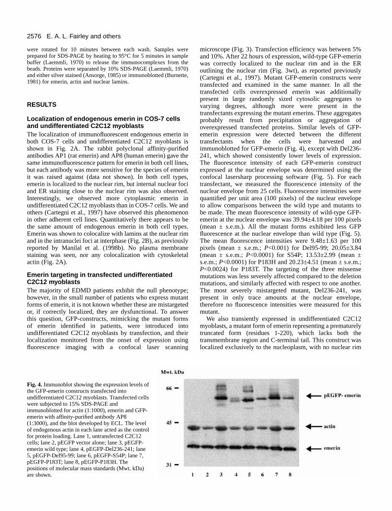
microscope (Fig. 3). Transfection efficiency was between 5%and 10%. After 22 hours of expression, wild-type GFP-emerinwas correctly localized to the nuclear rim and in the ERoutlining the nuclear rim (Fig. 3wt), as reported previously(Cartegni et al., 1997). Mutant GFP-emerin constructs weretransfected and examined in the same manner. In all thetransfected cells overexpressed emerin was additionallypresent in large randomly sized cytosolic aggregates tovarying degrees, although more were present in thetransfectants expressing the mutant emerins. These aggregatesprobably result from precipitation or aggregation ofoverexpressed transfected proteins. Similar levels of GFP-emerin expression were detected between the differenttransfectants when the cells were harvested andimmunoblotted for GFP-emerin (Fig. 4), except with Del236-241, which showed consistently lower levels of expression.The fluorescence intensity of each GFP-emerin constructexpressed at the nuclear envelope was determined using theconfocal lasersharp processing software (Fig. 5). For eachtransfectant, we measured the fluorescence intensity of thenuclear envelope from 25 cells. Fluorescence intensities werequantified per unit area (100 pixels) of the nuclear envelopeto allow comparisons between the wild type and mutants tobe made. The mean fluorescence intensity of wild-type GFP-emerin at the nuclear envelope was 39.94±4.18 per 100 pixels(mean ± s.e.m.). All the mutant forms exhibited less GFPfluorescence at the nuclear envelope than wild type (Fig. 5).The mean fluorescence intensities were 9.48±1.63 per 100pixels (mean ± s.e.m.; P<0.001) for Del95-99; 20.05±3.84(mean ± s.e.m.; P<0.0001) for S54P; 13.53±2.99 (mean ±s.e.m.; P<0.0001) for P183H and 20.23±4.51 (mean ± s.e.m.;P<0.0024) for P183T. The targeting of the three missensemutations was less severely affected compared to the deletionmutations, and similarly affected with respect to one another.The most severely mistargeted mutant, Del236-241, waspresent in only trace amounts at the nuclear envelope,therefore no fluorescence intensities were measured for thismutant.
We also transiently expressed in undifferentiated C2C12myoblasts, a mutant form of emerin representing a prematurelytruncated form (residues 1-220), which lacks both thetransmembrane region and C-terminal tail. This construct waslocalized exclusively to the nucleoplasm, with no nuclear rim
E. A. L. Fairley and others
Fig. 4. Immunoblot showing the expression levels ofthe GFP-emerin constructs transfected intoundifferentiated C2C12 myoblasts. Transfected cellswere subjected to 15% SDS-PAGE andimmunoblotted for actin (1:1000), emerin and GFP-emerin with affinity-purified antibody AP8(1:3000), and the blot developed by ECL. The levelof endogenous actin in each lane acted as the controlfor protein loading. Lane 1, untransfected C2C12cells; lane 2, pEGFP vector alone; lane 3, pEGFP-emerin wild type; lane 4, pEGFP-Del236-241; lane5, pEGFP-Del95-99; lane 6, pEGFP-S54P; lane 7,pEGFP-P183T; lane 8, pEGFP-P183H. Thepositions of molecular mass standards (Mwt. kDa)are shown.

2577Emerin and Emery-Dreifuss muscular dystrophy
staining (Fig. 6), suggesting that the major determinant for thenuclear membrane localization of emerin is within the regionspanning residues 221-254.
Overlay of bacterial emerin-fusion proteins onto celllysatesTo identify interacting proteins and the region in emerininvolved in these potential protein-protein interactions, C2C12cell lysate proteins were separated electrophoretically,transferred to nitrocellulose membranes and overlaid with thebacterially expressed emerin-fusion proteins. Only the fusionprotein FP9 (residues 1-220) bound to C2C12 cell lysates usingthis overlay procedure. Since no binding was seen with thefusion protein expressing residues 1-174, it suggests thatresidues 174-220 contain the sites that interact with otherproteins. Three bands of approx. 42, 64 and 74 kDa wereidentified as binding to emerin in the C2C12 cell lysates (Fig.7A). Similar results were obtained on purified rat hepatocytenuclei, human skeletal muscle, HeLa cells and lymphoblastoidcells (data not shown). The affinity-purified rabbit polyclonalantisera AP2, AP8 and AP9 gave the same results and couldbe used interchangeably, but AP5 (which recognizes residues244-254) did not. These interacting bands did not appear in agel overlay onto an E. coli cell lysate (Fig. 7A, lane 1), andcould be blocked by incubating affinity-purified antibody with1.25 µg of fusion protein FP9 (Fig. 7A, lane 2), prior to the geloverlay. The 42 kDa band predominated and no real increasein binding to this component was seen after 1.25 µg/ml fusionprotein was added (Fig. 7B, lane 5), whereas increased bindingwas seen for the 64 and 74 kDa bands as the fusion proteinconcentration was increased (Fig. 7A, lanes 3-7). Theinteraction with the 42 kDa protein was stable at 500 mMNaCl, but was severely reduced with the 64 and 74 kDaproteins. Alongside the gel overlay, we immunoblotted theC2C12 cell lysates for actin (Fig. 7A, lane 8) and on the basisof size we speculated that the 42 kDa is actin.
To see if the 42 kDa band was actin, the emerin bacterialfusion proteins FP2, FP3, FP8 and FP9 were overlaid ontorabbit skeletal muscle α-actin (prepared as described in Pardeeand Spudich, 1982). 2.5 µg F-actin was loaded per gel trackand each track overlaid with 1.25 µg/ml of each bacterialfusion protein (Fig. 7B). Binding to actin by the emerin-fusionproteins was analyzed by immunoblotting with the affinity-purified emerin antibodies. Only fusion protein FP9 bound to
actin (Fig. 7B, lane 7), and this was stable at 500 mM NaCl.This could be blocked either by allowing 2.5 µg F-actin to bindto 1.25 µg FP9 for 2 hours at room temperature, prior tooverlaying onto actin (Fig. 7B, lane 8) or by allowing 1.25 µgFP9 to bind to 10 µg C2C12 cell lysate for 2 hours at roomtemperature, prior to overlaying onto actin (Fig. 7B, lane 9). Inaddition, if we incubated 1.25 µg FP9 with 15 µg F-actin for2 hours at room temperature prior to overlaying onto C2C12cell lysates, and immunoblotted with emerin antibody AP9, nobands at all were visualized (data not shown).
These results suggest that emerin is able to interact withactin, and that residues 174-220 are involved in binding to actinand also to the 64 and 74 kDa bands. In addition, the interactionbetween emerin and all three proteins can be blocked by priorincubation of fusion protein FP9 with an excess of actin,suggesting that the actin, 64 kDa and 74 kDa binding sites onemerin are very close to one another or overlap.
Coimmunoprecipitation of proteins interacting withemerinTo identify proteins interacting with emerin, mild detergentsolubilisation is required to ensure that emerin is isolated undernon-denaturing conditions in order to retain protein-proteininteractions. We adapted extraction conditions reported for theLBR (Simos and Georgatos, 1992) and LAP1 proteins (Maisonet al., 1997) for extracting emerin, using a basic extractionbuffer of 1% (v/v) Triton X-100, 50 mM Tris-HCl, pH 7.4, 2mM MgCl2, with varying amounts of NaCl (100 mM to 1 M).The optimal extraction conditions chosen were those where wecould demonstrate by immunoblotting of the cell lysate that wehad maximum extraction of emerin and nuclear lamins, butwhich still retained protein-protein interactions in thesubsequent immunoprecipitation (data not shown). The finalextraction buffer contained 150 mM NaCl, and the capturedimmunocomplexes were washed at 300 mM NaCl.
A direct interaction between emerin and a nuclearcomponent has not been previously reported. We thereforeexamined our immunoprecipitates for lamin A/C, lamin B andactin. The size of the bands obtained were compared with animmunoblot of the cell lysate/extracted rat nuclei run alongside(Fig. 8A,C). Emerin antibody AP1 immunoprecipitated emerinfrom both C2C12 (Fig. 8A, lane 7) and purified rat hepatocytenuclei (Fig. 8C, lane 13). The pre-immune sera did notimmunoprecipitate emerin (Fig. 8A,C, lanes 5 and 10).
Del95-99 S54P P183H P183T Wild type
0
10
20
30
40
50Wild typeP183TP183HS54PDel95-99
***
***
**
***
GFP-emerin constructs
fluo
resc
ence
inte
nsity
/100
pix
els
Fig. 5. Bar chart showing the nuclear envelope fluorescenceintensities of the GFP-emerin constructs expressed inundifferentiated C2C12 cells. Undifferentiated C2C12myoblasts were transiently transfected with either wild typeor mutant GFP-emerin constructs as described in Materialsand methods. The nuclear envelope fluorescence intensitiesfrom 25 cells were quantified and statistically analyzed foreach transfectant, as described in Materials and methods.Values are means ± s.e.m. **P<0.01; ***P<0.001.

2578
Coimmunoprecipitated with emerin were lamin B and laminA/C from both C2C12 (Fig. 8A, lanes 6 and 7, respectively)and purified nuclei extracts (Fig. 8C, lanes 9, lamin A/C, 11and 13, lamin B). Actin was only coimmunoprecipitated withemerin in the C2C12 cell lysates (Fig. 8A, lane 7), and not from
purified rat nuclei (Fig. 8C, lane 11), even on lowering thewashing stringency to 200 mM, which increased the amountof emerin and lamin B being immunoprecipitated (Fig. 8C,lane 13). In this track we blocked the signal from the heavyIgG chain (by adding excess rabbit IgG-alkaline phosphataseantibody first) to increase the sensitivity of detecting a minorband. The immunocomplexes isolated with the pre-immunesera were also immunoblotted for nuclear lamins and actin, andwere negative (Fig. 8A,C, lanes 5 and 10). To examine whetherimmunoprecipitating with the nuclear lamin antibodies wouldisolate emerin, we performed the reciprocal experiment. Thelamin A/C antibody did not immunoprecipitate anyimmunocomplexes, but the lamin B antibodyimmunoprecipitated itself and emerin but not actin (Fig. 8C,lane 14), suggesting that the actin and lamin B binding sites inemerin are the same or at least in close proximity to oneanother. Silver staining SDS-PAGE gels of the immunoisolates,did not reveal any other protein bands that were specificallyimmunoprecipitated with any of the antibodies.
To examine the strength of any ionic interactions betweenemerin, actin and the lamins, the stringency of the washingconditions of the immunocomplexes isolated were altered.When we raised the salt concentration to 500 mM in thewashing buffer, we severely reduced the amounts of both laminA/C and emerin immunoprecipitated equally, but not theamount of actin (Fig. 8B). This somewhat surprising result maybe explained if the stoichiometry of binding of emerin to actinis only a few emerin molecules per actin filament (which maycontain many actin monomers), in which case a decrease in theamount of actin immunoprecipitated compared to emerinwould be difficult to detect. Alternatively, the actin-emerininteraction may be through covalent bonds or disulphidebridges. The strength of the emerin-protein interactions werenot affected by whether the immunoprecipitation wasperformed under reducing or non-reducing conditions. Wewere unable to detect lamin B when we washed at 500 mM salt(data not shown), suggesting the interaction between emerinand lamin B is substantially weaker than between lamin A andemerin.
DISCUSSION
We have previously shown, by immunoblottinglymphoblastoid cell lines derived from EDMD patients, thatthere are a small number of mutations in the emerin genewhich allow mutant emerin proteins to be expressed (Ellis etal., 1998). In the present paper we show that when cDNAconstructs reflecting these mutations are expressed inundifferentiated C2C12 myoblasts, all are targeted to thenuclear membrane, but less efficiently than wild type. It isgenerally believed that all EDMD patients exhibit the sameclinical phenotype, therefore X-linked EDMD can ariseregardless of whether emerin is totally absent or expressed ina modified form. The emerin mutant with a deletion in thetransmembrane region (Del236-241), was present in the leastamount at the nuclear envelope, in agreement with proteinstudies conducted on muscle biopsies and lymphoblastoidcell lines derived from two unrelated EDMD families whoboth possess this mutation (Manilal et al., 1998; Ellis et al.,1999). The muscle samples taken from patients expressing
E. A. L. Fairley and others
Fig. 6. Intracellular localization of the GFP-emerin 1-220 constructexpressed by transient transfection into undifferentiated C2C12myoblasts. The cells were transfected with GFP-emerin-1-220 (green)and double-labelled (red) for MAC256 (ER marker), LAMP1(lysosomal marker), or lamin A and lamin B (nuclear lamina), withthe overlay showing the extent of protein colocalization. Bars, 10 µm.

2579Emerin and Emery-Dreifuss muscular dystrophy
this emerin mutation were shown to possess normalemerin mRNA controls. This suggests that a functionaltransmembrane helix is required for emerin stability (Manilalet al., 1998), and may explain why we see a lot of thismodified form of emerin aggregated in our transfectants. Thelack of nuclear envelope localization of this construct, and ofthe construct lacking the transmembrane and C-terminal tail,suggest that the major nuclear envelope-targeting determinantof emerin lies within residues 221-254. Similar results havebeen reported by Cartegni et al. (1997), who demonstratedthat a Del227-254 construct is localized to the nucleoplasm,and that a construct of GFP-227-254 targets to the nuclearenvelope. Taken together these results would suggest thatresidues 236-241 of the transmembrane region contain themajor determinant for nuclear envelope targeting. Emerinalso contains a consensus bipartite nuclear localizationsequence (NLS; Fig.1; Dingwell and Laskey, 1991) in its N-terminal domain (Ellis et al., 1998) also present in LAP2(Furukawa et al., 1995). NLSs are non-operational inmembrane proteins, but once the restraint of thetransmembrane region is removed, they have been shown todirect the remaining portion of the protein into thenucleoplasm (Soullam and Worman, 1995). However, forsoluble proteins of >42-60 kDa, import into the nucleus canalso occur by passive diffusion through the lateral channelsof the nuclear pore complex (Paine, 1975). Construct GFP-1-
220, with its molecular mass of 51 kDa, may therefore enterthe nucleus by either of these mechanisms.
Multiple regions have been identified in both the aminodomain of LAP2 and LBR (Furukawa et al., 1995, 1998;Soullam and Worman 1993; 1995) and in the firsttransmembrane segment of LBR (Smith and Blobel, 1993),which promote localization to the nuclear rim. LAP2 has twonon-overlapping regions in its N-terminal domain, whichindependently promote nuclear rim localization. The first is inresidues 1-296, which also includes the chromatin bindingregion, and the second spans residues 298-409 and is involvedin associating with nuclear lamins (Furukawa et al., 1995,1998; Furukawa and Kondo, 1998). The first nuclear targetingsignal (residues 1-226) includes residues 114-152, which share41% identity with residues 6-44 of emerin (Bione et al., 1994),and contains a bipartite NLS. This suggests that this region ofidentity functions as a common nuclear targeting region. Thetransmembrane region in LAP2 is not required for nuclear rimlocalization, but is required for efficient membrane integration(Furukawa et al., 1995). The transmembrane region in LAP2(residues 410-433) lies within the carboxyl-terminal region(residues 409-442), which exhibits 41% identity to residues221-254 of emerin (Bione et al., 1994). We can thus assign afunction of membrane insertion to this sequence.
Mutations in the N-terminal domain of emerin may alsoaffect its ability to be retained at the nuclear membrane, by
Fig. 7. Gel overlay of bacterial fusionproteins onto cell lysates (A) andpurified sarcomeric α-actin (B). Proteinswere separated by 10% SDS-PAGE andimmunoblotted onto nitrocellulosemembranes for incubation with emerin-fusion proteins under a variety ofconditions, as described in Materials andmethods. (A) FP9 was overlaid onto abacterial cell lysate (lane 1) or C2C12cell lysates (lanes 2-7) in increasingamounts of FP9 (lane 3, zero; lane 4,0.25 µg/ml; lane 5, 1.25 µg/ml; lane 6,2.5 µg/ml; lane 7, 12.5 µg/ml).Preincubation of affinity-purifiedantibody AP9 (1:1000) with 1.25 µg ofFP9 prior to overlaying is shown in lane2. Lanes 1-7 were immunoblotted withAP8 at 1:3000 after fusion proteinbinding, and bands visualized by ECLand autoradiography. A C2C12 celllysate was immunoblotted directly foractin (lane 8) for size comparison.(B) Gel overlay of emerin-fusionproteins onto pure skeletal muscle α-actin. Fusion proteins were overlaid ontoa blot of actin (lane 4, FP8; lane 5, FP3;lane 6, FP2; lane 7, FP9). FP9 binding toactin could be blocked either bypreincubating 1.25 µg FP9 with 2.5 µgactin prior to overlay (lane 8) orpreincubating 1.25 µg FP9 with 10 µgC2C12 cell lysate (lane 9). Lanes 4-9were immunoblotted with affinity-purified antibody AP8 subsequent to fusion protein overlay. Controls included immunoblotting directly forpure actin (lane 2) and actin in C2C12 cell lysates (lane 3) with actin antibody, and overlaying FP9 onto C2C12 cell lysates andimmunoblotting with AP9 to show the position of the 42 kDa band in cell lysates with respect to actin (lane 1).

2580
affecting its interaction with other nuclear components. Theoverlay binding assay technique, in combination withcoimmunoprecipitation experiments, identified lamin A/C,lamin B and actin as binding to residues 174-220 in emerin.The inner nuclear membrane proteins LAP1, LAP2 and LBRhave all been shown to bind to lamin B (Maison et al., 1997;Furukawa et al., 1998; Ye and Worman, 1994) and LAP1 hasbeen shown in addition to bind to lamin A (Foisner and Gerace,1993). The lamin B binding region in LAP2 coincides with oneof its nuclear envelope targeting domains found in the amino-terminal domain (Furukawa et al., 1998; Furukawa and Kondo,1998), suggesting that a major mechanism for localization ofintegral membrane proteins at the inner nuclear membraneinvolves binding to lamins, thus preventing diffusion throughthe continuous nuclear envelope/endoplasmic reticulummembrane system. Two of our constructs with mutations atP183 are within the region identified in emerin as interactingwith lamins (residues 174-220) and they exhibited reducedtargeting and retention at the nuclear rim, but not as severelyas the Del95-99 and Del236-241 constructs. Further studies are
required to determine whether the N-terminal mutationsstudied here are mistargeted to the inner nuclear membranebecause of disruptions in lamin interactions affecting nuclearmembrane retention, or because of aberrant nuclear membranetargeting signals or due to contributions from both effects.
The observation that emerin antisera coimmunoprecipitatecytoplasmic actin was an unexpected finding. Physiologicallyit is most likely that emerin is binding to nuclear actin, butunder the conditions of cell lysis employed in ourimmunoprecipitation experiments emerin is exposed to thetotal pool of cellular actin (approximately 10% of total cellprotein is actin), which probably accounts for the large amountof actin being immunoprecipitated. Bundles of actin filamentswhose distribution changes with respect to changes in nuclearfunctional states and appear to maintain the linear integrity ofpolytene chromosomes have been reported (Parfenov et al.,1995). In addition it has been reported that the carboxylterminus of lamin A interacts with nuclear actin (Sasseville andLangelier, 1998), suggesting that actin is also a structuralcomponent of the nucleus. An actin-based motor link to the
E. A. L. Fairley and others
Fig. 8. Coimmunoprecipitation of proteins interacting with emerin in C2C12 cell lysates (A,B) and in purified rat hepatocyte nuclei (C). Theimmunocomplexes captured by immunoprecipitation with affinity-purified antibody AP1 (lanes 6, 7, 9, 11, 13) or pre-immune sera (lanes 5 and10) were subjected to SDS-PAGE and immunoblotted for emerin (lanes 5, 7, 10 and 13), lamin A/C (lanes 7 and 9), lamin B (lanes 6, 11 and13) and actin (lanes 7, 11 and 13). The size of the bands obtained were compared with an immunoblot of the cell lysate/rat nuclei run alongside;emerin (lane 1); actin (lanes 2 and 12); lamin A/C (lanes 3 and 8) and lamin B (lanes 4 and 12). Purified rat nuclei were alsoimmunoprecipitated with lamin B antibody and immunoblotted for lamin B and emerin (lane 14). (B) The strength of the interaction betweenemerin, actin and the nuclear lamins was investigated by washing the immunoprecipitates with AP1, at either 350 mM or 500 mM salt, andimmunoblotting for lamin A/C, emerin and actin. Where necessary, heavy chain IgG was either immunoblotted separately or blocked (lane 13),so as to prevent reduction of the ECL signal.

2581Emerin and Emery-Dreifuss muscular dystrophy
lamina and lamina-associated proteins could provide adynamic network which, by causing structural changes inchromatin, could be involved in the transduction of messagesfrom the cytoskeleton to various genes (Sasseville andLangelier, 1998).
It is likely that the interaction of inner nuclear membraneproteins with themselves and with other nuclear componentsplays a major role in regulating nuclear architecture. However,it is far from obvious how a disruption in location/function ofa protein with a nuclear location could influence muscle cellintegrity. Actin and lamin filaments have been reported tomechanically connect the plasma membrane to the nuclearenvelope (Maniotis et al., 1997). A mechanical tug on the cellsurface has been shown to change the molecular organizationof both the nucleus and the cytoplasm (Maniotis et al., 1997),but there is no evidence that a disruption of nuclear integrityaffects plasma membrane stability. However, because fullydifferentiated cardiac and skeletal muscle cells are non-dividing and long-lived, the nuclear membrane in these cells isrequired to provide stability over a long time. Emerin may beinvolved in the molecular interactions necessary to maintainthis stability.
The recent discovery that an autosomal dominant EDMD(Bonne et al., 1999) arises due to defects in the gene encodingfor nuclear lamin A, provides further support that nuclearcomponents are important in the pathogenesis ofneuromuscular disorders. Our report of emerin interacting withnuclear lamins and nuclear actin, suggests that emerin may bepart of a protein complex, possibly mimicking the function ofthe dystrophin-glycoprotein complex found at the sarcolemma.The dystrophin-glycoprotein complex is an essential functionalunit that links the actin-based membrane cytoskeleton with theextracellular matrix and is thought to play a structural role inmaintaining sarcolemma membrane integrity. Duchenne andother muscular dystrophies are associated with defects in oneof these components (Roberts, 1995). In this context, a defectin any of the components of the emerin-nuclear laminacomplex, may cause a muscular dystrophy. It is known thatduring skeletal muscle differentiation, an increase in A-typelamin expression is accompanied by changes in chromatinstructure and precedes the induction of muscle-specific geneexpression (Lorim and Lin, 1989). Trauma to healthy skeletalmuscle is characterized by myofibre degeneration, followed bythe proliferation and differentiation of satellite cells, leading toregeneration of the myofibres (Cullen, 1997). In musculardystrophies the regeneration process is defective, possibly dueto the inability to keep pace with the amount of degenerationoccurring. The satellite cells appear to have limitedproliferative capacity (Cullen, 1997) and after a few rounds ofcell division regeneration aborts. It is therefore feasible thatany defect in nuclear membrane function could specificallydisrupt the process of skeletal muscle regeneration. This wouldalso imply that either emerin is only functional in satellite cellsor that there is genetic redundancy in other tissues, which isabsent in skeletal muscle. In this context, LAP2 has beenshown to alter its nuclear localization and expression patternin spermatids during spermiogenesis (Alsheimer at al., 1998)and these changes are coordinately and differentially regulatedwith lamins during the differentiation process.
Recent studies have identified a subassembly of nuclearenvelope proteins termed the LBR complex (Nikolakaki et al.,
1996). At interphase this complex includes the LBR, LBRkinase, nuclear lamins A and B, p18 (an 18 kDa polypeptide)and p34/p32 (a 34 kDa protein). The LBR kinase regulatesLBRs interaction with p34/p32 at its N-terminal domain bysite-specific phosphorylation. The function of this complex isnot clear, although regulating its internal interactions maycontrol nuclear architecture or link the nuclear lamina toregulatory factors involved in different aspects of geneexpression (Nikolakaki et al., 1996). Emerin has been shownto be phosphorylated both at interphase and in a cell cycle-dependent manner (Ellis et al., 1998), and this modificationmay control the interactions between the components of theproposed emerin-nuclear protein complex. It may be thatregulation of the interactions between these componentsproduces changes in nuclear structure necessary for musclefunction and any defects in these interactions and/or regulationmay be the underlying cause of EDMD.
This work was funded by the Muscular Dystrophy Group of GreatBritain and Northern Ireland and the Medical Research Council (MRCstudentship to E.A.L.F.). We would like to thank Tony P. Hodge andSabine M. Gonsior for their help with the transfections and confocalimmunofluorescence imaging, to Sean Munro for immunolabellingreagents, to Roy Quinlan for lamin A antibodies, Rebecca J. Carterfor help with the statistics and to J. Paul Luzio for critically readingthe manuscript.
REFERENCES
Alsheimer, M., Fecher, E. and Benavente, R. (1998). Nuclear enveloperemodelling during rat spermatogenesis: distribution and expression patternof LAP2. J. Cell Sci. 111, 2227-2234.
Ansorge, W. (1985). Fast and sensitive detection of protein and DNA bandsby treatment with potassium permanganate. J. Biochem. Biophys. Meth. 11,13-20.
Bione S., Maestrini, E., Rivella, S., Mancini, M., Regis, S., Romeo, G. andToniolo, D. (1994). Identification of a novel X-linked gene responsible forEmery-Dreifuss muscular dystrophy. Nature Genet. 8, 323-327.
Bonne, G., Di Barletta, M. R., Varnous, S., Becane, H.-M., Hammouda,E-H., Merlini, L., Muntoni, F., Greenberg, C. R., Gary, F., Urtizbera,J.-A, Doboc, D., Fardeau, M., Toniolo, D. and Schwartz, K. (1999).Mutations in the gene encoding lamin A/C cause autosomal dominantEDMD. Nature Genet. 21, 285-288.
Burnette, W. N. (1981). ‘Western blotting’ Electrophoretic transfer of proteinsfrom SDS-PAGE gels to unmodified nitrocellulose and radiographicdetection with antibodies and radioiodinated protein A. Anal. Biochem. 112,195-303.
Cartegni, L., Raffaele de Barletta, M., Barresi, R., Sqarzoni, S., Sabatelli,P., Maraldi, N., Mora, M., Di Dlasi, C., Cornelio, F., Merlino, L., Villa,A., Cobianchi, F. and Toniolo, D. (1997). Heart-specific localization ofemerin: new insights into Emery-Dreifuss muscular dystrophy. Hum. Mol.Genet. 6, 2257-2264.
Cullen, M. J. (1997). Muscle regeneration in Dystrophin-Gene, Protein andCell Biology (ed. S. C. Brown and J. A. Lucy), pp. 233-273. Cambridge,Cambridge University Press.
Dingwell, C. and Laskey, R. A. (1991). Nuclear targeting sequences – aconsensus? Trends Biol. Chem. 16, 478-481.
Ellis, J. A., Craxton, M., Yates, J. R. W. and Kendrick-Jones, J. (1998).Aberrant intracellular targeting and cell cycle-dependent phosphorylation ofemerin contribute to the EMD phenotype. J. Cell Sci. 111, 781-792.
Ellis, J. A., Tilley, L. D., Yates, J. R. W., Kendrick-Jones, J. and Brown C.B. (1999). Changes at P183 of emerin weaken its protein-proteininteractions resulting in X-linked EDMD. Hum. Genet. 104, 262-268.
Emery, A. E. H. (1989). Emery-Dreifuss syndrome. J. Med. Genet. 26, 637-641.
Emery, A. E. H. (1996). Duchenne and other X-linked muscular dystrophies.In Principles and Practices of Medical Genetics, 3rd edn (ed. D. L. Rimon,J. M. Connor and R. E. Pyeritz), pp. 2337-2354. Churchill Livingstone.

2582
Foisner, R. and Gerace, L. (1993). Integral membrane proteins of the nuclearenvelope interact with lamins and chromosomes, and binding is modulatedby mitotic phosphorylation. Cell 73, 1267-1279.
Furukawa, K., Pane, N., Aebi, U. and Gerace, L. (1995). Cloning of acDNA for lamina-associated polypeptide 2 (LAP2) and identification ofregions that specify targeting to the nuclear envelope. EMBO J. 14, 1626-1636.
Furukawa, K., Fritze, C. E. and Gerace, L. (1998). The major nuclearenvelope targeting domain of LAP2 coincides with its lamin binding regionbut is distinct from its chromatin interaction domain. J. Biol. Chem. 273,4213-4219.
Furukawa, K. and Kondo, T. (1998). Identification of the lamina-associated-polypeptide-2-binding domain of B-type lamin. Eur. J. Biochem. 251, 729-733.
Gerace, L. and Foisner, R. (1994). Integral membrane proteins and dynamicorganization of the nuclear envelope. Trends. Cell Biol. 4, 127-131.
Georgatos, S. P., Meier, J. and Simos, G. (1994). Lamins and lamin-associated proteins. Curr. Opin. Cell Biol. 6, 347-353.
Harris, C. A., Andryuk, P. J., Cline, S. W., Mathew, S., Siekierka, J. J. andGoldstein, G. (1995). Structure and mapping of the human thymopoietin(TMPO) gene and relationship of the human TMPO-β to rat lamina-associated polypeptide 2. Genomics 28, 198-205.
Laemmli, U. K. (1970). Cleavage of structural proteins during assembly ofthe head of bacteriophage T4. Nature 227, 680-685.
Lorim, D. and Lin, J. J.-C. (1989). Expression of nuclear lamin A andmuscle-specific proteins in differentiating muscle cells in ovo and in vitro.J. Cell Biol. 109, 495-504.
Maison, C., Pyrpasopoulou, A., Theodoropoulos, P. A. and Georgatos, S.D. (1997). The inner nuclear membrane protein LAP1 forms a nativecomplex with B-type lamins and partitions with spindle-associated mitoticvesicles. EMBO J. 16, 4839-4850.
Manilal, S., Nguyen thi Man, Sewry, C. A. and Morris G. E. (1996). TheEmery-Dreifuss muscular dystrophy protein, emerin, is a nuclear protein.Hum. Mol. Genet. 5, 801-808.
Manilal, S., Sewry C. A., Nguyen thi Man, Muntoni, F. and Morris G. E.(1997). Diagnosis of Emery-Dreifuss muscular dystrophy by proteinanalysis of leucocytes with monocloncal antibodies. Neuromusc. Disorders7, 63-66.
Manilal, S., Recan, D., Sewry, C. A., Hoeltzenbein, M., Llense, S., Leturcq,F., Deburgrave, N., Barbot, J.-C., Nguyen thi Man, Muntoni, F.,Wehnert, M., Kaplan, J.-C. and Morris, G. E. (1998a). Mutations inEmery-Dreifuss muscular dystrophy and their effects on emerin proteinexpression. Hum. Mol. Genet. 7, 855-864.
Manilal, S., Nguyen thi Man and Morris G. E. (1998b). Co-localization ofemerin and lamins in interphase nuclei and changes in mitosis. Biochem.Biophys. Res. Commun. 249, 643-647.
Manilal, S., Sewry, C. A., Pereboev, A., Nguyen thi man, Gobbi, P.,Hawkes, S., Love, D. R. and Morris, G. E. (1999). Distribution of emerinand lamins in the heart and implications for EDMD. Hum. Mol. Genet. 8,353-359.
Maniotis, A. J., Chen, C. S. and Ingber, D. E. (1997). Demonstration ofmechanical connections between integrins, cytoskeletal filaments andnucleoplasm that stabilize nuclear structure. Proc. Natl. Acad. Sci. USA 94,849-854.
Martin, L., Crimaudo, C. and Gerace, L. (1995). cDNA cloning andcharacterization of lamina-associated polypeptide 1C (LAP1C), an integralprotein of the inner nuclear membrane. J. Biol. Chem. 270, 8822-8828.
McKeon, F. D., Kirschner, M. W. and Caput, D. (1986). Homologies in bothprimary and secondary structure between nuclear envelope and intermediatefilament proteins. Nature 319, 463-468.
Moir, R. D., Montag-Lowy, M. and Goldman, R. D. (1994). Intrinsic
properties of nuclear lamins: lamin B is associated with sites of DNAreplication. J. Cell Biol. 125, 1201-1212.
Mora, M., Carregni, L., Di Dlasi, C., Barresi, R., Bione, S., Raffaele diBarletta, M., Morandi, L., Merlini, L., Nigro, V., Politano, L., Donati,M. A., Cornelio, F., Cobianchi, F. and Toniolo, D. (1997). X-linkedEmery-Dreifuss muscular dystrophy can be diagnosed from skin biopsy orblood sample. Ann. Neurol. 42, 249-253.
Nagano, A., Koga, R., Ogawa, M., Kurano, Y., Kawada, J., Okada, R.,Hayashi, Y. K., Tsukahara, T. and Arahata, K. (1996). Emerin deficiencyat the nuclear membrane in patients with Emery-Dreifuss musculardystrophy. Nature Genet. 2, 254-259.
Nikolakaki, E., Simos, G., Georgatos, S. D. and Giannakouros, T. (1996).A nuclear envelope-associated kinase phosphorylates arginine-serine motifsand modulates interactions between the lamin B receptor and other nuclearproteins. J. Biol. Chem. 271, 8365-8372.
Paine, P. L. (1975). Nucleocytoplasmic movement of fluorescent tracersmicroinjected into living salivary gland cells. J. Cell Biol. 66, 652-657.
Pardee, J. D. and Spudich, J. A. (1982). Purification of muscle actin. Meth.Enzymol. 85,164-181.
Parfenov, V. N., Davis, D. S., Pochukalina, G. N., Sample, C. E., Bugaeva,E. A. and Muriti, K. G. (1995). Nuclear filaments and their topologicalchanges in frog oocytes. Exp. Cell Res. 217, 385-394.
Roberts, R.G. (1995). Dystrophin, its gene and the dystrophinopathies. Adv.Gen, 33, 177-231.
Sasseville, A. M.-J. and Langelier, Y. (1998). In vitro interaction of thecarboxy-terminal domain of lamin A with actin. FEBS Lett. 425, 485-489.
Simos, G. and Georgatos, S. D. (1992). The inner nuclear membrane proteinp58 associates in vivo with a p58 kinase and nuclear lamins. EMBO. J. 11,4027-4036.
Smith, S. and Blobel, G. (1993). The first membrane spanning region of theLBR is sufficient for sorting to the inner nuclear membrane. J. Cell Biol.120, 631-637.
Soullam, B. and Worman, G. (1993). The amino-terminal domain of thelamin-B receptor is a nuclear-envelope targeting signal. J. Cell Biol. 120,1093-1100.
Soullam, B. and Worman, G. (1995). Signals and structural features involvedin integral membrane targeting to the inner nuclear membrane. J. Cell Biol.130, 15-27.
Squarzoni, S., Sabatelli, P., Ognibene, A., Toniolo, D., Cartegni, L.,Cobianchi, F., Petrini, S., Merlini, L. and Maraldi, N. M. (1998).Immunocytochemical detection of emerin within the nuclear matrix.Neuromusc. Disorders 8, 338-344.
Yang, L., Guan, T. and Gerace, L. (1997a). Integral membrane proteins ofthe nuclear envelope are dispersed throughout the ER during mitosis. J. CellBiol. 137, 1199-1210.
Yang, L., Guan, T. and Gerace, L. (1997b). Lamin-binding fragment ofLAP2 inhibits increase in nuclear volume during the cell cycle andprogression into S-phase. J. Cell Biol. 139, 1077-1087.
Yates, J. R. W. and Wehnert, M. (1999). The Emery-Dreifuss musculardystrophy mutation database. Neuromusc. Disorders (in press).
Yates, J. R. W., Bagshaw, J., Aksmanovic, V. M. A., Coomber, E.,McMahon, R., Whittaker, J. L., Morrison, P. J., Kendrick-Jones, J. andEllis, J. A. (1999). Genotype-phenotype analysis in X-linked Emery-Dreifuss muscular dystrophy and identification of a missense mutationassociated with a milder phenotype. Neuromusc. Disorders 9, 159-165.
Ye, Q. and Worman, H. J. (1994). Primary structure analysis and DNAbinding of human LBR, an integral protein of the nuclear envelope innermembrane. J. Biol. Chem. 269, 11306-11311.
Yorifuji, H., Tadano, Y., Tsuchiya, Y., Ogawa, M., Goto, K., Umetani, A.,Asaka, Y. and Arahata, K. (1997). Emerin, deficiency of which causesEDMD, is localized at the inner nuclear membrane. Neurogenet. 1, 135-140.
E. A. L. Fairley and others