BAF is required for emerin assembly into the reforming...
11
INTRODUCTION The nucleus provides a physical framework for chromosome organization, yet is highly dynamic. The nuclear envelope (NE) disassembles at the onset of mitosis, and is reconstructed around the chromosomes during telophase, re-establishing the architecture of the nucleus for the next interphase. Disassembly and reassembly of the NE are crucial for the progression of mitosis in higher eukaryotic cells. Membrane proteins that bind to chromosomes are proposed to play an important role in nuclear assembly (Gant and Wilson, 1997). The best studied of these proteins are lamin B receptor (LBR) (Worman et al., 1990) and lamina-associated polypeptide-2 (LAP2) (Foisner and Gerace, 1993; Furukawa et al., 1995). Both LBR and LAP2 bind to chromosomes in vitro and are recruited to chromosomes at the earliest stage of NE assembly in vivo (Furukawa et al., 1997; Ellenberg et al., 1997; Haraguchi et al., 2000). LBR interacts with HP1, a protein required for repressive chromatin structure in Drosophila (Ye and Worman, 1996). LAP2 has a different chromatin partner, named barrier- to-autointegration factor (BAF) (Furukawa, 1999). BAF, a DNA binding protein, is highly conserved among multicellular animals from nematodes to humans. BAF was discovered as a cellular factor that prevents retroviral DNA from self- integrating in vitro, and is proposed to bridge double-stranded DNA molecules in a non-sequence-specific manner (Lee and Craigie, 1994; Lee and Craigie, 1998; Zheng et al., 2000). However, the biological role of BAF in uninfected cells remains unknown. LAP2β is related to another integral membrane protein of the nuclear envelope, named emerin (Manilal et al., 1996; Nagano et al., 1996; Yorifuji et al., 1997; Shumaker et al., 2001). The loss of emerin gene product is responsible for an X-linked recessive disease, Emery-Dreifuss muscular dystrophy (EDMD) (Bione et al., 1994). Most X-linked EDMD patients are null for emerin protein, although a few patients express mutated emerin that is mislocalized to the ER. Emerin binds both A- and B-type lamins (Clements et al., 2000; Fairley et al., 1999). Mutations in the gene encoding A-type lamins, LMNA, are responsible for an autosomal dominant form of EDMD and dominant lipodystrophy (Bonne et al., 1999; Raffaele Di Barletta et al., 2000; Cao and Hegele, 2000; Shackleton et al., 2000; Genschel and Schmidt, 2000; Wilson et al., 2001). In LMNA-null mouse cells, emerin is not specifically retained at the NE, but is also found in the ER, and the NE localization of emerin is restored by exogenous expression of LMNA (Sullivan et al., 1999). Thus, emerin localization depends at least in part on A-type lamins, and loss of emerin from the NE may contribute to the LMNA-null mouse phenotype. 4575 Mutations in emerin cause the X-linked recessive form of Emery-Dreifuss muscular dystrophy (EDMD). Emerin localizes at the inner membrane of the nuclear envelope (NE) during interphase, and diffuses into the ER when the NE disassembles during mitosis. We analyzed the recruitment of wildtype and mutant GFP-tagged emerin proteins during nuclear envelope assembly in living HeLa cells. During telophase, emerin accumulates briefly at the ‘core’ region of telophase chromosomes, and later distributes over the entire nuclear rim. Barrier-to- autointegration factor (BAF), a protein that binds nonspecifically to double-stranded DNA in vitro, co- localized with emerin at the ‘core’ region of chromosomes during telophase. An emerin mutant defective for binding to BAF in vitro failed to localize at the ‘core’ in vivo, and subsequently failed to localize at the reformed NE. In HeLa cells that expressed BAF mutant G25E, which did not show ‘core’ localization, the endogenous emerin proteins failed to localize at the ‘core’ region during telophase, and did not assemble into the NE during the subsequent interphase. BAF mutant G25E also dominantly dislocalized LAP2β and lamin A from the NE, but had no effect on the localization of lamin B. We conclude that BAF is required for the assembly of emerin and A-type lamins at the reforming NE during telophase, and may mediate their stability in the subsequent interphase. Key words: Nuclear envelope, Lamin A, Lamin-associated polypeptide 2, MAN1, Emery-Dreifuss muscular dystrophy, Barrier- to-autointegration factor, Chromosome SUMMARY BAF is required for emerin assembly into the reforming nuclear envelope Tokuko Haraguchi 1, * Takako Koujin 1 , Miriam Segura-Totten 2 , Kenneth K. Lee 2 , Yosuke Matsuoka 3 , Yoshihiro Yoneda 3 , Katherine L. Wilson 2 and Yasushi Hiraoka 1 1 CREST Research Project of the Japan Science and Technology Corporation, Kansai Advanced Research Center, Communications Research Laboratory, 588-2 Iwaoka, Iwaoka-cho, Nishi-ku, Kobe 651-2492, Japan 2 Department of Cell Biology, Johns Hopkins University School of Medicine, 725 N. Wolfe Street, Baltimore, MD 21205, USA 3 Department of Cell Biology and Neuroscience, Graduate School of Medicine, Osaka University, 2-2 Yamada-oka, Osaka 565-0871, Japan *Author for correspondence (e-mail: [email protected]) Accepted 15 October 2001 Journal of Cell Science 114, 4575-4585 (2001) © The Company of Biologists Ltd RESEARCH ARTICLE
Transcript of BAF is required for emerin assembly into the reforming...
INTRODUCTION
The nucleus provides a physical framework for chromosomeorganization, yet is highly dynamic. The nuclear envelope (NE)disassembles at the onset of mitosis, and is reconstructedaround the chromosomes during telophase, re-establishing thearchitecture of the nucleus for the next interphase. Disassemblyand reassembly of the NE are crucial for the progression ofmitosis in higher eukaryotic cells. Membrane proteins that bindto chromosomes are proposed to play an important role innuclear assembly (Gant and Wilson, 1997). The best studiedof these proteins are lamin B receptor (LBR) (Worman et al.,1990) and lamina-associated polypeptide-2 (LAP2) (Foisnerand Gerace, 1993; Furukawa et al., 1995). Both LBR andLAP2 bind to chromosomes in vitro and are recruited tochromosomes at the earliest stage of NE assembly in vivo(Furukawa et al., 1997; Ellenberg et al., 1997; Haraguchi etal., 2000). LBR interacts with HP1, a protein required forrepressive chromatin structure in Drosophila(Ye and Worman,1996). LAP2 has a different chromatin partner, named barrier-to-autointegration factor (BAF) (Furukawa, 1999). BAF, aDNA binding protein, is highly conserved among multicellularanimals from nematodes to humans. BAF was discovered asa cellular factor that prevents retroviral DNA from self-integrating in vitro, and is proposed to bridge double-stranded
DNA molecules in a non-sequence-specific manner (Lee andCraigie, 1994; Lee and Craigie, 1998; Zheng et al., 2000).However, the biological role of BAF in uninfected cellsremains unknown.
LAP2β is related to another integral membrane protein ofthe nuclear envelope, named emerin (Manilal et al., 1996;Nagano et al., 1996; Yorifuji et al., 1997; Shumaker et al.,2001). The loss of emerin gene product is responsible for anX-linked recessive disease, Emery-Dreifuss musculardystrophy (EDMD) (Bione et al., 1994). Most X-linked EDMDpatients are null for emerin protein, although a few patientsexpress mutated emerin that is mislocalized to the ER. Emerinbinds both A- and B-type lamins (Clements et al., 2000; Fairleyet al., 1999). Mutations in the gene encoding A-type lamins,LMNA, are responsible for an autosomal dominant form ofEDMD and dominant lipodystrophy (Bonne et al., 1999;Raffaele Di Barletta et al., 2000; Cao and Hegele, 2000;Shackleton et al., 2000; Genschel and Schmidt, 2000; Wilsonet al., 2001). In LMNA-null mouse cells, emerin is notspecifically retained at the NE, but is also found in the ER,and the NE localization of emerin is restored by exogenousexpression of LMNA (Sullivan et al., 1999). Thus, emerinlocalization depends at least in part on A-type lamins, and lossof emerin from the NE may contribute to the LMNA-null mousephenotype.
4575
Mutations in emerin cause the X-linked recessive form ofEmery-Dreifuss muscular dystrophy (EDMD). Emerinlocalizes at the inner membrane of the nuclear envelope(NE) during interphase, and diffuses into the ER whenthe NE disassembles during mitosis. We analyzed therecruitment of wildtype and mutant GFP-tagged emerinproteins during nuclear envelope assembly in living HeLacells. During telophase, emerin accumulates briefly atthe ‘core’ region of telophase chromosomes, and laterdistributes over the entire nuclear rim. Barrier-to-autointegration factor (BAF), a protein that bindsnonspecifically to double-stranded DNA in vitro, co-localized with emerin at the ‘core’ region of chromosomesduring telophase. An emerin mutant defective for bindingto BAF in vitro failed to localize at the ‘core’ in vivo, and
subsequently failed to localize at the reformed NE. In HeLacells that expressed BAF mutant G25E, which did not show‘core’ localization, the endogenous emerin proteins failedto localize at the ‘core’ region during telophase, and did notassemble into the NE during the subsequent interphase.BAF mutant G25E also dominantly dislocalized LAP2βand lamin A from the NE, but had no effect on thelocalization of lamin B. We conclude that BAF is requiredfor the assembly of emerin and A-type lamins at thereforming NE during telophase, and may mediate theirstability in the subsequent interphase.
Key words: Nuclear envelope, Lamin A, Lamin-associatedpolypeptide 2, MAN1, Emery-Dreifuss muscular dystrophy, Barrier-to-autointegration factor, Chromosome
SUMMARY
BAF is required for emerin assembly into thereforming nuclear envelopeTokuko Haraguchi 1,* Takako Koujin 1, Miriam Segura-Totten 2, Kenneth K. Lee 2, Yosuke Matsuoka 3,Yoshihiro Yoneda 3, Katherine L. Wilson 2 and Yasushi Hiraoka 1
1CREST Research Project of the Japan Science and Technology Corporation, Kansai Advanced Research Center, Communications ResearchLaboratory, 588-2 Iwaoka, Iwaoka-cho, Nishi-ku, Kobe 651-2492, Japan2Department of Cell Biology, Johns Hopkins University School of Medicine, 725 N. Wolfe Street, Baltimore, MD 21205, USA3Department of Cell Biology and Neuroscience, Graduate School of Medicine, Osaka University, 2-2 Yamada-oka, Osaka 565-0871, Japan*Author for correspondence (e-mail: [email protected])
Accepted 15 October 2001Journal of Cell Science 114, 4575-4585 (2001) © The Company of Biologists Ltd
RESEARCH ARTICLE
4576
Human emerin is a serine-rich 254-residue protein with anapparent mass of 34 kDa in SDS-PAGE (Bione et al., 1994),and is phosphorylated in a cell-cycle-dependent manner (Elliset al., 1998). Emerin belongs to the LEM (LAP2, Emerin,MAN1) family of nuclear proteins, which have the ~43-residue‘LEM’-domain (Lin et al., 2000). The LEM domain of LAP2mediates its binding to BAF, as shown by yeast two-hybridanalysis (Furukawa, 1999) and biochemical assays withpurified BAF dimers and BAF-DNA nucleoprotein complexes(Shumaker et al., 2001). Emerin also binds directly to BAF ina LEM-domain-dependent manner, suggesting that LEMproteins constitute a family of BAF-binding proteins, asdescribed in the accompanying paper (Lee et al., 2001).
To test the hypothesis that emerin and BAF interact in vivo,we analyzed the dynamic behavior of emerin and BAF duringNE assembly in telophase human (HeLa) cells. We usedcomputerized time-lapse fluorescence microscopy (Haraguchiet al., 1997; Haraguchi et al., 1999) to follow emerin and BAFtagged with green fluorescent protein (GFP-emerin and GFP-BAF) in living HeLa cells, in combination with indirectimmunofluorescence in fixed cells. We also analyzed a seriesof emerin truncations, and emerin and BAF proteins withbiochemically-characterized mutations. We found that emerinand BAF are co-enriched during telophase in the central regionof the assembling nuclear rim (designated the ‘core’ region),near spindle-attachment sites. However this ‘core’ localizationdid not depend on microtubules or the cytoskeleton.Importantly, we found that emerin localization depends on itsability to bind BAF. Furthermore, transient expression of aBAF mutant that does not localize at the ‘core’ regionselectively blocked the nuclear envelope localization ofendogenous emerin, LAP2β and lamin A, but not lamin B.
MATERIALS AND METHODS
Cells and reagentsHeLa cells were obtained from the Riken Cell Bank (Tsukuba ScienceCity, Tsukuba, Japan). Hoechst 33342 was purchased from Calbiochem(La Jolla, CA). GFP fusion constructs of full-length human emerin(Nagano et al., 1996) and truncated emerin (Tsuchiya et al., 1999), andanti-emerin antibodies (Yorifuji et al., 1997) were generous gifts ofK. Arahata and Y. Tsuchiya (National Institute of Neuroscience ofJapan). Rabbit polyclonal serum 3273 against human BAF wasprepared by injecting rabbits with a keyhole limpet hemocyanin (KLH)-conjugated synthetic peptide (NH3-CSQKHRDFVAEPMGEKPV-COOH) representing a region near the N-terminus of human BAF. BAFis almost identical between mammalian species, and this peptide waspreviously shown to be effective as an antigen (Furukawa, 1999). Serum3273 detected one major band with an apparent molecular weight of 10kDa (corresponding to BAF) on western blots of total HeLa cellextracts, and also recognized purified recombinant BAF protein on blots(data not shown). Serum 3273 antibodies were affinity purified usingnitrocellulose membranes immobilized with purified recombinanthuman full-length BAF protein. The purified antibody was concentratedon Centricon-10 spun columns (Amicon, MA), replacing the eluentbuffer with phosphate-buffered saline (Gibco BRL), and used forimmunofluorescence studies. An independent rabbit polyclonalantibody against the synthetic peptide (residues 4-20 of mammalianBAF) was a generous gift of Kazuhiko Furukawa (Niigata University,Japan). Plasmids encoding human BAF and BAF mutant G25E were agenerous gift of Robert Craigie (National Institutes of Health, USA).Mouse monoclonal antibody 101-B7 (IgG) against human lamin B was
purchased from Matritech, Inc. (Cambridge, MA). Mouse monoclonalanti-LAP2β antibody 6G11 against residues 313-330 of human LAP2βwere the generous gift of Crafford Harris (R. W. JohnsonPharmaceutical Research Institute, Raritan, NJ). Mouse monoclonalantibody (IgM) against lamin A/C was generated as follows: lamins A,B and C were purified from rat liver nuclei as described previously(Georgotos and Blobel, 1987), and 25 mg of a mixture of lamins A, Band C were injected intraperitoneally into an 8-week-old BDF1 mouse(Japan SLC, Inc., Japan). Two subsequent injections followed at 1-weekintervals with the same dose. One month after the third injection, themouse was given a booster injection of the same dose. Four days later,spleen cells from the mouse were fused with the mouse myeloma cellline P3U1 using standard methods. The hybridoma culture supernatantswere screened by immunofluorescence microscopy of HeLa cells andnormal rat kidney (NRK) cells, and by western blotting of rat livernuclear lamina fraction and HeLa cell extract. The monoclonalantibody, TiM92, stained the nuclear rim in HeLa cells and in NRKcells, and specifically recognized lamins A/C on immunoblots (data notshown). TiM92 (the IgM subclass) was purified from ascites fluid bychromatography on a hydroxyapatite column.
Plasmid constructionPlasmid pET15b-BAF was described previously (Lee and Craigie,1998). To construct GFP-BAF and GFP-BAF-G25E plasmids, thecoding regions of wild-type or mutant BAF were PCR-amplified usingthe following primers: 5′-CGGTCGACGAGCAGCCATCATC-ATCATCAT-3′ and 5′-CCGGATCCCTACAAGAAGGCATCACA-CCAT-3′. PCR products were digested with SalI and BamHI, andinserted into the CMV promoter-driven pEGFP-C3 vector (ClontechLaboratories, Inc., Palo Alto, CA) at the 3′ end of GFP using the SalIand BamHI sites in the vector. GFP-fused emerin and GFP-fusedtruncated emerins were obtained from the Arahata laboratory. PlasmidpET11c-emerin-m24 encodes a mutant in which four amino acids arechanged to alanines at residues 24-27, and lacks the C-terminaltransmembrane domain (Lee et al., 2001). To make a GFP fusion toemerin-m24 that included the transmembrane domain, the codingregion of pET11c-emerin-m24 was first PCR-amplified using primers5′-CGTCCGGACTCAGATCCATGGACAACTAC-3′ and 5′-GCGG-ATCCCTGGCGATCCTGGCCCAG-3′. Second, the PCR productwas digested with BspEI and BamHI, and inserted in the pEGFP-C1vector at the BspEI and BamHI sites. Finally, this construct wasdigested with SacI and BamHI, and ligated with the SalI/BamHIfragment from full length GFP-emerin plasmids that include thetransmembrane domain. The DNA sequence of all fusion plasmidswere confirmed using a ABI377 DNA sequencer (AppliedBiosystems, Norwalk, CT).
Microscope system setupA DeltaVision microscope system (Applied Precision Inc. Seattle,WA) was used in this study. For temperature control during liveobservation, the microscope was placed in a temperature-controlledroom, and kept at 37°C, as described previously (Haraguchi et al.,1997; Haraguchi et al., 1999).
Fluorescence microscopy in living cellsCells were grown in a glass-bottom culture dish (MatTech, USA).GFP fusion plasmids (1 µg) were transfected into cells withLipofectaminePlus (Gibco BRL) according to manufacturer’smethods except that the incubation time with DNA was reduced to1.5 hour. Cells expressing GFP fusion proteins were stained with 100ng/ml of Hoechst 33342 (a DNA-specific fluorescent dye) for 5-30minutes, then cultured in phenol red-free DME medium supplementedwith 10% fetal bovine serum in a CO2 incubator for at least 30 minutesbefore microscopic observation. For microscopic observation, HepespH 7.3 (final concentration 25 mM) was used to avoid the need forCO2 gas. Fluorescently stained living cells were imaged on a Peltier-cooled charge-coupled device (Photometrics) using an Olympus
JOURNAL OF CELL SCIENCE 114 (24)
4577Emerin-BAF interaction in mitosis
oil immersion objective lens with a high numerical aperture(40×/NA=1.35) as described previously (Haraguchi et al., 1997;Haraguchi et al., 1999; Haraguchi et al., 2000).
Indirect immunofluorescence stainingCells were fixed in a mixture of 3.7% formaldehyde and 0.2%glutaraldehyde (final concentrations) for 10 minutes at roomtemperature, and permeabilized with 0.1% Triton X-100 in PBS for 5minutes at room temperature. After fixation, cells were treated twicewith 0.1% sodium borohydride in PBS for 15 minutes at roomtemperature to block unreacted glutaraldehyde, and then incubated for1 hour at room temperature with 1% BSA in PBS. Antibodies againstthe primary antigen (emerin, BAF or LAP2β) were then added to cellsat dilutions of 1:100-500, and incubated for 18 hours at 4°C. Cellswere washed four times with 2 ml PBS and stained with a fluorescent(Cy3-, Texas Red-, rhodamine- or Alexa-conjugated) secondaryantibody at a dilution of 1:200-1000 for 3-4 hours at roomtemperature. Finally, the cells were washed three times with PBS, andthen incubated sequentially with 20, 40, 60 and 80% glycerol
containing PBS, 2.5% 1,4-diazabicyclo-2,2,2-octane (DABCO) and0.5 µg/ml DAPI. The cells were mounted in 90% glycerol, containing2.5% DABCO as an anti-fading reagent. Methanol fixation was usedfor immunofluorescence staining with anti-lamin A and anti-lamin Bantibodies as described previously (Haraguchi et al., 2000).
Fluorescent microscopic images were obtained with an Olympusmicroscope IX70 using an oil immersion objective lens (UApo 60UV,NA=1.4) and high-selectivity filters. Serial optical section data (15-30 focal planes at 0.5 µm intervals) were collected on a Peltier-cooledcharge-coupled device (Photometrics) and computationally processedby a 3D deconvolution method.
RESULTS
We first examined the subcellular localization of endogenousemerin in HeLa cells using a specific antibody (see Materialsand Methods). As expected for interphase cells, endogenous
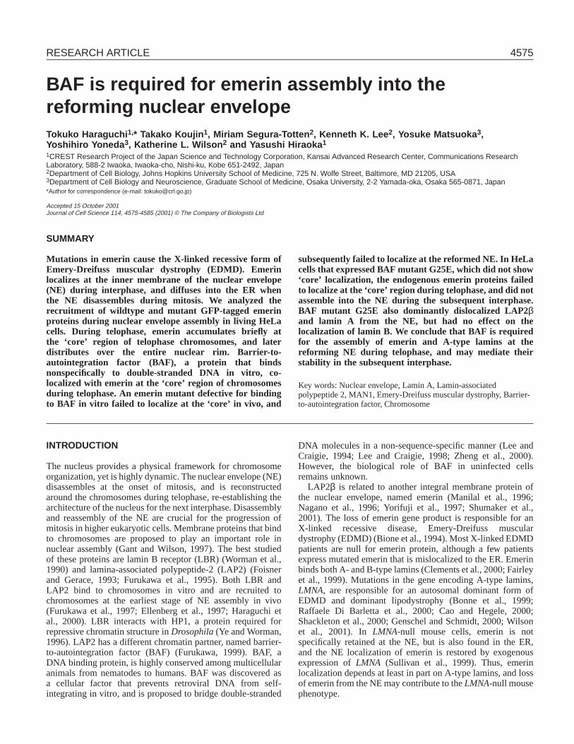
Fig. 1. Intracellular localization of emerin in HeLa cells. (a-c) Emerin stained with the antibody in fixed cells:(a) interphase; (b) metaphase; and(c) telophase stages of mitosis. (d-e) GFP-emerin in living cells: (d) interphase; (e) metaphase through telophase; time-lapse images in (e) wereobtained at one-minute intervals in the same cell. Chromosomes were stained with DAPI in fixed cells, and with Hoechst 33342 in living cells.Bar, 10 µm.
4578
emerin immunolocalized predominantly at the NE (‘rim’staining; Fig. 1a). During metaphase, emerin is dispersed intothe ER network upon NE breakdown (Fig. 1b) and reassemblesaround chromosomes in early telophase (Fig. 1c). Emerin isfirst localized at the central region of telophase chromosomes,near spindle attachment sites (the ‘core’ region; Fig. 1c). Thistelophase specific ‘core’ localization was previously seen inanother human cell type, HEp-2 (Dabauvalle et al., 1999), andis also seen in normal human fibroblast WI38 cells (T.Haraguchi, unpublished), suggesting that ‘core’ localization isa general property of emerin in dividing cells.
To examine the dynamic behavior of emerin in living cells,GFP-emerin was transiently expressed in living HeLa cells andmonitored by time-lapse fluorescence microscopy. Cellsexpressing GFP-emerin were double-labeled for chromosomesusing the DNA-specific fluorescent dye, Hoechst 33342. GFP-emerin was localized at the NE during interphase (Fig. 1d) andthe majority was dispersed into the ER upon NE disassembly(Fig. 1e, 0 minutes), as expected. The GFP-emerin reappearedon the assembling NE during early telophase (4 minutes afterthe metaphase-anaphase transition in Fig. 1e) and thenspecifically localized to the ‘core’ region of the telophasechromosome mass (Fig. 1e, 5 minutes) for 3-4 minutes, beforebecoming uniformly distributed in the NE (Fig. 1e, 8 minutes).This dynamic behavior of GFP-emerin was consistent with thatof endogenous emerin, demonstrating that the fusion to GFPdid not disrupt emerin dynamics.
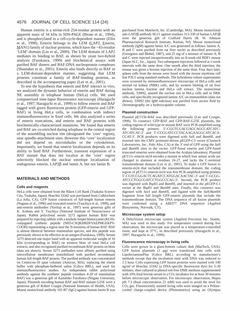
Since the ‘core’ region is close to the spindle attaching sites(see Fig. 7 in Haraguchi et al.) (Haraguchi et al., 2000), wetested whether microtubules mediatedthe ‘core’ localization. Surprisingly,treatment with nocodazole orvinblastin, under conditions thatdepolymerize microtubules and mitoticspindles in living HeLa cells(Haraguchi et al., 1997), did not disruptthe ‘core’ localization of emerin (Fig.2). This result suggested that ‘core’localization is independent ofmicrotubules and microtubule-mediatedevents, and therefore might reflecta chromosome-based structure.Treatment with cytochalasin D, anactin-depolymerizing agent, also had noeffect on ‘core’ localization (notshown), suggesting that the ‘core’structure was also independent of actin.
The LEM-domain is necessaryand sufficient for emerinlocalization at the ‘core’To identify regions in emerin responsiblefor its ‘core’ localization, we expresseda series of truncated emerins as GFP-fusion proteins in HeLa cells. Eachtruncation removed residues from the N-terminus (∆1-64, ∆1-106, ∆1-133, ∆1-155, ∆1-175 and ∆1-222) or C-terminus(∆226-254, ∆175-254, ∆109-254, ∆66-254 and ∆38-254) (Tsuchiya et al.,1999), as shown in Fig. 3a.
First, we determined the region needed for ‘core’localization. The GFP-emerin∆66-254 construct, which hasonly the N-terminal 65 residues (including the LEM domain),localized correctly at the ‘core’ region of telophasechromosomes (Fig. 3c-e), but failed to localize at theinterphase NE (Fig. 3b). All other constructs that includedresidues 1-65 also showed the ‘core’ localization, whereas theshortest construct, containing N-terminal residues 1-37 (∆38-254) did not (Fig. 3a). In striking accordance, the GFP-emerin∆1-64 construct, which lacks residues 1-64, did notlocalize at the ‘core’ region (Fig. 3g) although it did localizeat the NE (Fig. 3f). A point mutation of the leucine residue atposition 64 to phenylalanine also weakened the ‘core’localization of emerin (data not shown), consistent withevidence that residues outside the LEM domain can influencebinding to BAF (Shumaker et al., 2001). These results showedthat the N-terminal 65 residues, which include the LEM-domain, were both necessary and sufficient for the ‘core’localization of emerin. We concluded that ‘core’ localizationconstitutes an in vivo assay for emerin binding to BAF, sinceBAF is also present in the ‘core’ (see below), and becauseemerin residues 1-65 include the LEM domain, which directlymediates binding to BAF (residues 1-43) (Shumaker et al.,2001; Cai et al., 2001; Wolff et al., 2001; Lee et al., 2001).
Östlund et al. previously showed that the nucleoplasmicdomain of emerin is necessary and sufficient for its nuclearenvelope localization (Östlund et al., 1999). To dissect thenuclear envelope targeting domains in relation to the ‘core’binding domain, we expressed and localized each GFP-emerin
JOURNAL OF CELL SCIENCE 114 (24)
Fig. 2.Emerin ‘core’ localization does not depend on microtubules. Cells treated withnocodazole or vinblastine were fixed and stained with antibodies against endogenous emerin.Bar, 10 µm.

4579Emerin-BAF interaction in mitosis
construct during interphase. As shown in Fig. 3b, GFP-emerin∆66-254, which contains a putative bipartite nuclearlocalization signal (NLS) at residues 31-47, did not localize atthe NE. By contrast, the GFP-emerin∆1-64 construct, whichlacks the putative NLS, did localize at the NE (Fig. 3f). Thus,this putative NLS is not necessary for nuclear membranelocalization when emerin is expressed during interphase. By
contrast, GFP-emerin∆226-254, which lacksthe transmembrane domain and the C-terminaltail, localized mostly inside the nucleus, asreported previously (residues 3-228) (Östlundet al., 1999). However, this protein alsounexpectedly localized at the NE duringinterphase, albeit less prominently (Fig. 3h-j),and still localized at the ‘core’ region duringtelophase (Fig. 3k), consistent with having anintact LEM-domain. The weak NE localizationof GFP-emerin∆226-254 was totally lost inGFP-emerin∆174-254 (Fig. 3l-m), confirmingthat residues 174-225 can mediate NElocalization to some extent in the absence of thetransmembrane domain (Östlund et al., 1999).Altogether, our results suggest that emerin mayhave affinities for multiple partners at the innernuclear membrane. Emerin residues 174-225are not required to bind lamins in vitro, andare instead proposed to interact with anunidentified partner at the nuclear envelope(Lee et al., 2001).
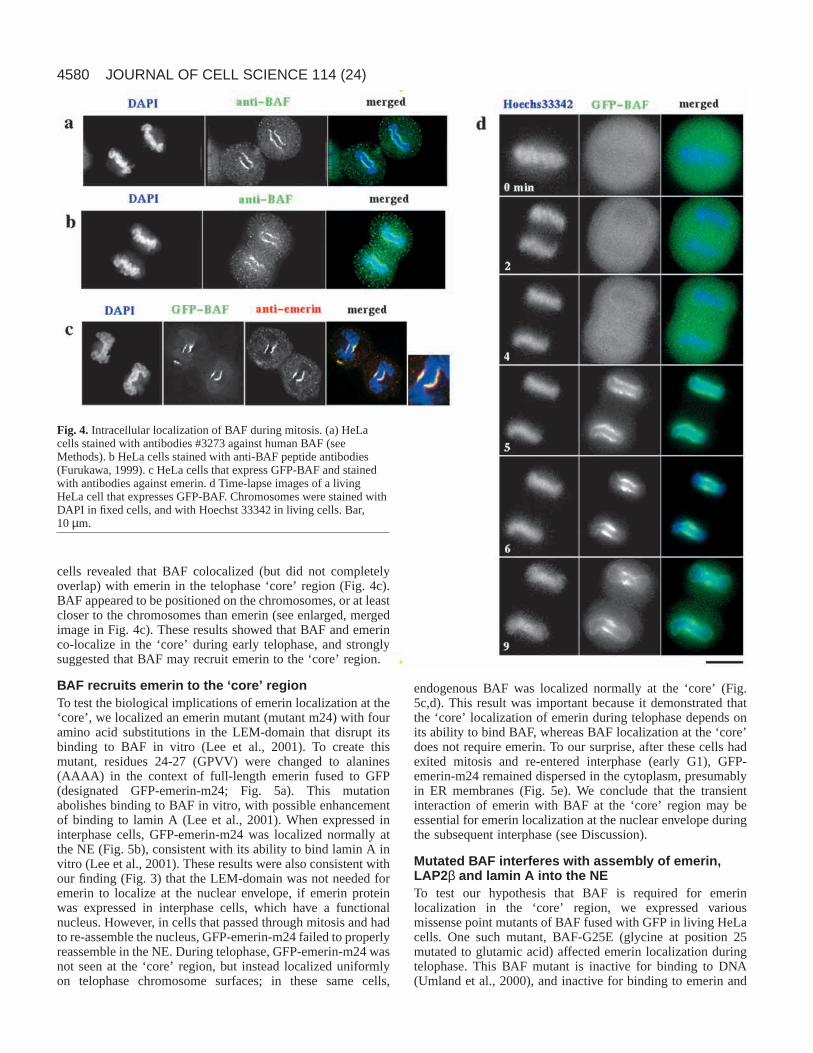
BAF accumulates at the telophase‘core’Emerin interacts directly with BAF through theLEM domain (Lee et al., 2001) and the aboveresults showed that the LEM-domain of emerinis required for its ‘core’ localization. Wetherefore tested the hypothesis that BAF itselflocalizes at the ‘core’ region, by using indirectimmunofluorescence to localize endogenousBAF during mitosis. Our hypothesis wassupported by two independent antisera againstBAF, both of which stained the ‘core’ region inearly telophase chromosomes (Fig. 4a,b).
To further understand the dynamic behaviorof BAF, we transiently expressed a GFP-BAFfusion protein in living HeLa cells, andlocalized BAF during the cell cycle. As shownin Fig. 4d, GFP-BAF was diffusely non-
localized during metaphase and early anaphase, but thendramatically localized at the ‘core’ region for a limited timeduring telophase – specifically, from 5 minutes to 9 minutesafter the metaphase-anaphase transition. By 9 minutes after themetaphase-anaphase transition, some GFP-BAF staining hadbegun to disperse into the cytoplasm. Simultaneous detectionof GFP-BAF and emerin (with anti-emerin antibodies) in fixed
Fig. 3.Molecular domains of emerin necessary forits localization. (a) Molecular structure of emerinand its truncations. GFP was fused to their C-terminus. Localization of each truncation at the NEand the ‘core’ is summarized: N-terminal 65residues are necessary and sufficient for the ‘core’localization of emerin; C-terminal domain isrequired for nuclear membrane localization.Examples are shown for ∆66-254 (b-e), for ∆1-64(f,g), for ∆226-254 (h-k), and for ∆175-254 (l-o). In(g) time-lapse images at 1 minute intervals areshown. Bar, 10 µm.
4580
cells revealed that BAF colocalized (but did not completelyoverlap) with emerin in the telophase ‘core’ region (Fig. 4c).BAF appeared to be positioned on the chromosomes, or at leastcloser to the chromosomes than emerin (see enlarged, mergedimage in Fig. 4c). These results showed that BAF and emerinco-localize in the ‘core’ during early telophase, and stronglysuggested that BAF may recruit emerin to the ‘core’ region.
BAF recruits emerin to the ‘core’ regionTo test the biological implications of emerin localization at the‘core’, we localized an emerin mutant (mutant m24) with fouramino acid substitutions in the LEM-domain that disrupt itsbinding to BAF in vitro (Lee et al., 2001). To create thismutant, residues 24-27 (GPVV) were changed to alanines(AAAA) in the context of full-length emerin fused to GFP(designated GFP-emerin-m24; Fig. 5a). This mutationabolishes binding to BAF in vitro, with possible enhancementof binding to lamin A (Lee et al., 2001). When expressed ininterphase cells, GFP-emerin-m24 was localized normally atthe NE (Fig. 5b), consistent with its ability to bind lamin A invitro (Lee et al., 2001). These results were also consistent withour finding (Fig. 3) that the LEM-domain was not needed foremerin to localize at the nuclear envelope, if emerin proteinwas expressed in interphase cells, which have a functionalnucleus. However, in cells that passed through mitosis and hadto re-assemble the nucleus, GFP-emerin-m24 failed to properlyreassemble in the NE. During telophase, GFP-emerin-m24 wasnot seen at the ‘core’ region, but instead localized uniformlyon telophase chromosome surfaces; in these same cells,
endogenous BAF was localized normally at the ‘core’ (Fig.5c,d). This result was important because it demonstrated thatthe ‘core’ localization of emerin during telophase depends onits ability to bind BAF, whereas BAF localization at the ‘core’does not require emerin. To our surprise, after these cells hadexited mitosis and re-entered interphase (early G1), GFP-emerin-m24 remained dispersed in the cytoplasm, presumablyin ER membranes (Fig. 5e). We conclude that the transientinteraction of emerin with BAF at the ‘core’ region may beessential for emerin localization at the nuclear envelope duringthe subsequent interphase (see Discussion).
Mutated BAF interferes with assembly of emerin,LAP2β and lamin A into the NETo test our hypothesis that BAF is required for emerinlocalization in the ‘core’ region, we expressed variousmissense point mutants of BAF fused with GFP in living HeLacells. One such mutant, BAF-G25E (glycine at position 25mutated to glutamic acid) affected emerin localization duringtelophase. This BAF mutant is inactive for binding to DNA(Umland et al., 2000), and inactive for binding to emerin and
JOURNAL OF CELL SCIENCE 114 (24)
Fig. 4. Intracellular localization of BAF during mitosis. (a) HeLacells stained with antibodies #3273 against human BAF (seeMethods). b HeLa cells stained with anti-BAF peptide antibodies(Furukawa, 1999). c HeLa cells that express GFP-BAF and stainedwith antibodies against emerin. d Time-lapse images of a livingHeLa cell that expresses GFP-BAF. Chromosomes were stained withDAPI in fixed cells, and with Hoechst 33342 in living cells. Bar,10µm.

4581Emerin-BAF interaction in mitosis
LAP2 in vitro (M.S.-T. and K.L.W., unpublished), but mightretain the ability to interact with BAF-DNA complexes. WhenGFP-BAF-G25E was transiently expressed in living cells, the‘core’ localization of endogenous emerin at telophase wasabolished (Fig. 6a; compare with Fig. 1c) even though GFP-BAF-G25E itself did not localize at the ‘core’ region (Fig. 6a).In addition, expression of GFP-BAF-G25E prevented emerinfrom localizing at the NE at a later stage of mitosis (Fig. 6b)and during the ensuing interphase (indicated by an arrow inFig. 6c), whereas normal NE localization was seen in non-expressing cells (Fig. 6d; arrowheads in Fig. 6c). Controlsshowed that emerin localized normally in cells thatexpressed GFP fused to wildtype BAF (Fig. 6e).
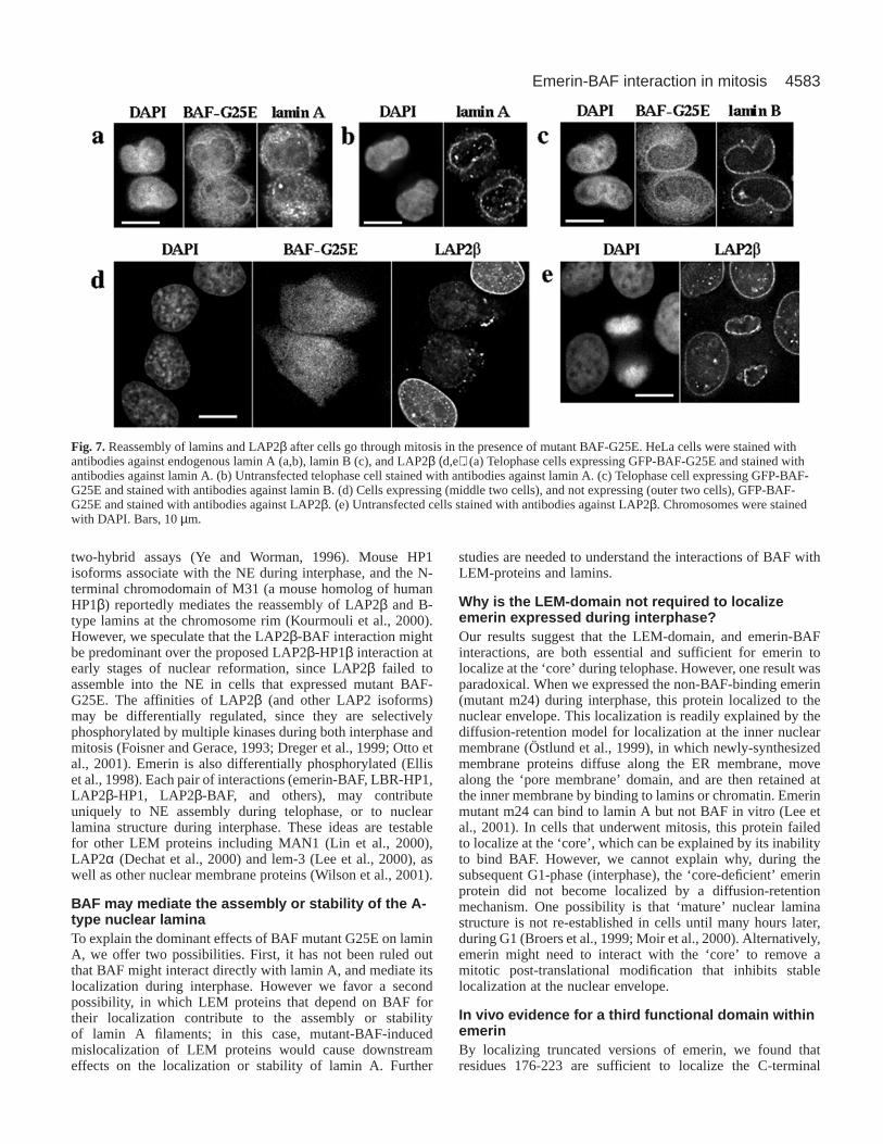
To test the possibility that mutant BAF G25E might alsodisrupt other nuclear proteins, we localized lamin A, laminB and LAP2β in cells that went through mitosis in thepresence of GFP-BAF-G25E (Fig. 7). We first compared thetiming of lamin A assembly in HeLa cells that were eitheruntransfected (Fig. 7b), or transfected with the BAF mutant(Fig. 7a). Untransfected controls were important becausemost lamin A is imported into nuclei after the nascentnuclear envelope has assembled; thus, the lamin A signal atearly times during nuclear envelope assembly can be weakor variable (Gant and Wilson, 1997; Broers et al., 1999;Moir et al., 2000). We found that the accumulation of laminA at reforming nuclear envelopes was significantly retardedin HeLa cells that expressed GFP-BAF-G25E, with mostlamin A remaining in the cytoplasm (Fig. 7a), compared tountransfected cells (Fig. 7b). Importantly, GFP-BAF-G25Ehad no detectable effect on the localization of lamin B (Fig.7c). However, the other LEM-domain protein tested,LAP2β, also failed to localize at the reforming nuclearenvelope in cells that had passed through mitosis in thepresence of GFP-BAF-G25E, and often formed smallpunctate aggregates in the cytoplasm (Fig. 7d). Emerin alsotended to form cytoplasmic aggregates in the presence ofGFP-BAF-G25E (Fig. 6b), and colocalized with the LAP2βaggregates (data not shown). Such aggregates may representthe ER because it has been reported that emerin accumulatesas aggregates within the ER when lamin A/C is eliminatedfrom the NE (Vaughan et al., 2001). Consistent withprevious reports (Foisner and Gerace, 1993), ouruntransfected controls showed rapid accumulation ofLAP2β at reforming nuclear envelopes during telophase(Fig. 7e; also note the two non-expressing cells in Fig. 7d).These results suggested that BAF may specifically recruitand mediate the nuclear envelope assembly of at least twoLEM-domain proteins, emerin and LAP2β, and theassembly of lamin A, but not lamin B, in vivo. In addition,we found that nuclear transport activity was not affected innuclei reformed in the presence of GFP-BAF-G25E asdetected by an NLS-conjugated fluorescent protein (data notshown), suggesting that nuclear transport machinery, suchas nuclear pore complex, is assembled normally into the NEin those cells.
DISCUSSION
Emerin and BAF are conserved among multicellulareukaryotes, from C. elegansto H. sapiens(Cai et al., 1998;
Cohen et al., 2001), suggesting that these proteins havefundamental biological roles. Both proteins (and lamins) areabsent from single-cell eukaryotes such as yeast (Cohen et al.,2001), suggesting that emerin and BAF contribute uniquely tonuclear architecture or function in ‘higher’ eukaryotes. Onesuch function can be NE reassembly during mitosis, becausedisassembly and reassembly of the NE are major events uniqueto ‘higher’ eukaryotes. Emerin accumulates transiently in a
Fig. 5.Localization of GFP-fused mutant emerin-m24. (a) Molecularstructure of GFP-fusion to mutant emerin-m24. (b) Localization of GFP-emerin-m24 in an interphase HeLa cell. (c-e) Localization of GFP-emerin-m24 and endogenous BAF in HeLa cells expressing the mutantemerin. GFP-tagged emerin-m24, and anti-BAF staining are shown in c,telophase in d and the next interphase in e. Chromosomes were stainedwith DAPI.

4582
specific region, designated the ‘core’ region, of the telophasechromosome rim in HeLa cells (Haraguchi et al., 2000) as wellas in human HEp-2 cells (Dabauvalle et al., 1999). We nowfind that emerin localization at the ‘core’ requires its BAF-binding region, and depends on BAF’s localization at the‘core’. Mutant BAF-G25E, which does not localize at the‘core’, dominantly disrupts the NE assembly of emerin, LAP2βand lamin A, but not lamin B, in the subsequent interphase.Thus, the BAF-dependent ‘core’ structure during earlytelophase is important for re-establishment of nuclear laminastructure. These findings have important implications. Wepropose that BAF, lamin A, emerin and other LEM domainproteins may constitute a discrete structural element of thenuclear lamina, since lamin B was sheltered from the dominanteffect of the BAF mutant. We also propose that BAF may bea structural element of the NE-chromatin interface, since BAFcan theoretically anchor all LEM proteins and their attachedlamin filaments to chromatin. Finally, our results suggest that
telophase interactions between emerin andBAF at the ‘core’ may define a novel earlyphase in the assembly of A-type lamins.
The ‘core’ region: a BAF-dependentchromatin structureThe ‘core’ region is close to the spindleattaching sites (Haraguchi et al., 2000).Despite this proximity to kinetochores, ourevidence strongly suggests that the ‘core’region is independent of the cytoskeleton,because it was insensitive todepolymerization of microtubules and actinfilaments. We propose that the ‘core’ regionrepresents a previously unrecognized BAF-dependent chromatin structure that is distinctfrom centromeres and telomeres, and whichmediates the reassembly of specific elementsof the reforming NE. These elements includelamin A, emerin and probably other LEMproteins, and exclude lamin B and possiblyLBR (see below). This idea is based on thedominant effects of mutant BAF-G25E,which interfered with the assembly orretention of endogenous emerin, lamin Aand LAP2β (but not lamin B) at the NE incells that went through mitosis. We think that
chromosome surfaces outside the ‘core’ might mediateattachments between B-type lamins and their other bindingpartner(s), since LBR (which binds B-type lamins) is foundoutside the ‘core’ (Haraguchi et al., 2000). Other results alsosupport this model. For example, A- and B-type lamins havedistinct pathways of assembly during interphase (Moir et al.,2000; Steen and Collas, 2001) which might mirror distinctpathways of assembly during nuclear reformation.
NE-chromatin interactions during nuclearreformationChromatin binding by nuclear membrane proteins is anessential first step in NE assembly. Our results suggest a modelin which BAF may selectively recruit membrane proteins to adefined surface or structure on telophase chromatin. Otherchromatin proteins that interact with NE proteins mayalso have roles in membrane recruitment or attachment.Heterochromatin protein (HP) 1 interacts with LBR in yeast
JOURNAL OF CELL SCIENCE 114 (24)
Fig. 6.Emerin does not accumulate at nuclearenvelopes reassembled in the presence of mutantBAF-G25E. HeLa cells were stained withantibodies against endogenous emerin. (a) Atelophase cell expressing GFP-BAF-G25E.(b) Cells expressing GFP-BAF immediately aftercytokinesis. (c) An interphase cell that wentthrough mitosis in the presence of GFP-BAF-G25E is indicated by the arrow; cells with noexpression of GFP-BAF-G25E are indicated bythe arrowhead. (d) A late telophase cell with noexpression of GFP-BAF-G25E observed in thesame culture dish as for b. (e) HeLa cellsexpressing GFP-BAF (wildtype) shortly aftercytokinesis. Chromosomes were stained withDAPI. Bars, 10 µm.
4583Emerin-BAF interaction in mitosis
two-hybrid assays (Ye and Worman, 1996). Mouse HP1isoforms associate with the NE during interphase, and the N-terminal chromodomain of M31 (a mouse homolog of humanHP1β) reportedly mediates the reassembly of LAP2β and B-type lamins at the chromosome rim (Kourmouli et al., 2000).However, we speculate that the LAP2β-BAF interaction mightbe predominant over the proposed LAP2β-HP1β interaction atearly stages of nuclear reformation, since LAP2β failed toassemble into the NE in cells that expressed mutant BAF-G25E. The affinities of LAP2β (and other LAP2 isoforms)may be differentially regulated, since they are selectivelyphosphorylated by multiple kinases during both interphase andmitosis (Foisner and Gerace, 1993; Dreger et al., 1999; Otto etal., 2001). Emerin is also differentially phosphorylated (Elliset al., 1998). Each pair of interactions (emerin-BAF, LBR-HP1,LAP2β-HP1, LAP2β-BAF, and others), may contributeuniquely to NE assembly during telophase, or to nuclearlamina structure during interphase. These ideas are testablefor other LEM proteins including MAN1 (Lin et al., 2000),LAP2α (Dechat et al., 2000) and lem-3 (Lee et al., 2000), aswell as other nuclear membrane proteins (Wilson et al., 2001).
BAF may mediate the assembly or stability of the A-type nuclear lamina To explain the dominant effects of BAF mutant G25E on laminA, we offer two possibilities. First, it has not been ruled outthat BAF might interact directly with lamin A, and mediate itslocalization during interphase. However we favor a secondpossibility, in which LEM proteins that depend on BAF fortheir localization contribute to the assembly or stabilityof lamin A filaments; in this case, mutant-BAF-inducedmislocalization of LEM proteins would cause downstreameffects on the localization or stability of lamin A. Further
studies are needed to understand the interactions of BAF withLEM-proteins and lamins.
Why is the LEM-domain not required to localizeemerin expressed during interphase?Our results suggest that the LEM-domain, and emerin-BAFinteractions, are both essential and sufficient for emerin tolocalize at the ‘core’ during telophase. However, one result wasparadoxical. When we expressed the non-BAF-binding emerin(mutant m24) during interphase, this protein localized to thenuclear envelope. This localization is readily explained by thediffusion-retention model for localization at the inner nuclearmembrane (Östlund et al., 1999), in which newly-synthesizedmembrane proteins diffuse along the ER membrane, movealong the ‘pore membrane’ domain, and are then retained atthe inner membrane by binding to lamins or chromatin. Emerinmutant m24 can bind to lamin A but not BAF in vitro (Lee etal., 2001). In cells that underwent mitosis, this protein failedto localize at the ‘core’, which can be explained by its inabilityto bind BAF. However, we cannot explain why, during thesubsequent G1-phase (interphase), the ‘core-deficient’ emerinprotein did not become localized by a diffusion-retentionmechanism. One possibility is that ‘mature’ nuclear laminastructure is not re-established in cells until many hours later,during G1 (Broers et al., 1999; Moir et al., 2000). Alternatively,emerin might need to interact with the ‘core’ to remove amitotic post-translational modification that inhibits stablelocalization at the nuclear envelope.
In vivo evidence for a third functional domain withinemerinBy localizing truncated versions of emerin, we found thatresidues 176-223 are sufficient to localize the C-terminal
Fig. 7.Reassembly of lamins and LAP2β after cells go through mitosis in the presence of mutant BAF-G25E. HeLa cells were stained withantibodies against endogenous lamin A (a,b), lamin B (c), and LAP2β (d,e). (a) Telophase cells expressing GFP-BAF-G25E and stained withantibodies against lamin A. (b) Untransfected telophase cell stained with antibodies against lamin A. (c) Telophase cell expressing GFP-BAF-G25E and stained with antibodies against lamin B. (d) Cells expressing (middle two cells), and not expressing (outer two cells), GFP-BAF-G25E and stained with antibodies against LAP2β. (e) Untransfected cells stained with antibodies against LAP2β. Chromosomes were stainedwith DAPI. Bars, 10 µm.
4584
region of emerin at the nuclear envelope. Mutations in thisregion do not interfere with emerin binding to either lamin Aor BAF in vitro (Lee et al., 2001). Because inner membranelocalization is thought to depend on binding to a stablecomponent within the nucleus, such as lamins (Östlund et al.,1999; Fairley et al., 1999; Vaughan et al., 2001), we proposethat residues 176-223 interact with a novel binding partner.This possibility will be tested in future work.
We would like to thank Y. Tsuchiya and K. Arahata for emerinconstructs, R. Craigie for BAF constructs, K. Furukawa for the BAFantibody, C. A. Harris for the LAP2β antibody, and the Riken CellBank for HeLa cells. We also thank H. Tanaka, T. Irisawa, and M.Moto-ori for their help in hybridoma production. This work wassupported by grants from the Human Frontier Science Program (toK.L.W. and Y.H.), the Japan Science and Technology Corporation(CREST; to Y.H.), Grant-in-Aid for Scientific Research B (to Y.Y.,T.H. and Y.H.), and National Institutes of Health grant GM48646 (toK.L.W.). This paper is dedicated to the memory and achievements ofDr Kiichi Arahata, a pioneer of EDMD studies.
REFERENCES
Bione, S., Maestrini, E., Rivella, S., Mancini, M., Regis, S., Romeo, G. andToniolo, D. (1994). Identification of a novel X-linked gene responsible forEmery-Dreifuss muscular dystrophy. Nat. Genet. 8, 323-327.
Bonne, G., Di Barletta, M. R., Varnous, S., Becane, H. M., Hammouda, E.H., Merlini, L., Muntoni, F., Greenberg, C. R., Gary, F., Urtizberea, J.A. et al. (1999). Mutations in the gene encoding lamin A/C cause autosomaldominant Emery- Dreifuss muscular dystrophy. Nat. Genet. 21, 285-288.
Broers, J. L. V., Machiels, B. M., van Eys, G. J. J. M., Kuijpers, H. J. H.,Manders, E. M. M., van Driel, R. and Ramaekers, F. C. S. (1999).Dynamics of the nuclear lamina as monitored by GFP-tagged A-type lamins.J. Cell Sci. 112, 3463-3475.
Cai, M., Huang, Y., Zheng, R., Wei, S. Q., Ghirlando, R., Lee, M. S.,Craigie, R., Gronenborn, A. M. and Clore, G. M. (1998). Solutionstructure of the cellular factor BAF responsible for protecting retroviralDNA from autointegration. Nat. Struct. Biol. 5, 903-909.
Cai, M., Huang, Y., Ghirlando, R., Wilson, K. L., Craigie, R. and Clore,G. M. (2001). Solution structure of the constant region of nuclear envelopeprotein LAP2 reveals two LEM-domain structures: one binds BAF and theother binds DNA. EMBO J. 20, 4399-4407.
Cao, H. and Hegele, R. A. (2000). Nuclear lamin A/C R482Q mutation inCanadian kindreds with Dunnigan-type familial partial lipodystrophy. Hum.Mol. Genet. 9, 109-112.
Clements, L., Manilal, S., Love, D. R. and Morris, G. E. (2000). Directinteraction between emerin and lamin A. Biochem. Biophys. Res. Commun.267, 709-714.
Cohen, C., Lee, K. K., Wilson, K. L. and Gruenbaum, Y. (2001).Transcriptional repression, apoptosis, human disease and the functionalevolution of the nuclear lamina. Trends Biochem. Sci.26, 41-47.
Dabauvalle, M. C., Muller, E., Ewald, A., Kress, W., Krohne, G. andMuller, C. R. (1999). Distribution of emerin during the cell cycle. Eur. J.Cell Biol. 78, 749-756.
Dechat, T., Vlcek, S. and Foisner, R. (2000). Review: lamina-associatedpolypeptide 2 isoforms and related proteins in cell cycle-dependent nuclearstructure dynamics. J. Struct. Biol. 129, 335-345.
Dreger, M., Otto, H., Neubauer, G., Mann, M. and Hucho, F. (1999).Identification of phosphorylation sites in native lamina-associatedpolypeptide 2 beta. Biochemistry38, 9426-9434.
Ellenberg, J., Siggia, E. D., Moreira, J. E., Smith, C. L., Presley, J. F.,Worman, H. J. and Lippincott-Schwartz, J. (1997). Nuclear membranedynamics and reassembly in living cells: targeting of an inner nuclearmembrane protein in interphase and mitosis. J. Cell Biol. 138, 1193-1206.
Ellis, J. A., Craxton, M., Yates, J. R. and Kendrick-Jones, J. (1998).Aberrant intracellular targeting and cell cycle-dependent phosphorylation ofemerin contribute to the Emery-Dreifuss muscular dystrophy phenotype. J.Cell Sci. 111, 781-792.
Fairley, E. A., Kendrick-Jones, J. and Ellis, J. A. (1999). The Emery-
Dreifuss muscular dystrophy phenotype arises from aberrant targeting andbinding of emerin at the inner nuclear membrane. J. Cell Sci. 112, 2571-2582.
Foisner, R. and Gerace, L. (1993). Integral membrane proteins of the nuclearenvelope interact with lamins and chromosomes and binding is modulatedby mitotic phosphorylation. Cell 73, 1267-1279.
Furukawa, K. (1999). LAP2 binding protein 1 (L2BP1/BAF) is a candidatemediator of LAP2-chromatin interaction. J. Cell Sci. 112, 2485-2492.
Furukawa, K., Pante, N., Aebi, U. and Gerace, L. (1995). Cloning of acDNA for lamina-associated polypeptide 2 (LAP2) and identification ofregions that specify targeting to the nuclear envelope. EMBO J. 14, 1626-1636.
Furukawa, K., Glass, C. and Kondo, T. (1997). Characterization of thechromatin binding activity of lamina-associated polypeptide (LAP) 2.Biochem. Biophys. Res. Commun. 238, 240-246.
Gant, T. M. and Wilson, K. L. (1997). Nuclear assembly. Annu. Rev. CellDev. Biol. 13, 669-695.
Genschel, J. and Schmidt, H. H. (2000). Mutations in the LMNA geneencoding lamin A/C. Hum. Mutat. 16, 451-459.
Georgatos, S. D. and Blobel, G. (1987). Lamin B constitutes anintermediate filament attachment site at the nuclear envelope. J. Cell Biol.105, 117-125.
Haraguchi, T., Kaneda, T. and Hiraoka, Y. (1997). Dynamics ofchromosomes and microtubules visualized by multiple-wavelengthfluorescence imaging in living mammalian cells: effects of mitotic inhibitorson cell cycle progression. Genes Cells2, 369-380.
Haraguchi, T., Ding, D. Q., Yamamoto, A., Kaneda, T., Koujin, T. andHiraoka, Y. (1999). Multiple-color fluorescence imaging of chromosomesand microtubules in living cells. Cell Struct. Funct.24, 291-298.
Haraguchi, T., Koujin, T., Hayakawa, T., Kaneda, T., Tsutsumi, C.,Imamoto, N., Akazawa, C., Sukegawa, J., Yoneda, Y. and Hiraoka, Y.(2000). Live fluorescence imaging reveals early recruitment of emerin, LBR,RanBP2, and Nup153 to reforming functional nuclear envelopes. J. Cell Sci.113, 779-794.
Kourmouli, N., Theodoropoulos, P. A., Dialynas, G., Bakou, A., Politou,A. S., Cowell, I. G., Singh, P. B. and Georgatos, S. D. (2000). Dynamicassociations of heterochromatin protein 1 with the nuclear envelope. EMBOJ. 19, 6558-6568.
Lee, M. S. and Craigie, R. (1994). Protection of retroviral DNA fromautointegration: involvement of a cellular factor. Proc. Natl. Acad. Sci. USA.91, 9823-9827.
Lee, M. S. and Craigie, R. (1998). A previously unidentified host proteinprotects retroviral DNA from autointegration. Proc. Natl. Acad. Sci. USA.95, 1528-1533.
Lee, K. K., Gruenbaum, Y., Spann, P., Liu, J. and Wilson, K. L. (2000).C. elegans nuclear envelope proteins emerin, MAN1, lamin, andnucleoporins reveal unique timing of nuclear envelope breakdown duringmitosis. Mol. Biol. Cell11, 3089-3099.
Lee, K. K., Haraguchi, T., Lee, R. S., Koujin, T., Hiraoka, Y. and Wilson,K. L. (2001). Distinct functional domains in emerin bind lamin A and DNA-bridging protein BAF. J. Cell Sci. 114, 4567-4573.
Lin, F., Blake, D. L., Callebaut, I., Skerjanc, I. S., Holmer, L., McBurney,M. W., Paulin-Levasseur, M. and Worman, H. J. (2000). MAN1, an innernuclear membrane protein that shares the LEM domain with lamina-associated polypeptide 2 and emerin. J. Biol. Chem. 275, 4840-4847.
Manilal, S., Nguyen, T. M., Sewry, C. A. and Morris, G. E. (1996). TheEmery-Dreifuss muscular dystrophy protein, emerin, is a nuclear membraneprotein. Hum. Mol. Genet. 5, 801-808.
Moir, R. D., Yoon, M., Khuon, S. and Goldman, R. D. (2000). Nuclearlamins A and B1. Different pathways of assembly during nuclear envelopeformation in living cells. J. Cell Biol. 151, 1155-1168.
Nagano, A., Koga, R., Ogawa, M., Kurano, Y., Kawada, J., Okada, R.,Hayashi, Y. K., Tsukahara, T. and Arahata, K. (1996). Emerin deficiencyat the nuclear membrane in patients with Emery-Dreifuss musculardystrophy. Nat. Genet. 12, 254-259.
Östlund, C., Ellenberg, J., Hallberg, E., Lippincott-Schwartz, J. andWorman, H. J. (1999). Intracellular trafficking of emerin, the emery-dreifuss muscular dystrophy protein. J. Cell Sci. 112, 1709-1719.
Otto, H., Dreger, M., Bengtsson, L. and Hucho, F. (2001). Identification oftyrosine-phosphorylated proteins associated with the nuclear envelope. Eur.J. Biochem. 268, 420-428.
Raffaele Di Barletta, M., Ricci, E., Galluzzi, G., Tonali, P., Mora, M.,Morandi, L., Romorini, A., Voit, T., Orstavik, K. H., Merlini, L. et al.(2000). Different mutations in the LMNA gene cause autosomal dominant
JOURNAL OF CELL SCIENCE 114 (24)

4585Emerin-BAF interaction in mitosis
and autosomal recessive Emery-Dreifuss muscular dystrophy. Am. J. Hum.Genet. 66, 1407-1412.
Shackleton, S., Lloyd, D. J., Jackson, S. N., Evans, R., Niermeijer, M. F.,Singh, B. M., Schmidt, H., Brabant, G., Kumar, S., Durrington, P. N. etal. (2000). LMNA, encoding lamin A/C, is mutated in partial lipodystrophy.Nat. Genet. 24, 153-156.
Shumaker, D. K., Lee, K. K., Tanhehco, Y. C., Craigie, R. and Wilson,K. L. (2001). LAP2 binds to BAF-DNA complexes: requirement for theLEM-domain and modulation by variable regions. EMBO J. 20, 1754-1764.
Steen, R. L. and Collas, P. (2001). Mistargeting of B-type lamins at the endof mitosis: Implications on cell survival and regulation of lamins A/Cexpression. J. Cell Biol. 153, 621-626.
Sullivan, T., Escalante-Alcalde, D., Bhatt, H., Anver, M., Bhat, N.,Nagashima, K., Stewart, C. L. and Burke, B. (1999). Loss of A-typelamin expression compromises nuclear envelope integrity leading tomuscular dystrophy. J. Cell Biol. 147, 913-920.
Tsuchiya, Y., Hase, A., Ogawa, M., Yorifuji, H. and Arahata, K. (1999).Distinct regions specify the nuclear membrane targeting of emerin, theresponsible protein for Emery-Dreifuss muscular dystrophy. Eur. J.Biochem. 259, 859-865.
Umland, T. C., Wei, S.-Q., Craigie, R. and Davies, R. (2000). Structuralbasis of DNA bridging by barrier-to-autointegration factor. Biochemistry39,9130-9138.
Vaughan, O. A., Alvarez-Reyes, M., Bridger, J. M., Broers, J. L. V.,Ramaekers, F. C. S., Wehnert, M., Morris, G. E., Whitfield, W. G. F.and Hutchison, C. J. (2001). Both emerin and lamin C depend on lamin Afor localization at the nuclear envelope. J. Cell Sci. 114, 2577-2590.
Wilson, K. L., Zastrow, M. and Lee, K. K. (2001). Nuclear lamins anddisease: insights into nuclear infrastructure. Cell 104, 647-650.
Wolff, N., Gilquin, B., Courchay, K., Callebaut, I., Worman, H. J. andZinn-Justin, S. (2001). Structural analysis of emerin, an inner nuclearmembrane proteins mutated in X-linked Emery-Dreifuss musculardystrophy. FEBS Lett. 501, 171-176.
Worman, H. J., Evans, C. D. and Blobel, G. (1990). The lamin B receptorof the nuclear envelope inner membrane: a polytopic protein with eightpotential transmembrane domains. J. Cell Biol. 111, 1535-1542.
Ye, Q. and Worman, H. J. (1996). Interaction between an integral protein ofthe nuclear envelope inner membrane and human chromodomain proteinshomologous to DrosophilaHP1. J. Biol. Chem. 271, 14653-14656.
Yorifuji, H., Tadano, Y., Tsuchiya, Y., Ogawa, M., Goto, K., Umetani, A.,Asaka, Y. and Arahata, K. (1997). Emerin, deficiency of which causesEmery-Dreifuss muscular dystrophy, is localized at the inner nuclearmembrane. Neurogenetics1, 135-140.
Zheng, R., Ghirlando, R., Lee, M. S., Mizuuchi, K., Krause, M. andCraigie, R. (2000). Barrier-to-autointegration factor (BAF) bridges DNA ina discrete, higher-order nucleoprotein complex. Proc. Natl. Acad. Sci. USA97, 8997-9002.