Electrodiagnostic Studies (more than you ever cared to know) Pete Vonderau, MD Physical Medicine and...
47
Electrodiagnosti Electrodiagnosti c Studies c Studies (more than you ever cared to (more than you ever cared to know) know) Pete Vonderau, MD Pete Vonderau, MD Physical Medicine and Physical Medicine and Rehabilitation Rehabilitation THE REHAB DOCTORS THE REHAB DOCTORS
-
Upload
jasper-gilmore -
Category
Documents
-
view
215 -
download
1
Transcript of Electrodiagnostic Studies (more than you ever cared to know) Pete Vonderau, MD Physical Medicine and...

Electrodiagnostic Electrodiagnostic StudiesStudies
(more than you ever cared to (more than you ever cared to know)know)
Pete Vonderau, MDPete Vonderau, MD
Physical Medicine and Physical Medicine and RehabilitationRehabilitation
THE REHAB DOCTORSTHE REHAB DOCTORS

ObjectivesObjectives
Review pertinent Review pertinent neuromusculoskeletal anatomyneuromusculoskeletal anatomy
Explain how nerve conduction Explain how nerve conduction studies and needle examinations studies and needle examinations are performedare performed
Review common indications for Review common indications for NCS/EMG studiesNCS/EMG studies

Electrodiagnostic Electrodiagnostic StudiesStudies
Two parts (often collectively referred to as “EMG”)Two parts (often collectively referred to as “EMG”) Nerve Conduction Studies = Nerve StimulationNerve Conduction Studies = Nerve Stimulation EMG (Electromyography) = Needle ExamEMG (Electromyography) = Needle Exam
Why is it performed?Why is it performed? To evaluate nerve To evaluate nerve function function (assess for nerve or muscle (assess for nerve or muscle
injury)injury) MRI only evaluates structureMRI only evaluates structure
Also for localization of injury, severity, prognosis, ruling Also for localization of injury, severity, prognosis, ruling out other diseaseout other disease
Commonly indicated for peripheral nerve injury Commonly indicated for peripheral nerve injury Performed by physiatrists and neurologistsPerformed by physiatrists and neurologists

Interpreting Interpreting EMG/NCS studies EMG/NCS studies requires a solid requires a solid
understanding of understanding of NeuromusculoskeleNeuromusculoskele
tal Anatomytal Anatomy

Spine AnatomySpine Anatomy Primary motor neuron is the Primary motor neuron is the
anterior horn cellanterior horn cell Resides in the ventral gray matter of Resides in the ventral gray matter of
SCSC Axons exit as motor roots, combine to Axons exit as motor roots, combine to
form peripheral nerves, innervate form peripheral nerves, innervate musclemuscle
Primary sensory neuron is DRGPrimary sensory neuron is DRG Resides in intervertebral foramenResides in intervertebral foramen Accepts sensory input from body via Accepts sensory input from body via
pre-ganglionic fiberspre-ganglionic fibers Spinal nervesSpinal nerves
Composed of ventral (motor) and Composed of ventral (motor) and dorsal (sensory) nerve rootsdorsal (sensory) nerve roots
Divides into dorsal and ventral ramiDivides into dorsal and ventral rami Ventral rami combine to form Ventral rami combine to form
plexusesplexuses

Nerve AnatomyNerve Anatomy Each motor (AHC) or sensory Each motor (AHC) or sensory
neuron (DRG) has one axon neuron (DRG) has one axon (fiber through which (fiber through which impulses are sent)impulses are sent)
Each axon surrounded by Each axon surrounded by Schwann cell (makes myelin)Schwann cell (makes myelin)
Motor axons and large sensory Motor axons and large sensory axons (proprioception) are axons (proprioception) are myelinated (electrically myelinated (electrically insulated)insulated)
Small nerves lack insulation Small nerves lack insulation (pain, temperature, autonomic)(pain, temperature, autonomic)
Axons are bundled into Axons are bundled into fasciclesfascicles
Peripheral Nerve is Peripheral Nerve is composed of many fasciclescomposed of many fascicles

Nerve PhysiologyNerve Physiology
All Cells (including axons of neurons) All Cells (including axons of neurons) maintain an Equilibrium Potentialmaintain an Equilibrium Potential Na/K Pump, cell membrane permeability, and Na/K Pump, cell membrane permeability, and
diffusion potentials work together, resulting in diffusion potentials work together, resulting in the inside of the cell having a negative potential the inside of the cell having a negative potential compared to outside (-70mV)compared to outside (-70mV)

Nerve Physiology: Nerve Physiology: ConductionConduction
Nerves transmit information Nerves transmit information via Action Potentialsvia Action Potentials Electrical stimulation of nerve Electrical stimulation of nerve
causes inside to become more causes inside to become more ++
Threshold reached at which Threshold reached at which voltage-gated Na+ channels voltage-gated Na+ channels open briefly, charge inside ↑ open briefly, charge inside ↑ rapidly (cell depolarizes)rapidly (cell depolarizes)
Na+ flows down nerve to Na+ flows down nerve to propagate the action potential propagate the action potential along nerve (voltage change)along nerve (voltage change)
Then gates close and Then gates close and membrane potential increases membrane potential increases againagain
This AP can be measuredThis AP can be measured

Nerve Physiology: Saltatory Nerve Physiology: Saltatory ConductionConduction
A Schwann cell surrounds A Schwann cell surrounds each axon and can each axon and can myelinate the axonmyelinate the axon
Myelin electrically insulates Myelin electrically insulates motor axons and larger motor axons and larger sensory axons (not small sensory axons (not small sensory fibers)sensory fibers)
Na+ channels are not Na+ channels are not continuous down nerve but continuous down nerve but are clustered at Nodes of are clustered at Nodes of RanvierRanvier
Conduction jumps from Conduction jumps from node to node, which is much node to node, which is much faster than unmyelinated faster than unmyelinated nerves (10-20x)nerves (10-20x)

Motor Nerves: Muscle Motor Nerves: Muscle ContractionContraction
Signal sent from AHC in Signal sent from AHC in spinal cord, down axonspinal cord, down axon
At the Neuromuscular At the Neuromuscular Junction, Ach is releasedJunction, Ach is released
Ach binds muscle Ach binds muscle membrane, opening Na+ membrane, opening Na+ channels, thus channels, thus depolarizing muscle fiber depolarizing muscle fiber membrane (voltage membrane (voltage change)change)
Sarcoplasmic reticulum Sarcoplasmic reticulum releases Ca++, which releases Ca++, which binds troponin to start binds troponin to start muscle contraction via muscle contraction via overlap of actin and overlap of actin and myosinmyosin

Motor UnitMotor Unit Skeletal muscle composed Skeletal muscle composed
of many fasciclesof many fascicles Fascicles contain thousands Fascicles contain thousands
of muscle fibers (muscle of muscle fibers (muscle cells)cells)
Motor UnitMotor Unit Defined as the motor Defined as the motor
neuron, its axon, and the neuron, its axon, and the muscle fibers it innervatesmuscle fibers it innervates
These fibers depolarize These fibers depolarize nearly simultaneously, nearly simultaneously, creating a motor unit creating a motor unit action potential (electric action potential (electric potential)potential)
We can place a needle We can place a needle into the muscle and into the muscle and measure this electric measure this electric potential potential

Sensory NervesSensory Nerves Activated by sensory Activated by sensory
inputinput MechanoreceptorsMechanoreceptors
Light touch, pressure, Light touch, pressure, muscle stretch muscle stretch (proprioception)(proprioception)
Myelinated fibersMyelinated fibers NociceptorsNociceptors
Pain (unmyelinated)Pain (unmyelinated) ThermoreceptorsThermoreceptors
Temperature Temperature (unmyelinated)(unmyelinated)
Action potentials travel Action potentials travel proximally to the DRG, proximally to the DRG, then on to spinal cordthen on to spinal cord

Nerve Nerve Conduction Conduction
StudiesStudies

Nerve StimulationNerve Stimulation We can stimulate a nerve by We can stimulate a nerve by
sending negative charges sending negative charges around the nervearound the nerve Reduces the resting membrane Reduces the resting membrane
potential and cause the action potential and cause the action potential to start anywhere potential to start anywhere along the nervealong the nerve
Motor or sensory nerves can Motor or sensory nerves can be stimulatedbe stimulated Stimulating a motor nerve can Stimulating a motor nerve can
result in muscle contractionresult in muscle contraction Stimulating a sensory nerve Stimulating a sensory nerve
gives a buzzing sensationgives a buzzing sensation We attempt to stimulate all of We attempt to stimulate all of
the axon fibers of the target the axon fibers of the target nervenerve

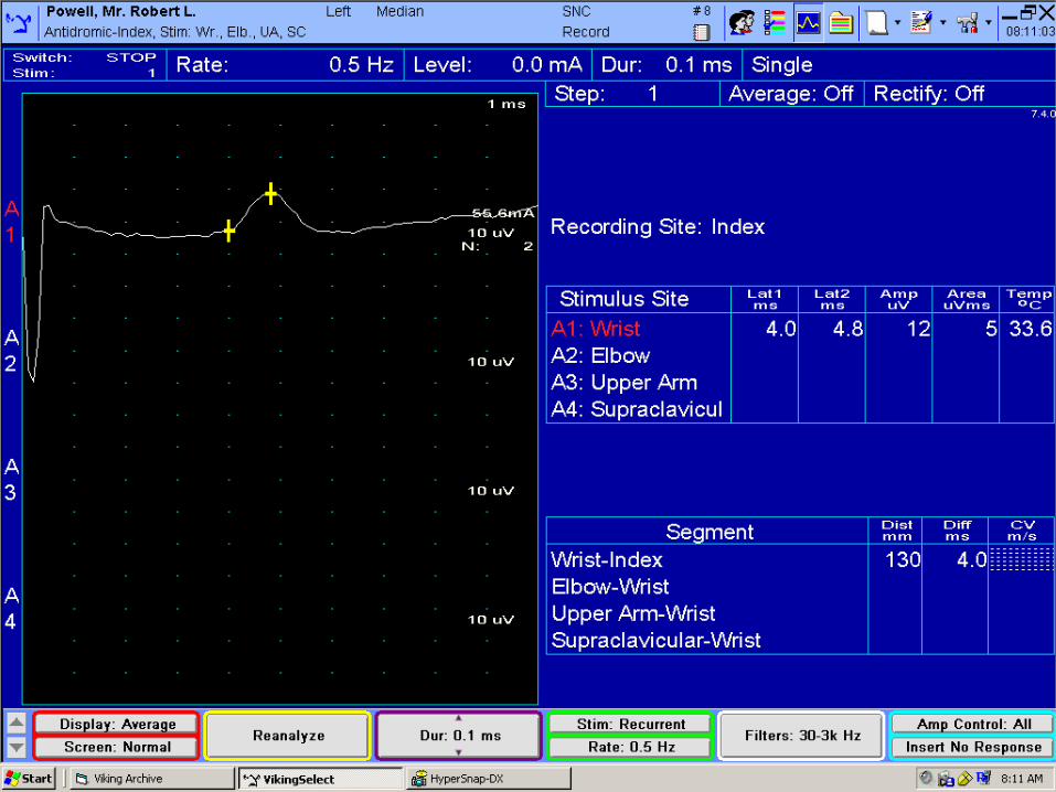
Sensory Nerve Conduction Sensory Nerve Conduction StudiesStudies
We measure the electric We measure the electric potentials farther along the potentials farther along the nerve with skin electrodesnerve with skin electrodes
The computer amplifies the The computer amplifies the potentials and records potentials and records them on the computer them on the computer screenscreen
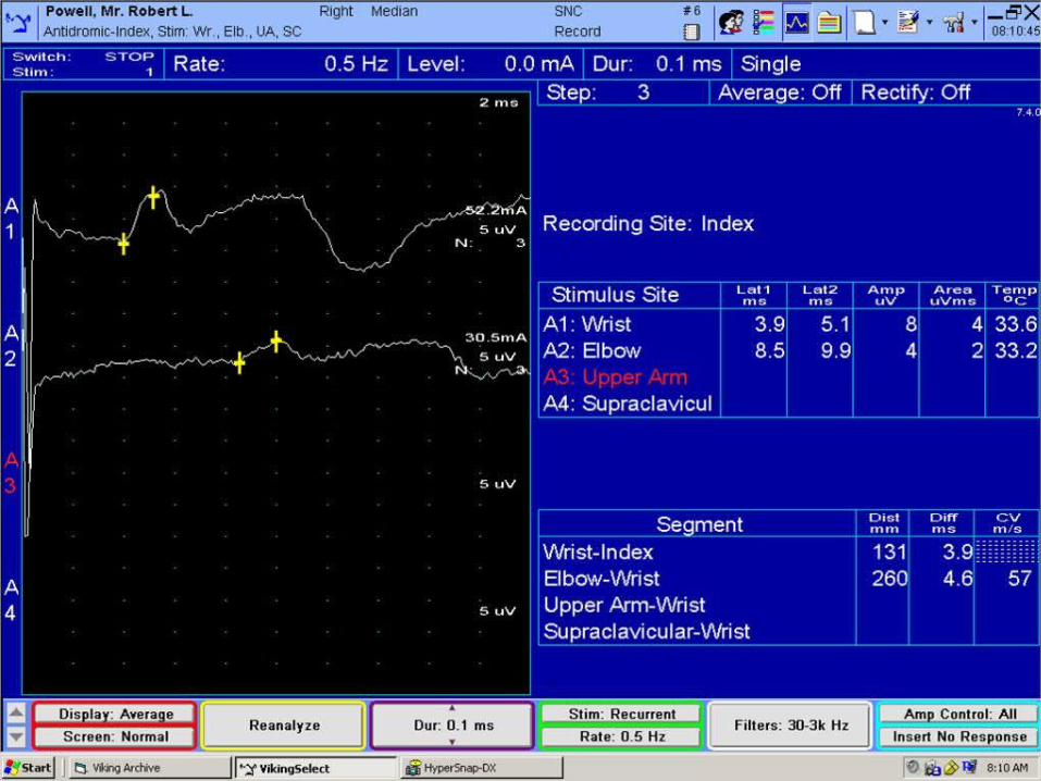
Recorded valuesRecorded values Distal latency (conduction Distal latency (conduction
time over distal limb to time over distal limb to electrodes)electrodes)
Amplitudes (functioning Amplitudes (functioning axons)axons)
Conduction Velocity Conduction Velocity (dist/time)(dist/time)
Compare to normal values Compare to normal values or other limbor other limb

Sensory Nerve Conduction Sensory Nerve Conduction Studies: UsesStudies: Uses
Most sensitive for focal Most sensitive for focal mononeuropathies (CTS)mononeuropathies (CTS) Sensory nerves more Sensory nerves more
prone to injuryprone to injury The only test for pure The only test for pure
sensory cutaneous nerve sensory cutaneous nerve (cheiralgia paresthetica)(cheiralgia paresthetica)
Sensory peripheral Sensory peripheral neuropathyneuropathy
Localization: Separating Localization: Separating pre-DRG from post-DRG pre-DRG from post-DRG injuryinjury Sensory NCS will be Sensory NCS will be
normal if lesion proximal normal if lesion proximal to DRGto DRG

Normal Values (Sensory Normal Values (Sensory NCS)NCS)
Sensory Nerve Distal Latency Amplitude Cond. Veloc.
Median Antidromic (II) <3.6 msec >15 μV >56 m/s
Ulnar Antidromic (V) <3.1 msec >10 μV >54 m/s
Median Palmar <2.3 msec >50 μV >56 m/s
Ulnar Palmar <2.3 msec >15 μV >55 m/s
Diff ≤ 0.3
Dorsal Ulnar Cutaneous <2.6 msec >8 μV ---------
Superficial Radial Antidromic <2.9 msec >20 μV >49 m/s

Motor Nerve Conduction Motor Nerve Conduction StudiesStudies
Stimulate over a motor nerveStimulate over a motor nerve Action Potential travels down Action Potential travels down
motor nerve, depolarizes motor nerve, depolarizes muscle, resulting in a muscle muscle, resulting in a muscle contractioncontraction
The electric potential (mV) The electric potential (mV) from muscle contraction is from muscle contraction is recorded on the skin over the recorded on the skin over the muscle bellymuscle belly
Attempt to activate all the Attempt to activate all the axons of the nerveaxons of the nerve Increase stimulation until Increase stimulation until
amplitude no longer increasesamplitude no longer increases
Motor Nerve Conduction Motor Nerve Conduction Studies: MeasurementsStudies: Measurements
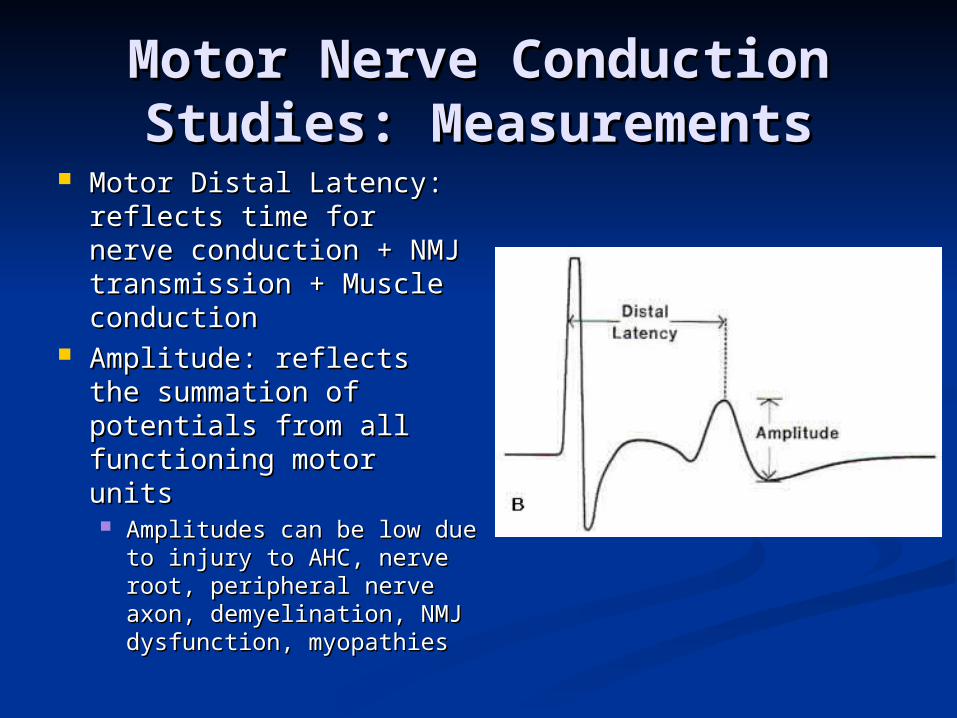
Motor Distal Latency: Motor Distal Latency: reflects time for nerve reflects time for nerve conduction + NMJ conduction + NMJ transmission + Muscle transmission + Muscle conductionconduction
Amplitude: reflects the Amplitude: reflects the summation of potentials summation of potentials from all functioning from all functioning motor unitsmotor units Amplitudes can be low Amplitudes can be low
due to injury to AHC, due to injury to AHC, nerve root, peripheral nerve root, peripheral nerve axon, nerve axon, demyelination, NMJ demyelination, NMJ dysfunction, myopathiesdysfunction, myopathies

Normal Values (Motor Normal Values (Motor NCS)NCS)
Motor Nerve Dist. Lat. Amplitude Cond Velocity
Median (APB) < 4.5 msec >4.0 mV >48 m/s
Ulnar (ADM) <3.6 msec >6.0 mV >51 m/s
Radial (EDC) <3.1 msec --------- >67 m/s
Peroneal (EDB) <6.6 msec >2.0 mV >41 m/s
Tibial (AH) <6.1 msec >4.0 mV >40 m/s


F wavesF waves This is a way of assessing the This is a way of assessing the
proximal motor nerveproximal motor nerve With distal stimulation, 1-2% With distal stimulation, 1-2%
of motor fibers will carry of motor fibers will carry signal proximally to SC, then signal proximally to SC, then back down to muscleback down to muscle
We can measure the latency We can measure the latency (response time)(response time)
Prolonged latency may be Prolonged latency may be due to proximal nerve injury: due to proximal nerve injury: radiculopathy, plexopathy, radiculopathy, plexopathy, GBSGBS
Not a very sensitive testNot a very sensitive test

Pathophysiology: Axonal Pathophysiology: Axonal InjuryInjury
AKA AxonotmesisAKA Axonotmesis ““Defective wires”Defective wires” Individual axons no longer Individual axons no longer
functionfunction Fewer axons remain Fewer axons remain
within each nerve fasciclewithin each nerve fascicle For sensory nerves, there For sensory nerves, there
may be some impairment may be some impairment of sensation (numbness, of sensation (numbness, tingling, absent reflexes)tingling, absent reflexes)
For motor nerves, there For motor nerves, there may be weakness and may be weakness and ATROPHYATROPHY Atrophy due to muscle cells Atrophy due to muscle cells
no longer being innervatedno longer being innervated

Pathophysiology: Axonal Pathophysiology: Axonal InjuryInjury
CausesCauses Nerve trauma or compresssion, Nerve trauma or compresssion,
metabolic disorders (DM), metabolic disorders (DM), congenital disease, etccongenital disease, etc
Nerve Conduction StudiesNerve Conduction Studies Low amplitudes, both proximal and Low amplitudes, both proximal and
distaldistal Amplitude loss proportional to axon Amplitude loss proportional to axon
lossloss Complete nerve trans-section results Complete nerve trans-section results
in no distal responsein no distal response Relatively preserved DL and CVRelatively preserved DL and CV
PrognosisPrognosis GuardedGuarded Intact axons can slowly sprout to Intact axons can slowly sprout to
denervated muscle fibersdenervated muscle fibers

Pathophysiology: Pathophysiology: Demyelinating InjuryDemyelinating Injury
AKA NeurapraxiaAKA Neurapraxia ““Defective insulation”Defective insulation” Axons are intact, but the Axons are intact, but the
myelin sheath (insulation) myelin sheath (insulation) is damagedis damaged
May result in numbness or May result in numbness or tingling (sensory nerves), tingling (sensory nerves), or weakness (motor or weakness (motor nerves) but NO atrophy nerves) but NO atrophy because the axon is still because the axon is still intactintact

Demyelinating Injury: NCSDemyelinating Injury: NCS Stimulating PROXIMAL to injury Stimulating PROXIMAL to injury
reveals slowed CV and prolonged reveals slowed CV and prolonged DLDL Low proximal amplitude if conduction Low proximal amplitude if conduction
block (complete conduction block block (complete conduction block possible=no proximal response)possible=no proximal response)
Stimulating distal to the lesion Stimulating distal to the lesion will elicit a normal responsewill elicit a normal response
If you cannot stimulate proximal If you cannot stimulate proximal to the lesion, NCS will be normal to the lesion, NCS will be normal (radic)(radic)
Causes: compression, ischemia, Causes: compression, ischemia, autoimmune, congenital disordersautoimmune, congenital disorders
Prognosis is good if nerve can be Prognosis is good if nerve can be decompressed (remyelination decompressed (remyelination often occurs over 4-6 weeks)often occurs over 4-6 weeks)

NCS NCS ExamplesExamples
A: NormalA: Normal B: Axonal LossB: Axonal Loss
Low amplitude prox & distal, Low amplitude prox & distal, Normal DLNormal DL
Poor prognosisPoor prognosis C: Uniform DemyelinationC: Uniform Demyelination
Prolonged DL, Slow CV, normal Prolonged DL, Slow CV, normal amplitude (seen in CMT)amplitude (seen in CMT)
D: Focal Demyelination with D: Focal Demyelination with Conduction BlockConduction Block Low amplitude with proximal Low amplitude with proximal
stimulation, slow CV, temporal stimulation, slow CV, temporal dispersiondispersion
Better prognosis than BBetter prognosis than B

Needle ExamNeedle Exam This is done to analyze This is done to analyze
the electrical activity of the electrical activity of voluntary musclevoluntary muscle
A needle electrode is A needle electrode is placed into the muscleplaced into the muscle
Extracellular potentials Extracellular potentials created by muscle created by muscle depolarization are depolarization are recorded, then amplified recorded, then amplified and converted to audible and converted to audible soundsound
Electrical activity is Electrical activity is assessed at rest and assessed at rest and while the muscle is while the muscle is contractedcontracted

Needle Exam: Spontaneous Needle Exam: Spontaneous ActivityActivity
Muscle at rest (not contracting) is at Muscle at rest (not contracting) is at electrochemical equilibriumelectrochemical equilibrium There are no action potentials, so the needle There are no action potentials, so the needle
should record no activity (silence)should record no activity (silence) Axonal Injury (Denervation injury)Axonal Injury (Denervation injury)
Due to injury to AHC, nerve root, peripheral nerveDue to injury to AHC, nerve root, peripheral nerve Muscle fibers that lack innervation will fire Muscle fibers that lack innervation will fire
spontaneously in an regular pattern called a spontaneously in an regular pattern called a fibrillation potential (abnormal)fibrillation potential (abnormal)
Takes 3-4 weeks for these to develop, resolve if sprouting Takes 3-4 weeks for these to develop, resolve if sprouting occursoccurs
Demyelinating disorders will NOT cause fibrillation Demyelinating disorders will NOT cause fibrillation potentialspotentials

Needle Exam: Fibrillation Needle Exam: Fibrillation PotentialsPotentials

Needle Exam: Voluntary Needle Exam: Voluntary ActivityActivity
Ask patient to contract Ask patient to contract musclemuscle
Needle records Needle records extracellular potentials extracellular potentials from one motor unit (all from one motor unit (all fibers innervated by one fibers innervated by one axon), all firing in near axon), all firing in near synchrony near the synchrony near the needle tipneedle tip
Amplified and displayed Amplified and displayed on monitoron monitor

Needle Exam: Voluntary Needle Exam: Voluntary ActivityActivity
Assess the following:Assess the following: Number of units Number of units
firingfiring FrequencyFrequency Duration of waveformDuration of waveform Configuration of Configuration of
waveformwaveform Biphasic, TriphasicBiphasic, Triphasic
PhasesPhases AmplitudeAmplitude RecruitmentRecruitment
Needle: Abnormal Needle: Abnormal Voluntary ActivityVoluntary Activity
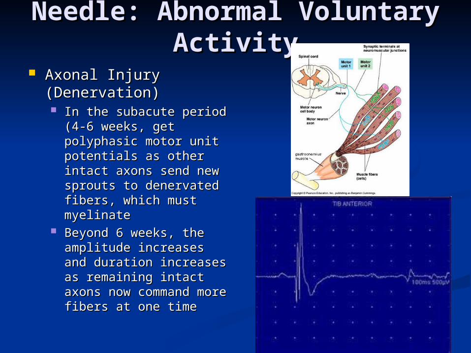
Axonal Injury Axonal Injury (Denervation)(Denervation) In the subacute period In the subacute period
(4-6 weeks, get (4-6 weeks, get polyphasic motor unit polyphasic motor unit potentials as other potentials as other intact axons send new intact axons send new sprouts to denervated sprouts to denervated fibers, which must fibers, which must myelinatemyelinate
Beyond 6 weeks, the Beyond 6 weeks, the amplitude increases amplitude increases and duration increases and duration increases as remaining intact as remaining intact axons now command axons now command more fibers at one timemore fibers at one time

RadiculopathyRadiculopathy This is injury to the spinal nerve This is injury to the spinal nerve
rootroot Many causes, but most common is Many causes, but most common is
nerve impingement by herniated nerve impingement by herniated discdisc Impingement can affect ventral Impingement can affect ventral
(motor) or dorsal (sensory) nerve root, (motor) or dorsal (sensory) nerve root, or bothor both
Routine NCS of Lumbar Radic:Routine NCS of Lumbar Radic: Peroneal and Tibial Motor NCS with FPeroneal and Tibial Motor NCS with F Sural and Peroneal Sensory NCSSural and Peroneal Sensory NCS Needle: Tibialis Anterior, Medial Needle: Tibialis Anterior, Medial
Gastrocnemius, Peroneus Longus, Gastrocnemius, Peroneus Longus, Vastus Medialis, TFL, Gluteus Vastus Medialis, TFL, Gluteus Maximus, Paraspinal musclesMaximus, Paraspinal muscles

RadiculopathyRadiculopathy Nerve Conduction Studies are often normalNerve Conduction Studies are often normal
Sensory studies are normal because the sensory Sensory studies are normal because the sensory nerve is proximal to the DRGnerve is proximal to the DRG
Motor are normal unless there is a severe axonal Motor are normal unless there is a severe axonal injury (because muscles are innervated by multiple injury (because muscles are innervated by multiple roots)roots)
So why do NCS? – to rule out plexus or peripheral So why do NCS? – to rule out plexus or peripheral nerve injurynerve injury
Needle examination is the keyNeedle examination is the key Axonal injury will demonstrate fibrillation potentials Axonal injury will demonstrate fibrillation potentials
after 3-4 weeksafter 3-4 weeks Later, polyphasic motor unit potentials are seen, Later, polyphasic motor unit potentials are seen,
followed by long duration/high amplitude motor followed by long duration/high amplitude motor unitsunits
Diagnosis requires abnormalities in at least 2 Diagnosis requires abnormalities in at least 2 muscles innervated by same root but different muscles innervated by same root but different nervesnerves

Myotomes and DermatomesMyotomes and DermatomesIliopsoas L 2 3 4
Rectus Femoris L 2 3 4
Adductor Longus L 2 3 4
Gracilis L 2 3 4
Vastus Lateralis L 2 3 4
Vastus Medialis L 2 3 4
Tibialis Anterior L 4 5
EHL L 4 5
Peroneus Tertius L 5 1
EDB L 4 5 1
Peroneus Longus L 5 1
Internal Hamstrings (SM/ST) L 4 5 1
External Hamstring (BFLH) L 5 1
External Hamstring (BFSH) L 5
Gluteus Medius L 4 5 1
TFL L 4 5
Gluteus Maximus L 5 1 2
Tibialis Posterior L 5 1
FDL L 5 1
Abductor Hallucis S 1 2
Abductor Digiti Minimi Pedis S 1 2
Gastrocnemius (Medial) L 5 1 2
Gastrocnemius (Lateral) S 1 2
Soleus S 1 2
Reflexes
Patellar L 3 4
Internal Hamstring L 5
Achilles S1

Radiculopathy: CaveatsRadiculopathy: Caveats Pure sensory Pure sensory
radiculopathy will radiculopathy will result in a normal studyresult in a normal study Pre-ganglionic fibers Pre-ganglionic fibers
intactintact Pure demyelinating Pure demyelinating
radiculopathy will radiculopathy will result in a normal studyresult in a normal study
What is the diagnostic What is the diagnostic sensitivity of EMG?sensitivity of EMG? Estimated to be 60-70% Estimated to be 60-70%
based upon AANEM based upon AANEM review of literaturereview of literature

Carpal Tunnel SyndromeCarpal Tunnel Syndrome Injury to the median nerve Injury to the median nerve
within the carpal tunnel of the within the carpal tunnel of the wrist, typically due to wrist, typically due to compressioncompression
Routine StudyRoutine Study Sensory NCS: Median and Ulnar Sensory NCS: Median and Ulnar
antidromics, Palmar studiesantidromics, Palmar studies Motor NCS: Median and Ulnar Motor NCS: Median and Ulnar
Motor with F wavesMotor with F waves Needle Exam: Deltoid, Triceps, Needle Exam: Deltoid, Triceps,
Pronator Teres, FDI, APBPronator Teres, FDI, APB Rule outRule out
ALS, Cervical radiculopathy, ALS, Cervical radiculopathy, brachial plexopathy, proximal brachial plexopathy, proximal median neuropathy, peripheral median neuropathy, peripheral neuropathyneuropathy


Carpal Tunnel Syndrome: Carpal Tunnel Syndrome: ClassificationClassification
Mild CTSMild CTS Prolonged sensory (or palmar) distal latencyProlonged sensory (or palmar) distal latency
Compare median and ulnar palmar DLCompare median and ulnar palmar DL Reduced sensory (or palmar) amplitudeReduced sensory (or palmar) amplitude
Moderate CTSModerate CTS Prolonged motor distal latencyProlonged motor distal latency
Severe CTSSevere CTS Absent sensory responseAbsent sensory response Reduced motor amplitudeReduced motor amplitude Needle evidence of fibrillation potentials or long Needle evidence of fibrillation potentials or long
duration/high amplitude motor unit potentialsduration/high amplitude motor unit potentials

Carpal Tunnel SyndromeCarpal Tunnel Syndrome
What is the diagnostic sensitivity of What is the diagnostic sensitivity of NCS?NCS? Sensory antidromics: 65% sensitivity for Sensory antidromics: 65% sensitivity for
CTSCTS Palmar studies: 74% sensitivity for CTS Palmar studies: 74% sensitivity for CTS
(Mayo)(Mayo)

Summary: What you need to Summary: What you need to knowknow
EMG/NCS is useful to further evaluate EMG/NCS is useful to further evaluate complaints of numbness, tingling, weakness, complaints of numbness, tingling, weakness, and atrophy thought to be related to the and atrophy thought to be related to the peripheral nervous systemperipheral nervous system
IndicationsIndications Peripheral nerve injuryPeripheral nerve injury
Carpal Tunnel Syndrome, Ulnar neuropathy, Peroneal Carpal Tunnel Syndrome, Ulnar neuropathy, Peroneal NeuropathyNeuropathy
Peripheral NeuropathyPeripheral Neuropathy Plexopathy (Brachial, Lumbosacral)Plexopathy (Brachial, Lumbosacral) RadiculopathyRadiculopathy Motor Neuron Disease (ALS)Motor Neuron Disease (ALS) MyopathyMyopathy NMJ Junction Disorders (Academic Centers?)NMJ Junction Disorders (Academic Centers?)

What you need to know What you need to know (Cont)(Cont)
Wait 3-4 weeks after nerve injury before ordering an Wait 3-4 weeks after nerve injury before ordering an EMG testEMG test Fibrillation potentials take time to developFibrillation potentials take time to develop
Anti-coagulation may limit needle examinationAnti-coagulation may limit needle examination Nerve conduction studies are falsely reduced in Nerve conduction studies are falsely reduced in
obese, edematous limbsobese, edematous limbs EMG/NCS does not evaluate injury to the central EMG/NCS does not evaluate injury to the central
nervous system (stroke, spinal cord injury, etc)nervous system (stroke, spinal cord injury, etc) EMG/NCS cannot assess for small fiber peripheral EMG/NCS cannot assess for small fiber peripheral
neuropathies (unmyelinated nerves)neuropathies (unmyelinated nerves) Demyelinating nerve injuries have a much better Demyelinating nerve injuries have a much better
prognosisprognosis Improvement over 4-6 weeks if decompressedImprovement over 4-6 weeks if decompressed
Axonal nerve injuries have a more guarded Axonal nerve injuries have a more guarded prognosisprognosis 1-2 years to see maximum improvement1-2 years to see maximum improvement

My Perspective on My Perspective on EMG/NCSEMG/NCS
A thorough history and A thorough history and comprehensive comprehensive musculoskeletal exam musculoskeletal exam will yield the diagnosis will yield the diagnosis >90% of time>90% of time
EMG is useful in the EMG is useful in the following situationsfollowing situations When the patient’s When the patient’s
presentation is non-presentation is non-physiologic or their effort physiologic or their effort on exam is unreliable on exam is unreliable (EMG is objective)(EMG is objective)
When imaging findings do When imaging findings do not match clinical findingsnot match clinical findings

Thank You!Thank You!