Effects Training the Distribution CardiacOutput Patients...
15
Effects of Training on the Distribution of Cardiac Output in Patients with Coronary Artery Disease By JAN PRAETORIUS CLAUSEN, M.D., AND JENS TRAP-JENSEN, M.D. SUMMARY In nine patients with coronary artery disease, cardiac output distribution was evaluated at rest and during exercise by measurement of cardiac output and regional blood flow parameters (hepatic and muscle blood flow). In seven patients repeated values were obtained after a physical training program of 4 to 10 weeks' duration. After training, cardiac output was reduced at moderate work loads (13.1%) causing a change of the relation between oxygen uptake and cardiac output from hyper- kinetic to normal. During heavy exercise the cardiac output was increased (5.5%) after training. Similar changes were observed in muscle blood flow, which was reduced at submaximal loads (14.9%) and increased at maximal (8.6%). Hepatic blood flow showed in contrast a less pronounced reduction at both work loads after training (difference, 7.2%). These effects of training could be explained as peripheral regulatory alterations without implying primary improvement in myocardial performance. They are consistent with the view that local changes in the trained muscles are important for the reduction in myocardial pressure-work caused by physical conditioning. Additional Indexing Words: Hepatic blood flow Muscle blood flow REVIOUS investigations have suggested that the immediate beneficial effects of physical conditioning on symptoms related to chronic coronary artery disease (CAD) could be explained by hemodynamic alterations which reduce myocardial pressure work.'-3 The finding of a more hypokinetic circulation at a given submaximal work load after training' indicated that redistribution of car- diac output might be of importance for this economizing effect on the heart. The mecha- nism responsible for the observed reduction in From the Department of Clinical Physiology and the Department of Medicine C, Bispebjerg Hospital, Copenhagen, Denmark. Study was supported by a grant from the King Christian X Foundation. Preliminary report of the present investigation has been given at the General Meetings of the Swedish Medical Association in November 1969. Received April 14, 1970; revision accepted for publication June 25, 1970. Circulation, Volume XLII, October 1970 Tension-time index cardiac output was, however, unclear. Some investigators ascribed such decrement to a more effective sympathetic vasoconstriction in the "nonworking" tissues favoring the blood supply to the working muscles.1 4 5 This assumption disagrees, however, with the observation that the perfusion of abdominal viscera is less reduced during exercise in trained subjects as compared to untrained.6' 7 In a previous study we observed that muscle blood flow during exercise was reduced after training.3 Hence we suggested that the reduction in cardiac output after training may be secondary to the reduced flow in the working muscles. In the present investigation the effect of training on the distribution of cardiac output to working as well as "nonwork- ing" tissues was studied. Muscle blood flow and hepatic blood flow were measured in patients with CAD before and after a physical conditioning program. 611 by guest on July 19, 2018 http://circ.ahajournals.org/ Downloaded from
Transcript of Effects Training the Distribution CardiacOutput Patients...

Effects of Training on the Distribution ofCardiac Output in Patients with
Coronary Artery DiseaseBy JAN PRAETORIUS CLAUSEN, M.D., AND JENS TRAP-JENSEN, M.D.
SUMMARYIn nine patients with coronary artery disease, cardiac output distribution was
evaluated at rest and during exercise by measurement of cardiac output and regionalblood flow parameters (hepatic and muscle blood flow). In seven patients repeatedvalues were obtained after a physical training program of 4 to 10 weeks' duration.After training, cardiac output was reduced at moderate work loads (13.1%) causing a
change of the relation between oxygen uptake and cardiac output from hyper-kinetic to normal. During heavy exercise the cardiac output was increased (5.5%)after training. Similar changes were observed in muscle blood flow, which was reducedat submaximal loads (14.9%) and increased at maximal (8.6%). Hepatic blood flowshowed in contrast a less pronounced reduction at both work loads after training(difference, 7.2%). These effects of training could be explained as peripheral regulatoryalterations without implying primary improvement in myocardial performance. Theyare consistent with the view that local changes in the trained muscles are importantfor the reduction in myocardial pressure-work caused by physical conditioning.
Additional Indexing Words:Hepatic blood flow Muscle blood flow
REVIOUS investigations have suggestedthat the immediate beneficial effects of
physical conditioning on symptoms related tochronic coronary artery disease (CAD) couldbe explained by hemodynamic alterationswhich reduce myocardial pressure work.'-3The finding of a more hypokinetic circulationat a given submaximal work load aftertraining' indicated that redistribution of car-diac output might be of importance for thiseconomizing effect on the heart. The mecha-nism responsible for the observed reduction in
From the Department of Clinical Physiology andthe Department of Medicine C, Bispebjerg Hospital,Copenhagen, Denmark.
Study was supported by a grant from the KingChristian X Foundation.
Preliminary report of the present investigation hasbeen given at the General Meetings of the SwedishMedical Association in November 1969.
Received April 14, 1970; revision accepted forpublication June 25, 1970.
Circulation, Volume XLII, October 1970
Tension-time index
cardiac output was, however, unclear. Someinvestigators ascribed such decrement to amore effective sympathetic vasoconstriction inthe "nonworking" tissues favoring the bloodsupply to the working muscles.1 4 5 Thisassumption disagrees, however, with theobservation that the perfusion of abdominalviscera is less reduced during exercise intrained subjects as compared to untrained.6' 7
In a previous study we observed that muscleblood flow during exercise was reduced aftertraining.3 Hence we suggested that thereduction in cardiac output after training maybe secondary to the reduced flow in theworking muscles. In the present investigationthe effect of training on the distribution ofcardiac output to working as well as "nonwork-ing" tissues was studied. Muscle blood flow andhepatic blood flow were measured in patientswith CAD before and after a physicalconditioning program.
611
by guest on July 19, 2018http://circ.ahajournals.org/
Dow
nloaded from

CLAUSEN, TRAP-JENSEN
MethodsGroup StudiedNine male patients with CAD were examined.
Their clinical data are given in table 1. The meanage was 53 years (range, 46 to 59 years). Thepatients were treated in the outpatient clinic andwere admitted to the training program not earlierthan 4 mo after myocardial infarction. None ofthe patients was in cardiac failure or had seriouspersistent rhythm disturbances. All had sedentaryhabits, and none had participated in athleticactivities during the last 10 years. The aim of thestudy and the experimental procedure was
explained to the patients to obtain their informedconsent.
ProceduresPrior to the training the patients underwent a
physical examination including a working capaci-ty test on bicycle ergometer with registration ofthe electrocardiogram. Fasting values for hemo-globin, erythrocyte sedimentation rate, serum
creatinine, serum transaminases (GOT andGPT), serum lactic acid dehydrogenase, andserum cholesterol were obtained. Chest x-rays andrespiratory function tests were also performed.These clinical routine determinations were re-
peated after the training program.
Hemodynamic MeasurementsThe blood flow in the vestus lateralis muscle
(MBF) was determined during bicycle ergometertests at several submaximal and at the maximalwork load. The determinations were performedthree to four times with an interval of at least 6days in order to avoid a pronounced trainingeffect. The MBF measurements were repeatedthree to four times within a week immediatelyafter the training.
Determination of cardiac output (CO), hepaticblood flow (HBF), ventilation (VE), oxygen
uptake (Vo2), heart rate (HR), intra-arterialblood pressure, blood oxygen content, and bloodlactate concentration at rest and during exerciseat two submaximal work loads were performedonce before and once after the training. Theinvestigation took place in the morning. Thesubjects were allowed a light breakfast, and no
premedication was administered. With the sub-jects in the supine position three catheters were
inserted at the antecubital fossae. One arterialcatheter (Odman-Ledin no. 6) was placed in thebrachial artery and advanced to the subclavianartery. The tip of a venous catheter (Intracath)was placed in the superior vena cava. A secondvenous catheter (Goodale-Lubin no. 6-8) wasinserted in the contralateral arm. The tip was
Table 1
Clinical Data
SubjectInitials & symbol Age Height Weightused in figures Occupation (yr) (cm) (kg) History Chest x-rays
L.T. 0 Provision 47 168 72.0 Infarct 14 mo before Normaldealer (70.1)*
G.J. * Clerk 57 175 70.3 Angina at severe exertion Normal(70.0) for 6 mo (mild)
V.B. Driver 59 170 72.0 Infarct 10 mo before Normal(72.5)
S.B. v Clerk 54 172 68.5 Infarct 12 mo before; Normal(66.8) angina for 12 mo (mild)
P.J. O Electrician 48 173 81.0 Arterial hypertension (moderate) Normal(76.5) for 3 yr; infarct 13 mo before;
angina for 13 mo (mild)H.T. * Workman 58 176 88.0 Infarct 15 mo before Heart slightly
(84.2) enlargedK.S. x Post office 51 181 87.7 Infarct 7 mo before; Normal
employee (86.2) mild angina on exertion since
H.J. El Clerk 46 165 69.3 Infarct 4 mo before Heart slightly(68.1) enlarged
R.H. G Dairyman 58 180 78.0 Angina pectoris on exertion Heart slightly(- ) (severe) for 3 yr enlarged
*Figures in parentheses are post-training values.Circulation, Volume XLII, October 1970
612
by guest on July 19, 2018http://circ.ahajournals.org/
Dow
nloaded from

TRAINING AND CARDIAC OUTPUT DISTRIBUTION
wedged into a right hepatic vein under fluoro-scopic guidance and then retracted just sufficient-ly to allow free withdrawal of blood. After a 30-min rest the different above-mentioned deter-minations were carried out with the subject stillresting supine. When these were completed, thesubject assumed the sitting position, the place-ment of the hepatic catheter was checked againwith the fluoroscope, and then he was transferredto an arm chair placed in front of a bicycleergometer (Elema-Schonander A 368). There-after the hemodynamic examinations were re-
peated during leg exercise in the sitting positionfirst at a work load of approximately 60% of thepretraining maximum and then, after a 5 to 10-min rest, at the highest load the subject couldtolerate before training for the 12 to 15 minnecessary to perform the measurements. Thesetwo submaximal work loads were identical (inkpm/min) before and after training and will bereferred to hereafter as "moderate" and "heavy"loads, respectively. A warm-up period lasting 5and 3 min preceded the respective exercisemeasurements. Lastly it was ensured that thehepatic catheter had not been displaced duringthe investigation. The protocol outlined abovewas repeated after the training program usingidentical time intervals. No patients complainedof precordial pain during these exercise studies.One patient (K.S.) had a vasovagal syncope afterthe first exercise period and therefore did notperform the second work load. Technical difficul-ties prevented measurement of cardiac output atthe first level in patient H.J. Furthermore in thispatient we failed to insert two venous catheters,and the determinations were therefore performedomitting measurement of HBF.
Technics
Hepatic Blood Flow (HBF)To determine hepatic blood flow during rest,
constant intravenous infusion of indocyaninegreen was used. Prior to the start of the infusionan initial dose of 18 mg was given. The infusionwas administered at a constant rate (average, 0.5mg/min) through the caval catheter by an
electrically driven pump. After 30 min of infusionat rest four pairs of blood samples for determina-tion of dye and oxygen saturation were simulta-neously drawn from the arterial and the hepaticcatheters at intervals of 5 min, each samplinglasting approximately 30 sec. Concentrations ofindocyanine green were determined spectropho-tometrically.8 HBF was calculated from the dyeinfusion rate and the arteriohepatic venous dyedifference.9Due to the difficulties in using the constant
infusion technic during short-term exercise, HBFduring exercise was calculated by using the actualCirculation, Volume XLII, October 1970
arteriohepatic venous oxygen difference and theestimated resting hepatic oxygen consumption.Blood sampling was started after 3 min of workwith three pairs of blood samples being collectedthereafter at intervals of 3 min. The repeateddeterminations of arteriohepatic venous oxygendifferences showed a good reproducibility withinthe same investigation both at rest and duringexercise (SD, 0.64 vol%).
Cardiac OutputCardiac output (CO) was determined immedi-
ately after the HBF measurements by theindicator-dilution technic with collection at 1-secintervals of multiple samples of arterial blood bymeans of an automatic sampling apparatus afterinjection of 10 to 20 ,uc of 131I-labeledo-iodohippuric acid* in 1 ml of isotonic salinesolution through the caval catheter. The radioac-tivity in the blood samples was counted to10,000 counts in a well-type scintillation counterusing a conventional scaler. CO was calculatedfrom the injected amount of tracer and the area
under the outflow time-concentration curve.10
Other DeterminationsIntra-arterial blood pressure (BP) and heart
rate (HR) were continuously recorded duringeach study.3 Peripheral vascular resistance(PVR) was calculated in mm Hg/L/min as thearterial mean blood pressure divided by CO. Thetension-time index (TTI) was calculated in mmHg * sec/min during 30-sec periods as theproduct of HR and the area under the systolicarterial pressure curves obtained by planimetry(paper speed, 50 mm/sec).The oxygen content (ml/100 ml) in the blood
samples was calculated from the spectrophoto-metrically measured oxygen saturation"1 and thehemoglobin concentration. Arterial blood lactateconcentrations were measured enzymatically(Boehringer kit:TC-B no. 15972).The hemotocrit in the arterial and hepatic
venous blood samples was determined by centri-fugation in a modified Wintrobe tube (3,000 rpm,radius, 15 cm - 1500 g, for 30 min). Thecorrection factor for trapped plasma was 0.96.
Ventilation, oxygen consumption, and carbondioxide elimination were determined as earlierdescribed.3The muscle blood flow (MBF) during exercise
was measured in both vasti laterales by the133xenon local clearance technic.3 12 In each testthe outwash from two to four individuallyintramuscularly injected 133Xe depots was fol-lowed by the same number of light weight
*Farbwerke Hoechst, Frankfurt, Germany.
613
by guest on July 19, 2018http://circ.ahajournals.org/
Dow
nloaded from

CLAUSEN, TRAP-JENSEN
Table 2
Training Program: Individual Data
Days of Work load (kpm/min) Difference Heart rate during training workSubject training Initial Final kpm/min % Initial Final
L.T. 37 640 800 160 25.0 158 141G.J. 30 720 940 220 30.5 138 136V.B. 36 650 940 290 44.6 140 138S.B. 16 600 760 160 26.7 153 150P.J. 23 540 600 60 11.1 138 141H.T. 21 650 720 70 10.8 155 141K.S. 17 600 900 300 50.0 150 147H.J. 47 600 975 375 62.5 129 130(R.H.) (2) (430) (142)
scintillation crystals strapped to the thigh. Onlycurves showing a monoexponential washout of atleast 75% of the initial counting rate were used.This procedure was followed to minimize therandom experimental error. An average of eight to11 MBF determinations were obtained from eachpatient at each work level.The Training Program
This consisted of intermittent bicycle ergometerwork 5 days a week for 4 to 10 weeks.3 The initialand final work loads and the number of trainingdays for each subject are shown in table 2.
1S0
LUI-
100
I-
Lu
50
CONTROL
ResultsEight of the nine patients examined in the
untrained state completed the training pro-gram. No serious complications were seenduring the training, but one patient (R.H.)discontinued after only 2 days due to a severeattack of angina pectoris at rest during theevening of the second day of training. Nosigns of myocardial infarction were found, butthe patient preferred to withdraw from the
TRAINI NG GROUP MEANS
0.2 0.6 1.0 1.4 0.2 0.6 1.0 1.4 0.2 0.6 1.0 1.4
O X Y G E N U PTA K EFigure 1
Heart rate in relation to oxygen uptake at supine rest and during upright exercise at twowork loads. Individual data before and after training and group means before (o o) andafter (. o) training. (Symbols explained in table 1.)
Circulation, Volume XLII, October 1970
L/MIN
614
by guest on July 19, 2018http://circ.ahajournals.org/
Dow
nloaded from

TRAINING AND CARDIAC OUTPUT DISTRIBUTION
z
CL2I-I-
0
0
u
16
14
12
10
a
6
4
CONTROL TRAINING GROUP MEANS
0.2 0.6 1.0 1.4
O XYG EN
0.2 -.. . 1 0.2 0 -6 I0 1.
0.2 0.6 1.0 1.4 0.2 0.6 1.0 1.4
U PTAKE L/M I NFigure 2
Cardiac output in relation to oxygen uptake at supine rest and during upright exercise attwo work loads. Individual data before and after training and group means before (o o)and after ( .) training. (Symbols explained in table 1.) The dotted line is the regressionline for normal subjects.
training. Another patient (K.S.) went throughthe entire training program but did not wantto participate in the investigations after thetraining.The clinical effect was similar to that
observed earlier. All patients enjoyed the dailyexercise sessions and reported increased gen-eral well-being during the training. Thepatients studied here were initially extremelyuntrained but in none of the patients whowere trained, was physical activity limited bysevere angina pectoris. Two patients (G.J. andK.S.) complained of discomfort in the chest atsevere exertion in the untrained state but notafter training. Two other patients (S.B. andP.J.) suffered from moderate anginal painsduring exercise. Attacks of pain during thetraining work were only seen the first fewdays. After training they still had attacks ofanginal pain occasionally during daily activi-ties involving other types of physical exercise.Circulation, Volume XLHI, October 1970
The attacks were, however, less frequent andless severe than before.Physical Working CapacityA direct measurement of the maximal
oxygen uptake was not performed in thepresent study. The maximal work load wasdefined as the work load on bicycle ergometerwhich the patients could sustain for just 5 min.The group mean value before training was 633kpm/min corresponding to an estimated oxy-gen uptake of approximately 20 ml/kg/min.After training the maximal work load wasincreased 32% to an average of 830 kpm/minor approximately 26 ml 02/kg/min (N = 8).Before training two patients (S.B. and P.J.)had anginal pain at the end of the determina-tion of maximal work load. After training nopatients had precordial pain during this test.
Hemodynamic StudiesIndividual values and group averages for
the most important respiratory and circulatory
615
by guest on July 19, 2018http://circ.ahajournals.org/
Dow
nloaded from

CLAUSEN, TRAP-JENSEN
Table 3
Mean Respiratory and Circulatory Data Before and After Training
VE (L/min VO, (L/min CO HR SVBTPS) STPD) (L/min) (beats/min) (ml)
RestN 7 7 7 7 7
Mean Before 5.60 0.255 6.60 71 94.2value After 5.19 0.241 6.92 66 106.1diff. -0.41 -0.014 0.32 -5 11.9SD 1.07 0.016 1.30 13.9 11.9P NS* NS NS NS <0.05
Exercise: Submaximal loadN 6 6 6 6 6
Mean Before 30.32 1.078 13.13 119 111.7value After 27.29 1.063 11.41 103 112.2diff. -3.03 -0.015 -1.72 -16 0.5SD b 1.89 0.132 1.50 9.6 14.8P <0.01 NS <0.05 <0.005 NS
Exercise: Heavy loadN 7 7 6t 6 6
Mean Before 44.57 1.489 12.99 145 91.8value After 38.68 1.462 13.71 132 104.7diff. -5.89 -0.027 0.72 -13 12.9SD L 5.80 0.126 2.04 7.5 15.0P <0.05 NS NS <0.005 <0.05
*NS not significant P > 0.05.tThe mean difference 7.2% for all exercise values taken together (24 determinations) is significant (P < 0.05).IThe patient (H.J.) who only performed one work load is not included.CO for this patient was 11.53 (L/min) both before and after training (compare fig. 6).
data are given in figures 1 through 6 and intables 3 and 4. The two patients who were notstudied after training are not included in pre-
training mean values listed here.The oxygen uptake at rest was within
normal limits in all patients before training(mean, 255 ml/min, SD, 16, N = 9). Theincrease in relation to the work load corre-
sponded well to that reported in other studiesinvolving exercise on the bicycle ergometer,'3the mechanical efficiency being about 20%.The training caused no significant changes inthe oxygen uptake at rest or during exercise(table 3).The ventilatory equivalent (VE/VO,) dur-
ing exercise was reduced at both work loadsafter training from 28.1 L/min to 25.6 L/minand from 29.8 L/min to 26.4 L/min, respec-
tively '(P < 0.05 and < 0.0125).The heart rate (fig. 1, table 3) at rest supine
was normal in all patients but one. Thispatient (R.H.) had marked sinus tachyeardia
( 120 beats per min) and his circulatoryresponse to exercise was essentially differentfrom that observed in the other patients.* Inthe eight patients who completed the pro-
gram, the training reduced heart rate both atrest (diff., 7.1%) and at the two work loads(diff., 12.7% and 10.3% respectively). Aftertraining in the patient (K.S.) who did notparticipate in the hemodynamic investigation,the decrease in heart rate was assessed fromthe values obtained during the daily trainingsessions.Before training the relation between cardiac
output (fig. 2, table 3) and oxygen consump-
tion was normal at rest but clearly hyperkine-tic during moderate submaximal work for themajority of patients. In seven patients cardiac
*The heart rate increased slightly during exercise,but his cardiac output was unaltered or slightlydecreased at both levels and the stroke volume duringexercise was much lower than at supine rest. Asmentioned, this patient was not trained.
Circulation, Volume XLII, October 1970
616
by guest on July 19, 2018http://circ.ahajournals.org/
Dow
nloaded from

TRAINING AND CARDIAC OUTPUT DISTRIBUTION
Brachial artery pressure (mm Hg) TTI PVR HBF HBF (% of Blood lactateSystolic Diastolic Mean (mm Hg sec/min) (mm Hg/L/min) (L/min) value at rest) (mEq/L)
Rest7 7 7 7 7 6 6 7
139 76 97 2764 14.8 1.380 100 0.66127 73 91 2063 14.0 1.320 100 0.58-12 -3 -6 -701 -0.8 -0.060 - -0.08
12.3 6.18 7.8 416 2.8 0.211 - 0.60<0.05 NS NS <0.005 NS NS - NS
Exercise: Submaximal load6 6 6 6 6 6 6 6
173 80 127 4301 9.74 1.067 77.0 1.87149 73 111 3192 9.88 1.129 84.4 0.75-24 -7 -16 -1109 0.14 0.062 7.4 -1.13
18.0 8.1 10.8 797 1.43 0.206 9.1 1.11<0.013 <0.05 <0.01 <0.01 NS NS >0.05t <0.05
Exercise: Heavy load7 7 7 7 6 6 6 7
200 90 147 5107 11.33 0.798 58.0 4.12170 74 126 3887 9.40 0.869 65.0 1.79-30 -16 -21 -1220 -1.93 0.071 7.0 -2.33
15.0 14.0 13.3 521 0.72 0.159 9.5 1.30<0.02 <0.05 <0.013 <0.005 <0.0025 NS >0.05t <0.005
output was determined at two exercise levels.All had higher heart rates and oxygen uptakesat the heavy submaximal work load comparedto the moderate, but a concomitant increase incardiac output occurred in only two (G.J. andV.B.). In accordance, the group mean strokevolume, which was greater during moderatesubmaximal work than at rest supine, de-creased significantly on transition to the heavywork load (diff. 20 ml, P < 0.013) (fig. 3).
After training, the stroke volume at restsupine increased 12.5%, corresponding tothe reduction in heart rate while the cardiacoutput was essentially unchanged. The train-ing caused marked alterations of the circula-tory regulation pattern during exercise in thesix patients who worked at two loads bothbefore and after training. The relation be-tween cardiac output and oxygen uptake waschanged from hyperkinetic to normal due to asignificant decrease of cardiac output at themoderate load and a minor insignificantincrease at the heavy load (fig. 2). The groupmean stroke volume was not altered at themoderate work load (fig. 3) and thus thereduction of cardiac output at this work levelCirculation, Volume XLHI, October 1970
was explained solely by the lower heart rate.In contrast a significant increase of the strokevolume was seen after training at the heavywork load, where the cardiac output washigher despite a lower heart rate.
Following training the systolic arterialblood pressure (table 3) decreased at rest,whereas the diastolic pressure and the meanblood pressure were unchanged. The increasein systolic, diastolic, and mean arterial bloodpressures during exercise was less pronouncedat both work loads after training (table 3).The calculated TTI (table 3) was reducedafter training both at rest and during exercise.The total peripheral vascular resistance wasunchanged at rest and during moderatesubmaximal work, but was significantly lowerat the heavy work load (table 3).Hepatic blood flow at rest supine (fig. 4 and
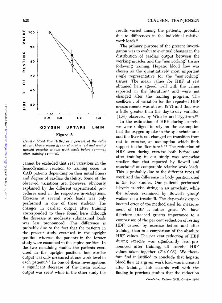
table 3) was within normal limits in allpatients examined in the untrained state. Thegroup mean value was 1421 ml (SD, + 261 ml,N = 8). During exercise HBF was reduced inproportion to the work intensity in allpatients. This reduction was most pronounced(to 30% of the resting value) in the patient
617
by guest on July 19, 2018http://circ.ahajournals.org/
Dow
nloaded from

CONTROL
130
-iE
CLAUSEN, TRAP-JENSEN
GROUP MEANS
0.2 0.6 1.0 1.4 0.2 0.6 1.0 1.4 0.2 0.6 1.0 1.4
OXYGEN UPTAKE L/MINFigure 3
Stroke volune in relation to oxygen uptake at supine rest and during upright exercise at twowork loads. Individual data before and after training and group means before (o o) andafter (. *) training. (Symbols explained in table 1.)
(R.H.) who was unable to increase cardiacoutput during exercise. Apart from this patientthe interindividual variations in the per centreduction of HBF were only small. Thetraining did not change HBF at rest (table 3).During exercise an increase of HBF was
observed compared to the pre-training valuesat both work loads (table 3). In absoluteterms these differences are, however, notstatistically significant, but after training HBFexpressed as a percentage of the value at restwas inoreased 7.2%, for all exercise determina-tions taken together, and this difference issignificant (N = 24; P < 0.05).Muscle blood flow (MBF) in vastus later-
alis muscle during exercise increased almostlinearly with the work intensity up to an
average 70% (range, 64 to 75%) of the maximalwork load. Beyond this level no furtherincrease of MBF was seen (fig. 6). Thisplateauing of MBF before maximal work load
is reached was observed both before and aftertraining. However, the training displaced thecurve relating blood flow to the absolute workload to the right (fig. 6). Thus MBF was
reduced at two submaximal work levels whileMBF was increased at maximal work loads(table 4).Blood lactate concentration increased less at
both work levels after training (table 3).The respiratory quotient (RQ) at rest was
0.68 before and after training. RQ duringexercise was reduced after training from0.82 to 0.78 (P < 0.05) and from 0.91 to0.82 (P < 0.01) at the moderate and heavywork loads, respectively.Other Measurements
The mean hemoglobin concentration was
15.4 g/100 ml (SD, 2.4 g/100 ml) and was
not altered by the training. Mean arterialoxygen saturation was 94% at rest and 97%during exercise both before and after training.
Circulation, Volume XLII, October 1970
618
TRAINI NG
110UJ
0
90
0
I-U,
70
by guest on July 19, 2018http://circ.ahajournals.org/
Dow
nloaded from

TRAINING AND CARDIAC OUTPUT DISTRIBUTION
e C-O O.-O~~~90 0
; AVV V
LO8 0 0
t o fl i L i f
CI
t4 x CDto
.t m a~L ooo.>
CI ¢ m LrO
0~~~
X 0 0C)
C~~~~~~C0
C a VE XLII, Oc 17
Circulation, Volume XLII, October 1970
zi
-J
0
-J
I.i
a00m
-
4IL
s
2.0 r
1.5
1.0
0.5
0.2 0.8 1.0 1.2 1.4 1.6
OXYGEN UPTAKE L/MI N
Figure 4Individual values (untrained state) for hepatic bloodflow at supine rest and during upright exercise at twowork loads. (Symbols explained in table 1.)
The training caused a slight improvement ofthe forced expiratory volume in 1 sec (diff.,4.5%, P<0.05) whereas all other pulmonaryfunction tests were unchanged. The fastingserum transaminases, lactic acid dehydrogen-ase, serum creatinine, and serum cholesterolwere identical at both occasions. A slightweight reduction was observed in mostpatients (mean diff., 1 kg) -
DiscussionThe question whether physical conditioning
changes the distribution of cardiac outputduring submaximal work arises from observa-tions of a reduced cardiac output aftertraining.'4 Also in the present study adecrement of cardiac output was seen atmoderate work intensities, whereas the cardi-ac output was unchanged or even increasedduring heavy exercise. Three previous studiesof the hemodynamic response to training inpatients with CAD are available. -3 Theresults from these studies differ somewhatwith respect to the effect on cardiac output. It
C3
P-1
9-
~-
*
619
by guest on July 19, 2018http://circ.ahajournals.org/
Dow
nloaded from
CLAUSEN, TRAP-JENSEN
Hepatic iat rest. Gupright e
after trai
cannot lhemodyCAD paand degobserve'explaineceduresExerciseperformchangescorrespcthe dec.was lesprobabl'the prepositionstudy wthe twocised ir
output v
each pata signifioutput s;
results varied among the patients, probablydue to differences in the individual relative\work loads.3
The primary purpose of the present investi-gation was to evaluate eventual changes in thedistribution of cardiac output between theworking muscles and the "nonworking" tissuesfollowing training. Hepatic blood flow waschosen as the quantitatively most importantsingle representative for the "nonworking"tissues. The mean values for HBF at restobtained here agreed well with the valuesreported in the literature'5 and were notchanged after the training program. Thecoefficient of variation for the repeated HBFmeasurements was at rest 19.7% and thus wasa little greater than the day-to-day variation
0.2 0.8 1.2 1.6 (15%) observed by Winkler and Tygstrup.'6In the estimation of HBF during exercise
OXYGEN UPTAKE L/M I N we were obliged to rely on the assumptionthat the oxygen uptake in the splanchnic area
Figure 5 and the liver is not changed on transition fromblood flow (HBF) as a percent of the value rest to exercise, an assumption which findsrroup means ± SEM at supine rest and during support in the literature.6" 17 The reduction of?xercise at two work loads before (oa o), HBF seen during exercise both before and
after training in our study was somewhatbe excluded that real variations in the smaller than that reported by Rowell andnamic reaction to training occur in associates6 at comparable relative work loads.itients depending on their initial fitness This is probably due to the different types of;ree of cardiac disability. Some of the work and the difference in body position usedd variations are, however, obviously in the two studies. Our patients performedd by the different experimental pro- bicycle exercise sitting in an armchair, whileused in the respective investigations. the subjects examined by Rowell's group6
at several work loads was only walked on a treadmill. The day-to-day exper-ed in one of these studies.2 The imental error of the method used for measure-
in cardiac output after training ment of HBF is rather great. We have)nded to those found here although therefore attached greater importance to arease at moderate submaximal loadsreaseat
pronounced. sThisadifferen s comparison of the per cent reduction of restingssduetronouthedf That thfferaent i HBF caused by exercise before and aftersentestudytexrcised i the upatiegtin training, than to a comparison of the absolutewhereas the patients in the former HBF values. The per cent reduction of HBFere examined in the supine position. In during exercise was significantly less pro-remaining studies the patients exer- nounced after training, all exercise HBFrthe upright position, but cardiac values taken together (P <0.05). We there-
vas only measured at one work level in fore find it justified to conclude that hepatictient.1, 3In one of these investigations blood flow at a given work load was increasedicant decrease of the mean cardiac after training. This accords well with thevas seen' while in the other study the finding in previous studies that the reduction
Circulation, Volume XLIl, October 1970
100-i
90
zI-U)AM
80
70U-
0
60
U-50
620
by guest on July 19, 2018http://circ.ahajournals.org/
Dow
nloaded from

TRAINING AND CARDIAC OUTPUT DISTRIBUTION
PERCENT OF
after 2i5before 3i2
MAXIMAL WORK LOAD
38 56
4,8 71
4 Q
I I200 300 400 500
WORK LOAD KPM/MIN
Figure 6
Blood flow in m-vastus lateralis (MBF) during exercise in relation to the work load (kpm/min).Group means + SEM before (o o) and after (e *) training. Relative work load (% ofmax.) is shown above. Q indicates the load at which cardiac output was determined.
of hepatic and renal blood flow during exer-
cise is proportional to the relative work load.6' 7
The hepatic flow and the renal flow duringexercise are controlled by the sympatheticvasoconstrictor system. It is assumed that thevasoconstrictor system during exercise acts en
masse.18 The results obtained in studies ofblood flow to the abdominal viscera are
therefore probably valid also for other "non-
working" tissues supplied with sympatheticvasoconstrictor nerves. It can thus be expectedthat at a given submaximal work load aftertraining a greater fraction of the cardiacoutput is directed to these regions.The skeletal muscles performed the same
submaximal work with a lower blood flowafter training. In the present study thisreduction (15%) was somewhat lower thanthat found in the previous investigation(21% ) .3 Varnauskas and co-workers'9 re-
ported an even greater decrement of 30% in
Circulation, Volume XLII, October 1970
MBF in seven young normal subjects after 6weeks of strenuous training. The more moder-ate decrement seen in the present study mightbe ascribed to the fact that the two patientsearlier studied by us were younger and more
trainable than the majority of the patientsexamined here. The mechanism responsiblefor the decrease in MBF is not clear, but theresults from a study by Linderholm andassociates20 may give an important clue. In a
group of patients with a hereditary abnormalmuscle inetabolism, hyperkinetic circulationduring exercise was caused by an abundantblood flow through the working muscles.According to these authors this could beexplained by an abnormally large accumula-tion of vasodilator metabolites in the activemuscles resulting from decreased oxidativecapacity of the muscle mitochondria. A similarregulatory pattern, although less pronounced,may exist in the untrained CAD patient, who
69
100
zi
.16-
a
40
30
2
20
600 700
j- %vi11 a.
i
#4
621i
8a.o
by guest on July 19, 2018http://circ.ahajournals.org/
Dow
nloaded from

CLAUSEN, TRAP-JENSEN
can be expected to have a low oxidativemetabolic capacity in the skeletal muscles.Physical conditioning is known to increase thecapacity of oxidative enzymes in the trainedmuscles.'9' 21 This might limit the release ofvasodilator metabolites and thus cause areduction of muscle blood flow.In a qualitative sense the changes in MBF
and cardiac output observed after trainingwere in good agreement. Both parametersdecreased at the moderate work load andincreased at the heavy work level (com-pare fig. 2 and fig. 6). The calculated abso-lute decrease in MBF of 4.7 ml/100 g/min,however, can hardly account for the 1.72L/min reduction in cardiac output at themoderate load since it would imply an activemuscle mass of 37 kg, that is, a value probablyexceeding the total muscle mass of a patient.It is discussed elsewhere that the '33Xe-clearance method probably somewhat under-estimates absolute MBF values.22 This how-ever, does not influence the evaluation of theflow in a qualitative sense and does notinvalidate the statement made above thatMBF is decreased during submaximal workafter training. Thus, according to our findingsthe change of the circulatory regulation fromhyperkinetic to normal induced by trainingcould be related to a reduction in the bloodflow through the working muscles, whichovershadowed inversely directed changes inthe perfusion of "nonworking" tissues.
Before training at the heavy work load itwas found that despite a normal increase ofthe heart rate cardiac output failed to increasedue to a marked decrease of the strokevolume. A similar abnormal hemodynamicresponse to exercise has been described earlierin a selected group of extremely untrainedmiddle-aged subjects.23 After training thecirculatory regulation was normalized due toan increase in cardiac output provided by a
greater stroke volume and a lower heartrate.
In normal subjects the lower heart rateinduced by training occurs together with an
increased stroke volume.24 In consequence thedeceleration of the heart rate during submax-
imal exercise is generally considered to besecondary to a direct improvement of myocar-dial function. It is therefore interesting, that areduction in heart rate could be observed inthese CAD patients at the moderate work loadwithout a concomitant increase of the strokevolume (compare figs. 1 and 3). This findingsuggests that peripheral factors contribute tothe training effect on the heart rate. We havetested this hypothesis in a training study ofnormal subjects.25 Two groups were traineddaily during 4 weeks by maximal dynamicexercise on bicycle ergometer. The one groupperformed the training work with the arms,the other group trained their legs. Thereduction in heart rate obtained by trainingthe arm muscles could not be transferred toexercise performed with the leg muscles; nordid leg training cause significant reduction ofthe heart rate during dynamic arm work. Thusit was concluded that local changes in thetrained muscles are of importance for theeffect of training on heart rate.*The myocardial pressure work expressed as
the tension-time index was reduced aftertraining at both work loads.t The decrease inthe heart rate and in the systolic bloodpressure contributed to this reduction. In theprevious studies concerning the hemodynamiceffects of training in CAD patients, the bloodpressure was also reduced during exercisewhen measured in the upright position" 3 butnot during supine exercise.2The results presented herein seem to
indicate that physical conditioning in additionto eventual direct improvement of respiratoryand cardiac performance causes alterations in
*In a study now in progress we trained a cardiacpatient with complete atrioventricular block treatedwith a fixed-rate artificial pacemaker (72 beats/min).Two weeks of bicycle training caused a marked reduc-tion of the atrial rate (P-wave frequency) from 146to 115 beats/min (work load, 300 kpm). This pre-liminary result seems to confirm the above conclusionthat peripheral factors are important for the reduc-tion in heart rate found after short-term physicaltraining.tThe justification of using peripheral blood pressure
tracings for evaluation of left ventricular work haspreviously been discussed.3
Circulation, Volume XLII, October 1970
622
by guest on July 19, 2018http://circ.ahajournals.org/
Dow
nloaded from

TRAINING AND CARDIAC OUTPUT DISTRIBUTION
the trained muscles which are essential to thegeneral circulatory regulation. When empha-sizing the importance of local training effectsin skeletal muscle we are in agreement withthe results recently presented by Kaijser.26 Inan extensive study of the limiting factors foraerobic muscle performance in normal man,he concluded that the upper limit normally isdetermined by the maximal metabolic rate ofthe muscles and not by cardiac output or thelocal perfusion. The assumption that thetraining effect on cardiac performance inpatients with CAD is to a large extentrestricted to exercise performed with thetrained muscles has important implications forthe planning of training programs for thesepatients. To be beneficial for the patients intheir daily activity the training must namelyinclude types of exercise normally performedby the patients at work and at leisure.Exercise on a bicycle ergometer makes thepatient a competent ergometer cyclist buthardly increases his tolerance for exerciseperformed with the arms, for example.
References1. VARNAUSKAS E, BERGMAN H, HOUK P, ET AL:
Hemodynamic effects of physical training incoronary patients. Lancet 2: 8, 1968
2. FRICK MH, KATILA M: Hemodynamic conse-quences of physical training after myocardialinfarction. Circulation 37: 192, 1968
3. CLAUSEN JP, LARSEN OA, TRAP-JENSEN J:Physical training in the management ofcoronary artery disease. Circulation 40: 143,1969
4. EKBLONI B, ASTRAND PO, SALTIN B, ET AL:Effects of training on the circulatory responseto exercise. J Appl Physiol 24: 518, 1968
5. HOLMGREN A, JONSSON B, LEVANDER M, ET AL:Physical training of patients with vasoregula-tory asthenia. Acta Med Scand 158: 437,1957
6. ROWELL LB, BLACKMON JR, BRUCE RA: Indo-cyanine green clearance and estimated hepaticblood flow during mild to maximal exercise inupright man. J Clin Invest 43: 1677, 1964
7. GRIMBY G: Renal clearances during prolongedsupine exercise at different loads. J ApplPhysiol 20: 1294, 1965
8. NIELSEN NC: Spectrophotometric determinationof indocyanine green in plasma especially witha view to an improved correction for blank
Circulation, Volume XLII, October 1970
density. Scand J Clin Lab Invest 15: 613,1963
9. BRADLEY SE, INGELFINGER FJ, BRADLEY GP, ETAL: The estimation of hepatic blood flow inman. J Clin Invest 24: 890, 1945
10. HAMILTON WF, RILEY RL, ATrYAH AM, ET AL:Comparison of the Fick and dye injectionmethods of measuring the cardiac output inman. Amer J Physiol 153: 309, 1948
11. HOLMGREN A, PERNOw B: Spectrophotometricmeasurement of oxygen saturation of blood inthe determination of cardiac output: Acomparison with the van Slyke method. ScandJ Clin Lab Invest 11: 143, 1959
12. LASSEN NA, LINDBJERG J. MUNCK 0: Measure-ment of blood flow through skeletal muscle byintramuscular-injection of xenon-133. Lancet1: 686, 1964
13. SHEPHARD RJ, ALLEN C, BENADE AJS, ET AL:Standardization of submaximal exercise tests.Bull WHO 38: 765, 1968
14. CLAUSEN JP: Effects of physical conditioning: Ahypothesis concerning circulatory adjustmentto exercise. Scand J Clin Lab Invest 24: 305,1969
15. WINKLER K: Studies on the elimination ofbromsulfalein in man. Thesis in Danish with anEnglish summary. University of Copenhagen,1966
16. WINKLER K, TYGSTRUP N: The day-to-dayvariations in bromsulfalein elimination curves.Scand J Clin Lab Invest 16: 481, 1964
17. WADE OL, COMBES B, CHILDS AW, ET AL: Theeffect of exercise on the splanchnic blood flowand splanchnic blood volume in normal man.Clin Sci 15: 457, 1956
18. BEVEGAR1D BS, SHEPHERD JT: Regulation of thecirculation during exercise in man. Physiol Rev47: 178, 1967
19. VARNAUSKAS E, BJORNTORP P, FAHLEN M, ET AL:Effects of physical training on exercise bloodflow and succinic dehydrogenase activity inskeletal muscle. Cardiovasc Res. In press
20. LINDERHOLM H, MULLER R, RINGQVIST T, ET AL:Hereditary abnormal muscle metabolism withhyperkinetic circulation during exercise. ActaMed Scand 185: 153, 1969
21. HOLLOSZY JO: Biochemical adaptations in muscle:Effects of exercise on mitochondrial oxygenuptake and respiratory enzyme activity inskeletal muscle. J Biol Chem 242: 2278,1967
22. CLAUSEN JP, LASSEN NA: Muscle blood flowduring exercise in normal man studied by the133Xe clearance method. Cardiovasc Res. Inpress.
23. HANSON JS, TABAKIN BS, LEVY AM, ET AL:
623
by guest on July 19, 2018http://circ.ahajournals.org/
Dow
nloaded from

CLAUSEN, TRAP-JENSEN
Long-term physical training and cardiovasculardynamics in middle-aged men. Circulation 38:783, 1968
24. SALTIN B, BLOMIQVIST G, MITCHELL JH, ET AL:Response to exercise after bed rest and aftertraining. Circulation 38 (suppl VII): VII-1,1968
25. CLAUSEN JP, TRAP-JENSEN J, LASSEN NA: Theeffect of training on the heart rate during armand leg exercise. Scand J Clin Lab Invest. Inpress.
26. KAIJSER L: Limiting factors for aerobic muscleperformance. Acta Physiol Scand Suppl 346,1970
Editor's Dilettantism
Cardiac Glycosides, Blue Jays, and Butterflies
Monarch butterflies Danaus plexippus, when reared on tropical asclepiads andapocynads, contain cardiac glycosides which make them unpalatable to avianpredators.
Glycosides were extracted from seeds [of the plants] . . . and insect tissuepurified on a Florisil column . . . and their identity was verified by thin-layerchromatography on silica gel G, by spectrophotometry, and by biological assay (ino-tropic response in rat heart, and antagonism of this by aldosterone)....The blue jay Cyanocitta cristata bromia is the major avian predator employed in
studies of palatability. . . We do not know whether Danaus feeding on Asclepiassimply accumulates glycosides according to the type and concentration in the plant,or if it has specific selective or concentration mechanisms.-DUFFEY SS: Cardiac glycosides and distastefulness: Some observations on the palat-ability spectrum of butterflies. Science 169: 78, 1970.(Courtesy the American Association for the Advancement of Science: Copyright 1970).
Circulation, Volume XLII, October 1970
624
by guest on July 19, 2018http://circ.ahajournals.org/
Dow
nloaded from

JAN PRAETORIUS CLAUSEN and JENS TRAP-JENSENCoronary Artery Disease
Effects of Training on the Distribution of Cardiac Output in Patients with
Print ISSN: 0009-7322. Online ISSN: 1524-4539 Copyright © 1970 American Heart Association, Inc. All rights reserved.
is published by the American Heart Association, 7272 Greenville Avenue, Dallas, TX 75231Circulation doi: 10.1161/01.CIR.42.4.611
1970;42:611-624Circulation.
http://circ.ahajournals.org/content/42/4/611located on the World Wide Web at:
The online version of this article, along with updated information and services, is
http://circ.ahajournals.org//subscriptions/
is online at: Circulation Information about subscribing to Subscriptions:
http://www.lww.com/reprints Information about reprints can be found online at: Reprints:
document. and Rights Question and Answer
Permissionsthe Web page under Services. Further information about this process is available in thewhich permission is being requested is located, click Request Permissions in the middle column ofClearance Center, not the Editorial Office. Once the online version of the published article for
can be obtained via RightsLink, a service of the CopyrightCirculationoriginally published in Requests for permissions to reproduce figures, tables, or portions of articlesPermissions:
by guest on July 19, 2018http://circ.ahajournals.org/
Dow
nloaded from