
Effects of long-term extended contact lens wear on the human cornea.
13
Effects of Long-Term Extended Contact Lens Wear on the Human Cornea Drien A. Holden,* Deborah F. Sweeney,* Antti Vannas,t Klas T. Nilsson,^: and Nathan Efron* The effects of long-term extended wear of soft contact lenses on the human cornea were determined by examining 27 patients who had worn a high water content hydrogel contact lens in 1 eye only for an average of 62 ± 29 months (mean ± SD). The other eye, which was either emmetropic or amblyopic, acted as a control. The lens-wearing eye showed a 14.8% reduction in epithelial oxygen uptake (P < 0.001), a 5.6% reduction in epithelial thickness (P < 0.05), a 2.3% reduction in stromal thickness (P < 0.05), the induction of epithelial microcysts, and a 22.0% increase in endothelial polymegathism (P < 0.001). Endothelial cell density was unaffected by extended lens wear. No interocular differences in any of these physiological characteristics were found in a matched control group of anisometropic and amblyopic subjects who did not wear contact lenses. The patients ceased lens wear for up to one month and recovery of corneal function was monitored during this period. Epithelial oxygen uptake and thickness recovered within 33 days of lens removal. The number of microcysts increased over the first 7 days, but decreased thereafter; some microcysts were still present 33 days after lens removal. Recovery from stromal thinning had not occurred after 33 days following lens removal. There was a slight reduction in polymegathism in some patients, but overall this was not statistically significant. These findings establish (1) that the extended wear of hydrogel lenses induces significant changes in all layers of the cornea; (2) that lens wear suppresses aerobic epithelial metabolism, which may compromise the epithelial barrier to infection; and (3) that changes to the stroma and endothelium are long-lasting. Lens-induced effects on corneal physiology can be minimized by fitting lenses that have greater oxygen transmissibility (are thinner), are more mobile, more frequently removed, and more regularly replaced. Invest Ophthalmol Vis Sci 26:1489-1501, 1985 Despite an immense body of literature detailing the short-term effects of contact lenses upon corneal struc- ture and function, few studies have addressed the ques- tion of changes that may occur after many years of lens wear. This question now assumes greater signifi- cance in view of the present popularity of extended lens wear and its preference to radial keratotomy 1 as an alternative form of vision correction. It is clear that contact lenses interfere with corneal metabolism; changes that have been reported include glucose 2 and glycogen 2 " 4 depletion and increased levels of lactic acid. 23 These changes have been attributed primarily to epithelial hypoxia, 5 although other influ- From the Cornea and Contact Lens Research Unit, School of Op- tometry, University of New South Wales, Kensington, New South Wales, Australia,* the University Eye Clinic, Helsinki, Finland,! and Contacta Optik, Gothenberg, Sweden.:}: This research was supported by grants from the Optometric Vision Research Foundation of Australia and the National Health and Medical Research Council of Australia. Submitted for publication: August 8, 1984. Reprint requests: Brien A. Holden, PhD, Cornea and Contact Lens Research Unit, School of Optometry, University of New South Wales, PO Box 1, Kensington, N.S.W. 2033, Australia ences such as mechanical stress may also affect corneal metabolism. 4 More recently, clinicians have observed that contact lenses can induce structural alterations to the cornea, including epithelial microcysts, 6 "" stromal striae and folds, 12 and endothelial blebs 1314 and poly- megathism. 15 " 17 We conducted a study to determine changes in cor- neal structure and function that occur as a result of long-term extended contact lens wear. This was achieved by examining the corneas of 27 patients who had each worn a contact lens in one eye only, on an extended wear basis, for an average of 62 ± 29 months (mean ± SD). The eye in which a lens was not worn acted as a control. A number of these patients were examined at regular intervals for up to 1 month fol- lowing cessation of lens wear to determine if such changes were permanent. Materials and Methods Subjects Patients were recruited from a large contact lens practice in Gothenberg, Sweden (KN). 18 The files of all contact lens patients attending this practice (ap- 1489 Downloaded From: http://iovs.arvojournals.org/pdfaccess.ashx?url=/data/journals/iovs/933120/ on 04/12/2018
Transcript of Effects of long-term extended contact lens wear on the human cornea.

Effects of Long-Term Extended Contact Lens Wear onthe Human Cornea
Drien A. Holden,* Deborah F. Sweeney,* Antti Vannas,t Klas T. Nilsson,̂ : and Nathan Efron*
The effects of long-term extended wear of soft contact lenses on the human cornea were determined byexamining 27 patients who had worn a high water content hydrogel contact lens in 1 eye only for anaverage of 62 ± 29 months (mean ± SD). The other eye, which was either emmetropic or amblyopic,acted as a control. The lens-wearing eye showed a 14.8% reduction in epithelial oxygen uptake(P < 0.001), a 5.6% reduction in epithelial thickness (P < 0.05), a 2.3% reduction in stromal thickness(P < 0.05), the induction of epithelial microcysts, and a 22.0% increase in endothelial polymegathism(P < 0.001). Endothelial cell density was unaffected by extended lens wear. No interocular differencesin any of these physiological characteristics were found in a matched control group of anisometropicand amblyopic subjects who did not wear contact lenses. The patients ceased lens wear for up to onemonth and recovery of corneal function was monitored during this period. Epithelial oxygen uptake andthickness recovered within 33 days of lens removal. The number of microcysts increased over the first7 days, but decreased thereafter; some microcysts were still present 33 days after lens removal. Recoveryfrom stromal thinning had not occurred after 33 days following lens removal. There was a slight reductionin polymegathism in some patients, but overall this was not statistically significant. These findingsestablish (1) that the extended wear of hydrogel lenses induces significant changes in all layers of thecornea; (2) that lens wear suppresses aerobic epithelial metabolism, which may compromise the epithelialbarrier to infection; and (3) that changes to the stroma and endothelium are long-lasting. Lens-inducedeffects on corneal physiology can be minimized by fitting lenses that have greater oxygen transmissibility(are thinner), are more mobile, more frequently removed, and more regularly replaced. Invest OphthalmolVis Sci 26:1489-1501, 1985
Despite an immense body of literature detailing theshort-term effects of contact lenses upon corneal struc-ture and function, few studies have addressed the ques-tion of changes that may occur after many years oflens wear. This question now assumes greater signifi-cance in view of the present popularity of extendedlens wear and its preference to radial keratotomy1 asan alternative form of vision correction.
It is clear that contact lenses interfere with cornealmetabolism; changes that have been reported includeglucose2 and glycogen2"4 depletion and increased levelsof lactic acid.23 These changes have been attributedprimarily to epithelial hypoxia,5 although other influ-
From the Cornea and Contact Lens Research Unit, School of Op-tometry, University of New South Wales, Kensington, New SouthWales, Australia,* the University Eye Clinic, Helsinki, Finland,! andContacta Optik, Gothenberg, Sweden.:}:
This research was supported by grants from the Optometric VisionResearch Foundation of Australia and the National Health andMedical Research Council of Australia.
Submitted for publication: August 8, 1984.Reprint requests: Brien A. Holden, PhD, Cornea and Contact Lens
Research Unit, School of Optometry, University of New South Wales,PO Box 1, Kensington, N.S.W. 2033, Australia
ences such as mechanical stress may also affect cornealmetabolism.4 More recently, clinicians have observedthat contact lenses can induce structural alterations tothe cornea, including epithelial microcysts,6"" stromalstriae and folds,12 and endothelial blebs1314 and poly-megathism.15"17
We conducted a study to determine changes in cor-neal structure and function that occur as a result oflong-term extended contact lens wear. This wasachieved by examining the corneas of 27 patients whohad each worn a contact lens in one eye only, on anextended wear basis, for an average of 62 ± 29 months(mean ± SD). The eye in which a lens was not wornacted as a control. A number of these patients wereexamined at regular intervals for up to 1 month fol-lowing cessation of lens wear to determine if suchchanges were permanent.
Materials and Methods
Subjects
Patients were recruited from a large contact lenspractice in Gothenberg, Sweden (KN).18 The files ofall contact lens patients attending this practice (ap-
1489
Downloaded From: http://iovs.arvojournals.org/pdfaccess.ashx?url=/data/journals/iovs/933120/ on 04/12/2018

1490 INVESTIGATIVE OPHTHALMOLOGY 6 VISUAL SCIENCE / November 1985 Vol. 26
Table 1. Patient details and lens wearing experience
No
01*02*030405*06*07*08*09*101112*13*1415*16*17*18*1920*21*22*23*24*2526*27*
Sex
FFMMFFMMFMMMMMMMMMFFFFMMFFF
F 12M 15
Age(yrs)
272733262630532429212732242533133022482932252737273715
m 29SD 8
Patient details
Lensweareye
LRLRLLLLRLLLLRRRLLRLRLRLRRR
R 12L 15
Rx lenswearingeye\ (D)
-2.00+3.63-1.25-4.62-2.50-5.50+ 1.12-3.87-1.37-4.25
'+6.87-1.75+5.62-2.00-4.25-6.87-3.12-9.50+5.25-1.75
-10.00+4.12-5.12-4.87+5.75+2.87-9.12
MYOPESm -4.41SD 2.77HYPEROPESm +4.40SD 1.85
Rx controleye\ (D)
-0.370.00
-0.75-0.37
0.00-3.5O|+5.00$-0.50+ 1.50*-6.00$+7.50$
0.00+7.00$-3.25$-0.25-0.75-0.62-7.50$+4.25$-1.00
-12.00$+4.50$
0.000.00
+6.12$+7.25$-0.25
E.W.E.§(mo)
1314222626363844465256606164677476787979818184919194
136
m 62SD 29
Lens wearing experience
PriorD.W.E.W
(mo)
4811601146H
62U
36**
72**
4211
T.W.E.tf(mo)
1314222626363844465280908464937476789779
117818491
11294
136
m 68SD 33
* Patient was examined on Day 0.t Best sphere refraction.% Amblyopic eye. All other patients were anisometropic.§ Extended wear experience.
proximately 20,000) were examined; 54 were found tobe unilateral hydrogel contact lens wearers. Of these,ten were wearing extended wear lenses to correctaphakia. These patients were excluded from this studybecause ocular surgery is known to alter corneal func-tion.19 Of the remaining 44, seven were daily wear pa-tients, eight could not be contacted, one declined ourinvitation to participate, and one had only one eye.Thus, a total of 27 unilateral extended wear patientswere examined after the nature of the procedures werefully explained. Fourteen patients wore a contact lensunilaterally because of anisometropia and 13 becauseof amblyopia.
In order to determine whether there is an interoculardifference in corneal characteristics in anisometropiaand amblyopia, we examined 20 matched anisome-tropic and amblyopic control subjects who did not wearcontact lenses.
|| Prior daily wear experience (in all cases, immediately preceding extendedlens wear).
H Soft lens wear.** Hard lens wear.f t Total wearing experience = extended wear + 0.5 (daily wear).
Both lens-wearing patients and control subjects weregiven a general ocular examination and found to befree from ocular disease. Details of the lens-wearingpatients and control subjects are presented in Tables1 and 2, respectively.
Lenses
All patients wore a 71% water content hydrogel lensconsisting of polyvinyl pyrrolidone cross-linked withpolymethyl methacrylate (Scanlens; Gothenberg,Sweden). Lenses were fitted in three base curves. (8.3,8.5 and 8.7 mm) and two diameters (13.5 and 14.0mm); the basic fitting philosophy was to allow formaximum possible movement consistent with goodcomfort and stable vision.
The patients had worn a contact lens in one eye onan extended wear basis for an average of 62 ± 29
Downloaded From: http://iovs.arvojournals.org/pdfaccess.ashx?url=/data/journals/iovs/933120/ on 04/12/2018

No. 11 EFFECTS OF EXTENDED CONTACT LENS WEAR ON THE HUMAN CORNEA / Holden er ol. 1491
months (range 13 to 136 months). Some patients hadprior daily wear experience with hard and/or soft lensesin both the eye that was currently wearing the lens andin the other eye. Details of the lens-wearing experienceof all patients are given in Table 1.
The patients were originally instructed to leave theirlens out overnight approximately twice per month andto clean it using an oxidizing system. On average, theyremoved their lens 2.3 ± 2.6 times each month (Table3). All patients were on a service contract which entitledthem to a new lens every six months. The lens replace-ment rate in our patient group was found to be 1.6± 0.8 lenses per year (Table 3).
The center thickness of all lenses being worn at thetime of examination was measured in vivo using anelectronic digital pachometer.19 Average lens thicknesswas determined using Brennan nomograms.20'21 Detailsof the lens used by each patient are presented inTable 3.
Experimental Protocol
Patients were asked to attend for an initial exami-nation wearing their lens. After assessing in-eye lensperformance, the lens was removed and measurementswere made on the corneas of both eyes over a two hourperiod (Table 4). In order to minimize observer bias,data was gathered by observers who were not informedas to which eye had been wearing the lens.
All patients were asked to cease wearing their lensfor approximately one month; however, some wereunable to comply with this request. Patients were ex-amined at regular intervals following lens removal. Tofacilitate data analysis, the time elapsed following lensremoval was divided into four groups: "Day 0" (20patients examined); "Day 2" (2.5 ± 0.7 days [mean± SD], range 2 to 4 days, 18 patients examined); "Day7" (7.3 ± 0.8 days, range 6 to 9 days, 19 patients ex-amined); and "Day 33" (33.6 ± 13.3 days, range 13 to65 days, 12 patients examined).
Some patients presented for their first examinationafter having not worn their lens for some time previ-ously, for reasons such as lens breakage or lens loss.Data obtained from these examinations were includedin one of the above four groups, as appropriate.
The physiological measurements described belowwere carried out on the 27 lens-wearing patients andon 20 non-lens-wearing control subjects who werematched to the 20 unilateral lens wearers examined onDay 0.
Measurements
Lens movement: The lens-wearing eye was observedunder low magnification with a slit lamp biomicro-scope. The patient fixated a target which was adjusted
Table 2. Control subject details
No
2829*30313233*3435*363738*39404142*4344454647
Sex
MFFMMFMMMFFFMMMFFMMF
F 9M 11
Age
(yr)
2243253520311925264155235844215749212424
m 33SD 14
Correctedeye\
RLRRLRRRLRRLLLLRLLRR
R 11L 9
Rx:correctedeye (D)%
-2.75-2.87-2.75-1.50-2.87-2.63-1.00-1.37-3.75+2.75
-11.87-2.37
-11.37-4.62+4.75+2.67+ 1.00-1.62-2.12+2.25
MYOPESm -3.70SD 3.35HYPEROPESm +2.68SD 1.35
Rx:uncorrected
eye(D)%
0.00-0.37
0.00+0.25+0.50
0.00+ 1.75§-0.25-0.63+0.87
0.00§-0.50+ 1.00+0.12+ 1.50-0.37§
0.00-0.62-0.50+ 1.00
* These subjects had prior contact lens experience, but ceased wear at least12 months prior to participation in this study,
t Spectacle correction.% Best sphere refraction.§ Amblyopic eye. All other subjects were anisometropic.
so that the eye was in the primary gaze position. Theamount of lens movement following each blink wasestimated to the nearest 0.5 mm.
Epithelial oxygen uptake: A polarographic oxygensensor was used to measure epithelial oxygen uptake;this technique has been described in detail previously.19
The time taken for the oxygen tension in the sensormembrane to drop from 140 to 100 mmHg was usedas a relative measure of oxygen uptake. The result isexpressed in units of mmHg/sec.
Epithelial thickness: Epithelial thickness was mea-sured at the central cornea using an electronic digitalpachometer that has been described previously.19 Tenreadings of epithelial thickness were obtained on eachmeasurement occasion; the average standard deviationfor ten readings was ±5 /xm (10% of epithelial thick-ness).
Epithelial microcysts: Microcysts were observed us-ing the slit lamp technique of marginal retro-illumi-nation." The approximate number of microcysts vis-ible in the central and paracentral corneal epitheliumwas recorded.
Stromal thickness: The electronic digital pachome-ter19 was also used to measure the thickness of the
Downloaded From: http://iovs.arvojournals.org/pdfaccess.ashx?url=/data/journals/iovs/933120/ on 04/12/2018

1492 INVESTIGATIVE OPHTHALMOLOGY & VISUAL SCIENCE / November 1985 Vol. 26
Table 3. Details of lenses worn
No
010203040506070809101112131415161718192021222324252627
Back centraloptic radius
(mm)
8.58.78.78.58.58.58.38.78.78.58.58.58.58.38.58.58.58.58.78.58.58.58.78.78.58.58.5
Lens diameter(mm)
14.014.014.01.3.513.5•13.51'3'.514.013.513.513.513.513.513.513.513.513.513.514.013.513.513.514.014.013.513.513.5
Back vertexpower (D)
-1 .25+4.00-1.25-4.00-2.50-4.75+ 1.75-3.25-1.25-5.00+8.25 •-1.50 .+5.75-1.75-3.75-3.75-2.75-8.00+5.25-1.50-7.50+3.75-4.50-4.50+8.25+3.25-8.00
MYOPESm -3.72SD 2.23HYPEROPESm +5.03SD 2.33
A verage lensthickness
(urn)
253310255198221186326211210194401247338202168267237203182219226353208163453349177
MINUSm 213SD 30PLUSm 339SD 78
Removalrate*
(mo'1)
2.74.01.00.02.02.01.34.01.34.02.31.53.31.31.00.02.00.71.01.01.01.31.3
13.01.37.0.0
m 2.3SD 2.6
Replacementraie\(yr1)
1.83.62.22.40.90.74.20.8
.02.30.91.61.01.71.62.31.11.2.5
0.91.31.31.71.51.91.31.4
m 1.6SD 0.8
LensmovemenlX
(mm)
0.51.51.0
- §0.52.01.01.00.51.01.01.02.00.52.01.02.02.0- §1.01.51.01.51.51.02.01.0
m 1.2SD 0.5
* Average number of days the lens was removed each month.t Average number of lenses replaced each year.
% Lens movement in the primary gaze position following each blink.§ Lens not seen on eye.
central corneal stroma. Ten repeated measurementswere made on each occasion. The average standarddeviation for ten readings was ±5 fim (1.0% of stromalthickness). Stromal thickness was measured immedi-ately after lens removal, first in the lens-wearing eye,then in the contralateral control eye.
Endothelial morphology: The endothelium wasphotographed with Kodak (Eastman Kodak; Roches-ter, NY) Tri-X Pan Film using a non-contact specularmicroscope that has been described elsewhere.14 Fifteenphotographs were taken of each eye at every exami-nation. The photographs were printed at 12.33X mag-nification from negatives which contained images ofendothelial cells at approximately 30X magnification.A precision microscopic grid of known dimensions wasphotographed at the corneal plane to enable exactquantification of the endothelial mosaic. Five photo-graphs of good quality were chosen from the 15 pho-tographs of each eye. Technicians manually traced 50cells from each photograph on to a plastic sheet usinga fine black marking pen, giving a total of 250 cells
traced per eye. The cell tracings were analysed usingan IBAS-2 Automatic Image Analysis System (Kon-tron; Munich, West Germany). This instrument mea-sured individual cell areas and calculated endothelialcell density (ECD) and the coefficient of variation ofmean cell area, which was used as an index of endo-thelial cell polymegathism. The IBAS-2 is generallyprogrammed to measure the area within the line trac-ings; however, cell boundaries occur at the centre ofthe traced lines. To overcome this problem, the IBAS-2 was programmed to adjust each individual cell areawith reference to the internal cell perimeter and halfthe average width of the pen line.
Data Analysis
The differences between the two eyes of both lens-wearing patients and control subjects were tested forstatistical significance using a t-test for matched pairs.A 95% confidence level was chosen as the criterion foraccepting a difference as being significant.
To test for statistical associations between variables
Downloaded From: http://iovs.arvojournals.org/pdfaccess.ashx?url=/data/journals/iovs/933120/ on 04/12/2018

No. 11 EFFECTS OF EXTENDED CONTACT LENS WEAR ON THE HUMAN CORNEA / Holden er ol. 1493
examined in this study, a multiple regression analysis,with stepwise inclusion of variables, was conducted onall of the Day 0 data; again, the 95% confidence levelwas chosen to denote statistical significance. A list ofall dependent and independent variables assessed inthe analysis is given in Table 4.
Table 4. Experimental variables investigated
Results
Control Subjects
The differences in corneal structure and functionbetween the two eyes of the 20 control subjects arepresented in Table 5. In all cases, data from the am-blyopic or less ametropic eye was subtracted from dataof the non-amblyopic or more ametropic eye, the latterbeing the eye that generally would have received a con-tact lens had that form of vision correction beenadopted. It is evident from Table 5 that there is nostatistically significant difference between the two eyeswith respect to any of the corneal characteristicsstudied.
Lens-Wearing Patients
The interocular differences in epithelial and endo-thelial characteristics on Day 0 for the lens-wearingpatients are presented in Table 6. Day 7 results forstromal thickness are also presented in this table; thesedata represent stromal thickness once lens-inducededema has subsided.
Lens movement: The amount of lens movement inthe primary gaze position following each blink is givenfor each patient in Table 3. Lens movement rangedfrom 0.5 mm to 2.0 mm, with a mean ± SD of 1.2±0.5 mm.
Epithelial oxygen uptake: A significant reduction inepithelial oxygen uptake of 14.8% was found in thelens-wearing eye compared to the control eye on Day0. This difference gradually reduced after ceasing lenswear, being statistically significant on Days 0, 2 and 7,but not on Day 33. The change in epithelial oxygenconsumption in the lens-wearing eye relative to thecontrol eye following lens removal is displayed in Fig-ure 1.
The statistical analysis revealed that three factorshave a significant effect upon epithelial oxygen uptake;lens center thickness (CT; /*m), lens movement in theprimary gaze position following each blink (LM; mm),and the average number of days the lens was removedeach month (DR). The following regression equationdescribes the percentage change in the epithelial oxygenuptake (A% O2) resulting from extended contact lens
Dependent variables
Epitheliumthicknessoxygen uptakemicrocystsstaining
Slromathicknessstriaefolds
Endotheliumcell densitypolymegathismblebsbedewing
Adverse reactionsred eye episodesvascularisationgiant papillary conjunctivitis
Visionacuitysubjective vision
Comfortsubjective comfort
Lens variablescentrationmovementcorneal coveragedepositswettingkeratometer mire quality
Independent variables
Lens variablesback central optic radiusdiameterback vertex poweraverage thicknessmovementlens tightnessrate of lens replacement
Patient variablesagesexrefractive errorcorneal curvaturemonths of weardays lens out per month
wear:O2 = -9.44 - 0.0657 CT
+ 5.61 LM + 0.94DR (1)
This equation accounts for 68.8% of the variance inthe measured difference in epithelial oxygen uptakerate between the two eyes induced by extended contactlens wear (r = 0.83, F = 11.76).
Epithelial thickness: The change in epithelial thick-ness of the lens-wearing eye, relative to the control eye,is presented in Figure 2. The epithelium of the lens-wearing eye was 5.6% thinner than that of the controleye on Day 0; this difference was statistically significant.After ceasing lens wear, the difference in epithelialthickness gradually decreased and was still statisticallysignificant on Day 2, but not on Days 7 or 33 (Fig. 2).
The difference in epithelial thickness (A ET; ^m)between the lens-wearing and control eyes could bepredicted by a combination of four variables: patientage (PA; years), the average number of lenses replacedeach year (LR), lens center thickness (CT; /zm) and theaverage number of days the lens was removed eachmonth (DR). The following equation describes the re-lationship between these factors:
AET = 9.68 - 0.50 PA + 4.77 LR
- 0.0388 CT + 0.73 DR (2)
This equation accounts for 47.8% of the variance inthe measured difference in epithelial thickness betweenthe two eyes induced by extended contact lens wear(r = 0.69, F = 3.437).
Downloaded From: http://iovs.arvojournals.org/pdfaccess.ashx?url=/data/journals/iovs/933120/ on 04/12/2018

1494 INVESTIGATIVE OPHTHALMOLOGY 6 VISUAL SCIENCE / November 1985 Vol. 26
Table 5. Interocular differences in control subjects
No
2829303132333435363738394041424344454647
MeanSD
Epithelial oxygen
(n
CE*
4.985.065.295.135.775.224.493.383.623.114.244.694.893.513.133.854.334.384.824.41
4.420.77
uptakevnHg' sec~')
UE\
5.065.285.175.005.645.304.733.273.343.224.384.604.813.583.103.924.244.424.894.35
4.420.78
CE-UE
-0.08-0.22
0.120.130.13
-0.08-0.24
0.110.28
-0.11-0.14
0.090.08
-0.070.03
-0.070.09
-0.04-0.07
0.06
0.000.13
X.% = 0.00df = 19
Not significant
Epithelial thickness
CE*
5866716851705464577465607559646353535759
627
(urn)
UE\
5863656453625867576462637565636455505759
616
4 =df =
CE-UE
036428
- 4- 3
0103
- 30
- 61
- 1- 2
300
14
0.9219
Not significant
Stromal thickness
CE*
469500458465435483411422484462490458484510429490449448447428
46128
(nm
C/£t
467497448480439491401435513473496454483483424495452452447422
46330
4 =df =
j
CE-UE
23
10-15- 4- 810
- 1 3-2 9-11
- 641
265
- 5- 3- 4
06
- 211
0.6019
Not significant
Endothelial cell density(cells - mm~2)
CE*
33153234334834134030337132362771251227892353280024662985376128931689360325463669
3039565
UEf
32653092359934073928324232723127243026122520291125102906346425471777314123143222
2964513
4 =df =
CE-UE
50142
-2516
102129
-36-356
82177
-167-111
- 4 479
297346
- 8 8462232447
75217
1.5119
Not significant
Coefficient of variation of•
CE*
0.2770.3370.3160.3280.2620.4120.2190.2470.3000.3480.2700.2490.3010.4140.4070.4520.2420.2650.2900.341
0.3140.066
mean cell area
UEf
0.2800.3510.2920.2970.2380.4520.1900.3150.3190.3150.2870.2590.2750.4980.3390.4630.2670.3370.2930.268
0.3170.076
4 =df =
CE-UE
-0.003-0.014
0.0240.0310.024
-0.0400.029
-0.068-0.019
0.033-0.017-0.010
0.026-0.084
0.068-0.011-0.025-0.072-0.003
0.073
-0.0030.043
0.29= 19
Not significant
* Corrected eye.t Uncorrected eye.
Epithelial microcysts: An average of 17 ± 21 (mean± SD) microcysts were observed in the lens-wearingeye on Day 0 (Fig. 3). The number of microcysts wasapproximately the same on Day 2, but then increasedto 34 ± 34 microcysts on Day 7. By Day 33, the numberof microcysts returned to the level observed on Day 0.The microcysts became smaller in size and less discreteover the 33-day observation period. A small numberof microcysts (usually fewer than three) were occa-sionally observed in the control eye. Figure 4 displaysthe pattern of change in epithelial microcysts afterceasing lens wear. The development of microcystscould not be correlated statistically with any of thevariables examined.
Stromal thickness: Data is presented in Figure 5 asthe difference in stromal thickness of the lens-wearingeye relative to the control eye. Stromal thickness was2.5% greater in the lens-wearing eye than the controleye on Day 0, but decreased following lens removal tobecome thinner than the control eye by Day 2. Stromalthickness in the eye that had been wearing the lenscontinued to decrease until Day 7 and remained con-tant thereafter, being 2.3% thinner than the controleye. This difference was statistically significant on Day
X Two-tailed t-test for matched pairs.
7 (t = 2.35, df = 18). None of the variables examinedcould be correlated with the observed changes instromal thickness.
Endothelial morphology: The ECD in the lens-wear-ing and control eyes on Day 0 is given for each patientin Table 6. There was no statistically significant differ-ence between the two eyes on this or any other mea-surement day (t-test for matched pairs).
Figure 6 gives the difference in endothelial poly-megathism between the lens-wearing and control eyesfor the 33 days following lens removal. The polymega-thism was 22.0% greater in the eye that had worn thelens, relative to the control eye, on Day 0; indeed, theinterocular difference in polymegathism was significanton all four measurement days (t-test for matched pairs).There was a slight overall decrease in mean polyme-gathism over the 33-day recovery period, but this trendwas not statistically significant. The changes in endo-thelial polymegathism could not be correlated with anyof the other variables measured in this study.
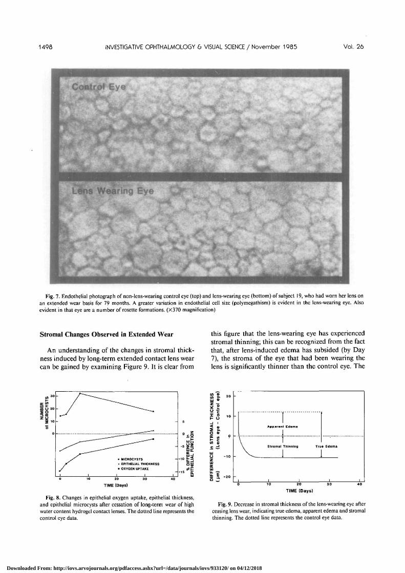
Figure 7 displays the endothelial mosaic of both eyesof patient 19, who had worn an extended wear lens inher right eye for 79 months. A greater variation in en-dothelial cell size in the lens-wearing (right) eye is ev-
Downloaded From: http://iovs.arvojournals.org/pdfaccess.ashx?url=/data/journals/iovs/933120/ on 04/12/2018

No. 11 EFFECTS OF EXTENDED CONTACT LENS WEAR ON THE HUMAN CORNEA / Holden er ol. 1495
Table 6. Interocular differences in lens-wearing patients following lens removal
No
010205060708091011121315161718192021222324252627
MeanSD
Epithelial oxygenuptake
CE*
3.794.653.943.883.414.363.94—
3.884.184.534.334.594.68
4.324.723.804.294.64
4.204.25
4.220.36
Significant:
* Corrected eye.
(mmHg-sec )
t/£t
5.165.304.694.444.855.024.86—
5.035.325.105.154.975.11
5.184.795.015.014.52
4.934.44
4.940.26
t§ =df =
; P <i
t Uncorrected eye.
CE-UE
-1.37-0.65-0.75-0.56-1.44-0.66-0.92
—
-1.15-1.14-0.57-0.82-0.38-0.43
-0.86-0.07-1.21-0.72
0.12
-0.73-0.19
-0.730.42
7.5819
3.001
Epithelial thickness
CE*
63736477646563—
645762847969
6962517376
7068
688
(urn)
{/£t
66686680747681——726268778375—6862707775—7759
727
t§ =df =
CE-UE
- 35
-2- 3
- 1 0-11-18——-8- 5- 6
7- 4- 6
10
-19- 4
1—- 7
9
- 47
= 2.52= 19
Significant; P < 0.05
Stromal thickness%
CE*
484—
458523465451516448448501—
485444—
467406423425——
453505413467
46234
(urn)
l/£t
475—
469543471497512468490508—
468419—
492419445460——
449512426460
47334
t§df
Significant; P
CE-UE
9—
-11-20-6
-464
- 2 0-42
- 7—
1725
—- 2 5- 1 3-22- 3 5
—4
- 7- 1 3
7
-1119
= 2.35= 18< 0 . 0 5
Endothelial cell density
CE*
3937355133943558241530763380—
3094—
3539322039213541
25503073387932153226
31694208
3366451
(cells • mm
l/£t
3905361629813463243732203765—
2957—
3767349335413288
21573348371132362981
32343864
3314465
t§ =df =
)
CE-UE
32-6541395
-22-144-385
—
137
-228-273
380253
393-275
168-21245
- 6 5344
52247
= 0.89= 18
Not significant
Coefficient of variation ofmean cell area
CE*
0.3970.3200.3280.3800.4060.3360.501
—
0.554
0.3600.2840.3980.453
0.5360.3480.2890.3730.355
0.3640.271
0.3820.080
£/£t
0.3830.3480.2760.3280.3600.2560.339
—
0.321
0.3160.2470.2700.384
0.3330.3210.2450.3220.335
0.3170.238
0.3130.045
t§ =df =
Significant; P <
% Day 7 results—all other data represents Day 0 measurements.§ Two-tailed f-test for matched pairs.
CE-UE
0.014-0.028
0.0520.0520.0460.0800.162
0.233
0.0440.0370.1280.069
0.2030.0270.0440.0510.020
0.0470.033
0.0690.066
= 4.44= 180.001
ident on inspection of this figure. Also present in thiseye are a number of rosette formations, which are con-stellations of 6-8 cells surrounding a single cell; typi-cally the border of the outer cell neighbouring the cen-tral cell is contracted.
Discussion
Control Group
We examined a matched control group of non-lens-wearing anisometropic and amblyopic subjects to as-certain whether these subjects display interocular dif-ferences in aspects of corneal anatomy and physiologythat are pertinent to this study. Since no difference wasfound between the two eyes of our control subjectswith respect to any of the physiological indicatorsmeasured, we can assume that any interocular differ-ences observed in our lens-wearing patients on Day 0can be attributed to the effects of extended contact lens
Fig. 1. Changes in epithelial oxygen uptake of the lens-wearingeye, relative to the control eye (dotted line), after ceasing lens wear.Data for Day 0 was obtained within 2 hr of lens removal. The dif-ference in epithelial oxygen uptake between the two eyes was statis-tically significant on Days 0,2, and 7. Error bars represent the standard
Downloaded From: http://iovs.arvojournals.org/pdfaccess.ashx?url=/data/journals/iovs/933120/ on 04/12/2018

1496 INVESTIGATIVE OPHTHALMOLOGY 6 VISUAL SCIENCE / November 1985 Vol. 26
TIME (Days)
Fig. 2. Changes in epithelial thickness of the lens-wearing eye,relative to the control eye (dotted line), after ceasing lens wear. Datafor Day 0 was obtained within 2 hr of lens removal. The differencein epithelial thickness between the 2 eyes was statistically significanton Days 0 and 2. Error bars represent the standard error.
wear. This assumption would only be invalid if themonocularly worn lens induced changes in the con-tralateral cornea; however, such a phenomenon is un-likely.22
Epithelial Changes Observed in Extended Wear
Epithelial oxygen consumption was reduced by long-term extended contact lens wear. Previous authors havedemonstrated that oxygen uptake can also be reducedas a result of corneal disease,23 systemic disease (suchas diabetes)24 and ocular surgery.19
We have also demonstrated that extended hydrogellens wear induces epithelial thinning. This finding can-not be attributed to the phenomenon of post-anoxicepithelial thinning described by O'Leary et al,25 sinceepithelial thickness was still reduced on Day 2; theO'Leary effect subsides within 3 hours of removal ofan anoxic stress.
Epithelial microcysts were observed in the lens-wearing eye of most patients. Microcysts have beenobserved in extended wear patients by a number ofauthors,6"11 they appear as small (15-50 Mm), irregu-larly shaped dots after approximately 8 weeks of ex-tended lens wear, and are thought to indicate pocketsof cellular debris."
The Etiology of Lens-Induced Epithelial Changes
We propose the following hypothesis to explain thestructural and functional changes observed in the ep-ithelium. The chronic hypoxia in extended lens wearcauses a decrease in aerobic metabolic activity in theepithelium. This is manifested by the decreased epi-thelial oxygen consumption. These changes hindernormal epithelial growth and cause epithelial thinning.
Indeed, Hamano and Hori26 have demonstrated thathydrogel lenses causes a 94.3% suppression of mitosisafter 48 hr in rabbit corneal epithelium. We believethat epithelial microcysts provide visible evidence ofthe altered growth patterns occurring in the epithelium.
Recovery of the Epithelium after Ceasing Lens Wear
It is interesting to observe that oxygen consumptionand epithelial thickness return to normal levels within33 days of lens removal. Holden et al19 found that sur-gical intervention of the cornea results in a permanentreduction in epithelial oxygen consumption. The dif-ference in these findings could be attributed to the dif-ferent effects of surgery and contact lens wear uponepithelial function. Partial corneal denervation, as oc-curs in surgery, is associated with a permanent depres-sion of epithelial metabolic activity,19 probably becauseincomplete nerve regeneration occurs across the sur-gical scar.27 We suggest that the reduced oxygen con-sumption and epithelial thickness during contact lens
Fig. 3. Slit lamp photograph of epithelial microcysts (arrows) inthe corneal epithelium of a lens-wearing eye. Note that the microcystsdisplay reversed illumination; that is, the distribution of light withinthe microcysts is opposite to that of the background. This suggeststhat microcysts represent pockets of cellular debris. (X200 magnifi-cation)
Downloaded From: http://iovs.arvojournals.org/pdfaccess.ashx?url=/data/journals/iovs/933120/ on 04/12/2018

No. 11 EFFECTS OF EXTENDED CONTACT LENS WEAR ON THE HUMAN CORNEA / Holden er ol. 1497
wear is due to a suppression of epithelial metabolismresulting from chronic hypoxic stress. Removing thecontact lens alleviates the epithelial hypoxia, allowinga resumption of normal cellular metabolism and mi-totic activity; consequently, epithelial thickness andoxygen consumption recover.
Epithelial microcysts were observed when the con-tact lens was removed on Day 0; they then increasedin number by Day 7 and decreased by Day 33. Thisphenomenon, which has been confirmed by other au-thors,81 ' can be explained as follows. When the lens isremoved, epithelial metabolism begins to return tonormal (as evidenced by the recovery of oxygen con-sumption and thickness) and regular cell mitosis re-sumes. This resurgence of epithelial metabolism andgrowth results in an accelerated removal of cellular de-bris (formation of microcysts) and a rapid movementof microcysts towards the surface; hence, more micro-cysts are observed a few days after removal of the lens.As the epithelium continues to function normally, theremaining microcysts are brought to the surface, andin the absence of further microcystic development, thenumber of microcysts gradually decreases. Figure 8 isa composite diagram, based on the findings of thisstudy, showing the time course of recovery of the cor-neal epithelium following cessation of lens wear.
These results suggest that epithelial metabolism iscompromised after a number of years of extended con-tact lens wear. Under such conditions, the epitheliummay be more susceptible to infection. This problem iscompounded by the very presence of the contact lens,which may trap cellular debris. Should contaminationoccur, the above factors in combination with the in-creased temperature of the tear film between corneaand lens may serve to create an environment that ismore conducive to bacterial growth, thereby exacer-
20
10
0
- 1 0
- 2 0
-
-
\
I I I I
o ®
</> «c d
u. E
TIME (Days)
Fig. 5. Changes in stromal thickness of the lens-wearing eye, relativeto the control eye (dotted line), after ceasing lens wear. Data for Day0 was obtained immediately following lens removal. Error bars rep-resent the standard error.
bating the infectious process. Indeed, these factors maywell have contributed towards serious corneal infec-tions that have been reported in extended wear pa-tients.28"32
The results of this study reveal a number of possiblestrategies for minimizing lens-induced epithelialchanges. Equation (I) indicates that the suppression ofcorneal oxygen consumption can be minimized by fit-ting lenses that have greater oxygen transmissibility (arethinner), have greater movement, and are replacedmore regularly. According to equation (2). lens-inducedepithelial thinning can be reduced by replacing lensesmore regularly, removing lenses more frequently, andreducing lens center thickness. Patient age also has asignificant effect; epithelial thinning will be greater inolder patients.
TIME (Days)
Fig. 4. Changes in the number of epithelial microcysts in the lens-wearing eye, after ceasing lens wear. Data for day 0 was obtainedwithin 2 hr of lens removal. Error bars represent the standard error.
TIME (Days)
Fig. 6. Change in polymegathism (co-efficient of variation of meancell area) in the lens-wearing eye, relative to the control eye (dottedline), after ceasing lens wear. Data for Day 0 was obtained within 2hr of lens removal. The interocular difference in polymegathism wasstatistically significant on all four measurement days (P < 0.05). Errorbars represent the standard error.
Downloaded From: http://iovs.arvojournals.org/pdfaccess.ashx?url=/data/journals/iovs/933120/ on 04/12/2018
1498 INVESTIGATIVE OPHTHALMOLOGY & VISUAL SCIENCE / November 1985 Vol. 26
Lens Wearing Eye
Fig. 7. Endothelial photograph of non-lens-wearing control eye (top) and lens-wearing eye (bottom) of subject 19, who had worn her lens onan extended wear basis for 79 months. A greater variation in endothelial cell size (polymegathism) is evident in the lens-wearing eye. Alsoevident in that eye are a number of rosette formations. (X37O magnification)
Stromal Changes Observed in Extended Wear
An understanding of the changes in stromal thick-ness induced by long-term extended contact lens wearcan be gained by examining Figure 9. It is clear from
this figure that the lens-wearing eye has experiencedstromal thinning; this can be recognized from the factthat, after lens-induced edema has subsided (by Day7), the stroma of the eye that had been wearing thelens is significantly thinner than the control eye. The
• WICROCVSTS* EPITHELIAL THICKNESS• OXYGEN UPTAKE
20
TIME (Days)
Fig. 8. Changes in epithelial oxygen uptake, epithelial thickness,and epithelial microcysts after cessation of long-term wear of highwater content hydrogel contact lenses. The dotted line represents thecontrol eye data.
o »
Uio -10zUJ(IHiU. -=•t I -20
Apparent Edema
20 30
TIME (Days)
Fig. 9. Decrease in stromal thickness of the lens-wearing eye afterceasing lens wear, indicating true edema, apparent edema and stromalthinning. The dotted line represents the control eye data.
Downloaded From: http://iovs.arvojournals.org/pdfaccess.ashx?url=/data/journals/iovs/933120/ on 04/12/2018

No. 11 EFFECTS OF EXTENDED CONTACT LENS WEAR ON THE HUMAN CORNEA / Holden er ol. 1499
amount of stromal thinning can be calculated from thedifference in stromal thickness between the lens-wear-ing and control eyes for the combined data of Days 7and 33; this was found to be 11 ^m (2.3% of stromalthickness).
On Day 0, stromal thickness was measured within5 min of lens removal; this measurement thus repre-sents the thickness of the stroma while experiencingcontact lens-induced edema. The difference betweenthis value and the stromal thickness of the control eyegives the "apparent edema," which in this study wasfound to be 12 jum (2.5% of stromal thickness). Thiscalculation does not take into account the fact thatlong-term extended contact lens wear has inducedstromal thinning. To determine the "true edema," itis necessary to calculate the difference between stromalthickness on Day 0 (when the stroma is experiencinglens-induced edema) and the stromal thickness of thesame eye after the edema has subsided (Day 7 and Day33 data). Calculation of this difference reveals that thetrue edema on Day 0 was 23 /im (4.8% of stromalthickness, which is equivalent to a 4.2% increase intotal corneal thickness). This level of edema is similarto that observed by Holden et al33 (3.8%) during thefirst week of extended wear of a similar 71% water con-tent hydrogel lens. These findings suggest that "adap-tation," which is defined as a reduction in edema withlens wear, may not occur. The concepts described here,and illustrated in Figure 9, can be encompassed in thefollowing equation:
True Edema = Apparent Edema + Stromal Thinning.Our results can at least partially explain the obser-
vations of Schoessler and Barr34 and Lebow andPlishka35 of an apparent reduction in corneal edemaduring the extended wear of hydrogel contact lenses.These authors monitored changes in total cornealthickness in patients who wore contact lenses contin-uously for 18 months and 11 months, respectively.Corneal thickness reached a maximum within the firstwk of wear, and gradually declined thereafter. By cal-culating the difference between corneal thickness priorto lens wear and corneal thickness immediately follow-ing lens removal, these authors were calculating theapparent edema. Had they accounted for stromal thin-ning, the gradual decline in corneal edema after 1 wkmay have been less pronounced or even absent.
It is interesting to consider the possible etiology ofstromal thinning resulting from the extended wear ofhydrogel contact lenses. It has been demonstrated inthis and previous studies33"35 that the cornea experi-ences chronic edema during extended lens wear.Stromal keratocytes undergo morphological alterationsin response to chronic edema36 and short-term hardlens wear.37 Since the bulk of the collagen, glycoproteinsand proteoglycans are synthesized by keratocytes in
the adult cornea,38 it is possible that the chronic lens-induced edema interferes with the synthesis of stromaltissue, resulting in gradual stromal thinning.
An alternative hypothesis to explain stromal thin-ning is that the chronic edema associated with extendedlens wear may lead to some dissolution of stromal tis-sue. This is possibly due to the action of lactic acid,which accumulates in the stroma under hypoxic con-ditions,39 on the stromal mucopolysaccharide groundsubstance.
We observed an average of 11 /um of stromal thin-ning after approximately 5 yr of extended lens wear,which represents a thinning rate of about 2 ^m per yr.Our statistical analysis failed to identify variables whichcould be manipulated to minimize contact lens-in-duced stromal thinning. Visual acuity was not alteredin patients exhibiting stromal thinning, nor was thereany evidence of stromal scarring or other anomalieson biomicroscopic examination. Thus, the clinical sig-nificance of contact lens-induced stromal thinning ispresently unclear.
Endothelial Changes Observed in Extended Wear
No statistically significant difference in ECD betweenthe lens-wearing and control eyes was found in thisstudy. Although Schoessler17 found a slightly reducedECD in a small group of extended wear patients relativeto a control group, he failed to provide statistical ver-ification of this difference. The study of Caldwell etal,40 which claimed that long-term hard lens wearcauses a reduction in ECD, can be criticized becausethe ECDs of their lens-wearing subjects were comparedto the ECDs from the control group of another author;consequently, any differences in ECD between the 2groups could be attributed to differences in patientsamples, experimental techniques and data analysis.Indeed, Schoessler and Woloschak15 and Hirst et al16
found no change in ECD in hard lens wearers.We found a significant increase in endothelial poly-
megathism in response to long-term extended wear ofhydrogel lenses, which is in agreement with the qual-itative analysis of Schoessler.17 Long-term hard lenswear also induces significant endothelial polymegath-ism.1516 The mechanism that causes an increase in en-dothelial polymegathism is unclear, although it maybe due to the effect of chronic edema on corneal tissuein response to lens-induced epithelial hypoxia.1517
Certainly, endothelial cells are known to have the ca-pacity to shift, elongate, expand, and rearrange afterminimal endothelial trauma.41 Such changes are at-tributed to the action of contractile microfilamentswithin the endothelial cells.42 Hirst et al16 have recentlyadvanced the hypothesis that the appearance of smallcells in a polymegathous endothelial mosaic may rep-resent active cellular mitosis.
Downloaded From: http://iovs.arvojournals.org/pdfaccess.ashx?url=/data/journals/iovs/933120/ on 04/12/2018

1500 INVESTIGATIVE OPHTHALMOLOGY & VISUAL SCIENCE / November 1985 Vol. 26
An interesting observation in this study is that lens-induced polymegathism appears to regress slightly insome patients after cessation of lens wear. However,we have been unable to provide statistical verificationof this phenomenon. Honda and Eguchi43 have de-scribed how polygonal cellular patterns tend to becomeregular in biological systems via a boundary shorteningmechanism, whereby the total cell boundary lengthtends towards a minimum. Any apparent reduction inpolymegathism following removal of the lens could beexplained by this mechanism.
Cells forming a rosette pattern were often observedin the lens-wearing eye. Such formations have previ-ously been correlated with disease activity in the eyein which they appear.44 Rosette formations are thoughtto occur following injury of a single endothelial cell,whereby neighbouring cells radiate towards the centreof the damaged cell.44 The appearance of rosettes mayindicate that contact lens wear has injured some en-dothelial cells, although this process is unlikely to beextensive and does not result in cell loss.
This study demonstrates that extended wear of hy-drogel contact lenses can induce endothelial poly-megathism. In view of the recent findings of Rao etal45 of significantly greater post-surgical complicationswith IOL patients who had greater polymegathism,such changes must be considered undesirable. How-ever, it is unclear whether this process affects the func-tional integrity of the endothelium in the absence ofthe added stress of surgery. We are currently monitoringthe endothelium over longer periods following cessa-tion of lens wear to determine whether these changesare reversible.
It is clear, then, that extended contact lens wear in-terferes with normal corneal function. However, theepithelial changes are reversible and can be minimizedby paying careful attention to patient education andby employing the strategies in lens design and fittingthat we have described. Continued efforts should bemade to develop lenses and management systems thatpresent less physiological challenge to the cornea andthereby advance the clinical application of extendedwear lenses.
Key words: hydrogel contact lens, extended wear, cornealepithelium, corneal stroma, corneal endothelium, human
Acknowledgments
We thank Helen Swarbrick for her help in the preparationof this manuscript, Arthur Ho for assisting with the dataanalysis, Stuart Ingham, Krys Zabkiewicz, Maki Shiobara,Teo Kirkinnen, Eeva Vannas, Sirkka Elomaa and Sue Dwyerfor their technical assistance, and Drs. Daniel O'Leary andSheila Crewther for their helpful comments. We also wish tothank Wolfgang Loeb and Markus Hilz of Carl Zeiss Pty Ltdfor their invaluable assistance with the endothelial cell areaanalyses. Finally we thank the staff of Contacta Optik.
References
1. Stark WJ and Martin NF: Extended wear contact lenses for my-opic correction. Arch Ophthalmol 99:1963, 1981.
2. Burns RP, Roberts H, and Rich LF: Effect of silicone contactlenses on corneal epithelial metabolism. Am J Ophthalmol 71:486, 1971.
3. Smelser GK and Chen DK: Physiological changes in cornea in-duced by contact lens. Arch Ophthalmol 53:676, 1955.
4. Thoft RA and Friend J: Biochemical aspects of contact lens wear.Am J Ophthalmol 80:139, 1975.
5. Hill RM and Fatt I: Oxygen deprivation of the cornea by contactlenses and lid closure. Am J Optom 41:382, 1964.
6. Ruben M, Brown N, Lobascher D, Chaston J, and Morris J:Clinical manifestations secondary to soft contact lens wear. BrJ Ophthalmol 60:529, 1976.
7. Zantos SG and Holden BA: Ocular changes associated with con-tinuous wear of contact lenses. Aust J Optom 61:418, 1978.
8. Josephson J: Extended wear lenses—avoiding the pitfalls.Ophthalmic Optician, May 26:408, 1979.
9. Josephson J: Coalescing microcysts after long-term use of ex-tended wear lenses. Int Contact Lens Clinic 6:24, 1979.
10. Humphreys JA, Larke JR, and Parrish ST: Microepithelial cystsobserved in extended contact-lens wearing subjects. Br JOphthalmol 64:888, 1980.
11. Zantos SG: Cystic formations in the corneal epithelium duringextended wear of contact lenses. Int Contact Lens Clinic 10:128,1983.
12. Sarver MD: Striate corneal lines among patients wearing hydro-philic contact lenses. Am J Optom 48:762, 1971.chives of the American Academy of Optometry 48:762, 1971.
13. Zantos SG and Holden BA: Transient endothelial changes soonafter wearing soft contact lenses. Am J Optom Physiol Opt 54:856, 1977.
14. Vannas A, Makitie J, Sulonen J, Ahonen R, and Jarvinen E:Contact lens induced transient changes in corneal endothelium.Acta Ophthalmol 59:552, 1981.
15. Schoessler JP and Woloschak MJ: Corneal endothelium in vet-eran PMMA contact lens wearers. Int Contact Lens Clinic 8:19,1981.
16. Hirst LW, Auer C, Cohen J, Tseng SCG, and Khodadoust AA:Specular microscopy of hard contact lens wearers. Ophthalmol-ogy 91:1147, 1984.
17. Schoessler JP: Corneal endothelial polymegathism associated withextended wear. Int Contact Lens Clinic 10:148, 1983.
18. Nilsson KT: Preventing extended wear problems, the Swedishway. Contact Lens Forum 8:21, 1983.
19. Holden BA, Poise KA, Fonn D, and MertzGW: Effects of cataractsurgery on corneal function. Invest Ophthalmol Vis Sci 22:343,1982.
20. Brennan NA: Average thickness of a hydrogel lens for gas trans-missibility calculations. Am J Optom Physiol Opt 61:627, 1984.
21. Brennan NA: Application of hydrogel lens average thickness.Am J Optom Physiol Opt 61:636, 1984.
22. Efron N, Kotow M, Martin DK, and Holden BA: Physiologicalresponse of the contralateral cornea to monocular hydrogel con-tact lens wear. Am J Optom Physiol Opt 61:517, 1984.
23. Morris J and Ruben M: Clinical aspects of the measurement ofoxygen flux into the cornea. Br J Ophthalmol 65:97, 1981.
24. Graham CR, Richards RD and Varma SD: Oxygen consumptionby normal and diabetic rat and human corneas. Ophthalmic Res13:65, 1981.
25. O'Leary DJ, Wilson G, and Henson DB: The effect of anoxiaon the human corneal epithelium. Am J Optom Physiol Opt 58:472, 1981.
26. Hamano H and Hori M: Effect of contact lens wear on the mitoses
Downloaded From: http://iovs.arvojournals.org/pdfaccess.ashx?url=/data/journals/iovs/933120/ on 04/12/2018

No. 11 EFFECTS OF EXTENDED CONTACT LENS WEAR ON THE HUMAN CORNEA / Holden er ol. 1501
of corneal epithelial cells: Preliminary report. CLAO Journal 9:133, 1983.
27. Lyne A: Corneal sensitivity after surgery. Trans Ophthalmol SocUK 102:302, 1982.
28. Adams CP, Cohen EJ, Laibson PR, Galentine P, and ArentsenJJ: Corneal ulcers in patients with cosmetic extended wear contactlenses. Am J Ophthalmol 96:705, 1983.
29. Weissman BA, Mondino BJ, Pettit TH, and Hofbauer JD: Cor-neal ulcers associated with extended wear soft contact lenses.Am J Ophthalmol 97:476, 1984.
30. Eichenbaum JW, Feldstein M, and Podos SM: Extended wearaphakic soft contact lenses and corneal ulcers. Br J Ophthalmol66:663, 1982.
31. Salz JJ and Schlanger JL: Complications of aphakic extendedwear lenses encountered during a seven-year period in 100 eyes.CLAO Journal 9:241, 1983.
32. Hassman G and Sugar J: Pseudomonas corneal ulcer with ex-tended wear soft contact lenses for myopia. Arch Ophthalmol101:1549, 1983.
33. Holden BA, Mertz GW, and McNally JJ: Corneal swelling re-sponse to contact lenses worn under extended wear conditions.Invest Ophthalmol Vis Sci 24:218, 1983.
34. Schoessler JP and Barr JT: Corneal thickness changes with ex-tended contact lens wear. Am J Optom Physiol Opt 57:729, 1980.
35. Lebow KA and Plishka K: Ocular changes associated with ex-tended wear contact lenses. Int Contact Lens Clinic 7:49, 1980.
36. Kanai A and Kaufman H: Electron microscopic studies of swollencorneal stroma. Ann Ophthalmol 5:178, 1973.
37. Bergmanson JPG and Chu LW-F: Corneal response to rigid con-tact lens wear. Br J Ophthalmol 66:667, 1982.
38. Hart GW: Corneal proteoglycans. In Cell Biology of the Eye,McDevitt DS, editor. New York, Academic Press, 1982, p. 2.
39. Klyce SD: Stromal lactate accumulation can account for cornealoedema osmotically following epithelial hypoxia in the rabbit. JPhysiol 321:49, 1981.
40. Caldwell DR, Kastl PR, Dabezies OH, Miller KR, and HawkTJ: The effect of long-term hard lens wear on corneal endothe-lium. Contact Intra-ocular Lens Med J 8:87, 1982.
41. Honda H, Ogita Y, Higuchi S, and Kani K: Cell movements ina living mammalian tissue: long-term observation of individualcells in wounded corneal endothelia of cats. J Morphol 174:25,1982.
42. Van Horn DL and Hyndiuk RA: Endothelial wound repair inprimate cornea. Exp Eye Res 21:113, 1975.
43. Honda H and Eguchi G: How much does the cell boundarycontract in a monolayered cell sheet? J Theor Biol 84:575, 1980.
44. Olsen T and Sperling S: Endothelial morphology related to diseaseactivity in human corneas. Acta Ophthalmol 58:103, 1980.
45. Rao GN, Aquavella JV, Goldberg SH, and Berk SL: Pseudo-phakic bullous keratopathy-relationship to preoperative cornealendothelial status. Ophthalmology 91:1135, 1984.
Downloaded From: http://iovs.arvojournals.org/pdfaccess.ashx?url=/data/journals/iovs/933120/ on 04/12/2018


