The Eye. Parts of the Eye: Cornea Cornea Iris Iris Pupil Pupil Lens Lens Retina Retina.
Upload
vuongtuyenCategory
view
236download
0
Chapter 4
Optical function of the cornea, crystalline lens and pupil
In order to see objects clearly and in detail, light rays coming from these objects must be
focused sharply on the retina. Several structures of the eye are specifically adapted to focus
light rays.
Fig. Focusing of light rays on the retina
The cornea and the lens, refract light rays passing through them so that they are focused
precisely on the retina. The total dioptric power of the normal eye is about 60 to 65 diopters.
Retina
Lens
Cornea
Ciliary Body
Refraction: change in the direction of light rays as they pass from one medium to another. The refracting power of a
surface is measured in diopters (D).
A diopter: is the unit used to describe a lens power. One diopter is the power of the lens whose focal length is one
meter. The higher the lens power the smaller its focal length and vice versa.
Converging light rays: Light rays moving towards each other. Lenses which converge light rays have convex surfaces
and are given a plus sign.
Diverging light rays: Light rays moving away from each other. Lenses which diverge light rays have concave surfaces
and are given a minus sign.

Fig. Convex surfaces cause light rays to converge
Fig. Concave surfaces cause light rays to diverge
Errors of refraction, accommodation and presbyopia
When parallel rays of light from a distant object are focused on the retina, in the non-
accommodating eye, this is known as emmetropia.
Fig. In emmetropia parallel light rays focus on the retina

Errors of refraction (Ametropia)
1) Hypermetropia
Hypermetropia occurs when the non-accommodating eye does not have enough refracting
power to focus parallel light rays on the retina and thus light rays focus behind the retina. This
can be due to a lesser than normal refracting power of the cornea or lens, or due to a shorter
than normal axial length of the eye. In young age, this is usually compensated for by
accommodation.
Fig. In hypermetropia parallel light rays focus behind the retina
Symptoms of Hypermetropia :
In young age the condition may not affect vision since accommodation is still quite active.
However the overuse of accommodation can give rise to a special type of eye strain (
accommodative asthenopia) .In older individuals, and with decreasing power of
accommodation, the patient will complain of blurring of vision especially near vision.
Complications :
1) In children excessive accommodation may result in convergent squint ( esotropia) . See
Chapter 5
2) The small hypermetropic eye usually has a narrow angle of the anterior chamber, which
predisposes to angle closure glaucoma. See chapter 7

2) Myopia
Myopia occurs when the non-accommodating eye has excessive refracting power and focuses
parallel light rays in front of the retina. It is due to a higher than normal refracting power of the
cornea or lens, or due to a longer than normal axial length of the eye. Myopia is probably
genetically determined but its increased prevalence in the past years has been at least partially
attributed to excessive near work. But this has not been proven.
Fig. In myopia parallel light rays focus in front of the retina
In its lower degrees, myopia is mostly considered a normal variant in eye dimension and is
called simple myopia. Pathological or high myopia is an entity in itself in which there is a high
degree of myopia which progressively increases with time due to excessive and progressive
elongation of the axial length of the eye. Due to excessive stretching of the retina , eyes with
pathological myopia are susceptible to certain serious retinal diseases such as retinal breaks,
retinal detachment , macular holes and foveal choroidal neovascular membrane.
Note :
Ophthalmoscopic examination of a highly myopic eye may show :
- Tigroid appearance
- Chorioretinal degenerations
- Peripapillary atrophy
- Posterior staphyloma
- Tilted optic discs
- Myopic chorioretinal membranes
- Posterior staphyloma

Fig. Fundus photography of a patient with pathological myopia showing tigroid fundus, a tilted
optic disc and peripapillary atrophy.
Symptoms of Myopia include:
1) Blurring of vision; especially for far objects.
2) Sometimes patients are seen to partially close their eyes in order to see better ( pin hole
like effect).
Complications:
1) Retinal complications ( see before)
2) Primary open angle glaucoma is a known association to myopia especially its higher
degrees. It is also much more difficult to confirm the diagnosis of early OAG in the more
myopic eyes.

Astigmatism
Astigmatism is the optical condition of the eye in which light rays from an object do not focus to
a single point because of variations in the curvature of the cornea or lens at different meridians.
It can be regular or irregular. Regular astigmatism, especially of small degree, is very common
(just like simple myopia or mild hypermetropia). Regular astigmatism is characterized by: 1) the
steep and flat meridians are perpendicular to each other, 2) the transition between the steep
and flat meridia is gradual and 3) can be perfectly corrected by cylindrical or sphero-cylindrical
eye glasses.
Irregular astigmatism occurs in certain pathologies such as keratoconus , corneal opacities or
lens subluxation.
Symptoms of astigmatism:
1) Blurring of vision for far and near objects
2) Headache and asthenopia (eye strain)
Fig. An example of simple myopic astigmatism ( one meridian is myopic and the other is
emmetropic).
Asthenopia= Eyestrain is of types:
1) Accommodative or refractive as in
hypermetropia and astigmatism and
2) Muscular as in heterophoria ( see
later)

Fig. An example of simple hypermetropic astigmatism ( one meridian is hypermetropic and the
other is emmetropic)
Diagnosis of astigmatism:
1) The patient may only see signs and misses others in the same line of the visual acuity
chart.
2) Keratometry ( measuring the corneal curvature/power in different meridian) is used to
detect corneal astigmatism
3) Corneal topography ( see later in keratoconus)
Anisometropia is a condition in which the refractive error of both eyes is different. In
children, it can result in amblyopia (see later), while in adults it can lead to an intolerance of
spectacles.
Aphakia is the absence of crystalline lens often due to cataract surgery or trauma. It usually
results in a high degree of hypermetropia due to loss of the refractive power of the lens

Accommodation:
Accommodation is the mechanism by which the eye increases its refractive power by altering
the shape of its crystalline lens. This allows better visualization of near objects by focusing their
image on the retina.
Fig. With accommodative effort light rays focused behind the retina and brought to focus on it
Mechanism of accommodation: Parasympathetic stimulation causes the ciliary muscle to
contract. This results in relaxation of the zonular fibers which allows the lens to become more
globular and its surface to become more convex thus increasing its refractive power.
Fig. Relaxation of the zonules allows the lens to become more globular with more convex
surface increasing its refractive power.
Presbyopia
It is the loss of the ability to accommodate that occurs with aging. It manifests as the decrease
in the ability to see near objects clearly with aging. It is corrected by glasses (plus lenses). These
may be dispensed as separate glasses for reading and near work or as bifocal or multifocal eye
glasses. The choice between these options is usually not a medical one. The choice depends on
the patient’s life style and his or her reading or near work habits.
Without
accommodation
With
accommodation

Methods of correction of refractive errors
1) Spectacles:
They are the basic method for refractive errors correction. Hypermetropia and
presbyopia are corrected by convex lenses, myopia by concave lenses and
astigmatism by cylindrical lenses.
They are comparatively less expensive, most safe, easy to get and use. Besides,
any error in prescription can be corrected without persistent harm to the
patient. However, some optical drawbacks exist especially in high powered
glasses. Also, with the availability of alternatives, some patients prefer to get rid
of their glasses for cosmetic or other paractical reasons.
2) Contact Lenses (CLs):
They are lenses that are placed on the cornea in direct contact with the eye.
Types include soft CLs (more comfortable) and rigid contact lenses ( more
healthy). All CLs especially soft CLs require utmost hygienic handling and should
never be used during sleep. Soft contact lenses can be disposable or reusable.
Both have a limited life span, however.
3) Refractive Surgeries:
Several refractive surgeries are becoming increasingly popular recently. These
include Laser assisted in situ keratomileusis (LASIK) and its variants as well as
Photorefractive keratectomy (PRK)
Refractive surgery is not suitable for any one or any age and therefore the key to
successful outcome of refractive surgery is proper patient selection. Patients
with thin corneas and higher errors are a contraindication for corneal refractive
surgery since pathological ectasia almost inevitably follows.
Other lines for refractive surgery include refractive lens exchange and phakic
IOLs implantation. Both procedures have their own selection criteria and
possible complications.

Fig. A contact lens fitted on the cornea
Diseases affecting proper image capture
Certain diseases of the transparent media of the eye (cornea, lens and
vitreous) do influence proper image capture and result in absence of
optimal image formation on the retina. These diseases affect image
capture due to either opacities or abnormal refraction or both.

I: The Cornea
A) Corneal opacification occurs with pathological situations affecting one or more of the
factors that normally maintain corneal transparency (see chapter 1).This may be due to:
1) Corneal Edema:
The most important physiological factor for corneal clarity is its dehydration (or deturgescence).
This is largely the function of the corneal endothelium present on the inner surface of the
cornea. Endothelial cells are responsible for pumping ions and fluid out of the cornea.
Unfortunately endothelial cells do not regenerate. Corneal edema and swelling results from loss
or damage of these cells. Clinically, corneal opacification due to corneal edema appears as
increase in the corneal thickness detectable by the slit lamp. Specular microscopy is a device
used to examine the endothelial cells and obtain a count of them.
Causes of endothelial cell loss include:
a) Fuch’s Dystrophy
b) Trauma or toxicity during surgeries
c) Damage by implanted intraocular lenses or tubes of glaucoma drainage devices
d) Endotheliitis as in herpetic eye disease
Fig. Post-operative corneal edema due to endothelial cell loss

2) Disturbances of the regular stromal structure:
The most common cause of loss of the regular arrangement of collagen fibers in the corneal
stroma is scarring. The newly formed collagen of the scar tissue is irregular and causes
scattering of the incident light. This often occurs secondary to healing of a corneal ulcers or
inflammation. A penetrating corneal trauma also heals by scarring. Scarring is commonly
accompanied by corneal neovascularization which, in turn, increases the opacification.
In Trachoma, there is scarring of the conjunctiva resulting in entropion and trichiasis which
results in repeated trauma to the cornea by the rubbing lashes and leads to scarring.
Fig. Corneal ulcer resulted in scarring and neovascularization.
Fig. Corneal neovascularization secondary to corneal ulcer and contributing to corneal
opacification.

3) Defective corneal epithelium:
Disturbances of the corneal epithelium and/or its underlying layer ( Bowman’s membrane)
can cause corneal opacification. Diabetes is known to weaken these layers and thus
predisposes to their opacification. Some cells at the limbus are important for epithelial
regeneration. These are called limbal stem cells . Their deficiency can result in persistent
epithelial defects and corneal opacification. An example for this is seen in chemical eye
injuries.
Fig. Corneal opacification with limbal stem cell deficiency following chemical injury
4) Tear film abnormalities
A normal tear film is necessary to nourish the corneal surface, maintain its integrity, and
surface out irregularities. Severe dry eye, as in ocular cicatricial pemphigoid, results in
surface keratinization and loss of transparency.
Fig. Keratinization in ocular cicatricial pemphigoid.

5) Deposition of abnormal material in the corneal stroma
Deposition of abnormal material inside the cornea also results in its opacification. A wide
variety of substances can deposit inside the cornea such as calcium (Band keratopathy), red
blood cells (corneal blood staining following long standing hyphema) and keratin (in some
corneal dystrophies). Corneal dystrophies are a group of genetically determined diseases
which show bilateral symmetrical involvement of the 2 eyes. Their effect on vision is
different depending on the specific type and the involved layer of the cornea.
Fig. Band Keratopathy due to calcium deposition
Fig. Corneal Dystrophy
6) Congenital corneal clouding : See Pediatric Ophthalmology.

Treatment:
The definitive treatment of many corneal opacities is surgical. The opaque cornea is totally or
partially exchanged in an operation called keratoplasy ( i.e. corneal transplantation)
It requires the proper storage and preservation of cadaveric corneas through an Eye bank.
The main types of keratoplasties include:
a) Penetrating Keratoplasty (PKP): Replaces all corneal layers.
b) Deep Anterior Lamellar Keratoplasty (DALK): Replaces all corneal layers except the
Descemet’s membrane and endothelium.
c) Descemet’s membrane/ Endothelial Keratoplasting (DMEK): Replaces the Descemet’s
membrane and endothelial cell layer.
All types of corneal tissue transplantation require the availability of properly stored and
preserved cadaveric corneas through an eye bank.
B) Keratoconus
Keratoconus (KC) is a condition in which there is progressive thinning and bulging of the
cornea until it is cone shaped.
Fig. Keratoconus
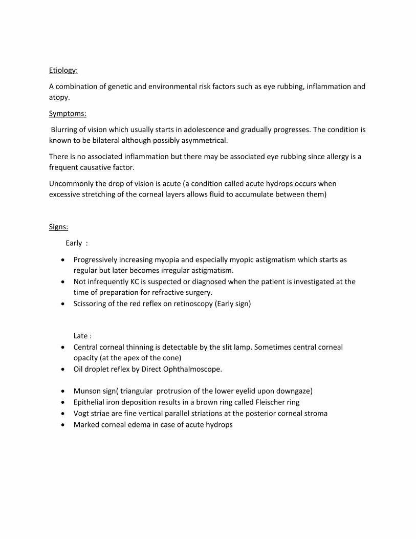
Etiology:
A combination of genetic and environmental risk factors such as eye rubbing, inflammation and
atopy.
Symptoms:
Blurring of vision which usually starts in adolescence and gradually progresses. The condition is
known to be bilateral although possibly asymmetrical.
There is no associated inflammation but there may be associated eye rubbing since allergy is a
frequent causative factor.
Uncommonly the drop of vision is acute (a condition called acute hydrops occurs when
excessive stretching of the corneal layers allows fluid to accumulate between them)
Signs:
Early :
Progressively increasing myopia and especially myopic astigmatism which starts as
regular but later becomes irregular astigmatism.
Not infrequently KC is suspected or diagnosed when the patient is investigated at the
time of preparation for refractive surgery.
Scissoring of the red reflex on retinoscopy (Early sign)
Late :
Central corneal thinning is detectable by the slit lamp. Sometimes central corneal
opacity (at the apex of the cone)
Oil droplet reflex by Direct Ophthalmoscope.
Munson sign( triangular protrusion of the lower eyelid upon downgaze)
Epithelial iron deposition results in a brown ring called Fleischer ring
Vogt striae are fine vertical parallel striations at the posterior corneal stroma
Marked corneal edema in case of acute hydrops

Fig. Munson Sign.
Associations:
Down Syndrome, Marfan Syndrome and Atopy.
Investigations:
Computerized videokeratography (Corneal Topography) is used in diagnosing keratoconus using
specific and sensitive indices and screen prospective refractive surgery patients for
keratoconus. Algorithms to diagnose subclinical, keratoconus ( forme fruste) are continually
being perfected to identify keratoconus suspects before refractive corneal surgery which is
absolutely contraindicated in these patients.
Fig. Videokeratography of a Keratoconus patient showing inferior steepening.

Management depends on whether the condition is early or late :
1) Spectacles or soft contact lenses may succeed to correct the refractive errors in early
cases
2) Rigid contact lenses can successfully correct the associated irregular astigmatism
3) Corneal collagen cross-linking using riboflavin is a method aiming at stopping the
progression of keratoconus in young individuals . The idea is to induce artificial aging (
hardening) of the corneal stroma.
4) Intrastromal corneal rings implantation may flatten the corneal center and thus improve
vision
5) Keratoplasty either PKP or DALK for advanced cases.
II ) Diseases of the Crystalline Lens:
A) Cataract
Cataract means opacity of the crystalline lens. Clinically the term cataract is often reserved to
visually significant opacities i.e. those which affect visual acuity.
It is the leading cause of blindness and visual impairment throughout the world.
Etiology:
The most common cause of cataract is age-related. Other factors include trauma,
uveitis, metabolic defects and the use of corticosteroids.
Description:
Cataract may be described by the zone of the lens involved e.g. Nuclear , cortical or
subcapsular. It may be anterior or posterior. It can be also described in terms of its stage of
development. A cataract with some clear lens remaining is described as immature whereas a
mature cataract has totally opacified cortex.

Fig. Slit lamp examination showing Nuclear Cataract
Fig. Slit lamp exam of Mature Cortical Cataract
Fig. Slit lamp exam of Immature Cortical Cataract

Fig. Posterior subcapsular cataract : a) Slit lamp exam and b) Red reflex
Symptoms:
Gradually progressive blurring of vision followed by defective vision is the classical symptom
Note :
The first symptom may be glare and/ or haloes around the light in posterior subcapsular
cataract due to light scattering
Sometimes the patient with nuclear cataract notices some improvement of near vision.
They may describe a “second sight” when they discover they are able to read without
their reading spectacles (This is due to increase in refractive index of the lens nucleus
resulting in a myopia called index myopia),
Signs:
Cataract can be directly visualized by the penlight when it is mature and by the slit lamp and/or
the red reflex if immature. It is then very important to determine if the diminution of vision can
be accounted for by the cataract alone or if there is discrepancy between the patient’s
symptoms and the extent of cataract. This is achieved by complete systematic eye examination(
see chapter 2) .
In mature cataract where there is no fundus visibility, the projection of light and the ability to
discriminate colors help to give an idea on the retinal and optic nerve function. This is
important for the prognosis of visual improvement after surgery. Other tests may be sometimes
needed such as ultrasonography (or rarely electroretinography and visually evoked potential)
Special attention is to be given to the anterior chamber (AC) depth in order not to miss an
intumescent cataract which causes a shallow AC. This occurs when the lens acutely accumulates
water droplets and swells. The cataract appears glistening due to capsule stretching. The
intraocular pressure (IOP) may become acutely elevated.

Treatment:
There is no medical treatment for cataract. Therefore, surgery is to be considered for cataracts
causing visual symptoms. Except for intumescent cataract, which must be operated promptly,
cataract surgery is non urgent. The timing of surgical intervention varies according to the
patient needs and lifestyle and is not based on a specific level of reduced vision. It is, however,
wise to try to discourage undue delay in order to avoid hypermature cataract or extremely hard
nuclear cataract which are more difficult to operate upon.
The techniques of cataract surgery have been evolving and improving markedly in the past few
decades. Nowadays, the standard technique of cataract extraction is phacoemulsification (PE).
This means the use of ultrasound waves to emulsify the cataract in order to extract it from a
small, often sutureless, incision. Intraocular lens (IOL) is then implanted in the capsular bag in
order to provide the exact refractive power needed after extraction of the cataractous
crystalline lens.
Conventional extracapsular cataract extraction ( ECCE) may be resorted to in some cases such
as extremely hard lens or opacified cornea. In subluxated lenses, intracapsular cataract
extraction ( ICCE) might be chosen by the surgeon and in this case the IOL may be placed in the
anterior chamber (AC) due to the absence of a capsular bag.
B) Lens Subluxation and Dislocation:
In addition to cataract, the crystalline lens can also cause disturbance in image formation on the
retina by virtue of change in its normal position. Subluxation is the displacement of the lens
from its normal position while remaining in the visual axis. The lens does not completely loose
its zonular attachments. Whereas in lens dislocation the whole zonular attachment is lost and
the lens usually falls posteriorly into the vitreous.

Fig. Inferiorly Subluxated Lens.
Fig. Completely Dislocated lens resting on the retina.
Etiology:
Most cases are either congenital or secondary to blunt trauma. Congenital subluxation
(rarely dislocation) is not an uncommon finding in certain systemic syndromes such
Marfan syndrome and homocystinuria.
Symptoms:
In addition to the blurring of vision, monocular diplopia may be a significant complaint
in cases of subluxation
Signs:
The findings in subluxation depend on its degree:

In mild cases the crystalline lens is completely present in the pupillary space. An
abnormal movement of the lens ( tremulous lens or phacodonesis) or the iris (tremulous
iris or iridodonesis) is often seen with eye movement
In more advanced cases the lens edge may appear in the pupil with or even without
dilatation
Astigmatism always present. Its degree depends on the degree of subluxation
In posterior lens dislocation the anterior chamber appears aphakic ( deep with jet black pupil
and loss of Purkinje-sanson images) and fundus examination shows the lens in the vitreous or
on the retina.
Both subluxation and dislocation may be complicated with cataract, uveitis or glaucoma. Also
Marfan syndrome cases are known to be complicated by retinal detachment.
Treatment:
In mild subluxation, spectacle correction may correct astigmatism induced by the slight
lens tilt in eyes with mild subluxation. In more advanced subluxation and in dislocation,
the definitive treatment is surgical removal of the crystalline lens with IOL implantation.
Surgery is mandatory in cases of complications.
III) Vitreous Opacities
A) Vitreous Hemorrhage
It is the presence of blood in the vitreous cavity.
Fig. Fundus exam showing Vitreous Hemorrhage and almost no retinal details

Etiology:
Commonest cause of spontaneous non traumatic vitreous hemorrhage are
*diabetic retinopathy
*posterior vitreous detachment inducing retinal tear
*neovascularization secondary to central or branch vein occlusion
( see retina chapter 8)
B) Vitritis
Vitreous opacity can be due to inflammatory cells. This is described as vitritis. The
transparent vitreous becomes hazy or even opaque. This may occur in certain types of
uveitis and in endophthalmitis. ( see chapter 7)
Fig. Vitreous haze due to Vitritis.

C) Asteroid Hyalosis
They are minute white opacities composed of calcium-containing particles that are deposited in
the vitreous. They are usually discovered accidentally since they rarely cause visual symptoms
unless dense. They are usually not associated with another local or systemic disorder. If really
dense and obscuring vision they can be removed by pars plana vitrectomy.
Fig. Fundus exam showing Asteroid Hyalosis.