Effects of fasting on the expression pattern of FGFs in ...
13
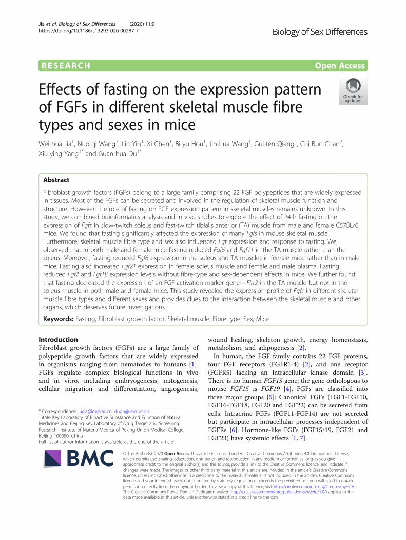
RESEARCH Open Access Effects of fasting on the expression pattern of FGFs in different skeletal muscle fibre types and sexes in mice Wei-hua Jia 1 , Nuo-qi Wang 1 , Lin Yin 1 , Xi Chen 1 , Bi-yu Hou 1 , Jin-hua Wang 1 , Gui-fen Qiang 1 , Chi Bun Chan 2 , Xiu-ying Yang 1* and Guan-hua Du 1* Abstract Fibroblast growth factors (FGFs) belong to a large family comprising 22 FGF polypeptides that are widely expressed in tissues. Most of the FGFs can be secreted and involved in the regulation of skeletal muscle function and structure. However, the role of fasting on FGF expression pattern in skeletal muscles remains unknown. In this study, we combined bioinformatics analysis and in vivo studies to explore the effect of 24-h fasting on the expression of Fgfs in slow-twitch soleus and fast-twitch tibialis anterior (TA) muscle from male and female C57BL/6 mice. We found that fasting significantly affected the expression of many Fgfs in mouse skeletal muscle. Furthermore, skeletal muscle fibre type and sex also influenced Fgf expression and response to fasting. We observed that in both male and female mice fasting reduced Fgf6 and Fgf11 in the TA muscle rather than the soleus. Moreover, fasting reduced Fgf8 expression in the soleus and TA muscles in female mice rather than in male mice. Fasting also increased Fgf21 expression in female soleus muscle and female and male plasma. Fasting reduced Fgf2 and Fgf18 expression levels without fibre-type and sex-dependent effects in mice. We further found that fasting decreased the expression of an FGF activation marker gene—Flrt2 in the TA muscle but not in the soleus muscle in both male and female mice. This study revealed the expression profile of Fgfs in different skeletal muscle fibre types and different sexes and provides clues to the interaction between the skeletal muscle and other organs, which deserves future investigations. Keywords: Fasting, Fibroblast growth factor, Skeletal muscle, Fibre type, Sex, Mice Introduction Fibroblast growth factors (FGFs) are a large family of polypeptide growth factors that are widely expressed in organisms ranging from nematodes to humans [1]. FGFs regulate complex biological functions in vivo and in vitro, including embryogenesis, mitogenesis, cellular migration and differentiation, angiogenesis, wound healing, skeleton growth, energy homeostasis, metabolism, and adipogenesis [2]. In human, the FGF family contains 22 FGF proteins, four FGF receptors (FGFR1-4) [2], and one receptor (FGFR5) lacking an intracellular kinase domain [3]. There is no human FGF15 gene; the gene orthologous to mouse FGF15 is FGF19 [4]. FGFs are classified into three major groups [5]: Canonical FGFs (FGF1-FGF10, FGF16-FGF18, FGF20 and FGF22) can be secreted from cells. Intracrine FGFs (FGF11-FGF14) are not secreted but participate in intracellular processes independent of FGFRs [6]. Hormone-like FGFs (FGF15/19, FGF21 and FGF23) have systemic effects [1, 7]. © The Author(s). 2020 Open Access This article is licensed under a Creative Commons Attribution 4.0 International License, which permits use, sharing, adaptation, distribution and reproduction in any medium or format, as long as you give appropriate credit to the original author(s) and the source, provide a link to the Creative Commons licence, and indicate if changes were made. The images or other third party material in this article are included in the article's Creative Commons licence, unless indicated otherwise in a credit line to the material. If material is not included in the article's Creative Commons licence and your intended use is not permitted by statutory regulation or exceeds the permitted use, you will need to obtain permission directly from the copyright holder. To view a copy of this licence, visit http://creativecommons.org/licenses/by/4.0/. The Creative Commons Public Domain Dedication waiver (http://creativecommons.org/publicdomain/zero/1.0/) applies to the data made available in this article, unless otherwise stated in a credit line to the data. * Correspondence: [email protected]; [email protected] 1 State Key Laboratory of Bioactive Substance and Function of Natural Medicines and Beijing Key Laboratory of Drug Target and Screening Research, Institute of Materia Medica of Peking Union Medical College, Beijing 100050, China Full list of author information is available at the end of the article Jia et al. Biology of Sex Differences (2020) 11:9 https://doi.org/10.1186/s13293-020-00287-7
Transcript of Effects of fasting on the expression pattern of FGFs in ...
RESEARCH Open Access
Effects of fasting on the expression patternof FGFs in different skeletal muscle fibretypes and sexes in miceWei-hua Jia1, Nuo-qi Wang1, Lin Yin1, Xi Chen1, Bi-yu Hou1, Jin-hua Wang1, Gui-fen Qiang1, Chi Bun Chan2,Xiu-ying Yang1* and Guan-hua Du1*
Abstract
Fibroblast growth factors (FGFs) belong to a large family comprising 22 FGF polypeptides that are widely expressedin tissues. Most of the FGFs can be secreted and involved in the regulation of skeletal muscle function andstructure. However, the role of fasting on FGF expression pattern in skeletal muscles remains unknown. In thisstudy, we combined bioinformatics analysis and in vivo studies to explore the effect of 24-h fasting on theexpression of Fgfs in slow-twitch soleus and fast-twitch tibialis anterior (TA) muscle from male and female C57BL/6mice. We found that fasting significantly affected the expression of many Fgfs in mouse skeletal muscle.Furthermore, skeletal muscle fibre type and sex also influenced Fgf expression and response to fasting. Weobserved that in both male and female mice fasting reduced Fgf6 and Fgf11 in the TA muscle rather than thesoleus. Moreover, fasting reduced Fgf8 expression in the soleus and TA muscles in female mice rather than in malemice. Fasting also increased Fgf21 expression in female soleus muscle and female and male plasma. Fastingreduced Fgf2 and Fgf18 expression levels without fibre-type and sex-dependent effects in mice. We further foundthat fasting decreased the expression of an FGF activation marker gene—Flrt2 in the TA muscle but not in thesoleus muscle in both male and female mice. This study revealed the expression profile of Fgfs in different skeletalmuscle fibre types and different sexes and provides clues to the interaction between the skeletal muscle and otherorgans, which deserves future investigations.
Keywords: Fasting, Fibroblast growth factor, Skeletal muscle, Fibre type, Sex, Mice
IntroductionFibroblast growth factors (FGFs) are a large family ofpolypeptide growth factors that are widely expressedin organisms ranging from nematodes to humans [1].FGFs regulate complex biological functions in vivoand in vitro, including embryogenesis, mitogenesis,cellular migration and differentiation, angiogenesis,
wound healing, skeleton growth, energy homeostasis,metabolism, and adipogenesis [2].In human, the FGF family contains 22 FGF proteins,
four FGF receptors (FGFR1-4) [2], and one receptor(FGFR5) lacking an intracellular kinase domain [3].There is no human FGF15 gene; the gene orthologous tomouse FGF15 is FGF19 [4]. FGFs are classified intothree major groups [5]: Canonical FGFs (FGF1-FGF10,FGF16-FGF18, FGF20 and FGF22) can be secreted fromcells. Intracrine FGFs (FGF11-FGF14) are not secretedbut participate in intracellular processes independent ofFGFRs [6]. Hormone-like FGFs (FGF15/19, FGF21 andFGF23) have systemic effects [1, 7].
© The Author(s). 2020 Open Access This article is licensed under a Creative Commons Attribution 4.0 International License,which permits use, sharing, adaptation, distribution and reproduction in any medium or format, as long as you giveappropriate credit to the original author(s) and the source, provide a link to the Creative Commons licence, and indicate ifchanges were made. The images or other third party material in this article are included in the article's Creative Commonslicence, unless indicated otherwise in a credit line to the material. If material is not included in the article's Creative Commonslicence and your intended use is not permitted by statutory regulation or exceeds the permitted use, you will need to obtainpermission directly from the copyright holder. To view a copy of this licence, visit http://creativecommons.org/licenses/by/4.0/.The Creative Commons Public Domain Dedication waiver (http://creativecommons.org/publicdomain/zero/1.0/) applies to thedata made available in this article, unless otherwise stated in a credit line to the data.
* Correspondence: [email protected]; [email protected] Key Laboratory of Bioactive Substance and Function of NaturalMedicines and Beijing Key Laboratory of Drug Target and ScreeningResearch, Institute of Materia Medica of Peking Union Medical College,Beijing 100050, ChinaFull list of author information is available at the end of the article
Jia et al. Biology of Sex Differences (2020) 11:9 https://doi.org/10.1186/s13293-020-00287-7

Studies show that fasting has several healthy benefits[8], such as increasing longevity, alleviating obesity andother non-infectious diseases [9], and anti-tumour [10].However, fasting can also lead to muscle wasting andcause life-threatening problems [11]. These effects areclosely related to the physiological response of the skel-etal muscle to fasting. The skeletal muscle is not only amotor organ, but also a metabolic organ, which storesmost of the body’s energy and regulates systemic metab-olism. During fasting or energy deficits, the skeletal mus-cles would adjust energy demands and preserve energyhomeostasis [12]. Ours and other studies found thatgrowth factors coordinate metabolic reprogramming inmuscle during fasting [13].FGFs have long been implicated in skeletal muscle
structure and function [14]. Several FGFs have beenproved to function as myokines, such as FGF2 [15] andFgF21 [15, 16]. A myokine is a protein or peptide that itis derived and secreted from the skeletal muscle and per-forms biological functions in an endocrine or paracrinemanner [17]. FGFs were initially associated with myo-genesis [18]. FGFs, FGFRs and their co-receptor (heparinor heparan sulfate) are expressed in a time- and space-dependent manner during all stages of skeletal develop-ment [19]. FGF15/19 was found to ameliorate skeletalmuscle atrophy [20]. Studies reported that Fgf21expressed by muscle controls muscle mass [16] and in-creases insulin sensitivity [21].The skeletal muscles of mammals are composed of
multiple muscle fibre types, including oxidative slow-twitch (type I), mixed oxidative-glycolytic fast-twitch(type IIa) and glycolytic fast-twitch (type IIb) myofibres,which differ in their metabolic properties [22]. The so-leus muscle has a higher proportion of type I muscle fi-bres than that of many other muscles [23], and thetibialis anterior (TA) muscle has significantly more typeIIb fibres [24]. Studies report that fasting influences FGFexpression in the skeletal muscle. It was observed thatfasting and metabolic disorders induced FGF21 releasefrom muscles [16], whereas the muscle Fgf6 mRNAlevels were not significantly affected by animal feedingstatus [25]. In addition, biological sexes have differentresponses to muscle health, largely due to the significantvariety of general muscle fibre type, satellite cell activa-tion and proliferation, anabolic and catabolic factors,and hormonal interactions between males and females[26]. Our previous study found that Fgf15 expression ishigher in the skeletal muscle of female mice than that ofmale mice. Fasting reduces Fgf15 expression in femalemuscles but had no effect on male muscles [27].However, the mechanisms by which fasting influences
the expression pattern of skeletal muscle FGFs is stillunknown. In this study, we investigated the expressionof all the 22 Fgfs in slow-twitch soleus and fast-twitch
tibialis anterior (TA) muscle, to explore the influence ofmuscle fibre types and sexes on Fgf expression duringfasting in female and male mice.
Materials and methodsAnimalsThis study and all the procedures using animals wereapproved by the Animal Care and Use Committee of theInstitute of Materia Medica, Chinese Academy of Med-ical Sciences. Female and male C57BL/6 mice (20–22 g),obtained from the Institute of Laboratory Animal Sci-ence, Chinese Academy of Medical Sciences (Beijing,China), were used in this study. Mice were housed in en-vironmentally controlled conditions with a 12-h light/dark cycle at a temperature of 22 ± 3 °C and humidity of55 ± 5%. Mice were provided free access to food andwater for 7 days before the experiment.
Animal fasting procedureAfter being housed for 7 days, female and male C57BL/6mice were randomly allocated either to a fed group(Fed) or a fasted group (Fast) with six mice in eachgroup. The fasted mice were placed in new cages with-out providing any food in the cage at 9 A.M. After fast-ing for 24 h [16, 28, 29], the Fed and Fast mice wereanaesthetised with isoflurane, blood samples were col-lected via the abdominal vein and the indicated skeletalmuscles from different anatomical positions were quicklyremoved and frozen in liquid nitrogen for RNA extrac-tion and further experiments.
Quantitative reverse transcription PCRTotal RNA was isolated from ~ 50mg muscles or awhole piece of the soleus muscle by homogenisation inTrizol Isolation Reagent (Invitrogen, USA), and thenpurified with Direct-zol RNA Kits (cat. no. R2052,ZYMO search, USA) as previously described [27, 30].Reverse transcription was performed using 1 μg of totalRNA with a reverse transcription reaction mix that con-tained Superscript III reverse transcriptase (Invitrogen,USA) and Oligo-dT17 as primers. Specific mRNA contentwas determined using AceQ qPCR SYBR Green MasterMix (cat. no. Q111-03, Vazyme, Biotech) on a CFX-96Real-time PCR System (Bio-Rad, USA) with gene-specificprimer pairs (Table 1). The total reaction volume was10 μl. The results were quantified after normalisation withTATA-box binding protein (Tbp) [31, 32].
Measurement of FGFs in mice plasmaPlasma concentrations of FGF15, FGF21, FGF23, andFLRT2 were quantified using the ELISA kits (cat.no.CSB-EL522052MO, CUSABIO; cat.no.ab212160, Abcam;cat.no.ab213863, Abcam; cat.no. JL38212, Shanghai
Jia et al. Biology of Sex Differences (2020) 11:9 Page 2 of 13

Jianglai Biotech) according to the manufacturer’s in-structions, respectively.
GEO data acquisition and FGF expression analysisWe first performed human tissue-specific FGF expres-sion analysis in non-diseased tissues using Genotype-Tissue Expression (GTEx) expression dataset (https://gtexportal.org/home/multiGeneQueryPage), which is acomprehensive public resource to study tissue-specificgene expression and regulation. For each gene, expres-sion values were normalised across samples using an in-verse normal transform. Next, we explored Fgfexpression in the skeletal muscle in mice using the GeneExpression Omnibus (GEO) database. The GEO is apublic functional genomics data repository that providesa multimodal data repository and retrieval system formicroarray and next-generation sequencing gene expres-sion profiles. This study queried and downloaded therelevant studies from the GEO website (The Gene Ex-pression Omnibus, https://www.ncbi.nlm.nih.gov/gds/)[33]. We identified a suitable dataset comparing expres-sion in the skeletal muscles of fed and fasting mice,which is an RNA sequencing dataset (accession numberGSE107787). In this dataset study, male C57BL/6 miceat 8 weeks of age were randomly divided into an ad libi-tum fed group (n = 18) or a 24-h fasted group (n = 18).For complete experimental details, please refer to a pre-vious publication [29].
Statistical analysisDifferences between the groups were tested using un-paired two-tailed Student’s t test or one-way ANOVA.Data were analysed using GraphPad Prism (GraphPadSoftware, USA). Data are presented as means ± S.E.M.The significance level was set at P ≤ 0.05.
ResultsBioinformatic analysis found FGFs expressed differentlyand could be influenced diversely by fastingWe compared expression levels of human FGFs in theskeletal muscle with that in other tissues. Bioinformaticresults from GTEx dataset show that FGFs wereexpressed divergently in different tissues. Different FGFsdemonstrated distinctive expression in the human
Table 1 Primers used for real-time PCR
Pairs Genes Primer sequence
1 Mouse Fgf1 5′ CCCTGACCGAGAGGTTCAAC
Mouse Fgf1 3′ GTCCCTTGTCCCATCCACG
2 Mouse Fgf2 5′ GCGACCCACACGTCAAACTA
Mouse Fgf2 3′ TCCCTTGATAGACACAACTCCTC
3 Mouse Fgf3 5′ TACAACGCAGAGTGTGAGTTTG
Mouse Fgf3 3′ CACCGACACGTACCAAGGTC
4 Mouse Fgf4 5′ TACCCCGGTATGTTCATGGC
Mouse Fgf4 3′ TTACCTTCATGGTAGGCGACA
5 Mouse Fgf5 5′ AAGTAGCGCGACGTTTTCTTC
Mouse Fgf5 3′ CTGGAAACTGCTATGTTCCGAG
6 Mouse Fgf6 5′ CAGGCTCTCGTCTTCTTAGGC
Mouse Fgf6 3′ AATAGCCGCTTTCCCAATTCA
7 Mouse Fgf7 5′ CTCTACAGATCATGCTTCCACC
Mouse Fgf7 3′ ACAGAACAGTCTTCTCACCCT
8 Mouse Fgf8 5′ AGAGCCTGGTGACGGATCA
Mouse Fgf8 3′ CTTCCAAAAGTATCGGTCTCCAC
9 Mouse Fgf9 5′ ATGGCTCCCTTAGGTGAAGTT
Mouse Fgf9 3′ TCCGCCTGAGAATCCCCTTT
10 Mouse Fgf10 5′ TTTGGTGTCTTCGTTCCCTGT
Mouse Fgf10 3′ TAGCTCCGCACATGCCTTC
11 Mouse Fgf11 5′ TAGCCTGATCCGACAGAAGC
Mouse Fgf11 3′ GGCAGAACAGTTTGGTGACG
12 Mouse Fgf12 5′ CAGGCCGTGCATGGTTTCTA
Mouse Fgf12 3′ TCGTGTAGTGATGGTTCTCTGT
13 Mouse Fgf13 5′ CTCATCCGGCAAAAGAGACAA
Mouse Fgf13 3′ TTGGAGCCAAAGAGTTTGACC
14 Mouse Fgf14 5′ CCCCAGCTCAAGGGCATAG
Mouse Fgf14 3′ TGATGGGTAGAGGTAACCTTCTC
15 Mouse Fgf15 5′ ATGGCGAGAAAGTGGAACGG
Mouse Fgf15 3′ CTGACACAGACTGGGATTGCT
16 Mouse Fgf16 5′ GTGTTTTCCGGGAACAGTTTGA
Mouse Fgf16 3′ GGTGAGCCGTCTTTATTCAGG
17 Mouse Fgf17 5′ GGCAGAGAGCGAGAAGTACAT
Mouse Fgf17 3′ CGGTGAACACGCAGTCTTTG
18 Mouse Fgf18 5′ CCTGCACTTGCCTGTGTTTAC
Mouse Fgf18 3′ TGCTTCCGACTCACATCATCT
19 Mouse Fgf20 5′ GGTGGGGTCGCACTTCTTG
Mouse Fgf20 3′ GATACCGAAGAGACTGTGATCCT
20 Mouse Fgf21 5′ CTGCTGGGGGTCTACCAAG
Mouse Fgf21 3′ CTGCGCCTACCACTGTTCC
21 Mouse Fgf22 5′ CCAGGACAGTATAGTGGAGATCC
Mouse Fgf22 3′ AGTAGACCCGCGACCCATAG
22 Mouse Fgf23 5′ ATGCTAGGGACCTGCCTTAGA
Mouse Fgf23 3′ AGCCAAGCAATGGGGAAGTG
Table 1 Primers used for real-time PCR (Continued)
Pairs Genes Primer sequence
23 Mouse Flrt2 5′ ATGGGCCTACAGACTACAAAGT
Mouse Flrt2 3′ CAGCGGCATACACTAGGGC
24 Mouse Flrt3 5′ CCTCATCGGGACTAAAATTGGG
Mouse Flrt3 3′ GCAAGTTCTTCAAATCGGAAGGA
25 Mouse Tbp 5′ ACCCTTCACCAATGACTCCTATG
Mouse Tbp 3′ ATGATGACTGCAGCAAATCGC
Jia et al. Biology of Sex Differences (2020) 11:9 Page 3 of 13

skeletal muscle, of which FGF13, FGF11, FGF6, FGF2and FGF7 had the top five expression levels (Fig. 1a).Next, fasting-induced expressions of Fgfs in the skeletalmuscle of male mice were analysed using a dataset fromNCBI Gene Expression Omnibus (GEO). In this dataset,mice were fasted for 24 h and the gastrocnemius skeletalmuscles were taken for RNA sequencing experiment (ac-cession numbers GSE107787) [29]. According to the
analysis of the dataset results, the top five Fgf genes withthe highest expression levels in mice gastrocnemius wereFgf13, Fgf1, Fgf6, Fgf11 and Fgf7. Meanwhile, in the caseof canonical FGFs, fasting induced Fgf20 and Fgf22 ex-pression by 13.2- and 2.5-fold and reduced Fgf2, Fgf5,Fgf6, Fgf7, Fgf9, Fgf10 and Fgf18 expression by 2.1-, 8.5-,6.0-, 2.1-, 1.4-, 1.6- and 4.6-fold, respectively. Regardingintracellular FGFs, fasting reduced Fgf11, Fgf13
Fig. 1 Bioinformatics analysis demonstrates that fasting influences FGF expression in skeletal muscles. a Human FGF expression levels in skeletalmuscles compared with those in other tissues. Left: The expression of FGFs in various tissues. Right: Heatmap of FGF expression in skeletalmuscles. b Expression of Fgfs in muscle from 24-h fasted mice. Left: Heatmap of basal expression of Fgfs in skeletal muscles. Right: Expression ofFgfs in gastrocnemius muscle. Data represent means ± S.E.M, n = 18; biological replicates. *P < 0.05, **P < 0.01 and *** P < 0.001 by one-wayANOVA followed by unpaired two-tailed Student’s t test
Jia et al. Biology of Sex Differences (2020) 11:9 Page 4 of 13

expression by 2.5- and 1.5-fold. The expression of anendocrine FGF—Fgf23—was reduced by 6.4-fold (Fig.1b). However, data on Fgf15 was not obtained from theRNA sequencing results.
Differential expression of Fgfs in male and female miceaccording to fibre type of the skeletal muscleOur previous study showed that slow-twitch soleus andfast-twitch TA have different myokine expressions [27].In this study, we found that the muscle fibre types alsoinfluenced expressions of Fgfs. The results showed thatin male C57BL/6 mice, expressions of canonical Fgf1–Fgf7, Fgf9, Fgf10, Fgf16, Fgf17 and Fgf20 in the soleus
muscle were 5.3-, 3.0-, 34.4-, 37.8-, 23.4-, 3.7-, 8.0-,10.3-, 36.3-, 38.0-, 41.9- and 112.4-fold higher comparedwith those in the TA muscle, respectively. IntracellularFgf12 and Fgf13 expressed in the soleus muscle were 2.8-and 29.8-fold higher than those in the TA muscle, whileFgf11 in the TA muscle was 21.2-fold higher than that inthe soleus muscle. Expressions of endocrine Fgf15, Fgf21and Fgf23 in the soleus muscle were 59.0-, 65.1- and39.7-fold higher than those of the TA muscle (Fig. 2a).In female C57BL/6 mice, canonical Fgf1-Fgf7, Fgf9,
Fgf10, Fgf16, Fgf17 and Fgf20 had 2.2-, 2.6-, 4.9-, 8.2-,5.2-, 3.3-, 2.9-, 3.7-, 6.3-, 6.8-, 9.4- and 6.6-fold higherexpression in the soleus than those in the TA muscle,
Fig. 2 Fgf expression levels in different muscle fibre types of male and female C57BL/6 mice. a Basal Fgf expression level in male mice. Left:Heatmap of basal Fgf expression in the TA and soleus muscles. Right: Comparison of Fgf expression levels between the soleus and TA muscles. bBasal Fgf expression level in female mice. Left: Heatmap of basal Fgf expression in the TA and soleus muscles. Right: Comparison of Fgf expressionlevels between the soleus and TA muscles. Data represent means ± S.E.M, n = 6; biological replicates. *P < 0.05, **P < 0.01 and ***P < 0.001 byone-way ANOVA followed by unpaired two-tailed Student’s t test
Jia et al. Biology of Sex Differences (2020) 11:9 Page 5 of 13

respectively, which is consistent with male mice. Fgf8and Fgf11 in the TA muscle were 2.1- and 43.5-foldhigher than those in the soleus muscle. IntracellularFgf12 was expressed in the soleus muscle as 2.1-fold asmuch as that in the TA muscle, while Fgf11 in the TAmuscle was 43.5-fold higher than that in the soleusmuscle. Expressions of endocrine Fgf15, Fgf21 and Fgf23in the soleus muscle were 6.5-, 4.8- and 6.0-fold higherthan those in the TA muscle, respectively (Fig. 2b).
Fgf expression in the TA and soleus muscles duringfasting in male C57BL/6 miceIn this study, we analysed all expression levels of Fgfs inthe TA and soleus muscles of fasting male C57BL/6mice. Our study showed that fasting influenced the Fgfexpression of the TA and soleus. In fast-twitch TAmuscle, fasting reduced canonical Fgf2, Fgf6 and Fgf18expression by 2.4-, 3.2- and 2.5-fold, respectively. Intra-cellular Fgf11 was reduced by 2.9-fold during fasting(Fig. 3a). On the contrary, in slow-twitch soleus muscle,24-h fasting induced the expression of Fgf2 and Fgf18 by4.0- and 2.8-fold, respectively (Fig. 3b).
Fgf expression in the TA and soleus muscles duringfasting in female C57BL/6 miceIn this study, the effects of fasting on the pattern of Fgfexpression were different in female C57BL/6 mice com-pared with those in male mice. In fast-twitch TA muscle,as for canonical FGFs, 24-h fasting reduced Fgf2, Fgf6,Fgf8, Fgf18 and Fgf20 by 3.4-, 2.3-, 4.4-, 3.4- and 2.3-fold,respectively. Intracellular Fgf11, Fgf12 and Fgf14 were re-duced by 4.2-, 1.5- and 4.2-fold, respectively. EndocrineFgf15 and Fgf23 were reduced by 2.0- and 1.6-fold (Fig. 4a).In slow-twitch soleus muscle, 24-h fasting reduced the ex-pression of Fgf2, Fgf8 and Fgf18 by 5.0-, 3.8- and 4.9-fold,respectively, while endocrine Fgf21 was 1.9-fold higher thanthat of fed soleus muscle (Fig. 4b).
Differential expression of Fgfs in the soleus and TAmuscles according to the sex of miceThe expression of Fgfs in the soleus muscle was similarin male and female mice (Fig. 5a), while they were dra-matically different in the TA muscle (Fig. 5b). Moreover,most of the Fgf expressions were higher in female TAmuscle compared with those of male TA muscle, includ-ing canonical Fgf1, Fgf3–Fgf5, Fgf7–Fgf10, Fgf16, Fgf17and Fgf20; intracellular Fgf12 and Fgf13; and endocrineFgf15, Fgf21 and Fgf23.
Differential expression of Flrts in the soleus and TAmuscles of male and female miceFibronectin-leucine-rich transmembrane proteins(FLRTs) are markers of FGF activity [34, 35]. Fasting re-duced Flrt2 and Flrt3 expression by 2.8- and 1.7-fold in
mice gastrocnemius skeletal muscle (Fig. 6a). In maleC57BL/6 mice, Flrt2 and Flrt3 in the soleus muscle were16.1- and 30.8-fold higher than those of the TA muscle,and so is in female mice, Flrt2 and Flrt3 in the soleusmuscle were 3.4- and 3.8-fold higher than those of theTA muscle (Fig. 6b). Furthermore, in male C57BL/6mice, fasting reduced Flrt2 expression in the TA muscle,while fasting did not affect Flrt2 expression in the soleusmuscle (Fig. 6c). Female C57BL/6 mice showed similarchanges, fasting also reduced Flrt2 expression by 1.8-foldin the TA muscle and had no effect on the soleus muscle(Fig. 6d). In addition, in the TA muscle, basal Flrt2 andFlrt3 express more in female mice than in male C57BL/6 mice, while there is no difference in the soleus muscle(Fig. 6e).
Endocrine FGF15, FGF21 and FGF23 levels in mice plasmaEndocrine FGFs have systemic effects via blood circula-tion, and plasma endocrine FGF concentration may alteraccording to condition changes or diseases. In severalsituations, they can act as biomarkers for some diseases[6]. Fasting significantly increased FGF21 circulatinglevel in both male and female mice by 11.9- and 23.2-fold, respectively. FGF23 increased by 1.6-fold in femalemice only. As for FLRTs, we measured FLRT2 andfound it decreased by 13.1-fold in respond to fasting inmale mice, but slightly decreased in female mice with nosignificant difference (Fig. 7).
DiscussionFasting and fed conditions divergently influence skeletalmuscle structure and metabolism [36]. In this study, wefound that fasting influenced expressions of Fgfs inmouse skeletal muscle. Furthermore, sex and skeletalmuscle fibre type also influenced Fgf expression and theresponse to fasting.The 15 canonical FGFs (FGF1–FGF10, FGF16–FGF18,
FGF20 and FGF22) act predominantly in an autocrineand paracrine fashion and bind to FGFRs in a complexwith heparan sulfate proteoglycans (HSPGs) [37]. In thisstudy, we found that fasting reduced Fgf2 and Fgf18 ex-pression levels in both fibre types and sexes in mice.This result is consistent with a previous study that re-vealed that in fish, fgf2 mRNA level increased by 2.5-foldat both 4 and 12 days after refeeding compared to thatin fasting conditions [38]. FGF2 released from skeletalmuscle cells plays important roles in myogenesis andmuscle development [39]. FGF2 contributes to musclecompensatory growth induced by refeeding [38]. Ex-tracts from slow-twitch muscles contained higher levelsof FGF2 than those from fast-twitch muscles [40]. Thisis in line with our results, that in both male and femalemice, the expression levels of all Fgfs were higher orcomparable in slow-twitch soleus muscle compared with
Jia et al. Biology of Sex Differences (2020) 11:9 Page 6 of 13

those in fast-twitch TA muscle. What’s more, fastingreduced Fgf6 expression in the TA muscle rather thanthe soleus in both the male and female. Fasting re-duced Fgf8 expression in female rather than male
mice in both the TA and soleus muscles. Thus far,effects of fasting on the expression levels of other ca-nonical Fgfs, except Fgf2, in the skeletal muscle havenot been reported.
Fig. 3 Fasting-stimulated Fgf expression pattern changes were different in the TA and soleus muscles in male C57BL/6 mice. a Fgf expression inthe TA muscle. b Fgf expression in the soleus muscle. Data represent means ± S.E.M, n = 6; biological replicates. *P < 0.05, **P < 0.01 and *** P <0.001 by one-way ANOVA followed by unpaired two-tailed Student’s t test
Jia et al. Biology of Sex Differences (2020) 11:9 Page 7 of 13

The intracellular FGFs (FGF11–FGF14) are non-secretory forms that bind and modulate voltage-gatedsodium channels (VGSCs) [41]. Fgf13 is highly expressedin muscle and inhibits C2C12 cell proliferation and
differentiation [42]. In our study, we found that fastingdecreased Fgf13 expression in gastrocnemius skeletalmuscle. In addition, it is of interest that Fgf11 isexpressed more in the TA than in the soleus, which
Fig. 4 Fasting-stimulated Fgf expression pattern changes were different in the TA and soleus muscles in female C57BL/6 mice. a Fgf expression inthe TA muscle. b Fgf expression in the soleus muscle. Data represent means ± S.E.M, n = 6; biological replicates. *P < 0.05, **P < 0.01 and ***P <0.001 by one-way ANOVA followed by unpaired two-tailed Student’s t test
Jia et al. Biology of Sex Differences (2020) 11:9 Page 8 of 13

differs from any other Fgfs. And fasting reduced Fgf11expression in both the male and female TA muscle ra-ther than the soleus. Fasting reduced Fgf12 and Fgf14 ex-pression levels in female soleus muscle other than TA.
However, there have no reports related to the effects offasting on intracellular FGF expression so far.The three endocrine FGFs (FGF15/19, FGF21 and
FGF23) act as hormones and lack affinity for HSPG
Fig. 5 Expression of Fgfs in skeletal muscles were divergent in male and female C57BL/6 mice. a Comparison of Fgf expression levels in the TAmuscle. b Comparison of Fgf expression levels in the soleus muscle. Data represent means ± S.E.M, n = 6; biological replicates. *P < 0.05, **P <0.01 and ***P < 0.001 by one-way ANOVA followed by unpaired two-tailed Student’s t test
Jia et al. Biology of Sex Differences (2020) 11:9 Page 9 of 13

binding, which enables their diffusion from the site ofproduction into the circulation. Endocrine FGFs areexpressed in various tissues and organs. They play rolesin cell growth, differentiation, bile acid, glucose and lipidmetabolism, as well as in the control of vitamin D andphosphate levels, thereby maintaining whole-bodyhomeostasis [43, 44]. Secretory forms of FGFs directlyand indirectly control the differentiation of fast- andslow-twitch muscle lineages, respectively [45]. The threeendocrine FGF mRNA expressions are higher in the so-leus than those in the TA in both sexes, and as for TA,
they are higher in female than those in male. Oestrogencan increase hepatic production of FGF21, suggestingthat sex influences gene expression [46]. In this study,we found that fasting reduced Fgf15 mRNA level in fe-male TA muscle, while had no notably influence on theprotein level in plasma. FGF15/19 induced skeletalmuscle hypertrophy and blocked muscle atrophy [20].FGF15/19 is mainly secreted from the small intestine inresponse to feed [47]. FGF21 has been reported nega-tively to regulate muscle mass and contribute towardsskeletal muscle atrophy [16]. In this study, we found that
Fig. 6 Fasting-stimulated Flrt expression pattern in C57BL/6 mice. a Expression of Flrts in gastrocnemius muscle from 24-h fasted mice. b BasalFlrt expression levels in different muscle fibre types of male (light) and female (right) C57BL/6 mice. c Fasting-stimulated Flrt expression in the TAmuscle of male (light) and female (right) C57BL/6 mice. d Fasting-stimulated Flrt expression in the soleus muscle of male (light) and female (right)C57BL/6 mice. e Fasting-stimulated Flrt expression in the TA (light) and soleus (right) muscles of male and female C57BL/6 mice. Data representmeans ± S.E.M, n = 6; biological replicates. *P < 0.05, **P < 0.01 and ***P < 0.001 by one-way ANOVA with unpaired two-tailed Student’s t test
Jia et al. Biology of Sex Differences (2020) 11:9 Page 10 of 13

fasting increased Fgf21 mRNA expression level in femalesoleus muscle and increased FGF21 level in plasma inboth female and male mice. Emerging evidence hasshown that fasting increases hepatic Fgf21 mRNA ex-pression and plasma FGF21 level in mice [47]. FGF21plays a role in fasting-induced muscle atrophy and weak-ness. However, a report suggested that fasting signifi-cantly decreased plasma FGF21 in obese subjects [48].Circadian regulation has a stronger impact on plasmaFGF21 than that of fasting within a 72-h period [49]. Wealso found that fasting decreased Fgf23 mRNA expres-sion in gastrocnemius skeletal muscle in male mice, butincreased FGF23 protein expression in female miceplasma. FGF23 is a bone-derived factor [50] and plays arole in metabolic diseases [51]. FGF23 induces cellularsenescence in human skeletal muscle mesenchymal stemcells [52]. A study indicated that the expression ofFGF23 is higher in females than in males [53], while we
found no significant difference in the protein expressionbetween two sexes.Regarding the FGFR activation marker gene, we found
that Flrts express more in the soleus than those in the TA,and fasting decreased the expression of Flrts in the TAmuscle in two sexes, while in the soleus, the expression ofFlrts was not significantly affected. As for FLRT2 inplasma, fasting reduced its concentration in male mice.What’s more, Flrt expression was sex-differentiated. Therewere more Flrts in female than in male mice TA muscle,while no sexual differences in the soleus muscle. However,in plasma, there is more FLRT2 protein expression inmale mice. Thus, our study shows that the effect of fastingon FGF activity is closely related to the fibre type ofskeletal muscle.However, the lack of the data on FGF protein expressions
in muscle is a limitation of this study. Our future work willfocus on the related mechanisms and implications of
Fig. 7 Expression of endocrine FGFs and FLRT2 in plasma. a Comparison of FGF15, FGF21, FGF23 and FLRT2 expression levels in response tofasting in male mice plasma. b Comparison of FGF15, FGF21, FGF23 and FLRT2 expression levels in response to fasting in female mice plasma. cComparison of FGF15, FGF21, FGF23 and FLRT2 expression levels in female and male mice plasma. Data represent means ± S.E.M, n = 6;biological replicates. *P < 0.05, **P < 0.01 and ***P < 0.001 by one-way ANOVA with unpaired two-tailed Student’s t test
Jia et al. Biology of Sex Differences (2020) 11:9 Page 11 of 13

individual FGFs on the function and structure of the skel-etal muscle.
Perspectives and significanceThis study provides a new insight into the effects of fast-ing on FGF expression in skeletal muscles. In addition,our study uncovers the expression profiles of FGFs in dif-ferent muscle fibre types and sexes. The knowledge of thebiological characteristics of FGF mRNA expression is crit-ical for the research on skeletal muscle. This may also helpto identify new biomarker in skeletal muscles and noveltherapeutics targeting on fasting related health benefits.
AbbreviationsFGFs: Fibroblast growth factors; FLRT: Fibronectin-leucine-richtransmembrane; GEO: Gene Expression Omnibus; GTEx: Genotype-TissueExpression; HSPGs: Heparan sulfate proteoglycans; TA: Tibialis anterior;VGSCs: Voltage-gated sodium channels
AcknowledgmentsThanks to the mice who have contributed to scientific research.
Authors’ contributionsDGH, YXY, CCB and QGF contributed to the study conception and design.JWH, WNQ, YL, CX, HBY and WJH contributed to the acquisition of data. JWHand YXY contributed to the analysis of data. JWH, YXY and DGH wrote themanuscript. All authors read and approved the final manuscript.
FundingThis work was supported by the following foundations: Chinese Academy ofMedical Sciences (CAMS-I2M 2016-I2M-3-007 and 2017-I2M-1-010), NationalMajor Science and Technology Projects of China (2018ZX09711001-012,2018ZX09711001-003-005 and 2017YFG0112900) and the National NaturalScience Foundation of China (81470159 and 81770847).
Availability of data and materialsThe datasets used and/or analysed during the current study are availablefrom the corresponding author on reasonable request.
Ethics approval and consent to participateAll mouse procedures were approved by the Animal Care and UseCommittee of the Institute of Materia Medica, Chinese Academy of MedicalSciences.
Consent for publicationNot applicable.
Competing interestsThe authors declare that they have no competing interests.
Author details1State Key Laboratory of Bioactive Substance and Function of NaturalMedicines and Beijing Key Laboratory of Drug Target and ScreeningResearch, Institute of Materia Medica of Peking Union Medical College,Beijing 100050, China. 2State Key Laboratory of PharmaceuticalBiotechnology, School of Biological Sciences, The University of Hong Kong,Hong Kong, China.
Received: 16 October 2019 Accepted: 2 March 2020
References1. Fon TK, Bookout AL, Ding X, et al. Research resource: comprehensive
expression atlas of the fibroblast growth factor system in adult mouse. MolEndocrinol. 2010;24(10):2050–64.
2. Tuzon CT, Rigueur D, Merrill AE. Nuclear fibroblast growth factor receptorsignaling in skeletal development and disease. Curr Osteoporos Rep. 2019;17:138–46.
3. Babina IS, Turner NC. Advances and challenges in targeting FGFR signallingin cancer. Nat Rev Cancer. 2017;17(5):318–32.
4. Somm E, Jornayvaz FR. Fibroblast growth factor 15/19: from basic functionsto therapeutic perspectives. Endocr Rev. 2018;39(6):960–89.
5. Matsuo I, Kimura-Yoshida C. Extracellular modulation of fibroblast growthfactor signaling through heparan sulfate proteoglycans in mammaliandevelopment. Curr Opin Genet Dev. 2013;23(4):399–407.
6. Itoh N, Ohta H, Konishi M. Endocrine FGFs: evolution, physiology,pathophysiology, and pharmacotherapy. Front Endocrinol (Lausanne). 2015;6:154..
7. Degirolamo C, Sabba C, Moschetta A. Therapeutic potential of theendocrine fibroblast growth factors FGF19, FGF21 and FGF23. Nat Rev DrugDiscov. 2016;15(1):51–69.
8. de Cabo R, Mattson MP. effects of intermittent fasting on health, aging, anddisease. N Engl J Med. 2019;381(26):2541–51.
9. Zouhal H, Saeidi A, Salhi A, et al. Exercise training and fasting: currentinsights. Open Access J Sports Med. 2020;11:1–28.
10. Nencioni A, Caffa I, Cortellino S, Longo VD. Fasting and cancer: molecularmechanisms and clinical application. Nat Rev Cancer. 2018;18(11):707–19.
11. Cohen S, Nathan JA, Goldberg AL. Muscle wasting in disease: molecularmechanisms and promising therapies. Nat Rev Drug Discov. 2015;14(1):58–74.
12. Cantó C, Jiang LQ, Deshmukh AS, et al. Interdependence of AMPK andSIRT1 for metabolic adaptation to fasting and exercise in skeletal muscle.Cell Metab. 2010;11(3):213–9.
13. Yang X, Brobst D, Chan WS, et al. Muscle-generated BDNF is a sexuallydimorphic myokine that controls metabolic flexibility. Sci Signal. 2019;12(594). https://doi.org/10.1126/scisignal.aau1468.
14. Groves JA, Hammond CL, Hughes SM. Fgf8 drives myogenic progression ofa novel lateral fast muscle fibre population in zebrafish. Development. 2005;132(19):4211–22.
15. Stranska Z, Svacina S. Myokines - muscle tissue hormones. Vnitr Lek. 2015;61(4):365–8.
16. Oost LJ, Kustermann M, Armani A, Blaauw B, Romanello V. Fibroblast growthfactor 21 controls mitophagy and muscle mass. Journal of cachexia,sarcopenia and muscle. 2019;10(3):630–42.
17. Whitham M, Febbraio MA. The ever-expanding myokinome: discoverychallenges and therapeutic implications. Nat Rev Drug Discov. 2016;15:719–29.
18. Lim RW, Hauschka SD. A rapid decrease in epidermal growth factor-bindingcapacity accompanies the terminal differentiation of mouse myoblastsin vitro. J. Cell Biol. 1984;98:739–47.
19. Ornitz DM, Marie PJ. Fibroblast growth factor signaling in skeletaldevelopment and disease. Genes Dev. 2015;29:1463–86.
20. Benoit B, Meugnier E, Castelli M, et al. Fibroblast growth factor 19 regulatesskeletal muscle mass and ameliorates muscle wasting in mice. Nat. Med.2017;23:990–6.
21. Lee P, Linderman JD, Smith S, et al. Irisin and FGF21 are cold-inducedendocrine activators of brown fat function in humans. Cell Metab. 2014;19:302–9.
22. Flück M, Hoppeler H. Molecular basis of skeletal muscle plasticity--from gene toform and function. Rev Physiol Biochem Pharmacol. 2003;146:159–216.
23. Gollnick PD, Sjodin B, Karlsson J, Jansson E, Saltin B. Human soleus muscle: acomparison of fiber composition and enzyme activities with other legmuscles. Pflugers Arch. 1974;348:247–55.
24. Tasić D, Dimov D, Gligorijević J, Veličković L, Katić K, Krstić M, Dimov I.Muscle fibre types and fibre morphometry in the tibialis posterior andanterior of the rat: a comparative study. Med Biol. 2003;10:16–21.
25. Terova G, Bernardini G, Binelli G, Gornati R, Saroglia M. cDNA encodingsequences for myostatin and FGF6 in sea bass (Dicentrarchus labrax, L.) andthe effect of fasting and refeeding on their abundance levels. Domest AnimEndocrinol. 2006;30(4):304–19.
26. Rosa-Caldwell ME, Greene NP. Muscle metabolism and atrophy: let’s talkabout sex. Biol Sex Differ. 2019;10(1):43.
27. Jia WH, Wang NQ, Lin Y, Chen X, Hou BY, Qiang GF, Chan CB, Yang XY, DuGH. Effect of skeletal muscle phenotype and gender on fasting-inducedmyokine expression in mice. Biochem. Biophys. Res. Commun. 2019;514:407–14.
28. Hakvoort TB, Moerland PD, Frijters R, et al. Interorgan coordination of themurine adaptive response to fasting. J Biol Chem. 2011;286(18):16332–43.
29. Kinouchi K, Magnan C, Ceglia N, et al. Fasting imparts a switch to alternativedaily pathways in liver and muscle. Cell Rep. 2018;25:3299–3314.e6.
Jia et al. Biology of Sex Differences (2020) 11:9 Page 12 of 13

30. Yang XY, MCL T, Hu X, Jia WH, Du GH, Chan CB. Interaction of CREB andPGC-1alpha induces fibronectin type III domain-containing protein 5expression in C2C12 myotubes. Cell. Physiol. Biochem. 2018;50:1574–84.
31. Radonić A, Thulke S, Mackay IM, Landt O, Siegert W, Nitsche A. Guideline toreference gene selection for quantitative real-time PCR. Biochem BiophysRes Commun. 2004;313(4):856–62.
32. Qiang G, Kong HW, Fang D, McCann M, Yang X, Du G, Blüher M, Zhu J, Liew CW.The obesity-induced transcriptional regulator TRIP-Br2 mediates visceral fatendoplasmic reticulum stress-induced inflammation. Nat Commun. 2016;7:11378.
33. Barrett T, Wilhite SE, Ledoux P, Evangelista C, Kim IF, Tomashevsky M, et al.NCBI GEO: archive for functional genomics data sets--update. Nucleic acidsresearch. 2013;41:D991–5.
34. Böttcher RT, Pollet N, Delius H, Niehrs C. The transmembrane proteinXFLRT3 forms a complex with FGF receptors and promotes FGF signalling.Nat. Cell Biol. 2004;6:38–44.
35. Haines BP, Wheldon LM, Summerbell D, et al. Regulated expression of FLRTgenes implies a functional role in the regulation of FGF signalling duringmouse development. Dev Biol. 2006;297(1):14–25.
36. Aird TP, Davies RW, Carson BP. Effects of fasted vs fed-state exercise onperformance and post-exercise metabolism: a systematic review and meta-analysis. Scandinavian journal of medicine & science in sports. 2018;28(5):1476–93.
37. Beenken A, Mohammadi M. The FGF family: biology, pathophysiology andtherapy. Nat Rev Drug Discov. 2009;8:235–53.
38. Chauvigne F, Gabillard JC, Weil C, Rescan PY. Effect of refeeding on IGFI,IGFII, IGF receptors, FGF2, FGF6, and myostatin mRNA expression in rainbowtrout myotomal muscle. Gen. Comp. Endocrinol. 2003;132:209–15.
39. Choi JS, Yoon HI, Lee KS, Choi YC, Yang SH, Kim IS, Cho YW. Exosomes fromdifferentiating human skeletal muscle cells trigger myogenesis of stem cellsand provide biochemical cues for skeletal muscle regeneration. J ControlRelease. 2016;222:107–15.
40. Anderson JE, Liu L, Kardami E. Distinctive patterns of basic fibroblast growthfactor (bFGF) distribution in degenerating and regenerating areas ofdystrophic (mdx) striated muscles. Dev Biol. 1991;147(1):96–109.
41. Wei EQ, Barnett AS, Pitt GS, et al. Fibroblast growth factor homologousfactors in the heart: a potential locus for cardiac arrhythmias. TrendsCardiovasc Med. 2011;21(7):199–203.
42. Lu H, Shi X, Wu G, et al. FGF13 regulates proliferation and differentiation ofskeletal muscle by down-regulating Spry1. Cell Prolif. 2015;48(5):550–60.
43. Fernandes-Freitas I, Owen BM. Metabolic roles of endocrine fibroblastgrowth factors. Curr Opin Pharmacol. 2015;5, 25:30.
44. Luo Y, Ye S, Li X, et al. Emerging structure-function paradigm of endocrineFGFs in metabolic diseases. Trends Pharmacol Sci. 2019;40(2):142–53.
45. Yin J, Lee R, Ono Y, et al. Spatiotemporal coordination of FGF and Shhsignaling underlies the specification of myoblasts in the zebrafish embryo.Dev Cell. 2018;46(6):735–750.e4.
46. Kim JH, Meyers MS, Khuder SS, et al. Tissue-selective estrogen complexeswith bazedoxifene prevent metabolic dysfunction in female mice. MolMetab. 2014;3(2):177–90.
47. Guan D, Zhao L, Chen D, Yu B, Yu J. Regulation of fibroblast growth factor 15/19and 21 on metabolism: in the fed or fasted state. J Transl Med. 2016;14:63.
48. Nygaard EB, Orskov C, Almdal TP, Vestergaard H, Andersen B. Fastingdecreases plasma FGF21 in obese subjects and the expression of FGF21receptors in adipose tissue in both lean and obese subjects. J. Endocrinol.2018;239:73–80.
49. Andersen B, Beck-Nielsen H, Hojlund K. Plasma FGF21 displays a circadianrhythm during a 72-h fast in healthy female volunteers. Clin Endocrinol(Oxf). 2011;75(4):514–9.
50. Quarles LD. Skeletal secretion of FGF-23 regulates phosphate and vitamin Dmetabolism. Nat Rev Endocrinol. 2012;8(5):276–86.
51. Bonnet N. Bone-derived factors: a new gateway to regulate glycemia. CalcifTissue Int. 2017;100(2):174–83.
52. Sato C, Iso Y, Mizukami T, et al. Fibroblast growth factor-23 induces cellularsenescence in human mesenchymal stem cells from skeletal muscle.Biochem Biophys Res Commun. 2016;470(3):657–62.
53. Ben-Aharon I, Levi M, Margel D, et al. Premature ovarian aging in BRCAcarriers: a prototype of systemic precocious aging. Oncotarget. 2018;9(22):15931–41.
Publisher’s NoteSpringer Nature remains neutral with regard to jurisdictional claims inpublished maps and institutional affiliations.
Jia et al. Biology of Sex Differences (2020) 11:9 Page 13 of 13