EFFECTS OF CRYOPRESERVATION CONDITIONS ON HUMAN ADIPOSE...
180
EFFECTS OF CRYOPRESERVATION CONDITIONS ON HUMAN ADIPOSE-DERIVED MESENCHYMAL STEM CELLS AND ITS POTENTIAL APPLICATION IN CARDIAC FIBROSIS YONG KAR WEY THESIS SUBMITTED IN FULFILMENT OF THE REQUIREMENTS FOR THE DEGREE OF DOCTOR OF PHILOSOPHY DEPARTMENT OF BIOMEDICAL ENGINEERING FACULTY OF ENGINEERING UNIVERSITY OF MALAYA KUALA LUMPUR 2016
-
Upload
phungnguyet -
Category
Documents
-
view
218 -
download
0
Transcript of EFFECTS OF CRYOPRESERVATION CONDITIONS ON HUMAN ADIPOSE...
EFFECTS OF CRYOPRESERVATION CONDITIONS ON
HUMAN ADIPOSE-DERIVED MESENCHYMAL STEM
CELLS AND ITS POTENTIAL APPLICATION
IN CARDIAC FIBROSIS
YONG KAR WEY
THESIS SUBMITTED IN FULFILMENT OF THE
REQUIREMENTS FOR THE DEGREE OF
DOCTOR OF PHILOSOPHY
DEPARTMENT OF BIOMEDICAL ENGINEERING
FACULTY OF ENGINEERING
UNIVERSITY OF MALAYA
KUALA LUMPUR
2016

ii
UNIVERSITY OF MALAYA
ORIGINAL LITERARY WORK DECLARATION
Name of Candidate: Yong Kar Wey (I.C. No: 880825-02-5383)
Registration/Matric No: KHA130037
Name of Degree: Doctor of Philosophy
Title of Thesis:
EFFECTS OF CRYOPRESERVATION CONDITIONS ON HUMAN
ADIPOSE-DERIVED MESENCHYMAL STEM CELLS AND ITS
POTENTIAL APPLICATION IN CARDIAC FIBROSIS
Field of Study: Biomedical Engineering
I do solemnly and sincerely declare that:
(1) I am the sole author/writer of this Work;
(2) This Work is original;
(3) Any use of any work in which copyright exists was done by way of fair dealing
and for permitted purposes and any excerpt or extract from, or reference to or
reproduction of any copyright work has been disclosed expressly and
sufficiently and the title of the Work and its authorship have been
acknowledged in this Work;
(4) I do not have any actual knowledge nor do I ought reasonably to know that the
making of this work constitutes an infringement of any copyright work;
(5) I hereby assign all and every rights in the copyright to this Work to the
University of Malaya (“UM”), who henceforth shall be owner of the copyright
in this Work and that any reproduction or use in any form or by any means
whatsoever is prohibited without the written consent of UM having been first
had and obtained;
(6) I am fully aware that if in the course of making this Work I have infringed any
copyright whether intentionally or otherwise, I may be subject to legal action
or any other action as may be determined by UM.
Candidate’s Signature Date:
Subscribed and solemnly declared before,
Witness’s Signature Date:
Name:
Designation:
iii
ABSTRACT
Human adipose-derived mesenchymal stem cells (hASCs) hold great potential for clinical
application (e.g., regenerative medicine and cell-based therapies) due to their
multilineage differentiation ability and paracrine function. To achieve sufficient numbers
of hASCs for off-the-shelf-use in an intensive clinical setting (e.g., cardiac fibrosis
therapy), hASCs at early passage should be capable of being cryopreserved in the long-
term with cell functionality maintained without raising biosafety concerns (e.g.,
tumourigenesis). The objectives of this study are to evaluate the effects of
cryopreservation conditions on hASCs and the potential application of cryopreserved
hASCs in cardiac fibrosis. In this study, hASCs were cryopreserved for 3 months using a
slow freezing method in various combinations of 3 general used cryoprotective agents
(CPAs), including dimethylsulfoxide (DMSO), trehalose, and fetal bovine serum (FBS).
Following rapid thawing, hASCs cryopreserved in a cryopreservation medium containing
DMSO at a reduced concentration without FBS (5% DMSO) were found to maintain high
viability and functional properties in terms of differentiation potential (including
adipogenic, osteogenic and chondrogenic), proliferation potential, and stemness.
Moreover, hASCs cryopreserved in 5% DMSO have a low risk of tumourigenesis, as
indicated by normal expression levels of tumour suppressor markers and human
telomerase reverse transcriptase (hTERT), normal telomere length, and normal
telomerase activity without significant DNA damage or p53 mutation. In addition, it was
found that fresh (non-cryopreserved) hASCs and hASCs cryopreserved in 5% DMSO
both display a similar potential to inhibit cardiac myofibroblast differentiation in vitro via
paracrine signalling, and thus may decrease the incidence of cardiac fibrosis. In summary,
5% DMSO without FBS may be an ideal CPA for efficient long-term cryopreservation of

iv
hASCs for clinical applications. Further, long-term cryopreserved hASCs demonstrate
their significant therapeutic value in cardiac fibrosis therapy.
v
ABSTRAK
Sel stem mesenkima dari tisu adipos manusia (hASCs) merupakan sel yang mempunyai
keupayaan untuk menjalani pembezaan ke pelbagai sel spesifik dan fungsi parakrin, maka
ia mempunyai potensi yang tinggi dalam penggunaan klinikal (contohnya, terapi sel dan
perubatan pertumbuhan semula). Demi mendapat sel yang mencukupi untuk tersedia
kepada penggunaan dalam rawatan intensif (contohnya, terapi fibrosis jantung), hASCs
pada kultur awal sepatutnya mempunyai keupayaan untuk dikriopreservasi dalam jangka
masa yang panjang supaya fungsi sel dapat dikekalkan tanpa menyebabkan kebimbangan
dalam keselamatan biologi (contohnya, pertumbuhan tumor). Tujuan kajian ini adalah
untuk menentukan kesan keadaan kriopreservasi terhadap hASCs dan potensi
penggunaan hASCs yang telah dikriopreservasikan dalam fibrosis jantung. Dalam kajian
ini, hASCs dikriopreservasi dalam pelbagai kombinasi agen perlindungan
penyejukbekuan yang biasa digunakan, iaitu dimethylsulfoxide (DMSO), trehalose, dan
serum fetus lembu (FBS), untuk jangka masa selama 3 bulan dengan menggunakan
kaedah penyejukbekuan secara perlahan. Berikutan dengan pencairan secara cepat,
hASCs yang dikriopreservasi dalam medium penyejukbekuan yang mengandungi DMSO
yang berkepekatan yang rendah tanpa FBS (5% DMSO) didapati mengekalkan viabiliti
yang tinggi dan fungsinya termasuk keupayaan menjalani pembezaan ke pelbagai sel
(termasuk adipos, tulang dan rawan), keupayaan proliferasi dan sifat stem. Tambahan
pula, hASCs yang dikriopreservasi dalam 5% DMSO menunjukkan ekspresi penanda
penindasan tumor dan human telomerase reverse transcriptase (hTERT) yang normal,
aktiviti telomerase dan panjang telomere yang normal tanpa mutasi pada p53 atau
kerosakan DNA yang signifikan, maka ia mempunyai risiko yang rendah dalam
pertumbuhan tumor. Selain itu, didapati bahawa hASCs yang segar (tanpa
dikriopreservasikan) dan dikriopreservasi dalam 5% DMSO mempunyai potensi yang

vi
sama dalam perencatan pembezaan miofibroblas jantung secara in vitro melalui isyarat
parakrin, maka ini mungkin dapat mengurangkan insiden fibrosis jantung. Secara
ringkasnya, 5% DMSO tanpa FBS merupakan CPA yang sesuai untuk kriopreservasi
hASCs secara berkesan dalam jangka masa yang panjang untuk penggunaan klinikal.
Tambahan pula, hASCs yang dikriopreservasi dalam jangka masa yang panjang
menunjukkan nilai terapi yang signifikan dalam terapi fibrosis jantung.

vii
ACKNOWLEDGEMENTS
I would like to express deepest gratitude to my supervisors, Assoc. Prof. Ir. Dr. Belinda
Murphy and Dr. Wan Safwani Wan Kamarul Zaman, for their constant support and
guidance throughout my studies. I am grateful to them who have spent their valuable time
to give me idea and suggestion for solving the problems I had during my research. Despite
their busy schedule, they are able to revise my manuscripts and improve their quality to
the level required for publication.
Special thanks to Prof. Dr. Xu Feng and Dr. Chua Kien Hui for allowing me to learn and
improve my research skills from their post-doctoral researchers and students. I
particularly acknowledge Prof. Dr. Xu Feng for inviting me to have attachment in his
center (BEBC) at Xi’an Jiaotong University, China. They are always available to give me
valuable suggestion and comment which helped me a lot in my manuscript writing.
Many thanks to my family for their endless prayer, encouragement and support all the
way. Thanks to my friends and colleagues in Tissue Engineering Lab and BEBC who
helped me in completing this research.
Finally, I would like to express my endless appreciation to Ms. Choi Jane Ru for her
unconditional love and support. She also worked with me in optimizing the assay for my
research. Thank you very much to you.
This research was supported by University of Malaya, High Impact Research Grant
(UM.C/HIR/MOHE/ENG/44) and Postgraduate Research Grant (PPP) (PG007-2014A)
from Ministry of Education Malaysia.

viii
TABLE OF CONTENTS
Page
ORIGINAL LITERARY WORK DECLARATION………………………………...ii
ABSTRACT……………………………………………………………………………iii
ABSTRAK………………………………………………………………………………v
ACKNOWLEDGEMENTS…………………………………………………………..vii
TABLE OF CONTENTS………………………………………………….................viii
LIST OF FIGURES…………………………………………………………………..xiv
LIST OF TABLES…………………………………………………………………...xvii
LIST OF SYMBOLS AND ABBREVIATIONS……………………….................xviii
LIST OF APPENDICES……………………………………………………………...xx
1 Introduction…………………………………………………………………….1
1.1 Background………………………………………………………………1
1.2 Research question……………………………………………………….4
1.3 Research hypothesis…………………………….……………………….5
1.4 Research objectives………………………………………………… …...5
1.5 General methodology……………………………………………………5
1.6 Thesis framework………………………………………………………..8
2 Literature Review……………………………………………………………10
2.1 Stem cells……………………………………………………………….10
2.1.1 Embryonic stem cells…………………………………………12
2.1.2 Adult stem cells…………………………………………………12
2.2 Mesenchymal stem cells………………………………………………13
2.3 Adipose-derived mesenchymal stem cells…………………...…………14

ix
2.4 Cryopreservation……………………………………………………….16
2.4.1 Benefits of cryopreservation……………………….…………...17
2.4.2 Methods of cryopreservation…………………………...............17
2.4.2.1 slow freezing……………………………………………17
2.4.2.2 rapid freezing/vitrification……………………………...18
2.4.3 Cryoprotective agents…………………………………………20
2.4.3.1 permeating CPAs……………………………………….21
2.4.3.2 non-permeating CPAs…………………………………..22
2.4.4 Methods of thawing…………………………………………….23
2.4.5 Challenges in cryopreservation………………………………...24
2.5 The effect of cryopreservation on hASCs……………………………...25
2.5.1 Cell phenotype………………………………………………….25
2.5.2 Cell proliferation………………………………………………..25
2.5.3 Cell differentiation……………………………………………...26
2.5.4 Cell viability……………………………………………………27
2.6 Biosafety assessments of mesenchymal stem cells………………….…..28
2.7 Cardiac fibrosis…………………………………………………………29
2.7.1 Cardiac myofibroblast differentiation………………………….30
2.7.2 Matrix-stiffness induced cardiac fibrosis………………………33
2.7.3 The effect of MSCs on matrix-stiffness induced cardiac………36
fibrosis
2.8 Literature review summary……………………………………………..39
3 Isolation and Characterization of Human Adipose-Derived Mesenchymal
Stem Cells………………………………………………………………….…..40
3.1 Introduction……………………………………………………….……40
3.2 Materials and methods………………………………………………….40
x
3.2.1 Isolation and culture of hASCs…………………………………40
3.2.2 Cell phenotyping………………………………………………..43
3.2.3 Tri-lineage differentiation (adipogenic, osteogenic and………..43
chondrogenic) assessment
3.2.4 RNA extraction, cDNA synthesis and quantitative Real-Time...44
polymerase chain reaction (PCR)
3.2.5 Statistical analysis………………………………………………47
3.3 Results………………………………………………………………….50
3.3.1 Phenotype of hASCs……………………………………………50
3.3.2 Tri-lineage differentiation of hASCs…………………………...52
3.4 Discussion………………………………………………………………54
3.5 Conclusion……………………………………………………………...56
4 Functionality and Biosafety Assessment of Cryopreserved Human Adipose-
Derived Mesenchymal Stem Cells……………………………………………57
4.1 Introduction…………………………………………………………….57
4.2 Materials and methods………………………………………………….59
4.2.1 Isolation and culture of hASCs…………………………………59
4.2.2 Cryopreservation……………………………………………….59
4.2.3 Cell phenotyping………………………………………………..60
4.2.4 Cell proliferation assay (Resazurin red reduction assay and…...60
population doubling time)
4.2.5 Tri-lineage differentiation assessment………………………….61
4.2.6 Cell viability assay……………………………………………...61
4.2.7 RNA extraction, cDNA synthesis and Real-Time PCR………..61
4.2.8 Telomerase assay……………………………………………….62

xi
4.2.9 Genomic DNA extraction………………………………………63
4.2.10 Telomere length analysis……………………………………….63
4.2.11 p53 nucleotide sequence mutation detection…………………...64
4.2.12 Comet assay…………………………………………………….70
4.2.13 Statistical analysis………………………………………………70
4.3 Results………………………………………………………………….71
4.3.1 The effect of cryopreservation on phenotype of hASCs………..71
4.3.2 The effect of cryopreservation on viability of hASCs…………..73
4.3.3 The effect of cryopreservation on proliferation of hASCs………73
4.3.4 The effect of cryopreservation on adipogenic potential of………75
hASCs
4.3.5 The effect of cryopreservation on osteogenic capacity of……….77
hASCs
4.3.6 The effect of cryopreservation on chondrogenic potential of…...79
hASCs
4.3.7 The effect of cryopreservation on stemness of hASCs………….81
4.3.8 The effect of cryopreservation on expression of tumor………...81
suppressor markers in hASCs
4.3.9 The effect of cryopreservation on hTERT expression,………….83
telomerase activity and telomere length in hASCs
4.3.10 The effect of cryopreservation on p53 nucleotide……………….84
sequence mutation in hASCs
4.3.11 The effect of cryopreservation on DNA damage in hASCs……86
4.4 Discussion………………………………………………………………88
4.5 Conclusion……………………………………………………………...94

xii
5. Paracrine Effects of Human Adipose-Derived Mesenchymal Stem Cells on
Cardiac Fibrosis……………………………………………………………….95
5.1 Introduction…………………………………………………………….95
5.2 Materials and Methods…………………………………………………98
5.2.1 Isolation and culture of cardiac fibroblasts……………………..98
5.2.2 Isolation, culture and cryopreservation of hASCs……………...98
5.2.3 Fabrication of PA hydrogels……………………………………99
5.2.4 PA hydrogel characterization: atomic force microscopy………100
(AFM) and scanning electron microscopy (SEM)
5.2.5 Evaluation of cardiac myofibroblast differentiation in………101
cardiac fibroblasts cultured on collagen coated PA hydrogels
5.2.6 Evaluation of paracrine effects of fresh and cryopreserved……102
hASCs on cardiac myofibroblast differentiation
5.2.7 Immunofluorescence staining…………………………………102
5.2.8 RNA extraction, cDNA synthesis and Real-Time PCR………103
5.2.9 ELISA…………………………………………………………103
5.2.10 Statistical analysis…………………………………………104
5.3 Results………………………………………………………………104
5.3.1 Characterization of the PA hydrogels…………………………104
5.3.2 Characterization of cardiac fibroblasts……………………106
5.3.3 The effect of pore size and substrate stiffness on…………107
cardiac myofibroblast differentiation
5.3.4 Mechanism of matrix stiffness-induced cardiac……………110
myofibroblast differentiation
5.3.5 Paracrine effects of fresh and cryopreserved hASCs on……...113
cardiac fibrosis

xiii
5.3.6 Inhibitory mechanism of hASCs on cardiac fibrosis………….116
5.4 Discussion……………………………………………………………..122
5.5 Conclusion…………………………………………………………….125
6. Conclusion and Future Work……………………………………………….126
6.1 Thesis contribution…………………………………………………126
6.2 Conclusion……………………………………………………………126
6.3 Limitation of the study and future work………………………………128
7. References…………………………………………………………………….130
8. Supplementary……………………………………………………………….153
- List of publications…………………………………………….153
- Appendix……………………………………………………...154

xiv
LIST OF FIGURES
Figure Page
1.1: Flow chart of general methodology………………………………………….7
2.1: Classification of stem cells based on potency……………………….……..11
2.2: The composition of adipose tissue…………………………….…………...15
2.3: Morphology of ASCs. ASCs present spindle shaped morphology (Scale bar:
200 μm)…………………………………………………………………..16
2.4: Carrier/channel-based and carrier-free (microdroplet-based) vitrification
system……………………………………………………………………...19
2.5: The role of CPAs in cell cryopreservation. .……..…………………………21
2.6: Criteria for assessment of cardiac myofibroblast differentiation. …………31
2.7: The existing mechanism of matrix-stiffness induced cardiac myofibroblast
differentiation…………………………………………..…………………..33
3.1: A flow chart demonstrates the procedures for isolation and culture of
hASCs……………………………………………………………………...42
3.2: Phenotype of hASCs…………………….…………………………………51
3.3: Surface marker expression of hASCs……………………………………...51
3.4: Tri-lineage differentiation of hASCs……………………………………….53
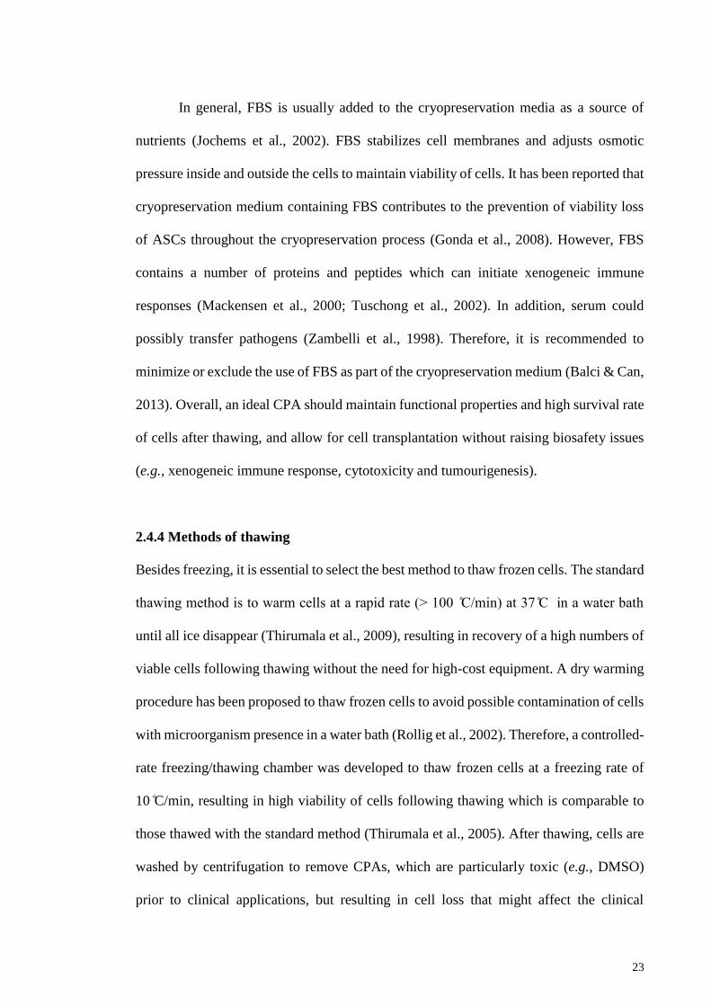
4.1: A flow chart demonstrates the procedures of cryopreservation of hASCs using
a slow freezing method……………………………………………..............60
4.2: Electrophoresis revealed that exons 5 to 8 of p53 nucleotide sequences with
the correct base pair (bp) respectively, were amplified successfully using
PCR..……………………..………………………………..……………….66
4.3: Blue/white colony screening…..…………………………………………...68
4.4: EcoR1 restriction…………………………………………………………..69
4.5: Morphology of hASCs was maintained following cryopreservation
(magnification 100 ×; scale bar: 100 µm)……………..…………………...71
4.6: Cryopreservation maintain surface markers expression of hASCs...............72
4.7: The effect of cryopreservation on the viability of hASCs………….……...73

xv
4.8: Proliferation rate of hASCs was maintained following cryopreservation….74
4.9: A d i p o gen i c p o t en t i a l o f h A S C s w as m a i n t a i n ed f o l l o w in g
cryopreservation…………………………………………………………...76
4.10: Cryopreservation maintained osteogenesis of hASCs……………………...78
4.11: Cryopreservation maintained chondrogenesis of hASCs………...………...80
4.12: Cryopreservation maintained stemness of hASCs…………………….…...81
4.13: Expression of tumour suppressor markers (p53, p21, p16 and pRb) were
maintained following the long-term cryopreservation……………………..82
4.14: Cryopreservation maintained hTERT expression, telomerase activity and
telomere length of hASCs………………………………………………….83
4.15: Cryopreserved and fresh hASCs showed no sign of p53 mutation…………85
4.16: Low levels of DNA damage were observed in cryopreserved hASCs……..87
5.1: Polyacrylamide (PA) hydrogel substrate fabrication……………………100
5.2: Characterization of Polyacrylamide (PA) hydrogels……………….…105
5.3: Characterization of cardiac fibroblasts and myofibroblasts wi th
immunofluorescence staining…………………………………………..107
5.4: Cardiac myofibroblast differentiation was dependent on substrate stiffness
instead of pore size……………………………………………………….109
5.5 Expression of Col I, Col III and TGF-β1 was dependent on substrate
stiffness…………………………………………………………………...110
5.6: Mechanism of matrix st iffness -induced cardiac myofibroblast
differentiation…………………………………………………………….112
5.7: Treatment of losartan on cardiac fibroblasts cultured on 4 kPa substrates, 13
kPa substrates and cell culture plate………………………………………113
5.8: Fresh and cryopreserved hASCs inhibited matrix stiffness-induced cardiac
myofibroblast differentiation through paracrine effect…………………...115
5.9: Treatment with only conditioned medium of fresh and cryopreserved human
adipose-derived mesenchymal stem cells (hASCs) or conditioned medium of
hASCs + anti-HGF on cardiac fibroblasts cultured on 4 kPa substrates, 13 kPa
substrates and cell culture plate…………………………………………...116
5.10 Conditioned medium of fresh and cryopreserved hASCs reduced
expression of AT1R while enhanced expression of Smad7………………..118

xvi
5.11: hASCs secreted HGF to inhibit cardiac myofibroblast differentiation via
AT1R and Smad7…………………………………………………………119
5.12: A similar concentration of HGF was observed in conditioned medium of
fresh and cryopreserved human adipose-derived mesenchymal stem cells
(hASCs)…………………………………………………………………..120
5.13: Summary of proposed mechanism of matrix stiffness-induced cardiac
myofibroblast differentiation and anti-fibrotic effects of undifferentiated
human adipose-derived mesenchymal stem cells (hASCs)……………….123

xvii
LIST OF TABLES
Table Page
2.1: Comparison between slow freezing and vitrification method…………….20
3.1: Concentration and absorbance at 260 nm/280 nm of total RNA of cells….48
3.2: Sample preparation for cDNA synthesis…………………………………..48
3.3: RNA-to-cDNA conversion setting………………………………………...49
3.4: Sample preparation for Real-Time PCR…………………………………..49
3.5: Thermal cycling profile for Real-Time PCR……………………………...50
4.1 Thermal cycling profile for PCR in telomerase assay……………………..63
4.2: Sample preparation for PCR in p53 nucleotide sequence mutation detection
assay………………………………………………………………………..65
4.3: p53 primer sequences, primer annealing positions, and expected length of
PCR products………………………………………………………………65
4.4: Thermal cycling profile for PCR in p53 nucleotide sequence mutation
detection assay……………………………………………………………..66
4.5: Sample preparation for DNA ligation……………………………………...67

xviii
LIST OF SYMBOLS AND ABBREVIATIONS
2D Two dimensional
3D Three dimensional
ACAN Aggrecan
AFM Atomic force microscopy
ALPL Alkaline phosphatase
anti-HGF HGF antibody
ASCs Adipose-derived stem cells
AT1R Angiotensin II type 1 receptor
α-SMA Alpha smooth muscle actin
Col I Collagen type I
COL-2 Collagen type II
Col III Collagen type III
CPAs Cryoprotective agents
DAPI 4'-6-diamidino-2-phenylindole
DMEM Dulbecco's Modified Eagle Medium
DMSO Dimethylsulfoxide
ECM Extracellular matrix
ESCs Embryonic stem cells
FABP4 Fatty acid binding protein 4
FBS Fetal bovine serum
GAPDH Glyceraldehyde 3-phosphate dehydrogenase
hASCs Human adipose-derived stem cells

xix
HGF Hepatocyte growth factor
hMSCs Human mesenchymal stem cells
IGF-1 Insulin-like growth factor 1
LPL Lipoprotein lipase
M mol/L
MBA N,N methylene-bis-acrylamide
Min minute
MSCs Mesenchymal stem cells
OSC Osteocalcin
PA Polyacrylamide
PBS Phosphate buffered saline
PCR Polymerase chain reaction
PPAR- γ Peroxisome proliferator-activated receptor γ
qPCR Quantitative Real-Time polymerase chain reaction
RUNX2 Runt-related transcription factor 2
SEM Scanning electron microscopy
SOX-9 Sry-related HMG box-9
SVF Stromal vascular fraction
TERT Telomerase reverse transcriptase
TGF-β1 Transforming growth factor beta 1
˚C Degree celsius

xx
LIST OF APPENDICES
Appendix Page
A: Human medical ethic approval letter……………………………………..154
B: Patient consent form……………………………………………………...155
C: Standard curve of percentage of Resazurin reduction versus cell number
(R2 = 0.999)………………………………………………………………156
D: Published paper 1 – Phenotypic and functional characterization of long-term
cryopreserved human adipose-derived stem cells…………………..........157
E: Published paper 2 – Cryopreservation of human mesenchymal stem cells for
clinical applications: current methods and challenges……………………158
F: Published paper 3 – Assessment of tumourigenic potential in long-term
cryopreserved human adipose-derived stem cells………………………..159
G: Published paper 4 – Mechanoregulation of cardiac myofibroblast
differentiation: implications for cardiac fibrosis and therapy……………..160

1
CHAPTER 1: INTRODUCTION
1.1 Background
Human stem cells, particularly human mesenchymal stem cells (hMSCs), are an ideal
candidate for many biomedical applications such as regenerative medicine and cell-based
therapies (Arthur et al., 2009; Mimeault et al., 2007). hMSCs obtained from various
locations of the human body (including adipose tissue, bone marrow and periosteum) are
capable of self-renewing and differentiating into multiple specific types of cell, such as
those in bone or cartilage (Callihan et al., 2011; Doulatov & Daley, 2013). Further,
hMSCs also have paracrine function and immunosuppressive property (Baraniak &
McDevitt, 2010; De Miguel et al., 2012; Nauta & Fibbe, 2007), which support their use
in various clinical settings. Implantation of such multi-functional hMSCs may overcome
the problems of organ shortage crisis worldwide to treat the fatal diseases, e.g., heart
failure and liver failure. Among the hMSCs, human adipose-derived mesenchymal stem
cells (hASCs) have gained great attraction due to the abundance and readily accessibility
of adipose tissues (Gomillion & Burg, 2006; Kolle et al., 2013; Mizuno et al., 2012).
In general, each clinical therapy may require a huge amount of cells (including
hASCs), approximately 200 million cells (Faustini et al., 2010). Therefore, the cells need
to be expanded in vitro extensively (Pittenger et al., 1999). During the extensive cell
expansion, hASCs may be at a risk of tumourigenesis and chromosomal aberration
(Froelich et al., 2013; Roemeling-van Rhijn et al., 2013) and thus may raise biosafety
concerns in clinical application (Sharpe et al., 2012). For instance, it has been reported
that hASCs displayed spontaneous malignant transformation and chromosomal aberration
after culture beyond 10 passages (Froelich et al., 2013; Roemeling-van Rhijn et al., 2013).
Such adverse changes may be caused by a potential contamination with an external cell

2
source such as tumour cells and the stressful culture conditions (e.g., frequent enzymatic
cell dissociation and long-term culture) (Barkholt et al., 2013; de la Fuente et al., 2010).
In addition, long-term culture of hASCs was associated with senescence, reduced
differentiation potential, and alteration of stemness and phenotype (Safwani et al., 2012,
2013; Wan Safwani et al., 2011). These concerns limit the wide use of long-term cultured
hASCs in clinical applications. In fact, hASCs at an early passage (within passage 4) are
safe for clinical use (Barkholt et al., 2013). Therefore, a method for long-term storage of
hASCs at an early passage is required for off-the-shelf clinical use. Cryopreservation
represents the potential storage method which maintains functional properties of hASCs
in long-term (Gonda et al., 2008; Yong et al., 2015c).
Among the cryopreservation methods (e.g., slow freezing and vitrification), slow
freezing is the most preferable method for hASCs due to easier processing and low risk
of contamination, whereas vitrification requires a good manipulation skill and has a high
risk of causing contamination (Guven & Demirci, 2012). To achieve an efficient
cryopreservation using a slow freezing method, optimization of the use of cryoprotective
agents (CPAs) is essential to protect hASCs from freezing injury (e.g., cell death and
compromised functional properties). Among the CPAs, 10% dimethylsulfoxide (DMSO)
and fetal bovine serum (FBS) is the most widely used for cryopreservation of hASCs.
However, clinical uses of cells preserved in such CPA formulations have raised the issues
of adverse reactions (e.g., respiratory depression and neurotoxicity and xenogeneic
immune response) in recipients after implantation of cryopreserved cells (Benekli et al.,
2000; Tuschong et al., 2002; Windrum & Morris, 2003). This has resulted the need for
development of an alternative CPA or a CPA formulation consisting of DMSO in a
reduced concentration and without FBS. To date, a standardized cryopreservation
protocol of hASCs for clinical applications has not been fully established. As the

3
biosafety profile of cryopreserved hASCs has not been established, it is also essential to
evaluate the biosafety (e.g., tumourigenic potential and chromosomal abnormality) of
cryopreserved hASCs prior to clinical applications (Yong et al., 2015b). Understanding
the storage conditions and characteristics of cryopreserved hASCs following thawing is
essential prior to clinical application to ensure a good and safe product.
Cardiac fibrosis, an initial healing process essential for heart repair after damage
due to heart diseases such as myocardial infarction, is often dysregulated once it has
begun, causing adverse remodeling of cardiac tissues that may ultimately lead to cardiac
failure (Wynn & Ramalingam, 2012; Zeisberg et al., 2007). To date, cardiac fibrosis
cannot be effectively halted or reversed by surgery or drug therapies (e.g., anti-fibrotic
agents) (Daskalopoulos et al., 2012). Recently, undifferentiated MSCs with an ability to
secrete soluble factors (e.g., hepatocyte growth factor (HGF) and insulin growth factor-1
(IGF-1)), have been found to be capable of altering the behaviors of neighboring cells
(e.g., myofibroblast differentiation (a hallmark of fibrosis) and cardiomyocyte
regeneration) through their paracrine functions (Ramkisoensing et al., 2014), indicating
their great potential for treating cardiac fibrosis. The existing studies showed that MSCs
could suppress cardiac fibrosis by many mechanisms, e.g., ECM degradation and
suppression of alpha-smooth muscle actin (α-SMA) expression (a defining marker of
cardiac myofibroblast differentiation) (Li et al., 2015; Mao et al., 2013; Mias et al., 2009;
Ohnishi et al., 2007; Wang et al., 2011). However, these studies were performed on
cardiac fibroblasts cultured in non-physiological or mechanically irrelevant conditions
(e.g., cell culture plastic plates and glass slides). To date, the paracrine effects of MSCs
on cardiac myofibroblast differentiation in conditions mimicking the stiffness of in vivo
normal and fibrotic cardiac tissues has not been explored, which would provide a better
understanding for the therapeutic use of MSCs in cardiac fibrosis. In addition,

4
cryopreserved hASCs might hold great potential for cardiac fibrosis therapy as such an
intensive clinical setting might require a large number of cells for off-the-shelf use.
In the present study, the types and concentrations of CPAs used to cryopreserve
hASCs for 3 months using a slow freezing method were optimized by evaluating the
effect of cryopreservation on the phenotype, viability, functional properties (e.g.,
proliferation and differentiation potential) and biosafety (e.g., tumourigenic potential) of
hASCs. Based on these evaluation, the ideal CPA used to cryopreserve hASCs was
determined. Further, an in vitro cardiac fibrosis model was developed based on evaluation
of cardiac myofibroblast differentiation markers in cardiac fibroblasts cultured on
hydrogels mimicking the stiffness of native fibrotic cardiac tissues. Then, conditioned
medium of fresh (non-cryopreserved) and cryopreserved hASCs was applied to the model
to investigate the paracrine effects of hASCs on cardiac fibrosis. The findings of this
study would impact the establishment of a standardized cryopreservation protocol for
hASCs and therapeutic use of long-term cryopreserved hASCs in cardiac fibrosis in future.
1.2 Research question
i. Does cryopreservation have any effect on hASCs in terms of cell phenotype,
viability, proliferation, and differentiation?
ii. Do cryopreserved hASCs raise any biosafety concerns (e.g., tumourigenesis)?
iii. Do cryopreserved hASCs have the potential to be used in therapeutic application
of cardiac fibrosis?

5
1.3 Research hypotheses
i. Cryopreservation maintains cell phenotype, high viability, proliferation potential,
multilineage differentiation potential of hASCs.
ii. Cryopreserved hASCs do not undergo tumourigenesis.
iii. Cryopreserved hASCs have a high potential to be used in therapeutic application
of cardiac fibrosis.
1.4 Research objectives
The objectives of this study include:
i. To determine the cryoprotective effect of various CPAs e.g., on cell
phenotype, cell viability, cell proliferation and differentiation (including
adipogenic, osteogenic and chondrogenic) of hASCs.
ii. To evaluate the biosafety of cryopreserved hASCs through tumourigenic
potential assessment.
iii. To develop an in vitro cardiac fibrosis model.
iv. To evaluate the paracrine effects of cryopreserved hASCs on cardiac
fibrosis.
1.5 General methodology
In general, this study was approved by the Medical Ethics Committee of University
Malaya Medical Centre (UMMC) (reference no. 996.46) (Appendix A), and carried out
in accordance with the approved guidelines and experimental protocols which conform
the declaration of Helsinki. The scopes of the ethical clearance in this study are as follows:
i. A written informed consent can be obtained from the donor of adipose tissues in
the presence of a physician. If the donor is physically or mentally incapable of

6
giving consent, one of his/her relative is allowed to give consent on the behalf of
him/her.
ii. Isolation of adipose tissues must be performed by a physician.
iii. Adipose tissues of the donor can only be used for research purpose.
iv. The personal information of donor are confidential. The results of research can
only be presented and published without including the personal information of
donor.
After collection of adipose tissues, the tissues were processed to obtain hASCs.
hASCs were cryopreserved using various combinations of general used CPAs (e.g.,
DMSO, trehalose and FBS) for 3 months. After 3 months of cryopreservation, hASCs
were subjected to the evaluation of cell phenotype, viability, proliferation, differentiation
and tumourigenic potential. Fresh or non-cryopreserved hASCs were used as control.
Based on these evaluations, an ideal CPA used to cryopreserve hASCs was determined.
On the other hand, an in vitro cardiac fibrosis model made by rat cardiac
fibroblasts cultured on a hydrogel which mimics the stiffness of native fibrotic cardiac
tissues was developed. Cardiac fibroblasts were isolated from the hearts of neonatal rats
(1-3 day old), which conform the National Institutes of Health (NIH) guidelines (Guide
for the care and use of laboratory animals). Further, hASCs which was cryopreserved in
the ideal CPA, was applied to the model to evaluate the potential therapeutic application
of cryopreserved hASCs in cardiac fibrosis. Flow chart of the general methodology was
shown in the Fig. 1.1. The research methodology will be elaborated in detail in the section
of materials and methods in chapter 3, 4 and 5.

7
Figure 1.1. Flow chart of general methodology
Collection of human adipose tissues
Tissue processing
hASCs
Cryopreservation
CPAs 1. 0.25 M Trehalose 2. 5% DMSO 3. 10% DMSO 4. 5% DMSO + 20% FBS 5. 10% DMSO + 20% FBS 6. 10% DMSO + 90% FBS
After 3 months
Assessment 1. Cell phenotype 2. Cell proliferation 3. Cell differentiation 4. Tumourigenic potential
Isolation of rat cardiac fibroblasts
Hydrogel mimicking the stiffness of native fibrotic cardiac tissues
Culture
In vitro cardiac fibrosis model

8
1.6 Thesis framework
The thesis is written in accordance with the article style format. It is comprised of six
chapters as described as follows:
i. Chapter 1 gives a brief overview of the research background, research question,
research hypotheses, research objectives, general methodology and the content of
each chapter.
ii. Chapter 2 provides a critical review on the research background, including stem
cells, mesenchymal stem cells, ASCs, cryopreservation, the existing studies of
cryopreservation of hASCs, biosafety assessments of mesenchymal stem cells,
and cardiac fibrosis. This chapter contains selected figures and table reprinted in
part with permission from my review articles as follows:
- Yong, K. W., Wan Safwani, W. K., Xu, F., Wan Abas, W. A., Choi, J. R., &
Pingguan-Murphy, B. (2015). Cryopreservation of Human Mesenchymal
Stem Cells for Clinical Applications: Current Methods and Challenges.
Biopreserv Biobank, 13(4), 231-239. Copyright (2016) Mary Ann Liebert.
- Yong, K. W., Li, Y., Huang, G., Lu, T. J., Safwani, W. K., Pingguan-Murphy,
B., & Xu, F. (2015). Mechanoregulation of Cardiac Myofibroblast
Differentiation: Implications for Cardiac Fibrosis and Therapy. Am J Physiol
Heart Circ Physiol, 309(4), 19. Copyright (2016) American Physiological
Society.
iii. Chapter 3 presents the characterization of hASCs isolated from human adipose
tissues.
iv. Chapter 4 presents the effect of cryopreservation on hASCs in terms of cell
phenotype, viability, proliferation, differentiation (adipogenesis, osteogenesis and
chondrogenesis), stemness and biosafety (tumourigenic potential). Chapter 3 and
9
4 contains selected figures and table reprinted in part with permission from my
research articles as follows:
- Yong, K. W., Pingguan-Murphy, B., Xu, F., Abas, W. A., Choi, J. R., Omar,
S. Z., . . . Wan Safwani, W. K. (2015). Phenotypic and Functional
Characterization of Long-Term Cryopreserved Human Adipose-Derived
Stem Cells. Sci Rep, 5(9596). Copyright (2016) Nature Publishing Group.
- Yong, K. W., Safwani, W. K., Xu, F., Zhang, X., Choi, J. R., Abas, W. A., . . .
Pingguan-Murphy, B. (2016). Assessment of tumourigenic potential in long-
term cryopreserved human adipose-derived stem cells. J Tissue Eng Regen
Med, 12(10). Copyright (2016) John Wiley & Sons.
v. Chapter 5 presents the development of an in vitro cardiac fibrosis model and the
evaluation of paracrine effects of cryopreserved hASCs on cardiac fibrosis.
vi. Chapter 6 includes thesis contribution, conclusion, limitation of the study and
future work.
10
CHAPTER 2: LITERATURE REVIEW
2.1 Stem cells
Stem cells are unspecialized and undifferentiated cells that have the ability to undergo
self-renewal and multilineage differentiation potential (Conrad & Huss, 2005). Due to
these characteristics, stem cells have become an ideal candidate for regenerative medicine
and tissue engineering, as well as cell-based and gene therapies (Mimeault et al., 2007).
Ideally, to meet the requirement of regenerative medicinal applications, stem cells should
fulfill the following set of criteria: First, stem cells should be harvested by a minimally
invasive procedure and cultivated to obtain millions to billions of cells. Second, stem cells
can be differentiated into multiple specific types of cells in a reproducible manner. Last
but not least, stem cells should be effectively and safely implanted into either an
allogeneic or autologous host (Gimble, 2003).
In general, stem cells can be divided into four categories based on their potency.
These categories are totipotent, pluripotent, multipotent and unipotent (Callihan et al.,
2011; Eisenberg & Eisenberg, 2003). Zygotes, an example of totipotent stem cells, are
created by egg fertilization by sperm, have the greatest differentiation potential, thus
capable of producing all kinds of cells including embryonic stem cells (ESCs) (Callihan
et al., 2011). Pluripotent stem cells can give rise to almost all the specialized cells of the
three germ layers, including endoderm (gastrointestinal tract and lungs), mesoderm
(urogenital, bone, blood and muscle) and ectoderm (nervous system and epidermal
tissues) (Binder et al., 2009). Multipotent stem cells have the ability to differentiate into
multiple but limited cell types, e.g., mesenchymal stem cells (MSCs) can differentiate
into specialized cells such as those in bones and cartilages but cannot differentiate into
blood cells such as lymphocytes and monocytes (Fig. 2.1). Stem cells that can only

11
differentiatie into one type of cells are termed unipotent stem cells (Eisenberg &
Eisenberg, 2003).
Figure 2.1. Classification of stem cells based on potency. Blastocyst totipotent stem
cells produce pluripotent stem cells (also namely embryonic stem cells) that
give rise to adult stem cells (including hematopoietic stem cells (HSC),
neural stem cells (NSC) and mesenchymal stem cell (MSCs)) which can
differentiate into various types of specialized cells. Reproduced from
Corsten and Shah (2008).
Stem cells can also be divided into three broad types, including ESCs which are
isolated from the inner cell mass of blastocysts, adult stem cells which are found in adult
tissues, and induced-pluripotent stem cells (iPSCs) (Estrov, 2009). iPSCs are derived
from somatic cells that are genetically reprogrammed into cells mimicking ESCs by
possessing the pluripotency of ESCs. These cells require pluripotency-associated genes
or transcription factors (e.g., OCT-4, SOX-2, cMyc and Klf4) to maintain their ESCs-like
properties (Power & Rasko, 2011).

12
2.1.1 Embryonic stem cells
ESCs can be induced in vitro and in vivo in a simple culture condition to differentiate into
specific progenitor cells or specialized cells (Mimeault & Batra, 2006), e.g.,
hematopoietic cell lineages, neuron-like cells, hepatocytes, pancreatic islet-like cells,
osteocytes, chondrocytes, adipocytes, and cardiomyocytes (Trounson, 2006; Wu et al.,
2007). Three markers, particularly SOX-2, NANOG, and OCT-4, are expressed in human
ESCs to maintain their pluripotency (Callihan et al., 2011). However, the implantation of
differentiated cells generated from ESCs into recipients may cause adverse effects such
as, teratoma formation and immune rejection, due to the presence of residual
undifferentiated and pluripotent ESCs (Andrews et al., 2005; Mimeault & Batra, 2006).
Further, the destruction of embryos to obtain ESCs is ethically unacceptable (Young,
2000).
2.1.2 Adult stem cells
The use of stem cells derived from adult tissues avoids many ethical concerns related to
the use of ESCs (Gomillion & Burg, 2006). Adult stem cells are capable of self-renewing
and differentiating into the major specialized cells of the tissue in which they reside (Vats
et al., 2002). Adult stem cells typically are divided into hematopoietic stem cells, neural
stem cells and MSCs (Gomillion & Burg, 2006). Hematopoietic stem cells are primitive
and undifferentiated cells that are capable of self-renewing and differentiating into all
types of blood cells such as white blood cells and red blood cells (Mayani, 2003). Neural
stem cells can differentiate into almost all the cells in the adult central nervous system
such as, neurons and glia (Price & Williams, 2001; Temple, 2001).

13
2.2 Mesenchymal stem cells
Among the adult stem cells, MSCs hold great promise for cell therapy (Chagastelles et
al., 2010). In addition to multipotent differentiation potential, MSCs have paracrine
functions and immunosuppressive effects, which support their use in various clinical
settings (Baraniak & McDevitt, 2010; Caplan, 2007; Caplan & Bruder, 2001).
Morphologically, MSCs resemble fibroblasts in their spindle shape morphology
(Pittenger et al., 1999). MSCs should possess the following criteria as proposed by
Mesenchymal and Tissue Stem Cell Committee of the International Society for Cellular
Therapy such as adherence to plastic, positively express specific surface antigens for
MSCs (e.g., CD90, CD105, CD73 and CD44), negatively express specific antigens for
hematopoietic cells (e.g., CD14, CD19, CD34 and CD45), and multipotent differentiation
(including adipogenic, osteogenic and chondrogenic) (Dominici et al., 2006). MSCs can
be isolated from various locations in the human body, such as bone marrow (Ginis et al.,
2012; Pravdyuk et al., 2013), adipose tissue (De Rosa et al., 2009; Minonzio et al., 2014),
periosteum (Ferretti et al., 2012), amniotic fluid (Angelo et al., 2012) and umbilical cord
blood (Liu et al., 2011).
Bone marrow is the most recognized source of MSCs (Ringden & Le Blanc, 2005),
which can be found in the bone marrow stroma. These cells are able to differentiate into
multiple lineages of cells such as adipocytes, osteocytes, myocytes and neural cells (Barry
& Murphy, 2004; Jiang et al., 2002). During bone marrow biopsy, about 10-40 mL bone
marrow is aspirated from the iliac crest, and MSCs are then isolated in culture by their
adherence properties (Caplan, 2000; Pittenger et al., 1999). However, isolation of bone
marrow often causes morbidity donor site pain, and yields low numbers of MSCs (0.01%
of the total nucleated cells in marrow) that thus requires extensive in vitro cultivation
(Pittenger et al., 1999; Tholpady et al., 2006). Therefore, adipose tissue, which harbours
14
a high yield of MSCs, has been identified as a potential source of MSCs to replace bone
marrow. Adipose tissue is particularly attractive because of its abundance and readily
accessibility (Gomillion & Burg, 2006).
2.3 Adipose-derived mesenchymal stem cells
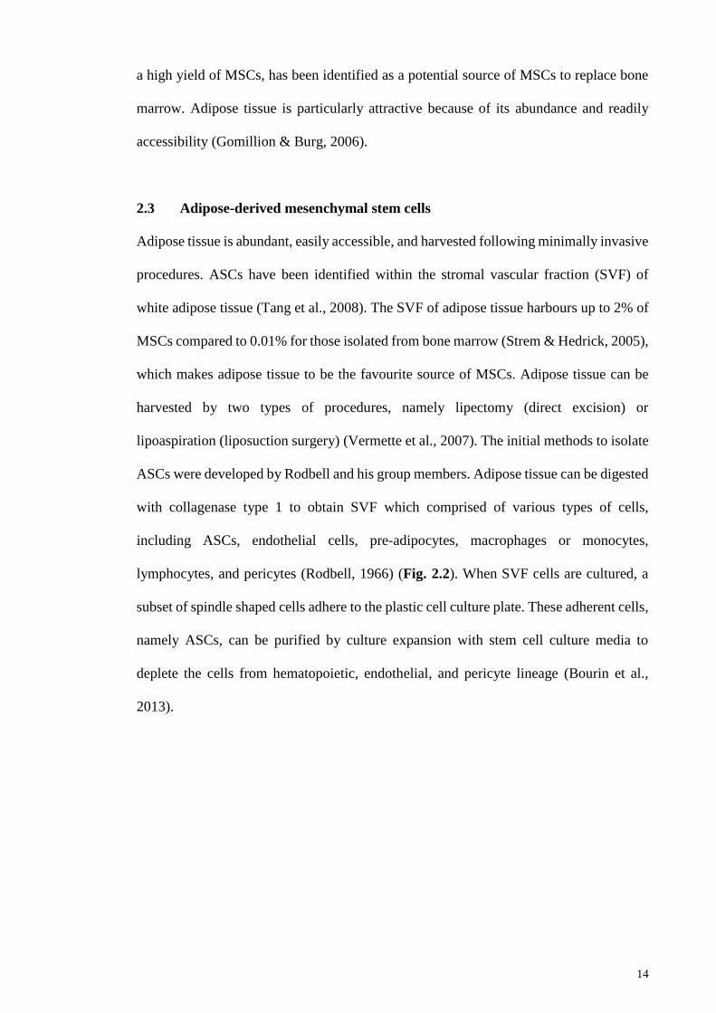
Adipose tissue is abundant, easily accessible, and harvested following minimally invasive
procedures. ASCs have been identified within the stromal vascular fraction (SVF) of
white adipose tissue (Tang et al., 2008). The SVF of adipose tissue harbours up to 2% of
MSCs compared to 0.01% for those isolated from bone marrow (Strem & Hedrick, 2005),
which makes adipose tissue to be the favourite source of MSCs. Adipose tissue can be
harvested by two types of procedures, namely lipectomy (direct excision) or
lipoaspiration (liposuction surgery) (Vermette et al., 2007). The initial methods to isolate
ASCs were developed by Rodbell and his group members. Adipose tissue can be digested
with collagenase type 1 to obtain SVF which comprised of various types of cells,
including ASCs, endothelial cells, pre-adipocytes, macrophages or monocytes,
lymphocytes, and pericytes (Rodbell, 1966) (Fig. 2.2). When SVF cells are cultured, a
subset of spindle shaped cells adhere to the plastic cell culture plate. These adherent cells,
namely ASCs, can be purified by culture expansion with stem cell culture media to
deplete the cells from hematopoietic, endothelial, and pericyte lineage (Bourin et al.,
2013).

15
Figure 2.2: The composition of adipose tissue. Adapted from Cao (2010).
ASCs have spindle shaped morphology (Wan Safwani et al., 2011) (Fig. 2.3),
which is similar in appearance with bone marrow-derived MSCs. Further, ASCs are
similar with bone marrow-derived MSCs in terms of cell surface phenotype and
differentiation potential (De Ugarte et al., 2003). As a type of MSCs, ASCs highly express
surface markers such as CD90, CD73, CD105 and CD44 (Zaman et al., 2012). In addition,
ASCs are capable of in vitro differentiation into adipocytes (Ogawa et al., 2004;
Rodriguez et al., 2004), osteocytes (Hattori et al., 2004; Hicok et al., 2004), chondrocytes
(Estes & Guilak, 2011), cardiomyocytes (Lee et al., 2009; van Dijk et al., 2008), neuron-
like cells (Dhar et al., 2007; Ning et al., 2006) and pancreatic endocrine-like insulin-
producing cells (Silva et al., 2012; Timper et al., 2006). They also possess an ability to
maintain their stemness and multipotency by expressing stemness markers, such as OCT-
4, REX-1, SOX-2 and NANOG (Izadpanah et al., 2006; Zuk et al., 2002).

16
Figure 2.3. Morphology of ASCs. ASCs present spindle shaped morphology
(Magnification: 40 ×; Scale bar: 200 μm).
2.4 Cryopreservation
Cryopreservation is a process of maintaining biological function and viability of cells by
freezing and storing them below -80 ⁰C, e.g., at the temperature of liquid nitrogen (-196
⁰C) (Karlsson & Toner, 1996). In general, the first step in cryopreservation is harvesting
the cells followed by the addition of cryopreservation medium containing cryoprotective
agents (CPAs). Then, ice crystal induction is performed with an optimum cooling rate.
Finally, the cells are stored in liquid nitrogen (Hubel, 1997). Long-term cryopreservation
of living tissues and cells offers a great potential for clinical applications, including blood
transfusion (Wagner et al., 2002), bone marrow transplantation (Rowley et al., 2003), in
vitro fertilization (Anger et al., 2003), vascular grafts and bone grafts (Kofron et al., 2003).
In the case of MSCs, preclinical trials demonstrated that implantation of cryopreserved
MSCs was capable of treating intestinal inflammation (Castelo-Branco et al., 2012) and
restoring myocardial function (Chin et al., 2010), but their therapeutic use is still not well
established yet.

17
2.4.1 Benefits of cryopreservation
Cryopreservation allows transportation and pooling of cells to reach cell numbers
required for cell therapy while maintaining their functional properties. It also allows the
completion of quality control and safety testing of cells prior to clinical applications
(Hubel, 1997). Without cryopreservation, the cells are forced to be continuously
subcultured, which may accumulate genetic changes and result in tumorigenicity or
heterogeneity (Stubban et al., 2007). On the other hand, at -196 ⁰C, the cells have no
metabolic demands, thus avoiding biological variation due to genetic drift for years
(Rowley, 1992). Therefore, cryopreservation produces a bank of cells at specific passages
with intact functional properties and genetic characteristics. These validated cells can be
used to initiate new experiments, maximize the long-term use of cells, and minimize
experimental variation (Stubban et al., 2007).
2.4.2 Methods of cryopreservation
2.4.2.1 Slow freezing
The slow freezing method is the preferred method for cryopreservation of high volume
cells such as ASCs and cell lines. With a freezing rate of 1 ºC/min using a non-
programmable time freezing protocol in a a commercially available freezing container
“Mr. Frosty” (temperature is lowered at such rate due to the slow freezing property of
isopropanol), a large number of cells can be frozen in one cryovial at a low concentration
(<1.5 M) of CPAs (Zhang et al., 2011c). To achieve such a low freezing rate, direct
contact of cells with non-sterile liquid nitrogen is not required during freezing, thus
avoiding potential contamination with other microorganism or pathogens. By using a
slow freezing method, a large amount of cryopreserved cells which are contamination-
free can be pooled to provide sufficient cells for off-the-shelf clinical use. In order to
further improve the efficiency of cryopreservation, a high-cost controlled

18
freezing/warming rate freezer which adopts a programmable freezing time protocol was
developed. However, it has been reported that both protocols which have been used to
freeze MSCs display similar potential to maintain phenotype, viability, and functional
properties of MSCs (Janz Fde et al., 2012). With the advance of technology, “Cell Alive
System” (CAS), a programmed freezer which adopts an approach of vibrating the water
molecules and cells during freezing to prevent intra- and extra-cellular ice formation
using alternating magnetic field and electric field, has been developed (Wowk, 2012). It
has been reported that the risk of freeze injury to MSCs can be further reduced by using
this system (Lee et al., 2012). Among the slow freezing protocols, a nonprogrammable
freezing time protocol is favorable for cryopreservation of ASCs due to its low cost and
high cryopreservation efficiency. To preserve ASCs efficiently using a slow freezing
method, it is essential to optimize and determine the ideal CPAs used to preserve ASCs.
2.4.2.2 Rapid freezing/vitrification
Vitrification is a process which requires a very high freezing rate to convert a cell-laden
CPA suspension directly from its aqueous phase to a glass state upon in contact with
liquid nitrogen (Rall & Fahy, 1985). Vitrification is usually applied to small volume cells
such as ESCs, embryo and oocyte because it requires low volume of cell suspension to
achieve high freezing rate (Song et al., 2010). Therefore, it is ill-suited to cryopreservation
of ASCs in large volumes. Further, it requires a high concentration of CPAs (6-8 M)
which can cause osmotic shock and chemical toxicity to cells (Karlsson & Toner, 1996;
Zhang et al., 2011c). Vitrification systems can be divided into carrier-based systems and
carrier-free systems (Fig. 2.4). Cryoloops (700,000 ºC/min), quartz microcapillaries
(250,000 ºC/min) and plastic straws (2500 ºC/min) are among the carriers that have been
developed for vitrification with each of them offering a different freezing rate (Zhang et
al., 2011c). The use of higher freezing rate allows vitrification using CPA at a lower
concentration, thereby reducing the risk of osmotic damage and chemical toxicity to cells.

19
On the other hand, carrier-free systems which adopt an approach of generating cell-laden
CPAs in microdroplets followed by ejection to liquid nitrogen, were developed to further
increase freezing rate for vitrification. (Zhang et al., 2011c).
Figure 2.4. Carrier/channel-based and carrier-free (microdroplet-based)
vitrification system. Adapted from Xu et al. (2010).
Post–thaw viability of cells was reported to be higher using vitrification compared
to slow freezing method as the high freezing rate reduces the time of intra- and
extracellular ice formation (Li et al., 2010). However, vitrification which adopts an
approach of direct cell-to-liquid nitrogen contact, might lead to potential pathogenic
contamination (Tedder et al., 1995). The Hepatitis B virus and Aspergillus sp. are among
the pathogens which have been reported to contaminate liquid nitrogen (Fountain et al.,
1997; Tedder et al., 1995). The contamination may come from non-sterile liquid nitrogen
itself or cross-contamination from infected samples in the liquid nitrogen storage tank
(AbdelHafez et al., 2011). Moreover, good manipulation skill is required for vitrification
and it is impractical on a large scale of cell cryopreservation. The recycling of cells is
labor-consuming as it needs manual picking up of individual cell colonies (Li et al., 2010),

20
potentially causing cell loss. The advantages and disadvantages of slow freezing and
vitrification are described in brief in Table 2.1.
Table 2.1 Comparison between slow freezing and vitrification method
No Aspect Slow freezing Rapid freezing
1 Concentration of
CPAs required
Low High
2 Risk of freeze injury High Low
3 Post-thaw viability High Higher
4 Risk of toxicity of
cryoprotective agens
Low High
5 Potential
contamination with
pathogenic agents
Low High
6 Manipulation Skill Easy Good manipulation skill
is needed.
Reproduced from Yong et al. (2015b)
Overall, the slow freezing method is preferable because cryopreservation of ASCs
can be done on a large scale, the post-thaw viability of cells is high, and it avoids the risk
of pathogenic contamination, allowing cryopreserved ASCs to be used for clinical
applications.
2.4.3 Cryoproctective agents
The roles of CPAs in cell cryopreservation are to stabilize the cell membrane, minimize
osmotic stress to the cells, and protect cells against intracellular and extracellular ice
crystal formation which are both harmful to cells (McGann, 1978; Rudolph & Crowe,
1985) (Fig. 2.5). CPAs are divided into two broad classes: 1) permeating CPAs that freely
permeate cell membranes and 2) nonpermeating CPAs (McGann, 1978).

21
Figure 2.5. The role of CPAs in cell cryopreservation. Cryopreservation requires CPAs
(either permeating or non-permeating) to protect cells from freeze injury (e.g.,
cell death) by avoiding the formation of intra- and extra-cellular ice crystals
which are harmful to cells during freezing. Reproduced from Martin-Ibanez
et al. (2012).
2.4.3.1 Permeating CPAs
The most commonly used permeating CPAs are DMSO (dimethylsulfoxide) and glycerol
(Buchanan et al., 2004). Permeating CPAs can penetrate the cell membrane due to their
low molecular weight (Karlsson, 2002; Karlsson & Toner, 1996). Permeating CPAs
prevent cell dehydration and physical injury induced by intracellular ice formation by
reducing the incidence of increasing concentrations of electrolytes in the extracellular
space during freezing (Rowley, 1992). DMSO is the most widely used CPA for large
numbers of tissue and cell cryopreservation. DMSO can penetrate the cell membrane by
removing the water within the cells to prevent the formation of intracellular ice that can
cause cell rupture (Berz et al., 2007). Further, DMSO is able to stabilize the plasma
membrane by electrostatic interaction (Anchordoguy et al., 1991). Generally, DMSO with

22
a concentration of 10% (v/v) combined with FBS (20% - 90%) (v/v) is used to preserve
MSCs (Liu et al., 2008; Miranda-Sayago et al., 2012; Zhang et al., 2011b).
.
However, DMSO is toxic at temperatures beyond 4 ⁰C (Zambelli et al., 1998). The
clinical uses of cells preserved in 10% DMSO have caused many adverse effects in
recipients, such as neurotoxicity and respiratory depression (Benekli et al., 2000;
Windrum & Morris, 2003). Nevertheless, DMSO is not only cytotoxic but it also induces
undesired differentiation of stem cells to neuronal-like cells (Woodbury et al., 2000).
Therefore, the transplantation protocol has included the step of post-thaw washing of the
cells to remove DMSO prior to transplantation (Fraser et al., 1998; Rubinstein et al.,
1995). However, washing cells by centrifugation prior to removal of DMSO can cause
significant cell loss. Further, total removal of DMSO from cryopreserved cells is time-
consuming and complex (Thirumala et al., 2009). Therefore, the development of an
alternative cryopreservation medium or a cryopreservation medium consisting of a
reduced concentration of DMSO is required to avoid such adverse effects (Son et al.,
2010).
2.4.3.2 Non-permeating CPAs
The commonly used non-permeating CPAs are polymers (e.g., polyvinylpyrollidone /
PVP) and sugars (e.g., trehalose and sucrose), which cannot enter the cells (Buchanan et
al., 2004). Non-permeating CPAs cannot move accross the cell membrane but protect the
cell membrane by forming a viscous glassy shell around the outer surface of cells
(Karlsson, 2002; Karlsson & Toner, 1996). Sucrose and PVP have been used for
cryopreservation of MSCs to replace DMSO, but the results demonstrated that they are
inferior to DMSO in terms of maintaining viability of MSCs (Janz Fde et al., 2012;
Thirumala et al., 2010a).
23
In general, FBS is usually added to the cryopreservation media as a source of
nutrients (Jochems et al., 2002). FBS stabilizes cell membranes and adjusts osmotic
pressure inside and outside the cells to maintain viability of cells. It has been reported that
cryopreservation medium containing FBS contributes to the prevention of viability loss
of ASCs throughout the cryopreservation process (Gonda et al., 2008). However, FBS
contains a number of proteins and peptides which can initiate xenogeneic immune
responses (Mackensen et al., 2000; Tuschong et al., 2002). In addition, serum could
possibly transfer pathogens (Zambelli et al., 1998). Therefore, it is recommended to
minimize or exclude the use of FBS as part of the cryopreservation medium (Balci & Can,
2013). Overall, an ideal CPA should maintain functional properties and high survival rate
of cells after thawing, and allow for cell transplantation without raising biosafety issues
(e.g., xenogeneic immune response, cytotoxicity and tumourigenesis).
2.4.4 Methods of thawing
Besides freezing, it is essential to select the best method to thaw frozen cells. The standard
thawing method is to warm cells at a rapid rate (> 100 ̊C/min) at 37 ̊C in a water bath
until all ice disappear (Thirumala et al., 2009), resulting in recovery of a high numbers of
viable cells following thawing without the need for high-cost equipment. A dry warming
procedure has been proposed to thaw frozen cells to avoid possible contamination of cells
with microorganism presence in a water bath (Rollig et al., 2002). Therefore, a controlled-
rate freezing/thawing chamber was developed to thaw frozen cells at a freezing rate of
10 ̊C/min, resulting in high viability of cells following thawing which is comparable to
those thawed with the standard method (Thirumala et al., 2005). After thawing, cells are
washed by centrifugation to remove CPAs, which are particularly toxic (e.g., DMSO)
prior to clinical applications, but resulting in cell loss that might affect the clinical

24
outcome. In the future, it is essential to develop a controlled-rate dry warming device
which is low cost, reliable and portable for thawing frozen cells, and a method which can
remove CPAs while minimizing cell loss (Fleming & Hubel, 2006) for efficient clinical
applications.
2.4.5 Challenges in cryopreservation
The issue of adverse effects in recipients of cryopreserved cells is a challenge to be
addressed to cryopreserve cells using a slow freezing method for clinical applications (Li
et al., 2010; Martin-Ibanez et al., 2012). To date, the introduction of non-toxic polymers,
such as PVP, and disaccharides, such as sucrose, as a CPA (Janz Fde et al., 2012;
Thirumala et al., 2010b), did not achieve expectation in replacing DMSO for
cryopreservation of MSCs because they are less efficient than DMSO in maintaining the
viability of MSCs. Further investigation is required to explore alternative CPAs to replace
DMSO completely. On the other hand, cryopreservation of cells using FBS-free
cryopreservation medium seems to be possible, for instance, 5% human albumin solution
and sericin have been proposed to replace FBS for cryopreservation of MSCs (Minonzio
et al., 2014; Verdanova et al., 2014). Taken together, an effective and safe cell
cryopreservation protocol with an optimal concentration of CPAs is still under
investigation.
To vitrify cells for clinical applications, many issues need to be addressed,
including small scale cryopreservation, high risk of contamination and cell loss due to
inefficient collection of frozen cells. With the advance of technology, an ejection-based
micro-droplet generation system which permits efficient vitrification of micro-droplets in
a continuous manner has been developed, improving throughput capability of vitrification
required for cryopreservation of MSCs. A closed, sterile, and fully-automated

25
vitrification system (e.g., ejection-based micro-droplet generation system) with a high
throughput capability which allows vitrification of cells via non-contact with liquid
nitrogen should be developed to avoid potential contamination and reduce cell loss
(Guven & Demirci, 2012; Shi et al., 2015). On the other hand, a sterile
polyetrafluoroethylene cartridge with filteration and ultraviolet radiation can be used to
sterilize liquid nitrogen prior to vitrification of cells (Parmegiani et al., 2009).
2.5 The effect of cryopreservation on hASCs
2.5.1 Cell phenotype
The cell phenotype of hASCs can be determined through assessment of their
morphological appearance and expression of specific surface markers. Morphologically,
hASCs have spindle-like or fibroblast-like shapes (Wan Safwani et al., 2011). It has been
reported that cryopreservation does not cause any morphological changes to hASCs (De
Rosa et al., 2009). Gonda et al. (2008) and Liu et al. (2008) reported that cryopreserved
hASCs maintain a similar expression level of negative (CD14 and CD45) and positive
(CD44, CD90 and CD105) CD markers as fresh (non-cryopreserved) hASCs. However,
the effect of cryopreservation on full panels of specific surface markers suggested by the
Mesenchymal and Tissue Stem Cell Committee of the International Society for Cellular
Therapy has not been evaluated elsewhere.
2.5.2 Cell proliferation
The proliferative potential of hASCs can be determined through the Resazurin reduction
assay and their growth kinetics (Choi et al., 2015). The degree of enzymatic reduction of
Resazurin corresponds to the numbers of viable cells is a valuable parameter to assess
cell proliferation (Czekanska, 2011). Growth kinetics can be analyzed to calculate
population doubling time of cells (Martinello et al., 2011; Seo et al., 2011). It has been

26
reported that cryopreserved and fresh hASCs have a similar population doubling time (De
Rosa et al., 2009; Gonda et al., 2008). These results show that the proliferative potential
of hASCs is maintained following the cryopreservation.
2.5.3 Cell differentiation
The differentiation of hASCs into chondrocytes, osteocytes and adipocytes can be
evaluated by histochemical staining methods. Cryopreserved hASCs were found to
display formation of round lipid droplets which are positively stained by Oil red O upon
adipogenic induction, indicating adipogenic differentiation (Minonzio et al., 2014;
Thirumala et al., 2010b). Upon osteogenic induction, cryopreserved hASCs showed
formation of calcium deposits which are positively stained by Alizarin red. (Liu et al.,
2008; Minonzio et al., 2014). Upon chondrogenic induction, the presence of proteoglycan
in cryopreserved hASCs was confirmed by Safranin O or Alcian blue staining, indicating
chondrogenic differentiation (Gonda et al., 2008; Minonzio et al., 2014). Besides
histochemical staining, gene expression analysis of specific differentiation markers
through polymerase chain reaction (PCR) and gel agarose electrophoresis also can be
performed to evaluate differentiation potential of cryopreserved hASCs. For instance,
osteogenic-like cells differentiated from cryopreserved hASCs expressed osteogenic
markers such as alkaline phosphatase, osteocalcin and osteopontin (Liu et al., 2008).
However, most of the data were analyzed in a qualitative manner, which cannot be
accurately compared with those of fresh hASCs and also among hASCs preserved in
various CPAs. For instance, quantitative Real-Time PCR analysis revealed that hASCs
preserved in 10% DMSO and 90% FBS show low gene expression levels of osteogenic
and adipogenic markers when compared to fresh hASCs (James et al., 2011). Therefore,
it is also essential to assess differentiation ability of cryopreserved hASCs in a

27
quantitative manner, e.g., through Real-Time PCR assay to observe molecular changes
that might occur following cell freezing (Davies et al., 2014).
Stemness markers such as OCT-4, SOX-2, REX-1 and NANOG can be used to
analyze the multipotent differentiation potential of stem cells (Choi et al., 2014; Chua et
al., 2014). It have been reported that the reduction in differentiation potential of hASCs
was associated with reduced expression of such markers (Chua et al., 2014; Wan Safwani
et al., 2011). Therefore, stemness markers in cryopreserved hASCs should be assessed as
cryopreservation might compromise differentiation potential of hASCs by
downregulating those markers. To date, analysis of stemness markers in hASCs following
the cryopreservation has not been evaluated yet.
2.5.4 Cell viability
Viability assays such as live-dead cell staining (acetomethoxy derivate of calcein (calcein-
AM)/ethidium bromide), annexin V-propidium iodide (annexin V-PI) and trypan blue
exclusion assays can be used to assess cell viability following cryopreservation. These
assays have consistently demonstrated that DMSO (at a concentration of ≤ 10%) gives a
high viability of MSCs (> 75%) following cryopreservation (Ginis et al., 2012; Janz Fde
et al., 2012; Pravdyuk et al., 2013; Thirumala et al., 2010a). Among the assays, trypan
blue exclusion assay is more preferable due to its high reliability, low cost and ease of
processing (without the need of sophisticated external analyzing systems such as flow
cytometry system and fluorescence imaging system). In general, types and concentration
of CPAs are the main factors which affect cell viability. For instance, amnion-derived
MSCs preserved in DMSO display a higher cell viability than those preserved in sucrose
(Janz Fde et al., 2012), indicating a relatively high efficiency of DMSO in sustaining the
viability of amnion-derived MSCs throughout the cryopreservation. Methylcellulose and

28
PVP which were used to replace DMSO in cryopreservation of hASCs, were found to
display lower viability than those preserved in 10% DMSO. On the other hand, Pravdyuk
et al. (2013) showed that bone marrow derived-MSCs preserved in 10% DMSO have a
higher post-thaw cell viability than those preserved in 5% DMSO. Therefore, it is
essential to assess the cell viability of hASCs following cryopreservation to ensure that a
sufficient therapeutic effect from instantly applied cryopreserved hASCs can be achieved.
2.6 Biosafety assessments of MSCs
Knowledge of the biosafety of stem cells including MSCs is still limited, therefore it is
essential to assess their biosafety in addition to their therapeutic efficacy (Goldring et al.,
2011). Potential risks in terms of chromosomal abnormalities and tumourigenicity in stem
cells should be evaluated prior to clinical application (Fink 2009). Adult stem cells may
become tumorigenic cells due to mutations acquired during long term expansion
(Goldring et al., 2011). For instance, hMSCs have been shown to undergo spontaneous
malignant transformation and chromosomal aberration after culture beyond 10 passages
(Froelich et al., 2013; Roemeling-van Rhijn et al., 2013; Rosland et al., 2009). As genetic
aberrations are strongly associated with formation of tumours, stem cells for clinical use
must be free from tumour-associated genomic alteration (Goldring et al., 2011; Laurent
et al., 2011). To this end, genetic characterization and culture conditions of stem cells
should be clearly defined. Further, parameters that can be used to assess biosafety of cells
from the aspect of tumourigenicity are telomerase activity, telomere length, tumour
suppressor gene expression level, and detection of p53 mutation (Zaman et al., 2012).
Recently, it has been reported that cells (e.g., ESCs and lymphocytes)
cryopreserved in DMSO may display chromosomal abnormality and changes in telomere
length, which in turn might lead to formation of tumour (Diaferia et al., 2008; Jenkins et

29
al., 2012; Yong et al., 2015b). These concerns have established the need for assessment
of tumourigenic potential and chromosomal abnormality in cryopreserved hMSCs
including hASCs prior to clinical applications. Chromosomal abnormality can be detected
through karyotyping which allows detection of abnormal structures (e.g., duplication,
translocation, depletions or inversion) and numbers (e.g., gain or loss) of chromosomes
(Dittmar et al., 2010). It has been reported that cryopreservation does not alter
chromosomes’ structures and numbers in hMSCs (Angelo et al., 2012; de Lima Prata et
al., 2012; Luetzkendorf et al., 2015; Miranda-Sayago et al., 2012) Angelo et al., 2012.
However, the tumourigenic potential of cryopreserved hMSCs has not been evaluated
elsewhere.
2.7 Cardiac fibrosis
Heart injury from many causes, e.g., hypertension and myocardial infarction, can trigger
cardiac fibrosis, a healing process for heart repair. During cardiac fibrosis, extracellular
matrix (ECM) (e.g., collagen) is actively secreted by cardiac fibroblasts and
myofibroblasts to replace damaged heart tissues. However, this process is often
dysregulated once it has begun, causing adverse remodeling of cardiac tissues which leads
to permanent scarring, impair cardiac function and congestive heart failure (Davis &
Molkentin, 2014; Weber, 1997; Wynn & Ramalingam, 2012; Zeisberg et al., 2007). Heart
failure is a major public health issue with a prevalence of over 23 million worldwide and
approximately 300 thousand deaths per year are directly attributable to it (Lloyd-Jones et
al., 2010).
In general, cardiac fibrosis is characterized by increased α-SMA expression and
collagen production which indicate cardiac myofibroblast differentiation (differentiation
of cardiac fibroblasts to myofibroblasts), a hallmark of cardiac fibrosis (Thompson et al.,

30
2011; van den Borne et al., 2010). Besides resident cardiac fibroblasts, cardiac
myofibroblasts can be derived from cells from other lineages, e.g., endothelial cells and
bone marrow-derived precursors in response to profibrotic growth factors and cytokines
released by immune cells and cardiac cells (Kis et al., 2011; Lajiness & Conway, 2014).
To date, cardiac fibrosis is a substantial problem that is difficult to manage, as it
cannot be effectively halted or reversed by surgery or drug therapies (e.g., anti-fibrotic
agents) once it has begun (Daskalopoulos et al., 2012). Recently it has been realized that
activities (e.g., collagen secretion) and differentiation of cardiac myofibroblasts should
be controlled in order to treat cardiac fibrosis. Therefore, it is essential to understand
cardiac myofibroblast differentiation to explore several possible targets for intervention
as effective cardiac fibrosis therapy (Yong et al., 2015a).
2.7.1 Cardiac myofibroblast differentiation
In general, myofibroblasts present spindle-shaped and extensive areas of endoplasmic
reticulum with protruding dendrite-like processes (Eyden, 2008; Hinz et al., 2007). The
defining marker of myofibroblasts is a relatively high expression of α-SMA that
incorporates into stress fibers which contribute to their contractile function (Dobaczewski
et al., 2012). The α-SMA expression can be determined using protein based (e.g., Western
Blotting or immunofluorescence staining) or molecular-based (e.g., quantitative Real-
Time PCR) assays (Galie et al., 2012; Watson et al., 2014; Watson et al., 2012). Further,
soluble factors (e.g., transforming growth factor-beta 1 (TGF-β1)) and ECM proteins such
as collagen I (Col I), collagen III (Col III) and fibronectin-extra domain A (EDA) are
actively secreted by myofibroblasts (Daskalopoulos et al., 2012; van den Borne et al.,
2010). The function of Col I and III (major components of ECM in heart) is to replace the
damaged heart tissue (Ma et al., 2014), whereas fibronectin EDA connects integrins and

31
stress fibers in myofibroblasts to ECM, allowing myofibroblasts to contract and generate
traction force on the ECM (Santiago et al., 2010). Cell traction forces (tensile forces
exerted by the cells to ECM through focal adhesions such as integrins) are important for
migration, maintenance of shaped and mechanotransduction (Balaban et al., 2001). The
criteria for assessment of cardiac myofibroblast differentiation are described in brief in
Fig. 2.6.
Figure 2.6. Criteria for assessment of cardiac myofibroblast differentiation. Adapted
from Yong et al. (2015a).
Initially, cardiac myofibroblasts are known to be derived from cardiac fibroblasts
in response to biochemical cues. Soluble factors (e.g., angiotensin II (AngII) and TGF-
β1), extracellular proteins (e.g., fibronectin-EDA), matricellular proteins (e.g., connective
tissue growth factor), and cytokines (e.g., interleukin-6), are found to induce cardiac
myofibroblast differentiation (Daniels et al., 2009; Leask, 2010). Among the biochemical
cues, TGF-β1, a major inducer of cardiac myofibroblast differentiation (Ma et al., 2014),
binds to TGF-β1 receptor on cardiac fibroblasts to enhance α-SMA expression and
synthesis of ECM molecules (e.g., collagen and fibronectin EDA) which further mediate
myofibroblast differentiation in a positive feedback loops manner (Serini et al., 1998).

32
Recently, it has been realized that biochemical cues are not the only inducer for cardiac
myofibroblast differentiation.
Over the past decade, cardiac myofibroblast differentiation is also found to be
regulated by the mechanical properties of ECM (e.g., matrix stiffness) (Hinz, 2010).
Matrix stiffness is defined as a measure of matrix resistance to mechanical deformation
(Hinz, 2009). Cardiac fibroblasts have been found to differentiate into cardiac
myofibroblasts in response to mechanical stimuli (e.g., ECM stiffness) (Galie et al., 2011;
Zhao et al., 2014). Stiff ECM generates mechanical stress to the cells which in turn
activates TGF-β1 to induce myofibroblast differentiation (Galie et al., 2012). The
prolonged existence of fibrosis is due to increasing secretion of TGF-β1 in response to
persistence of stiff injured and fibrotic tissues (Wells, 2013). These demonstrate that
matrix stiffness and biochemical cues are actually interdependent in myofibroblast
differentiation (MacKenna et al., 2000). Generally, TGF-β1 produced by the cells (e.g.,
cardiac fibroblasts) is stored in the ECM as part of a latent complex. One of the
components of latent complex called latency-associated peptide (LAP) which connects
with integrins (e.g., αvβ5) controls the release of TGF-β1 from the latent complex (Wells,
2013). TGF-β1 is released when the latent complex undergoes conformational changes in
response to traction forces generated by the cells to ECM (Wipff et al., 2007). When cells
apply traction to soft ECM, soft ECM can deform easily, leaving no conformational
changes to the latent complex, thus disables the release of TGF-β1. However, when cells
apply traction to stiff ECM, stiff ECM can resist deformation, causing distortion of the
latent complex which in turn releases the active TGF- β1. TGF- β1 binds to its receptor at
the cell membrane and phosphorylates or activate Smad2 to increase expression of α-
SMA protein or myofibroblast differentiation (Wells & Discher, 2008). These proteins
interact with myosin to generate increased traction to stiff ECM, which in turn activates

33
more and more TGF-β1 to maintain myofibroblast phenotype and promote myofibroblast
differentiation (Fig. 2.7). The whole process is a positive feedback loop that incorporates
both mechanical and biochemical signals (Wipff et al., 2007). In short, myofibroblast
differentiation requires stiff ECM, cell traction force, and activation of TGF-β1. In
addition to extracellular release of TGF-β1 from the latent complex, cardiac fibroblasts
can secrete TGF-β1 in a paracrine or autocrine manner through mechanical stress to induce
myofibroblast differentiation (Dalla Costa et al., 2010; Galie et al., 2012).
Figure 2.7. The existing mechanism of matrix-stiffness induced cardiac
myofibroblast differentiation. LAP: latency-associated peptide. p-
Smad2: phosphorylated Smad2. Adapted from Yong et al. (2015a).
2.7.2 Matrix stiffness-induced cardiac fibrosis model
In normal hearts, a stable cross-linked ECM network at healthy myocardium protects
cardiac fibroblasts from mechanical stress. Upon myocardial infarction, prolonged cardiac
remodeling disrupts the structural integrity of ECM network at the infarcted areas. The
surrounding cardiac fibroblasts were exposed to increased mechanical stress due to stiff
and disorganized matrix at the infarcted areas, thus leading to cardiac fibrosis and
myofibroblast differentiation (Dobaczewski et al., 2012). Therefore, animal models of
myocardial infarction were used to develop matrix stiffness-induced in vivo cardiac
fibrosis models as they showed increased expression of Col I, Col III and TGF-β1, which
can be seen in cardiac fibrosis (Xu et al., 2005). Though such models, it is possible to

34
explore signaling pathways, integrins and inducers involved in cardiac fibrosis and
myofibroblast differentiation. For instance, it was found that cardiac myofibroblast
differentiation is associated with elevation of collagen IV in rat myocardium 20 weeks
after myocardial infarction (Naugle et al., 2006). Further, cardiac function was shown to
be improved while cardiac fibrosis was shown to be decreased in animal models of
myocardial infarction following the intervention on collagen IV (Bryant et al., 2009;
Luther et al., 2012). It seems that cardiac myofibroblast differentiation might be mediated
by interaction of collagen IV with α3 integrin acquired by cardiac fibroblasts (Bryant et
al., 2009; Shamhart & Meszaros, 2010). Although animal models may give deep insights
into the cell response in vivo, however, to avoid ethical issues of discomfort or pain to
animals, in vitro models which are more conducive to repetitive and systematic
investigation of cell pathophysiology and allow high-throughput testing with less time-
consuming, are advantageous and preferable (Elliott & Yuan, 2011).
In the past decade, cardiac fibroblasts which spontaneously differentiate into
cardiac myofibroblasts on stiff plastic cell culture plate or glass slide were used as an in
vitro cardiac fibrosis model (Li et al., 2015; Mao et al., 2013; Mias et al., 2009; Ohnishi
et al., 2007; Wang et al., 2011). However, it was realized that the stiffness of such plastic
and glass substrates (around 1 GPa) does not represent the stiffness of the native fibrotic
cardiac tissues, which could not accurately reflect the cell response in vivo. To study
cardiac fibrosis and cardiac myofibroblast differentiation in an accurate manner, it is
essential to develop a cardiac fibrosis model mimicking the stiffness of native fibrotic
cardiac tissues. For instance, a two dimensional (2D) in vitro cardiac fibrosis model which
is made of rat cardiac fibroblasts cultured on PEGDA hydrogels with patterned stiffness
(10 kPa and 40 kPa) which correspond to the stiffness of native normal and fibrotic cardiac
tissues respectively, was created. The stiffness of normal rat cardiac tissues is 10-20 kPa,

35
whereas the stiffness of fibrotic rat cardiac tissues is 30-70 kPa (Berry et al., 2006; Engler
et al., 2008). This model revealed that cardiac fibroblasts migrate across the border from
the soft hydrogel substrate (stiffness of 10 kPa) to the stiff hydrogel substrates (stiffness
of 40 kPa) which in turn differentiate into myofibroblasts on the stiff substrate, as
indicated by a relatively high expression of α-SMA and fibronectin. Cardiac fibroblasts
were found to maintain their inactivated phenotype on soft substrates (Zhao et al., 2014).
Recently, it has been suggested that cell signaling and differentiation which result
from responses to mechanical stress induced by ECM in 2D culture, might arise from
matrix protein (e.g., collagen) tethering besides matrix stiffness (Chaudhuri & Mooney,
2012; Trappmann et al., 2012). For instance, varying collagen tethering degree has been
found to induce different cell behaviors (e.g., differentiation and spreading) in human
epidermal stem cells cultured on substrates with various stiffness (Trappmann et al.,
2012). Collagen tethering can be controlled through varying the distance between two
adjacent collagen anchoring points, by adjusting concentration of protein-substrate linker
or substrate pore size (Trappmann et al., 2012; Wen et al., 2014). Therefore, it is essential
to develop a 2D in vitro cardiac fibrosis model which incorporates substrate pore size and
substrate stiffness mimicking native fibrotic cardiac tissues which may regulate cardiac
myofibroblast differentiation. For instance, Engler and his group members have
developed a mechanical tunable cell culture system which decouples substrate stiffness
and pore size to investigate the independent effect of each factor on adipogenic and
osteogenic differentiation of hASCs (Wen et al., 2014).
To date, certain challenges in engineering a three-dimensional (3D) in vitro matrix
stiffness-induced cardiac fibrosis still remain. For instance, the properties of hydrogels
such as stiffness and pore size are still coupled to each other, disabling the investigation

36
on the independent effect of each factor on cardiac myofibroblast differentiation. In future,
new fabrication techniques of 3D cell-laden hydrogels are needed to decouple such
factors.
2.7.3 The effect of MSCs on matrix stiffness-induced cardiac fibrosis
The beneficial effects of MSCs on cardiac fibrosis have been evaluated mostly in animal
models of myocardial infarction for preclinical trials. Most of the studies demonstrated
that MSCs derived either from bone marrow or adipose tissue were found to attenuate
cardiac fibrosis following the transplantation of MSCs near infarcted or myofibroblast-
rich areas (Badimon et al., 2015; Ramkisoensing et al., 2014). The inhibitory effects of
MSCs on cardiac fibrosis in the infarcted heart were indicated by improved left
ventricular heart function, decreased ECM production and decreased scar size (Ishikane
et al., 2013; Miyahara et al., 2006; Song et al., 2013). It seems that MSCs decrease ECM
(e.g., collagen) production by attenuating the secretion of TGF-β1 to reduce the size of
scar formed post-myocardial infarction, thus restoring heart function (Xu et al., 2005).
Taken together, the existing evidence found in the preclinical trials demonstrates the
potential of MSCs to treat cardiac fibrosis post-myocardial infarction by restoring cardiac
function and reducing scar size, which encourages more clinical trials in this area. To date,
the use of MSCs in such clinical trials is still in phase I-II. The results seem to be
promising as they are in accordance with the findings of preclinical trials, which could
prompt the clinical trial into the next phase (Badimon et al., 2015). Although such in vivo
studies have been conducted extensively, these studies restrict mechanistic investigations
of anti-fibrotic effects of MSCs, which can help to optimize therapeutic benefit of MSCs
in cardiac fibrosis therapy. The effects could be mediated by either paracrine signaling,
direct cell-to-cell contact or differentiation to cardiomyocytes (Ramkisoensing et al.,

37
2014). Therefore, in vitro studies are required to further investigate the exact and
comprehensive inhibitory mechanism of MSCs on cardiac fibrosis.
The therapeutic effect based on cardiomyogenic differentiation potential of MSCs
on cardiac fibrosis has been shown to be greatly reduced under stiff ECM. For instance,
when undifferentiated MSCs were implanted into the heart of a rat model of cardiac
fibrosis post myocardial infarction, they showed bone tissue formation instead of
cardiomyogenic differentiation (Breitbach et al., 2007). MSCs were shown to present low
expressions of early cardiac transcription factors (e.g., GATA4 and Nkx2.5) in response
to stiff ECM upon cardiomyogenic induction (Sullivan et al., 2014). These suggest that
the therapeutic effect of MSCs on cardiac fibrosis might not mediated by differentiation
to cardiomyocytes.
On the other hand, a conditioned medium of bone marrow-derived MSCs has been
applied to in vitro cardiac fibrosis models made of rat cardiac fibroblasts cultured on
plastic cell culture plate (Mao et al., 2013; Mias et al., 2009; Ohnishi et al., 2007; Wang
et al., 2011). Bone marrow-derived MSCs were found to attenuate production of Col I
and Col III (Ohnishi et al., 2007). It was also observed that MSCs decrease viability of
cardiac fibroblasts and α-SMA expression, suggesting the loss or phenotypic change of
myofibroblasts. Further, ECM degradation was activated in response to upregulation of
matrix metalloproteinase-2 and -9 (MMP-2 and MMP-9) and downregulation of MMP
endogenous inhibitor (TIMP) upon treatment with conditioned medium of MSCs (Mias
et al., 2009). On the other hand, bone marrow derived-MSCs transfected by integrin-
linked kinase were found to reduce expression of Col I and Col III, TIMP, α-SMA and
connective tissue growth factor through paracrine signaling (Mao et al., 2013). Several
soluble factors with anti-fibrotic effects secreted by MSCs were determined, such as HGF,
IGF-1, basic fibroblast growth factor (bFGF) and adrenomedullin (ADM)

38
(Ramkisoensing et al., 2014). HGF and IGF-1 may suppress pro-fibrotic signaling by
miR-155 and miR-21 to reduce cardiac fibrosis (Iekushi et al., 2012; Kishore et al., 2013).
bFGF may enhance the secretion of HGF which in turn increases the anti-fibrotic effects
of MSCs (Tang et al., 2015). ADM was found to inhibit the synthesis of Col I and Col III
(Li et al., 2009). Among the soluble factors, HGF is well-known as major contributor for
MSCs anti-fibrotic function to reduce fibrosis in multiple organs (Gorji et al., 2012;
Semedo et al., 2009; Tsai et al., 2009; Zhang et al., 2011a). However, to date, the
comprehensive inhibitory mechanism of soluble factors, particularly HGF, secreted by
MSCs on cardiac fibrosis still remain elusive. Investigation of the paracrine effects of
MSCs toward an in vitro cardiac fibrosis model mimicking the stiffness of native fibrotic
cardiac tissues should be performed to elucidate such mechanism.
Besides paracrine signaling, MSCs may interact with cardiac myofibroblasts to
reduce cardiac fibrosis through direct cell-to-cell contact. To study such a mechanism, an
engineered microfluidic-based co-culture platform was developed to co-culture hASCs
with rat cardiac myofibroblasts using a gap co-culture method. Tube-like structures which
connect hASCs with cardiac myofibroblasts were observed, suggesting the formation of
tunnels for transporting vesicles containing anti-fibrotic factor from hASCs to
myofibroblasts. It was showed that hASCs decrease viability and α-SMA expression of
myofibroblasts via cell-to-cell contact. By using the same platform, conditioned medium
of hASCs was used to culture cardiac myofibroblasts, resulting in low viability and α-
SMA expression of myofibroblasts, but this method seems to be less potent than the gap
co-culture method (Li et al., 2015). Notably, the existing in vitro studies focused on the
reduction of interstitial fibrosis (development of myofibroblasts) rather than scar
elimination which can only be observed in animal-based cardiac fibrosis model upon
treatment with MSCs.

39
2.8 Literature review summary
hASCs gain special attention due to the abundance and readily accessibility of adipose
tissues in human body. To date, cryopreservation is the existing method used to preserve
and store hASCs for clinical applications. However, a standardized protocol of
cryopreservation of hASCs is not well established yet. Optimization of type and
concentration of CPA used in the cryopreservation of hASCs still remains challenging.
In addition, the biosafety (e.g., risk of tumourigenesis) of cryopreserved hASCs is also
not well established. Taken together, this has strengthened the need to fully evaluate the
effects of cryopreservation conditions (e.g., CPA) on hASCs in term of their cell viability,
functional properties and tumourigenic potential.
Cardiac fibrosis is a pathological condition that cannot be completely stopped by any
treatment to date, resulting in heart failure which is a leading cause of mortality
worldwide. It has been suggested that MSCs which can secrete soluble factors to alter
behavior of neighbouring cells (e.g., cardiac myofibroblast differentiation and
cardiomyocyte regeneration), holding great potential in cardiac fibrosis therapy. Since
cardiac fibrosis may require intensive cell therapies, hASCs with a high capability of
being cryopreserved in long-term for off-the-shelf clinical use, would be an ideal cell
source for such therapy. To elucidate the therapeutic potential and mechanism of hASCs
in cardiac fibrosis therapy, it is essential to investigate the paracrine effects of hASCs on
an in vitro cardiac fibrosis model which mimics the stiffness of the native fibrotic cardiac
tissues.

40
CHAPTER 3: ISOLATION AND CHARACTERIZATION OF HUMAN
ADIPOSE-DERIVED MESENCHYMAL STEM CELLS
3.1 Introduction
In the past, investigators used to report studies of hMSCs obtained from various sources
of the human body and using differing cell isolation and characterization approaches (Zuk
et al., 2001). In order to promote the trade of data among investigators for comparison of
biological properties and experimental outcomes in hMSCs, characterization of hMSCs
should be perform in accordance to the standard criteria of hMSCs defined by the
Mesenchymal and Tissue Stem Cell Committee of the International Society for Cellular
Therapy (Dominici et al., 2006). Assessment of surface antigen/marker expression, which
allows identification of cell population, can be performed using immunophenotyping
through a flow cytometry assessment (Diaz‐Romero et al., 2005). Multipotent
differentiation can be induced by culturing cells in various cell differentiation induction
culture media, and the outcome can be assessed through a histochemical staining method
(e.g., Oil red O, Alizarin red and Alcian blue) (Choi et al., 2015). In the present study, the
cells isolated from human adipose tissues were characterized based on the proposed
criteria of hMSCs prior to further experiments. In addition to histochemical staining,
quantitative Real-Time PCR was performed to evaluate multipotent differentiation
potential of the cells.
3.2 Material and methods
3.2.1 Isolation and culture of hASCs
Human adipose tissue samples were obtained from 6 distinct female donors undergoing
caesarean section, aged between 25 and 35 years old, with prior informed written consent
(Appendix B). The size for each sample was within 5 – 15 mL. Procedures for isolation
and culture of hASCs were described briefly in Fig. 3.1. First, adipose tissues were

41
cleaned using phosphate buffer saline (PBS) (Sigma Aldrich, St. Louis, USA) to remove
the blood and oil. Blood vessels were removed and adipose tissues were cut into small
pieces. Digestion of 5 mL of adipose tissue was conducted using 5 mL of 0.3% (w/v)
collagenase type I enzyme (Worthington, New Jersey, USA) solution with 250 rpm
agitation at 37°C for 10 mins in an incubator shaker (LSI-3016A, Daihan Labtech, India).
Following centrifugation with 1000 rpm at room temperature, 3 fractions of digested
tissues (oil, collagenase solution and pellet) were seen. Oil and collagenase solution were
removed followed by washing to obtain the pellets. Finally, the pellets were resuspended
with complete culture medium composed of Dulbecco’s Modified Eagle’s medium
(DMEM)/Ham F-12, 10% FBS, 1% glutamax, 1% antibiotic/antimycotic (Gibco, New
York, USA) and 1% vitamin C solution (5 mg/mL) (Sigma-Aldrich, USA), and seeded in
a cell culture flask. Cells were cultured in a CO2-controlled incubator (Galaxy® 170 R,
New Brunswick Scientific, USA) at 37 ̊C and 5% CO2. The number of mononuclear cells
for each isolation was approximately 2 × 10⁵ cells per mL of adipose tissue. Cells were
subcultured to passage 3 prior to being used for all assays for characterization of hMSCs,
including cell phenotyping and differentiation (adipogenic, osteogenic, and
chondrogenic) assessment.

42
Figure 3.1. A flow chart demonstrates the procedures for isolation and culture of
hASCs.

43
3.2.2 Trypsinization
Culture medium from culture vessel was removed and the monolayer of cells was washed
with PBS to remove all traces of FBS. PBS was then removed and trypsinization solution
(Accutase cell detachment solution (Innovative Cell Technologies, Inc., San Diego, USA)
or 0.25% trypsin solution (Gibco)) was added into culture vessel to completely cover the
monolayer of adherent cells (hASCs or cardiac fibroblasts). Culture vessel was placed
into a cell incubator (37 ˚C, 5% CO2) for 5 mins. Progress was checked by microscopic
examination. The cells in suspension appeared rounded when trypsinization process is
complete. Culture medium containing FBS was added to the cell suspension to inhibit
further trypsinization activity which is harmful to cells. Cell suspension was then
centrifuged with 1000 rpm at room temperature. All supernatant was removed and the
pellet was resuspended with fresh culture medium for cell count, assays, subculturing or
further experiment.
3.2.3 Cell phenotyping
Cell phenotyping of hASCs was performed through microscopic observation and
immunophenotyping. Microscopic examination was conducted to assess the
morphological appearance and adherent properties of hASCs, whereas surface markers
expressed by hASCs were determined by immunophenotyping. To conduct
immunophenotyping, hASCs were subjected to trypsinization using Accutase cell
detachment solution (Innovative Cell Technologies, Inc.) following method as described
in the section 3.2.2, and then washed and centrifuged (1000 rpm, room temperature) with
0.5% (w/v) bovine serum albumin solution (BSA) (Amresco, Ohio, USA) in PBS. Cell
suspension in BSA was filtered using a cell strainer with a pore size of 70 µm (Becton
Dickinson, San Jose, USA) to eliminate the cell clumps followed by cell counting. About
5 × 10⁵ cells were transferred into a 5 mL falcon tube (Becton Dickinson) and centrifuged.

44
The cell pellets were resuspended in 100 µL sheath fluid (Becton Dickinson). Cells were
then incubated with antibodies labeled with fluorochrome (Becton Dickinson) as follows:
CD 105-FITC, CD 14-PE, CD 90-FITC, CD 34-FITC, HLA DRDPDQ-FITC, CD 73-PE,
CD19-PE, CD 44-FITC, CD 45-FITC and HLA ABC-FITC, each for 30 mins on ice in
the dark. Cells also were incubated with negative control antibodies (Becton Dickinson)
as follows: FITC-conjugated mouse IgG1 and IgG2α isotypes, and PE-conjugated mouse
IgG1 and IgG2α isotypes. After 30 mins, cells were washed and centrifuged with sheath
fluid to remove unbound and residual antibody. Finally, cells were resuspended in 500
µL sheath fluid, and the data were immediately acquired using a flow cytometry system
(BD FACSCanto II, Becton Dickinson). For each sample, a minimum of 10,000 events
were acquired. The data were then analyzed using FlowJo software (Treestar, OR, USA).
3.2.4 Tri-lineage differentiation (adipogenic, osteogenic and chondrogenic)
assessment
The tri-lineage differentiation potential (adipogenesis, osteogenesis and chondrogenesis)
of hASCs was determined using histochemical staining and Real-Time PCR. To induce
adipogenesis, hASCs were cultured in adipogenic induction medium composed of
complete culture medium, 1 μM dexamethasone (Sigma), 0.5 μM isobutyl-1-
methylxanthine (Sigma), insulin-transferrin-selenium (ITS) (Becton Dickinson) and 200
μM indomethacin (Sigma) for 21 days. Adipogenic induction medium was changed
alternately with complete culture medium every 2 days, as cells incubated in the
adipogenic induction medium for more than 3 consecutive days may die due to the toxic
effect of adipogenic induction medium. After 21 days, the cells were fixed in 4% neutral
buffered formalin (Sigma) at room temperature for 10 mins. After the removal of formalin,
the cells were incubated in 60% isopropanol at room temperature for 5 mins. A fresh 60%
Oil red O (Sigma) working solution was prepared by diluting a stock solution (0.3% (w/v)

45
Oil Red O in 60% isopropanol (Sigma)) using deionized water followed by filtration using
syringe filter with a pore size of 20 µm. After discarding the isopropanol, the cells were
stained with Oil Red O working solution at room temperature for 5 mins. Finally, the cells
were washed with deionized water to remove the excess stain, and then observed using a
light microscope (Nikon Eclipse TS100, Tokyo, Japan). Differentiation of hASCs into
adipogenic-like cells was indicated by the formation of lipid droplets stained by Oil Red
O. Further, the adipogenic potential of hASCs before and after adipogenic induction was
compared in terms of adipogenic markers expression determined using Real-Time PCR.
Lipoprotein lipase (LPL) (Hs00173425_m1), fatty acid binding protein (FABP4)
(Hs01086177_m1) and peroxisome proliferator-activated receptor-γ (PPARG)
(Hs01115513_m1) serve as markers for adipogenic differentiation.
For inducing osteogenic differentiation, hASCs were cultured in osteogenic
induction medium composed of complete culture medium, 0.5 mM ascorbic acid-2-
phosphate (Sigma), 10 mM b-glycerophosphate (Sigma) and 100 nM dexamethasone
(Sigma) for 21 days. Osteogenic induction medium was changed every 3 days. After 21
days, the cells were fixed in 4% neutral buffered formalin at room temperature for 10
mins. After the removal of formalin, the cells were incubated in deionized water at room
temperature for 5 mins. After discarding deionized water, the cells were stained with 2%
(w/v) Alizarin Red S (Sigma) solution at room temperature for 5 mins. Finally, the cells
were washed with deionized water to remove the excess stain, and then observed using a
light microscope. Differentiation of hASCs into osteogenic-like cells was indicated by
the presence of calcium deposits stained by Alizarin Red (Sigma). Further, osteogenic
potential of hASCs before and after osteogenic induction was compared in terms of
osteogenic markers expression. Runt-related transcription factor 2 (RUNX2)

46
(Hs00231692_m1), Osteocalcin (OSC) (Hs015878914_m1) and Alkaline phosphatase
(ALPL) (Hs01029144_m1) serve as markers for osteogenic differentiation.
For inducing chondrogenesis, hASCs in pellet forms were cultured in
chondrogenic induction medium composed of complete culture medium with 50 ng/ml
IGF-1 (Peprotech, New Jersey, USA), 100 nM dexamethasone (Sigma), 10 ng/ml TGF-
β1 (Peprotech), 40 μg/ml L-proline (Sigma), 50 μg/ml ascorbate-2-phosphate (Sigma)
and 1% ITS (Becton Dickinson) for 21 days. Chondrogenic induction medium was
changed every 3 days. After 21 days, the pellets were fixed in 4% neutral buffered
formalin overnight at room temperature. Then, the pellets were dehydrated in the series
of ascending concentrations of ethanol, cleared with xylene, infiltrated with paraffin and
moulded into tissue block. Then, the tissue block were cut into 5 mm thick section and
placed on a glass slide. Tissue section on the slide were deparaffinized with xylene and
rehydrated through series of descending concentrations of ethanol. The tissue section was
stained with 1% (w/v) Alcian blue (Sigma) in 3% acetic acid with pH 2.5. Nuclear fast
red solution at 0.1% (Sigma) was used as a counterstain to stain cytoplasm of the cells.
Tissue sections were then dehydrated through series of ascending concentrations of
ethanol. Finally, tissue section was mounted with DPX (Sigma) and then observed using
a light microscope. Further, the chondrogenic potential of hASCs before and after
chondrogenic induction was compared in terms of chondrogenic markers expression. Sry-
related HMG box-9 (SOX-9) (Hs00165814_m1), Collagen type II (COL-2)
(Hs00264051_m1) and Aggrecan (ACAN) (Hs00153936_m1) serve as markers for
chondrogenic differentiation. Cells before differentiation induction were used as negative
control in staining and Real-Time PCR assays.

47
3.2.5 RNA extraction, cDNA synthesis and quantitative Real-Time polymerase
chain reaction (PCR)
RNA of cells was extracted using TRI reagent (Ambion, Austin, USA) according to the
manufacturer’s recommendations. Briefly, cells were trypsinized using trypsin following
method as described in the section 3.2.2, and then resuspended in 1 mL TRI reagent
followed by grinding using a pestle to lyse the cells. Phase separation was performed
using 200 µL chloroform (Fisher Scientific, Pittsburg, USA), with 15 mins incubation at
room temperature followed by centrifugation (12,000 × g, 15 mins, 4 ̊C). Three solution
phases were seen: clear aqueous (contained RNA), white cloudy (contained DNA) and
red organic (contained protein) phase. Clear aqueous solution was collected followed by
RNA precipitation with 500 µL isopropanol (Sigma) and 5 µL polyacryl carrier
(Molecular Research Center, Ohio, USA). After centrifugation (12,000 × g, 10 mins, 4 ̊C),
RNA pellets were washed with 1 mL 75% absolute ethanol (Fisher Scientific) and
centrifuged (12,000 × g, 5 mins, 4 ̊C). Finally, RNA pellets were dried at room
temperature for 3 mins and then resuspended in 15 uL ultrapure nuclease-free water
(Invitrogen, Carlsbad, USA). The purity and concentration of the total RNA were
determined using a Nanodrop (Thermo Scientific, Waltham, USA). The RNA
concentration of each total RNA of cells is within 500 – 1000 ng/µL. On the other hand,
the ratio of absorbance at 260 nm/280 nm was used to assess the purity of RNA. The ratio
of absorbance at 260 nm/280 nm for each total RNA of cells is within 1.9 – 2.0. A ratio
of ~2.0 is generally accepted as “pure” for RNA, indicating only RNA was isolated. The
representative data were shown in the Table 3.1.

48
Table 3.1: Concentration and absorbance at 260 nm/280 nm of total RNA of cells
Concentration
(ng/µL)
Absorbance at 260
nm/280 nm
Fresh hASCs
before differentiation induction
632.5 1.93
hASCs preserved in 5% DMSO
before differentiation induction
939.3 1.97
Fresh hASCs
after adipogenic induction
935.1 1.96
hASCs preserved in 5% DMSO
after adipogenic induction
949.3 1.93
Fresh hASCs
after osteogenic induction
667.0 1.94
hASCs preserved in 5% DMSO
after osteogenic induction
984.3 1.98
Fresh hASCs
after chondrogenic induction
543.6 1.94
hASCs preserved in 5% DMSO
after chondrogenic induction
570.9 1.92
The high capacity RNA-to-cDNA kit (Applied Biosystems, Foster City, USA)
was used to convert RNAs to cDNAs. Samples for RNA-to-cDNA conversion were
prepared in a 0.2 ml microcentrifuge tube, as shown in Table 3.2. The tubes were then
placed into a thermal cycler (MyGene TM Series Peltier, Hangzhou, China) for RNA-to-
cDNA conversion using the setting recommended by manufacturer (Table 3.3).
Table 3.2: Components for cDNA synthesis
Component Volume (µL)
RNA sample (2 µg) Up to 9 μL
20 × reverse transcriptase enzyme 1
2 × reaction buffer 10
Nuclease-free water
Top up to a final
reaction volume
of 20 μL
Total 20

49
Table 3.3: RNA-to-cDNA conversion setting
Process Condition
(temperature,
duration)
cDNA synthesis 37 oC, 60 mins
Enzyme denaturation 95 oC, 5 mins
Cooling 4 oC, 30 mins
Real-Time PCR was conducted for gene expression analysis using the TaqMan
gene expression assay (Applied Biosystems) and the Real-Time PCR system
(StepOnePlus, Applied Biosystems). Briefly, samples for Real-Time PCR were prepared
and loaded into an optical 96-wells reaction plate (Applied Biosystems), as shown in
Table 3.4. Once all the samples were loaded, the reaction plate was covered with an
optical adhesive film (Applied Biosystems) and then placed into the Real-Time PCR
system for gene expression analysis. The thermal cycling profile of Real-Time PCR is
shown in Table 3.5, following the manufacturer’s recommendation. The genes to be
evaluated include differentiation markers as mentioned earlier. The housekeeping gene
used for normalization was GAPDH (Hs99999905_m1). The gene expression level of
the control group (hASCs before differentiation) was normalized to 1. Data were
expressed as fold change in relative to the control.
Table 3.4: Sample preparation for Real-Time PCR
Component Volume (µL)
cDNA 1
Taqman gene expression assay (target marker) 1
Taqman fast advanced master mix 10
Nuclease-free water 8
Total per well 20

50
Table 3.5: Thermal cycling profile for Real-Time PCR
Process Conditions (temperature,
duration)
Polymerase activation 95 oC, 20 secs
Denaturation
+ 40 cycles
Primer annealing/extending
95 oC, 1 secs
+ 40 cycles
60 oC, 20 secs
3.2.6 Statistical analysis
Statistical analysis was performed using a paired t-test to compare data before and after
the differentiation induction in the gene expression. Each datum was expressed as mean
± standard error of mean of six different donors (n = 6). Each experiment was conducted
in triplicates per donor. Statistical significance was accepted at p<0.05. All data analyses
were performed using SPSS 18.0 software.
3.3 Results
3.3.1 Phenotype of hASCs
Through microscopic examination, it was observed that cells at passage 3 presented
fibroblast-like or spindle shaped and adherent property (Fig. 3.2). Flow cytometry
analysis showed that cells express high levels of CD90 (99.60 ± 0.23%), CD73 (98.88 ±
0.24%), CD105 (84.67 ± 0.90%), CD44 (98.15 ± 0.76%) and HLA ABC (97.83 ± 0.27%),
and expressed low level of CD14 (0.117 ± 0.040%), CD19 (0.083 ± 0.031%), CD34
(0.183 ± 0.054%), CD45 (0.317 ± 0.142%) and HLA DQDPDR (0.233 ± 0.072%) (Fig.
3.3).

51
v
Figure 3.2. Morphology of hASCs. Spindle-shaped cells were observed (magnification:
40×). Scale bar: 200 µm.
Figure 3.3. Surface marker expression of hASCs. Cells were positive for surface
markers such as CD90, CD73, CD105, CD44 and HLA ABC, while
negative for CD14, CD19, CD34, CD45 and HLA DQDPDR. a) 10,000
events within the gated area P1 were acquired. b) The representative dot
plot for the expression of each surface marker of hASCs. c) Mean expression
of each surface marker of hASCs (n = 6).

52
3.3.2 Tri-lineage differentiation of hASCs
Upon adipogenic induction, cells presented the formation of lipid droplets which are
positively stained by Oil red O. Real-Time PCR analysis revealed that cells after
adipogenic induction expressed significantly (p<0.05) higher gene expression levels of
adipogenic markers (PPARG, FABP4 and LPL) than those before adipogenic induction
(Fig. 3.4a). Upon osteogenic induction, many dark red regions stained by Alizarin red,
indicating the presence of calcium deposits, were observed. In addition, cells after
osteogenic induction expressed significantly (p<0.05) higher gene expression levels of
osteogenic markers (ALPL, RUNX2 and OSC) than those before osteogenic induction
(Fig. 3.4b). Upon chondrogenic induction, histological section of cells showed the
formation of proteoglycans which are stained positively by Alcian blue. Further, cells
after chondrogenic induction expressed significantly (p<0.05) higher gene expression
levels of the chondrogenic markers ACAN, COL-2 and SOX-9 than those before
chondrogenic induction (Fig. 3.4c). As cells before chondrogenic induction were
incapable to form pellet in the absence of chondrogenic induction medium, cells in
monolayer were used as negative control for Alcian Blue staining. As a result, the cells
were not stained by Alcian Blue and only positively stained by nuclear fast red.

53
Figure 3.4. Tri-lineage differentiation of hASCs. a) Cells differentiated to adipogenic-
like cells as indicated by lipid droplets formation (magnification: 400×) and
a significant (p<0.05) high gene expression level of adipogenic markers. No
sign of lipid droplet was observed in the cells before adipogenic induction. b)
Cells differentiated to osteogenic-like cells as indicated by calcium deposition
(magnification: 100×) and a significantly (p<0.05) higher gene expression
level of osteogenic markers. No sign of calcium deposits was observed in the
cells before osteogenic induction. c) Cells differentiated to chondrogenic-like
cells as indicated by proteoglycan formation (magnification: 100×) and a
significantly (p<0.05) higher gene expression level of chondrogenic markers.
No sign of proteoglycan was seen in the cells before chondrogenic induction.

54
3.4 Discussion
A minimal criteria has been proposed by the Mesenchymal and Tissue Stem Cell
Committee of the International Society for Cellular Therapy to define hMSCs (Dominici
et al., 2006). First, hMSCs must be plastic-adherent when cultured in tissue culture flasks.
In the present study, it was observed that cells isolated from human adipose tissues
possess spindle shaped and adherent properties when cultured in plastic cell culture flasks.
Second, hMSCs should highly express CD105, CD73, CD90 and CD44 while lacking
expression of CD45, CD34, CD14, CD19 and HLA DQDPDR. CD105, CD73, CD90 and
CD44 represent panels of surface marker which are usually expressed on the cells
possessing mesenchymal phenotype, e.g., hMSCs (Dominici et al., 2006). CD34
represents marker of endothelial cells and primitive hematopoietic cells (Lin et al., 1995),
whereas CD45 is a leukocyte marker (Nakano et al., 1990). CD14 is usually expressed on
monocytes and macrophages (Jersmann, 2005), whereas CD19 is a marker of B
lymphocytes (Wang et al., 2012). HLA DQDPDR is not expressed on hMSCs unless they
are stimulated by interferon-γ (Dominici et al., 2006). The results of immunophenotyping
were in accordance with the second criteria of hMSCs, suggesting that most of the cells
at passage 3 possess mesenchymal phenotype while lack of hematopoietic phenotype. In
addition, cells expressed a high level of HLA ABC (only expressed by human nucleated
cells including hMSCs) (Jones et al., 1988), excluding the presence of non-nucleated cells
(e.g., red blood cells) in culture. These findings indicate the presence of MSCs in culture.
Third, hMSCs have the ability to undergo trilineage differentiation (adipogenic,
osteogenic and chondrogenic). Formation of lipid droplets is a main feature of adipogenic
differentiation as lipid droplets are lipid-rich organelles which are found mostly in
adipose tissue to regulate storage and hydrolysis of lipid (Ducharme & Bickel, 2008). In
addition to histochemical staining, we also performed Real-Time PCR to verify the

55
differentiation by analyzing the gene expression of differentiation markers after
differentiation induction. PPARG is an adipogenic marker which regulates adipogenic
differentiation and maintains adipocyte phenotype (Kawai & Rosen, 2010), whereas LPL
which acts as a catalyzer for hydrolyzing triglyceride, is an early marker that indicates
the formation of adipocytes (Gonzales & Orlando, 2007). Further, FABP4 which is
mainly expressed in adipocytes to facilitate metabolism and transport of fatty acids, acts
as an intermediate marker of adipocyte differentiation (Scifres et al., 2011). Upon
adipogenic induction, it was observed that cells display formation of lipid droplets and
expressed high levels of adipogenic markers, suggesting cells possess adipogenic
differentiation ability.
Calcium, which is produced and deposited to form a strong and dense bone tissue
(Clarke, 2008), is a typical feature to indicate osteogenic differentiation. ALPL, an early
indicator of osteogenic differentiation, is highly expressed upon active formation of bone
to allow calcium and phosphate deposition for bone mineralization (Ding et al., 2009).
RUNX2 is a transcription factor which regulates skeletal morphogenesis and osteoblastic
differentiation (Caetano-Lopes et al., 2007), whereas OSC acts as a late marker of
osteogenic differentiation which plays essential role in bone mineralization and matrix
synthesis (Caetano-Lopes et al., 2007). Upon osteogenic induction, it was observed that
cells display calcium deposition and expressed high levels of osteogenic markers,
suggesting cells are capable to differentiate into osteogenic-like cells.
ACAN, a chondroitin sulfate proteoglycan, is a structural component of cartilage
(Kiani et al., 2002) which can be stained by Alcian blue to indicate formation of
chondrocytes. Further, ACAN represents a late chondrogenic marker which provides
osmotic resistance to the cartilage to resist compressive forces (Chandran & Horkay,

56
2012), whereas SOX-9 is a transcription factor which regulates cartilage formation and
maintain chondrocyte phenotype, acts as an early chondrogenic marker (Amarilio et al.,
2007). COL-2, another late indicator of chondrocyte formation, provides tensile strength
to the cartilage for resisting shearing force (Martel-Pelletier et al., 2008). Upon
chondrogenic induction, it was observed that cells display formation of proteoglycan and
expressed high levels of chondrogenic markers, indicating the chondrogenic
differentiation potential of cells.
3.5 Conclusion
In conclusion, cultured passage 3 cells isolated from human adipose tissues showed a
plastic-adherent property, expression of specific surface markers and trilineage
differentiation, suggesting that these cells are hASCs. Therefore, hASCs at passage 3 can
be used for further experiments.

57
CHAPTER 4: FUNCTIONALITY AND BIOSAFETY ASSESSMENT OF
CRYOPRESERVED HUMAN ADIPOSE-DERIVED MESENCHYMAL STEM
CELLS
4.1 Introduction
Cryopreservation using a slow freezing method involves issue of freeze injury (e.g., cell
death) induced by extra- and intra-cellular ice crystal formation during the freezing
process (Gao & Critser, 2000; Li et al., 2010; Samot et al., 2011). Therefore, it is essential
to optimize the types and concentrations of CPAs used to preserve hASCs to avoid ice
formation. In the present study, hASCs were cryopreserved for 3 months in various
combinations of 3 generally used CPAs, including DMSO, trehalose and FBS.
Among the CPAs, 10% DMSO and FBS is the most widely used for
cryopreservation of hASCs (De Rosa et al., 2009; Gonda et al., 2008; James et al., 2011;
Liu et al., 2008). However, many adverse effects, including respiratory depression and
neurotoxicity, have been caused by the clinical use of cells preserved in 10% DMSO
(Benekli et al., 2000; Windrum & Morris, 2003), resulting a need to reduce the
concentration of DMSO in cryopreservation medium to decrease the risk of such adverse
effects. Moreover, FBS should be reduced or excluded in cryopreservation medium due
to issues such as transmission of pathogens or xenogeneic immune response in recipient
following the transplantation of cryopreserved cells (Tuschong et al., 2002; Zambelli et
al., 1998). Although significant efforts have been put to explore new CPAs for preserving
hASCs to replace DMSO, but they appeared to be less effective than DMSO. For instance,
CPAs such as methylcellulose and PVP, were found to be less effective than DMSO in
terms of sustaining the viability of hASCs (Thirumala et al., 2010a; Thirumala et al.,
2010b). To date, there is still an unmet need for an alternative cryopreservation medium
to completely replace FBS and DMSO. Therefore, trehalose at a concentration of 0.25 M

58
has been suggested, as it was found to be effective in preserving functional properties and
viability of adipocytes from human adipose tissue, a source which hASCs are isolated
from (Pu et al., 2005).
Besides evaluation of functional properties, it is also essential to assess the
biosafety of cryopreserved hASCs. Recently, it has been reported that cells (e.g.,
embryonic stem cells and lymphocytes) cryopreserved in DMSO may display
chromosome abnormality and changes in telomere length, which in turn might lead to
tumour formation (Diaferia et al., 2008; Jenkins et al., 2012; Yong et al., 2015b). These
concerns have established the need for assessment of tumourigenic potential and
chromosomal abnormality in cryopreserved hMSCs including hASCs prior to clinical
applications. Through karyotyping, it has been reported that there are no alteration in
chromosome structures and numbers in cryopreserved hMSCs, indicating no
chromosomal abnormality in hMSCs after cryopreservation (Angelo et al., 2012; de Lima
Prata et al., 2012; Luetzkendorf et al., 2015; Miranda-Sayago et al., 2012). However, the
tumourigenic potential of hMSCs cryopreserved in various CPAs has not been evaluated
yet.
Herein, quantitative assessments were conducted to accurately compare the effect
of various CPAs on hASCs in terms of cell phenotype, viability, proliferation potential,
differentiation (including adipogenic, osteogenic and chondrogenic) potential, and
stemness. Further, the tumourigenic potential of hASCs preserved in various CPAs were
evaluated, in terms of changes in tumour suppressor marker and human telomerase
reverse transcriptase (hTERT) expression, telomere length and telomerase activity, as
well as the analysis of DNA damage and p53 mutation. Based on the comparison, the
ideal CPA to preserve hASCs effectively for clinical applications was determined. The

59
findings from this study would impact the establishment of a standardized
cryopreservation protocol of hASCs and therapeutic use of long-term cryopreserved
hASCs in future clinical applications.
4.2 Materials and methods
4.2.1 Isolation and culture of hASCs
Isolation and culture of hASCs were performed using the methods as described in section
3.2.1.
4.2.2 Cryopreservation
About 1 x 10⁶ hASCs at passage 2 were trypsinized using trypsin following method as
described in the section 3.2.2, and then suspended with 1 mL DMEM/Ham F-12
containing CPA (which was kept at -20 ˚C followed by thawing at room temperature) in
a cryovial. The CPAs tested were: 1) 0.25 M trehalose (Sigma); 2) 5% DMSO (Sigma);
3) 10% DMSO; 4) 5% DMSO + 20% FBS; 5) 10% DMSO + 20% FBS and 6) 10%
DMSO + 90% FBS. All cryovials were moved into “Mr. Frosty (Nalgene), a freezing
container, and kept at -80 °C overnight. All cryovials were then moved to liquid nitrogen
(-196 °C) the next day and stored for 3 months. Procedures of cryopreservation were
described in Fig. 4.1. After 3 months, the frozen hASCs were thawed rapidly at 37 ̊C in
a water bath, washed with complete cell culture medium and centrifuged to remove the
CPAs. Finally, hASCs were resuspended with complete cell culture medium and
subcultured to passage 3 prior to be used for all assays.
60
Figure 4.1. A flow chart demonstrates the procedures of cryopreservation of hASCs
using a slow freezing method.
4.2.3 Cell phenotyping
Cell phenotyping of cryopreserved hASCs was performed following the method as
described in section 3.2.2.
4.2.4 Cell proliferation assay (Resazurin red reduction assay and population
doubling time)
hASCs were plated with cell density of 2 x 10⁴ cells / cm2 into the 24 well plate and left
overnight in an incubator for cell attachment. Resazurin red reduction assays were
performed after 24 hrs (day 1), and on days 3, 7, 10 and 14. hASCs were incubated with
Resazurin (Sigma) working solution (14 mg/L Resazurin in PBS)) for 3 hours prior to
absorbance measurement. A microplate reader (Fluostar Optima, BMG Labtech,
Germany) was used to measure the absorbance of Resazurin at 570 nm and 595 nm. The
percentage of Resazurin reduction of hASCs on each day was calculated and analyzed.
In the meantime, a standard curve of percentage of Resazurin reduction versus cell
number was generated (Appendix C). Briefly, cells were seeded into a 24 wells plate
with a series of cell numbers as follows: 1.25 × 10⁴, 2.5 × 10⁴, 5.0 × 10⁴, 7.5 × 10⁴, 10 ×

61
104 and 12.5 × 10⁴ cells per well. After overnight incubation for cell attachment, the
percentage of Resazurin reduction on each well was calculated. Finally, cell number on
days 1, 3, 7, 10 and 14 respectively was determined.
On the other hand, population doubling time (PDT) of hASCs at passage 3 were
calculated using the formulas PD = log10 [(A)/(B)]/log10 2 and PDT = t/PD, where A
represents number of cells harvested; B represents number of cells seeded; and t
represents the time between seeding at the previous passage and harvest at the subsequent
passage (Luetzkendorf et al., 2015).
4.2.5 Cell differentiation potential assay
The tri-lineage differentiation potential (adipogenesis, osteogenesis and chondrogenesis)
of cryopreserved hASCs was performed using methods as described in section 3.2.3.
4.2.6 Cell viability assay
Cell viability of hASCs post-thawing was determined using 0.4% trypan blue solution
(Gibco) and haemocytometer (Neubauer, USA). The total number of dead cells (cells
stained with trypan blue) and live cells (bright cells without trypan blue stain) was
counted under a light microscope (Eclipse TS100, Nikon, USA). Cell viability of hASCs
preserved in various CPAs was calculated using the formula as follows.
Cell viability = Total of live cell X 100%
Total of live cell + dead cell
4.2.7 RNA extraction, cDNA synthesis and Real-Time PCR
RNA extraction, cDNA synthesis and Real-Time PCR were conducted in accordance with
the methods as described in section 3.2.4. Genes to be evaluated include differentiation
markers (LPL, FABP4, PPARG, Runx2, OSC, ALP, SOX9, COL-2, and ACAN),

62
stemness markers such as SOX-2 (Hs01053049_s1), REX-1 (Hs01938187_s1), NANOG
(Hs01060663_m1) and OCT-4 (Hs04260367_g1), tumour suppressor genes such as
RB1/pRb (Hs01078066_ml), CDKN2A/p16 (Hs00923894_ml), CDKN1A/p21
(Hs00355782_m1), TP53/p53 (Hs00153349_m1), and hTERT (Hs00972656_ml). The
housekeeping gene used for normalization was GAPDH (Hs99999905_m1). The gene
expression level of the control group (fresh hASCs or hASCs before differentiation) was
normalized to 1. Data were expressed as fold change in relative to the control.
4.2.8 Telomerase Assay
TeloTAGGG PCR ELISAPLUS kit (Roche Applied Science, Indianapolis, USA) was
used to determine the telomerase activity of hASCs, based on telomere repeat
amplification protocol (TRAP) recommended by manufacturer. Briefly, 2 × 10⁵ hASCs
were suspended in 200 µL lysis reagent followed by incubation on ice for 30 mins for cell
lysis. The lysates obtained were subjected to subsequent PCR using reagents and primers
provided in the kit. The thermal cycling profile of PCR is shown in Table 4.1. PCR
products were denatured at room temperature for 10 mins followed by hybridization with
digoxigenin-(DIG)-labelled, telomeric repeat-specific detection probes at 37 ̊C with 300
rpm agitation for 2 hrs. These hybridized products were immobilized to a streptavidin-
coated microplate via the biotin conjugated with PCR products. The hybridized products
were incubated in a solution containing anti-DIG antibody, which is tagged with
horseradish peroxidase (HRP) enzyme at room temperature for 30 mins with agitation
followed by incubation in 3', 3', 5, 5'-Tetramethylbenzidine (TMB) substrate solution at
room temperature for 20 mins with agitation. The color of solution changed from blue to
yellow when stop reagent was added to stop the activity of HRP enzyme. By using a
microplate reader (Fluostar Optima, BMG Labtech), the absorbance of the solution at 450

63
nm was measured. Relative telomerase activity (RTA) of hASCs was calculated using the
formula given by the manufacturer.
Table 4.1: Thermal cycling profile for PCR in telomerase assay
Process Conditions (temperature,
duration)
Primer elongation 25 oC, 20 mins
Telomerase inactivation 94 oC, 5 mins
Amplification:
i. Denaturation
ii. Annealing 30 cycles
iii. Polymerization
94 °C, 30 secs
50 °C, 30 secs 30 cycles
72 °C, 90 secs
Hold 72 °C, 10 mins
4.2.9 Genomic DNA extraction
PureLink® Genomic DNA Mini Kit (Invitrogen), a spin column-based DNA extraction
kit, was used to extract genomic DNA from hASCs according to manufacturer’s
instruction. Briefly, hASCs were suspended in 200 µL PBS followed by the addition of
20 µL proteinase K (for digestion of protein), 20 µL RNase A (for removal of RNA) and
200 µL lysis/binding buffer. Cell digestion was performed in a water bath at 55 ̊C for 10
mins. The lysates obtained were mixed with 200 µL absolute ethanol and subjected to
DNA binding using a spin column, which contains a silica-based membrane to which
only DNA can bind. DNA was washed with washing buffers and eventually eluted in an
elution buffer into a 1.5 mL microcentrifuge tube. Genomic DNA obtained was used for
telomere length analysis and p53 mutation detection.
4.2.10 Telomere length analysis
A TeloTAGGG Telomere Length Assay kit (Roche) was performed to determine the
telomere length of hASCs in accordance with the manufacturer’s recommendation.
Briefly, 1 µg genomic DNA of hASCs was digested with a solution mixture of Hinf I/

64
RSA I (restriction enzymes) at 37 °C for 2 hrs. These enzymes only digest non-telomeric
DNA to low molecular weight fragments and do not digest telomeric DNA. Following
DNA digestion, these DNA were subjected to 0.8% (w/v) agarose gel electrophoresis in
1× Tris-acetate-EDTA buffer (Roche) to separate telomeric DNA from the other DNA
fragments. These DNA fragments were transferred to a positively charged nylon
membrane (Roche) by Southern blotting overnight. Then, the transferred DNA fragments
on the membrane were fixed by baking the membrane at 120 ̊C for 20 mins. These DNA
fragments were subjected to DNA hybridization using a solution containing DIG-labelled
probes specific for telomeric repeats with gentle agitation at 42 °C for 3 hrs. The
hybridized DNA on the membrane was washed with washing buffers followed by
incubation in a solution containing anti DIG-specific antibody tagged with alkaline
phosphatase enzyme at room temperature for 30 mins with gentle agitation. Following
washing, 40 drops of substrate solutions were added and spread evenly on the wet
membrane followed by incubation at room temperature for 5 mins. Alkaline phosphatase
enzyme metabolized CDP-Star which is contained in a substrate solution to produce
chemiluminescence signals. Chemiluminescence signals produced in this assay were
detected by a gel documentation imaging system (Vilber Lourmat, Marne la Vallee,
France). The terminal restriction fragment (TRF) length of hASCs was determined by
comparing the signals relative to a molecular weight standard.
4.2.11 p53 nucleotide sequence mutation detection
Briefly, Platinum® Taq DNA Polymerase (Invitrogen) was used to perform PCR on
genomic DNA of hASCs in accordance to the manufacturer’s instructions. The sample
for PCR was prepared in a 0.2 mL tube, as shown in Table 4.2 and then the tube was
placed into a thermal cycler for PCR. The details of p53 primers (Biobasic, Ontario,

65
Canada) and thermal cycling profile for PCR were described in Table 4.3 and Table 4.4
respectively.
Table 4.2: Sample preparation for PCR in p53 nucleotide sequence mutation detection
assay
Component Volume (µL)
10 × PCR buffer 5.0
50 mM MgCl2 1.5
10 mM dNTP mix 1.0
Taq DNA polymerase 0.2
10 µM forward primer 1.0
10 µM reverse primer 1.0
Genomic DNA (up to 500 ng) Up to 40.3 µL
Nuclease-free water Top up to a final reaction
volume of 50 μL
Total per tube 50
Table 4.3: p53 primer sequences, primer annealing positions, and expected length of
PCR products
p53 Primer Sequence1 Annealing
position
Product length
(bp)
Exon 5
forward
5’-gctgccgtgttccagttgct-3’ 12980 - 12999 294
Exon 5
reverse
5’-ccagccctgtcgtctctcca-3’ 13254 - 13273
Exon 6
forward
5’-ggcctctgattcctcactga-3’ 13290 - 13309 199
Exon 6
reverse
5’-gccactgacaaccaccctta-3’ 13469 - 13488
Exon 7
forward
5’-tgccacaggtctccccaagg-3’ 13943 - 13962 196
Exon 7
reverse
5’-agtgtgcagggtggcaagtg-3’ 14119 - 14138
Exon 8
forward
5’-ccttactgcctcttgcttct-3’ 14413 - 14432 225
Exon 8
reverse
5’-ataactgcacccttggtctc-3’ 14618 - 14637
1European Molecular Biology Laboratory (EMBL) accession X54156.1 (p53 gene
sequence)
Reproduced from Yong et al. (2016)

66
Table 4.4: Thermal cycling profile for PCR in p53 nucleotide sequence mutation
detection assay
Process Conditions (temperature,
duration)
Initial denaturation 94 °C, 3 mins
Amplification:
i. Denaturation
ii. Annealing 35 cycles
iii. Extension
94 °C, 30 secs
55 °C, 30 secs 35 cycles
72 °C, 1 min
About 10 µL PCR products were subjected to 4% gel agarose electrophoresis for
45 mins to confirm the existence of p53 nucleotide sequence in PCR product by
comparing the PCR product length to a molecular weight standard (Fig. 4.2). PCR
products were then purified using a Purelink® PCR Purification Kit (Invitrogen) via a
spin column-based method. The spin column contains a silica-based membrane which is
selectively bound to double stranded DNA. Briefly, 40 µL PCR products mixed with
binding buffers were loaded into a spin column followed by centrifugation at room
temperature at 10,000 × g for 1 min. Following washing and centrifugation to remove the
impurities and residual washing buffer, purified PCR products were eluted in 50 µL
elution buffer (10 mM Tris-HCl, pH 8.5) into a 1.5 mL microcentrifuge tube.
Figure 4.2. Electrophoresis revealed that exons 5 to 8 of p53 nucleotide sequences
with the correct base pair (bp) respectively, were amplified successfully
using PCR. MW: molecular weight standard.

67
A TOPcloner™ TA core kit (Enzynomics, Daejeon, Korea) was used to clone the
purified PCR products in accordance with the manufacturer’s instruction. Briefly, the
purified PCR products were ligated with vectors (Plasmid DNA) followed by incubation
at 4 ̊C for overnight. Sample preparation for DNA ligation is shown in Table 5.5. After
being held overnight, the ligated DNA was inserted into DH5α chemically competent
Escherichia coli (Enzynomics) cells via a process called transformation. Briefly, 6 µL
ligated DNA was mixed with a solution containing DH5α chemically competent E. coli
cells in a 1.5 mL microcentrifuge tube followed by incubation on ice for 30 mins. Then,
the tube was subjected to heat (a transformation method called heat-shock) in a water bath
at 42 ̊C for 30 secs followed by cooling on ice for 2 mins. About 400 µl SOC media was
added and the tube was then incubated in an incubator shaker at 37 ̊C for 1 hr to ensure
maximum recovery of DH5α chemically competent E. coli cells. About 200 µL solution
containing E. coli cells were inoculated onto a pre-warmed Lucia Broth (LB) agar (Sigma)
mixed with 20 mg/ml X-gal (Corning Cellgro Manassas, USA), 500 mM IPTG (Corning
Cellgro) and 50 mg/ml ampicillin (Sigma). After being kept overnight at 37 ̊C,
examination of colonies was performed. Positive clones, colonies containing ligated DNA
(white colonies) (Fig. 4.3), were picked and inoculated into a 5 mL pre-warmed LB broth
(Sigma) mixed with 100 µg/mL ampicillin, followed by incubation overnight at 37 °C.
Table 4.5: Sample preparation for DNA ligation
Component Volume (µl)
6 × TOPclonerTM buffer 1
Vector (pTOP TA V2) (10 ng/µl) 1
PCR products Up to 4 µl
Nuclease-free water Top up to a final
reaction volume of 6 μl
Total per tube 6

68
Figure 4.3. Blue/white colony screening. White colony represent the positive clone
which contains plasmid DNA ligated with PCR products of p53, while blue
colony represent the negative clone which contains only plasmid DNA
without PCR products of p53.
After being held overnight, the positive clones were confirmed with PCR using
specific primers for p53 exons respectively (Table 4.3) with the PCR profile shown in
Table 4.4, followed by 4% agarose gel electrophoresis (Fig. 4.2). Following confirmation
of positive clones, a Purelink® Quick Plasmid Miniprep Kit (Invitrogen) was used to
extract plasmid DNA from E. coli cells via a spin column method. A spin column contains
a silica-based membrane which selectively binds to plasmid DNA. Briefly, 5 mL of the
overnight LB-culture containing E. coli cells were centrifuged to obtain cell pellets
followed by resuspension in 250 µl resuspension buffer containing RNase A in a 1.5 mL
microcentrifuge tube for the removal of RNA. About 250 µL lysis buffer was added into
the tube and the tube was then incubated at room temperature for 5 mins to lyse the cells.
The lysates obtained were mixed with 350 µL precipitation buffer and then centrifuged
at 12,000 × g at room temperature for 10 mins. Supernatant was loaded into a spin column
followed by centrifugation at 12,000 × g at room temperature for 1 min. Following
washing and centrifugation to remove impurities and residual washing buffers, plasmid
DNA was eluted in 75 µL Tris-EDTA (TE) buffer into a 1.5 mL microcentrifuge tube.

69
To confirm the existence of p53 nucleotide sequences in Plasmid DNA, 10 µL
Plasmid DNA was subjected to EcoR1 (Invitrogen) restriction followed by 4% gel
electrophoresis (Fig. 4.4). Finally, the rest of the plasmid DNA was sent to Bio Basic Inc,
Canada, for DNA sequencing. The nucleotide sequences of plasmid DNA were aligned
with the wild-type p53 DNA sequences to detect p53 mutation, using online software
called Basic Local Alignment Search Tool (BLAST).
Figure 4.4. EcoR1 restriction. In brief, 10 µL of plasmid DNA mixed with 1 µL
restriction enzyme EcoR1, 2 µL reaction buffer and 7 µl deionized water
were incubated at 37 ºC for 1 hr. a) EcoR1 was used to cut plasmid DNA at
EcoR1 restriction site to release the p53 nucleotides. b) The resulting
products are p53 nucleotides + the remaining 17 base pair (bp) of plasmid
DNA which were not been cut. MW: molecular weight standard.
70
4.2.12 Comet assay
A Comet assay was performed in an alkaline condition using a Comet Assay Kit
(Trevigen, Gaithersburg, MD, USA) in accordance with the manufacturer’s
recommendation. Briefly, 1 × 10⁵ hASCs in 50 µL PBS were mixed with 500 µL pre-
warmed low melting point agarose. About 50 µL cell-laden agarose was spread evenly
onto a frosted microscope slide followed by gelling at 4 ̊C for 30 mins in the dark. All of
the following steps were carried out in the dark. The slide was incubated in a pre-cooled
lysis solution at room temperature for 45 mins for cell lysis to generate DNA. The slide
was then incubated in an alkaline unwinding solution at room temperature for 20 mins
followed by electrophoresis using a pre-cooled alkaline electrophoresis solution for 30
mins. The slide was washed twice with deionized water for 5 mins and then 70% alcohol
for 5 mins. Following drying, DNA in the gel was stained with ethidium bromide
(Invitrogen) at room temperature for 30 mins followed by observation using an inverted
fluorescence microscope (Nikon ECLIPSE TI-S, Tokyo, Japan). The images of DNA
were then captured by a digital camera connected to the inverted fluorescence microscope.
A software called Open Comet (Gyori et al., 2014) was used to analyze 100 nuclei for
each sample. The parameters tested include tail moment, tail DNA % and tail length.
4.2.13 Statistical analysis
One-Way ANOVA with tukey post hoc test was employed to compare data among
cryopreserved and fresh hASC groups. Data before and after the differentiation induction
in the gene expression study were compared using a paired t-test. Each datum was
expressed as mean ± standard error of mean of six different donors (n = 6). Each
experiment was conducted in triplicates per donor. Statistical significance was accepted
at p<0.05.

71
4.3 Results
4.3.1 The effect of cryopreservation on phenotype of hASCs
To evaluate the effect of cryopreservation on the phenotype of hASCs, microscopic
observation and flow cytometry assessment were performed. Through microscopic
observation, it was found that hASCs preserved in various CPAs and fresh hASCs (non-
cryopreserved hASCs at passage 3) displayed spindle or fibroblast-like shape and
adherent properties (Fig. 4.5). These results indicate that there is no alteration in the
morphology of cryopreserved hASCs.
Figure 4.5. Morphology of hASCs was maintained following cryopreservation
(magnification 100 ×; scale bar: 100 µm). Cryoprotective agent: 1) 0.25 M
trehalose; 2) 5% dimethylsulfoxide (DMSO); 3) 10% DMSO; 4) 5% DMSO
+ 20% fetal bovine serum (FBS); 5) 10% DMSO + 20% FBS; 6) 10%
DMSO + 90% FBS.
Flow cytometry analysis revealed that cryopreserved and fresh hASCs are
negative for CD14, CD19, CD34, CD45 and HLA DRDPDQ while positive for CD90,
HLA ABC, CD44, CD105 and CD73 (Fig. 4.6). These results are in accordance with the
findings reported by Liu et al. (2008) and Gonda et al. (2008) Further, there was no
significant (p<0.05) changes in the expression of all surface markers among hASCs
preserved in various CPAs and fresh hASCs (Fig. 4.6), indicating cryopreserved and fresh

72
hASCs expressed a similar pattern of cell surface markers. Overall, cryopreservation
process (freezing and thawing) and the use of CPAs do not affect the phenotypes of
hASCs.
Figure 4.6. Cryopreservation maintain surface markers expression of hASCs.
Cryoprotective agent: 1) 0.25 M trehalose; 2) 5% dimethylsulfoxide
(DMSO); 3) 10% DMSO; 4) 5% DMSO + 20% fetal bovine serum (FBS);
5) 10% DMSO + 20% FBS; 6) 10% DMSO + 90% FBS.

73
4.3.2 The effect of cryopreservation on viability of hASCs
To determine the viability of hASCs preserved in various CPAs, a trypan blue exclusion
assay was performed. hASCs preserved in 0.25 M trehalose demonstrated the
significantly lowest cell viability (p<0.05, 11.93% ± 0.96) (Fig. 4.7). The viability of
hASCs preserved in DMSO was significantly (p<0.05) higher compared than those
preserved in trehalose, indicating the relatively high efficiency of DMSO in sustaining
the survival rate of hASCs throughout the cryopreservation process. Interestingly, it was
found that cryopreservation medium containing a reduced concentration of DMSO
without FBS (5% DMSO) maintained a high viability of hASCs (75.96% ± 2.62), as
compared (p>0.05) to those preserved in 10% DMSO and 90% FBS (standard
cryopreservation medium) (81.28% ± 2.34).
Figure 4.7. The effect of cryopreservation on the viability of hASCs. Cell viability of
hASCs preserved in 5% dimethylsulfoxide (DMSO) is similar to those
preserved in 10% DMSO + 90% fetal bovine serum (FBS) (standard
cryopreservation medium), and significantly (p<0.05) higher than those
preserved in 0.25 M trehalose (*p<0.05 relative to 0.25 M trehalose).
Cryoprotective agent: 1) 0.25 M trehalose; 2) 5% DMSO; 3) 10% DMSO; 4)
5% DMSO + 20% FBS; 5) 10% DMSO + 20% FBS; 6) 10% DMSO + 90%
FBS.
4.3.3 The effect of cryopreservation on proliferation of hASCs
To determine the proliferation capacity of hASCs preserved in various CPAs, a Resazurin
reduction assay was performed and their population doubling time was determined. It was
found that hASCs preserved in various CPAs display a similar cell number (p>0.05) to
fresh hASCs from day 1 until day 14 (Fig. 4.8a). In addition, it was found that population

74
doubling time of hASCs preserved in various CPAs is similar (p>0.05) to that of fresh
hASCs (Fig. 4.8b), indicating that cryopreserved and fresh hASCs have a similar
proliferation rate. These findings show that the proliferative potential of hASCs is
maintained following long-term cryopreservation, as supported by De Rosa et al. (2009)
and Gonda et al. (2008).
Figure 4.8. Proliferation rate of hASCs was maintained following cryopreservation.
a) Fresh and cryopreserved hASCs demonstrated a similar cell number
cultured from day 1 to day 14. b) A similar population doubling time was
observed in fresh and cryopreserved hASCs. Cryoprotective agent: 1) 0.25 M
trehalose; 2) 5% DMSO; 3) 10% DMSO; 4) 5% DMSO + 20% FBS; 5) 10%
DMSO + 20% FBS; 6) 10% DMSO + 90% FBS.

75
4.3.4 The effect of cryopreservation on adipogenic potential of hASCs
To evaluate the adipogenic potential of hASCs preserved in various CPAs, Oil red O
staining and adipogenic gene expression analysis were conducted. It was observed that
cryopreserved and fresh ASCs after adipogenic induction showed the formation of round
lipid droplets that are positively stained by Oil red O (Fig. 4.9a), which is in accordance
with the findings reported by Thirumala et al. (2010b) and Gonda et al. (2008). Further,
it was found that both cryopreserved and fresh hASCs display significantly increased
(p<0.05) expression levels of adipogenic markers (LPL, FBP4 and PPAR-γ) upon
adipogenic induction (Fig. 4.9b). These results show that the adipogenic potential of
hASCs is maintained after cryopreservation. It was observed that fresh hASCs and hASCs
preserved in various CPAs express similar (p>0.05) expression levels of adipogenic
markers following adipogenic induction, indicating that they have a similar adipogenic
capacity.

76
Figure 4.9. Adipogenic potential of hASCs was maintained following
cryopreservation. (a) Adipogenesis evaluated by Oil Red O staining
(magnification 400×) (scale bars: 20 µm.). (b) hASCs preserved in
various CPAs expressed similar level of adipogenic markers (PPAR-γ,
FABP4 and LPL) compared to fresh hASCs (*p<0.05 relative to before
induction). Cryoprotective agent: 1) 0.25 M trehalose; 2) 5%
dimethylsulfoxide (DMSO); 3) 10% DMSO; 4) 5% DMSO + 20% fetal
bovine serum (FBS); 5) 10% DMSO + 20% FBS; 6) 10% DMSO + 90%
FBS.

77
4.3.5 The effect of cryopreservation on osteogenic capacity of hASCs
To assess the osteogenic potential of hASCs preserved in various CPAs, Alizarin red
staining and osteogenic gene expression analysis were performed. Many dark red regions
stained by Alizarin red were observed, indicating the presence of calcium deposits in
cryopreserved and fresh ASCs upon osteogenesis (Fig. 4.10a). Similar findings have been
reported in literature (Gonda et al., 2008; Liu et al., 2008; Thirumala et al., 2010b).
Further, it was found that both cryopreserved and fresh hASCs display significantly
elevated (p<0.05) expression levels of osteogenic markers (RUNX2, OSC and ALPL)
following the osteogenic induction (Fig. 4.10b). These results show that cryopreservation
maintains the osteogenic potential of hASCs. Further, similar (p>0.05) expression levels
of osteogenic markers among hASCs preserved in various CPAs and fresh hASCs after
osteogenic induction were observed, suggesting that they possess similar osteogenic
potential.

78
Figure 4.10. Cryopreservation maintained osteogenesis of hASCs. a) Osteogenesis
assessed by Alizarin red staining (magnification 100×) (scale bars: 100
µm). b) Similar osteogenic gene (ALPL, OSC and RUNX2) expression
level in hASCs preserved in various CPAs compared to fresh hASCs
(*p<0.05 relative to before induction). Cryoprotective agent: 1) 0.25 M
trehalose; 2) 5% dimethylsulfoxide (DMSO); 3) 10% DMSO; 4) 5%
DMSO + 20% fetal bovine serum (FBS); 5) 10% DMSO + 20% FBS; 6)
10% DMSO + 90% FBS.

79
4.3.6 The effect of cryopreservation on chondrogenic potential of hASCs
To determine the chondrogenic potential of hASCs preserved in various CPAs, Alcian
blue staining and chondrogenic gene expression analysis were conducted. It was observed
that histological sections of cryopreserved and fresh hASCs after chondrogenic induction
display the formation of proteoglycan, that is positively stained by Alcian blue (Fig.
4.11a), which is in accordance with the results reported by Gonda et al. (2008). Further,
it was found that cryopreserved and fresh hASCs show significantly increased (p<0.05)
expression level of chondrogenic markers (SOX-9, COL-2 and ACAN) following the
chondrogenic induction (Fig. 4.11b). These results indicate that the chondrogenic
potential of hASCs is maintained following cryopreservation. Further, there was no
significant difference (p>0.05) in the expression level of chondrogenic markers among
hASCs preserved in various CPAs and fresh hASCs after chondrogenic induction,
suggesting that they have similar chondrogenic capacity.

80
Figure 4.11. Cryopreservation maintained chondrogenesis of hASCs. (a)
Chondrogenesis indicated by Alcian blue staining (magnification 100×)
(scale bars: 100 µm). (b) A similar chondrogenic gene (ACAN, COL-2
and SOX-9) expression level in hASCs preserved in various CPAs
compared to fresh hASCs (*p<0.05 relative to before induction).
Cryoprotective agent: 1) 0.25 M trehalose; 2) 5% dimethylsulfoxide
(DMSO); 3) 10% DMSO; 4) 5% DMSO + 20% fetal bovine serum
(FBS); 5) 10% DMSO + 20% FBS; 6) 10% DMSO + 90% FBS.

81
4.3.7 The effect of cryopreservation on stemness of hASCs
To date, stemness markers of cryopreserved hASCs have not been evaluated elsewhere.
It was found that hASCs preserved in various CPAs display significantly higher (p<0.05)
expression level in stemness markers (NANOG, REX-1, SOX-2 and OCT-4) compared
to fresh ASCs (Fig. 4.12), suggesting that cryopreserved hASCs have a greater ability to
maintain their stemness.
Figure 4.12. Cryopreservation maintained stemness of hASCs. hASCs preserved in
various CPAs displayed significantly (p<0.05) higher expression level of
stemness markers (NANOG, OCT-4, SOX-2 and REX-1) (*p<0.05 relative
to fresh hASCs). Cryoprotective agent: 1) 0.25 M trehalose; 2) 5%
dimethylsulfoxide (DMSO); 3) 10% DMSO; 4) 5% DMSO + 20% fetal
bovine serum (FBS); 5) 10% DMSO + 20% FBS; 6) 10% DMSO + 90%
FBS.
4.3.8 The effect of cryopreservation on expression of tumor suppressor markers
in hASCs
Tumour suppressor activity in the cryopreserved hASCs was evaluated by analyzing the
gene expression level of tumour suppressors p53, p21, p16 and pRb. It was found that
there was no significant (p>0.05) difference in terms of expression levels of p53, p21,

82
p16 and pRb among hASCs preserved in various CPAs and fresh hASCs (Fig. 4.13),
suggesting that there is no indication of an uncontrolled proliferation in cryopreserved
hASCs. This result is supported by the cell proliferation data, where a similar proliferation
rate in fresh and cryopreserved hASCs was observed.
Figure 4.13. Expression of tumour suppressor markers (p53, p21, p16 and pRb)
were maintained following the long-term cryopreservation.
Cryoprotective agent: 1) 0.25 M trehalose; 2) 5% dimethylsulfoxide
(DMSO); 3) 10% DMSO; 4) 5% DMSO + 20% fetal bovine serum (FBS);
5) 10% DMSO + 20% FBS; 6) 10% DMSO + 90% FBS.

83
4.3.9 The effect of cryopreservation on hTERT expression, telomerase activity
and telomere length in hASCs
No detectable expression of hTERT was observed in fresh hASCs and hASCs preserved
with various CPAs (Fig. 4.14a), indicating the extremely low levels of hTERT in fresh
and cryopreserved hASCs. Further, it was observed that hASCs preserved in various
CPAs and fresh hASCs display a similar (p>0.05) relative telomerase activity (Fig. 4.14b)
and TRF length (Fig. 4.14c), suggesting that fresh and cryopreserved hASCs have a
similar telomere shortening rate.
Figure 4.14. Cryopreservation maintained hTERT expression, telomerase activity
and telomere length of hASCs. a) No detectable expression level of
hTERT was observed in cryopreserved and fresh hASCs. b)
Cryopreserved and fresh hASCs displayed a similar relative telomerase
activity. c) By comparing the chemiluminescence signal of sample to
molecular weight (MW) standard through Southern blotting, it was found
that telomere length of hASCs is maintained following the long-term
cryopreservation. Cryoprotective agent: 1) 0.25 M trehalose; 2) 5%
dimethylsulfoxide (DMSO); 3) 10% DMSO; 4) 5% DMSO + 20% fetal
bovine serum (FBS); 5) 10% DMSO + 20% FBS; 6) 10% DMSO + 90%
FBS.

84
4.3.10 The effect of cryopreservation on p53 nucleotide sequence mutation in
hASCs
DNA sequence analysis was performed to detect p53 mutation in cryopreserved hASCs,
Exons 5 to 8 of p53 were analyzed, as 95% - 98% of p53 mutations exist mainly in these
regions (Berloco et al., 2003; Lleonart et al., 1998). BLAST analysis revealed that exons
5 to 8 of p53 nucleotide sequences of fresh hASCs and hASCs preserved in various CPAs
are 100% matched with those of wild-type p53 (NCBI accession no. X54156.1) (Fig.
4.15), indicating no sign of p53 mutation in cryopreserved and fresh hASCs.

85
Figure 4.15. Cryopreserved and fresh hASCs showed no sign of p53 mutation. A
representative BLAST data demonstrated that exons 5 to 8 of p53
nucleotide sequences in either cryopreserved or fresh hASCs are 100%
matched with those of wild-type p53 (NCBI accession no. X54156.1).

86
4.3.11 The effect of cryopreservation on DNA damage in hASCs
An Alkaline Comet assay was performed to assess the DNA damage pattern in
cryopreserved hASCs. More DNA with short or without comet tail were observed in
hASCs preserved in various CPAs and fresh hASCs (Fig. 4.16a). Further, hASCs
preserved in various CPAs and fresh hASCs displayed a similar (p>0.05) comet tail length,
DNA %, and moment (Fig. 4.16b), indicating low levels of DNA damage in long-term
cryopreserved hASCs.

87
Figure 4.16. Low levels of DNA damage were observed in cryopreserved hASCs. a)
Fresh and cryopreserved hASCs displayed more DNA with short or without
comet tails (magnification: 100×) (scale bars: 100 µm). (b) Fresh and
cryopreserved hASCs showed a similar comet tail length, tail DNA % and
tail moment. Cryoprotective agent: 1) 0.25 M trehalose; 2) 5%
dimethylsulfoxide (DMSO); 3) 10% DMSO; 4) 5% DMSO + 20% fetal
bovine serum (FBS); 5) 10% DMSO + 20% FBS; 6) 10% DMSO + 90%
FBS.

88
4.4 Discussion
In the present study, hASCs were cryopreserved for 3 months (long-term) (Katkov et al.,
2006; Lee et al., 2013) using a slow freezing method in various CPAs, including 1) 0.25
M trehalose; 2) 5% DMSO; 3) 10% DMSO; 4) 5% DMSO + 20% FBS; 5) 10% DMSO
+ 20% FBS and 6) 10% DMSO + 90% FBS. In general, 10% DMSO + 90% FBS which
was used to preserve and maintain high viability of many types of cells during slow
freezing, acts as a standard cryopreservation medium (Li & Ma, 2012; Zeisberger et al.,
2011). However, many adverse effects, e.g., respiratory depression and neurotoxicity
have been raised due to the clinical uses of cells preserved with 10% DMSO (Benekli et
al., 2000; Windrum & Morris, 2003). Therefore, it is desirable to reduce the concentration
of DMSO in cryopreservation medium to 5% to preserve cells for clinical use (Windrum
et al., 2005). On the other hand, the use of FBS, an animal-based product, should be
minimized or excluded from the cryopreservation media to lower the risk of xenogeneic
immune response to recipients after implantation of cryopreserved cells, as suggested by
regulatory guidelines (Balci & Can, 2013). So, it is desirable to reduce the concentration
of FBS in cryopreservation medium to 20% (FBS concentration used for cryopreservation
of many hMSCs) (Liu et al., 2011; Liu et al., 2008; Zhang et al., 2011b) or 0% (xeno-free
cryopreservation medium). Trehalose at a concentration of 0.25 M is suggested as an
alternative CPA to completely replace DMSO and FBS. It was found to be effective in
preserving the functional properties and viability of adipocytes from human adipose
tissue, a source from which hASCs are isolated (Pu et al., 2005). hASCs preserved in
these 6 groups of CPAs were subjected to evaluation of functional properties and
biosafety after thawing and subculture to passage 3.

89
In general, the maintenance of the viability and functional properties of hASCs in
long-term storage is important because the therapeutic effects of hASCs might be affected
by reduction in their functional capacity and cell viability (Davies et al., 2014). Therefore,
CPA choice is important in order to maintain the functional properties and cell viability
when the cells are stored in liquid nitrogen (-196 ̊C) (Karlsson & Toner, 1996; Xu et al.,
2010). Various combinations of general used CPAs were tested in this study, including
one permeable agent (DMSO) that penetrates the cell membranes and avoid the formation
of intracellular ice crystal (Berz et al., 2007), and two non-permeable agents (trehalose
and FBS) that sustain the osmotic pressure and cell membrane without penetrating the
cell membranes (Karlsson, 2002). Low viability of hASCs preserved in trehalose was
observed, possibly because of the inability of trehalose to penetrate the cell membranes
to avoid intracellular ice crystal formation, disrupting the cell membranes and thus
inducing cell death due to physical damage and dehydration caused by intracellular ice
(Karlsson & Toner, 1996; Muldrew & McGann, 1990). On the other hand, the viability
of hASCs preserved in DMSO was higher than those preserved in trehalose, indicating
the relatively high efficiency of DMSO in sustaining the survival rate of hASCs
throughout the cryopreservation process. This is due to the fact that DMSO can penetrate
the cell membranes and prevent the formation of intracellular ice, which can cause rupture
of cell membranes, by removing water within the cells (Berz et al., 2007). These findings
are supported by Janz et al. (2012), which has demonstrated that DMSO is better than
trehalose in sustaining the cell viability of amniotic fluid-derived stem cells. Therefore,
this suggests that CPA containing trehalose alone is a sub-optimal alternate to DMSO in
cryopreservation of hASCs. However, recently, when trehalose encapsulated in genipin-
cross-linked Pluronic F127-chitosan nanoparticles was delivered into hMSCs for
cryopreservation, it was found that cells are capable to maintain their functional properties
and high viability following cryopreservation (Rao et al., 2015). This indicates that

90
trehalose may be more efficient when they are delivered intracellularly for
cryopreservation of hMSCs. Meanwhile, it was observed that hASCs preserved in FBS-
free DMSO do not suffer from any harmful effects. Given that cryopreservation medium
containing only 5% DMSO without FBS is less cytotoxic than 10% DMSO and induces
a low risk of xenogeneic immune responses, this raises the potential of 5% DMSO as a
sole CPA in cryopreservation medium to store hASCs for clinical applications.
To date, the evaluation of differentiation potential in cryopreserved hASCs has
been performed mostly in a qualitative manner, which was only assessed with a
histochemical staining method (Thirumala et al., 2010b). However, the gene expression
levels of differentiation markers among hASCs preserved in various CPAs following the
differentiation induction has not been compared yet. Therefore, it is essential to analyze
the gene expression levels of differentiation markers to observe the molecular changes
that may occur after cryopreservation. James et al. (2011) reported that osteogenic and
adipogenic potential of hASCs were decreased following the cryopreservation, as
indicated by downregulation of the expression of osteogenic and adipogenic genes. In the
present study, gene expression analysis revealed that hASCs preserved in various CPAs
and fresh hASCs display a similar potential in adipogenic, osteogenic and chondrogenic
differentiation, suggesting that cryopreserved hASCs maintain their capability to
differentiatie into adipocytes, osteocytes and chondrocytes. Therefore, cryopreserved
hASCs, particularly hASCs preserved in cryopreservation medium containing 5% DMSO
as the sole CPA, could be potentially used in various clinical applications, e.g., cartilage
and bone regeneration.

91
Stemness (a typical feature of MSCs) plays a crucial role in coordinating
differentiation and self-renewal activity of MSCs (Boyer et al., 2005). It has been reported
that knockdown of SOX-2 or OCT-4 significantly lowered multipotency and cell
proliferation of MSCs (Greco et al., 2007; Seo et al., 2009; Yoon et al., 2011).
Interestingly, the increased stemness markers expression in cryopreserved hASCs is
reported for the first time, therefore, this requires further investigation. Overexpression
of SOX-2 or OCT-4 in MSCs has been found increasing proliferation rate or
differentiation potential (e.g., adipogenesis and osteogenesis) (Fan et al., 2013; Liu et al.,
2009). However, in the present study, although the enhanced stemness markers
expression has been seen in cryopreserved hASCs, they still showed a similar
differentiation potential and proliferation rate as fresh hASCs. Pierantozzi et al. (2011)
and Choi et al. (2014) have demonstrated that the enhanced differentiation potential and
proliferation rate of MSCs is not directly associated with the increased expression level
of stemness markers. In fact, the differentiation and proliferation activity of MSCs is
mainly influenced by other factors, e.g., culture environment (2D or 3D) (Han et al., 2012)
and oxygen tension level (Choi et al., 2014). In short, cryopreserved hASCs including
hASCs preserved in xeno-free cryopreservation medium consists of 5% DMSO,
maintained their stemness.
Besides evaluation of phenotype, viability, and functional properties in hASCs
preserved in various CPAs, it is also essential to assess their biosafety in terms of
tumourigenic potential. To date, the tumourigenic potential of hASCs preserved in
various CPAs has not been evaluated elsewhere. Although tumourigenic transformation
of hASCs may not happen immediately after cryopreservation, but observing the early
changes in terms of expression of tumour suppressor markers (including p53, p21, p16
and pRb), hTERT expression, telomerase activity, telomere length, p53 mutation status,

92
and DNA damage of cells in response to cryopreservation is significant to assess their
tumourigenesis risk. For instance, lymphocytes displayed a significantly reduced
telomere length right after cryopreservation (Jenkins et al., 2012), increasing the risk of
chromosomal aberration which might lead to tumourigenesis. This proves that data
collected at our time point is valuable to assess the tumourigenic potential of
cryopreserved cells (de Lima Prata et al., 2012; Jenkins et al., 2012; Luetzkendorf et al.,
2015).
Cellular senescence and cell cycle are regulated by tumour suppressor markers
(including p53, p21, p16 and pRb) through 2 main tumour suppressor pathways (e.g.,
p16-pRb and p53-p21-pRb) (Pelicci, 2004). When cells encounter oncogenic stimuli that
trigger uncontrolled cell proliferation (one of the hallmarks of human cancer), the tumour
suppressor markers (e.g., p16, p53 and p21) will be upregulated, which in turn actuate
their downstream target, pRb. pRb will be maintained in its hypophosphorylated state,
resulting in apoptosis and cell cycle arrest to suppress cell proliferation, thus preventing
tumour formation (Chuaire-Noack et al., 2010; Pelicci, 2004). In the present study,
hASCs preserved in various CPAs and fresh hASCs displayed similar expression levels
of tumour suppressor markers, suggesting no sign of uncontrolled cell proliferation in
cryopreserved hASCs.
Generally, a telomere-maintenance mechanism is usually engaged through
upregulation of telomerase to transform normal cells (including hASCs) to tumour cells
(Xu et al., 2013). Telomeres, specialized structures at chromosome end, are important for
regulation of cell proliferation and genome stability. Telomere length is maintained by an
enzyme called telomerase (Blackburn, 1991; Gomez et al., 2012). Telomere dysfunction
or shortening can lead to genome instability which induce tumour initiation (Raynaud et

93
al., 2008). It has been reported that telomeres in tumour cells are shorter than in normal
cells (Blasco & Hahn, 2003). Following tumour initiation, subsequent upregulation of
telomerase and hTERT (a catalytic subunit of telomerase for protecting the telomeres),
and function restoration of telomere activate tumour progression (Ding et al., 2012; Hu
et al., 2012; Xu et al., 2013). During tumour progression, telomere length is not shortened,
resulting in uncontrolled cell proliferation (Artandi & DePinho, 2010). Increased
telomerase activity has been found in 85% - 90% of all human tumours (Elenitoba-
Johnson, 2001; Kim et al., 1994). Therefore, hTERT expression, telomere length and
telomerase activity were performed to evaluate tumourigenic potential of hASCs in
response to cryopreservation. As a result, hTERT was undetected in fresh and
cryopreserved hASCs because it is usually undetectable in normal human cells but easily
to be detected in tumour cells (Elenitoba-Johnson, 2001; Murofushi et al., 2006).
Generally, the upregulation of hTERT maintains tumour phenotype (e.g., increase
telomerase activity) and immortality by blocking cell apoptosis (Lamy et al., 2013). For
instance, hMSCs isolated from bone marrow were found to display an overexpressed
hTERT after long-term culture, resulting in spontaneous malignant transformation
(Rosland et al., 2009). In the present study, fresh hASCs and hASCs preserved in various
CPAs have a similar telomerase activity and telomere length, indicating no changes in
those parameters in response to cryopreservation. Taken together, cryopreserved hASCs
are at a low risk of telomere dysfunction-induced tumourigenesis.
Mutation at p53 nucleotide sequences can alter the cell growth regulation,
accelerating cell proliferation which may lead to tumour formation (Muller & Vousden,
2013). For instance, in the absence of p53, MSCs derived from bone marrow displayed
spontaneous malignant transformation and accelerated cell proliferation (Armesilla-Diaz
et al., 2009). It has been reported that p53 mutation was occurred in 60% of human

94
malignancies (Kusser et al., 1993). In the present study, there is no sign of p53 mutation
in hASCs preserved in various CPAs, suggesting that cryopreserved hASCs are at a low
risk of malignant transformation.
Upon encounter with oncogenic stresses, p53 and p21 are activated followed by
DNA damage response (DDR) (a guardian of genomic integrity) which in turn promotes
apoptosis of tumour cells to suppress uncontrolled cell proliferation (Bartek et al., 2007).
On the other hand, many factors in the cryopreservation processes, e.g., osmotic shock,
reactive oxygen species or CPA toxicity, may cause DNA damage in cells (Kopeika et
al., 2015; Stachowiak et al., 2009), potentially inducing chromosomal aberration which
may in turn lead to formation of tumour (van Gent et al., 2001). In the present study, fresh
and cryopreserved hASCs showed a low level of DNA damage, indicating no sign of
oncogenic stress to hASCs in response to cryopreservation.
4.5 Conclusion
In conclusion, hASCs maintained their cell phenotype, differentiation, proliferation and
stemness after long-term cryopreservation. Further, xeno-free 5% DMSO (without FBS),
which is less cytotoxic and induce low risk of xenogeneic immune response while
maintaining functional properties and high viability of hASCs, may be an ideal CPA in
long-term preservation of hASCs for clinical applications. In addition, hASCs preserved
in various CPAs including hASCs preserved in 5% DMSO displayed a low risk of
tumourigenesis as they maintain normal expressions of tumour suppressor markers and
hTERT, telomere length and telomerase activity without significant DNA damage and
p53 mutation. Taken together, hASCs preserved in 5% DMSO without FBS could be an
ideal cell source for clinical applications.

95
CHAPTER 5: PARACRINE EFFECTS OF HUMAN ADIPOSE-DERIVED
MESENCHYMAL STEM CELLS ON CARDIAC FIBROSIS
5.1 Introduction
Differentiation of cardiac fibroblasts to myofibroblasts is a hallmark of cardiac fibrosis,
in which cardiac myofibroblasts actively secrete collagen, resulting in excessive
accumulation of ECM that leads to permanent scarring of the heart and impact heart
function (van den Borne et al., 2010; Weber et al., 2013). Cardiac fibrosis is a substantial
problem that is difficult to manage, as it cannot be effectively halted or reversed by surgery
or drug therapies (e.g., anti-fibrotic agents) once it has begun (Daskalopoulos et al., 2012).
In order to treat cardiac fibrosis, it is important to understand and explore the regulation
mechanism of cardiac myofibroblast differentiation, which may offer possible targets for
intervention in cardiac fibrosis, leading to the development of an effective treatment for
cardiac fibrosis (Yong et al., 2015a).
Cardiac myofibroblasts are likely to be derived from quiescent cardiac fibroblasts
in response to biochemical cues, e.g., TGF-β1, endothelin-1 and angiotensin II (Leask,
2010). Over the past decade, it was realized that the mechanical properties of ECM (e.g.,
stiffness) also plays an important role in cardiac myofibroblast differentiation (Yong et
al., 2015a). For instance, polyethylene glycol diacrylate (PEGDA) hydrogels with
physiologically relevant stiffness (10-20 kPa) inhibit myofibroblast differentiation, better
maintaining the inactivated phenotype of cardiac fibroblasts, whereas PEGDA hydrogels
with stiffness mimicking fibrotic cardiac tissues (40 kPa) promote cardiac myofibroblast
differentiation (Zhao et al., 2014). However, recent literature suggests that the mechanical
resistance provided by ECM in planar culture, that results in cell signaling and
differentiation, could arise from matrix protein (e.g., collagen) tethering apart from
substrate stiffness (Chaudhuri & Mooney, 2012; Trappmann et al., 2012). For instance,

96
collagen tethering can be manipulated through modulation of the distance between two
adjacent anchoring points for collagen, by changing substrate pore size or concentration
of protein-substrate linker (Trappmann et al., 2012; Wen et al., 2014), which could induce
different cell behaviors of human epidermal stem cells (e.g., cell spreading and
differentiation) on substrates with various stiffness (Trappmann et al., 2012). Therefore,
it is important to develop a model which decouples the effect of ECM stiffness and pore
size on cardiac myofibroblast differentiation.
To date, significant effort has been put into cardiac fibrosis therapy. For instance,
treatment with TGF-β1 antibodies that block the activation of TGF-β1 has been proposed
as a potential therapeutic method. However, this non-specific treatment has not been
routinely used in clinics due to the potential issue of its adverse effects on other cell
populations (e.g., immune cells such as T cells and B cells) (Arslan et al., 2011).
Therefore, more promising strategies are needed to attenuate the secretion of TGF-β1 or
TGF-β1 signaling in a cell-type specific manner rather than blocking the active TGF-β1
(Hinz & Gabbiani, 2010). To this end, angiotensin II type 1 receptor (AT1R) and Smad7,
two signaling pathways that are located upstream and downstream of the TGF-β1 receptor,
respectively, are selected as targets for implementing such strategies. It was suggested
that undifferentiated MSCs which display paracrine effects on the behavior of cardiac
cells (e.g., myofibroblast differentiation and cardiomyocyte regeneration) through
secretion of soluble factors (e.g., HGF and IGF-1) (Ramkisoensing et al., 2014), may
work through AT1R and Smad7 to reduce cardiac fibrosis. However, to date, the
inhibitory mechanism of MSCs on cardiac myofibroblast differentiation still remains
elusive. To better understand the therapeutic use of MSCs in cardiac fibrosis, the
investigation of the paracrine effects of MSCs on cardiac myofibroblast differentiation
should be performed in the conditions mimicking the stiffness of in vivo normal and
97
fibrotic cardiac tissues. In addition, the paracrine effects of cryopreserved hASCs on
cardiac myofibroblast differentiation was also investigated as intensive clinical settings
such as cardiac fibrosis therapy might require a large number of cryopreserved cells for
off-the-shelf use.
In the present study, a mechanically tunable cell culture system developed by
Engler’s group (Wen et al., 2014) was used to independently investigate the effect of
substrate stiffness and pore size on cardiac myofibroblast differentiation. Normal cardiac
tissue models and cardiac fibrosis models were developed, based on evaluation of cardiac
myofibroblast differentiation markers such as α-SMA, Col I, Col III and TGF-β1, in
cardiac fibroblasts cultured on collagen coated polyacrylamide (PA) hydrogels with
various stiffness (4 kPa, 13 kPa and 30 kPa) and pore sizes. Following the development
of such models, conditioned medium of fresh and cryopreserved hASCs was applied to
the models to determine the role and inhibitory mechanism of hASCs on cardiac
myofibroblast differentiation and fibrosis. The aim of this work is more focused on the
reduction of interstitial fibrosis (development of myofibroblasts) rather than scar
elimination, as scar elimination effect can only be observed in animal-based cardiac
fibrosis model upon treatment with MSCs. The development of such normal cardiac tissue
models and cardiac fibrosis models would encourage research into cardiac myofibroblast
differentiation in vitro in an accurate and controlled manner. The findings from this study
could promote the establishment of the therapeutic use of hMSCs in cardiac fibrosis in
future.
98
5.2 Materials and methods
5.2.1 Isolation and culture of cardiac fibroblasts
Cardiac fibroblasts were isolated from the hearts of neonatal Sprague-Dawley rats (1-3
day old). First, heart tissues of the rats were excised following euthanasia by cervical
dislocation. The heart tissues were washed with PBS (Sigma) and minced into small
pieces. Digestion of 2 mL of tissue was performed using 5 mL of 0.8% (w/v) collagenase
type II enzyme (MP Biomedicals, Aurora, Ohio) solution at 37 °C with agitation for 20
min. The digested heart tissues were centrifuged and washed to obtain the pellets. The
pellets were resuspended with cell culture medium composed of DMEM/Ham F-12, 10%
FBS and 1% Penicillin/Streptomycin (Gibco,), and plated for 45 min in a cell culture plate
at 37 °C and 5% CO2. This allows preferential attachment of cardiac fibroblasts to the
cell culture plate. Then, cell culture medium containing cells (mainly cardiomyocytes)
were removed and replaced with fresh cell culture medium. Cardiac fibroblasts cultured
on day 1 to day 9 after isolation, were characterized using α-SMA immunofluorescence
staining. Based on the results of characterization, cardiac fibroblasts on day 4 after
isolation were selected, and seeded on PA substrates for evaluation of cardiac
myofibroblast differentiation.
5.2.2 Isolation, culture and cryopreservation of hASCs
The method for isolation and culture of hASCs was described in section 3.2.1.
Cryopreservation of hASCs was performed following the method as described in section
4.2.2. Passage 3 fresh hASCs and cryopreserved hASCs in 5% DMSO were seeded with
a cell number of 3×10⁵ into a tissue culture plate. Then 3 days after seeding, medium was
collected and used as a conditioned medium of hASCs for evaluating the paracrine effects
of fresh and cryopreserved hASCs on cardiac fibrosis.

99
5.2.3 Fabrication of PA hydrogels
Glass coverslips and slides were cleaned of organics with a dish washing detergent (30
min), 100% acetone (30 mins), 100% methanol (30 mins) and 0.05 N NaCl (1 hr)
successively, then rinsed with water and dried in an oven. The surface of a clean coverslip
was functionalized with 2% [3-(trimethoxysilyl)propyl methacrylate] (Sigma) in ethanol
to enhance the hydrophilic property of the surface for facilitating the attachment of PA
hydrogel substrate to glass. Meanwhile, the surface of a glass slide was treated with
dichloromethylsilane (DCMS) (Sigma) to enhance the hydrophobic properties of glass
for facilitating easy detachment of PA hydrogel after polymerizing on the above treated
coverslip. Fabrication of PA hydrogels was performed following the protocols as
described elsewhere (Wen et al., 2014). In brief, a polymer solution containing
acrylamide (monomer) (MP Biomedicals, Aurora, Ohio), N,N methylene-bis-acrylamide
(crosslinker) (MBA) (Sigma), 1/100 volume of 10% (w/v) aluminium persulfate (APS)
(Sigma) and 1/1000 volume of N,N,N’,N’-tetramethylethylenediamine (TEMED) (Sigma)
was prepared. This solution was sandwiched between a DCMS-treated slide and a
functionalized coverslip, and allowed to polymerize at room temperature for 5 min.
Following polymerization, PA hydrogel was incubated in 1 mg/mL N-
sulphosuccinimidyl-6-(4’-azido-2’-nitrophenylamino) hexanoate (sulfo-SANPAH)
(Pierce, Rockford, IL) activated with ultraviolet (UV) light for 10 min, washed with 50
mM HEPES at pH 8.5 (Sigma), and then incubated in 50 µg/mL rat tail collagen type I
(Corning, Manassas, USA) in deionized water overnight at room temperature. Sulfo-
SANPAH acts as a protein-substrate linker to couple collagen type I to the surface of PA
hydrogel to facilitate cell adhesion. Collagen-coated PA substrate was kept in PBS at 4 ̊C
and UV sterilized prior to being used for cell culture. The ratio of acrylamide (%)/MBA
(%) was varied in order to adjust PA hydrogel substrate stiffness (4, 13 and 30 kPa) and
pore size. Ratios of 4/0.4, 6/0.06, and 10/0.02 were used for 4 kPa gels. For 13 kPa gels,

100
ratios of 6/0.45, 10/0.1, and 20/0.03 were used. Ratios of 8/0.55, 10/0.3, and 20/0.15 were
used for 30 kPa gels. Fabrication of PA hydrogel substrates was demonstrated in brief in
Fig. 5.1.
Figure 5.1. Polyacrylamide (PA) hydrogel substrate fabrication. Polymer solutions
containing various ratios of acrylamide (%)/MBA (%) were allowed to
polymerized and adhered on coverslips followed by crosslinking with Sulfo-
SANPAH in the presence of ultraviolet (UV) light and collagen coating.
MBA: N,N methylene-bis-acrylamide.
5.2.4 PA hydrogel characterization: atomic force microscopy (AFM) and
scanning electron microscopy (SEM)
An AFM (Innova, Veeco, Santa Barbara, USA) was used to determine the stiffness of PA
hydrogel by indentation. PA hydrogels on glass coverslips were swollen to equilibrium
in PBS and indented at a velocity of 2 μm/s until a trigger of 2 nN was detected. All AFM
data were analysed using SPIP 6.3.3 software (Image Metrology, Denmark) to determine
the Young’s modulus, which represents the stiffness of PA hydrogel.

101
The PA hydrogels polymerized on glass coverslip were left to swell in deionized
water overnight, followed by freezing in liquid nitrogen. Then, the frozen hydrogel was
lyophilized at overnight -20 ˚C using a freeze dryer (Heto PowerDry LL 1500, Thermo
Scientific, Rockford, IL). Lyophilized samples were sputter coated with Iridium followed
by observation using a SEM (S-3000N, Hitachi, Japan). The images were taken at 700×
at 15 kV.
5.2.5 Evaluation of cardiac myofibroblast differentiation in cardiac fibroblasts
cultured on collagen coated PA hydrogels
To determine the effect of substrate stiffness and pore size on cardiac myofibroblast
differentiation, cardiac fibroblasts were seeded at a concentration of 2×10⁵ cells/cm2 on
collagen-coated PA hydrogels with different ratios of acrylamide (%)/MBA (%). Cardiac
fibroblasts cultured on a cell culture plate (with stiffness of ~1 GPa) were used as a
positive control. Following the 5-day culture on PA hydrogels, these cells were subjected
to evaluation of cardiac myofibroblast differentiation through α-SMA
immunofluorescence staining and gene (α-SMA, Col I, Col III, TGF-β1, AT1R and
Smad7) expression analysis (described later). Further, to explore the role of AT1R in
cardiac myofibroblast differentiation, losartan (AT1R inhibitor) (Selleckchem, Houston,
USA) at 10-7 M was given to cardiac fibroblasts cultured on PA hydrogels on day 4. The
treatment lasted for 1 day. The cells were then subjected to cardiac myofibroblast
differentiation assessment.

102
5.2.6 Evaluation of paracrine effects of fresh and cryopreserved hASCs on
cardiac myofibroblast differentiation
Conditioned medium of fresh and cryopreserved hASCs was given respectively to cardiac
fibroblasts following 5 days of culture on PA hydrogels, to determine the paracrine effects
of fresh and cryopreserved hASCs on cardiac myofibroblast differentiation. The
treatment lasted for 3 days. To explore the potential role of HGF on anti-fibrotic activity
of conditioned medium of fresh hASCs, the conditioned medium was incubated with 3
ng/mL HGF antibody (anti-HGF) (R & D System, Minneapolis, MN, USA) at 37 ̊C for 1
hr prior to being given to cardiac fibroblasts. After 3 day-treatment, myofibroblast
differentiation evaluation of these cells was performed. The concentration of HGF in
conditioned medium of fresh and cryopreserved hASCs was determined by enzyme-
linked immunosorbent assay (ELISA) (described later).
5.2.7 Immunofluorescence staining
Cardiac fibroblasts were subjected to fixation using 10% formaldehyde for 10 mins
followed by cell membrane permeabilization with 0.5% triton X-100 for 5 mins.
Immunofluorescence staining was performed using Rhodamine Phalloidin (Cytoskeleton,
Denver, Colorado, USA), α-SMA-FITC antibody (Sigma Aldrich, St. Louis, USA) and
4'-6-diamidino-2-phenylindole (DAPI) (Southern Biotechnology Associates,
Birmingham, Alabama). Following cell membrane permeabilization, cells were incubated
in 0.7% (v/v) Rhodamine Phallodin (stain F-actin) for 30 mins. Then, cells were incubated
with 1% (v/v) α-SMA-FITC antibody for 30 mins followed by incubation in 10% (v/v)
DAPI (stain nucleus) for 10 mins. Cells were washed three times with PBS after
incubation in each immunofluorescence dye to remove the residual dye. The images were
captured by an imaging system (Olympus IX81, Tokyo, Japan). Image overlay was
performed using ImagePro Plus 6.0 (Media cybernetics. Inc., Bethesda, MD).

103
5.2.8 RNA extraction, cDNA synthesis and Real-Time PCR
Cardiac fibroblasts were subjected to RNA extraction followed by cDNA synthesis and
Real-Time PCR using methods as described in section 3.2.4. The genes to be analyzed
include Col I (Rn00584426_m1), Col III (Rn01437681_m1), α-SMA/Acta2
(Rn01759928_g1), TGF-β1 (Rn00572010_m1), AT1R (Rn00578456_m1) and Smad7
(Rn00578319_m1). The housekeeping gene used for normalization was GAPDH
(Rn99999916_s1). All data were expressed as fold change in gene expression relative to
a group as stated in figure captions.
5.2.9 ELISA
ELISA was performed to determine the concentration of HGF in conditioned medium of
fresh and cryopreserved hASCs using a human HGF ELISA kit (Neobioscience, Beijing,
China) following the manufacturer’s instruction. In brief, standards with concentration of
8000 pg/mL, 4000 pg/mL, 2000 pg/mL, 1000 pg/mL, 500 pg/mL, 250 pg/mL, 125 pg/mL
and 0 pg/mL were prepared. About 100 µL of each standard and sample (conditioned
medium of fresh hASCs or cryopreserved hASCs) were incubated in each well of
microplate coated with HGF antibody at 37 ̊C for 90 mins to enhance binding of standard
and sample to antibody. Then, the resulted products were incubated in a solution
containing secondary HGF antibody tagged with biotin at 37 ̊C for 60 mins followed by
incubation in a solution containing streptavidin conjugated with horseradish peroxidase
(HRP) enzyme at 37 ̊C for 30 mins. 3', 3', 5, 5'-Tetramethylbenzidine (TMB) substrate
solution was added to each well and the microplate was incubated at 37 ̊C for 15 mins in
the dark. Color of solution was changed from blue to yellow when Stop reagent was added
to stop the activity of HRP enzyme. Absorbance of standards and samples at 450 nm were
measured using a microplate reader (Multiskan G0, Thermo Scientific, Rockford, IL). A
standard curve with known concentration of HGF versus absorbance at 450 nm was

104
plotted. By referring to the standard curve, the concentration of HGF in conditioned
medium of fresh and cryopreserved hASCs corresponds to its absorbance at 450 nm was
determined.
5.2.10 Statistical analysis
All statistical analysis were performed with One-Way ANOVA with tukey post hoc test,
independent t-test, or paired t-test accordingly using SPSS 18.0 software. Each datum
was expressed as mean ± standard error of mean of six independent experiments (n = 6).
Each experiment was conducted in triplicates. Statistical significance was accepted at
p<0.05.
5.3 Results
5.3.1 Characterization of the PA hydrogels
AFM and SEM were performed to determine the stiffness and pore size of PA hydrogels,
respectively. Through AFM analysis, it was found that by varying the ratio of acrylamide
monomer and MBA crosslinker, PA hydrogel substrates with different stiffness, i.e., ~4
kPa, ~13 kPa and ~30 kPa, corresponding to the stiffness of immature (1-6 kPa), normal
(10-20 kPa) and fibrotic (30-70 kPa) rat cardiac tissues (Berry et al., 2006; Engler et al.,
2008) respectively, were observed (Fig. 5.2a). Moreover, SEM images show that the pore
size of the PA hydrogel substrate can be adjusted without altering the stiffness. The
relative pore size increases with increasing concentration of acrylamide and decreasing
concentration of MBA for the 4 kPa, 13 kPa and 30 kPa hydrogel formulations (Fig. 5.2b).
These results indicate that the stiffness and pore size can be independently controlled for
PA hydrogel substrate, which are in accordance with the findings reported by Wen et al.
(2014). Thus, the above substrate can be applied to decouple the effect of substrate
stiffness and pore size on cardiac myofibroblast differentiation.
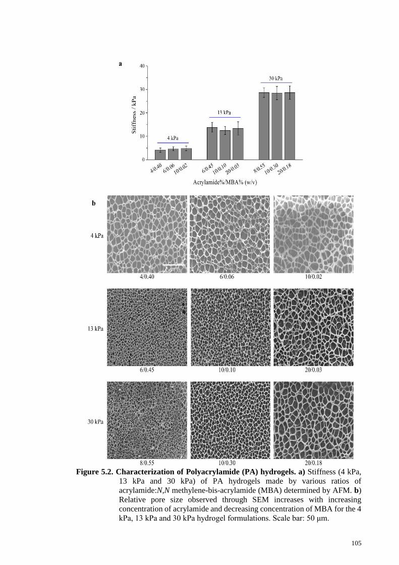
105
Figure 5.2. Characterization of Polyacrylamide (PA) hydrogels. a) Stiffness (4 kPa,
13 kPa and 30 kPa) of PA hydrogels made by various ratios of
acrylamide:N,N methylene-bis-acrylamide (MBA) determined by AFM. b)
Relative pore size observed through SEM increases with increasing
concentration of acrylamide and decreasing concentration of MBA for the 4
kPa, 13 kPa and 30 kPa hydrogel formulations. Scale bar: 50 μm.

106
5.3.2 Characterization of cardiac fibroblasts
Due to limited sources of primary human cardiac fibroblasts, rat cardiac fibroblasts were
selected in this study. Since the neonatal heart is less prone to injury compared to the
adult rat and heart injury may increase cardiac myofibroblast population (Norris et al.,
2008), neonatal rat was used. To characterize cardiac fibroblasts isolated from neonatal
rat, microscopic examination and α-SMA immunofluorescence staining were performed.
It was observed that the isolated cells display a spindle shape with no spontaneous beating
on day 1 until day 9, indicating the absence of cardiomyocytes in the culture. Further,
there is absence of α-SMA (defining marker of myofibroblast) positive cells in the culture
from day 1 till day 4, suggesting the presence of only cardiac fibroblasts without
myofibroblast differentiation (Fig. 5.3). However, starting from day 5, cardiac fibroblasts
were spontaneously differentiated into myofibroblasts (large cells with spindle shape and
protruding dendrite-like processes) (Hinz et al., 2007) on the cell culture plate, as
confirmed by the presence of α-SMA positive cells with enhanced assembly of stress
fibers (F-actin) (Fig. 5.3). These results are in accordance with the findings reported in
literature (Santiago et al., 2010; Wang et al., 2003). These findings suggest that cardiac
fibroblasts isolated and cultured on cell culture plates should be used before day 5.

107
Figure 5.3. Characterization of cardiac fibroblasts and myofibroblasts with
immunofluorescence staining. Cardiac fibroblasts cultured on cell
culture plate before day 5 were negative to α-SMA, whereas those cultured
after day 5 were positively stained by α-SMA. Magnification: 100 ×.Scale
bar: 100 μm.
5.3.3 Effect of pore size and substrate stiffness on cardiac myofibroblast
differentiation
To evaluate the effect of pore size and substrate stiffness on cardiac myofibroblast
differentiation, α-SMA immunofluorescence staining and gene expression analysis were
performed. There is no significant difference (p>0.05) in terms of α-SMA expression
among the cells cultured on PA hydrogels with various pore sizes (Fig. 5.4a-b). These
findings suggest that varying substrate pore size or collagen tethering distance does not
seem to affect cardiac myofibroblast differentiation. Similar results that indicate
osteogenic and adipogenic differentiation of MSCs are not affected by such changes have
been reported in literature (Wen et al., 2014). Taken together, cardiac myofibroblast
differentiation, a mechanoresponsive cellular behavior in cardiac fibroblasts, might not
be regulated by pore size or collagen tethering in planar culture.
108
On the other hand, it was found that cardiac myofibroblast differentiation (as
indicated by positive α-SMA immunofluorescence staining) was dependent on substrate
stiffness, as 30 kPa substrates and cell culture plate (with stiffness of ~1 GPa) were
positive for α-SMA while 4 kPa and 13 kPa substrates were negative for α-SMA (Fig.
5.4a). Gene expression analysis showed that cardiac fibroblasts cultured on 30 kPa
substrates and cell culture plate both display significantly (p<0.05) higher expression
levels of α-SMA than those cultured on 4 kPa and 13 kPa substrates (Fig. 5.4b), further
implying that substrate stiffness play essential role in cardiac myofibroblast
differentiation. These findings suggest that cardiac myofibroblast differentiation is
dependent on ECM stiffness rather than pore size.
Substrates with similar pore size but different stiffness (6/0.06 for 4 kPa, 20/0.03
for 13 kPa, and 20/0.18 for 30 kPa) were selected for further experiments. It was observed
that cardiac fibroblasts cultured on 30 kPa substrates show significantly (p<0.05) higher
expression levels of Col I, Col III and TGF-β1 than those cultured on 4 kPa and 13 kPa
substrates, but lower than those cultured on cell culture plate (Fig. 5.5). These results
show that stiff ECM can induce differentiation of cardiac fibroblasts to more active
cardiac myofibroblasts, which actively secrete collagen, through upregulation and
activation of TGF-β1 (a potent inducer of myofibroblast differentiation). It has been
proven that myofibroblast differentiation and the secretion of collagen and TGF-β1 were
elevated during cardiac fibrosis (Daskalopoulos et al., 2012; van den Borne et al., 2010).
These findings suggest that cardiac fibroblasts cultured on 30 kPa substrates mimicking
the stiffness of native fibrotic cardiac tissue can be used as an in vitro cardiac fibrosis
model while those cultured on 13 kPa substrates mimicking native normal cardiac tissue
can be used as an in vitro normal cardiac model, for pathophysiological and therapeutic
studies.

109
Figure 5.4. Cardiac myofibroblast differentiation was dependent on substrate
stiffness instead of pore size. a) Cardiac fibroblasts cultured on 30 kPa
substrates and cell culture plate were stained positively by α-SMA.
Magnification: 200 ×.Scale bar: 50 μm. b) Varying pore sizes did not affect
gene expression levels of α-SMA in cardiac fibroblasts. Cardiac fibroblasts
cultured on 30 kPa substrates and cell culture plate display significantly
(p<0.05) higher gene expression levels of α-SMA than those cultured on 4
kPa and 13 kPa substrates. Fold change was expressed in relative to 4 kPa
substrates with formulation of 4/0.4. MBA: N,N methylene-bis-acrylamide.

110
Fig. 5.5. Expression of Col I, Col III and TGF-β1 was dependent on substrate
stiffness. A significantly (p<0.05) higher gene expression level of Col I, Col III and TGF-
β1 was observed in cardiac fibroblasts cultured on 30 kPa substrates and cell culture plate.
Fold change was expressed in relative to 4 kPa substrates. * p<0.05 relative to 4 kPa; #
p<0.05 relative to 13 kPa; + p<0.05 relative to 30 kPa.
5.3.4 Mechanism of matrix stiffness - induced cardiac myof ibroblast
differentiation
To date, the comprehensive mechanism of matrix stiffness-induced cardiac myofibroblast
differentiation remains elusive, and only TGF-β1 is known to enhance such
differentiation in response to stiff ECM (Wells, 2013; Yong et al., 2015a; Zhao et al.,
2014). AT1R, a mechanical-induced signaling pathway (activated in the absence of
angiotensin II) (Zablocki & Sadoshima, 2013), has been proven to be activated in cardiac
fibroblasts subjected to interstitial fluid shear stress to induce myofibroblast
differentiation (Galie et al., 2012). However, the role of AT1R in matrix stiffness-induced
cardiac myofibroblast differentiation is unknown. In the present study, we determined the
gene expression level of AT1R in cardiac fibroblasts in response to various substrate
stiffness. It was found that cardiac fibroblasts cultured on 30 kPa substrates display
significantly (p<0.05) higher gene expression level of AT1R than those cultured on soft 4
kPa and 13 kPa substrates, but lowered than those cultured on cell culture plate (Fig. 5.6a).
These results indicate that AT1R expression is dependent on substrate stiffness. Further,
losartan was used to block AT1R-mediated signaling to determine the role of this pathway
in the cellular responses such as α-SMA expression, secretion of Col I, Col III and TGF-

111
β1. Through immunofluorescence staining, it was observed that losartan reduces α-SMA
expression in cardiac fibroblasts cultured on 30 kPa substrates and cell culture plate (Fig.
5.6b & 5.7). Moreover, losartan significantly (p<0.05) lowers the gene expression levels
of α-SMA, Col I, Col III and TGF-β1 in cardiac fibroblasts cultured on 30 kPa substrates
and cell culture plate (Fig. 5.6c). Blocking of AT1R did not affect the responses of cardiac
fibroblasts cultured on 4 kPa and 13 kPa substrates (Fig. 5.5c & 5.7). Taken together,
these findings suggest that stiff ECM induces upregulation and activation of AT1R, which
in turn upregulates and activates TGF-β1 to enhance cardiac myofibroblast differentiation.

112
Figure 5.6. Mechanism of matrix stiffness-induced cardiac myofibroblast
differentiation. Upregulation and activation of AT1R induced by stiff
substrate enhanced cardiac myofibroblast differentiation. a) A
significantly (p<0.05) higher gene expression level of AT1R was observed
in cardiac fibroblasts cultured on 30 kPa substrates and cell culture plate
(* p<0.05 relative to 4 kPa; # p<0.05 relative to 13 kPa; + p<0.05 relative to
30 kPa). Fold change was expressed relative to 4 kPa substrates. b) The α-
SMA in cardiac fibroblasts cultured on 30 kPa appeared to be reduced in
the presence of losartan (AT1R inhibitor). Magnification: 200 ×. Scale bar:
50 μm. c) Losartan significantly (p<0.05) lowered gene expression levels
of α-SMA, Col I, Col III and TGF-β1 in cardiac fibroblasts cultured on 30
kPa substrates and cell culture plate (* p<0.05 relative to - losartan). Fold
change was expressed in relative to 4 kPa substrates with – losartan.

113
Figure 5.7. Treatment of losartan on cardiac fibroblasts cultured on 4 kPa substrates,
13 kPa substrates and cell culture plate. Blocking AT1R by losartan did not
affect α-SMA expression in cardiac fibroblasts cultured on 4 kPa and 13 kPa
substrates. Losartan appeared to reduce expression of α-SMA in cardiac
fibroblasts cultured on cell culture plate. Magnification: 200 ×. Scale bar: 50
μm.
5.3.5 Paracrine effects of fresh and cryopreserved hASCs on cardiac
fibrosis
To determine the paracrine effects of fresh and cryopreserved hASCs on cardiac fibrosis,
conditioned medium of fresh and cryopreserved hASCs were applied respectively to
cardiac fibroblasts for 3 days following 5-day culture on PA hydrogels. It was observed
that conditioned medium of both fresh and cryopreserved hASCs reduces the expression
of α-SMA in cardiac fibroblasts cultured on 30 kPa substrates (Fig. 5.8a) and cell culture
plate (Fig. 5.9). Further, it was found that conditioned medium of fresh and cryopreserved
hASCs significantly (p<0.05) lower the gene expression levels of α-SMA, Col I, Col III
and TGF-β1 in cardiac fibroblasts cultured on 30 kPa substrates and cell culture plate
(Fig. 5.8b), while conditioned medium of fresh and cryopreserved hASCs does not have
any effect on the responses of cardiac fibroblasts cultured on 4 kPa and 13 kPa substrates

114
(Fig. 5.8b & Fig. 5.9). These results indicate that conditioned medium of fresh and
cryopreserved hASCs contain at least one kind of anti-fibrotic factors that can inhibit
matrix stiffness-induced cardiac fibrosis.

115
Figure 5.8. Fresh and cryopreserved hASCs inhibited matrix stiffness-induced
cardiac myofibroblast differentiation through paracrine effect. a) The
α-SMA in cardiac fibroblasts cultured on 30 kPa substrates appeared to be
reduced in the presence of conditioned medium of fresh and cryopreserved
hASCs. Magnification: 200 ×. Scale bar: 50 μm. b) Conditioned medium of
fresh and cryopreserved hASCs significantly (p<0.05) lowered gene
expression levels of α-SMA, Col I, Col III and TGF-β1 in cardiac fibroblasts
cultured on 30 kPa substrates and cell culture plate (* p<0.05 relative to
before treatment with conditioned medium of hASCs). Fold change was
expressed in relative to 4 kPa substrates before treatment with conditioned
medium of hASCs.

116
Figure 5.9. Treatment with only conditioned medium of fresh and cryopreserved
hASCs or conditioned medium of hASCs + anti-HGF on cardiac
fibroblasts cultured on 4 kPa substrates, 13 kPa substrates and cell
culture plate. No change of α-SMA expression in cardiac fibroblasts
cultured on 4 kPa and 13 kPa substrates. Conditioned medium of fresh and
cryopreserved hASCs appeared to reduce expression of α-SMA in cardiac
fibroblasts cultured on cell culture plate. Such effect appeared to be
abrogated in the presence of anti-HGF. Magnification: 200 ×. Scale bar: 50
μm.
5.3.6 Inhibitory mechanism of hASCs on cardiac fibrosis
Although significant effort has been put into the study of the anti-fibrotic effects of MSCs
(Li et al., 2015; Mao et al., 2013; Mias et al., 2009; Ohnishi et al., 2007; Wang et al.,
2011), the mechanism for inhibiting cardiac fibrosis through reduction of cardiac
myofibroblast differentiation remains elusive. In the present study, it was found that the
conditioned medium of hASCs significantly (p<0.05) lowers the gene expression level of
AT1R in cardiac fibroblasts cultured on stiff substrates (Fig. 5.10), indicating that the

117
conditioned medium of hASCs might downregulate AT1R which in turn decreases the
secretion of TGF-β1, resulting in lesser cardiac myofibroblast differentiation. In the
present study, it was found that cardiac fibroblasts cultured on stiff substrates display a
significantly higher gene expression level of Smad7 than those cultured on 4 kPa and 13
kPa substrates. These results demonstrate that Smad7 in cardiac fibroblasts is upregulated
in response to substrate stiffness-induced increase in TGF-β1 to antagonize TGF-β1
signalling by negative feedback loops. Interestingly, with the addition of conditioned
medium of hASCs, cardiac fibroblasts showed a significantly (p<0.05) sharp increase in
the gene expression level of Smad7 (Fig. 5.10), resulting in lesser cardiac myofibroblast
differentiation. Taken together, these findings suggest that the soluble factor in
conditioned medium of hASCs might inhibit cardiac myofibroblast differentiation via
AT1R and Smad7.
Among soluble factors secreted by MSCs, HGF, a growth factor which is known
to work as an antagonist to decrease TGF-β1 expression and counteract TGF-β1 signaling
(Inoue et al., 2003; Okayama et al., 2012), may work to inhibit cardiac myofibroblast
differentiation. To determine the role of HGF in the conditioned medium of hASCs for
inhibition of cardiac myofibroblast differentiation, the active HGF in conditioned
medium of hASCs was blocked using anti-HGF, prior to its being given to cardiac
fibroblasts. Through immunofluorescence staining, it was observed that cardiac
fibroblasts cultured on 30 kPa substrates and cell culture plate with anti-HGF treated
conditioned medium of hASCs expressed more α-SMA than those cultured with only
conditioned medium of hASCs (Fig. 5.11a & 5.9). Further, the gene expression levels of
of α-SMA, Col I, Col III, TGF-β1 and AT1R are significantly (p<0.05) higher, while the
expression level of Smad7 is significantly (p<0.05) lower in cardiac fibroblasts cultured
on 30 kPa substrates and cell culture plate with anti-HGF treated conditioned medium of

118
hASCs than those cultured with only conditioned medium of hASCs (Fig. 5.11b). These
results demonstrate that HGF secreted by hASCs plays an essential role on attenuating
cardiac fibrosis possibly through the downregulation of AT1R and upregulation of Smad7.
Figure 5.10. Conditioned medium of fresh and cryopreserved hASCs reduced
expression of AT1R while enhanced expression of Smad7. Conditioned
medium of fresh and cryopreserved hASCs significantly (p<0.05) lowered
the gene expression level of AT1R and enhanced the gene expression level
of Smad7 in cardiac fibroblasts cultured on 30 kPa substrates and cell
culture plate (* p<0.05 relative to before treatment with conditioned
medium of hASCs; # p<0.05 relative to 4 kPa; + p<0.05 relative to 13 kPa).

119
Figure 5.11: hASCs secreted HGF to inhibit cardiac myofibroblast differentiation
via AT1R and Smad7. HGF antibody (anti-HGF) appeared to abrogate the
inhibition of conditioned medium of hASCs on the expression of α-SMA in
cardiac fibroblasts cultured on 30 kPa substrates. Magnification: 200×.Scale
bar: 50 μm. c) Anti-HGF negated the anti-fibrotic activity of conditioned
medium of hASCs, as indicated by significant (p<0.05) higher gene
expression levels of α-SMA, Col I, Col III, TGF-β1 and AT1R, and lower
gene expression level of Smad7 in cardiac fibroblasts cultured on 30 kPa
substrates and cell culture plate (* p<0.05 relative to after treatment with
conditioned medium of hASCs; # p<0.05 relative to before treatment with
conditioned medium of hASCs). Fold change was expressed in relative to 4
kPa substrates before treatment with conditioned medium of hASCs.

120
ELISA was used to determine the concentration of HGF in the conditioned
medium of fresh and cryopreserved hASCs. A reliable standard curve of known
concentration of HGF versus absorbance at 450 nm with a high coefficient of
determination (R2 = 0.99) was plotted (Fig. 5.12a). It was found that the absorbance of
conditioned medium of fresh and cryopreserved hASCs at 450 nm is significantly (p<0.05)
higher than that of medium without cell and conditioned medium of cardiac fibroblasts
(Fig. 5.12b), suggesting that conditioned medium of fresh and cryopreserved hASCs
contains higher concentration of HGF. There is no significant (p>0.05) difference in terms
of absorbance at 450 nm between medium without cell and conditioned medium of
cardiac fibroblasts (Supplemental Fig. 5.12b), indicating that cardiac fibroblasts did not
secrete HGF. Conditioned medium of fresh and cryopreserved hASCs displayed a similar
(p>0.05) concentration of HGF (Fig. 5.12c).
Figure 5.12. A similar concentration of HGF was observed in conditioned medium
of fresh and cryopreserved hASCs. a) Standard curve of known
concentration of HGF versus absorbance at 450 nm measured through
ELISA with coefficient of determination R2 = 0.99. b) Absorbance at 450
nm was significantly (p<0.05) higher in conditioned medium of fresh and
cryopreserved hASCs compared to medium without cells and conditioned
medium of cardiac fibroblasts (CFs) (* p<0.05 relative to medium without
cell; # p<0.05 relative to conditioned medium of CFs). c) By referring to the
standard curve, the mean concentration of HGF in conditioned medium of
fresh and cryopreserved hASCs are shown to be 135 ± 10.75 pg/mL and
141.7 ± 3.01 pg/mL respectively.

121
5.4 Discussion
A mechanical tunable cell culture system developed by Engler’s group was used in the
present study, which allows us to decouple the effect of substrate stiffness and pore size
on cardiac myofibroblast differentiation. Col I was selected as the matrix protein coated
on PA hydrogels as it represents the majority of ECM (~75%) in the heart (Dobaczewski
et al., 2012). As a result, it was demonstrated that cardiac myofibroblast differentiation is
dependent on substrate stiffness instead of substrate pore size (represents matrix protein
tethering). Substrates mimicking stiffness of native fibrotic cardiac tissues (30 kPa) were
found to induce cardiac myofibroblast differentiation, suggesting that cardiac fibroblasts
cultured on 30 kPa substrates for 5 days can be used as an in vitro cardiac fibrosis model.
Further, it was found that gene expression of AT1R is dependent on substrate
stiffness, which is reported for the first time. AT1R, a receptor located upstream of TGF-
β1 receptor, is usually activated in response to angiotensin II or shear stress, which in turn
increases secretion of TGF-β1. By blocking AT1R using losartan, it was found that
expressions of TGF-β1 and α-SMA are reduced, suggesting that losartan might attenuate
substrate stiffness-induced increase in TGF-β1 and α-SMA to inhibit cardiac
myofibroblast differentiation through AT1R. It has been proved that losartan can attenuate
AT1R-mediated signaling induced by angiotensin II or interstitial fluid shear stress to
inhibit cardiac myofibroblast differentiation (Campbell & Katwa, 1997; Galie et al.,
2012). Taken together, stiff substrate upregulates and activates AT1R which in turn
upregulates TGF-β1 to promote cardiac myofibroblast differentiation.
Undifferentiated MSCs hold great potential in cardiac fibrosis therapy due to their
paracrine functions. MSCs were found to secrete soluble factors that might contribute to
anti-fibrotic and endogenous cardiomyogenesis, which aid in cardiac repair and restoring

122
cardiac function. Among the MSCs derived from different types of human tissue, hASCs,
in particular, have attracted special attention due to the ready accessibility and abundance
of adipose tissues (Gomillion & Burg, 2006; Kolle et al., 2013; Mizuno et al., 2012).
Analyses of the soluble factors released from hASCs have revealed that cultured
undifferentiated hASCs at relatively early passages (within passage 5) secrete growth
factors (e.g., HGF, IGF-1 and basic fibroblast growth factor (bFGF)), tumor necrosis
factor (TNF)-α, cytokines (e.g., interleukin-6 and interleukin-7) and others (Choi et al.,
2015; J Salgado et al., 2010; Mizuno et al., 2012). It has been proven that there is a
possible cross-species interaction between hASCs and rat cardiac fibroblasts, in which
hASCs can reduce rat cardiac myofibroblast differentiation through paracrine effects (Li
et al., 2015). However, the inhibitory mechanism of hASCs on cardiac myofibroblast
differentiation remains elusive. Following development of an in vitro rat cardiac fibrosis
model that incorporates stiffness mimicking native fibrotic cardiac tissues, conditioned
medium of hASCs was applied to the model to investigate their paracrine effects on
cardiac myofibroblast differentiation. Interestingly, it was demonstrated that
undifferentiated hASCs can secrete HGF to inhibit matrix stiffness-induced cardiac
myofibroblast differentiation via downregulation of AT1R and upregulation of Smad7,
thus reducing cardiac fibrosis. The proposed mechanism of matrix stiffness-induced
cardiac myofibroblast differentiation and the inhibitory mechanism of hASCs on such
differentiation are depicted in Fig. 5.13. It has been reported that conditioned media
collected from MSCs cultured on both tissue culture plate and substrates mimicking the
stiffness of native fibrotic cardiac tissues contain more HGF than those cultured on
substrates mimicking the physiological stiffness of cardiac tissues (Sullivan et al., 2014).
These findings suggest that HGF could be secreted by MSCs in response to stiff ECM,
which in turn suppresses cardiac fibrosis.

123
Figure 5.13. Summary of proposed mechanism of matrix stiffness-induced cardiac
myofibroblast differentiation and anti-fibrotic effects of
undifferentiated hASCs. HGF: hepatocyte growth factor; AT1R:
angiotensin II type 1 receptor; TGF-β1: transforming growth factor beta-
1; P-Smad2: phosphorylated Smad2; α-SMA: alpha-smooth muscle actin.
ECM: extracellular matrix.
It has been demonstrated that HGF inhibits the increase in AT1R expression to
reduce myofibroblast differentiation of glomerular mesangial cells in the kidney (Iekushi
et al., 2011). HGF might bind to HGF receptor (c-met) to attenuate AT1R expression
through inhibition of phosphatidylinositol 3-kinase/Akt signalling pathway (Iekushi et al.,
2011). On the other hand, HGF also activates Smad7 to inhibit myofibroblast
differentiation of alveolar epithelial cells in lungs (Shukla et al., 2009). Smad7, an
inhibitory Smad, is upregulated in response to increasing TGF-β1, and binds to TGF-β1
receptor to prevent interaction of Smad2 with the receptor (Hayashi et al., 1997; Nakao
et al., 1997), reducing the expression of α-SMA or myofibroblast differentiation. HGF-
induced increase in Smad7 could be contributed by mitogen-activated protein kinase –
extracellular-signal-regulated kinase (MEK - ERK) signalling, which in turn blocks

124
phosphorylation or activation of Smad2 (Hayashi et al., 1997; Shukla et al., 2009). Further
studies are needed to explore the roles of these pathways on regulation of AT1R and
Smad7 by HGF in cardiac myofibroblasts. Recently, it has been showed that hASCs may
exert anti-fibrotic effects by directly communicating with myofibroblasts (Li et al., 2015).
Taken together, hASCs may hold great promise for treating cardiac fibrosis though both
paracrine effects and direct cell-to-cell contact.
Although evidence continues to augment that hASCs have great potential as a
cardiac fibrosis therapy, but there are many challenges that need to be addressed. First,
cardiac fibrosis therapy is an intensive clinical setting which requires a massive amount
of cells which in vitro cell expansion might not able to supply in a short duration. Further,
hASCs under long-term in vitro culture could be prone to contamination, malignant
transformation, phenotypic instability and chromosomal aberration (Froelich et al., 2013;
Roemeling-van Rhijn et al., 2013). Therefore, a bank of cryopreserved cells is an ideal
option which offers a ready off-the-shelf supply of cells for transplant while allows better
timing for therapy. To achieve this, it is essential to cryopreserve hASCs at an early
passage (within passage 4) for long-term in order to supply a pool of efficacious and safe
cells for transplant, as their therapeutic efficacy might decrease with biosafety concerns
raised at late passage (Marquez-Curtis et al., 2015). In the present study, conditioned
medium of long-term cryopreserved hASCs (passage 3) also was applied to the in vitro
cardiac fibrosis model to evaluate their inhibitory effects on cardiac myofibroblast
differentiation. It was found that conditioned medium of hASCs cryopreserved in 5%
DMSO display a similar potential to inhibit cardiac myofibroblast differentiation as
compared to fresh hASCs. Further, a similar concentration of HGF was observed in the
conditioned medium of cryopreserved and fresh hASCs, suggesting that cryopreservation
do not affect the paracrine function of hASCs. In conjunction with the previous findings

125
indicating cell functionality of hASCs cryopreserved in 5% DMSO was maintained with
a low risk of tumourigenesis, long-term cryopreserved hASCs would be an ideal cell
source for cardiac fibrosis therapy in future.
Second, the therapeutic effects of implanted MSCs have been shown greatly
reduced under stiff ECM during cardiac fibrosis. For instance, when undifferentiated
MSCs were implanted into the heart of rat model of myocardial infarction, they showed
bone tissue formation in response to stiff ECM (Breitbach et al., 2007). Last but not least,
engraftment or retention of MSCs in the heart is low by any method of delivery (e.g.,
intramyocardiac and intracoronary) (Hou et al., 2005). Therefore, further investigation is
needed to assess the delivery time and mode for therapeutic use of hASCs in human body
to achieve an efficient and successful cardiac fibrosis therapy.
5.5 Conclusion
Cardiac fibroblasts cultured on 30 kPa hydrogels, which mimic the stiffness of native
fibrotic cardiac tissues, can be used as an in vitro cardiac fibrosis model. Fresh and
cryopreserved hASCs can secrete HGF to reduce cardiac fibrosis and myofibroblast
differentiation possibly through the downregulation of AT1R and upregulation of Smad7.
With the development of stem cell delivery method, hASCs may hold great potential for
cardiac fibrosis therapy and heart regeneration in future.

126
CHAPTER 6: CONCLUSION AND FUTURE WORK
6.1 Thesis contribution
This thesis is contributed principally to knowledge. The contributions are as follows:
i. A cryopreservation method, which adopts an approach using a low concentration
of DMSO (5% DMSO), can be used widely to efficiently store hASCs for clinical
application.
ii. Cryopreserved hASCs are at low risk of raising biosafety concerns, which
supports their use in various clinical settings.
iii. Cardiac myofibroblast differentiation is regulated by substrate stiffness
independent of substrate pore size.
iv. An in vitro cardiac fibrosis model which incorporates stiffness mimicking native
fibrotic cardiac tissues has been developed, allowing the pathophysiological and
therapeutic intervention study of cardiac fibrosis in an accurate manner.
v. hASCs preserved in 5% DMSO could be an ideal cell source for various clinical
applications, including but not limited to cardiac fibrosis.
6.2 Conclusion
In chapter 3, the cells isolated from human adipose tissue were characterized. It has been
showed that the cells are hASCs, as indicated by their adherent property, positive
expression of surface markers of MSCs (CD90, CD73 and CD105 and CD44) and
nucleated cells (HLA ABC), negative expression of surface markers of hematopoietic
cells (CD14, CD19, CD34, CD45 and HLA DQDPDR) and ability of trilineage
differentiation (adipogenesis, osteogenesis and chondrogenesis).

127
In chapter 4, the viability, functional properties and biosafety of hASCs following
the long-term cryopreservation have been evaluated. The findings showed that long-term
cryopreserved hASCs maintained their functional properties (e.g., stemness, proliferation
and trilineage differentiation potential) and a low risk of tumourigenesis. Among the
CPAs, 5% DMSO without FBS which is less cytotoxic and free of xeno-agent while
maintaining high viability of hASCs, would be an ideal CPA to cryopreserve hASCs in
long-term for clinical applications.
In chapter 5, an in vitro cardiac fibrosis model has been developed and used for
the evaluation of potential therapeutic application of hASCs in cardiac fibrosis. Cardiac
fibroblasts cultured on 30 kPa hydrogels can be used as an in vitro cardiac fibrosis model,
as such stiffness, which mimics the stiffness of native fibrotic cardiac tissues, induces
cardiac myofibroblast differentiation. In addition, fresh hASCs and hASCs cryopreserved
in 5% DMSO have been assessed of their potential in reducing cardiac fibrosis using the
in vitro cardiac fibrosis model. The findings demonstrated that hASCs cryopreserved in
5% DMSO display similar potential as fresh hASCs in reducing cardiac fibrosis through
inhibition of cardiac myofibroblast differentiation via downregulation of AT1R and
upregulation of Smad7 in a paracrine manner. hASCs cryopreserved in 5% DMSO could
be an ideal cell source for cardiac fibrosis therapy.
Collectively, 5% DMSO could be an ideal CPA for long-term cryopreservation of
hASCs for clinical applications. hASCs cryopreserved in such formulation demonstrates
their potential therapeutic application in cardiac fibrosis. These findings would help to
standardize the cryopreservation protocol of hASCs and optimize the therapeutic benefit
of cryopreserved hASCs in cardiac fibrosis.

128
6.3 Limitation of the study and future work
The viability of cryopreserved hASCs (75% - 85%) can be further improved.
Cryopreserved hASCs can be pre-cultured in a hypoxic condition for a short period prior
to being used in clinical applications, as hypoxia has been showed to enhance the viability
of fresh hASCs in culture. On the other hand, the development of an alternative CPA to
completely replace DMSO remains challenging, which requires an extensive
investigation. Since hASCs cryopreserved in 5% DMSO have displayed their potential
therapeutic application in cardiac fibrosis, their therapeutic potential in other various
clinical settings (e.g., diabetes and cancer therapy) should be explored.
The in vitro cardiac fibrosis model developed in this study is still lack of paracrine
factors arising from cardiomyocytes. In future, current model can be adapted to co-culture
cardiac fibroblasts and cardiomyocytes, which allows the mechanistic investigation of
their interaction in cardiac fibrosis. Meanwhile, paracrine effects of hASCs on
cardiomyocyte regeneration also can be investigated. However, there is a limited assay to
distinguish the response of different cells in a co-culture setting. This issue should be
addressed prior to the development of an in vitro cardiac fibrosis model incorporating
both cardiac fibroblasts and cardiomyocytes. With the advance in the development of
induced pluripotent stem cells (iPSCs) and hydrogel fabrication techniques (e.g.,
bioprinting), a 3D in vitro cardiac fibrosis model made of human cardiac fibroblasts and
cardiomyocytes which both are derived from iPSCs, allowing deep insights into human
cell response in vivo in an accurate manner. In addition, mechanical strain (e.g.,
mechanical stretch and interstitial fluid flow), which also play a role in cardiac
myofibroblast differentiation, can be incorporated into the model to develop a
hypertension-induced cardiac fibrosis model for studying hypertension.

129
The engraftment or retention of MSCs in the heart is low by any mode of delivery
(e.g., intramyocardiac and intracoronary). Therefore, the delivery time and mode of
cryopreserved hASCs into patients with cardiac fibrosis should be optimized to achieve
a successful and efficient cardiac fibrosis therapy.

130
References
AbdelHafez, F., Xu, J., Goldberg, J., & Desai, N. (2011). Vitrification in open and closed
carriers at different cell stages: Assessment of embryo survival, development,
DNA integrity and stability during vapor phase storage for transport. BMC
Biotechnology, 11(29), 1472-6750.
Amarilio, R., Viukov, S. V., Sharir, A., Eshkar-Oren, I., Johnson, R. S., & Zelzer, E.
(2007). Hif1alpha regulation of sox9 is necessary to maintain differentiation of
hypoxic prechondrogenic cells during early skeletogenesis. Development, 134(21),
3917-3928.
Anchordoguy, T. J., Cecchini, C. A., Crowe, J. H., & Crowe, L. M. (1991). Insights into
the cryoprotective mechanism of dimethyl sulfoxide for phospholipid bilayers.
Cryobiology, 28(5), 467-473.
Andrews, P. W., Matin, M. M., Bahrami, A. R., Damjanov, I., Gokhale, P., & Draper, J.
S. (2005). Embryonic stem (es) cells and embryonal carcinoma (ec) cells:
Opposite sides of the same coin. Biochem Soc Trans, 33(Pt 6), 1526-1530.
Angelo, P. C., Ferreira, A. C., Fonseca, V. D., Frade, S. P., Ferreira, C. S., Malta, F. S., . . .
Cabral, A. C. (2012). Cryopreservation does not alter karyotype, multipotency, or
nanog/sox2 gene expression of amniotic fluid mesenchymal stem cells. Genet Mol
Res, 11(2), 1002-1012. doi: 10.4238/2012.April.19.5
Anger, J. T., Gilbert, B. R., & Goldstein, M. (2003). Cryopreservation of sperm:
Indications, methods and results. J Urol, 170(4 Pt 1), 1079-1084.
Armesilla-Diaz, A., Elvira, G., & Silva, A. (2009). P53 regulates the proliferation,
differentiation and spontaneous transformation of mesenchymal stem cells. Exp
Cell Res, 315(20), 3598-3610. doi: 10.1016/j.yexcr.2009.08.004
Arslan, F., de Kleijn, D. P., & Pasterkamp, G. (2011). Innate immune signaling in cardiac
ischemia. Nat Rev Cardiol, 8(5), 292-300. doi: 10.1038/nrcardio.2011.38
Artandi, Steven E., & DePinho, Ronald A. (2010). Telomeres and telomerase in cancer.
Carcinogenesis, 31(1), 9-18. doi: 10.1093/carcin/bgp268
Arthur, A., Zannettino, A., & Gronthos, S. (2009). The therapeutic applications of
multipotential mesenchymal/stromal stem cells in skeletal tissue repair. J Cell
Physiol, 218(2), 237-245. doi: 10.1002/jcp.21592
Badimon, L., Onate, B., & Vilahur, G. (2015). Adipose-derived mesenchymal stem cells
and their reparative potential in ischemic heart disease. Rev Esp Cardiol (Engl
Ed), 68(7), 599-611. doi: 10.1016/j.rec.2015.02.025
Balaban, N. Q., Schwarz, U. S., Riveline, D., Goichberg, P., Tzur, G., Sabanay, I., . . .
Geiger, B. (2001). Force and focal adhesion assembly: A close relationship
studied using elastic micropatterned substrates. Nat Cell Biol, 3(5), 466-472. doi:
10.1038/35074532

131
Balci, D., & Can, A. (2013). The assessment of cryopreservation conditions for human
umbilical cord stroma-derived mesenchymal stem cells towards a potential use for
stem cell banking. Curr Stem Cell Res Ther, 8(1), 60-72.
Baraniak, P. R., & McDevitt, T. C. (2010). Stem cell paracrine actions and tissue
regeneration. Regen Med, 5(1), 121-143.
Barkholt, L., Flory, E., Jekerle, V., Lucas-Samuel, S., Ahnert, P., Bisset, L., . . .
Salmikangas, P. (2013). Risk of tumorigenicity in mesenchymal stromal cell-
based therapies--bridging scientific observations and regulatory viewpoints.
Cytotherapy, 15(7), 753-759. doi: 10.1016/j.jcyt.2013.03.005
Barry, F. P., & Murphy, J. M. (2004). Mesenchymal stem cells: Clinical applications and
biological characterization. Int J Biochem Cell Biol, 36(4), 568-584.
Bartek, J., Bartkova, J., & Lukas, J. (2007). DNA damage signalling guards against
activated oncogenes and tumour progression. Oncogene, 26(56), 7773-7779. doi:
10.1038/sj.onc.1210881
Benekli, M., Anderson, B., Wentling, D., Bernstein, S., Czuczman, M., & McCarthy, P.
(2000). Severe respiratory depression after dimethylsulphoxide-containing
autologous stem cell infusion in a patient with al amyloidosis. Bone Marrow
Transplant, 25(12), 1299-1301. doi: 10.1038/sj.bmt.1702452
Berloco, P., Russo, F., Cariola, F., Gentile, M., Giorgio, P., Caruso, M. L., . . . Di Leo, A.
(2003). Low presence of p53 abnormalities in h pylori-infected gastric mucosa
and in gastric adenocarcinoma. J Gastroenterol, 38(1), 28-36. doi:
10.1007/s005350300003
Berry, M. F., Engler, A. J., Woo, Y. J., Pirolli, T. J., Bish, L. T., Jayasankar, V., . . .
Sweeney, H. L. (2006). Mesenchymal stem cell injection after myocardial
infarction improves myocardial compliance. Am J Physiol Heart Circ Physiol,
290(6), H2196-2203. doi: 10.1152/ajpheart.01017.2005
Berz, D., McCormack, E. M., Winer, E. S., Colvin, G. A., & Quesenberry, P. J. (2007).
Cryopreservation of hematopoietic stem cells. Am J Hematol, 82(6), 463-472. doi:
10.1002/ajh.20707
Binder, Marc D, Hirokawa, Nobutaka, & Windhorst, Uwe. (2009). Encyclopedia of
neuroscience (Vol. 3166): Springer Berlin, Heidelberg.
Blackburn, E. H. (1991). Telomeres. Trends Biochem Sci, 16(10), 378-381.
Blasco, M. A., & Hahn, W. C. (2003). Evolving views of telomerase and cancer. Trends
Cell Biol, 13(6), 289-294.
Bourin, P., Bunnell, B. A., Casteilla, L., Dominici, M., Katz, A. J., March, K. L., . . .
Gimble, J. M. (2013). Stromal cells from the adipose tissue-derived stromal
vascular fraction and culture expanded adipose tissue-derived stromal/stem cells:
A joint statement of the international federation for adipose therapeutics and
science (ifats) and the international society for cellular therapy (isct). Cytotherapy,
15(6), 641-648. doi: 10.1016/j.jcyt.2013.02.006

132
Boyer, L. A., Lee, T. I., Cole, M. F., Johnstone, S. E., Levine, S. S., Zucker, J. P., . . .
Young, R. A. (2005). Core transcriptional regulatory circuitry in human
embryonic stem cells. Cell, 122(6), 947-956.
Breitbach, Martin, Bostani, Toktam, Roell, Wilhelm, Xia, Ying, Dewald, Oliver, Nygren,
Jens M, . . . Hescheler, Juergen. (2007). Potential risks of bone marrow cell
transplantation into infarcted hearts. Blood, 110(4), 1362-1369.
Bryant, J. E., Shamhart, P. E., Luther, D. J., Olson, E. R., Koshy, J. C., Costic, D. J., . . .
Meszaros, J. G. (2009). Cardiac myofibroblast differentiation is attenuated by
alpha(3) integrin blockade: Potential role in post-mi remodeling. J Mol Cell
Cardiol, 46(2), 186-192. doi: 10.1016/j.yjmcc.2008.10.022
Buchanan, S. S., Gross, S. A., Acker, J. P., Toner, M., Carpenter, J. F., & Pyatt, D. W.
(2004). Cryopreservation of stem cells using trehalose: Evaluation of the method
using a human hematopoietic cell line. Stem Cells & Development, 13(3), 295-
305.
Caetano-Lopes, J., Canhao, H., & Fonseca, J. E. (2007). Osteoblasts and bone formation.
Acta Reumatol Port, 32(2), 103-110.
Callihan, P., Mumaw, J., Machacek, D. W., Stice, S. L., & Hooks, S. B. (2011).
Regulation of stem cell pluripotency and differentiation by g protein coupled
receptors. Pharmacol Ther, 129(3), 290-306.
Campbell, S. E., & Katwa, L. C. (1997). Angiotensin ii stimulated expression of
transforming growth factor-beta1 in cardiac fibroblasts and myofibroblasts. J Mol
Cell Cardiol, 29(7), 1947-1958.
Cao, Y. (2010). Adipose tissue angiogenesis as a therapeutic target for obesity and
metabolic diseases. Nat Rev Drug Discov, 9(2), 107-115.
Caplan, A. I. (2000). Mesenchymal stem cells and gene therapy. Clin Orthop Relat
Res(379 Suppl), S67-70.
Caplan, A. I. (2007). Adult mesenchymal stem cells for tissue engineering versus
regenerative medicine. Journal of Cellular Physiology, 213(2), 341-347.
Caplan, A. I., & Bruder, S. P. (2001). Mesenchymal stem cells: Building blocks for
molecular medicine in the 21st century. Trends Mol Med, 7(6), 259-264.
Castelo-Branco, M. T., Soares, I. D., Lopes, D. V., Buongusto, F., Martinusso, C. A., do
Rosario, A., Jr., . . . Souza, H. S. (2012). Intraperitoneal but not intravenous
cryopreserved mesenchymal stromal cells home to the inflamed colon and
ameliorate experimental colitis. PLoS One, 7(3), e33360. doi:
10.1371/journal.pone.0033360
Chagastelles, P. C., Nardi, N. B., & Camassola, M. (2010). Biology and applications of
mesenchymal stem cells. Sci Prog, 93(Pt 2), 113-127.
Chandran, P. L., & Horkay, F. (2012). Aggrecan, an unusual polyelectrolyte: Review of
solution behavior and physiological implications. Acta Biomater, 8(1), 3-12.

133
Chaudhuri, O., & Mooney, D. J. (2012). Stem-cell differentiation: Anchoring cell-fate
cues. Nat Mater, 11(7), 568-569. doi: 10.1038/nmat3366
Chin, S. P., Poey, A. C., Wong, C. Y., Chang, S. K., Teh, W., Mohr, T. J., & Cheong, S.
K. (2010). Cryopreserved mesenchymal stromal cell treatment is safe and feasible
for severe dilated ischemic cardiomyopathy. Cytotherapy, 12(1), 31-37.
Choi, J. R., Pingguan-Murphy, B., Wan Abas, W. A., Noor Azmi, M. A., Omar, S. Z.,
Chua, K. H., & Wan Safwani, W. K. (2014). Impact of low oxygen tension on
stemness, proliferation and differentiation potential of human adipose-derived
stem cells. Biochem Biophys Res Commun, 448(2), 218-224.
Choi, J. R., Pingguan-Murphy, B., Wan Abas, W. A., Yong, K. W., Poon, C. T., Noor
Azmi, M. A., . . . Wan Safwani, W. K. (2015). In situ normoxia enhances survival
and proliferation rate of human adipose tissue-derived stromal cells without
increasing the risk of tumourigenesis. PLoS One, 10(1), e0115034. doi:
10.1371/journal.pone.0115034
Chua, K. H., Zaman Wan Safwani, W. K., Hamid, A. A., Shuhup, S. K., Mohd Haflah,
N. H., & Mohd Yahaya, N. H. (2014). Retropatellar fat pad-derived stem cells
from older osteoarthritic patients have lesser differentiation capacity and
expression of stemness genes. Cytotherapy, 16(5), 599-611. doi:
10.1016/j.jcyt.2013.08.013
Chuaire-Noack, L., Sánchez-Corredor, M. C., & Ramírez-Clavijo, S. R. (2010). The dual
role of senescence in tumorigenesis. Int. J. Morphol, 28(1), 37-50.
Clarke, B. (2008). Normal bone anatomy and physiology. Clinical journal of the
American Society of Nephrology, 3(Supplement 3), S131-S139.
Conrad, C., & Huss, R. (2005). Adult stem cell lines in regenerative medicine and
reconstructive surgery. The Journal of surgical research, 124(2), 201-208. doi:
10.1016/j.jss.2004.09.015
Corsten, M. F., & Shah, K. (2008). Therapeutic stem-cells for cancer treatment: Hopes
and hurdles in tactical warfare. Lancet Oncol, 9(4), 376-384. doi: 10.1016/s1470-
2045(08)70099-8
Czekanska, E. M. (2011). Assessment of cell proliferation with resazurin-based
fluorescent dye Mammalian cell viability (pp. 27-32): Springer.
Dalla Costa, A. P., Clemente, C. F., Carvalho, H. F., Carvalheira, J. B., Nadruz, W., Jr.,
& Franchini, K. G. (2010). Fak mediates the activation of cardiac fibroblasts
induced by mechanical stress through regulation of the mtor complex. Cardiovasc
Res, 86(3), 421-431. doi: 10.1093/cvr/cvp416
Daniels, A., van Bilsen, M., Goldschmeding, R., van der Vusse, G. J., & van
Nieuwenhoven, F. A. (2009). Connective tissue growth factor and cardiac fibrosis.
Acta Physiol (Oxf), 195(3), 321-338. doi: 10.1111/j.1748-1716.2008.01936.x

134
Daskalopoulos, E. P., Janssen, B. J., & Blankesteijn, W. M. (2012). Myofibroblasts in the
infarct area: Concepts and challenges. Microsc Microanal, 18(1), 35-49. doi:
10.1017/s143192761101227x
Davies, O. G., Smith, A. J., Cooper, P. R., Shelton, R. M., & Scheven, B. A. (2014). The
effects of cryopreservation on cells isolated from adipose, bone marrow and dental
pulp tissues. Cryobiology, 69(2), 342-347.
Davis, J., & Molkentin, J. D. (2014). Myofibroblasts: Trust your heart and let fate decide.
J Mol Cell Cardiol, 70, 9-18. doi: 10.1016/j.yjmcc.2013.10.019
de la Fuente, R., Bernad, A., Garcia-Castro, J., Martin, M. C., & Cigudosa, J. C. (2010).
Retraction: Spontaneous human adult stem cell transformation. Cancer Res,
70(16), 6682. doi: 10.1158/0008-5472.can-10-2451
de Lima Prata, K., de Santis, G. C., Orellana, M. D., Palma, P. V., Brassesco, M. S., &
Covas, D. T. (2012). Cryopreservation of umbilical cord mesenchymal cells in
xenofree conditions. Cytotherapy, 14(6), 694-700. doi:
10.3109/14653249.2012.677820
De Miguel, M. P., Fuentes-Julian, S., Blazquez-Martinez, A., Pascual, C. Y., Aller, M.
A., Arias, J., & Arnalich-Montiel, F. (2012). Immunosuppressive properties of
mesenchymal stem cells: Advances and applications. Curr Mol Med, 12(5), 574-
591.
De Rosa, A., De Francesco, F., Tirino, V., Ferraro, G. A., Desiderio, V., Paino, F., . . .
Papaccio, G. (2009). A new method for cryopreserving adipose-derived stem cells:
An attractive and suitable large-scale and long-term cell banking technology.
Tissue Eng Part C Methods, 15(4), 659-667. doi: 10.1089/ten.TEC.2008.0674
De Ugarte, D. A., Morizono, K., Elbarbary, A., Alfonso, Z., Zuk, P. A., Zhu, M., . . .
Hedrick, M. H. (2003). Comparison of multi-lineage cells from human adipose
tissue and bone marrow. Cells Tissues Organs, 174(3), 101-109.
Dhar, S., Yoon, E. S., Kachgal, S., & Evans, G. R. (2007). Long-term maintenance of
neuronally differentiated human adipose tissue-derived stem cells. Tissue Eng,
13(11), 2625-2632.
Diaferia, G. R., Dessi, S. S., Deblasio, P., & Biunno, I. (2008). Is stem cell chromosomes
stability affected by cryopreservation conditions? Cytotechnology, 58(1), 11-16.
doi: 10.1007/s10616-008-9163-y
Diaz‐Romero, J., Gaillard, J. P., Grogan, S. P., Nesic, D., Trub, T., & Mainil‐Varlet, P.
(2005). Immunophenotypic analysis of human articular chondrocytes: Changes in
surface markers associated with cell expansion in monolayer culture. J Cell
Physiol, 202(3), 731-742.
Ding, J., Ghali, O., Lencel, P., Broux, O., Chauveau, C., Devedjian, J. C., . . . Magne, D.
(2009). Tnf-alpha and il-1beta inhibit runx2 and collagen expression but increase
alkaline phosphatase activity and mineralization in human mesenchymal stem
cells. Life Sci, 84(15-16), 499-504.
135
Ding, Z., Wu, C. J., Jaskelioff, M., Ivanova, E., Kost-Alimova, M., Protopopov, A., . . .
DePinho, R. A. (2012). Telomerase reactivation following telomere dysfunction
yields murine prostate tumors with bone metastases. Cell, 148(5), 896-907. doi:
10.1016/j.cell.2012.01.039
Dittmar, K. E., Simann, M., Zghoul, N., Schon, O., Meyring, W., Hannig, H., . . .
Lindenmaier, W. (2010). Quality of cell products: Authenticity, identity, genomic
stability and status of differentiation. Transfus Med Hemother, 37(2), 57-64. doi:
10.1159/000284401
Dobaczewski, M., de Haan, J. J., & Frangogiannis, N. G. (2012). The extracellular matrix
modulates fibroblast phenotype and function in the infarcted myocardium. J
Cardiovasc Transl Res, 5(6), 837-847. doi: 10.1007/s12265-012-9406-3
Dominici, M., Le Blanc, K., Mueller, I., Slaper-Cortenbach, I., Marini, F., Krause, D., . . .
Horwitz, E. (2006). Minimal criteria for defining multipotent mesenchymal
stromal cells. The international society for cellular therapy position statement.
Cytotherapy, 8(4), 315-317.
Doulatov, S., & Daley, G. Q. (2013). Development. A stem cell perspective on cellular
engineering. Science, 342(6159), 700-702.
Ducharme, N. A., & Bickel, P. E. (2008). Minireview: Lipid droplets in lipogenesis and
lipolysis. Endocrinology, 149(3), 942-949.
Eisenberg, L. M., & Eisenberg, C. A. (2003). Stem cell plasticity, cell fusion, and
transdifferentiation. Birth Defects Res C Embryo Today, 69(3), 209-218. doi:
10.1002/bdrc.10017
Elenitoba-Johnson, K. S. (2001). Complex regulation of telomerase activity: Implications
for cancer therapy. Am J Pathol, 159(2), 405-410.
Elliott, N. T., & Yuan, F. (2011). A review of three-dimensional in vitro tissue models
for drug discovery and transport studies. J Pharm Sci, 100(1), 59-74.
Engler, A. J., Carag-Krieger, C., Johnson, C. P., Raab, M., Tang, H. Y., Speicher, D.
W., . . . Discher, D. E. (2008). Embryonic cardiomyocytes beat best on a matrix
with heart-like elasticity: Scar-like rigidity inhibits beating. J Cell Sci, 121(Pt 22),
3794-3802. doi: 10.1242/jcs.029678
Estes, B. T., & Guilak, F. (2011). Three-dimensional culture systems to induce
chondrogenesis of adipose-derived stem cells. Methods Mol Biol, 702, 201-217.
Estrov, Z. (2009). Stem cells and somatic cells: Reprogramming and plasticity. Clinical
lymphoma & myeloma, 9 Suppl 3, S319-328. doi: 10.3816/CLM.2009.s.031
Eyden, B. (2008). The myofibroblast: Phenotypic characterization as a prerequisite to
understanding its functions in translational medicine. J Cell Mol Med, 12(1), 22-
37. doi: 10.1111/j.1582-4934.2007.00213.x
Fan, Y. X., Gu, C. H., Zhang, Y. L., Zhong, B. S., Wang, L. Z., Zhou, Z. R., . . . Wang,
F. (2013). Oct4 and sox2 overexpression improves the proliferation and
136
differentiation of bone mesenchymal stem cells in xiaomeishan porcine. Genet
Mol Res, 12(4), 6067-6079. doi: 10.4238/2013.December.2.5
Faustini, M., Bucco, M., Chlapanidas, T., Lucconi, G., Marazzi, M., Tosca, M. C., . . .
Torre, M. L. (2010). Nonexpanded mesenchymal stem cells for regenerative
medicine: Yield in stromal vascular fraction from adipose tissues. Tissue
engineering. Part C, Methods, 16(6), 1515-1521.
Ferretti, C., Borsari, V., Falconi, M., Gigante, A., Lazzarini, R., Fini, M., . . . Mattioli-
Belmonte, M. (2012). Human periosteum-derived stem cells for tissue
engineering applications: The role of vegf. Stem Cell Rev, 8(3), 882-890.
Fleming, K. K., & Hubel, A. (2006). Cryopreservation of hematopoietic and non-
hematopoietic stem cells. Transfus Apher Sci, 34(3), 309-315. doi:
10.1016/j.transci.2005.11.012
Fountain, D., Ralston, M., Higgins, N., Gorlin, J. B., Uhl, L., Wheeler, C., . . . Benjamin,
R. J. (1997). Liquid nitrogen freezers: A potential source of microbial
contamination of hematopoietic stem cell components. Transfusion, 37(6), 585-
591.
Fraser, J. K., Cairo, M. S., Wagner, E. L., McCurdy, P. R., Baxter-Lowe, L. A., Carter,
S. L., . . . Kurtzberg, J. (1998). Cord blood transplantation study (coblt): Cord
blood bank standard operating procedures. J Hematother, 7(6), 521-561.
Froelich, K., Mickler, J., Steusloff, G., Technau, A., Ramos Tirado, M., Scherzed, A., . . .
Kleinsasser, N. (2013). Chromosomal aberrations and deoxyribonucleic acid
single-strand breaks in adipose-derived stem cells during long-term expansion in
vitro. Cytotherapy, 15(7), 767-781.
Galie, P. A., Russell, M. W., Westfall, M. V., & Stegemann, J. P. (2012). Interstitial fluid
flow and cyclic strain differentially regulate cardiac fibroblast activation via at1r
and tgf-beta1. Exp Cell Res, 318(1), 75-84.
Galie, P. A., Westfall, M. V., & Stegemann, J. P. (2011). Reduced serum content and
increased matrix stiffness promote the cardiac myofibroblast transition in 3d
collagen matrices. Cardiovasc Pathol, 20(6), 325-333. doi:
10.1016/j.carpath.2010.10.001
Gao, D., & Critser, J. K. (2000). Mechanisms of cryoinjury in living cells. ILAR J, 41(4),
187-196.
Gimble, J. M. (2003). Adipose tissue-derived therapeutics. Expert Opin Biol Ther, 3(5),
705-713.
Ginis, I., Grinblat, B., & Shirvan, M. H. (2012). Evaluation of bone marrow-derived
mesenchymal stem cells after cryopreservation and hypothermic storage in
clinically safe medium. Tissue Eng Part C Methods, 18(6), 453-463.
Goldring, C. E., Duffy, P. A., Benvenisty, N., Andrews, P. W., Ben-David, U., Eakins,
R., . . . Park, B. K. (2011). Assessing the safety of stem cell therapeutics. Cell
Stem Cell, 8(6), 618-628.

137
Gomez, D. E., Armando, R. G., Farina, H. G., Menna, P. L., Cerrudo, C. S., Ghiringhelli,
P. D., & Alonso, D. F. (2012). Telomere structure and telomerase in health and
disease (review). Int J Oncol, 41(5), 1561-1569.
Gomillion, C. T., & Burg, K. J. (2006). Stem cells and adipose tissue engineering.
Biomaterials, 27(36), 6052-6063.
Gonda, K., Shigeura, T., Sato, T., Matsumoto, D., Suga, H., Inoue, K., . . . Yoshimura, K.
(2008). Preserved proliferative capacity and multipotency of human adipose-
derived stem cells after long-term cryopreservation. Plast Reconstr Surg, 121(2),
401-410.
Gonzales, A. M., & Orlando, R. A. (2007). Role of adipocyte-derived lipoprotein lipase
in adipocyte hypertrophy. Nutrition & Metabolism, 4(1), 1-9. doi: 10.1186/1743-
7075-4-22
Gorji, S. ., Malekshah, A. A. K., Hashemi-Soteh, M. B., Rafiei, A., Parivar, K., &
Aghdami, N. (2012). Effect of mesenchymal stem cells on doxorubicin-induced
fibrosis. Cell Journal (Yakhteh), 14(2), 142.
Greco, S. J., Liu, K., & Rameshwar, P. (2007). Functional similarities among genes
regulated by oct4 in human mesenchymal and embryonic stem cells. Stem Cells,
25(12), 3143-3154. doi: 10.1634/stemcells.2007-0351
Guven, S., & Demirci, U. (2012). Integrating nanoscale technologies with cryogenics: A
step towards improved biopreservation. Nanomedicine (Lond), 7(12), 1787-1789.
doi: 10.2217/nnm.12.158
Gyori, B. M., Venkatachalam, G., Thiagarajan, P. S., Hsu, D., & Clement, M. V. (2014).
Opencomet: An automated tool for comet assay image analysis. Redox Biol, 2,
457-465. doi: 10.1016/j.redox.2013.12.020
Han, S., Zhao, Y., Xiao, Z., Han, J., Chen, B., Chen, L., & Dai, J. (2012). The three-
dimensional collagen scaffold improves the stemness of rat bone marrow
mesenchymal stem cells. J Genet Genomics, 39(12), 633-641. doi:
10.1016/j.jgg.2012.08.006
Hattori, H., Sato, M., Masuoka, K., Ishihara, M., Kikuchi, T., Matsui, T., . . . Fujikawa,
K. (2004). Osteogenic potential of human adipose tissue-derived stromal cells as
an alternative stem cell source. Cells Tissues Organs, 178(1), 2-12.
Hayashi, H., Abdollah, S., Qiu, Y., Cai, J., Xu, Y., Grinnell, B. W., . . . Wrana, J. L.
(1997). The mad-related protein smad7 associates with the tgfβ receptor and
functions as an antagonist of tgfβ signaling. Cell, 89(7), 1165-1173.
Hicok, K. C., Du Laney, T. V., Zhou, Y. S., Halvorsen, Y. D., Hitt, D. C., Cooper, L. F.,
& Gimble, J. M. (2004). Human adipose-derived adult stem cells produce osteoid
in vivo. Tissue Eng, 10(3-4), 371-380.
Hinz, B. (2009). Tissue stiffness, latent tgf-beta1 activation, and mechanical signal
transduction: Implications for the pathogenesis and treatment of fibrosis. Curr
Rheumatol Rep, 11(2), 120-126.

138
Hinz, B. (2010). The myofibroblast: Paradigm for a mechanically active cell. J Biomech,
43(1), 146-155. doi: 10.1016/j.jbiomech.2009.09.020
Hinz, B., & Gabbiani, G. (2010). Fibrosis: Recent advances in myofibroblast biology and
new therapeutic perspectives. F1000 Biol Rep, 2, 78. doi: 10.3410/b2-78
Hinz, B., Phan, S. H., Thannickal, V. J., Galli, A., Bochaton-Piallat, M. L., & Gabbiani,
G. (2007). The myofibroblast: One function, multiple origins. Am J Pathol, 170(6),
1807-1816. doi: 10.2353/ajpath.2007.070112
Hou, D., Youssef, E. A., Brinton, T. J., Zhang, P., Rogers, P., Price, E. T., . . . March, K.
L. (2005). Radiolabeled cell distribution after intramyocardial, intracoronary, and
interstitial retrograde coronary venous delivery: Implications for current clinical
trials. Circulation, 112(9 Suppl), I150-156. doi:
10.1161/circulationaha.104.526749
Hu, J., Hwang, S. S., Liesa, M., Gan, B., Sahin, E., Jaskelioff, M., . . . DePinho, R. A.
(2012). Antitelomerase therapy provokes alt and mitochondrial adaptive
mechanisms in cancer. Cell, 148(4), 651-663. doi: 10.1016/j.cell.2011.12.028
Hubel, A. (1997). Parameters of cell freezing: Implications for the cryopreservation of
stem cells. Transfusion Medicine Reviews, 11(3), 224-233.
Iekushi, K., Seeger, F., Assmus, B., Zeiher, A. M., & Dimmeler, S. (2012). Regulation of
cardiac micrornas by bone marrow mononuclear cell therapy in myocardial
infarction. Circulation, 125(14), 1765-1773, S1761-1767. doi:
10.1161/circulationaha.111.079699
Iekushi, K., Taniyama, Y., Kusunoki, H., Azuma, J., Sanada, F., Okayama, K., . . .
Morishita, Ryuichi. (2011). Hepatocyte growth factor attenuates transforming
growth factor-β-angiotensin ii crosstalk through inhibition of the pten/akt pathway.
Hypertension, 58(2), 190-196.
Inoue, T., Okada, H., Kobayashi, T., Watanabe, Y., Kanno, Y., Kopp, J. B, . . . Nakamura,
T.. (2003). Hepatocyte growth factor counteracts transforming growth factor-β1,
through attenuation of connective tissue growth factor induction, and prevents
renal fibrogenesis in 5/6 nephrectomized mice. The FASEB Journal, 17(2), 268-
270.
Ishikane, S., Hosoda, H., Yamahara, K., Akitake, Y., Kyoungsook, J., Mishima, K., . . .
Kangawa, K. (2013). Allogeneic transplantation of fetal membrane–derived
mesenchymal stem cell sheets increases neovascularization and improves cardiac
function after myocardial infarction in rats. Transplantation, 96(8), 697-706.
Izadpanah, R., Trygg, C., Patel, B., Kriedt, C., Dufour, J., Gimble, J. M., & Bunnell, B.
A. (2006). Biologic properties of mesenchymal stem cells derived from bone
marrow and adipose tissue. J Cell Biochem, 99(5), 1285-1297.
James, A. W., Levi, B., Nelson, E. R., Peng, M., Commons, G. W., Lee, M., . . . Longaker,
M. T. (2011). Deleterious effects of freezing on osteogenic differentiation of

139
human adipose-derived stromal cells in vitro and in vivo. Stem Cells Dev, 20(3),
427-439.
Janz Fde, L., Debes Ade, A., Cavaglieri Rde, C., Duarte, S. A., Romao, C. M., Moron, A.
F., . . . Bydlowski, S. P. (2012). Evaluation of distinct freezing methods and
cryoprotectants for human amniotic fluid stem cells cryopreservation. J Biomed
Biotechnol, 2012, 649353. doi: 10.1155/2012/649353
Jenkins, E. C., Ye, L., & Silverman, W. P. (2012). Does the cryogenic freezing process
cause shorter telomeres? Cryobiology, 65(1), 72-73. doi:
10.1016/j.cryobiol.2012.03.005
Jersmann, H. P. A. (2005). Time to abandon dogma: Cd14 is expressed by non-myeloid
lineage cells. Immunology and cell biology, 83(5), 462-467.
Jiang, Y., Jahagirdar, B. N., Reinhardt, R. L., Schwartz, R. E., Keene, C. D., Ortiz-
Gonzalez, X. R., . . . Verfaillie, C. M. (2002). Pluripotency of mesenchymal stem
cells derived from adult marrow. Nature, 418(6893), 41-49.
Jochems, C. E., van der Valk, J. B., Stafleu, F. R., & Baumans, V. (2002). The use of fetal
bovine serum: Ethical or scientific problem? Altern Lab Anim, 30(2), 219-227.
Jones, R. A., Scott, C. S., & Child, J. A. (1988). Expression of mhc class i and class i-like
gene products on the cell membrane of mature and immature t cells. Leukemia
research, 12(10), 799-804.
Karlsson, J. O. (2002). Cryopreservation: Freezing and vitrification. Science, 296(5568),
655-656.
Karlsson, J. O., & Toner, M. (1996). Long-term storage of tissues by cryopreservation:
Critical issues. Biomaterials, 17(3), 243-256.
Katkov, II, Kim, M. S., Bajpai, R., Altman, Y. S., Mercola, M., Loring, J. F., . . . Levine,
F. (2006). Cryopreservation by slow cooling with dmso diminished production of
oct-4 pluripotency marker in human embryonic stem cells. Cryobiology, 53(2),
194-205.
Kawai, M., & Rosen, C. J. (2010). Pparγ: A circadian transcription factor in adipogenesis
and osteogenesis. Nature Reviews Endocrinology, 6(11), 629-636.
Kiani, C., Chen, L., Wu, Y. J., Yee, A. J., & Yang, B. B. (2002). Structure and function
of aggrecan. Cell Res, 12(1), 19-32. doi: 10.1038/sj.cr.7290106
Kim, N. W., Piatyszek, M. A., Prowse, K. R., Harley, C. B., West, M. D., Ho, P. L., . . .
Shay, J. W. (1994). Specific association of human telomerase activity with
immortal cells and cancer. Science, 266(5193), 2011-2015.
Kis, K., Liu, X., & Hagood, J. S. (2011). Myofibroblast differentiation and survival in
fibrotic disease. Expert Rev Mol Med, 13, e27. doi: 10.1017/s1462399411001967
Kishore, R., Verma, S. K., Mackie, A. R., Vaughan, E. E., Abramova, T. V., Aiko, I., &
Krishnamurthy, P. (2013). Bone marrow progenitor cell therapy-mediated

140
paracrine regulation of cardiac mirna-155 modulates fibrotic response in diabetic
hearts. PLoS One, 8(4), e60161. doi: 10.1371/journal.pone.0060161
Kofron, M. D., Opsitnick, N. C., Attawia, M. A., & Laurencin, C. T. (2003).
Cryopreservation of tissue engineered constructs for bone. J Orthop Res, 21(6),
1005-1010.
Kolle, S. F., Fischer-Nielsen, A., Mathiasen, A. B., Elberg, J. J., Oliveri, R. S., Glovinski,
P. V., . . . Drzewiecki, K. T. (2013). Enrichment of autologous fat grafts with ex-
vivo expanded adipose tissue-derived stem cells for graft survival: A randomised
placebo-controlled trial. Lancet, 382(9898), 1113-1120. doi: 10.1016/s0140-
6736(13)61410-5
Kopeika, J., Thornhill, A., & Khalaf, Y. (2015). The effect of cryopreservation on the
genome of gametes and embryos: Principles of cryobiology and critical appraisal
of the evidence. Hum Reprod Update, 21(2), 209-227. doi:
10.1093/humupd/dmu063
Kusser, W. C., Levin, D. B., & Glickman, B. W. (1993). Sensitive two-stage pcr of p53
genomic DNA exons 5-9. PCR Methods Appl, 2(3), 250-252.
Lajiness, J. D., & Conway, S. J. (2014). Origin, development, and differentiation of
cardiac fibroblasts. J Mol Cell Cardiol, 70, 2-8.
Lamy, E., Goetz, V., Erlacher, M., Herz, C., & Mersch-Sundermann, V. (2013). Htert:
Another brick in the wall of cancer cells. Mutat Res, 752(2), 119-128. doi:
10.1016/j.mrrev.2012.12.005
Laurent, L. C., Ulitsky, I., Slavin, I., Tran, H., Schork, A., Morey, R., . . . Loring, J. F.
(2011). Dynamic changes in the copy number of pluripotency and cell
proliferation genes in human escs and ipscs during reprogramming and time in
culture. Cell Stem Cell, 8(1), 106-118.
Leask, A. (2010). Potential therapeutic targets for cardiac fibrosis tgfβ, angiotensin,
endothelin, ccn2, and pdgf, partners in fibroblast activation. Circ Res, 106(11),
1675-1680.
Lee, E. Y., Xia, Y., Kim, W. S., Kim, M. H., Kim, T. H., Kim, K. J., . . . Sung, J. H.
(2009). Hypoxia-enhanced wound-healing function of adipose-derived stem cells:
Increase in stem cell proliferation and up-regulation of vegf and bfgf. Wound
Repair Regen, 17(4), 540-547.
Lee, S. Y., Huang, G. W., Shiung, J. N., Huang, Y. H., Jeng, J. H., Kuo, T. F., . . . Yang,
W. C. (2012). Magnetic cryopreservation for dental pulp stem cells. Cells Tissues
Organs, 196(1), 23-33. doi: 10.1159/000331247
Lee, Y. A., Kim, Y. H., Kim, B. J., Kim, B. G., Kim, K. J., Auh, J. H., . . . Ryu, B. Y.
(2013). Cryopreservation in trehalose preserves functional capacity of murine
spermatogonial stem cells. PLoS One, 8(1), 22.
Li, L., Zhang, S., Zhang, Y., Yu, B., Xu, Y., & Guan, Z. (2009). Paracrine action mediate
the antifibrotic effect of transplanted mesenchymal stem cells in a rat model of
141
global heart failure. Mol Biol Rep, 36(4), 725-731. doi: 10.1007/s11033-008-
9235-2
Li, X., Zhao, H., Qi, C., Zeng, Y., Xu, F., & Du, Y. (2015). Direct intercellular
communications dominate the interaction between adipose-derived mscs and
myofibroblasts against cardiac fibrosis. Protein Cell. doi: 10.1007/s13238-015-
0196-7
Li, Y., & Ma, T. (2012). Bioprocessing of cryopreservation for large-scale banking of
human pluripotent stem cells. Biores Open Access, 1(5), 205-214.
Li, Y., Tan, J. C., & Li, L. S. (2010). Comparison of three methods for cryopreservation
of human embryonic stem cells. Fertil Steril, 93(3), 999-1005.
Lin, G., Finger, E., & Gutierrez-Ramos, J. C. (1995). Expression of cd34 in endothelial
cells, hematopoietic progenitors and nervous cells in fetal and adult mouse tissues.
Eur J Immunol, 25(6), 1508-1516. doi: 10.1002/eji.1830250606
Liu, G., Ye, X., Zhu, Y., Li, Y., Sun, J., Cui, L., & Cao, Y. (2011). Osteogenic
differentiation of gfp-labeled human umbilical cord blood derived mesenchymal
stem cells after cryopreservation. Cryobiology, 63(2), 125-128. doi:
10.1016/j.cryobiol.2011.05.005
Liu, G., Zhou, H., Li, Y., Li, G., Cui, L., Liu, W., & Cao, Y. (2008). Evaluation of the
viability and osteogenic differentiation of cryopreserved human adipose-derived
stem cells. Cryobiology, 57(1), 18-24. doi: 10.1016/j.cryobiol.2008.04.002
Liu, T. M., Wu, Y. N., Guo, X. M., Hui, J. H., Lee, E. H., & Lim, B. (2009). Effects of
ectopic nanog and oct4 overexpression on mesenchymal stem cells. Stem Cells
Dev, 18(7), 1013-1022. doi: 10.1089/scd.2008.0335
Lleonart, M. E., Garcia-Foncillas, J., Sanchez-Prieto, R., Martin, P., Moreno, A., Salas,
C., & Ramon y Cajal, S. (1998). Microsatellite instability and p53 mutations in
sporadic right and left colon carcinoma: Different clinical and molecular
implications. Cancer, 83(5), 889-895.
Lloyd-Jones, D., Adams, R. J., Brown, T. M., Carnethon, M., Dai, S., De Simone, G., . . .
Wylie-Rosett, J. (2010). Heart disease and stroke statistics--2010 update: A report
from the american heart association. Circulation, 121(7), e46-e215.
Luetzkendorf, J., Nerger, K., Hering, J., Moegel, A., Hoffmann, K., Hoefers, C., . . .
Mueller, L. P. (2015). Cryopreservation does not alter main characteristics of
good manufacturing process-grade human multipotent mesenchymal stromal cells
including immunomodulating potential and lack of malignant transformation.
Cytotherapy, 17(2), 186-198. doi: 10.1016/j.jcyt.2014.10.018
Luther, D. J., Thodeti, C. K., Shamhart, P. E., Adapala, R. K., Hodnichak, C., Weihrauch,
D., . . . Meszaros, J. G. (2012). Absence of type vi collagen paradoxically
improves cardiac function, structure, and remodeling after myocardial infarction.
Circ Res, 110(6), 851-856. doi: 10.1161/circresaha.111.252734
Ma, Y., de Castro Bras, L. E., Toba, H., Iyer, R. P., Hall, M. E., Winniford, M. D., . . .
Lindsey, M. L. (2014). Myofibroblasts and the extracellular matrix network in
142
post-myocardial infarction cardiac remodeling. Pflugers Arch, 466(6), 1113-1127.
doi: 10.1007/s00424-014-1463-9
MacKenna, D., Summerour, S. R., & Villarreal, F. J. (2000). Role of mechanical factors
in modulating cardiac fibroblast function and extracellular matrix synthesis.
Cardiovasc Res, 46(2), 257-263.
Mackensen, A., Drager, R., Schlesier, M., Mertelsmann, R., & Lindemann, A. (2000).
Presence of ige antibodies to bovine serum albumin in a patient developing
anaphylaxis after vaccination with human peptide-pulsed dendritic cells. Cancer
Immunol Immunother, 49(3), 152-156.
Mao, Q., Lin, C. X., Liang, X. L., Gao, J. S., & Xu, B. (2013). Mesenchymal stem cells
overexpressing integrin-linked kinase attenuate cardiac fibroblast proliferation
and collagen synthesis through paracrine actions. Mol Med Rep, 7(5), 1617-1623.
doi: 10.3892/mmr.2013.1348
Marquez-Curtis, L. A., Janowska-Wieczorek, A., McGann, L. E., & Elliott, J. A. (2015).
Mesenchymal stromal cells derived from various tissues: Biological, clinical and
cryopreservation aspects. Cryobiology, 71(2), 181-197.
Martel-Pelletier, J., Boileau, C., Pelletier, J. P., & Roughley, P. J. (2008). Cartilage in
normal and osteoarthritis conditions. Best Pract Res Clin Rheumatol, 22(2), 351-
384.
Martin-Ibanez, R, Hovatta, O., & Canals, J. M. (2012). Cryopreservation of human
pluripotent stem cells: Are we going in the right direction?
Martinello, T., Bronzini, I., Maccatrozzo, L., Mollo, A., Sampaolesi, M., Mascarello,
F., . . . Patruno, M. (2011). Canine adipose-derived-mesenchymal stem cells do
not lose stem features after a long-term cryopreservation. Research in veterinary
science, 91(1), 18-24. doi: 10.1016/j.rvsc.2010.07.024
Mayani, H. (2003). A glance into somatic stem cell biology: Basic principles, new
concepts, and clinical relevance. Arch Med Res, 34(1), 3-15.
McGann, L. E. (1978). Differing actions of penetrating and nonpenetrating cryoprotective
agents. Cryobiology, 15(4), 382-390.
Mias, C., Lairez, O., Trouche, E., Roncalli, J., Calise, D., Seguelas, M. H., . . . Cussac, D.
(2009). Mesenchymal stem cells promote matrix metalloproteinase secretion by
cardiac fibroblasts and reduce cardiac ventricular fibrosis after myocardial
infarction. Stem Cells, 27(11), 2734-2743. doi: 10.1002/stem.169
Mimeault, M., & Batra, S. K. (2006). Concise review: Recent advances on the
significance of stem cells in tissue regeneration and cancer therapies. Stem Cells,
24(11), 2319-2345.
Mimeault, M., Hauke, R., & Batra, S. K. (2007). Stem cells: A revolution in therapeutics-
recent advances in stem cell biology and their therapeutic applications in
regenerative medicine and cancer therapies. Clinical pharmacology &
therapeutics, 82(3), 252-264. doi: 10.1038/sj.clpt.6100301
143
Minonzio, G., Corazza, M., Mariotta, L., Gola, M., Zanzi, M., Gandolfi, E., . . . Soldati,
G. (2014). Frozen adipose-derived mesenchymal stem cells maintain high
capability to grow and differentiate. Cryobiology, 69(2), 211-216.
Miranda-Sayago, J. M., Fernandez-Arcas, N., Benito, C., Reyes-Engel, A., Herrero, J. R.,
& Alonso, A. (2012). Evaluation of a low cost cryopreservation system on the
biology of human amniotic fluid-derived mesenchymal stromal cells. Cryobiology,
64(3), 160-166. doi: 10.1016/j.cryobiol.2012.01.003
Miyahara, Y., Nagaya, N., Kataoka, M., Yanagawa, B., Tanaka, K., Hao, H., . . . Mori,
H. (2006). Monolayered mesenchymal stem cells repair scarred myocardium after
myocardial infarction. Nat Med, 12(4), 459-465. doi: 10.1038/nm1391
Mizuno, H., Tobita, M., & Uysal, A. C. (2012). Concise review: Adipose-derived stem
cells as a novel tool for future regenerative medicine. Stem Cells, 30(5), 804-810.
doi: 10.1002/stem.1076
Muldrew, K., & McGann, L. E. (1990). Mechanisms of intracellular ice formation.
Biophys J, 57(3), 525-532.
Muller, P. A. J., & Vousden, K. H. (2013). P53 mutations in cancer. Nat Cell Biol, 15(1),
2-8.
Murofushi, Y., Nagano, S., Kamizono, J., Takahashi, T., Fujiwara, H., Komiya, S., . . .
Kosai, K. (2006). Cell cycle-specific changes in htert promoter activity in normal
and cancerous cells in adenoviral gene therapy: A promising implication of
telomerase-dependent targeted cancer gene therapy. Int J Oncol, 29(3), 681-688.
Nakano, A., Harada, T., Morikawa, S., & Kato, Y. (1990). Expression of leukocyte
common antigen (cd45) on various human leukemia/lymphoma cell lines. Acta
Pathol Jpn, 40(2), 107-115.
Nakao, A., Afrakhte, M., Morn, A., Nakayama, T., Christian, J. L., Heuchel, R., . . .
Heldin, C. (1997). Identification of smad7, a tgfβ-inducible antagonist of tgf-β
signalling. Nature, 389(6651), 631-635.
Naugle, J. E., Olson, E. R., Zhang, X., Mase, S. E., Pilati, C. F., Maron, M. B., . . .
Meszaros, J. G. (2006). Type vi collagen induces cardiac myofibroblast
differentiation: Implications for postinfarction remodeling. Am J Physiol Heart
Circ Physiol, 290(1), H323-330. doi: 10.1152/ajpheart.00321.2005
Nauta, A. J., & Fibbe, W. E. (2007). Immunomodulatory properties of mesenchymal
stromal cells. Blood, 110(10), 3499-3506. doi: 10.1182/blood-2007-02-069716
Ning, H., Lin, G., Lue, T. F., & Lin, C. S. (2006). Neuron-like differentiation of adipose
tissue-derived stromal cells and vascular smooth muscle cells. Differentiation,
74(9-10), 510-518.
Norris, R. A., Borg, T. K., Butcher, J. T., Baudino, T. A., Banerjee, I., & Markwald, R.
R. (2008). Neonatal and adult cardiovascular pathophysiological remodeling and
repair. Annals of the New York Academy of Sciences, 1123(1), 30-40.

144
Ogawa, R., Mizuno, H., Watanabe, A., Migita, M., Hyakusoku, H., & Shimada, T. (2004).
Adipogenic differentiation by adipose-derived stem cells harvested from gfp
transgenic mice-including relationship of sex differences. Biochem Biophys Res
Commun, 319(2), 511-517.
Ohnishi, S., Sumiyoshi, H., Kitamura, S., & Nagaya, N. (2007). Mesenchymal stem cells
attenuate cardiac fibroblast proliferation and collagen synthesis through paracrine
actions. FEBS Lett, 581(21), 3961-3966. doi: 10.1016/j.febslet.2007.07.028
Okayama, K., Azuma, J., Dosaka, N., Iekushi, K., Sanada, F., Kusunoki, H., . . . Morishita,
R. (2012). Hepatocyte growth factor reduces cardiac fibrosis by inhibiting
endothelial-mesenchymal transition. Hypertension, 59(5), 958-965.
Parmegiani, L., Cognigni, G. E., & Filicori, M. (2009). Ultra-violet sterilization of liquid
nitrogen prior to vitrification. Hum Reprod, 24(11), 2969. doi:
10.1093/humrep/dep329
Pelicci, P. G. (2004). Do tumor-suppressive mechanisms contribute to organism aging by
inducing stem cell senescence? J Clin Invest, 113(1), 4-7.
Pierantozzi, E., Gava, B., Manini, I., Roviello, F., Marotta, G., Chiavarelli, M., &
Sorrentino, V. (2011). Pluripotency regulators in human mesenchymal stem cells:
Expression of nanog but not of oct-4 and sox-2. Stem Cells Dev, 20(5), 915-923.
doi: 10.1089/scd.2010.0353
Pittenger, M. F., Mackay, A. M., Beck, S. C., Jaiswal, R. K., Douglas, R., Mosca, J. D., . . .
Marshak, D. R. (1999). Multilineage potential of adult human mesenchymal stem
cells. Science, 284(5411), 143-147.
Power, C., & Rasko, J. E. J. (2011). Promises and challenges of stem cell research for
regenerative medicine. Annals of Internal Medicine, 155(10), 706-713. doi:
10.7326/0003-4819-155-10-201111150-00010
Pravdyuk, A. I., Petrenko, Y. A., Fuller, B. J., & Petrenko, A. Y. (2013). Cryopreservation
of alginate encapsulated mesenchymal stromal cells. Cryobiology, 66(3), 215-222.
doi: 10.1016/j.cryobiol.2013.02.002
Price, J., & Williams, B. P. (2001). Neural stem cells. Curr Opin Neurobiol, 11(5), 564-
567.
Pu, L. L., Cui, X., Fink, B. F., Cibull, M. L., & Gao, D. (2005). Cryopreservation of
adipose tissues: The role of trehalose. Aesthet Surg J, 25(2), 126-131. doi:
10.1016/j.asj.2005.01.003
Rall, W. F., & Fahy, G. M. (1985). Ice-free cryopreservation of mouse embryos at -196
degrees c by vitrification. Nature, 313(6003), 573-575.
Ramkisoensing, A. A., de Vries, A. A., Atsma, D. E., Schalij, M. J., & Pijnappels, D. A.
(2014). Interaction between myofibroblasts and stem cells in the fibrotic heart:
Balancing between deterioration and regeneration. Cardiovasc Res, 102(2), 224-
231. doi: 10.1093/cvr/cvu047

145
Rao, W., Huang, H., Wang, H., Zhao, S., Dumbleton, J., Zhao, G., & He, X. (2015).
Nanoparticle-mediated intracellular delivery enables cryopreservation of human
adipose-derived stem cells using trehalose as the sole cryoprotectant. ACS Appl
Mater Interfaces, 7(8), 5017-5028.
Raynaud, C. M., Sabatier, L., Philipot, O., Olaussen, K. A., & Soria, J. C. (2008).
Telomere length, telomeric proteins and genomic instability during the multistep
carcinogenic process. Crit Rev Oncol Hematol, 66(2), 99-117. doi:
10.1016/j.critrevonc.2007.11.006
Ringden, O., & Le Blanc, K. (2005). Allogeneic hematopoietic stem cell transplantation:
State of the art and new perspectives. Apmis, 113(11-12), 813-830.
Rodbell, M. (1966). Metabolism of isolated fat cells. Ii. The similar effects of
phospholipase c (clostridium perfringens alpha toxin) and of insulin on glucose
and amino acid metabolism. J Biol Chem, 241(1), 130-139.
Rodriguez, A. M., Elabd, C., Delteil, F., Astier, J., Vernochet, C., Saint-Marc, P., . . .
Ailhaud, G. (2004). Adipocyte differentiation of multipotent cells established
from human adipose tissue. Biochem Biophys Res Commun, 315(2), 255-263.
Roemeling-van Rhijn, M., de Klein, A., Douben, H., Pan, Q., van der Laan, L. J.,
Ijzermans, J. N., . . . Hoogduijn, M. J. (2013). Culture expansion induces non-
tumorigenic aneuploidy in adipose tissue-derived mesenchymal stromal cells.
Cytotherapy, 15(11), 1352-1361.
Rollig, C., Babatz, J., Wagner, I., Maiwald, A., Schwarze, V., Ehninger, G., & Bornhauser,
M. (2002). Thawing of cryopreserved mobilized peripheral blood--comparison
between waterbath and dry warming device. Cytotherapy, 4(6), 551-555. doi:
10.1080/146532402761624719
Rosland, G. V., Svendsen, A., Torsvik, A., Sobala, E., McCormack, E., Immervoll, H., . . .
Schichor, C. (2009). Long-term cultures of bone marrow-derived human
mesenchymal stem cells frequently undergo spontaneous malignant
transformation. Cancer Res, 69(13), 5331-5339.
Rowley, S. D. (1992). Hematopoietic stem cell cryopreservation: A review of current
techniques. journal of hematotherapy, 1(3), 233-250.
Rowley, S. D., Feng, Z., Chen, L., Holmberg, L., Heimfeld, S., MacLeod, B., & Bensinger,
W. I. (2003). A randomized phase iii clinical trial of autologous blood stem cell
transplantation comparing cryopreservation using dimethylsulfoxide vs
dimethylsulfoxide with hydroxyethylstarch. Bone Marrow Transplant, 31(11),
1043-1051.
Rubinstein, P., Dobrila, L., Rosenfield, R. E., Adamson, J. W., Migliaccio, G., Migliaccio,
A. R., . . . Stevens, C. E. (1995). Processing and cryopreservation of
placental/umbilical cord blood for unrelated bone marrow reconstitution. Proc
Natl Acad Sci U S A, 92(22), 10119-10122.

146
Rudolph, A. S., & Crowe, J. H. (1985). Membrane stabilization during freezing: The role
of two natural cryoprotectants, trehalose and proline. Cryobiology, 22(4), 367-377.
Safwani, W. K., Makpol, S., Sathapan, S., & Chua, K. H. (2012). The impact of long-
term in vitro expansion on the senescence-associated markers of human adipose-
derived stem cells. Appl Biochem Biotechnol, 166(8), 2101-2113.
Safwani, W. K., Makpol, S., Sathapan, S., & Chua, K. H. (2013). Alteration of gene
expression levels during osteogenic induction of human adipose derived stem
cells in long-term culture. Cell Tissue Bank, 14(2), 289-301. doi: 10.1007/s10561-
012-9309-1
Salgado, J., Antonio, L. R., Rui, S, Nuno, & Gimble, J. M.. (2010). Adipose tissue derived
stem cells secretome: Soluble factors and their roles in regenerative medicine.
Current stem cell research & therapy, 5(2), 103-110.
Samot, J., Moon, S., Shao, L., Zhang, X., Xu, F., Song, Y., . . . Demirci, U. (2011). Blood
banking in living droplets. PLoS One, 6(3), e17530. doi:
10.1371/journal.pone.0017530
Santiago, J. J., Dangerfield, A. L., Rattan, S. G., Bathe, K. L., Cunnington, R. H., Raizman,
J. E., . . . Dixon, I. M. (2010). Cardiac fibroblast to myofibroblast differentiation
in vivo and in vitro: Expression of focal adhesion components in neonatal and
adult rat ventricular myofibroblasts. Dev Dyn, 239(6), 1573-1584. doi:
10.1002/dvdy.22280
Scifres, C. M., Chen, B., Nelson, D. M., & Sadovsky, Y. (2011). Fatty acid binding
protein 4 regulates intracellular lipid accumulation in human trophoblasts. J Clin
Endocrinol Metab, 96(7), 2010-2084.
Semedo, P., Correa‐Costa, M., Antonio Cenedeze, M., Maria Avancini Costa Malheiros,
D., Antonia dos Reis, M., Shimizu, M. H., . . . Olsen, N. (2009). Mesenchymal
stem cells attenuate renal fibrosis through immune modulation and remodeling
properties in a rat remnant kidney model. Stem Cells, 27(12), 3063-3073.
Seo, J. M., Sohn, M. Y., Suh, J. S., Atala, A., Yoo, J. J., & Shon, Y. H. (2011).
Cryopreservation of amniotic fluid-derived stem cells using natural
cryoprotectants and low concentrations of dimethylsulfoxide. Cryobiology, 62(3),
167-173. doi: 10.1016/j.cryobiol.2011.02.003
Seo, K. W., Lee, S. R., Bhandari, D. R., Roh, K. H., Park, S. B., So, A. Y., . . . Kang, K.
S. (2009). Oct4a contributes to the stemness and multi-potency of human
umbilical cord blood-derived multipotent stem cells (hucb-mscs). Biochem
Biophys Res Commun, 384(1), 120-125. doi: 10.1016/j.bbrc.2009.04.094
Serini, G., Bochaton-Piallat, M. L., Ropraz, P., Geinoz, A., Borsi, L., Zardi, L., &
Gabbiani, G. (1998). The fibronectin domain ed-a is crucial for myofibroblastic
phenotype induction by transforming growth factor-beta1. J Cell Biol, 142(3),
873-881.

147
Shamhart, P. E., & Meszaros, J. G. (2010). Non-fibrillar collagens: Key mediators of
post-infarction cardiac remodeling? J Mol Cell Cardiol, 48(3), 530-537. doi:
10.1016/j.yjmcc.2009.06.017
Sharpe, M. E., Morton, D., & Rossi, A. (2012). Nonclinical safety strategies for stem cell
therapies. Toxicol Appl Pharmacol, 262(3), 223-231.
Shi, M., Ling, K., Yong, K. W., Li, Y., Feng, S., Zhang, X., . . . Xu, F. (2015). High-
throughput non-contact vitrification of cell-laden droplets based on cell printing.
Sci Rep, 5(17928).
Shukla, M. N., Rose, J. L., Ray, R., Lathrop, K. L., Ray, A., & Ray, P. (2009). Hepatocyte
growth factor inhibits epithelial to myofibroblast transition in lung cells via smad7.
American journal of respiratory cell and molecular biology, 40(6), 643-653.
Silva, A. C., Percegona, L. S., Franca, A. L., Dos Santos, T. M., Perini, C. C., Gonzalez,
P., . . . Aita, C. A. (2012). Expression of pancreatic endocrine markers by
mesenchymal stem cells from human adipose tissue. Transplant Proc, 44(8),
2495-2496.
Son, J. H., Heo, Y. J., Park, M. Y., Kim, H. H., & Lee, K. S. (2010). Optimization of
cryopreservation condition for hematopoietic stem cells from umbilical cord
blood. Cryobiology, 60(3), 287-292.
Song, L., Yang, Y., Dong, Q., Qian, H., Gao, R., Qiao, S., . . . Zhao, S.. (2013).
Atorvastatin enhance efficacy of mesenchymal stem cells treatment for swine
myocardial infarction via activation of nitric oxide synthase.
Song, Y. S., Adler, D., Xu, F., Kayaalp, E., Nureddin, A., Anchan, R. M., . . . Demirci,
U. (2010). Vitrification and levitation of a liquid droplet on liquid nitrogen. Proc
Natl Acad Sci U S A, 107(10), 4596-4600.
Stachowiak, E. M., Papis, K., Kruszewski, M., Iwanenko, T., Bartlomiejczyk, T., &
Modlinski, J. A. (2009). Comparison of the level(s) of DNA damage using comet
assay in bovine oocytes subjected to selected vitrification methods. Reprod
Domest Anim, 44(4), 653-658. doi: 10.1111/j.1439-0531.2007.01042.x
Strem, B. M., & Hedrick, M. H. (2005). The growing importance of fat in regenerative
medicine. Trends Biotechnol, 23(2), 64-66.
Stubban, C., Katkov, I., Loring, J. F., & Wesselschmidt, R. L. (2007). Chapter 4 -
cryopreservation of human embryonic stem cells. In Jeanne, F. L., Robin, L. W.
& Philip, H. S. (Eds.), Human stem cell manual (pp. 47-55). Oxford: Academic
Press.
Sullivan, K. E., Quinn, K. P., Tang, K. M., Georgakoudi, I., & Black, L. D., 3rd. (2014).
Extracellular matrix remodeling following myocardial infarction influences the
therapeutic potential of mesenchymal stem cells. Stem Cell Res Ther, 5(1).
Tang, W. P., Akahoshi, T., Piao, J. S., Narahara, S., Murata, M., Kawano, T., . . .
Hashizume, M. (2015). Basic fibroblast growth factor-treated adipose tissue-
derived mesenchymal stem cell infusion to ameliorate liver cirrhosis via paracrine

148
hepatocyte growth factor. J Gastroenterol Hepatol, 30(6), 1065-1074. doi:
10.1111/jgh.12893
Tang, W., Zeve, D., Suh, J. M., Bosnakovski, D., Kyba, M., Hammer, R. E., . . . Graff, J.
M. (2008). White fat progenitor cells reside in the adipose vasculature. Science,
322(5901), 583-586.
Tedder, R. S., Zuckerman, M. A., Goldstone, A. H., Hawkins, A. E., Fielding, A., Briggs,
E. M., . . . et al. (1995). Hepatitis b transmission from contaminated
cryopreservation tank. Lancet, 346(8968), 137-140.
Temple, S. (2001). The development of neural stem cells. Nature, 414(6859), 112-117.
Thirumala, S., Gimble, J. M., & Devireddy, R. V. (2010a). Evaluation of methylcellulose
and dimethyl sulfoxide as the cryoprotectants in a serum-free freezing media for
cryopreservation of adipose-derived adult stem cells. Stem Cells Dev, 19(4), 513-
522.
Thirumala, S., Goebel, W. S., & Woods, E. J. (2009). Clinical grade adult stem cell
banking. Organogenesis, 5(3), 143-154.
Thirumala, S., Wu, X., Gimble, J. M., & Devireddy, R. V. (2010b). Evaluation of
polyvinylpyrrolidone as a cryoprotectant for adipose tissue-derived adult stem
cells. Tissue Eng Part C Methods, 16(4), 783-792. doi:
10.1089/ten.TEC.2009.0552
Thirumala, S., Zvonic, S., Floyd, E., Gimble, J. M., & Devireddy, R. V. (2005). Effect of
various freezing parameters on the immediate post-thaw membrane integrity of
adipose tissue derived adult stem cells. Biotechnol Prog, 21(5), 1511-1524. doi:
10.1021/bp050007q
Tholpady, S. S., Llull, R., Ogle, R. C., Rubin, J. P., Futrell, J. W., & Katz, A. J. (2006).
Adipose tissue: Stem cells and beyond. Clin Plast Surg, 33(1), 55-62, vi. doi:
10.1016/j.cps.2005.08.004
Thompson, S. A., Copeland, C. R., Reich, D. H., & Tung, L. (2011). Mechanical coupling
between myofibroblasts and cardiomyocytes slows electric conduction in fibrotic
cell monolayers. Circulation, 123(19), 2083-2093.
Timper, K., Seboek, D., Eberhardt, M., Linscheid, P., Christ-Crain, M., Keller, U., . . .
Zulewski, H. (2006). Human adipose tissue-derived mesenchymal stem cells
differentiate into insulin, somatostatin, and glucagon expressing cells. Biochem
Biophys Res Commun, 341(4), 1135-1140.
Trappmann, B., Gautrot, J. E., Connelly, J. T., Strange, D. G. T., Li, Y., Oyen, M. L., . . .
Vogel, V. (2012). Extracellular-matrix tethering regulates stem-cell fate. Nature
materials, 11(7), 642-649.
Trounson, A. (2006). The production and directed differentiation of human embryonic
stem cells. Endocr Rev, 27(2), 208-219.

149
Tsai, P., Fu, T., Chen, Y. A., Ko, T., Chen, T., Shih, Y., . . . Fu, Y. (2009). The therapeutic
potential of human umbilical mesenchymal stem cells from wharton's jelly in the
treatment of rat liver fibrosis. Liver Transplantation, 15(5), 484-495.
Tuschong, L., Soenen, S. L., Blaese, R. M., Candotti, F., & Muul, L. M. (2002). Immune
response to fetal calf serum by two adenosine deaminase-deficient patients after t
cell gene therapy. Hum Gene Ther, 13(13), 1605-1610. doi:
10.1089/10430340260201699
van den Borne, S. W. M., Diez, J., Blankesteijn, W. M., Verjans, J., Hofstra, ., & Narula,
J. (2010). Myocardial remodeling after infarction: The role of myofibroblasts.
Nature Reviews Cardiology, 7(1), 30-37.
van Dijk, A., Niessen, H. W., Zandieh Doulabi, B., Visser, F. C., & van Milligen, F. J.
(2008). Differentiation of human adipose-derived stem cells towards
cardiomyocytes is facilitated by laminin. Cell Tissue Res, 334(3), 457-467.
van Gent, D. C., Hoeijmakers, J. H., & Kanaar, R. (2001). Chromosomal stability and the
DNA double-stranded break connection. Nat Rev Genet, 2(3), 196-206. doi:
10.1038/35056049
Vats, A., Tolley, N. S., Polak, J. M., & Buttery, L. D. (2002). Stem cells: Sources and
applications. Clin Otolaryngol Allied Sci, 27(4), 227-232.
Verdanova, M., Pytlik, R., & Kalbacova, M. H. (2014). Evaluation of sericin as a fetal
bovine serum-replacing cryoprotectant during freezing of human mesenchymal
stromal cells and human osteoblast-like cells. Biopreserv Biobank, 12(2), 99-105.
doi: 10.1089/bio.2013.0078
Vermette, M., Trottier, V., Menard, V., Saint-Pierre, L., Roy, A., & Fradette, J. (2007).
Production of a new tissue-engineered adipose substitute from human adipose-
derived stromal cells. Biomaterials, 28(18), 2850-2860.
Wagner, C. T., Martowicz, M. L., Livesey, S. A., & Connor, J. (2002). Biochemical
stabilization enhances red blood cell recovery and stability following
cryopreservation. Cryobiology, 45(2), 153-166.
Wan Safwani, W. K., Makpol, S., Sathapan, S., & Chua, K. H. (2011). The changes of
stemness biomarkers expression in human adipose-derived stem cells during long-
term manipulation. Biotechnol Appl Biochem, 58(4), 261-270. doi:
10.1002/bab.38
Wang, J., Chen, H., Seth, A., & McCulloch, C. A. (2003). Mechanical force regulation of
myofibroblast differentiation in cardiac fibroblasts. Am J Physiol Heart Circ
Physiol, 285(5), H1871-1881. doi: 10.1152/ajpheart.00387.2003
Wang, K., Wei, G., & Liu, D. (2012). Cd19: A biomarker for b cell development,
lymphoma diagnosis and therapy. Exp Hematol Oncol, 1(1), 36.
Wang, Y., Hu, X., Xie, X., He, A., Liu, X., & Wang, J. A. (2011). Effects of mesenchymal
stem cells on matrix metalloproteinase synthesis in cardiac fibroblasts. Exp Biol
Med (Maywood), 236(10), 1197-1204. doi: 10.1258/ebm.2011.010317

150
Watson, C. J., Phelan, D., Collier, P., Horgan, S., Glezeva, N., Cooke, G., . . . Baugh, J.
A. (2014). Extracellular matrix sub-types and mechanical stretch impact human
cardiac fibroblast responses to transforming growth factor beta. Connect Tissue
Res, 55(3), 248-256. doi: 10.3109/03008207.2014.904856
Watson, C. J., Phelan, D., Xu, M., Collier, P., Neary, R., Smolenski, A., . . . Baugh, J.
(2012). Mechanical stretch up-regulates the b-type natriuretic peptide system in
human cardiac fibroblasts: A possible defense against transforming growth factor-
beta mediated fibrosis. Fibrogenesis Tissue Repair, 5(1), 9. doi: 10.1186/1755-
1536-5-9
Weber, K. T. (1997). Extracellular matrix remodeling in heart failure: A role for de novo
angiotensin ii generation. Circulation, 96(11), 4065-4082.
Weber, K. T., Sun, Y., Bhattacharya, S. K., Ahokas, R. A., & Gerling, I. C. (2013).
Myofibroblast-mediated mechanisms of pathological remodelling of the heart.
Nat Rev Cardiol, 10(1), 15-26.
Wells, R. G., & Discher, D. E. (2008). Matrix elasticity, cytoskeletal tension, and tgf-beta:
The insoluble and soluble meet. Sci Signal, 1(10), pe13. doi:
10.1126/stke.110pe13
Wells, R. G. (2013). Tissue mechanics and fibrosis. Biochimica et Biophysica Acta
(BBA)-Molecular Basis of Disease, 1832(7), 884-890.
Wen, J. H., Vincent, L. G., Fuhrmann, A., Choi, Y. S., Hribar, K. C., Taylor-Weiner,
H., . . . Engler, A. J. (2014). Interplay of matrix stiffness and protein tethering in
stem cell differentiation. Nature materials, 13(10), 979-987.
Windrum, P., & Morris, T. C. (2003). Severe neurotoxicity because of dimethyl
sulphoxide following peripheral blood stem cell transplantation. Bone Marrow
Transplant, 31(4), 315. doi: 10.1038/sj.bmt.1703848
Windrum, P., Morris, T. C. M., Drake, M. B., Niederwieser, D., & Ruutu, T. (2005).
Variation in dimethyl sulfoxide use in stem cell transplantation: A survey of ebmt
centres. Bone Marrow Transplant, 36(7), 601-603.
Wipff, P. J., Rifkin, D. B., Meister, J. J., & Hinz, B. (2007). Myofibroblast contraction
activates latent tgf-beta1 from the extracellular matrix. J Cell Biol, 179(6), 1311-
1323. doi: 10.1083/jcb.200704042
Woodbury, D., Schwarz, E. J., Prockop, D. J., & Black, I. B. (2000). Adult rat and human
bone marrow stromal cells differentiate into neurons. Journal of Neuroscience
Research, 61(4), 364-370.
Wowk, B. (2012). Electric and magnetic fields in cryopreservation. Cryobiology, 64(3),
301-303; author reply 304-305. doi: 10.1016/j.cryobiol.2012.02.003
Wu, D. C., Boyd, A. S., & Wood, K. J. (2007). Embryonic stem cell transplantation:
Potential applicability in cell replacement therapy and regenerative medicine.
Front Biosci, 12, 4525-4535.

151
Wynn, T. A., & Ramalingam, T. R. (2012). Mechanisms of fibrosis: Therapeutic
translation for fibrotic disease. Nat Med, 18(7), 1028-1040. doi: 10.1038/nm.2807
Xu, F., Moon, S., Zhang, X., Shao, L., Song, Y. S., & Demirci, U. (2010). Multi-scale
heat and mass transfer modelling of cell and tissue cryopreservation. Philos Trans
A Math Phys Eng Sci, 368(1912), 561-583. doi: 10.1098/rsta.2009.0248
Xu, L., Li, S., & Stohr, B. A. (2013). The role of telomere biology in cancer. Annu Rev
Pathol, 8, 49-78. doi: 10.1146/annurev-pathol-020712-164030
Xu, X., Xu, Z., Xu, Y., & Cui, G. (2005). Selective down-regulation of extracellular
matrix gene expression by bone marrow derived stem cell transplantation into
infarcted myocardium. Circ J, 69(10), 1275-1283.
Yong, K. W., Li, Y., Huang, G., Lu, T. J., Safwani, W. K., Pingguan-Murphy, B., & Xu,
F. (2015a). Mechanoregulation of cardiac myofibroblast differentiation:
Implications for cardiac fibrosis and therapy. Am J Physiol Heart Circ Physiol,
309(4), H532-542. doi: 10.1152/ajpheart.00299.2015
Yong, K. W., Safwani, W. K., Xu, F., Zhang, X., Choi, J. R., Abas, W. A., . . . Pingguan-
Murphy, B. (2016). Assessment of tumourigenic potential in long-term
cryopreserved human adipose-derived stem cells. J Tissue Eng Regen Med, 12(10).
Yong, K. W., Wan Safwani, W. K., Xu, F., Wan Abas, W. A., Choi, J. R., & Pingguan-
Murphy, B. (2015b). Cryopreservation of human mesenchymal stem cells for
clinical applications: Current methods and challenges. Biopreserv Biobank, 13(4),
231-239.
Yong, K. W., Pingguan-Murphy, B., Xu, F., Abas, W. A. B. W., Choi, J. R., Omar, S.
Z., . . . Safwani, W. K. Z. W. (2015c). Phenotypic and functional characterization
of long-term cryopreserved human adipose-derived stem cells. Sci. Rep., 5, 9596.
doi: 10.1038/srep09596
Yoon, D. S., Kim, Y. H., Jung, H. S., Paik, S., & Lee, J. W. (2011). Importance of sox2
in maintenance of cell proliferation and multipotency of mesenchymal stem cells
in low-density culture. Cell Prolif, 44(5), 428-440.
Young, F. E. (2000). A time for restraint. Science, 287(5457).
Zablocki, D., & Sadoshima, J. (2013). Solving the cardiac hypertrophy riddle the
angiotensin ii–mechanical stress connection. Circ Res, 113(11), 1192-1195.
Zaman, W. S., Makpol, S., Sathapan, S., & Chua, K. H. (2012). Long-term in vitro
expansion of human adipose-derived stem cells showed low risk of
tumourigenicity. J Tissue Eng Regen Med, 8(1), 67-76. doi: 10.1002/term.1501
Zambelli, A., Poggi, G., Da Prada, G., Pedrazzoli, P., Cuomo, A., Miotti, D., . . .
Robustelli della Cuna, G. (1998). Clinical toxicity of cryopreserved circulating
progenitor cells infusion. Anticancer Res, 18(6B), 4705-4708.
152
Zeisberg, E. M., Tarnavski, O., Zeisberg, M., Dorfman, A. L., McMullen, J. R.,
Gustafsson, E., . . . Roberts, A. B. (2007). Endothelial-to-mesenchymal transition
contributes to cardiac fibrosis. Nature medicine, 13(8), 952-961.
Zeisberger, S. M., Schulz, J. C., Mairhofer, M., Ponsaerts, P., Wouters, G., Doerr, D., . . .
Zimmermann, H. (2011). Biological and physicochemical characterization of a
serum- and xeno-free chemically defined cryopreservation procedure for adult
human progenitor cells. Cell Transplant, 20(8), 1241-1257. doi:
10.3727/096368910x547426
Zhang, D., Jiang, M., & Miao, D. (2011a). Transplanted human amniotic membrane-
derived mesenchymal stem cells ameliorate carbon tetrachloride-induced liver
cirrhosis in mouse. PLoS One, 6(2), e16789.
Zhang, H. T., Chen, H., Zhao, H., Dai, Y. W., & Xu, R. X. (2011b). Neural stem cells
differentiation ability of human umbilical cord mesenchymal stromal cells is not
altered by cryopreservation. Neurosci Lett, 487(1), 118-122. doi:
10.1016/j.neulet.2010.10.008
Zhang, X., Catalano, P. N., Gurkan, U. A., Khimji, I., & Demirci, U. (2011c). Emerging
technologies in medical applications of minimum volume vitrification.
Nanomedicine (Lond), 6(6), 1115-1129. doi: 10.2217/nnm.11.71
Zhao, H., Li, X., Zhao, S., Zeng, Y., Zhao, L., Ding, H., . . . Du, Y. (2014).
Microengineered in vitro model of cardiac fibrosis through modulating
myofibroblast mechanotransduction. Biofabrication, 6(4), 045009. doi:
10.1088/1758-5082/6/4/045009
Zuk, P. A., Zhu, M., Ashjian, P., De Ugarte, D. A., Huang, J. I., Mizuno, H., . . . Hedrick,
M. H. (2002). Human adipose tissue is a source of multipotent stem cells. Mol
Biol Cell, 13(12), 4279-4295. doi: 10.1091/mbc.E02-02-0105
Zuk, P. A., Zhu, M., Mizuno, H., Huang, J., Futrell, J. W., Katz, A. J., . . . Hedrick, M. H.
(2001). Multilineage cells from human adipose tissue: Implications for cell-based
therapies. Tissue Eng, 7(2), 211-228.

153
LIST OF PUBLICATIONS
1. Yong, K. W., Pingguan-Murphy, B., Xu, F., Abas, W. A., Choi, J. R., Omar, S.
Z., . . . Wan Safwani, W. K. (2015). Phenotypic and Functional Characterization
of Long-Term Cryopreserved Human Adipose-Derived Stem Cells. Sci Rep,
5(9596).
2. Yong, K. W., Wan Safwani, W. K., Xu, F., Wan Abas, W. A., Choi, J. R., &
Pingguan-Murphy, B. (2015). Cryopreservation of Human Mesenchymal Stem
Cells for Clinical Applications: Current Methods and Challenges. Biopreserv
Biobank, 13(4), 231-239.
3. Yong, K. W., Safwani, W. K., Xu, F., Zhang, X., Choi, J. R., Abas, W. A., . . .
Pingguan-Murphy, B. (2016). Assessment of tumourigenic potential in long-term
cryopreserved human adipose-derived stem cells. J Tissue Eng Regen Med, 12(10).
4. Yong, K. W., Li, Y., Huang, G., Lu, T. J., Safwani, W. K., Pingguan-Murphy, B.,
& Xu, F. (2015). Mechanoregulation of Cardiac Myofibroblast Differentiation:
Implications for Cardiac Fibrosis and Therapy. Am J Physiol Heart Circ Physiol,
309(4), 19.

154
APPENDIX A
Human Medical Ethic Approval Letter

155
APPENDIX B
Patient consent form

156
APPENDIX C
Standard curve of percentage of resazurin reduction versus cell number (R2 =
0.999)

157
APPENDIX D
Published paper 1
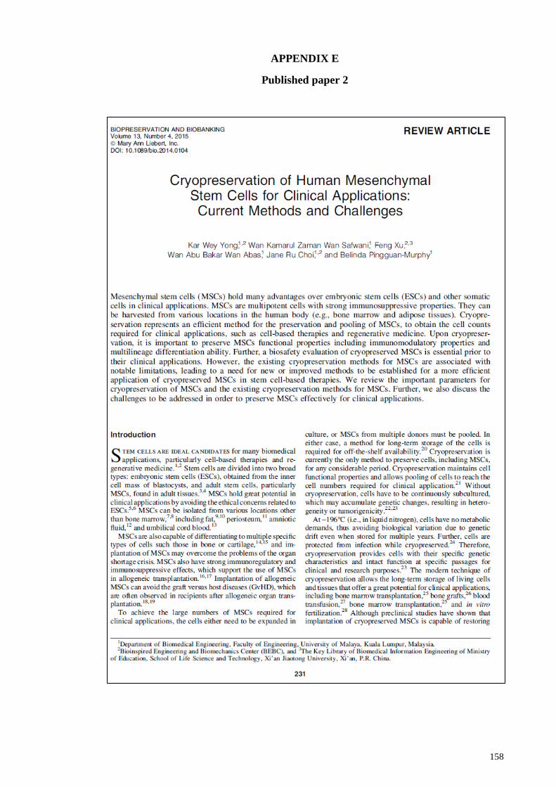
158
APPENDIX E
Published paper 2

159
APPENDIX F
Published paper 3

160
APPENDIX G
Published paper 4