Effect of Electrical Stimulation on Bacterial Growth · Effect of Electrical Stimulation on...
21
Effect of Electrical Stimulation on Bacterial Growth By Jerrold Petrofsky Ph D Michael Laymon DPTSc* Wendy Chung DPTSc Kelly Collins BS* Tien-Ning Yang BS* Depts. of Physical Therapy Loma Linda University Loma Linda, California and Azusa PACific University* Azusa California Send reprint requests to: Dr. Jerrold Petrofsky Professor and Director of Research Department of Physical Therapy Loma Linda University Loma Linda California, 92350 (909) 558 7274 E-mail: [email protected] Pag e
Transcript of Effect of Electrical Stimulation on Bacterial Growth · Effect of Electrical Stimulation on...

Effect of Electrical Stimulation onBacterial Growth
ByJerrold Petrofsky Ph DMichael Laymon DPTSc*Wendy Chung DPTScKelly Collins BS*Tien-Ning Yang BS*
Depts. of Physical Therapy Loma Linda UniversityLoma Linda, California andAzusa PACific University*Azusa California
Send reprint requests to:Dr. Jerrold PetrofskyProfessor and Director of ResearchDepartment of Physical TherapyLoma Linda UniversityLoma Linda California, 92350(909) 558 7274E-mail: [email protected]
Page
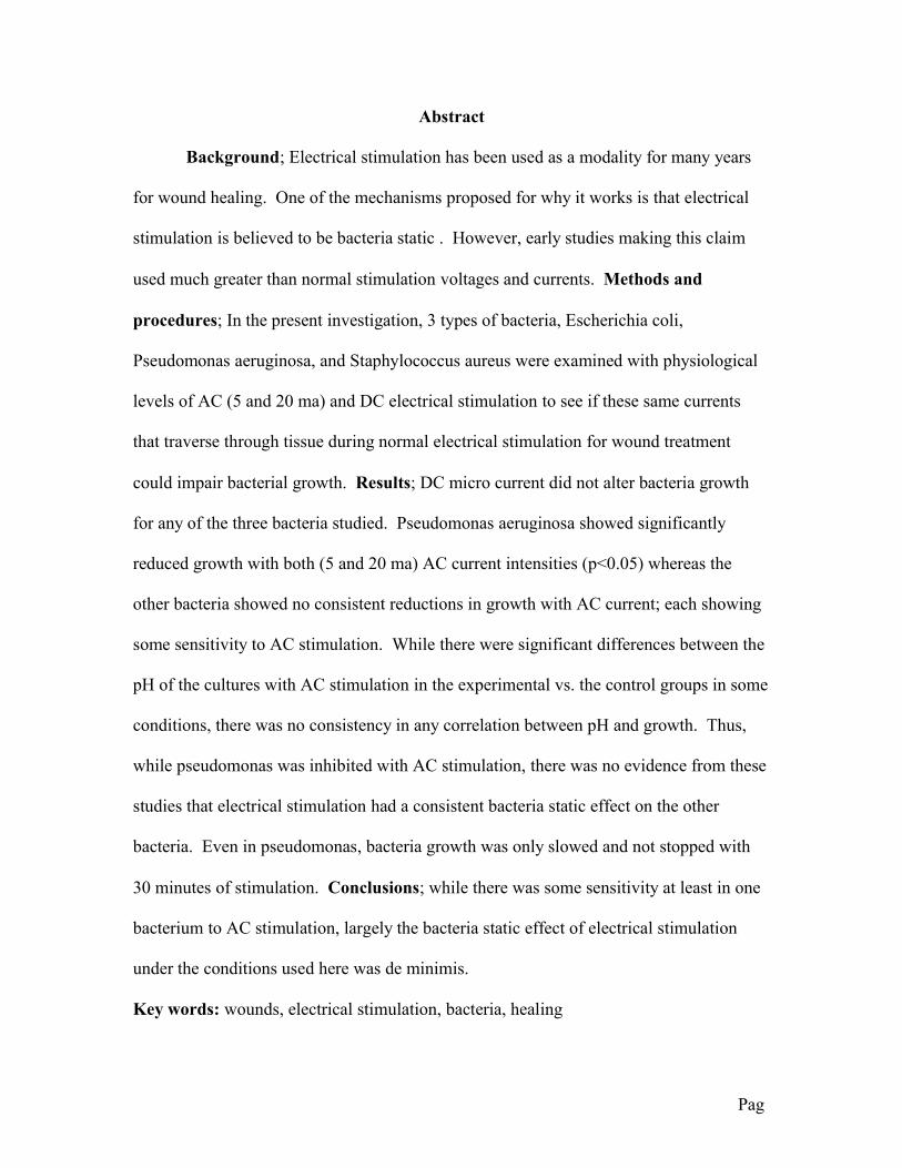
Abstract
Background; Electrical stimulation has been used as a modality for many years
for wound healing. One of the mechanisms proposed for why it works is that electrical
stimulation is believed to be bacteria static . However, early studies making this claim
used much greater than normal stimulation voltages and currents. Methods and
procedures; In the present investigation, 3 types of bacteria, Escherichia coli,
Pseudomonas aeruginosa, and Staphylococcus aureus were examined with physiological
levels of AC (5 and 20 ma) and DC electrical stimulation to see if these same currents
that traverse through tissue during normal electrical stimulation for wound treatment
could impair bacterial growth. Results; DC micro current did not alter bacteria growth
for any of the three bacteria studied. Pseudomonas aeruginosa showed significantly
reduced growth with both (5 and 20 ma) AC current intensities (p<0.05) whereas the
other bacteria showed no consistent reductions in growth with AC current; each showing
some sensitivity to AC stimulation. While there were significant differences between the
pH of the cultures with AC stimulation in the experimental vs. the control groups in some
conditions, there was no consistency in any correlation between pH and growth. Thus,
while pseudomonas was inhibited with AC stimulation, there was no evidence from these
studies that electrical stimulation had a consistent bacteria static effect on the other
bacteria. Even in pseudomonas, bacteria growth was only slowed and not stopped with
30 minutes of stimulation. Conclusions; while there was some sensitivity at least in one
bacterium to AC stimulation, largely the bacteria static effect of electrical stimulation
under the conditions used here was de minimis.
Key words: wounds, electrical stimulation, bacteria, healing
Page

Introduction
Wound healing, especially in people with diabetic neuropathies, can be extremely
difficult to accomplish.1 For example, of people who have diabetic ulcers on their feet,
inability of wounds to heal causes an average amputation rate to be about 25%2,3. Many
diabetic ulcers and even decubitus ulcers can take months, years, or never heal at all.2, 3, 4
This inability of conventional medical practice to heal wounds has been termed “a
failure of the health care system”.5 Even newer therapies such as myocutaneous flaps,
water debridement, and other techniques have not increased the healing rate appreciably
in the past fifty years or the rate of reoccurrence.5
One modality that has been studied is electrical stimulation.6 Many papers point
to the use of electrical stimulation across wounds to increase wound healing.7, 8 However,
even this technology does not always work. Some papers show wound healing with DC
micro currents while others show only healing with strong AC currents.9, 10, 11 Some
papers report that in the early stages of wound healing the polarity needs to be positive
near the wound and negative away from the wound and other papers report the opposite.12,
13, 14 In fact, in a summary of different studies on wound healing, Yarkony reported
nothing conclusive in setting the intensity of currents, whether it was DC or AC
stimulation or the frequency of electrical stimulation in the ability of electrical
stimulation to heal wounds.12 Recent evidence, however, links this disparity to possibly
being linked to room temperature. Vasoconstriction of the circulation during treatment
due to a cool or cold examination room blocks the blood flow increase due to electrical
stimulation of the wound. If the room is warmed, blood flow increases and wounds heal
well with electrical stimulation.15, 16
Page
Numerous mechanisms have been suggested on why electrical stimulation
promotes wound healing including increased circulation,1,17 increased angiogenesis18, 19, 20
increased proliferation of epidermal tissue, and a bacteria static effect of electrical
stimulation.21
The possible bacteria static effect of electrical stimulation was first reported over
30 years ago by Rowley & McKenna.22, 23 Using high voltage electrical stimulation (300
volts), bacteria (E. coli) died after a brief session of electrical stimulation. While a few
studies have used similar voltages for clinical electrical stimulation of wounds11, 24 most
studies that report healing use a fraction of these voltages and currents. 12, 13, 25, 17 The
stimulation voltages used by Rowley et al. would be extremely painful for the patient.
Further, since DC micro current has been shown, in some studies, to aid in healing of
wounds, when the voltages used by Rowley and colleagues are compared to DC micro
currents, they are thousands of times higher. Thus, the currents used in these studies were
not clinically relevant and, therefore, the application of these studies to real life situations
is questionable.
The present investigation was conducted to study the effect of electrical
stimulation on bacterial growth in three common bacteria that are seen in wounds,
Escherichia coli, Pseudomonas aeruginosa, and Staphylococcus Aureus.23, 26 These
bacteria were grown in culture and then exposed to either DC microcurrent (100 µamps)
or biphasic sine wave stimulation at 5 or 20 ma for 30 minutes. These currents are
normal currents used in wound healing.16, 15 Bacterial growth was then assessed after 24
hours of incubation to see if the normal currents that are used with electrical stimulation
clinically to heal wounds can alter bacterial growth.
Page

Methods
Bacteria-
Bacteria were grown in a soy broth (tryptic soy broth, soybean- casein digest) from Hardy
Diagnostics (Cat # C7141, Santa Monica, CA). The bacteria used were Escherichia coli,
Pseudomonas aeruginosa, and Staphylococcus aureus. These bacteria were obtained
from stock concentrations (Micro biologics, ST Cloud, Minnesota). Bacteria were grown
in broth at a temperature of 37o C until the concentration of bacteria was between 4-5 on
the McFarland Scale.27 All bacteria were grown and tested for activity by standard
microbiological light microscopy. The broth was tested for growth in an absorption
spectrometer at a wavelength of 580 nanometers; the relationship between bacterial
density and broth absorption was established. Once this was established, stock broth
cultures were placed between two 2 X 2 cm carbonized rubber electrodes, so that they
could be subjected to electrical stimulation for a period of thirty minutes. The separation
distance between the electrodes was 10.3cm. The covered culture size was 10.3 X 8.5
cm and 6.75 cm deep. The volume was 400ml. Using these electrodes and electrode
sizes, the impedance of the broth was 1280 ohms during electrical stimulation, a valve
similar to that recorded across wounds.15 The size of the broth holder was selected so that
if current was measured over a 0.5 cm distance in the broth, the recorded current was
similar to that recorded in human wounds.15, 16
Page

Electrical Stimulation- AC electrical stimulation was provided by a challenge 8000
powered muscle stimulator (MPTS Tustin, CA). This stimulator provided 4 channels of
current controlled output with a biphasic sine wave at 250 µsec pulse width and a
frequency of 30 Hz. DC stimulation was provided at a current of 1 ma by a battery with a
regulated current controlled output.
pH- The pH was measured with an Accruement Basic AB15 pH meter (Fisher Scientific,
Singapore).
Page

Procedures
Using the stock broth bacterial cultures described above, each of the 3 bacteria were
placed in a bacterial broth in the rectangular cultures. After the broth had been cultured,
electrical stimulation was applied for thirty minutes. AC current was delivered with
biphasic electrical stimulation (sine wave) at a frequency of 30 Hz and a pulse width of
250 microseconds at intensities of either 5 or 20 ma for a period of 30 minutes. The
stimulation output was current controlled. DC stimulation was also applied for 30
minutes. The bacterial culture along with cultures where no stimulation was applied,
were then placed back into the incubator for another 24 hours and growth was assessed.
The process was repeated on 8 cultures for each bacterium.
Statistical Analysis-
Statistical analysis involved the calculation of means, standard deviations, and t tests.
Values in the text are shown as the mean+/-SD. The level of significance was p < 0.05.
Page

Results
Initially, the McFarland Scale was calculated at a frequency of 580 nanometers for each
of the bacteria. Absorbency at this frequency was plotted against set concentrations of
bacteria to obtain a calibration curve. The correlation between the concentrations of
bacteria to the absorbency was 0.98. This yielded an R2 of 0.978 and, the equation related
concentration to absorbency was Y = 260.87x. Given the calibration on the McFarland
Scale, broth was mixed and the studies were completed as described under procedures.
The results for each bacterium are listed below.
Escherichia coli - The results showing growth in Escherichia coli are shown in figure 1,
panel A and B. Panel A, in figure 1, shows the bacterial colony growth from the
beginning to the end of the study (24 hours after the initial incubation). As shown in
panel A of figure 1, with a current of 5 milliamps, the increase in bacterial colony growth
over 24 hours was 365 +/- 57 x 106 bacteria per milliliter whereas for 20 milliamps
stimulation the increase in growth was 366 +/- 16 x 106 bacteria per milliliter. For the
controls, where no stimulation was accomplished, bacterial colony growth increased by
401 +/- 7.5 X 10⁶ bacteria per milliliter over the 24 hours. There was no statistical
difference between growth in the 5 and 20 ma and control groups (p < 0.01). Bacterial
colony growth for DC stimulation was 460 +/- 74 x 106 bacteria per milliliter. The only
significant growth reduction compared to the controls was for stimulation at 20 ma
(p<0.01). Here, however, the reduction in growth was only 9.8%.
Page

pH was also measured at the beginning and at the end of studies. For the control bacteria
colonies (no stimulation) pH started at 7.38 +/- 0.04. At the end of the 24 hour period,
pH averaged 6.16 +/- 0.18, a reduction of an average of 1.22 pH units. For bacterial
stimulation at 5 and 20 milliamps, there was a significantly greater reduction in pH over
the 24 hours compared to the control colonies (p > 0.01). The change in pH was small,
averaging 0.4 pH units. For DC stimulation, the change in pH, over the 24 hour period
was not significantly different than that of the controls (p > 0.05). There was no
correlation for any group in pH and bacterial growth (p>0.05).
Pseudomonas aeruginosa- As shown in the top panel of figure 2, under all four
conditions, there was an increase in bacterial growth over the 24 hour period for these
colonies. As shown in panel A of figure 2, the increase in bacterial growth with 5
milliamps of stimulation averaged 204 +/- 116 x 106 per milliliter, for 20 milliamps
stimulation averaged 228 +/- 121 x 106 bacteria per milliliter and after DC stimulation
averaged 331 +/- 161 x 106 bacteria per milliliter over the 24 hour period. The growth
for all conditions was significantly lower than growth for E. coli (p< 0.05). Compared to
the control group, the bacterial growth over the 24 hour period was significantly less for
the AC stimulation than for the control group (p<0.05). Bacterial growth averaged a
38% reduction in growth with AC stimulation. For DC stimulation, there was no
significant difference to the growth of the control group (p>0.05).
As shown in the bottom panel of figure 2, the overall change in pH was less than
that observed for E. coli (p < 0.01). However, the reduction in pH, which averaged 1.25
+/- 0.5 in the control group, was only statistically less with stimulation at DC and 20 ma
Page

AC (p<0.05). There was a significant correlation between pH and growth only for
stimulation at 20ma. (p<0.01)
Staphylococcus aureus- Figure 3 shows the results of the Staphylococcus experiments.
As shown in the top panel of figure 3, the average change in growth for these bacteria was
358 +/- 8 x 10⁶ bacteria per milliliter after stimulation with currents of 5 milliamps, 376
+/- 20 x 106 bacteria per milliliter after stimulation at 20 milliamps and 405 +/- 69 x 106
after DC stimulation. Only the growth after 5ma AC stimulation was different than that
of the controls (p < 0.01). However, the reduction in growth was only 3.9% compared to
the control group.
As shown in the bottom of the panel, the pH was different in the 5ma group
compared to the control group (p<0.01). There was no difference between the DC and
AC 20 ma groups compared to the control groups (p>0.05). There was a significant
correlation between pH and growth in the 20ma and DC groups (p<0.01).
Page

Discussion
One of the mechanisms causing wounds to not heal is bacterial invasion.21 Bacteria
rupture cell membranes and maintain chronic inflammation to prevent wound healing27
Many studies show that either DC microcurrent or low current AC stimulation has the
ability to increase wound healing.6, 9, 10, 17 In earlier studies, Rowley and colleagues 22, 23
published data showing that electrical stimulation has a bacteria static effect on
Escherichia coli and several other bacteria. Barranco et al. 28 showed inhibition in
Staphylococcus aureus growth with 400µA DC stimulation. But the electrodes were
metal and at 400 µamps, corrosion was noted. Other in vitro studies with 100 µA DC
stimulation also showed a bacteria static effect but here silver electrodes were also used.
29, 30 Other studies showed a bacteria static effect of DC at 1, 5, or 10 ma but not AC
stimulation. 31 These studies showed large changes in pH with DC stimulation, probably
due to the fact that, unlike clinical situations where stimulation is a 30 minute modality,
the current was left on for hours.32 This pH change was seen also with AC stimulation at
500 volts. 33 But these studies were conducted in vitro with a small separation distance
between the electrodes. Thus the current per mm of distance on the bacterial cultures
were large. Further, metal ions like silver can kill bacteria in themselves.
In addition, these studies were conducted at very high stimulation voltages (over 300
volts). In wounds, nerve endings are sensitized by the release of cytokines.34 Even mild
electrical stimulation can therefore be painful. In previous studies, 16, 1, 15 the current used
to heal wounds was less than 20 ma. Therefore this was the current used in these studies.
Page

In the present investigations, the density of the broth, size of the chamber and size
of electrodes were adjusted so that the electrodes were separated by the distance seen in a
typical wound treatment.15, 16 The impedance of the broth matched that of a human
wound, about 1200 ohms between the electrodes. This, in itself, is different than
previous studies where large current densities were seen due to high voltages being
applied to small bacteria cultures. While we used a large chamber for the broth, the
current density may have still been somewhat higher then in a limb due to the higher
volume in a limb. However, while current density may have been somewhat higher than
in a wound here, earlier in vitro studies used a fraction of the volume used here between
the electrodes and electrodes were closer together making current density in e.g. Rowley
et al 22, 23 much higher than in wounds.
When currents were applied in the present experiments for a 30 minute
stimulation period, similar to a standard treatment period for wound healing, DC
stimulation had no effect on bacteria growth. AC had some effect on each of the bacteria
but the only real effect on growth with electrical stimulation was on pseudomonas. Any
effect seen on Escherichia coli and Staphylococcus aureus was small. For pseudomonas,
the growth reduction at either AC current was large.
However, bacteria can still be destroyed by electrical stimulation through another
mechanism in wounds. Electrical stimulation and, electrical stimulation of wounds
causes the release of prostaglandins and other cytokines.35, 36, 37, 38 These prostaglandins
and other cytokines attract macrophages to the area where electrical stimulation is
applied.31 Therefore, although electrical stimulation may not have a direct effect on
bacteria in wounds, there is some evidence that white blood cells such as macrophages
Page

are attracted as a secondary effect of the electrical stimulation, this may explain why
electrical stimulation may be associated with bacterial death.
The present studies still remain in direct contradiction to the studies by Rowley et al 22, 23
showing a direct effect of electrical stimulation on inhibiting growth in Escherichia coli.
However, while Escherichia coli and Staphylococcus aureus showed either a statistically
insignificant change in growth or an insignificant clinical change in growth with electrical
stimulation, this is not to say that electrical current does not alter bacterial behavior.
Bacterial motility and attachment to substrates appears to be impaired by stimulation with
electric currents (39). This is especially true for pseudomonas where flagella are needed
for mobility (40). The fact that pseudomonas here did have some impaired growth may
be due to the fact that there are two flagella proteins involved in its mobility and
attachment- if either is impaired, growth is reduced 41. Here, these flagella, since they use
contractile proteins and ion channels for movement, may be impaired with electric
current. This makes these bacteria different from the other 2 which do not move by this
mechanism.
However, there are conditions where electrical stimulation will inhibit growth in all 3
bacteria. While currents of up to 20ma in themselves with these same bacteria did not
reduce bacterial growth 39,42, in combination with antibiotics, the reduced ability of the
bacteria to attach to substrates made them more susceptible to attack by the antibiotics.
Thus while antibiotics alone or electrical stimulation alone did not kill these bacteria, the
combination of the two did 39,42. Thus, electrical stimulation with either DC or weak AC
currents may have an unseen effect which may kill bacteria in vivo but not under these
circumstances in vitro. Further investigation is warranted. Here, for example, current
Page

was used for 30 minutes, a standard treatment modality. Perhaps longer exposure to
current would have a more dramatic effect in vivo.
Page

References
1. Petrofsky, J.S. Schwab, E., Lo, T., Cuneo, M., George, J., Kim, J., & AlMarty, A.(2005). Effects of Electrical stimulation on Skin Blood Flow in Controls and inand around Stage III and IV Wounds in Hairy and Non Hairy Skin. Med SciMonit, 11, 309 -316.
2. Kennedy, E.J. (1999). Spinal cord injury; the fACts and figures. The University atAlabama Press: Birmingham, Alabama.
3. Senet, P., & Meaume, S. (1999). Decubitus sores in geriatric medicine. Local andgeneral treatment of pressure sores in the aged. Presse Med, 28, 1840-5.
4. De Astis, V., Corbella, A., Bafico, F., Spinelli, E., Porcu, G., Bottari, L., Petrini,M., & Madeddu, V. (1999). Decubitus lesions in patients referred to acute andpost-acute home nursing care for the elderly in Genova. Assist Inferm Ric, 18, 20-4.
5. Meehan, M. (2000). Beyond the pressure ulcer blame game: reflections for thefuture. Ostomy Wound Manage, 46, 46-52.
6. Kloth, L.C. (2005). Electrical stimulation for wound healing: a review of evidencefrom in vitro studies, animal experiments, and clinical trials. Int J Low ExtremWounds, 4, 1, 23-44.
7. Ennis WJ, Lee C, Meneses P. (2007) A biochemical approach to wound healingthrough the use of modalities. Clin Dermatol, 25(1): 63-72
8. Bogie KM, Triolo RJ (2003) Effects of regular use of neuromuscular electricalstimulation on tissue health. J Rehabil Res Dev. 40(6): 469-75
9. Feedar, J., Kloth, L., & Gentzkow, G. (1992). Chronic dermal ulcer healingenhanced with monophasic pulsed electrical stimulation. Phys Ther, 72, 539.
10. Franek, A., Franek, E., & Grzesik, J. (1999). Electrically enhanced damagedtissues healing. Part II: direct and pulse current in soft tissue healing. PolMerkuriusz Lek, 40, 198-201.
11. Houghton, P.E., Kincaid, C.B., Lovell, M., Cambell, K.E., Keast, D.H.,Woodbury, M.G., & Harris, K.A. (2003). Effect of electrical stimulation onchronic leg ulcer size and appearance. Phys Ther, 83, 1, 17-28.
Page

12. Yarkony, G.M. (1994). Pressure ulcers: a review. Arch Phys Med Rehabil, 75,908-17.
13. Bogie, K.M., Reger, S.I., Levine, S.P, & Sahgal, V. (2000). Electrical stimulationfor pressure sore prevention. J Assist Technol, 12, 50-66.
14. Demir, H., Balay, H., & Kirnap, M. (2004). A comparison study of the effects ofelectrical stimulation and laser treatment on experimental wound healing in rats. JRehabil Res Dev, 42, 2, 147-54.
15. Petrofsky, J.S., Schwab, E., Cuneo, M., George, J., Kim, J., AlMarty, A., &Lawson, D. (2007). Interaction between resting skin blood flow and the bloodflow response to electrical stimulation in normal and wounded skin. SubmittedMed Sci Monit
16. Lawson, D., Petrofsky, J.S., (2007). A randomized control study of the effect ofbiphasic electrical stimulation in a warm room on blood flow and healing rates inchronic wounds of patients with and without diabetes. Med Sci Monit
17. Kloth, L.C. (2002). How to use electrical stimulation for wound healing. Nursing,32, 12, 17.
18. Bai H, McCaig C.D, Forrester J.V, & Zhao M. (2004). DC electrical fields inducedistinct preangiogenic responses in microvascular and macrovascular cells.Arterioscler Thromb Vasc Biol, 24, 7, 1234-9.
19. Zhao, M., Bai, H., Wang, E., Forrester, J.V., & McCaig, C.D. (2004). Electricalstimulation directly induces pre-angiogenic responses in vascular endothelial cellsby signaling through VEGF receptors. J Cell Sci, 117, 397-405.
20. Ojingwa, J.C., & Isseroff, R.R. (2003). Electrical stimulation in wound healing. JInvest Dermatol, 121, 1, 1-12.
21. Rowley BA, McKenna JM, Chase GR, Wolcott LE: (1974) The influence ofelectrical current on an infecting microorganism in wounds. Ann NY ACad Sci1974b 238: 543-51
22. Rowley BA, McKenna JM, Wolocott LE (1974) Proceedings: The use of lowlevel electrical current for enhancement of tissue healing. Biomed Sci Instrum,10: 111-4.
23. Rowley BA (1972) Electrical current effects on E. coli growth rates. Proc Soc ExpBiol Med 139(3): 929-34
24. Polak, A., Franek, A., Hunka-Zurawinska, W., Kucharzewski, M., & Swist, D.(2000). High voltage stimulation in leg ulcer’s treatment. Wiad Lek, 53, 7-8, 417-26.
Page

25. Speilholz, N.I., & Kloth, L.C. (2000). Electrical stimulation and pulsedelectromagnetic energy: differences in opinion. Ostomy Wound Manage, 5, 8-12.
26. Hosseini SV, Tanideh N, Kohanteb J, Ghodrati Z, Mehrabani D, YarmohammadiH. (2007) Comparision between Alpha and silver sulfadiazine ointments intreatment of Pseudomonas infections in 3rd degree burns. Int J Surg, 5(1): 23-6
27. Neubauer T, Bayer GS, Wagner M. (2006) Open fractures and infection. ACtaChir Orthop Traumotol Cech. 73(5): 301-12
28. Barranco S, Spadero J, Berger T (1974) In vitro effect of weak direct current onStaphylococcus aureus. ClinOrthop 100: 250-255.
29. Ong P, Laatsch L, Kloth L. (1994) Antibaterial effects of a silver electrodecarrying microampearage direct current in vitro. J Clin Electrophysiol 6(1): 14-18.
30. Laatsch L, Ong P, Kloth L. (1995) In vitro effects of two silver electrodes onselect wound pathogens. J Clin Electrophysiol 7(1): 10-15.
31. Guffey J, Asmussen M. (1989) In vitro bACtericidal effects of high voltagepulsed current versus direct current against Staphylococcus aureus. J ClinElectrophysiolo 1: 5-9.
32. Newton R, Karselis T. (1983) Skin pH following high voltage pulsed galvanicstimulation. Phys Ther 63(10): 1593-1596.
33. Szuminsky N, Albers A, Unger P (1994). Effect of narrow, pulsed high voltageson bacterial viability. Phys Ther 74: 660-667.
34. Hernandez R (2006). The use of systemic antibiotics in the treatment of chronicwounds. Dermatol Ther. 19(6):326-37.
35. Hofbauer R, Moser D, Kaye AD, Knapp S, Gmeiner B, Kapiotis S, Wagner O,Frass M. (2000) Prostaglandin E(1) is able to increase migration of leukocytesthrough endothelial cell monolayers. Microvasc Res, 59(3): 354-60
36. Anderson SI, Hudlicka O, Brown MD. (1997) Capillary red blood cell flow andACtivation of white blood cells in chronic muscle ischemia in the rat. Am JPhysiol. 272(6 Pt 2): H2757-64
37. McLoughlin TJ, Mylona E, Hornberger TA, Esser KA, Pizza FX. (2003)Inflammatory cells in rat skeletal muscle are elevated after electrically stimulatedcontrACtions. J Appl Physiol, 94(3): 876-82
Page

38. Dusterhoft S, Putman CT, Pette D. (1999) Changes in FGF and FGF receptorexpression in low frequency-stimulated rat muscles and rat satellite cell cultures.Differentiation 65(4): 203-8
39. Jass J , Costerton JW, Lappin-Scott HM. The effect of electrical currents andtobramycin on Pseudomonas aeruginosa biofilms. J Ind Microbiol. 1995 Sep;15(3):234-42.
40. Murray TS, Kazmierczak BI. FlhF is required for swimming and swarming inPseudomonas aeruginosa. J Bacteriol. 2006 Oct;188(19):6995-7004.
41. Toutain CM, Zegans ME, O'Toole GA. Evidence for two flagellar stators andtheir role in the motility of Pseudomonas aeruginosa. J Bacteriol. 2005 Jan;187(2):771-7.
42. Costerton JW, Ellis B, Lam K, Johnson F, Khoury AE. Mechanism of electricalenhancement of efficacy of antibiotics in killing biofilm bacteria. Antimicrob AgentsChemother. 1994 Dec;38(12):2803-9.
Figure 1
Page

A
B
Page
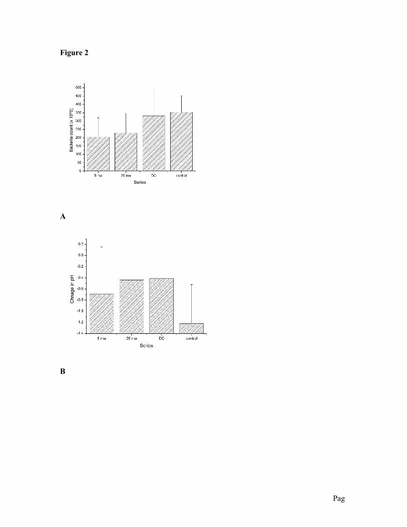
Figure 2
A
B
Page

Figure 3
A
B
Page