Edizioni - CNReprints.bice.rm.cnr.it/10413/1/article.pdf · In primary dentition intrusions are the...
10
Annali di Stomatologia 2013; IV (2): 174-183 174 Post-traumatic impaction of maxillary incisors: diagnosis and treatment Case report Introduction Trauma to oral and facial structures is a significant problem that may have serious medical, esthetic and psychologic consequences on both children and their parents. Studies have shown that approximately 30% of all children under the age of 7 years experience in- juries to ≥ 1 of their primary incisors and that most seri- ous injuries to primary teeth occur between the ages of 1 and 3 years (1). This high incidence is related to the passage to the up- right posture, the early stages of walking, a lack of mo- tor coordination and the unconsciousness of the child. The majority of the trauma occurs as a result of fall ac- cidents at home or during sporting activities (1, 2). According to gender, boys were injured more frequently in all age than girls (2) and owing to their exposed posi- tion in the dental arch, the upper central incisors are the teeth most commonly affected by traumatic injury in both primary and permanent dentition (1). In primary dentition intrusions are the most common type of trauma. These are luxation injuries which usual- ly results from an axially directed impact, displacing the incisor deeper into the alveolar socket. The primary in- cisors is driven deeply into the alveolar bone invading the follicle of permanent germ which lies palatally in close proximity to its root. In fact there is a close anatomic relationship between the apices of the prima- ry incisors and the germs of the succeeding teeth: the hard tissue barrier between them has a thickness of less than 3 mm and it might simply consist of connec- tive fibrous tissue (3). This close relationship explains why injuries to primary teeth are easily transmitted to the permanent dentition (4). The displacement results in compression of and dam- age to the periodontal ligament, contusion to the socket walls and injury to the pulp of the intruded incisor. The reported prevalence of intrusion injuries affecting pri- mary incisors varies among different studies and ranges from 4.4% to 22% (3). This high incidence is be- cause of the pliability of the facial skeleton and of the periodontal ligament, the large volume of teeth in rela- tion to the bone in primary and mixed dentition and fi- nally the shorter roots of primary teeth (5). Therefore traumas to primary dentition may interfere with further odontogenesis of permanent teeth and ac- cording to site and extension of the injury, different mal- formations may occur ranging from a slight disturbance in the mineralization of enamel to a sequestration of the entire tooth germ (4). They may lead to abnormality in the path of eruption of permanent series which may re- sult in impaction or ectopic eruption (6). The percent- Valeria Paoloni, DDS 1 Chiara Pavoni, DDS 1 Manuela Mucedero, DDS 1 Patrizio Bollero, DDS, PhD 2 Giuseppina Laganà, DDS 1 Paola Cozza, MD, DDS, MS 1 1 Department of Orthodontics, University of Rome “Tor Vergata”, Italy 2 Department of Oral Pathology, University of Rome “Tor Vergata”, Italy Corresponding author: Giuseppina Laganà Department of Orthodontics, University of Rome “Tor Vergata” Via G. Baglivi, 5/E 00161 Rome, Italy Phone: +39 06 44232321 E-mail: [email protected] Summary Aim. To provide clinicians with useful information for immediate diagnosis and management of im- pacted maxillary incisors due to trauma. Methods. We present a case of post-traumatic im- paction of a central right maxillary incisor in a young patient. The treatment plan consisted in the interceptive management (surgical and orthodon- tic), the valuation of the necessary space to move the impacted tooth in the normal position and the biomechanical approach for anchorage, avoiding prosthetic/implants replacement. Results. The therapy of an impacted maxillary in- cisor due to trauma requires a multidisciplinary ap- proach: orthodontic, surgical, endodontic and peri- odontal considerations are essential for successful treatment. Conclusions. Surgical exposure and orthodontic traction is the treatment most often used in case of posttraumatic impacted incisor: this technique in fact can lead to suitable results at the peri- odontal, occlusal and esthetics levels at an early stage and more definitively than with other treat- ment options. Key words: eruption disturbances, impacted incisor, oral trauma, orthodontic traction, early diagnosis. © CIC Edizioni Internazionali
Transcript of Edizioni - CNReprints.bice.rm.cnr.it/10413/1/article.pdf · In primary dentition intrusions are the...

Annali di Stomatologia 2013; IV (2): 174-183174
Post-traumatic impaction of maxillary incisors:diagnosis and treatment
Case report
Introduction
Trauma to oral and facial structures is a significantproblem that may have serious medical, esthetic andpsychologic consequences on both children and theirparents. Studies have shown that approximately 30%of all children under the age of 7 years experience in-juries to ≥ 1 of their primary incisors and that most seri-ous injuries to primary teeth occur between the ages of1 and 3 years (1). This high incidence is related to the passage to the up-right posture, the early stages of walking, a lack of mo-tor coordination and the unconsciousness of the child.The majority of the trauma occurs as a result of fall ac-cidents at home or during sporting activities (1, 2).According to gender, boys were injured more frequentlyin all age than girls (2) and owing to their exposed posi-tion in the dental arch, the upper central incisors arethe teeth most commonly affected by traumatic injury inboth primary and permanent dentition (1). In primary dentition intrusions are the most commontype of trauma. These are luxation injuries which usual-ly results from an axially directed impact, displacing theincisor deeper into the alveolar socket. The primary in-cisors is driven deeply into the alveolar bone invadingthe follicle of permanent germ which lies palatally inclose proximity to its root. In fact there is a closeanatomic relationship between the apices of the prima-ry incisors and the germs of the succeeding teeth: thehard tissue barrier between them has a thickness ofless than 3 mm and it might simply consist of connec-tive fibrous tissue (3).This close relationship explains why injuries to primaryteeth are easily transmitted to the permanent dentition(4).The displacement results in compression of and dam-age to the periodontal ligament, contusion to the socketwalls and injury to the pulp of the intruded incisor. Thereported prevalence of intrusion injuries affecting pri-mary incisors varies among different studies andranges from 4.4% to 22% (3). This high incidence is be-cause of the pliability of the facial skeleton and of theperiodontal ligament, the large volume of teeth in rela-tion to the bone in primary and mixed dentition and fi-nally the shorter roots of primary teeth (5).Therefore traumas to primary dentition may interferewith further odontogenesis of permanent teeth and ac-cording to site and extension of the injury, different mal-formations may occur ranging from a slight disturbancein the mineralization of enamel to a sequestration of theentire tooth germ (4). They may lead to abnormality inthe path of eruption of permanent series which may re-sult in impaction or ectopic eruption (6). The percent-
Valeria Paoloni, DDS1
Chiara Pavoni, DDS1
Manuela Mucedero, DDS1
Patrizio Bollero, DDS, PhD2
Giuseppina Laganà, DDS1
Paola Cozza, MD, DDS, MS1
1 Department of Orthodontics, University of Rome “TorVergata”, Italy
2 Department of Oral Pathology, University of Rome“Tor Vergata”, Italy
Corresponding author: Giuseppina LaganàDepartment of Orthodontics, University of Rome “Tor Vergata”Via G. Baglivi, 5/E00161 Rome, ItalyPhone: +39 06 44232321E-mail: [email protected]
Summary
Aim. To provide clinicians with useful informationfor immediate diagnosis and management of im-pacted maxillary incisors due to trauma. Methods. We present a case of post-traumatic im-paction of a central right maxillary incisor in ayoung patient. The treatment plan consisted in theinterceptive management (surgical and orthodon-tic), the valuation of the necessary space to movethe impacted tooth in the normal position and thebiomechanical approach for anchorage, avoidingprosthetic/implants replacement.Results. The therapy of an impacted maxillary in-cisor due to trauma requires a multidisciplinary ap-proach: orthodontic, surgical, endodontic and peri-odontal considerations are essential for successfultreatment.Conclusions. Surgical exposure and orthodontictraction is the treatment most often used in caseof posttraumatic impacted incisor: this techniquein fact can lead to suitable results at the peri-odontal, occlusal and esthetics levels at an earlystage and more definitively than with other treat-ment options.
Key words: eruption disturbances, impacted incisor,oral trauma, orthodontic traction, early diagnosis.
2-Laganà_- 21/06/13 10:36 Pagina 174
© C
IC Ed
izion
i Int
erna
ziona
li

Annali di Stomatologia 2013; IV (2): 174-183 175
Post-traumatic impaction of maxillary incisors: diagnosis and treatment
age of developmental disturbances of the permanentincisors that could be attributed to injuries of their pre-decessors ranges from 12% to 74% (3). Early diagno-sis is very important to monitor and prevent complica-tions (6).
Sequelae
The effects of trauma on the permanent tooth germ be-come clinically manifest only after the normal exfoliationperiod is over. It is possible, however, that in an earlierstage a malformation may be discovered radiographi-cally (7). The age of the child at the time of injury (germs of thepermanent teeth are particularly sensitive in the earlystages of their development, which occurs between theages of 4 months and 4 years), the developmentalphase of the permanent tooth germ, the direction andseverity of the trauma, the type of injury sustained andthe presence of alveolar bone fracture are importantvariable influencing the effect on the developing perma-nent germ (1, 3, 4, 7). The consequences for permanent dentition after a trau-ma to primary dentition may affect the coronal or rootregion or the whole of the permanent tooth germ (5). Many sequelae can be found in the coronal region: 1)white or yellow-brown discoloration of enamel; 2) whiteor yellow-brown discoloration of enamel and circularenamel hypoplasia, that represented the borderline be-tween hard tissue formed before and after injury; 3)crown dilaceration, that is described as an acute devia-tion in the long axis of the crown originating from a non-axial displacement of already formed hard tissue in re-lation to the developing noncalcified tissue (crown di-laceration can result from an intrusion of the primary in-cisor when a child is around that age of 2 years, whenhalf the crown would be formed) (3-5).Sequelae affecting the root region include: 1) root dupli-cation; 2) root dilaceration, these lesions appeared as amarked curvature confined to the root portion and mayresult from intrusion of a primary incisor after comple-tion of permanent crown formation between the ages of2 and 5 years; 3) partial or complete arrest of root for-mation, rare sequelae resulting from injury to primaryincisors between the ages of 4 and 7 years (3-5).When the entire permanent tooth germ is affected, al-terations to the process of eruption of the permanenttooth may be noted: 1) delayed eruption due to earlyloss of a primary incisor and formation of thick, fibrousgingival tissue; 2) accelerated eruption, when the pri-mary incisor is lost after the child is aged 5 years espe-cially in the presence of alveolar bone resorption fol-lowing an infection of the injured tooth; 3) ectopic erup-tion or (4) impaction that can be explained by the phys-ical displacement of the permanent germ with or with-out dilaceration at the time of the injury, the lack oferuption guidance from prematurely lost primary in-cisor, ankylosis or alterations of root resorption; 5) mal-formation of the permanent germ giving the appear-ance of an odontoma; 6) sequestration of the perma-nent tooth germ (3-5).
Diagnosis
The impaction of maxillary permanent incisor is oftenclinically and radiologically diagnosed in early ages be-cause the non-eruption of the anterior tooth causesconcern to parents during early mixed dentition phase(8). Clinical signs of an impacted tooth include asymmetriceruption of more than 6 months in relation to its homo-logue, change of the sequence and chronology of nor-mal eruption, retention of the primary tooth, midline de-viation, space closure and elevation of the soft tissue ofthe palatal or labial mucosa (8, 9).Diagnosis of impacted tooth is verified and its locationdetermined through radiographic evaluation. Panoramicradiograph is considered the standard radiographicfirst-step examination for treatment planning of impact-ed teeth because (10, 11) it is unique in that it will showthe entire dentition as a whole (12) and it may revealthe existence of an impacted tooth. Unfortunately, le-sions of permanent teeth resulting from previous trau-ma to the deciduous dentition are not always clearlyobservable on panoramic radiograph because the de-ciduous teeth are projected onto the permanent teethand the direction of projection is unfavorable in regardto the root curvature (7). Due to the highly detailed three-dimension informationobtained, computerized tomography is the method ofchoice for accurately defining the position of anunerupted tooth and identifying any root resorption ofadjacent teeth not detectable by other methods (12).The highly detailed information and the excellent tissuecontrast without blurring and overlapping of adjacentstructures outweighs the high radiation dose, limitedavailability, and high cost (13, 14).Three-dimensional imagery enables analysis of the pre-cise location and orientation of impacted teeth, their sit-uation relative to obstacles to eruption, their externaland internal anatomy, the labial and palatal bone thick-ness; any resorption of the adjacent teeth or pathologi-cal bone loss; the presence or absence of a continuousradiolucent line between the root and the bone (possi-ble ankylosis) (15).Recently cone beam CT (CBCT) has been introducedas a technique dedicated to the imaging of dental andmaxillofacial structures. It has one-sixth of the radiationof computed tomography, is more time efficient, morecost effective, is still able to provide three dimensionalimages, excellent bone differentiation and an unlimitednumber of views (11, 12). Its disadvantages includespatial resolution of subtle structures that is slightly in-ferior to that of CT and limited representation of soft tis-sues (due to the lower radiation dose) (11).
Treatment
The therapy of an impacted maxillary incisor due totrauma requires a multidisciplinary approach: orthodon-tic, surgical, endodontic and periodontal considerationsare essential for successful treatment (16). Careful
2-Laganà_- 21/06/13 10:36 Pagina 175
© C
IC Ed
izion
i Int
erna
ziona
li

planning is required especially because these are oftendilacerated incisors (17).If there is a retained, necrotic, ankylosed primary in-cisor it must be surgically removed because it repre-sents an obstacle to spontaneous eruption of the per-manent one. Sometimes after primary tooth’s removal,the permanent incisor erupts without any further treat-ment. When the displacement is severe and preventsthe spontaneous eruption, an orthodontic treatment isnecessary. Depending on the localization of the tooth and the de-gree of dilacerations different treatment options havebeen suggested in literature (8): surgical excision of theimpacted incisor and subsequent restoration with abridge or implant after orthodontic space opening whengrowth had stabilized; surgical excision of the impactedincisor, orthodontic space closure and prosthodonticsrestoration of the left lateral incisor as the central in-cisor at a later stage; orthodontic space opening, un-covering the impacted tooth using the apical reposi-
Annali di Stomatologia 2013; IV (2): 174-183176
V. Paoloni et al.
Figures 1 a-d. Pretreatment extraoralphotographs.
a b
c d
tioned flap and orthodontic traction into proper align-ment (18).The most commonly solution is surgical extraction ofthe impacted incisor (19) followed by an orthodontictreatment either to close the space or to keep it openuntil the patient reaches an age when definitiveprosthodontic treatments may be used. Both methods have associated problems: orthodonticspace closure substituting the lateral incisor for thecentral one with subsequent resin restoration is seldomindicated nor aesthetically satisfactory, while removableprosthetic replacement during childhood and adoles-cence may be unsatisfactory for psychological reasons(18). Moreover early loss of a maxillary incisor may re-sult in loss of alveolar height in the anterior region ofthe maxilla (20). In some cases extraction of the toothis unavoidable because of the severity of the inversionof the tooth (8).The surgical-orthodontic approach is the solution mostwidely adopted to save an impacted incisor (20) and it
2-Laganà_- 21/06/13 10:36 Pagina 176
© C
IC Ed
izion
i Int
erna
ziona
li

Annali di Stomatologia 2013; IV (2): 174-183 177
Post-traumatic impaction of maxillary incisors: diagnosis and treatment
Figures 2 a-e. Pretreatment intraoral photographs.
would yield satisfactory results with carefully plannedprocedures (18).This technique is commonly directed to surgical expo-sure of the crown and to apply an orthodontic traction.The strategy adopted for the surgical exposure is mini-mal bone removal and closed eruption after placing anattachment on the unerupted incisor. This is considereda good surgical choice considering the long-term es-thetic-periodontal status (9).The degree and level of dilacerations, tooth’s vertical po-sition, tooth’s maturity, flap design and orthodontic trac-tion are factors determining the success rate of orthodon-tic-surgical management of impacted incisors (18).The chances of failure could be due to ankylosis, exter-nal root resorption, root exposure after orthodontic trac-tion, loss of attachment (17, 21).Autotransplantation with premolars or supernumeraryteeth and surgical repositioning of impacted incisor hasbeen reported in dental literature (20).
Aim of this report was to show the interceptive manage-ment (surgical and orthodontic) of a case with an im-pacted central maxillary incisor caused by trauma in ayoung patient, avoiding prosthetic/implants replace-ment.
Case report
A 9-year-old Caucasian girl was referred by his generaldentist to the Department of Orthodontics of the Univer-sity of Rome “Tor Vergata” for an orthodontic evalua-tion. The chief complaint was concern about an erup-tion disturbance, which had resulted in an unaestheticappearance. The child was in excellent physical healthand had no history of disease, but there was a historyof anterior dental trauma at age 4 to its primary in-cisors. This trauma induced the necrosis and ankylosisof the maxillary primary right central incisor.
a b
c d
e
2-Laganà_- 21/06/13 10:36 Pagina 177
© C
IC Ed
izion
i Int
erna
ziona
li

Annali di Stomatologia 2013; IV (2): 174-183178
V. Paoloni et al.
Picture 1. Smailiene et al. measurement (22).
Picture 2. Bryan et al. measurement (23).
Diagnosis
The patient had balanced facial pattern with a goodprofile, and an asymmetric ugly smile. Intraoral clinicalexamination showed a mixed dentition, an altered se-quence of eruption, the absence of the maxillary per-manent right central incisor and the ankylosis andnecrosis of the maxillary primary right central incisor(Figs. 1a-1d).Occlusal analysis revealed a molar Class I relationship.There was not significant dental crowding in both arch-es. The maxillary right central incisor was absent whilethe maxillary right lateral incisor was erupting. Overbitewas reduced (Figs. 2a-2e).Radiological examinations were performed to completeclinical evaluation.
Panoramic radiograph showed the complete set of per-manent teeth in different stage of formation and the im-paction of the upper permanent right central incisor whichshowed a severe intraosseous displacement (Fig. 3).The vertical position of the delayed permanent incisorin relation to the contralaterally erupted central incisorwas in sector v3 (22) (Pict. 1), while its angulation tothe mid-sagittal plane was 90° (23) (Pict. 2).CT-Dentascan evaluation defined exactly the place ofthe impacted incisor: the tooth was positioned horizon-tally and its crown was close to the anterior nasal spineand across the midline, while the root was displacedpalatally (6) (Figs. 4a, 4b).Cephalometric analysis revealed a skeletal Class I mal-occlusion (ANB T1: 3°) and a normofacial pattern (FMAT1: 26°). Lower incisor showed good inclination onmandibular plane (IMPA T1: 89°) (Figs. 5a, 5b).
Figure 3. Pretreatment panoramic radiograph.
2-Laganà_- 21/06/13 10:36 Pagina 178
© C
IC Ed
izion
i Int
erna
ziona
li

Post-traumatic impaction of maxillary incisors: diagnosis and treatment
Figures 5 a-b. Pretreatment cephalometric radiographs.
a b
Figures 4 a-b. Pretreatment CT-Dentascan.
a b
Treatment objectives
The purpose of this treatment was to guide the im-pacted incisor into proper alignment with the adja-cent teeth, without root damage, and to re-create acomplete anterior dentition and a stable functionalocclusion. The treatment aimed also to extrude theincisor with all its supporting tissues (alveolar boneand attached gingiva), to investigate the effects thatsurgical exposure, orthodontic movements and finaltooth position would have had on them and to eval-uate the long-term gingival and periodontal condi-tions (19).
Treatment progress
The multidisciplinary approach involved a combinedsurgical/orthodontic treatment. After evaluating the ad-vantages and disadvantages of the therapeutic options,the following treatment steps were established: 1) sur-gical removal of the ankylosed and necrotic maxillaryprimary right central incisor which represented an ob-stacle to permanent incisor’s eruption, 2) monitoring forspontaneous eruption, 3) orthodontic traction, 4) fixedappliance to obtain the proper alignment.First of all the primary incisor was surgically removed.After 6 months the maxillary right incisor erupted in an
Annali di Stomatologia 2013; IV (2): 174-183 179
2-Laganà_- 21/06/13 10:36 Pagina 179
© C
IC Ed
izion
i Int
erna
ziona
li
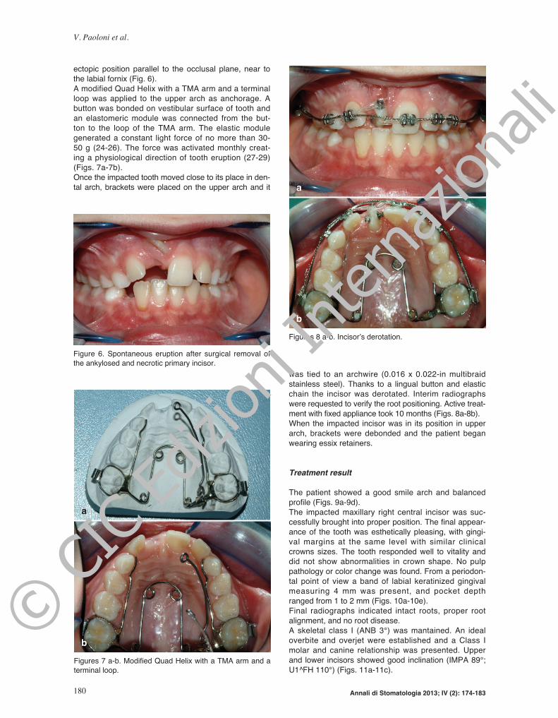
ectopic position parallel to the occlusal plane, near tothe labial fornix (Fig. 6).A modified Quad Helix with a TMA arm and a terminalloop was applied to the upper arch as anchorage. Abutton was bonded on vestibular surface of tooth andan elastomeric module was connected from the but-ton to the loop of the TMA arm. The elastic modulegenerated a constant light force of no more than 30-50 g (24-26). The force was activated monthly creat-ing a physiological direction of tooth eruption (27-29)(Figs. 7a-7b).Once the impacted tooth moved close to its place in den-tal arch, brackets were placed on the upper arch and it
was tied to an archwire (0.016 x 0.022-in multibraidstainless steel). Thanks to a lingual button and elasticchain the incisor was derotated. Interim radiographswere requested to verify the root positioning. Active treat-ment with fixed appliance took 10 months (Figs. 8a-8b).When the impacted incisor was in its position in upperarch, brackets were debonded and the patient beganwearing essix retainers.
Treatment result
The patient showed a good smile arch and balancedprofile (Figs. 9a-9d).The impacted maxillary right central incisor was suc-cessfully brought into proper position. The final appear-ance of the tooth was esthetically pleasing, with gingi-val margins at the same level with similar clinicalcrowns sizes. The tooth responded well to vitality anddid not show abnormalities in crown shape. No pulppathology or color change was found. From a periodon-tal point of view a band of labial keratinized gingivalmeasuring 4 mm was present, and pocket depthranged from 1 to 2 mm (Figs. 10a-10e).Final radiographs indicated intact roots, proper rootalignment, and no root disease. A skeletal class I (ANB 3°) was mantained. An idealoverbite and overjet were established and a Class Imolar and canine relationship was presented. Upperand lower incisors showed good inclination (IMPA 89°;U1^FH 110°) (Figs. 11a-11c).
Annali di Stomatologia 2013; IV (2): 174-183180
V. Paoloni et al.
Figure 6. Spontaneous eruption after surgical removal ofthe ankylosed and necrotic primary incisor.
Figures 7 a-b. Modified Quad Helix with a TMA arm and aterminal loop.
a
b
Figures 8 a-b. Incisor’s derotation.
b
a
2-Laganà_- 21/06/13 10:36 Pagina 180
© C
IC Ed
izion
i Int
erna
ziona
li

Annali di Stomatologia 2013; IV (2): 174-183 181
Post-traumatic impaction of maxillary incisors: diagnosis and treatment
Figures 10 a-e. Post-treatment intrao-ral photographs.
a
d e
b c
Figures 9 a-d. Post-treatment extraoral photographs.
a b
c d
2-Laganà_- 21/06/13 10:36 Pagina 181
© C
IC Ed
izion
i Int
erna
ziona
li

Annali di Stomatologia 2013; IV (2): 174-183182
V. Paoloni et al.
Conclusions
A traumatic injury in early age can realize a delay of erup-tion and eventually an impacted tooth. Upper incisors arethe most frequent impacted teeth due to trauma (6).Surgical exposure and orthodontic traction is the treat-ment most often used: this technique in fact can lead tosuitable results at the periodontal, occlusal and esthet-ics levels at an early stage and more definitively thanwith other treatment options.Sometimes the surgical removal of the retained trauma-tized primary incisor alone can lead to spontaneouseruption of the permanent tooth. However long-term monitoring of the stability and peri-odontal health of the impacted incisor is very importantafter orthodontic traction (30).
References
1. Altun C, Cehreli ZC, Güven G, Acikel C. Traumatic intrusionof primary teeth and its effects on the permanent successors:a clinical follow-up study. Oral Surg Oral Med Oral Pathol OralRadiol Endod 2009; 107(4):493-498.
2. Kargul B, Caglar E, Tanboga I. Dental trauma in Turkish chil-dren, Istanbul. Dent Traumatol 2003; 19:72-75.
3. Diab M, elBadrawy HE. Intrusion injuries of primary incisors.Part III: Effects on the permanent successors. QuintessenceInt 2000; 31(6):377-384.
4. Andreasen JO, Sundström B, Ravn JJ. The effect of trau-matic injuries to primary teeth on their permanent succes-sors. I. A clinical and histologic study of 117 injured permanentteeth. Scand J Dent Res 1971; 79(4):219-83.
5. Arenas M, Barberia E, Lucavechi T, Maroto M. Severe trau-ma in the primary dentition - diagnosis and treatment of se-quelae in permanent dentition. Dent Traumat 2006; 22(4):226-230.
6. Cozza P, Mucedero M, Ballanti F, De Toffol L. A case of anunerupted maxillary central incisor for indirect trauma localizedhorizontally on the anterior nasal spine. J Clin Pediatr Dent2005; 29(3):201-203.
7. von Gool AV. Injury to the permanent tooth germ after trau-ma to the deciduous predecessor. Oral Surg Oral Med OralPathol 1973; 35(1):2-12.
8. Kuvvetli SS, Seymen F, Gencay K. Management of anunerupted dilacerated maxillary central incisor: a case report.Dent Traumatol 2007; 23(4):257-261.
9. Valladares Neto J, de Pinho Costa S, Estrela C. Orthodon-tic-surgical-endodontic management of unerupted maxillarycentral incisor with distoangular root dilaceration. J Endod2010; 36(4):755-759.
Figures 11 a-c. Radiograph-ic records: two months be-fore debonding.
a
b c
2-Laganà_- 21/06/13 10:36 Pagina 182
© C
IC Ed
izion
i Int
erna
ziona
li

Annali di Stomatologia 2013; IV (2): 174-183 183
Post-traumatic impaction of maxillary incisors: diagnosis and treatment
10. Becker A. The orthodontic treatment of impacted teeth. Unit-ed Kingdom: Informa Healthcare Editor; 2006.
11. Chaushu S, Chaushu G, Becker A. The role of digital vol-ume tomography in the imaging of impacted teeth. World JOrthod 2004; 5(2):120-132.
12. Huber KL, Suri L, Taneja P. Eruption disturbances of the max-illary incisors: a literature review 1: J Clin Pediatr Dent 2008;32(3):221-230.
13. Sawamura T, Minowa K, Nakamura M. Impacted teeth in themaxilla:usefulness of 3D dental-CT for preoperative evalu-ation. Eur J Radiol 2003; 47:221-226.
14. Walker L, Enciso R, Mah J. Three-dimensional localiza-tion of maxillary canines with cone-beam computed to-mography. Am J Orthod Dentofacial Orthop 2005;128(4):418-423.
15. Chokron A, Reveret S, Salmon B. Vermelin L. Strategies fortreating an impacted maxillary central incisor. Int Orthod 2010;8(2):152-176.
16. Machtei EE, Zyskind K, Ben-Yehouda A. Periodontal con-siderations in the treatment of dilacerated maxillary incisors.Quintessence Int 1990; 21(5):357-360.
17. Uematsu S, Uematsu T, Furusawa K, Deguchi T, KuriharaS. Orthodontic treatment of an impacted dilacerated maxil-lary central incisor combined with surgical exposure and api-coectomy. Angle Orthod 2004; 74(1):132-136.
18. Chew MT, Ong M M-A. Orthodontic-surgical managementof an impacted dilacerated maxillary central incisor: a clin-ical case report. Pediatric Dent 2004; 26:341-344.
19. Farronato G, Maspero C, Farronato D. Orthodontic move-ment of a dilacerated maxillary incisor in mixed dentition treat-ment. Dent Traumatol 2009; 25(4):451-456.
20. Tsai TP. Surgical repositioning of an impacted dilacerated in-
cisor in mixed dentition. J Am Dent Assoc 2002; 133(1):61-66.21. Lin YT. Treatment of an impacted dilacerated maxillary cen-
tral incisor. Am J Orthod Dentofacial Orthop 1999; 115(4):406-409.
22. Smailiene D, Sidlauskas A, Bucinskiene J. Impaction of thecentral maxillary incisor associated with supernumerary teeth:initial position and spontaneous eruption timing. Stomatologija2006; 8(4):103-107.
23. Bryan RA, Cole BO, Welbury RR. Retrospective analysis offactors influencing the eruption of delayed permanent incisorsafter supernumerary tooth removal. Eur J Paediatr Dent 2005;6(2):84-89.
24. Kearns HP. Dilacerated incisors and congenitally displacedincisors: three case reports. Dent Update 1998; 25:339-342.
25. Kokavi D, Becker A, Zilbermann Y. Surgical exposure, or-thodontic movement and final tooth position as factor in pe-riodontal breakdown treated palatally impacted canines. AmJ Orthod Dentofacial Orthop 1984; 85:72-77.
26. Crawford LB. Impacted maxillary central incisor in mixed den-tition treatment. Am J Orthod Dentofacial Orthop 1997;76:310-315.
27. Farronato GP, Santoro F, Pignanelli M. Terapia chirurgicoortodontica di denti inclusi in soggetti adulti. Mondo Ortodon-tico 1982; 1:38-49.
28. Vanarsdall RL, Corn H. Soft-tissue management of labiallypositioned unerupted teeth. Am J Orthod 1977; 72:53-64.
29. Vermette ME, Kokich VG; Kennedy DB. Uncovering labial-ly impacted teeth: apically positioned flap and closed-erup-tion techniques. Angle Orthod 1995; 65:23-32.
30. Cozza P, Marino A, Condò R. Orthodontic treatment of animpacted dilacerated maxillary incisor: a case report. J ClinPediatr Dent 2005; 30(2):93-98.
2-Laganà_- 21/06/13 10:36 Pagina 183
© C
IC Ed
izion
i Int
erna
ziona
li