ECG Interpretation for Veterinary · PDF fileECG Interpretation for Veterinary Technicians Ori...
45
ECG Interpretation for Veterinary Technicians Ori D. Scislowicz, BS, LVT, Team Leader CVCA- Cardiac Care for Pets Dogwood Veterinary Emergency & Specialty Center, Richmond, VA Presentation Credit to Dr.Jess Weidman, DVM, Diplomate, ACVIM (Cardiology)
Transcript of ECG Interpretation for Veterinary · PDF fileECG Interpretation for Veterinary Technicians Ori...

ECG Interpretation for Veterinary Technicians
Ori D. Scislowicz, BS, LVT, Team Leader CVCA- Cardiac Care for Pets Dogwood Veterinary Emergency & Specialty Center, Richmond, VA Presentation Credit to Dr.Jess Weidman, DVM, Diplomate, ACVIM (Cardiology)

About CVCA Founded in 1987 Largest veterinary cardiology practice in the world 12 board certified cardiologists, Residency program Over 120 years combined experience Weekly rounds to optimize case management 1 hour appointments Client stay present for echocardiogram Cardiologist on-call at all times

Outline
Conduction system of the heart ECG lead system Genesis of ECG tracing Steps for ECG interpretation Example arrhythmias Questions
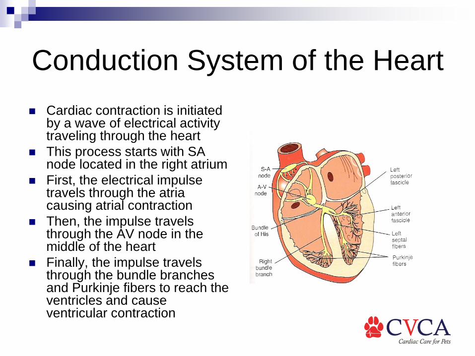
Conduction System of the Heart Cardiac contraction is initiated
by a wave of electrical activity traveling through the heart
This process starts with SA node located in the right atrium
First, the electrical impulse travels through the atria causing atrial contraction
Then, the impulse travels through the AV node in the middle of the heart
Finally, the impulse travels through the bundle branches and Purkinje fibers to reach the ventricles and cause ventricular contraction

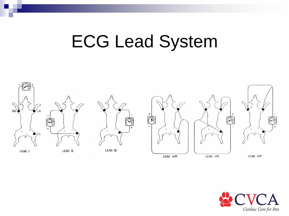
ECG Lead System The electrocardiogram (ECG or EKG) lead system consists of
several electrodes placed on specific locations of the body Animals are placed in right lateral Front limb leads are placed at level of elbow, hind limb leads are
placed at level of stifle The limb leads are color coded
White: RFL Black: LFL Green: RHL Red: LHL
Grass & snow on the ground, Christmas comes at the end of the year, white on right, etc…

ECG Lead System

ECG Lead System

ECG Lead System The ECG leads compare the electrical activity at one electrode
compared to another electrode Each lead has one electrode designate as positive and another
designated as negative Lead I: LFL (+) compared to RFL (-) Lead II: LHL (+) compared to RFL (-) Lead III: LHL (+) compared to LFL (-) Lead aVR: RFL (+) compared to average of LFL & LHL (-) Lead aVL: LFL (+) compared to average of RFL & LHL (-) Lead aVF: LHL (+) compared to average of RFL and LFL (-)
ECG Lead System
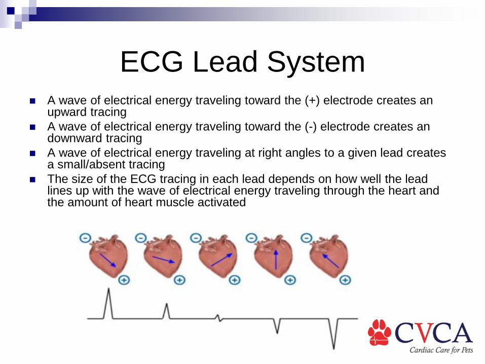
ECG Lead System A wave of electrical energy traveling toward the (+) electrode creates an
upward tracing A wave of electrical energy traveling toward the (-) electrode creates an
downward tracing A wave of electrical energy traveling at right angles to a given lead creates
a small/absent tracing The size of the ECG tracing in each lead depends on how well the lead
lines up with the wave of electrical energy traveling through the heart and the amount of heart muscle activated

Genesis of the Normal ECG
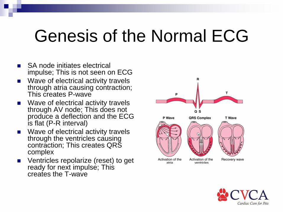
Genesis of the Normal ECG SA node initiates electrical
impulse; This is not seen on ECG Wave of electrical activity travels
through atria causing contraction; This creates P-wave
Wave of electrical activity travels through AV node; This does not produce a deflection and the ECG is flat (P-R interval)
Wave of electrical activity travels through the ventricles causing contraction; This creates QRS complex
Ventricles repolarize (reset) to get ready for next impulse; This creates the T-wave

Steps for ECG Interpretation What is the heart rate?
Pen times 10 on 25 mm/s Pen times 20 on 50 mm/s
Instantaneous rate: R-R divided into 1500 for 25 mm/s
R-R divided into 3000 for 50 mm/s
Is the rate too fast or too slow? Normal is 70-160 for dogs
and 150-240 in cats

Steps for ECG Interpretation
What is the heart rate? What is the configuration of the QRS complexes? Is the rhythm regular or irregular? Are there complexes that appear to come too early or too late? Is every P-wave followed by a QRS complex? Is every QRS complex preceded by a P-wave?

Example Arrhythmias

Example Arrhythmias
Sinus rhythm: normal rhythm in dogs and cats Normal rate, regular rhythm, normal QRS
complexes No treatment needed

Example Arrhythmias

Example Arrhythmias
Sinus arrhythmia: normal variation in dogs Normal rate, normal QRS complexes Irregular rhythm: speeds up and slows down Speeds up on inspiration slows down on expiration = Respiratory
sinus arrhythmia Wandering pacemaker: variation in P-wave configuration No treatment needed

Example Arrhythmias

Example Arrhythmias
Sinus tachycardia Regular rhythm, normal QRS complexes, but faster than normal Seen with excitement, fear, pain, shock, systemic illness Often does not need treatment; Focus on underlying condition

Example Arrhythmias

Example Arrhythmias
Sinus bradycardia Regular rhythm, normal QRS complexes, but slower than normal Seen in anesthetic overdose, hypothermia, increased vagal tone Often does not need treatment, but will likely respond to Atropine

Example Arrhythmias

Example Arrhythmias
Marked sinus arrhythmia with episodes of sinus arrest Called Sick Sinus Syndrome May improve with Atropine, but many cases require
pacemaker implantation

Example Arrhythmias

Example Arrhythmias
Supraventricular premature contraction May originate from atria or AV node Similar to normal complexes but comes early May or may not see P-wave Can be seen in animals with atrial enlargement, or in animals with normal hearts Does not require treatment

Example Arrhythmias

Example Arrhythmias
Supraventricular tachycardia Normal QRS complexes, regular rhythm, rate is too fast May or may not see P-waves Often cause weakness or collapse Treatment options include: Digoxin, Diltiazem, Atenolol

Example Arrhythmias
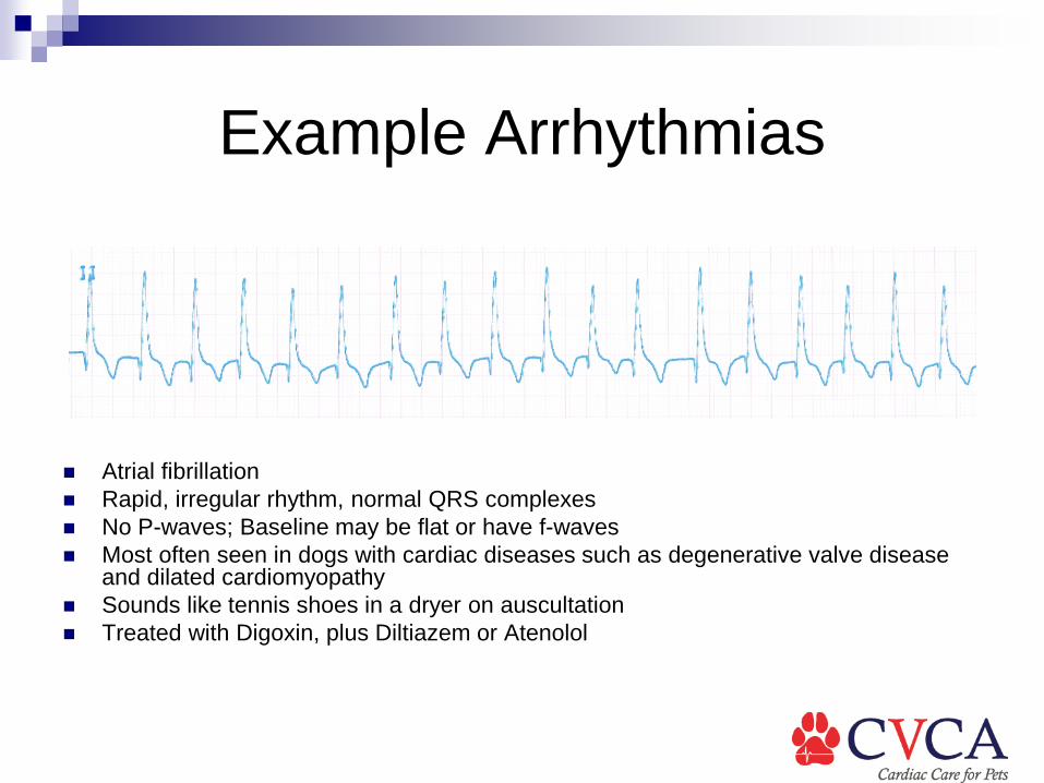
Example Arrhythmias
Atrial fibrillation Rapid, irregular rhythm, normal QRS complexes No P-waves; Baseline may be flat or have f-waves Most often seen in dogs with cardiac diseases such as degenerative valve disease
and dilated cardiomyopathy Sounds like tennis shoes in a dryer on auscultation Treated with Digoxin, plus Diltiazem or Atenolol
Example Arrhythmias
Example Arrhythmias
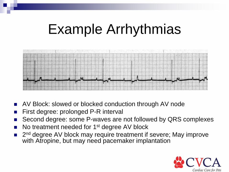
AV Block: slowed or blocked conduction through AV node First degree: prolonged P-R interval Second degree: some P-waves are not followed by QRS complexes No treatment needed for 1st degree AV block 2nd degree AV block may require treatment if severe; May improve
with Atropine, but may need pacemaker implantation

Example Arrhythmias

Example Arrhythmias
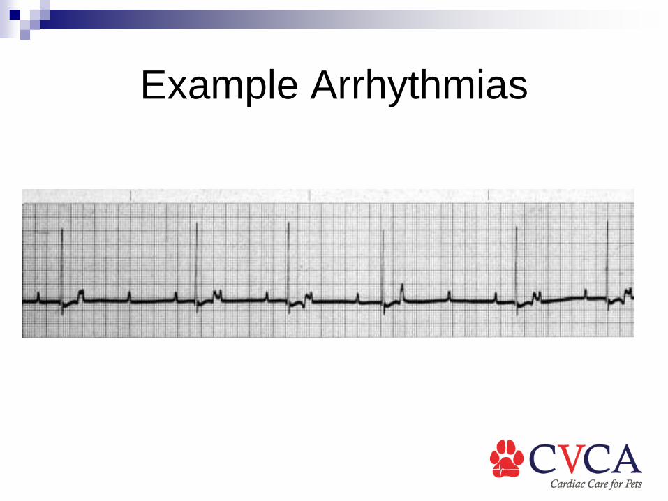
Third degree AV block: none of the P-waves are conducted through AV node
Two separate and independent rhythms: atrial and ventricular Heart rate is typically very slow; 30-40 beats per minute in dogs Causes weakness or collapse Requires pacemaker implantation

Example Arrhythmias

Example Arrhythmias
Ventricular premature contractions QRS complexes come early, are not preceded by a P-wave QRS complexes are wide and bizarre Wide T-wave opposite in polarity to QRS complex VPCs can go downward or upward in Lead II Downward = LV origin Upward = RV origin Can be seen in heart disease, abdominal neoplasia, GDV, systemic disease
Example Arrhythmias

Example Arrhythmias
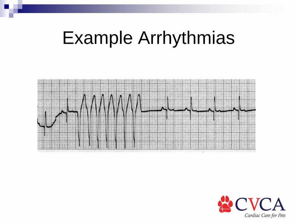
Ventricular tachycardia: 3 or more VPCs in a row Instantaneous heart rate can be over 400 bpm Sustained ventricular tachycardia is life-threatening Emergency treatment with IV Lidocaine

Example Arrhythmias

Example Arrhythmias
Ventricular fibrillation: seen during CPR Lethal arrhythmia, no ventricular contractions
occur Requires electrical defibrillation

Example Arrhythmias
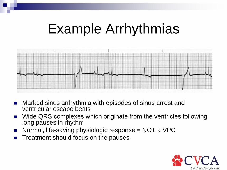
Example Arrhythmias
Marked sinus arrhythmia with episodes of sinus arrest and ventricular escape beats
Wide QRS complexes which originate from the ventricles following long pauses in rhythm
Normal, life-saving physiologic response = NOT a VPC Treatment should focus on the pauses

Example Arrhythmias

Example Arrhythmias Sinus tachycardia with
left bundle branch block Wide QRS complexes
which look like VPCs, but are all preceded by a P-wave
Rare arrhythmia which can be confused for ventricular tachycardia

Questions?

![Scone Veterinary Hospital [SVH]: 1990 – 1999sconevetdynasty.com.au/pdf/vet book/Scone Veterinary Hospital New.pdfScone Veterinary Hospital: 1990 – 1999 . Scone Veterinary Hospital](https://static.fdocuments.in/doc/165x107/5ea8e986cc0545149b1ee474/scone-veterinary-hospital-svh-1990-a-bookscone-veterinary-hospital-newpdf.jpg)