ABDOMINAL EXAMINATION Zhu Liangru Division of Gastroenterology, Union Hospital.

EBGH Sample Questions (revised August 2016)
EBGH Examination Gastroenterology Sample Questions 1.
A 50-year-old man presented with haematemesis and melaena. He had a history of excess alcohol intake for many years.
On examination, he was jaundiced with bilateral parotid enlargement, spider naevi and Dupuytren’s contractures. His pulse was 100 beats per minute and his blood pressure was 95/60 mmHg. He had ascites and peripheral oedema.
While awaiting endoscopy, what is the most appropriate management? A insert a Sengstaken–Blakemore tube B intravenous pantoprazole C intravenous terlipressin D nasogastric tube and aspiration E oral sucralfate
Comments There should be a high index of suspicion of a variceal bleed in this man who is shocked and has stigmata of chronic liver disease. In addition to resuscitation, intravenous terlipressin is the most appropriate treatment to reduce portal pressure whilst awaiting endoscopy.

EBGH Sample Questions (revised August 2016)
2.
A 67-year-old man with dysphagia was found at endoscopy to have lower oesophageal carcinoma.
For staging of local invasion in oesophageal cancer, which investigation is most sensitive? A contrast-enhanced CT scan of oesophagus B laparoscopy C MR scan of chest D PET scan E radial endoscopic ultrasound scan
Comments Endoscopic ultrasound is the most sensitive modality for local staging of oesophageal carcinoma. CT and CT-PET are modalities for assessing the presence of distant metastases. Comments: This study (Räsänen JV1, Sihvo EI, Knuuti MJ, Minn HR, Luostarinen ME, Laippala P, Viljanen T, Salo JA. Prospective analysis of accuracy of positron emission tomography, computed tomography, and endoscopic ultrasonography in staging of adenocarcinoma of the esophagus and the esophagogastric junction. Ann Surg Oncol. 2003 Oct;10(8):954-60) confirms the primary role of EUS for staging of esophageal cancer
EBGH Sample Questions (revised August 2016)
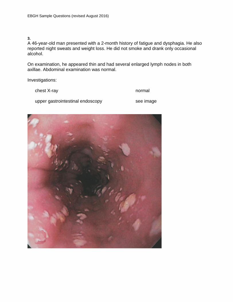
3.
A 46-year-old man presented with a 2-month history of fatigue and dysphagia. He also reported night sweats and weight loss. He did not smoke and drank only occasional alcohol.
On examination, he appeared thin and had several enlarged lymph nodes in both axillae. Abdominal examination was normal.
Investigations:
chest X-ray normal
upper gastrointestinal endoscopy see image

EBGH Sample Questions (revised August 2016)
What is the most appropriate next investigation? A bone marrow aspirate B CT scan of abdomen C HIV serology D lymph node biopsy E tuberculin test
Comments The image shows oesophageal candidiasis, which should always alert the physician to the possibility of underlying immunodeficiency (in the absence of inhaled corticosteroids). Additional pointers in this patient are systemic symptoms and axillary lymphadenopathy.

EBGH Sample Questions (revised August 2016)
4.
A 55-year-old man with Crohn’s disease underwent an ileocaecal resection. The surgical procedure was technically straightforward. Three months later, he was reviewed in the clinic. His appetite remained good and the abdominal pain had settled, but he was troubled by diarrhoea with a daytime stool frequency of six per day. He also experienced faecal urgency 20–40 minutes after eating. The stool was watery but there was no blood or pus.
Investigations:
haemoglobin 125 g/L (130–180)
white cell count 5.6 109/L (4.0–11.0)
platelet count 256 109/L (150–400) erythrocyte sedimentation rate 12 mm/1st h (<20) serum vitamin B12 340 ng/L (160–760) red cell folate 420 µg/L (160–640)
serum C-reactive protein 8 mg/L (<10)
What is the most likely cause for the diarrhoea? A bacterial overgrowth B bile salt malabsorption C enterocolic fistula D lactase deficiency E recurrent Crohn’s disease
Comments Resection of the distal ileum (depending on extent) prevents reabsorption of bile salts, which then enter the colon and induce diarrhoea. Although a recrudescence of Crohn’s disease is a possibility, it is less likely given the normal inflammatory markers. Similarly, the normal B12 and folate make bacterial overgrowth less likely.
EBGH Sample Questions (revised August 2016)
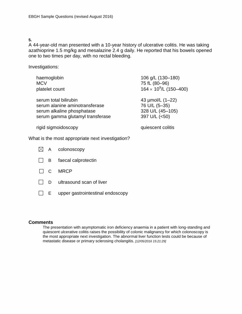
5.
A 44-year-old man presented with a 10-year history of ulcerative colitis. He was taking azathioprine 1.5 mg/kg and mesalazine 2.4 g daily. He reported that his bowels opened one to two times per day, with no rectal bleeding.
Investigations:
haemoglobin 106 g/L (130–180) MCV 75 fL (80–96)
platelet count 164 109/L (150–400)
serum total bilirubin 43 µmol/L (1–22) serum alanine aminotransferase 76 U/L (5–35) serum alkaline phosphatase 328 U/L (45–105) serum gamma glutamyl transferase 397 U/L (<50)
rigid sigmoidoscopy quiescent colitis
What is the most appropriate next investigation? A colonoscopy B faecal calprotectin C MRCP D ultrasound scan of liver E upper gastrointestinal endoscopy
Comments The presentation with asymptomatic iron deficiency anaemia in a patient with long-standing and quiescent ulcerative colitis raises the possibility of colonic malignancy for which colonoscopy is the most appropriate next investigation. The abnormal liver function tests could be because of metastatic disease or primary sclerosing cholangitis. [12/05/2016 15:21:29]

EBGH Sample Questions (revised August 2016)
6.
A 68-year-old man was found to have positive faecal occult blood tests (FOBT) in a national bowel cancer screening programme. He was offered colonoscopy, but before making his decision he wanted to know what the chances were of actually having a colonic carcinoma.
What is the likelihood of colonic carcinoma in a patient of this age with a positive FOBT? A 2% B 8% C 16% D 24% E 48%
Comments The prevalence of colorectal carcinoma is 8–10% following a positive faecal occult blood screen in the bowel cancer screening programme.

EBGH Sample Questions (revised August 2016)
7.
A 56-year-old man with established cirrhosis secondary to genetic haemochromatosis was found to have a 3-cm focal lesion in the right lobe of his liver at a surveillance ultrasound scan of his abdomen. When reviewed in the outpatient clinic, he was well with no new symptoms.
Investigations:
international normalised ratio 1.3 (<1.4)
serum albumin 32 g/L (37–49) serum total bilirubin 37 μmol/L (1–22) serum alanine aminotransferase 23 U/L (5–35) serum alkaline phosphatase 125 U/L (45–105)
serum α-fetoprotein 8 kU/L (<10)
What is the most appropriate next step in management? A further surveillance screening in 6 months B referral for consideration of resection of the hepatic lesion C repeat ultrasound scan of his liver in 6 weeks D triple-phase CT scan of liver E ultrasound scan-guided biopsy of the lesion
Comments Cirrhosis complicating genetic haemochromatosis is a particularly high risk situation for the development of hepatocellular carcinoma. Serum α-fetoprotein may be negative in >20% of hepatomas. A CT scan or MR scan of liver would be helpful in further defining the nature of the mass lesion. Biopsy may give the diagnosis but because of potential seeding may prevent curative resection. Hepatic resection is premature until a clearer diagnosis is reached.
EBGH Sample Questions (revised August 2016)
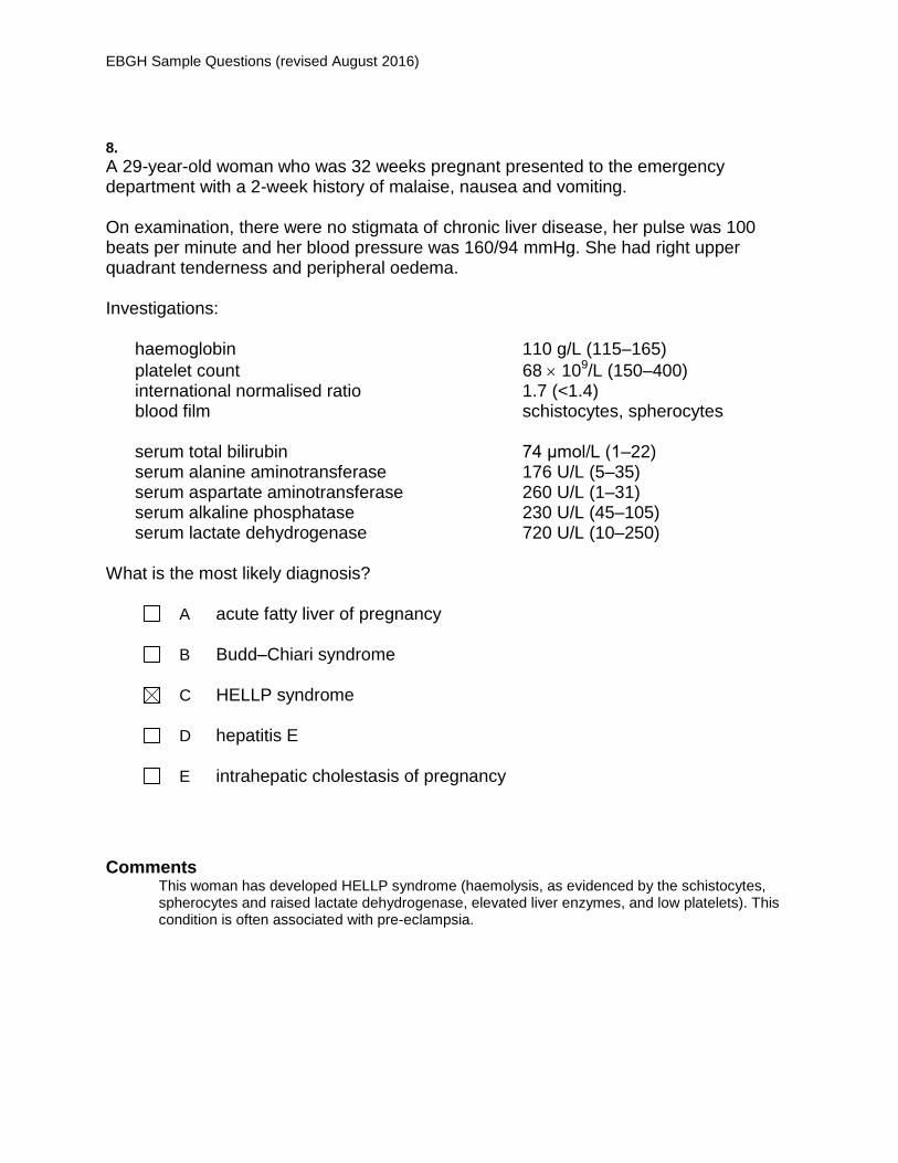
8.
A 29-year-old woman who was 32 weeks pregnant presented to the emergency department with a 2-week history of malaise, nausea and vomiting.
On examination, there were no stigmata of chronic liver disease, her pulse was 100 beats per minute and her blood pressure was 160/94 mmHg. She had right upper quadrant tenderness and peripheral oedema.
Investigations:
haemoglobin 110 g/L (115–165)
platelet count 68 109/L (150–400) international normalised ratio 1.7 (<1.4) blood film schistocytes, spherocytes
serum total bilirubin 74 μmol/L (1–22) serum alanine aminotransferase 176 U/L (5–35) serum aspartate aminotransferase 260 U/L (1–31) serum alkaline phosphatase 230 U/L (45–105) serum lactate dehydrogenase 720 U/L (10–250)
What is the most likely diagnosis? A acute fatty liver of pregnancy B Budd–Chiari syndrome C HELLP syndrome D hepatitis E E intrahepatic cholestasis of pregnancy
Comments This woman has developed HELLP syndrome (haemolysis, as evidenced by the schistocytes, spherocytes and raised lactate dehydrogenase, elevated liver enzymes, and low platelets). This condition is often associated with pre-eclampsia.

EBGH Sample Questions (revised August 2016)
Item I25430.2
9.
A 68-year-old woman was referred for investigation of iron deficiency anaemia. She was taking warfarin for atrial fibrillation.
On examination, she had atrial fibrillation with a ventricular rate of 76 beats per minute. No other abnormality was detected.
Investigations:
international normalised ratio 2.1
coeliac serology positive
echocardiography normal left ventricular systolic function; no valvular abnormality
Upper gastrointestinal endoscopy to obtain duodenal biopsies was planned.
What is the most appropriate plan for anticoagulation before this endoscopy? A no alteration of therapy B stop warfarin C substitute aspirin for warfarin D substitute clopidogrel for warfarin E substitute low-molecular-weight heparin for warfarin
Comments Warfarin does not need to be stopped for simple diagnostic biopsies.
EBGH Sample Questions (revised August 2016)
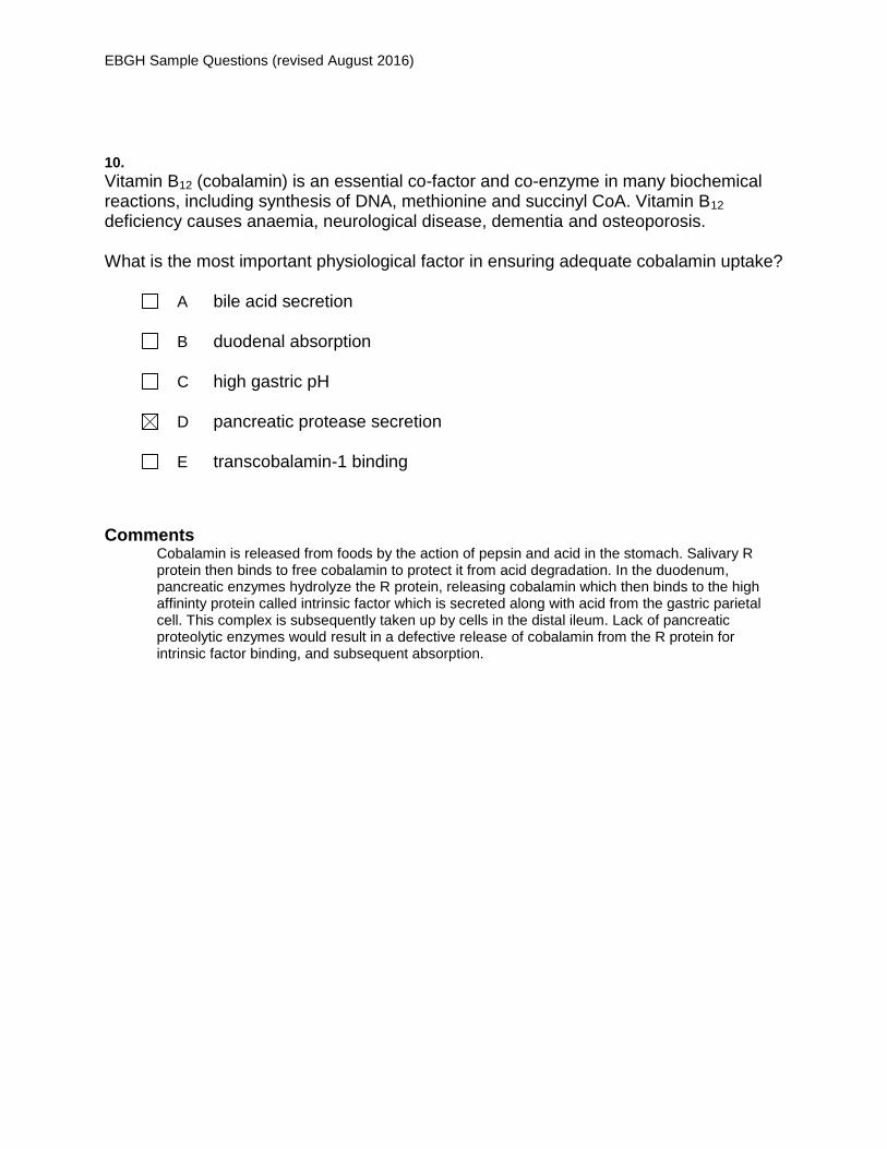
10.
Vitamin B12 (cobalamin) is an essential co-factor and co-enzyme in many biochemical reactions, including synthesis of DNA, methionine and succinyl CoA. Vitamin B12 deficiency causes anaemia, neurological disease, dementia and osteoporosis.
What is the most important physiological factor in ensuring adequate cobalamin uptake? A bile acid secretion B duodenal absorption C high gastric pH D pancreatic protease secretion E transcobalamin-1 binding
Comments Cobalamin is released from foods by the action of pepsin and acid in the stomach. Salivary R protein then binds to free cobalamin to protect it from acid degradation. In the duodenum, pancreatic enzymes hydrolyze the R protein, releasing cobalamin which then binds to the high affininty protein called intrinsic factor which is secreted along with acid from the gastric parietal cell. This complex is subsequently taken up by cells in the distal ileum. Lack of pancreatic proteolytic enzymes would result in a defective release of cobalamin from the R protein for intrinsic factor binding, and subsequent absorption.

EBGH Sample Questions (revised August 2016)
11.
A 60-year-old man with a 35-year history of well-controlled ulcerative colitis was seen for review. His maintenance treatment was sulfasalazine.
On what does the mechanism of action of sulfasalazine depend? A cleavage of 5-ASA dimers by colonic bacteria B cleavage of an azo bond by colonic bacteria C pH-dependent release in the ileocaecal region D slow release in the small and large intestine through an ethylcellulose
coating E timed release following alkalinisation in the duodenum
Comments Sulfasalazine is a dimer of sulfapyridine linked to 5-ASA by an azo bond. This bond is split by colonic bacteria to release 5-ASA.

EBGH Sample Questions (revised August 2016)
Item I26071.2
12.
A 64-year-old woman presented with a 3-year history of intermittent diarrhoea. She was otherwise well and had not lost weight. She had a past medical history of osteoarthritis, which had been treated with naproxen with omeprazole as gastroprotection. Treatment with loperamide had not improved her bowel symptoms.
On examination, she looked well. She had a body mass index of 34 kg/m2 (18–25).
Investigations:
colonoscopy normal
histology from colonic biopsies a mononuclear infiltrate with a few neutrophils and eosinophils in the lamina propria
Stopping naproxen did not improve her symptoms.
What is the most appropriate next step in management? A budesonide B colestyramine C octreotide D prednisolone E sulfasalazine
Comments The histological description in the context of a normal colonoscopy is consistent with microscopic colitis (lymphocytic colitis). This condition has an association with NSAID usage. Oral budesonide is the best treatment option.

EBGH Sample Questions (revised August 2016)
13.
A 43-year-old man with acromegaly was referred for colonoscopy. Pan-colonoscopy with terminal ileal intubation was achieved. A solitary, sessile, 5-mm polyp was found in the transverse colon. This was removed completely. Histology revealed a tubular adenoma with low-grade dysplasia.
Investigations:
haemoglobin 144 g/L (130–180)
platelet count 254 109/L (150–400)
serum insulin-like growth factor 1 30.3 nmol/L (5.6–23.3)
After how long should he undergo further colonoscopy? A 1 year B 2 years C 3 years D 5 years E 10 years
Comments Patients with acromegaly have an increased risk of colon cancer. The British Society of Gastroenterology guideline from 2010 indicates that those with an adenoma at first screening visit (offered from the age of 40) or with raised insulin-like growth factor (ILGF) levels should have 3-yearly colonoscopy. Those without a polyp at initial screening colonoscopy, or with hyperplastic polyps, or with normal ILGF levels should be screened at 5–10 yearly intervals.

EBGH Sample Questions (revised August 2016)
14.
The portal vein is formed by the confluence of which veins? A hepatic and superior mesenteric B inferior and superior mesenteric C splenic and hepatic D splenic and renal E splenic and superior mesenteric
Comments Knowledge of anatomy required. This knowledge is clinically useful when managing portal hypertension.

EBGH Sample Questions (revised August 2016)
15.
A 61-year-old man with Barrett’s oesophagus was found to have high-grade dysplasia in four out of eight biopsies taken from the Barrett’s segment. He had residual mild left-sided weakness from a cerebrovascular accident 2 years previously. A further upper gastrointestinal endoscopy was arranged and similar histological features were reported; there were no visible nodules.
Investigations:
CT scan of thorax and abdomen normal
What is the most appropriate management? A high-dose proton pump inhibitor B intensified endoscopic surveillance C laser ablation of Barrett’s segment D photodynamic therapy E radiofrequency ablation of Barrett’s segment
Comments High-grade dysplasia in Barrett’s on two separate endoscopy examinations is best treated by radiofrequency ablation. None of the remaining options is as reliable in achieving a cure.

EBGH Sample Questions (revised August 2016)
16.
A 32-year-old man attended for follow-up 2 months after presenting with a bleeding duodenal ulcer. As part of his treatment, he had been given a course of Helicobacter pylori eradication therapy, and had continued taking the proton pump inhibitor until 4 weeks before his appointment.
Which is the most appropriate test to confirm eradication of H. pylori infection? A gastric antral biopsy for culture B gastric antral biopsy for histology C gastric antral biopsy for rapid urease test D H. pylori antibody in serum E stool H. pylori antigen Comments
The urea breath test or faecal antigen test are the best non-invasive tests for confirming successful eradication treatment for Helicobacter pylori. Serology may take many months or never turn negative and repeat endoscopy is unnecessarily invasive.

EBGH Sample Questions (revised August 2016)
17.
A 24-year-old woman presented with a 3-month history of lethargy, fatigue and weight loss. She had abdominal bloating and passed loose stools.
On examination, there were no abnormal findings.
Investigations:
haemoglobin 85 g/L (115–165)
platelet count 164 109/L (150–400) MCV 70 fL (80–96)
white cell count 11.0 109/L (4.0–11.0) serum ferritin 9 µg/L (15–300) serum vitamin B12 180 ng/L (160–760) red cell folate 86 µg/L (160–640)
serum albumin 35 g/L (37–49)
serum IgG 7.2 g/L (6.0–13.0) serum IgA 0.1 g/L (0.8–3.0) serum IgM 0.3 g/L (0.4–2.5)
anti-tissue transglutaminase IgA antibodies 2 U/mL (<15)
What is the most appropriate next step in management? A CT scan of abdomen B duodenal biopsy C faecal elastase estimation D lactulose–hydrogen breath test E small bowel barium studies
Comments The symptoms together with a combined iron and folate deficiency anaemia would make coeliac disease highly likely. Patients with coeliac disease and IgA deficiency will have false negative serology as the antibody is of the IgA class.

EBGH Sample Questions (revised August 2016)
18.
A 53-year-old man had been admitted with moderately severe pancreatitis 1 week previously. Despite regular analgesia and antiemetics, he remained nauseated and uncomfortable, with no appetite and poor oral intake.
What is the most appropriate management of his nutrition? A encourage oral intake B nasogastric tube feeding C oral elemental diet D peripheral intravenous nutrition E total parenteral nutrition
Comments Enteral nutrition support is important here and recent data suggest nasogastric feeding is as effective as nasojejunal feeding and associated with fewer problems in this situation. Sufficient oral intake is unlikely given his nausea and intravenous nutrition is inappropriate.

EBGH Sample Questions (revised August 2016)
19.
A 23-year-old secretary presented with a 12-month history of intermittent epigastric and right upper quadrant pain occurring up to six times per month and lasting for 30 to 45 minutes. The most recent episode of pain had occurred 24 hours previously. She had been obliged to leave work on several occasions and, during one episode, had presented to the emergency department. The symptoms were unrelated to diet, eating or bowel movement. Antacids had been unhelpful and she took codeine at home for the pain. She was otherwise well with no other history.
Examination was normal.
Investigations:
serum total bilirubin 22 µmol/L (1–22) serum alanine aminotransferase 48 U/L (5–35) serum aspartate aminotransferase 52 U/L (1–31) serum alkaline phosphatase 200 U/L (45–105) serum gamma glutamyl transferase 80 U/L (4–35)
ultrasound scan of abdomen normal
What is the most appropriate next investigation? A CT scan of abdomen B ERCP C HIDA scan D MRCP E repeat ultrasound scan when in pain
Comments The clinical history is of biliary colic. Given her abnormal liver function tests, a common bile duct stone needs to be considered and this should be done non-invasively with MR imaging.
EBGH Sample Questions (revised August 2016)
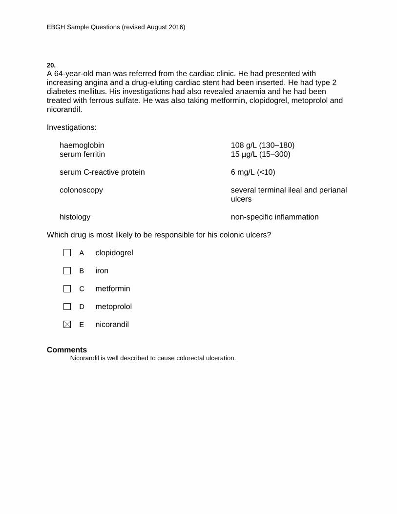
20.
A 64-year-old man was referred from the cardiac clinic. He had presented with increasing angina and a drug-eluting cardiac stent had been inserted. He had type 2 diabetes mellitus. His investigations had also revealed anaemia and he had been treated with ferrous sulfate. He was also taking metformin, clopidogrel, metoprolol and nicorandil.
Investigations:
haemoglobin 108 g/L (130–180) serum ferritin 15 µg/L (15–300)
serum C-reactive protein 6 mg/L (<10)
colonoscopy several terminal ileal and perianal ulcers
histology non-specific inflammation
Which drug is most likely to be responsible for his colonic ulcers? A clopidogrel B iron C metformin D metoprolol E nicorandil
Comments Nicorandil is well described to cause colorectal ulceration.

EBGH Sample Questions (revised August 2016)
21.
An 18-year-old man presented with a 5-day history of moderate abdominal pain, bloating, diarrhoea associated with mucus, and blood spotting on the toilet paper.
On examination, the abdomen was soft but he was mildly tender in both iliac fossae.
Investigations:
stool culture normal
Flexible sigmoidoscopy showed mucosal erythema and congestion. Histological examination of rectal biopsies showed crypt abscesses, mucin depletion and normal crypt architecture with neutrophilic infiltration.
What is the most likely diagnosis? A collagenous colitis B Crohn’s colitis C infectious colitis D microscopic colitis E ulcerative colitis
Comments The short history and normal crypt architecture would favour an infective aetiology.

EBGH Sample Questions (revised August 2016)
22.
A 35-year-old man with corticosteroid-resistant Crohn’s disease was treated with azathioprine. After 3 weeks he became severely leucopenic. Subsequent tests revealed an extremely low concentration of thiopurine methyltransferase (TPMT).
In approximately what proportion of the population does homozygous TPMT deficiency occur? A 1 in 10 B 1 in 50 C 1 in 100 D 1 in 300 E 1 in 1000
Comments 0.3% of the population have very low/insignificant levels of TPMT. This group are at a far greater risk of leucopenia with thiopurine analogues and dose reduction should be considered.

EBGH Sample Questions (revised August 2016)
23.
A 76-year-old woman was admitted with haematemesis and melaena. She was taking ibuprofen for osteoarthritis, but had no history of dyspepsia. There was a history of hypertension, severe COPD and stroke.
On examination, she was comfortable, but rather pale and sweaty. Her pulse was 104 beats per minute and her blood pressure was 108/75 mmHg. Abdominal examination was normal.
Investigations:
haemoglobin 85 g/L (115–165)
serum urea 15.4 mmol/L (2.5–7.0) serum creatinine 106 µmol/L (60–110)
What is her pre-endoscopy Rockall risk score for severity of upper gastrointestinal haemorrhage? A 3 B 4 C 5 D 6 E 7
Comments The patient scores 1 for her age; 1 for tachycardia; and 2 for ‘any major comorbidity’ (ie severe COPD).

EBGH Sample Questions (revised August 2016)
24.
A 35-year-old man was admitted with haematemesis. He had a 4-year history of chronic pancreatitis caused by excess alcohol. His stated alcohol intake over the previous 12 months was zero.
Investigations:
upper gastrointestinal endoscopy normal oesophagus; abnormality in gastric fundus (see image)

EBGH Sample Questions (revised August 2016)
What is the most likely explanation for this presentation? A hepatic cirrhosis B hepatic vein thrombosis C pancreatic pseudocyst D portal vein thrombosis E splenic vein thrombosis
Comments Pancreatitis is a risk factor for splenic vein thrombosis because of the proximity of the vessel to the pancreas. Splenic vein thrombosis is typically associated with isolated gastric varices

EBGH Sample Questions (revised August 2016)
25.
A 37-year-old man was referred from the haematology/oncology unit before starting treatment for non-Hodgkin’s lymphoma. He was originally from Hong Kong.
Investigations:
serum albumin 31 g/L (37–49) serum total bilirubin 19 µmol/L (1–22) serum alanine aminotransferase 41 U/L (5–35) serum alkaline phosphatase 155 U/L (45–105)
hepatitis B surface antigen positive hepatitis B e antigen positive HBV viral load 160 000 IU/mL (lower detection limit 250)
liver biopsy modified Ishak score necroinflammatory score 1/18 fibrosis score 2/6
What is the most appropriate treatment of the hepatitis B during chemotherapy? A adefovir B interferon alfa C no treatment indicated D prednisolone E tenofovir
Comments Treatment for non-Hodgkin’s lymphoma will provoke viral replication. Tenofovir will prevent an increase in viral load while minimising the risk of mutation.
EBGH Sample Questions (revised August 2016)
26.
A 76-year-old woman with a history of stroke had a percutaneous endoscopic gastrostomy (PEG) tube inserted. Four hours later, she complained of pain at the site of tube insertion.
On examination, her temperature was normal and her abdomen was soft and non-tender, but slightly distended. The wound was clean, dry and not hot to touch, but the surrounding skin felt as though it had air bubbles in it.
Erect X-rays of abdomen and chest showed free gas under both domes of the diaphragm and within the anterior abdominal wall.
What is the most likely diagnosis? A benign pneumoperitoneum B colonic perforation C enterocutaneous fistula D gastrocolic fistula E necrotising fasciitis
Comments Air is introduced into the peritoneum during the uncomplicated placement of a PEG feeding tube. Colonic perforation would produce signs of peritonism. Fistulas do not form after such a short time period. Similarly necrotising fasciitis would not develop so quickly, and there are no supportive signs of this condition in this case.

EBGH Sample Questions (revised August 2016)
27.
A 45-year-old man with a 15-year history of extensive ulcerative colitis underwent a surveillance colonoscopy. He had been well since his most recent colonoscopy 5 years previously. He had a normal bowel habit and was taking oral mesalazine only. His brother, who was aged 48, did not have inflammatory bowel disease, but had recently had a colorectal cancer resected.
The patient’s colonoscopy demonstrated quiescent changes, which were confirmed by the biopsies.
After what interval should a further colonoscopy be performed?
A 1 year B 2 years C 3 years D 5 years E 10 years
Comments Macroscopically and histologically quiescent disease should be surveyed at 5-yearly intervals. The family history in a first-degree relative means this patient should have annual surveillance (British Society of Gastroenterology guidelines 2010).

EBGH Sample Questions (revised August 2016)
28.
A 28-year-old man was referred after recurrent food bolus obstruction. He had a history of hay fever, mild asthma and bipolar disorder. He was taking venlafaxine and inhaled salbutamol. On examination, there were no abnormal findings. Investigations: barium swallow multiple rings and mucosal irregularities (see image)

EBGH Sample Questions (revised August 2016)
oesophageal biopsy >50 eosinophils per high-power field

EBGH Sample Questions (revised August 2016)
What is the most appropriate treatment? A chlorphenamine B fluticasone C omeprazole
D salmeterol E sodium cromoglicate
Comments The clinical history, barium swallow and biopsies strongly suggest eosinophilic oesophagitis . The initial treatment for this disorder is acid suppression with a proton pump inhibitor: In an open label study of omeprazole 20mg twice a day, Clinical improvement occurred in 43 (71.6%), endoscopic signs were reduced in 34 (61.8%) and normalised in 12 (21.8%), and histologically, 34 (56.6%) improved, while 15 (25%) obtained complete resolution. Overall, 22 patients (36.7%) obtained both complete clinical and histological remission. Treatment with corticosteroids or immune suppression should be reserved for non-responders. Vazquez-Elizondo G, Ngamruengphong S, Khrisna M, Devault KR, Talley NJ, Achem SR. The outcome of patients with oesophageal eosinophilic infiltration after an eight-week trial of a proton pump inhibitor. Aliment Pharmacol Ther. 2013 Nov;38(10):1312-9.

EBGH Sample Questions (revised August 2016)
29.
A 64-year-old man presented with jaundice and was found to have a carcinoma of the head of pancreas. He had undergone a mitral valve replacement and was taking warfarin. An ERCP and placement of a biliary stent was planned in 2 days’ time.
Investigations:
international normalised ratio 3.0
What is the most appropriate management of his anticoagulation? A continue warfarin B give intravenous vitamin K 2 mg C stop warfarin D stop warfarin and start intravenous unfractionated heparin E stop warfarin and start low-molecular-weight heparin
Comments Biliary or pancreatic stenting is regarded as a low-risk procedure, and the warfarin can be continued as long as the INR is within the therapeutic range (British Society of Gastroenterology guidelines 2016).

EBGH Sample Questions (revised August 2016)
30.
A 56-year-old woman with a 3-year history of ulcerative colitis presented with an increasing number of relapses that had responded well to oral prednisolone. Her past medical history included hypertension and recurrent urinary tract infections. She was taking ramipril 5 mg daily and mesalazine 1.6 g daily in divided doses. A decision was made to treat her with azathioprine therapy as a corticosteroid-sparing agent.
Investigations:
haemoglobin 114 g/L (115–165)
white cell count 8.3 109/L (4.0–11.0)
neutrophil count 3.1 109/L (1.5–7.0)
platelet count 456 109/L (150–400)
serum creatinine 135 µmol/L (60–110) serum albumin 32 g/L (37–49)
serum C-reactive protein 16 mg/L (<10)
What is the most useful test for monitoring toxicity in patients taking azathioprine? A erythrocyte 6-thioguanine nucleotide concentration B erythrocyte thiopurine methyltransferase activity C microalbuminuria D serum alanine aminotransferase concentration E white cell count
Comments Bone marrow suppression occurs in up to 5% of patients receiving azathioprine and necessitates careful full blood count monitoring. Thiopurine methyltransferase (TPMT) levels are often taken before starting azathioprine to identify the 0.3% who have negligible levels and should not be given the drug, or those with low/intermediate levels in whom much smaller doses should be given if the drug is used. Most patients who develop leucopenia, however, have normal TPMT levels. 6-thioguanine nucleotides are metabolites of 6-mercaptopurine. Liver function tests should also be measured regularly as there is a small incidence of hepatotoxicity.
EBGH Sample Questions (revised August 2016)
31.
A 49-year-old man presented with abdominal pain.
On examination, there was hepatosplenomegaly.
Investigations:
serum iron 48 µmol/L (12–30) serum ferritin 2055 µg/L (15–300) transferrin saturation 82% (20–50)
What abnormality is most likely to be detected in his HFE gene? A C282Y and H63D heterozygosity B C282Y heterozygosity C C282Y homozygosity D H63D heterozygosity E H63D homozygosity
Comments C282Y homozygosity is the most common gene mutation associated with genetic haemochromatosis.

EBGH Sample Questions (revised August 2016)
32.
A 67-year-old man presented with melaena while taking warfarin for atrial fibrillation. He was transfused and later underwent an upper gastrointestinal endoscopy and ileocolonoscopy, which were unremarkable. The melaena settled and so his warfarin was restarted and he was allowed home.
He re-presented 21 days later with further melaena and again required transfusion. His upper gastrointestinal endoscopy and colonoscopy were repeated but no cause for his melaena was discovered.
What is the most appropriate next investigation? A double balloon enteroscopy B MR scan of small bowel C push enteroscopy D red cell scintigraphy E small bowel capsule endoscopy
Comments It is likely that the source of bleeding in this patient is from the small bowel. Capsule endoscopy is the best modality to locate the possible bleeding source in this case before potential targeted treatment via either push enteroscopy or double balloon enteroscopy.

EBGH Sample Questions (revised August 2016)
33.
A 77-year-old man presented with a 6-hour history of profuse fresh rectal bleeding. There had been no pain or preceding gastrointestinal symptoms. He had COPD and ischaemic heart disease.
On examination, he was pale, with a pulse of 110 beats per minute and a blood pressure of 95/70 mmHg. Rectal examination revealed fresh blood but no masses palpable.
Investigations:
haemoglobin 96 g/L (130–180)
platelet count 464 109/L (150–400)
serum sodium 143 mmol/L (137–144) serum potassium 4.4 mmol/L (3.5–4.9) serum urea 11.1 mmol/L (2.5–7.0) serum creatinine 142 µmol/L (60–110)
He was partially resuscitated with intravenous fluids and blood, but continued to pass significant quantities of blood and remained haemodynamically unstable.
Upper gastrointestinal endoscopy was normal and limited flexible sigmoidoscopy was unrewarding, with poor views obtained.
What is the most appropriate next step in management? A colonoscopy B CT angiography C MR angiography D red cell scintigraphy
E small bowel capsule endoscopy Comments
CT angiography (with potential interventional mesenteric artery embolisation) is indicated in patients with an unclear source of gastrointestinal bleeding who are cardiovascularly unstable because of continued significant blood loss.

EBGH Sample Questions (revised August 2016)
34.
A 58-year-old man was admitted with a 2-week history of abdominal pain. The pain was widespread and associated with progressive abdominal distension. He had cirrhosis and a history of excess alcohol intake.
On examination, he appeared pale and cachectic. There was no palpable lymphadenopathy. The abdomen was tender, and shifting dullness was elicited. The liver was palpable 3 cm below the right costal margin.
Investigations:
haemoglobin 103 g/L (130–180)
platelet count 64 109/L (150–400)
serum sodium 131 mmol/L (137–144) serum potassium 4.2 mmol/L (3.5–4.9) serum creatinine 89 µmol/L (60–110) serum albumin 32 g/L (37–49) serum total bilirubin 51 µmol/L (1–22) serum alanine aminotransferase 72 U/L (5–35) serum alkaline phosphatase 187 U/L (45–105)
ascitic albumin 23 g/L ascitic neutrophil count 268 cells/mm3
ascitic lymphocyte count 34 cells/mm3
What is the most likely diagnosis? A Budd–Chiari syndrome B hepatocellular carcinoma C hepatopulmonary syndrome D spontaneous bacterial peritonitis E tuberculosis
Comments Abdominal pain in association with ascites should alert the clinician to a diagnosis of spontaneous bacterial peritonitis (SBP). SBP is confirmed with an ascitic neutrophil count of >250 cells/mm
3 .

EBGH Sample Questions (revised August 2016)
35.
A 35-year-old man with Crohn’s disease presented complaining of recurrent oral ulceration.
What is the most appropriate initial treatment? A chlorhexidine mouthwash B prednisolone C thalidomide D topical hydrocortisone E topical tacrolimus
Comments Crohn’s oral aphthous ulceration is best treated in the first instance with topical corticosteroid (eg hydrocortisone lozenges).

EBGH Sample Questions (revised August 2016)
36.
A 54-year-old man presented to the emergency department with dysphagia and stridor, and was found to have a large proximal oesophageal cancer. Because of external compression of the trachea by the oesophageal tumour, his stridor was treated initially with an endotracheal stent and then a T-tube tracheostomy.
A CT scan of chest showed a 7-cm mass from the suprasternal notch to the aortic arch arising from the oesophagus. Radical chemoradiotherapy was planned. Although he was able to swallow small amounts of semi-liquid food, nutritional support was required.
What means of nutritional support is most appropriate during chemoradiotherapy? A additional oral supplements B nasogastric tube C parenteral nutrition via a central vein D percutaneous endoscopic gastrostomy E radiologically inserted gastrostomy
Comments His dysphagia is likely to worsen on starting chemoradiotherapy and so he will need enteral feeding for several weeks. A radiologically inserted gastrostomy is preferred to percutaneous endoscopic gastrostomy as the latter is not technically possible with a tight oesophageal stricture. Any endoscopic procedure is high risk because of the high location of the tumour and the presentation of stridor.

EBGH Sample Questions (revised August 2016)
37.
A 43-year-old man presented with swelling of both ankles and a history of several weeks’ intermittent dyspepsia. He stated that he was eating well and had noticed no alteration in bowel habit.
Physical examination was normal apart from the finding of peripheral oedema.
Investigations:
haemoglobin 130 g/L (130–180)
white cell count 7.6 109/L (4.0–11.0)
serum creatinine 78 µmol/L (60–110) serum albumin 26 g/L (37–49)
upper gastrointestinal endoscopy giant mucosal folds in fundus and body of stomach; otherwise normal
What investigation is most likely to determine the cause of his hypoalbuminaemia? A 24-h urinary protein B anti-tissue transglutaminase antibodies C faecal elastase
D faecal to serum 1-antitrypsin ratio
E plasma gastrin
Comments The endoscopic findings suggest a diagnosis of Menetrier’s disease (giant hypertrophic gastropathy) which is the commonest gastric lesion causing severe protein loss. α1-antitrypsin is similar in size to albumin and is a useful marker of intestinal protein loss. It is resistant to proteolysis and not actively secreted nor absorbed, with low levels normally present in stools. Protein-losing enteropathies result in elevated levels in stool.

EBGH Sample Questions (revised August 2016)
38.
A 55-year-old man presented with upper abdominal pain and jaundice. He had had little appetite for a few days and had lost 5 kg in weight. He also complained of increased frequency of bowel movements, which had contained a little fresh blood. He drank 25 units of alcohol per week.
On examination, he was jaundiced. Abdominal examination showed slight enlargement of the liver, two fingers below the costal margin, and the gallbladder was palpable. Urinalysis showed blood 2+, protein 1+.
Investigations:
haemoglobin 137 g/L (130–180)
serum total protein 75 g/L (61–76) serum albumin 30 g/L (37–49) serum total bilirubin 80 µmol/L (1–22) serum alkaline phosphatase 920 U/L (45–105)
chest X-ray infiltration in right upper lobe
CT scan of abdomen swollen pancreas with prominence of pancreatic head; dilated intrahepatic bile ducts
What is the most appropriate treatment? A antituberculous therapy B endoscopic biliary stent placement C pancreatic enzyme supplements D prednisolone E ursodeoxycholic acid
Comments Diffusely swollen pancreas with pulmonary infiltrates are described in IgG4 disease. The low albumin and high normal total protein suggests an increase in immunoglobulin levels.

EBGH Sample Questions (revised August 2016)
39.
A 16-year-old boy with cystic fibrosis presented with an episode of severe, cramping abdominal pain. He had experienced similar less severe episodes previously. He felt bloated. There was no other significant medical history. His only regular medication was pancreatic enzyme supplements. His father had been found to have colorectal carcinoma at the age of 42.
On examination, he was uncomfortable but not distressed. He was apyrexial, his pulse was 84 beats per minute and his blood pressure was 105/60 mmHg. Abdominal examination showed a vague mass and mild tenderness in the right iliac fossa.
Investigations:
haemoglobin 155 g/L (130–180)
white cell count 8.4 109/L (4.0–11.0)
platelet count 233 109/L (150–400)
What is the most likely diagnosis? A appendix abscess B colorectal carcinoma C Crohn’s disease D distal intestinal obstruction syndrome E slow-transit constipation
Comments Distal intestinal obstruction syndrome is the adult equivalent of infant meconium ileus. It is thought in part to be because of inspissated intestinal secretions. See: Maus J, Mana F, Reynaert H, Urbain D. Distal intestinal obstruction in CF patients. Acta Gastroenterol Belg. 2015 Jan-Mar;78(1):49-52.

EBGH Sample Questions (revised August 2016)
40.
A 34-year-old woman presented with a 12-month history of intolerable watery diarrhoea. She had no significant past medical history and was not taking any medication. She was a non-smoker and drank 10 units of alcohol per week. Her mother had coeliac disease.
Physical examination was unremarkable.
Investigations:
serum sodium 139 mmol/L (137–144) serum potassium 2.1 mmol/L (3.5–4.9) serum creatinine 62 µmol/L (60–110) serum corrected calcium 2.77 mmol/L (2.20–2.60)
colonoscopy normal
She was admitted to the planned investigations unit for measurement of stool weight for 3 days with a fourth day fasting.
Stool weight investigations:
What is the most likely diagnosis? A alactasia B coeliac disease C laxative misuse D microscopic colitis E vasoactive intestinal peptide-secreting tumour
Comments The stool weights are abnormally high. There is no reduction in the stool weight on day 4 (fasting) which is characteristic of a secretory diarrhoea. VIPoma causes secretory diarrhoea.

EBGH Sample Questions (revised August 2016)
41.
A 23-year-old man was admitted with severe, colicky, left-sided abdominal pain of sudden onset, which required treatment with morphine. He had been discharged 5 days previously after 1 week’s treatment for a relapse of left-sided ulcerative colitis. On admission, he was taking prednisolone 40 mg daily, azathioprine 150 mg daily and mesalazine 1200 mg three times daily.
On examination, he was restless, in obvious pain, with guarding over the left iliac fossa. He was apyrexial, his pulse was 82 beats per minute and his blood pressure was normal. Urinalysis showed non-visible haematuria.
Investigations:
haemoglobin 146 g/L (130–180)
white cell count 13.8 109/L (4.0–11.0)
platelet count 164 109/L (150–400)
serum sodium 143 mmol/L (137–144) serum potassium 4.4 mmol/L (3.5–4.9) serum creatinine 123 µmol/L (60–110)
erect and supine X-rays of abdomen normal
What is the most appropriate next investigation? A CT scan of abdomen B flexible sigmoidoscopy C labelled white cell scan D laparoscopy E ultrasound scan of abdomen
Comments Severe abdominal pain with non-visible haematuria suggests a renal tract stone. The other differential diagnosis of severe pain and guarding over the left iliac fossa would be perforation or abscess formation. A CT scan of abdomen is the most appropriate initial investigation in this case to determine the diagnosis.

EBGH Sample Questions (revised August 2016)
42.
A 63-year-old woman reported severe thirst and jejunostomy losses of 2 L per day. Three months previously, she had undergone an intestinal resection for radiation enteritis and had been left with a jejunostomy. The operative note recorded that approximately 170 cm of jejunum had been preserved. The colon distal to the hepatic flexure had been brought out as a mucous fistula.
Investigations:
haemoglobin 109 g/L (115–165)
white cell count 7.2 109/L (4.0–11.0)
platelet count 163 109/L (150–400)
serum sodium 135 mmol/L (137–144) serum potassium 3.2 mmol/L (3.5–4.9) serum creatinine 148 µmol/L (60–110) serum corrected calcium 2.15 mmol/L (2.20–2.60) serum magnesium 0.63 mmol/L (0.75–1.05)
What is the best oral option for managing her thirst? A calcium supplements B glucose–sodium chloride solution C magnesium supplements D sodium supplements E unrestricted fluids
Comments Isotonic oral fluid is indicated in short bowel syndrome. Glucose – sodium maximises sodium absorption via the co-transporter. Unrestricted hypotonic fluids result in net sodium loss into the gut that results in failure of water absorption and increased water excretion by the short gut.

EBGH Sample Questions (revised August 2016)
43.
A 35-year-old woman presented for investigation of possible coeliac disease. She had been found to have raised anti-tissue transglutaminase antibodies on routine testing after the recent diagnosis of coeliac disease in her brother.
Examination showed no abnormal signs.
An upper gastrointestinal endoscopy was normal and duodenal biopsies were taken.
What histological feature on the duodenal biopsy specimen would most strongly support a diagnosis of coeliac disease? A eosinophilic infiltration B intraepithelial lymphocytosis C lymphangiectasia D neutrophil infiltration E plasma cell infiltration
Comments Intraepithelial lymphocytosis in the presence of normal villi and crypts is Marsh classification1. It may be seen in family members of patients with Coeliac or those with Coeliac on a gluten free diet. It is a characteristic of all forms of Coeliac Disease.

EBGH Sample Questions (revised August 2016)
44.
A 62-year-old man was referred for investigation of asymptomatic iron deficiency anaemia. His history included hypertension and hypercholesterolaemia. He was taking ramipril, simvastatin and low-dose aspirin.
Urinalysis showed blood 2+.
Investigations:
haemoglobin 102 g/L (130–180) MCV 73 fL (80–96)
platelet count 445 109/L (150–400)
upper gastrointestinal endoscopy normal, including duodenal biopsies colonoscopy sigmoid diverticular disease
What is the most appropriate next step in management? A iron replacement and observe haemoglobin B MR scan of small bowel C repeat upper gastrointestinal endoscopy
D small bowel capsule endoscopy E ultrasound scan of abdomen Comments The key diagnosis that should be considered is a renal cell carcinoma given the microscopic haematuria.

EBGH Sample Questions (revised August 2016)
45.
A 35-year-old man presented with his third episode of pancreatitis within 1 year. He was taking allopurinol for gout.
On examination, he was slim with no abnormal physical signs apart from epigastric tenderness and tachycardia.
Investigations:
haemoglobin 126 g/L (130–180)
white cell count 12.4 109/L (4.0–11.0) MCV 101 fL (80–96)
serum albumin 34 g/L (37–49) serum total bilirubin 16 µmol/L (1–22) serum alanine aminotransferase 33 U/L (5–35) serum alkaline phosphatase 92 U/L (45–105) serum amylase 850 U/L (60–180) fasting serum triglycerides 1.80 mmol/L (0.45–1.69) serum C-reactive protein 85 mg/L (<10)
ultrasound scan of abdomen normal pancreas, liver and biliary tree
What is the most likely cause of his pancreatitis? A alcoholic pancreatitis B biliary pancreatitis C drug-induced pancreatitis D hypertriglyceridaemia E pancreas divisum
Comments Recurrent pancreatitis in the absence of other risk factors is very suggestive of alcohol misuse. The raised MCV further supports this.
EBGH Sample Questions (revised August 2016)
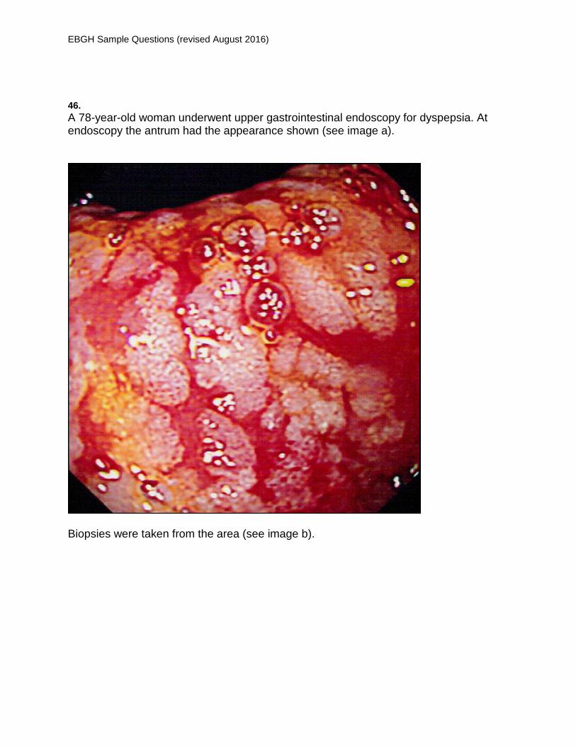
46.
A 78-year-old woman underwent upper gastrointestinal endoscopy for dyspepsia. At endoscopy the antrum had the appearance shown (see image a).
Biopsies were taken from the area (see image b).

EBGH Sample Questions (revised August 2016)
What is the most likely diagnosis? A diffuse type adenocarcinoma B gastric antral vascular ectasia C gastric lichen planus D Helicobacter pylori atrophic gastritis E intestinal metaplasia
Comments Rhe white plaque like lesions in the stomach are typical for intestinal metaplasia. On biopsy this is confirmed by the presence of goblet cells – suggestive of a small intestinal pattern of mucosal differentiation rather than the normal gastric mucosa. Intestinal metaplasia is seen in Helocobacter pylori infection and chronic bile reflux and carried an increased risk of gastric adenocarcinoma.

EBGH Sample Questions (revised August 2016)
47.
A 54-year-old woman was referred by her general practitioner with a history of epigastric discomfort and recurrent vomiting of recent onset. A direct-access upper gastrointestinal endoscopy was arranged.
Investigations:
upper gastrointestinal endoscopy 1-cm stalked polyp seen in the gastric antrum
histology tubular adenoma
What is the most appropriate next step in management? A CT scan of abdomen B endoscopic follow-up at 1 year C endoscopic ultrasound scan D no further action required E polypectomy
Comments Gastric polyps may be cystic fundal, associated with PPI therapy and FAP, hyperplastic (usually associated with chronic inflammation), hamartomatous and adenomatous. The adenomatous polyps are rare but carry a significant malignant potential and should be resected endoscopically. Goddard A, et al The management of gastric polyps Gut 2010;59:1270-1276