EARLY SENESCENCE1 Encodes a SCAR-LIKE PROTEIN2 · to identify ES1, which encodes a SCAR-LIKE...
15
EARLY SENESCENCE1 Encodes a SCAR-LIKE PROTEIN2 That Affects Water Loss in Rice 1[OPEN] Yuchun Rao 2 , Yaolong Yang 2 , Jie Xu 2 , Xiaojing Li, Yujia Leng, Liping Dai, Lichao Huang, Guosheng Shao, Deyong Ren, Jiang Hu, Longbiao Guo, Jianwei Pan, and Dali Zeng* State Key Laboratory of Rice Biology, China National Rice Research Institute, Hangzhou 310006, China (Y.R., Y.Y., J.X., Y.L., L.D., L.H., G.S., D.R., J.H., L.G., D.Z.); College of Chemistry and Life Sciences, Zhejiang Normal University, Jinhua 321004, China (Y.R., X.L., J.P.); and Key Laboratory of Crop Physiology, Ecology, and Genetic Breeding, Ministry of Education, Jiangxi Agricultural University, Nanchang 330045, China (Y.Y., J.X.) ORCID ID: 0000-0003-2349-8633 (D.Z.). The global problem of drought threatens agricultural production and constrains the development of sustainable agricultural practices. In plants, excessive water loss causes drought stress and induces early senescence. In this study, we isolated a rice (Oryza sativa) mutant, designated as early senescence1 (es1), which exhibits early leaf senescence. The es1-1 leaves undergo water loss at the seedling stage (as reflected by whitening of the leaf margin and wilting) and display early senescence at the three-leaf stage. We used map-based cloning to identify ES1, which encodes a SCAR-LIKE PROTEIN2, a component of the suppressor of cAMP receptor/Wiskott-Aldrich syndrome protein family verprolin-homologous complex involved in actin polymerization and function. The es1-1 mutants exhibited significantly higher stomatal density. This resulted in excessive water loss and accelerated water flow in es1-1, also enhancing the water absorption capacity of the roots and the water transport capacity of the stems as well as promoting the in vivo enrichment of metal ions cotransported with water. The expression of ES1 is higher in the leaves and leaf sheaths than in other tissues, consistent with its role in controlling water loss from leaves. GREEN FLUORESCENT PROTEIN-ES1 fusion proteins were ubiquitously distributed in the cytoplasm of plant cells. Collectively, our data suggest that ES1 is important for regulating water loss in rice. Rice (Oryza sativa) is a major worldwide food crop, but it consumes more water than most crops (Linquist et al., 2015), with water consumption for rice cultivation accounting for approximately 65% of agricultural water usage. Rice provides a staple food for about 3 billion people while using an estimated 24% to 30% of the world’s developed freshwater resources (Bouman et al., 2007). Severe water shortages restrict the expansion of rice production and hinder the irrigation of existing paddy fields (Zhu and Xiong, 2013). One effective way to overcome water shortages is to reduce water loss in rice plants, thus allowing the cultivation of this key crop in environments with less water (Nguyen et al., 1997). In plants, the stomata on the leaf surface work as the main channels for the discharge of water and the entry of carbon dioxide, thus strongly affecting physiological processes such as transpiration and photosynthesis. Previous studies showed that mutations in some genes could affect stomatal density or differentiation, such as the Arabidopsis (Arabidopsis thaliana) genes TOO MANY MOUTHS (AtTMM; Yang and Sack, 1995), SCREAM2 (SCRM2; Kanaoka et al., 2008), STOMATA DENSITY AND DISTRIBUTION1 (AtSDD1; Von Groll et al., 2002), and AtYODA2, a mitogen-activated protein kinase kinase kinase (Bergmann et al., 2004). Stomata show a regular distribution on rice leaves (Huang et al., 2009) during plant growth and development, and var- ious environmental factors affect the density and size of stomata as well as the chlorophyll contents of rice leaves. For example, rice leaves that develop under water stress show substantially fewer stomata com- pared with leaves that develop under well-watered conditions (Huang et al., 2009). Changes in stomatal density and morphology affect water loss (Boonrueng et al., 2013). In rice, SIMILAR TO RADICAL-INDUCED CELL DEATH1 enhances drought tolerance by regu- lating stomatal closure (You et al., 2013), while the zinc finger protein DROUGHT AND SALT TOLERANCE functions in drought and salt tolerance by adjusting 1 This work was supported by the National Natural Science Foun- dation of China (grant nos. 31201183, 31221004, 31171531, 31171520, and 91435105), the State Key Basic Research Program (grant no. 2013CBA01403), the Ministry of Agriculture of China for Transgenic Research (grant no. 2014ZX08009003–001), and the China Postdoc- toral Science Foundation (grant no. 2014M561108). 2 These authors contributed equally to the article. * Address correspondence to [email protected]. The author responsible for distribution of materials integral to the findings presented in this article in accordance with the policy de- scribed in the Instructions for Authors (www.plantphysiol.org) is: Dali Zeng ([email protected]). Y.R. conceived the original screening and research plans; D.Z., J.P., and L.G. supervised the experiments; Y.R., Y.Y., and J.X. performed most of the experiments; X.L., Y.L., L.D., L.H., G.S., D.R., and J.H. provided technical assistance to Y.R., Y.Y., and J.X.; Y.R. and D.Z. designed the experiments and analyzed the data; Y.R. conceived the project and wrote the article with contributions of all the authors; D.Z. supervised and complemented the writing. [OPEN] Articles can be viewed without a subscription. www.plantphysiol.org/cgi/doi/10.1104/pp.15.00991 Plant Physiology Ò , October 2015, Vol. 169, pp. 1225–1239, www.plantphysiol.org Ó 2015 American Society of Plant Biologists. All Rights Reserved. 1225 www.plantphysiol.org on June 30, 2018 - Published by Downloaded from Copyright © 2015 American Society of Plant Biologists. All rights reserved.
Transcript of EARLY SENESCENCE1 Encodes a SCAR-LIKE PROTEIN2 · to identify ES1, which encodes a SCAR-LIKE...

EARLY SENESCENCE1 Encodes a SCAR-LIKE PROTEIN2That Affects Water Loss in Rice1[OPEN]
Yuchun Rao2, Yaolong Yang2, Jie Xu2, Xiaojing Li, Yujia Leng, Liping Dai, Lichao Huang, Guosheng Shao,Deyong Ren, Jiang Hu, Longbiao Guo, Jianwei Pan, and Dali Zeng*
State Key Laboratory of Rice Biology, China National Rice Research Institute, Hangzhou 310006, China (Y.R., Y.Y.,J.X., Y.L., L.D., L.H., G.S., D.R., J.H., L.G., D.Z.); College of Chemistry and Life Sciences, Zhejiang NormalUniversity, Jinhua 321004, China (Y.R., X.L., J.P.); and Key Laboratory of Crop Physiology, Ecology, andGenetic Breeding, Ministry of Education, Jiangxi Agricultural University, Nanchang 330045, China (Y.Y., J.X.)
ORCID ID: 0000-0003-2349-8633 (D.Z.).
The global problem of drought threatens agricultural production and constrains the development of sustainable agricultural practices.In plants, excessive water loss causes drought stress and induces early senescence. In this study, we isolated a rice (Oryza sativa) mutant,designated as early senescence1 (es1), which exhibits early leaf senescence. The es1-1 leaves undergo water loss at the seedling stage (asreflected by whitening of the leaf margin and wilting) and display early senescence at the three-leaf stage. We used map-based cloningto identify ES1, which encodes a SCAR-LIKE PROTEIN2, a component of the suppressor of cAMP receptor/Wiskott-Aldrich syndromeprotein family verprolin-homologous complex involved in actin polymerization and function. The es1-1mutants exhibited significantlyhigher stomatal density. This resulted in excessive water loss and accelerated water flow in es1-1, also enhancing the water absorptioncapacity of the roots and the water transport capacity of the stems as well as promoting the in vivo enrichment of metal ionscotransported with water. The expression of ES1 is higher in the leaves and leaf sheaths than in other tissues, consistent with itsrole in controlling water loss from leaves. GREEN FLUORESCENT PROTEIN-ES1 fusion proteins were ubiquitously distributed in thecytoplasm of plant cells. Collectively, our data suggest that ES1 is important for regulating water loss in rice.
Rice (Oryza sativa) is a major worldwide food crop,but it consumes more water than most crops (Linquistet al., 2015), withwater consumption for rice cultivationaccounting for approximately 65% of agricultural waterusage. Rice provides a staple food for about 3 billionpeople while using an estimated 24% to 30% of theworld’s developed freshwater resources (Bouman et al.,2007). Severe water shortages restrict the expansion ofrice production and hinder the irrigation of existing
paddy fields (Zhu and Xiong, 2013). One effective wayto overcome water shortages is to reduce water loss inrice plants, thus allowing the cultivation of this key cropin environments with less water (Nguyen et al., 1997).
In plants, the stomata on the leaf surface work as themain channels for the discharge of water and the entryof carbon dioxide, thus strongly affecting physiologicalprocesses such as transpiration and photosynthesis.Previous studies showed that mutations in some genescould affect stomatal density or differentiation, suchas the Arabidopsis (Arabidopsis thaliana) genes TOOMANY MOUTHS (AtTMM; Yang and Sack, 1995),SCREAM2 (SCRM2; Kanaoka et al., 2008), STOMATADENSITY AND DISTRIBUTION1 (AtSDD1; Von Grollet al., 2002), andAtYODA2, a mitogen-activated proteinkinase kinase kinase (Bergmann et al., 2004). Stomatashow a regular distribution on rice leaves (Huang et al.,2009) during plant growth and development, and var-ious environmental factors affect the density and size ofstomata as well as the chlorophyll contents of riceleaves. For example, rice leaves that develop underwater stress show substantially fewer stomata com-pared with leaves that develop under well-wateredconditions (Huang et al., 2009). Changes in stomataldensity and morphology affect water loss (Boonruenget al., 2013). In rice, SIMILAR TORADICAL-INDUCEDCELL DEATH1 enhances drought tolerance by regu-lating stomatal closure (You et al., 2013), while the zincfinger protein DROUGHT AND SALT TOLERANCEfunctions in drought and salt tolerance by adjusting
1 This work was supported by the National Natural Science Foun-dation of China (grant nos. 31201183, 31221004, 31171531, 31171520,and 91435105), the State Key Basic Research Program (grant no.2013CBA01403), the Ministry of Agriculture of China for TransgenicResearch (grant no. 2014ZX08009003–001), and the China Postdoc-toral Science Foundation (grant no. 2014M561108).
2 These authors contributed equally to the article.* Address correspondence to [email protected] author responsible for distribution of materials integral to the
findings presented in this article in accordance with the policy de-scribed in the Instructions for Authors (www.plantphysiol.org) is:Dali Zeng ([email protected]).
Y.R. conceived the original screening and research plans; D.Z., J.P.,and L.G. supervised the experiments; Y.R., Y.Y., and J.X. performedmost of the experiments; X.L., Y.L., L.D., L.H., G.S., D.R., and J.H.provided technical assistance to Y.R., Y.Y., and J.X.; Y.R. and D.Z.designed the experiments and analyzed the data; Y.R. conceived theproject and wrote the article with contributions of all the authors;D.Z. supervised and complemented the writing.
[OPEN] Articles can be viewed without a subscription.www.plantphysiol.org/cgi/doi/10.1104/pp.15.00991
Plant Physiology�, October 2015, Vol. 169, pp. 1225–1239, www.plantphysiol.org � 2015 American Society of Plant Biologists. All Rights Reserved. 1225 www.plantphysiol.orgon June 30, 2018 - Published by Downloaded from
Copyright © 2015 American Society of Plant Biologists. All rights reserved.

stomatal aperture (Huang et al., 2009). In Arabidopsis,overexpression of the magnesium chelatase H subunitin guard cells confers drought tolerance by promotingstomatal closure (Tsuzuki et al., 2013). Thus, stomatalclosure and low stomatal density enhance droughttolerance by reducing water loss in plants.
The epicuticular wax layer in plants acts as the firstbarrier to environmental conditions by reducing waterloss due to transpiration and preventing plant dam-age due to excessively strong sunlight (Riederer andSchreiber, 2001). Leaf transpiration involves both sto-matal and cuticular transpiration. Stomatal conductancecontrols stomatal transpiration, but the physicochemicalproperties of the leaf surface mainly control cuticulartranspiration. For example, the composition, thickness,and microstructure of the cuticular wax affect waterpermeability and transport (Svenningsson, 1988; Xuet al., 1995; Buschhaus and Jetter, 2012). RiceDROUGHT-INDUCED WAX ACCUMULATION1, GLOSSY1, andWAX SYNTHESIS REGULATORY GENE1 affect droughttolerance by regulating the deposition or biosynthesisof cuticular wax (Islam et al., 2009; Wang et al., 2012;Zhou et al., 2013; Zhu and Xiong, 2013).
Besides, leaf trichomes can also affect water loss(Konrad et al., 2015) and leaf trichomes closely linkedwith the actin cytoskeleton. The involvement of the actincytoskeleton in controlling directional cell expansion intrichomes has received much attention (Zhang et al.,2005). Generally, genes that affect cytoplasmic organi-zation can be studied by screening leaf trichomemutants(Qiu et al., 2002). In Arabidopsis, a reproducible mor-phogenetic program directs the polarized developmentof trichome branches (Mathur et al., 1999; Szymanskiet al., 1999; Le et al., 2006). Some of these genes affect thecytoskeleton and also affect the morphology of normalplant cells, especially epidermal cells. For example, mu-tation of SPIKE1 in Arabidopsis causes epidermal cells toshow simple arrangements and morphologies (i.e. allcells dividing along a single axis; Qiu et al., 2002).
To study the molecular mechanisms underlyingwater loss in rice, we isolated and characterized theearly senescence1-1 (es1-1) rice mutant, which showedexcessive water loss and early senescence phenotypes.Map-based cloning data showed that ES1 encodes aSCAR-LIKE PROTEIN2, and its Arabidopsis homologaffects the polymerization of actin. The es1-1 mutantsshowed obvious changes in leaf trichomes, similar toArabidopsis. However, few studies have reported aconnection between the actin cytoskeleton and waterloss in Arabidopsis. Our results demonstrated a criticalrole of the actin cytoskeleton in regulating water lossin rice.
RESULTS
Identification and Characterization of EarlySenescence Mutants
To study the mechanisms of senescence in rice, wescreened a large pool of mutants generated in the japonica
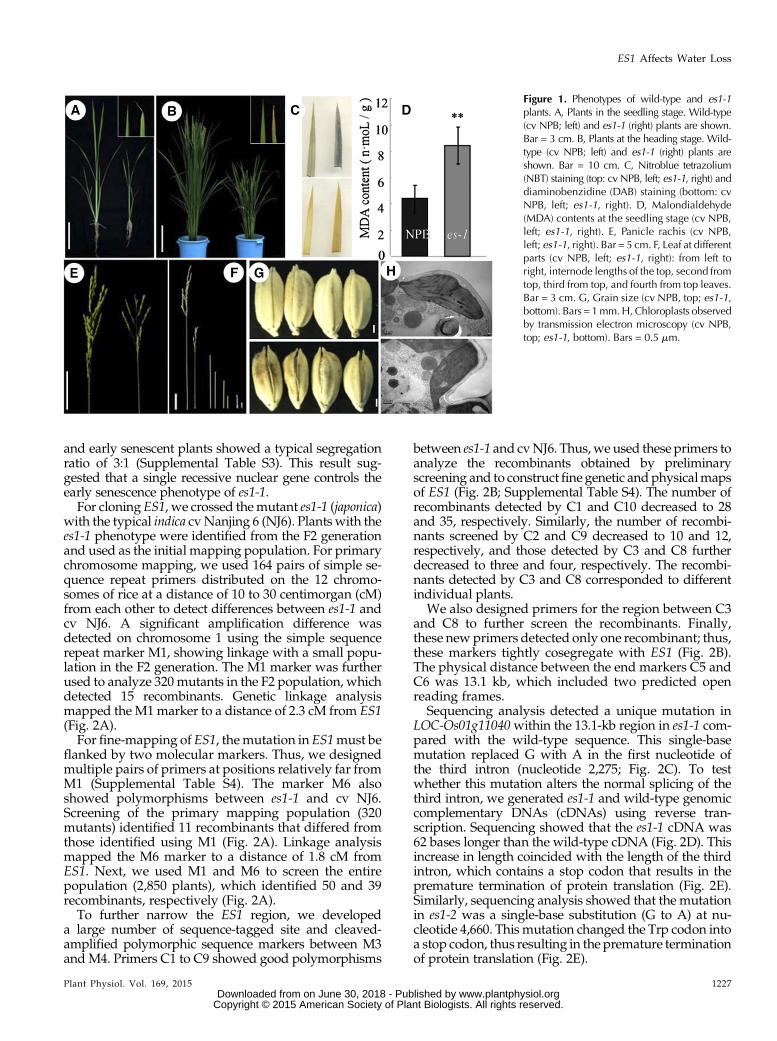
rice ‘Nipponbare’ (NPB) background by mutagenesisusing ethyl methanesulfonate. From this pool, we iden-tified two mutants with an early senescence phenotype.The two mutants showed similar phenotypes under thesame growth conditions, and F1 hybrid individualsproduced by the two parental mutants exhibited phe-notypes like the parental line with the weaker phenotype(subsequently named es1-1; see below), indicating thatthe two mutants are allelic (Supplemental Table S1);therefore, we termed these mutants es1-1 and es1-2. Un-der normal growth conditions, es1-1 plants showed se-verely retarded development. At the seedling stage, thees1-1 mutants displayed whitish and yellowish leaf tips(Fig. 1A), a hallmark of early senescence (Li et al., 2014);this phenotype increased with increasing leaf age, be-comingmore severe at the heading stage (Fig. 1B). Newlydeveloped leaves showed yellowing margins that rolledinward or formed a spiral, and old leaves showed spotswith a rusty color and water deficiency phenotypes, in-cluding wilting (Fig. 1, A and B). Histochemical analysisshowed that concentrations of senescence-related sub-stances, including hydrogen peroxide, superoxide radi-cal, and malondialdehyde, were higher in es1-1 leavesthan in wild-type leaves (Fig. 1, C and D), indicating thatthe mutant does show senescence phenotypes (Moradiand Ismail, 2007). Moreover, we measured the expres-sion of two senescence marker genes, STAYGREEN andOs185, in cv NPB and es1-1 (Supplemental Fig. S1, F andG).We found that the transcript levels of these two geneswere substantially higher in es1-1 than in cv NPB, sug-gesting that es1-1 caused senescence (Lee et al., 2001; Parket al., 2007). We also measured plant height, finding thates1-1 plants were much shorter than wild-type plantsfrom the seedling to the mature stages (Fig. 1, A and B;Supplemental Table S2). At the mature stage, es1-1 mu-tants showed degraded or white panicles with a lowseed-setting rate (only 2.4%; Fig. 1E; Supplemental TableS2), short internodes (Fig. 1F; Supplemental Table S2),and brown, open glumes (Fig. 1G). Scanning transmis-sion electron microscopy showed that es1-1mutants haddisordered thylakoids compared with the neat and well-ordered thylakoids observed in wild-type plants; more-over, es1-1 mutants had lower chlorophyll contents thanwild-type cv NPB.
Compared with es1-1, es1-2 plants had more tillers(Supplemental Fig. S1, A and C), were taller (SupplementalFig. S1, A and D), and showed a stronger early senes-cence phenotype in the leaves (Supplemental Fig. S1B).In this study, we mainly focused on es1-1 mutants,unless specified otherwise.
Map-Based Cloning of ES1
To clarify whether es1-1 phenotypes were caused by asingle gene or multiple genes and dominant or recessivegenes, we crossed the mutant plants directly and recip-rocally with three wild-type cultivars. All the F1 plantsshowed no symptoms of early senescence. In the F2segregating populations of these three crosses, normal
1226 Plant Physiol. Vol. 169, 2015
Rao et al.
www.plantphysiol.orgon June 30, 2018 - Published by Downloaded from Copyright © 2015 American Society of Plant Biologists. All rights reserved.
and early senescent plants showed a typical segregationratio of 3:1 (Supplemental Table S3). This result sug-gested that a single recessive nuclear gene controls theearly senescence phenotype of es1-1.For cloning ES1, we crossed themutant es1-1 (japonica)
with the typical indica cvNanjing 6 (NJ6). Plants with thees1-1 phenotype were identified from the F2 generationand used as the initial mapping population. For primarychromosome mapping, we used 164 pairs of simple se-quence repeat primers distributed on the 12 chromo-somes of rice at a distance of 10 to 30 centimorgan (cM)from each other to detect differences between es1-1 andcv NJ6. A significant amplification difference wasdetected on chromosome 1 using the simple sequencerepeat marker M1, showing linkage with a small popu-lation in the F2 generation. The M1 marker was furtherused to analyze 320mutants in the F2 population, whichdetected 15 recombinants. Genetic linkage analysismapped the M1marker to a distance of 2.3 cM from ES1(Fig. 2A).For fine-mapping of ES1, themutation in ES1must be
flanked by two molecular markers. Thus, we designedmultiple pairs of primers at positions relatively far fromM1 (Supplemental Table S4). The marker M6 alsoshowed polymorphisms between es1-1 and cv NJ6.Screening of the primary mapping population (320mutants) identified 11 recombinants that differed fromthose identified using M1 (Fig. 2A). Linkage analysismapped the M6 marker to a distance of 1.8 cM fromES1. Next, we used M1 and M6 to screen the entirepopulation (2,850 plants), which identified 50 and 39recombinants, respectively (Fig. 2A).To further narrow the ES1 region, we developed
a large number of sequence-tagged site and cleaved-amplified polymorphic sequence markers between M3andM4. Primers C1 to C9 showed good polymorphisms
between es1-1 and cvNJ6. Thus,we used these primers toanalyze the recombinants obtained by preliminaryscreening and to constructfinegenetic and physicalmapsof ES1 (Fig. 2B; Supplemental Table S4). The number ofrecombinants detected by C1 and C10 decreased to 28and 35, respectively. Similarly, the number of recombi-nants screened by C2 and C9 decreased to 10 and 12,respectively, and those detected by C3 and C8 furtherdecreased to three and four, respectively. The recombi-nants detected by C3 and C8 corresponded to differentindividual plants.
We also designed primers for the region between C3and C8 to further screen the recombinants. Finally,these newprimers detected only one recombinant; thus,these markers tightly cosegregate with ES1 (Fig. 2B).The physical distance between the end markers C5 andC6 was 13.1 kb, which included two predicted openreading frames.
Sequencing analysis detected a unique mutation inLOC-Os01g11040within the 13.1-kb region in es1-1 com-pared with the wild-type sequence. This single-basemutation replaced G with A in the first nucleotide ofthe third intron (nucleotide 2,275; Fig. 2C). To testwhether this mutation alters the normal splicing of thethird intron, we generated es1-1 and wild-type genomiccomplementary DNAs (cDNAs) using reverse tran-scription. Sequencing showed that the es1-1 cDNA was62 bases longer than the wild-type cDNA (Fig. 2D). Thisincrease in length coincided with the length of the thirdintron, which contains a stop codon that results in thepremature termination of protein translation (Fig. 2E).Similarly, sequencing analysis showed that the mutationin es1-2 was a single-base substitution (G to A) at nu-cleotide 4,660. This mutation changed the Trp codon intoa stop codon, thus resulting in the premature terminationof protein translation (Fig. 2E).
Figure 1. Phenotypes of wild-type and es1-1plants. A, Plants in the seedling stage. Wild-type(cv NPB; left) and es1-1 (right) plants are shown.Bar = 3 cm. B, Plants at the heading stage. Wild-type (cv NPB; left) and es1-1 (right) plants areshown. Bar = 10 cm. C, Nitroblue tetrazolium(NBT) staining (top: cv NPB, left; es1-1, right) anddiaminobenzidine (DAB) staining (bottom: cvNPB, left; es1-1, right). D, Malondialdehyde(MDA) contents at the seedling stage (cv NPB,left; es1-1, right). E, Panicle rachis (cv NPB,left; es1-1, right). Bar = 5 cm. F, Leaf at differentparts (cv NPB, left; es1-1, right): from left toright, internode lengths of the top, second fromtop, third from top, and fourth from top leaves.Bar = 3 cm. G, Grain size (cv NPB, top; es1-1,bottom). Bars = 1mm.H,Chloroplasts observedby transmission electron microscopy (cv NPB,top; es1-1, bottom). Bars = 0.5 mm.
Plant Physiol. Vol. 169, 2015 1227
ES1 Affects Water Loss
www.plantphysiol.orgon June 30, 2018 - Published by Downloaded from Copyright © 2015 American Society of Plant Biologists. All rights reserved.

We then used genetic complementation to verify theidentity of ES1. A 10.6-kb fragment containing ES1 (in-cluding the upstream 2-kb and downstream 1-kb se-quences)was introduced into es1-1 and es1-2mutants, and52 and 36 transgenic plants, respectively, were re-covered. Exogenous ES1 rescued the phenotypes ofes1-1 and es1-2 (Fig. 2F), and the complementationreversed the mutant phenotype of es1-1. Taken to-gether, these data demonstrate that LOC-Os01g11040(Os01g0208600) is ES1.
ES1 Encodes a SCAR-Like Protein of theACTIN-RELATED2/3-SCAR Pathway
We performed a BLAST search to identify ES1 ho-mologs in more than 20 different plant species. ES1showed high amino acid sequence similarity to proteinsin other species, such as Oryza brachyantha (85%), Bra-chypodium distachyon (68%), maize (Zea mays; 60%), andSetaria italica (64%; Fig. 3E). ES1 also shares high aminoacid sequence similarity to proteins in both monocotsand dicots, suggesting that ES1 and its homologs may
perform similar functions. Examination of ES1 homo-logs in Arabidopsis showed that ES1 (Os01g0208600)encodes a SCAR-LIKE PROTEIN2; its closest homologin Arabidopsis forms part of the ACTIN-RELATED2/3(ARP2/3) complex, which functions in regulatingactin filament polymerization and has been examinedin detail in Arabidopsis. ARP2/3 alone is inactive inArabidopsis. A domain formed by the SCAR-likefamily proteins, and members of other protein fami-lies (e.g. SRA1 and ABSCISIC ACD-INSENSITIVE[ABI] families), are responsible for binding to andactivation of the ARP2/3 complex (Zhang et al.,2005). In Arabidopsis, mutation of the gene encodingSCAR-LIKE PROTEIN2 leads to changes in leaf tri-chomes and stomata (Basu et al., 2005). In our study,we observed substantial changes in leaf trichomes ofes1-1, mainly reflected by the serious degradation ofthe trichome tip (Fig. 3, A and B). The leaves of es1-1had smooth leaf margins without a jagged pattern(Fig. 3A).
To examine the possible role of ES1 in regulatingactin filaments, we examined the organization of F-actinin the roots of the wild type and es1-1mutants by Alexa
Figure 2. Map-based cloning of ES1, and phenotype of an es1-1 complementation transgenic line. A, Location of es1-1 on ricechromosome 1. B, Coarse linkage map of es1-1. C, es1-1 gene structure. D, Reverse transcription of es1-1. E, Schematic diagramof es1-1 and mutated proteins encoded by es1-1 and es1-2. F, Phenotype of the complementation transgenic line: es1-1 (left), thewild type (cv NPB; middle), and the transgenic line (right). Bar = 10 cm.
1228 Plant Physiol. Vol. 169, 2015
Rao et al.
www.plantphysiol.orgon June 30, 2018 - Published by Downloaded from Copyright © 2015 American Society of Plant Biologists. All rights reserved.

Fluor 488-phalloidin staining. The actin filaments werewell organized in the wild type (Fig. 3C). The actin fil-aments in the wild type were complete and orderlyarranged, while es1-1 showed an irregular arrangement.Moreover, the actin filaments in es1-1 were shorterthan those in the wild type (Fig. 3D, arrows), whichindicated that ES1 likely functions in the ARP2/3-SCAR pathway in rice and that a mutation in ES1 af-fects the actin cytoskeleton in rice cells (Dyachok et al.,2011).
Expression of ES1 and Subcellular Localization of ES1
To monitor the tissue-specific expression of ES1, wegenerated an ES1:GUS reporter construct containing the
2-kb genomic region upstream of the start codon of ES1.Wedetected highGUS activity in leaves, leaf sheaths, androot tips and also detected lower GUS activity in stamensand vascular bundles (Fig. 4, A–E). Reverse transcription-quantitative PCR (RT-qPCR) assays showed that ES1was ubiquitously expressed in various tissues at the til-lering and heading stages, including roots, culms,leaves, sheaths, and spikelets. We observed the highestlevels of ES1 expression in the leaf and leaf sheath (Fig.4F). Consistent with the RT-qPCR data, ES1:GUS ex-pression was higher in rice leaves and leaf sheaths thanin other plant tissues.
To investigate the subcellular localization of ES1, weconstructed a construct to express enhanced green flu-orescent protein (eGFP)-ES1 (35S::eGFP-ES1 DNA) and
Figure 3. ES1 is a SCAR homolog. A, Leaf margins (left, cv NPB; right, es1-1). Arrows indicate the trichome tip. B, Trichomephenotype (macro hairs; top, cv NPB; bottom, es1-1). C and D, F-actin organization was visualized with Alexa Fluor 488-phalloidin in root cells of the wild type (C) and es1 (D). Over 40 roots were used in this analysis. Arrows indicate the continuity ofF-actin. Bars = 5 mm. E, Phylogenetic tree of SCAR-LIKE PROTEIN2 in several species.
Plant Physiol. Vol. 169, 2015 1229
ES1 Affects Water Loss
www.plantphysiol.orgon June 30, 2018 - Published by Downloaded from Copyright © 2015 American Society of Plant Biologists. All rights reserved.

introduced it into onion (Allium cepa) and tobacco(Nicotiana tabacum) epidermal cells. Fluorescencesignals were detected in both cell types, and thesignals surrounded the cytoplasm, nucleus, andcell membrane (Fig. 4, G–J). Consistent results were
obtained by examining protoplasts extracted fromtobacco leaves after the expression of the fusionprotein (Fig. 4, K and L). These results indicated thatES1 was ubiquitously distributed in the cytoplasm ofplant cells.
Figure 4. Expression of ES1 and subcellular localization of ES1. A to E,GUS staining of leaf (A), leaf sheath (B), fibrous root (C), stamen(D), and culm vascular bundles (E). F, Quantitative real-time PCR analysis of ES1 expression in different tissues at the tillering stage (T)and the heading stage (H). The results are given as means of three independent assays, and each error bar represents the percentage SD
of the mean. G to L, GFP fluorescence visualized by confocal microscopy. G and H, Transient expression of eGFP (G) and eGFP-ES1cDNA (H) in epidermal cells of onion. 35S::eGFP-ES1 cDNA and 35S::GFPwere transiently expressed in onion epidermal cells. I to L,Transient expression of eGFP in epidermal cells (I) and protoplasts (K) and expression of eGFP-ES1 in epidermal cells (J) and protoplasts(L) of Nicotiana benthamiana leaves. 35S::eGFP-ES1 and 35S::GFP were transiently expressed in N. benthamiana leaves.
1230 Plant Physiol. Vol. 169, 2015
Rao et al.
www.plantphysiol.orgon June 30, 2018 - Published by Downloaded from Copyright © 2015 American Society of Plant Biologists. All rights reserved.

ES1 Affects the Density of Leaf Stomata in Rice
Scanning electron microscopy of es1-1 leaves showedthat these leaves had smoother surfaces than the leavesof wild-type plants. In addition, these leaves showedsubstantially smaller siliceous protrusions at the seedlingstage and substantially more stomata per unit of leaf areacompared with wild-type leaves. The number of semi-open stomata in es1-1was also larger than that in thewildtype (Fig. 5,A andB).Moreover, the tips of trichomes and
the waxy layer were severely degraded on the leaf sur-face of es1-1 mutants (Fig. 5A). Statistical data showedthat, regardless of the position, stomatal density wassignificantly higher in es1-1 than in the wild type. Whileboth stomatal conductance and transpiration rate werealso detected, they were also significantly higher in es1-1;these data indirectly suggested that stomatal density ines1-1was higher (Fig. 5B). The maximum and minimumstomatal apertures were greater in the wild type com-pared with es1-1 (Fig. 5C). At the seedling stage, the
Figure 5. Observation of stomata between cvNPBand es1-1. A, Stomata density of thewild-type (left) and es1-1 (right) leaves. The redtriangles indicate the positions of stomata. Bars = 30mm.B, Statistical analysis of stomatal parameters, such as stomata density, partiallyopen stomata, stomatal conductance, and transpiration rate. C, Morphological characteristics (left) and maximum opening size (right)of stomata between wild-type (top) and es1-1 (bottom) plants. Bars = 5 mm. D, Comparison of water loss rate between wild-type andes1-1 plants with and without 300 mM ABA treatment at the three-leaf-stage. E, RT-qPCR analysis of stomata density-related genes(OsTMM, OsSCRM, OsSDD1, and OsYODA2), ES1, and some ABA response-related genes (OsDREB1A, OsDREB2A, OsMYB2,OsABI4,OsABI5, and a basic leucine zipper transcription factor,OsABF4) in wild-type and es1-1 leaves before and after 300mM ABAtreatment at the three-leaf-stage. The values were normalized to the ACTIN1 gene. Asterisks indicate significance (Student’s t test) atP , 0.01. Different letters indicate significant differences at the 1% level (Duncan’s multiple range test).
Plant Physiol. Vol. 169, 2015 1231
ES1 Affects Water Loss
www.plantphysiol.orgon June 30, 2018 - Published by Downloaded from Copyright © 2015 American Society of Plant Biologists. All rights reserved.

transcriptional levels of several genes that could affectthe increase of stomatal density were analyzed by real-time PCR, including OsTMM, OsSCRM, OsSDD1, andOsYODA2 (Du et al., 2014). The expression levels ofOsTMM, OsSCRM, and OsSDD1 were significantlyhigher in es1-1 than in the wild type, and the ES1 genealso showed a higher expression level in es1-1 (Fig. 5E).
Leaves of the wild type and es1-1 at the seedling stagewere sprayed with 300 mmol L21 abscisic acid (ABA),and after 3 h, quantitative analysis ofOsTMM,OsSCRM,OsSDD1, andOsYODA2was taken. In the wild type, theexpression level of these genes almost did not change,but in es1-1, the majority of these genes showed a sig-nificantly decreased expression level; this indicated ines1-1 that those genes that could affect the increase ofstomatal density were sensitive to ABA. We also detec-ted the rate of in vitro water loss (RWL) of seedlingleaves from the same position of the wild type and es1-1before and after ABA treatment. The results showedthat, nomatterwith orwithout ABA treatment, the RWLvalue was always higher in es1-1 than in the wild type.The RWL value in the wild type was reduced after ABAtreatment, and the value in es1-1 was also reduced afterABA treatment, but itwas not so obvious comparedwiththe wild type (Fig. 5D). Scanning electron microscopyassay on the leaves of the wild type and es1-1with ABAtreatment showed that nearly all stomata of both wild-type and es1-1 plants were closed. Moreover, we alsoexamined the expression levels of multiple ABAresponse-related genes (ABA signaling genes) in thewild type and es1-1 before and after ABA treatment(Shang et al., 2010) and discovered that, after ABAtreatment, OsDREB2A (a transcription factor) was up-regulated while OsABI4 was down-regulated. These re-sults suggest that both wild-type and es1-1 plants weresensitive to ABA (Fig. 5E).
The es1-1 Plants Showed Higher Water Loss Rates ThanWild-Type Plants
In vitro, the rate of water loss of es1-1was higher thanthat of cv NPB at the seedling stage (Fig. 6A), as thedetached leaves of es1-1 shrunk into a line at 30 min butthe cv NPB leaves remained nearly normal in width(Fig. 6A). To confirm this phenomenon, we also mea-sured water loss per unit of time (g cm22 1023) at thetillering stage in vivo and in vitro (Fig. 6, B and C),which gave similar results: the water loss per unit oftime in vitro (detached leaves) in different periods wasuniformly higher in es1-1 than in cv NPB (Fig. 6B). Thewater loss per unit of time in vivo was also uniformlyhigher in es1-1 than in cv NPB at 4, 8, and 14 d aftertillering (Fig. 6C).
To verify excessive water loss in es1-1, we performeda guttation experiment, where we looked for the exu-dation of drops of xylem sap on the leaf periphery inplants grown at different humidities. Hydroponicseedlings were cultured in the light, in an incubator atdifferent humidities, from 30% to 90%, and es1-1 andwild-type plants were examined for guttation. At the
seedling stage (two-leaf stage), both the wild-type andes1-1 plants were capable of guttation at 30% humidity.However, higher humidity was required for guttationwith plant growth. Until the strong seedling stage (two-tiller stage), guttation was observed in wild-type plantsbut not in es1-1 (Supplemental Fig. S2). This result in-dicated that water loss occurred more rapidly in es1-1mutants than in the wild-type plants.
Water Absorption and Transport Capacity Were Increasedin es1-1 Mutants
It is unknown whether excessive water loss throughleaves affects water absorption in the root. To addressthis, we measured root traits in es1-1 and wild-typeplants (Fig. 7, A–D). The es1-1 plants had smaller pri-mary and total root lengths (Fig. 7, E and F) but higheraverage root diameters, total root volumes, and totalnumbers of fibrous roots compared with wild-typeplants (Fig. 7, G–J). As fibrous roots are a major tissuefor water adsorption in rice (Smith and De Smet, 2012),an increase in fibrous root number indicated an increase
Figure 6. Observation of water loss rate between wild-type and es1-1plants. A,Water loss rate of detached leaves at the seedling stage (left, cvNPB; right, es1-1). B, Water loss rate of detached leaves at the tilleringstage. C,Water loss rate per unit of time of living leaves between cvNPBand es1-1 plants at the tillering stage. Error bars indicate SD (n = 15).Asterisks indicate the significance of differences between cv NPB andes1-1 plants, as determined by Student’s t test: *, 0.01 # P , 0.05 and**, P , 0.01.
1232 Plant Physiol. Vol. 169, 2015
Rao et al.
www.plantphysiol.orgon June 30, 2018 - Published by Downloaded from Copyright © 2015 American Society of Plant Biologists. All rights reserved.

in the water absorption capacity of the roots in es1-1mutants.Examination of cross sections of the culm and pri-
mary root from es1-1 and cv NPB showed that the es1-1stems had three to four additional vascular bundles(Fig. 8, A–C) and the es1-1 primary roots had two tothree additional conduits compared with cv NPB (Fig.8, D–F). Vascular bundles and conduits are necessaryfor water transport (Kim et al., 2014). Moreover, thewater potential values were all higher in es1-1 than inthe wild type in various tissues, including roots, stems,leaves, and sheath, which indicated that the ability totransport water was stronger in es1-1 than in the wild
type (Fig. 8G; Da Silva et al., 2011). Therefore, our re-sults indicate that es1-1 mutants have an enhancedability to transport water.
Excessive Water Loss Causes in Situ Enrichment ofMinerals in es1-1
In es1-1 mutants, excessive water loss through theleaves enhances the water absorption capacity of theroots and the water transportation capacity of the rootsand culms. This, in turn, accelerates water circulation inthe plant, thus leading to predicted in situ enrichmentof metal ions cotransported with water (Kim et al.,2014). To test this hypothesis in this mutant, we deter-mined the mineral concentrations of different tissues ofes1-1 and wild-type plants at the mature stage. Con-sistent with the hypothesis, the concentrations of allfour minerals tested (copper [Cu], iron [Fe], manganese[Mn], and zinc [Zn]) in the examined tissues were muchhigher in es1-1, especially in the roots and new leaves,than in wild-type plants (Table I). The Mn concentra-tion in es1-1 leaves (719 mg kg21) was seven timeshigher than that in wild-type plants (100 mg kg21). Theconcentrations of the four minerals were also higher ines1-1 seeds than in cv NPB (Table I). These resultssuggested that mutation in ES1 promoted the accu-mulation of minerals in plants. The substantial accu-mulation of metal ions in es1-1 plants was likely due toexcessive water loss and higher transpiration pull in theupper part of the plant.
DISCUSSION
In this study, we characterized es1-1, an early senes-cence mutant that experienced excessive water loss.Map-based cloning showed that ES1 encodes a SCAR-LIKEPROTEIN2, a component of the suppressor of cAMPreceptor/Wiskott-Aldrich syndrome protein familyverprolin-homologous (SCAR/WAVE) complex that isinvolved in actin polymerization. This suggests that amutation in ES1 affects the polymerization of actin andcauses an abnormality in the cytoskeleton that pro-motes higher stomatal density (Huang et al., 2000; Yuet al., 2001; Xiao et al., 2003; Zhang et al., 2012), thusleading to excessive water loss.
Under stress conditions, plants develop a wax layeron the leaf surface or moderately roll their leaves toreduce transpiration, thereby protecting themselvesfrom excessive water loss (Hu et al., 2010; Jäger et al.,2014). Although stomata account for only 1% to 2% ofthe leaf area, approximately 90% of water loss occursthrough stomata (Buckley, 2005). Under drought con-ditions, rice cultivars that can maintain relatively highwater potential and low transpiration rate in theirleaves have strong drought resistance (Luo and Zhang,2001). These drought-resistant cultivars commonly re-tain water in the plant body by closing their stomata.Small stomatal aperture and large diffusion resistancecan jointly reduce water loss due to transpiration, thus
Figure 7. Phenotype comparison of the roots between cv NPB andes1-1 plants. A, Root scanning of cv NPB plants. Bar = 5 cm. B, Rootscanning of es1-1 plants. Bar = 5 cm. C, Enlarged image of roots in cvNPBplants. Bar = 5 cm.D, Enlarged image of roots in es1-1 plants. Bar =5 cm. E to J, Statistical analysis of the main root length (E), total rootlength (F), total area of the root (G), average diameter of the root (H),total volume of the root (I), and total number of fibrous roots (J) of cvNPB and es1-1 plants. The results are given as means of three inde-pendent assays, and error bars indicate SD. Asterisks indicate the sig-nificance of differences between cv NPB and es1-1 plants, asdetermined by Student’s t test: *, 0.01 # P , 0.05 and **, P , 0.01.
Plant Physiol. Vol. 169, 2015 1233
ES1 Affects Water Loss
www.plantphysiol.orgon June 30, 2018 - Published by Downloaded from Copyright © 2015 American Society of Plant Biologists. All rights reserved.

avoiding the adverse effects of drought. Grill and Ziegler(1998) proposed that controlling the stomata is the mostimportant way to improve the efficiency of water usein plants. In this study, scanning electron microscopyshowed that the es1-1 mutants had substantially morestomata than wild-type plants. The expression levelsof OsTMM, OsSCRM, and OsSDD1 were significantly
higher in es1-1 than in the wild type (Fig. 5E), which wasclosely related to stomatal density. Stomatal density wassignificantly higher in es1-1 than in the wild type. Whileboth stomatal conductance and transpiration were alsosignificantly higher in es1-1 than in thewild type (Fig. 5B),these data suggested that es1-1 definitely loses watermore quickly than the wild type. Nearly all stomata of
Figure 8. Number of vascular bundles and con-duits in wild-type and es1-1 plants. A, Number ofvascular bundles in cv NPB plants. B, Numberof vascular bundles in es1-1 plants. C, Comparisonof the number of vascular bundles between cvNPB and es1-1 plants. D, Number of conduits in cvNPB plants. E, Number of conduits in es1-1 plants.F, Comparison of the number of conduits betweencvNPB and es1-1 plants. G, Detection of thewaterpotential value in the wild type and es1-1. Therelative water potential value in different tissues atthe seedling stage of the wild type and es1-1 wasanalyzedwith theWP4-TDewpoint PotentiaMeter(Decagon Devices, Inc.). The results are given asmeans of three independent assays, and error barsindicate SD. Asterisks indicate significance at thelevel of 5% (*) and 1% (**).
Table I. Comparison of mineral contents between wild-type and es1-1 plants
The results are given as means 6 SD of three independent assays. Asterisks indicate significance at the level of 1%.
Tissue Plant Cu Fe Mn Zn
mg kg21
Root cv NPB 23.5 6 3.83 3,757.7 6 122.55 17.75 6 1.75 28.49 6 1.12es1-1 446.3 6 18.52** 18,155.3 6 326.27** 58.97 6 3.88** 91.26 6 5.88**
Culm cv NPB 8.61 6 1.21 35.52 6 3.7 32.19 6 1.3 28.75 6 1.81es1-1 20.18 6 4.7** 46.05 6 3.54** 147.99 6 10.06** 85.83 6 6.35**
Sheath cv NPB 6.59 6 1.15 136.2 6 8.59 92.331 6 5.9 42.52 6 3.4es1-1 16.88 6 2.43** 240.2 6 14.65** 582.2 6 13.22** 65.36 6 7.92**
Old leaf cv NPB 9.32 6 2.1 228.8 6 15.1 100.01 6 7.1 29.89 6 2.56es1-1 18.27 6 3.88** 358.3 6 21.94** 719.05 6 34.8** 64.04 6 7.5**
Young leaf cv NPB 8.02 6 1.16 102.1 6 6.78 27.51 6 3.32 30.48 6 3.44es1-1 17.07 6 2.91** 223.1 6 11.4** 175.81 6 12.24** 62.22 6 4.28**
Seed cv NPB 4.26 6 0.64 4.05 6 0.35 31.62 6 2.86 24.23 6 2.6es1-1 5.91 6 0.72** 6.98 6 1.06** 50.36 6 4.1** 53.13 6 8.12**
1234 Plant Physiol. Vol. 169, 2015
Rao et al.
www.plantphysiol.orgon June 30, 2018 - Published by Downloaded from Copyright © 2015 American Society of Plant Biologists. All rights reserved.

both wild-type and es1-1 plants were closed after ABAtreatment, and the RWL value in the wild type was re-duced after ABA treatment; the value in es1-1 was alsoreduced after ABA treatment, but it was not so obviouscompared with the wild type (Fig. 5D). Moreover, thewax layer on the leaf surface of es1-1 mutants was con-siderably degraded. Therefore, we thought the excessivewater loss in es1-1 mainly due to increased stomataldensity; meanwhile, the degradation of the waxy layerand leaf hairs may aggravate the rate of water loss.Excessive water loss in the leaves inevitably depletes
water from the body of es1-1 plants. Thus, more waterneeds to be absorbed through the roots to meet themetabolic requirements of es1-1 plants. In this study,weanalyzed the roots of es1-1 and found that the totalvolume of the root and the number of fibrous rootsincreased compared with the wild type (Fig. 7, I and J),indicating that the water absorption capacity of theroots was enhanced in es1-1. Cross sections of the pri-mary root and stems also showed that the number ofvascular bundles in the stems increased by four (Fig.8C) and those in the roots increased by two to three ines1-1 compared with wild-type plants (Fig. 8F). More-over, the water potential values were all higher in es1-1than in the wild type in various tissues (Fig. 8G). Theseobservations confirmed that the water transport ca-pacity of the roots was enhanced in es1-1.All the above changes inevitably accelerate water
circulation and cause minerals commonly cotransportedwith water to accumulate in the various tissues of es1-1plants. Indeed, in different tissues, the concentrations offour minerals were higher in es1-1 than in cv NPB (TableI). For example, the Cu concentration in es1-1 (446.3 mgkg21) was 18 times higher than in wild-type plants(23.5 mg kg21), which could cause Cu overload in es1-1plants. Enrichment of low-mobility metal elements (e.g.Cu, Fe, Mn, and Zn) in various tissues of es1-1 may, inturn, accelerate the onset of senescence. Moreover, thehigher concentrations of mineral nutrients in seeds fromthe es1-1 mutants might produce more nutritious food;however, the accumulation of toxic elements such asarsenic might produce food that could adversely affecthuman health.In our study, we found that actin filaments were
shorter in es1-1, and the continuity was poor comparedwith the wild type (Fig. 3, C and D). Actin filamentsfunction in many endomembrane processes, such asendocytosis of plasma membrane, vacuole formation,plasma membrane protrusion in crawling cells, andvesicle transport from the Golgi complex (Svitkina andBorisy, 1999; Eitzen et al., 2002; Kaksonen et al., 2003;Mathur et al., 2003; Chen et al., 2004). The ARP2/3complex functions as a key regulator of actin filamentnucleation in plants (Zhang et al., 2008). ARP2/3 isinactive by itself, as actin polymerization requires ac-tivation by the SCAR/WAVE complex, formed byproteins of the SCAR family. The SCAR/WAVE com-plex is highly conserved in plants. A deficiency of theSCAR/WAVE complex often leads to morphologicalchanges in leaf trichomes and epidermal cells. This
phenomenon has been reported in several studies inArabidopsis (Li et al., 2003; Mathur et al., 2003; Saedleret al., 2004) but not in rice. In our study, es1-1 showedevident morphological changes in leaf trichomes (Figs.3, A and B, and 5C), confirming that the function ofthe SCAR/WAVE complex is conserved in differentspecies. Additional components of the SCAR/WAVEcomplex can be identified by screening for mutantsthat affect the leaf trichomes of the es1-1 mutant. Acompanion paper reveals that TUTOU1 (TUT1), iden-tical to ES1, also plays a conserved role in trichomedevelopment (Bai et al., 2015).
Actin polymerization occurs in the cytoplasm. In thisstudy, the fluorescent signal of eGFP-ES1 was detectedin the cytoplasm and plasma membrane of tobacco andonion epidermal cells, a localization consistent with thepredicted function of ES1 in affecting the actin cyto-skeleton. After fusing the ES1 promoter with the GUSreporter gene, GUS staining was observed in varioustissues, including the roots, root hairs, stems, leaves,leaf sheath, and vascular bundles, which are associatedwith water absorption, transportation, and loss. TheES1 expression level was relatively high in the leavesand leaf sheaths, the two major tissues involved inwater loss. All these results are consistent with theproposed function of ES1.
The increased number of stomata would increasewater loss and induce drought stress in es1-1 plants.Drought stress affects photosynthetic capacity, respi-ration, membrane permeability, osmotic adjustment,enzyme activity, and hormone levels in rice leaves, thusreducing leaf area, leaf area index, and dry weight.Drought stress also prevents the accumulation of drymatter and accelerates senescence (Hu et al., 2010; Dinget al., 2014; Sellammal et al., 2014). In this study, es1-1plants showed early senescence characterized by yel-lowish leaf color and wilting of leaf margins at theseedling stage. Transmission electron microscopyshowed that the volume percentage of chloroplasts inthe es1-1 cells was lower than that in wild-type cells;however, the number of chloroplasts (irregular and fineprolate) was higher in es1-1 cells than in wild-typecells (Fig. 1H). This might have caused the decline inchlorophyll content in es1-1, causing the leaves to turnyellowish. In es1-1, the chloroplasts contained disor-ganized thylakoids (Fig. 1H) as opposed to the well-organized thylakoids in wild-type plants. The disorderedthylakoids may have reduced the capacity to absorblight energy, which, in turn, would decrease the pho-tosynthetic rate, thus causing early senescence in plantleaves.
Early senescence in rice (especially in functionalleaves) is extremely unfavorable for rice growth andproduction because of its serious effects on yield andquality (Zhu et al., 2012). Therefore, rice breeders gen-erally avoid lines showing early senescence. Similarly,the stay-green trait in rice also does not improve yield;although the stay-green trait promotes the ability ofsource tissues to fix carbon, it does not necessarilyenhance the contents of sink tissues in plants. For
Plant Physiol. Vol. 169, 2015 1235
ES1 Affects Water Loss
www.plantphysiol.orgon June 30, 2018 - Published by Downloaded from Copyright © 2015 American Society of Plant Biologists. All rights reserved.

example, a study on a stay-green mutant of rice (sgr[t])reported that late photosynthesis and yield traits (seed-setting rate and thousand-grain weight) were not en-hanced in sgr[t]mutants (Cha et al., 2002). In this study,the early-senescing mutant es1-1 showed other pheno-types, such as decreased number of tillers and substan-tially reduced seed-setting rate. Our data provide newinsights into themolecularmechanismsunderlying earlysenescence in plants, which will help genetically im-prove rice yield.
MATERIALS AND METHODS
Plant Growth and Sampling
The rice (Oryza sativa) es1-1 and es1-2mutantswere isolated from the japonicacv NPB that was subjected to ethyl methanesulfonate treatment. After multiplegenerations of inbreeding, the phenotypes of these two mutants were stablyinherited, and homozygous mutant strains were obtained. The homozygousmutant es1-1 was directly and reciprocally crossed with two typical japonicacultivars (cv Zhonghua 11 and cv Chunjiang 06) and one typical indica ricecultivar (cv NJ6), providing a relatively clear genetic background to constructthe genetic population. The obtained seeds of the F1 generation were sown andtransplanted as individual plants to generate the F2 generation by inbreeding.The numbers of individuals showing wild-type and early senescence pheno-types were counted at the seedling stage. The segregation ratio was calculatedusing statistical methods. Genetic analysis was performed on es1-1, and reces-sive individuals were retrieved for preliminary mapping of the mutation. Plantmaterials were bred in an experimental field of the China National Rice Re-search Institute, in Hangzhou.
Detection of Reactive Oxygen Speciesand Malondialdehyde
Hydrogen peroxide and superoxide radical were detected using NBT andDAB, respectively, according to Li et al. (2010) and Wi et al. (2010), with somemodifications. Leaves of 3-week-old plants that were grown in growth cham-bers were incubated in 0.05% (w/v) NBT (Duchefa) or 0.1% (w/v) DAB (Sigma)at room temperature in darkness with gentle shaking for 12 h Chlorophyll wascleared by treating with 90% (v/v) ethanol at 80°C. The concentration ofmalondialdehyde was measured according to the method described byMoradiand Ismail (2007) with minor modifications.
Determination of Water Potential
Ten seeds of cv NPB and es1-1 were selected, germinated for 3 d, and thencultured in an incubator at 28°C for 60 d. The WP4-T Dewpoint PotentiaMeterwas preheated for 30min until the temperature remained stable. Various tissuessuch as leaf, sheath, culm, and root of cvNPB and es1-1were harvested from thesame position on each plant and cut into short pieces. Aftermaking sure that thesample temperature was lower than the room temperature, the samples wereput in the measuring apparatus, which showed the water potential value of thesample. Every sample measurement had six independent repeats, and theweight of the samples was equal.
Map-Based Cloning of ES1
The mutation in es1-1 was mapped in the F2 population of a cross betweenes1-1 and the indica cv NJ6. To finely map the ES1 locus, newmolecular markerswere developed using public databases (Supplemental Table S4). Furthermore,ES1 was sequenced using specific primers (Supplemental Table S4).
Fluorescence Microscopy of Actin Filaments
For F-actin observation, plants were grown in a culture chamber with a 16/8-h photoperiod at 30°C. Seedling rootswere used to observe actin organization.F-actin was stained using the previously described method (Olyslaegers and
Verbelen, 1998; Yang et al., 2011; Zhang et al., 2011). Seven-day-old roots werefixed in PEM buffer (100 mM PIPES, 10 mM EGTA, 0.3 M mannitol, and 5 mM
MgSO4, pH 6.9) with 3% paraformaldehyde for 30 min and washed three timeswith PEM. After washing, roots were incubated in PEM buffer with 0.66 mM
Alexa Fluor 488-phalloidin (Life Technologies). After overnight incubation at4°C, samples were washed three times with PEM buffer and then observed in50% glycerol with a confocal microscope (Olympus Fluoview FV1000).
Phylogenetic Analyses
A BLAST search of the SCAR-LIKE PROTEIN2 amino acid sequence wasperformed against the database (http://www.ebi.ac.uk/Tools/sss/ncbiblast/).The phylogenetic tree was constructed based on the amino acid sequences. Max-imum likelihood phylogenetic analysis was performed by MEGA6.0 (Tamuraet al., 2013). Maximum likelihood analysis of the SCAR-LIKE PROTEIN2 familywas based on the amino acid sequences using the Jones-Taylor-Thornton model,which was chosen after model testing. Support for the maximum likelihood treeswas evaluated by 1,000 bootstrap replicates.
Plasmid Construction and Plant Transformation
The cDNA sequence of ES1 was amplified using the primers cES1-F andcES1-R (Supplemental Table S3). This cDNA was fused in frame with eGFP inpCambia1300 (http://www.cambia.org) with minor modifications to generatethe transient expression vector 35S::eGFP-cES1. The 35S::eGFP-cES1 plasmidswere transferred into Agrobacterium tumefaciens strain EHA105 by electropor-ation; the cells of rice ES1, tobacco (Nicotiana tabacum), and onion (Allium cepa)were transformed according to a previously described method (Hiei et al., 1994;Lv, 2010).
For the molecular complementation assay, a 10.6-kb genomic fragmentcomprising the ES1 promoter region, coding sequence, and 39 untranslatedregion was PCR amplified from the genomic DNA of wild-type plants usingpES-F and pES-R primers (Supplemental Table S3). The PCR amplificationproduct was cloned into pCambia1300 to generate the ES1:ES1 construct.Briefly, the pCambia1300::ES1 vector was first digested with the restriction en-zymes PstI and XbaI. Next, the fragment containing ES1 was digested withrestriction enzymes Sse83871 and NheI. All the restriction enzymes used aboveare isocaudomers. The pCambia1300::ES1 plasmids were transformed intoA. tumefaciens strain EHA105 by electroporation. The es1-1 and es1-2 plantsweretransformed according to a published method (Hiei et al., 1994).
To generate the ES1:GUS fusion construct, a 2-kb upstream genomic regionof the ES1 start codonwas amplifiedwith the primers ES1-gus-F and ES1-gus-R(Supplemental Table S3). The amplified fragment was linked with the reporterGUS gene and together inserted in the pCambia1305 vector (http://www.cambia.org).
GUS Staining
Samples (proES1::GUS) were stained with a solution containing 50 mM
NaPO4 buffer, 1 mM 5-bromo-4-chloro-3-indolyl-b-D-GlcA, 0.4 mM K3Fe(CN)6,0.4 mM K4Fe(CN)6, and 0.1% (v/v) Triton X-100. The samples were then incu-bated at 37°C in the dark for 10 h. Chlorophyll was removed with 70% ethanolfor observation.
RNA Extraction and RT-qPCR
Total RNAwas extracted from the roots, culms, leaves, sheaths, and spikeletsof wild-type and es1-1 plants at the seedling, tillering, and heading stages usingthe RNeasy Plant Mini Kit (Qiagen). Reverse transcription was handled withthe ReverTra Ace qPCR-RT Kit (Toyobo); the primers are described inSupplemental Table S4. RT-qPCR analysis was conducted with the StepOne-Plus System (Applied Biosystems) using Thunderbird qPCR Mix (Toyobo); thereaction procedurewas 95°C for 30 s, 95°C for 5 s, 55°C for 10 s, and 72°C for 15 sfor 40 cycles. OsACTIN was used as a control. The 22DDCt method was used forquantification (Livak and Schmittgen, 2001).
Microscopy
In different periods, especially at noonwhen the sunlightwas strongest (and,thus, the stomata are likely tobe closed), fresh leaf specimenswere collected fromthe sameposition (one-fourth, one-half, and three-fourths of the leaf blade) in the
1236 Plant Physiol. Vol. 169, 2015
Rao et al.
www.plantphysiol.orgon June 30, 2018 - Published by Downloaded from Copyright © 2015 American Society of Plant Biologists. All rights reserved.

mutant es1-1 and the control cv NPB from plants that showed consistentgrowth. The specimens were cut into approximately 1- 3 2-mm pieces andprocessed as follows for microscopic examination.
For dual fixation, the specimens were fixed in 2.5% glutaraldehyde solution,vacuumed in a vacuum apparatus for approximately 20 to 30 min (whichallowed the specimens to be immersed in the fixative as completely as possible),and kept at 4°C overnight. Thereafter, the fixative was discarded, and thespecimens were rinsed with 0.1 M phosphate-buffered saline, pH 7. The speci-mens were rinsed three to four times (15 min each). The specimens were thenfixed in 1% osmium tetroxide for at least 1 h. The fixativewas discarded, and thespecimenswere rinsedwith 0.1 M phosphate-buffered saline three times (15mineach) as described above.
For dehydration, the specimens were first dehydrated with a gradient ofethanol (50%, 70%, 80%, 90%, and 95%; 15–20min at each gradient), processed inanhydrous ethanol for 20 min, and treated in pure acetone for 20 min.
After dehydration, the specimenswere immersed in amixture of ethanol andisoamyl acetate esters (volume ratio, 1:1) for 30 min and then treated with pureisoamyl acetate for 1 to 2 h. The specimens were dried, coated, and examinedusing the Hitachi TM-1000 scanning electron microscope.
For transmission electron microscopy of the leaves and cross-sectional exami-nation of the leaves and roots, the specimens were pretreated as described above.After dehydration, the specimens were treatedwith amixture of embedding agentand pure acetone at a volume ratio of 1:1 for 1 h, followed by treatment with amixture of embedding agent and pure acetone at a volume ratio of 3:1 for 3 h andpure embedding agent overnight. The specimens were then embedded in smallboxes and heated at 70°C for approximately 9 h. The obtained embedded sampleswere sliced into 70- to 90-nm sections using a Reichert ultramicrotome and stainedwith lead citrate solution and saturated uranyl acetate solution in 50% ethanol for15 min each. The sections were examined and photographed using the HitachiH-7650 transmission electron microscope.
Determination of Minerals
Wild-type and es1-1 plants were cultured in complete hydroponic nutrientsolution up to the heading stage. Tissue specimens were taken from the roots,stems, old leaves, leaf sheaths, and new leaves. The weight of fresh specimenswas more than 10 g for the different tissues. The specimens were oven dried at80°C for 3 d to achieve a constantweight. After grinding, the dry samples (0.25 gfor both es1-1 and cv NPB) were digested with a mixture of concentrated nitricacid and perchloric acid (volume ratio, 1:3). The volume of the digestion solu-tion was adjusted to 25 mL with single-distilled water. The treated sampleswere then used for elemental analysis. Concentrations of elements were de-termined using full-spectrum direct reading inductively coupled plasma-atomic emission spectroscopy (IRIS Intrepid II XSP ICP-OES PE-2100; ThermoElectron; Shao et al., 2008).
Detection of the Rate of Water Loss
In Vitro Detection
The same leaf parts were selected from cv NPB and es1-1 plants at theseedling stage. These leaf parts were left at room temperature and observed andphotographed every 10min to examine their dehydration. At the tillering stage,the same leaf parts of cv NPB and es1-1 were quickly weighed (W1), and thetotal area (S) was measured. Moreover, the weights of the leaf blade (W2, W3,andW4)were alsomeasured every 1 h. The formula (W12W2) S21 was used tocalculate the rate of water loss. Accordingly, the rate of water loss at differenttime points was obtained.
In Vivo Detection
After accelerating germination, wild-type and es1-1 seeds were cultured infine sand and watered twice per day (in the morning and evening). At the one-to two-leaf stage, rice seedlings were washed off andwrapped in a long sponge,then placed in a closed bottle with clean water. At the three-leaf stage, cleanwater was replaced by complete nutrient solution. After the seedlings devel-oped new roots, all the bottles were filled with a sufficient volume of nutrientsolution, and the level was marked for subsequent measurement. The leaveswere weighed on days 1 (W1), 4 (W2), 8 (W3), and 14 (W4). The correspondingleaf areas were also measured (S2, S3, and S4). Water loss per unit of time in4 d was calculated as (W1 2 W2) S221. Six plants were used for every timeinterval measurement (the results are presented as mean values). Analogously,
(W12W3) S321 was the amount of water loss per unit of time in 8 d, and (W12W4) S421 was the amount of water loss per unit of time in 14 d.
Measurement of Root Traits
Hydroponic experiments were conducted using wild-type and es1-1 plants.The seeds were soaked for 2 d, during which time the water was changed once.Next, the seeds were transferred into a germination box to accelerate germi-nation. Three days later, seeds with radicles breaking through the testa weretransferred into fine sand for cultivation. After incubation for 7 d, the seedlingswere washed carefully to avoid damage to the roots. The plants were transferredinto a black bucket containing tap water. After the transition, the plants weresupplied with complete nutrient solution. The seedlings entered the two-leaf stageafter 3 d. At the five-leaf stage, seedlings of the mutant and wild-type plants(10 each) were taken out for root measurements. The hydroponic seedlings wererinsed andplaced in a transparent rectangular box containing single-distilledwaterand loaded onto a scanner (ScanWizard Pro: ScanMarker 1000XL) for transparentscanning. The obtained imageswere loaded intoWin-RHIZOPro.V. 2002c for dataanalysis, and the results were exported to Excel 2011 (Microsoft) for analysis.
Sequence data from this article are available at the National Center for Bio-technology Information Web site (http:www.ncbi.nlm.nih.gov) with the follow-ing accession numbers: LOC_Os01g11040 (NP_001172227.1), OB01G1689(XP_006643892.1), Bradi2g06610 (XP_003565325.1), rice indica (OsI_00845), S. italica(Si000056m.g), Hordeum vulgare var distichum (M0XEC2), Aegilops tauschii(F775_07293), Triticum aestivum (W5CZS7), maize (ZEAMMB73_679274), Triticumurartu (TRIUR3_31093), Musa acuminata ssp. malaccensis (M0UB73), Medicagotruncatula (MTR_8g086300), Theobroma cacao (TCM_029698), Phaseolus vulgaris(PHAVU_002G238500g), Jatropha curcas (JCGZ_21566), Glycine max (I1K3Z5),Populus trichocarpa (POPTR_0004s05600g), Prunus persica (PRUPE_ppa000443mg),Citrus sinensis (CISIN_1g001053mg), Citrus clementina (CICLE_v10014081mg),Eucalyptus grandis (EUGRSUZ_D00489), Solanum lycopersicum (Solyc02g076840.2),Erythranthe guttata (MIMGU_mgv1a000504mg), Vitis vinifera (VIT_10s0003g01270),Ricinus communis (RCOM_0850090), Amborella trichopoda (AMTR_s00021p00061660),Morus notabilis (L484_002198), andArabidopsis lyrata ssp. lyrata (ARALYDRAFT_903078).
Supplemental Data
The following supplemental materials are available.
Supplemental Figure S1. Phenotypes of allelic mutants.
Supplemental Figure S2. Comparison of guttation between wild-type andes1-1 plants.
Supplemental Table S1. Allelic analysis of es1-1 and es1-2.
Supplemental Table S2. Phenotypic data of cv NPB and es1-1.
Supplemental Table S3. Reciprocal cross experiments between es1-1 andthree crop strains (cv Zhonghua 11, cv Chunjiang 06, and cv NJ6).
Supplemental Table S4. Primers used for PCR in this study.
ACKNOWLEDGMENTS
We thank Jianru Zuo (Institute of Genetics and Developmental Biology,Chinese Academy of Sciences) for sharing unpublished data and LizhongXiong(National Key Laboratory of Crop Genetic Improvement, Huazhong Agricul-tural University) for critical reading and suggestions for our manuscript.
Received June 30, 2015; accepted August 1, 2015; published August 4, 2015.
LITERATURE CITED
Bai J, Zhu X, Wang Q, Zhang J, Chen H, Dong G, Zhu L, Zheng H, Xie Q,Nian J, et al (2015) Rice TUTOU1 Encodes a Suppressor of cAMPReceptor-Like Protein That Is Important for Actin Organization andPanicle Development. Plant Physiol 169: 1179–1191
Basu D, Le J, El-Essal Sel-D, Huang S, Zhang C, Mallery EL, Koliantz G,Staiger CJ, Szymanski DB (2005) DISTORTED3/SCAR2 is a putative
Plant Physiol. Vol. 169, 2015 1237
ES1 Affects Water Loss
www.plantphysiol.orgon June 30, 2018 - Published by Downloaded from Copyright © 2015 American Society of Plant Biologists. All rights reserved.

Arabidopsis WAVE complex subunit that activates the Arp2/3 complexand is required for epidermal morphogenesis. Plant Cell 17: 502–524
Bergmann DC, Lukowitz W, Somerville CR (2004) Stomatal developmentand pattern controlled by a MAPKK kinase. Science 304: 1494–1497
Boonrueng N, Anuntalabhochai S, Jampeetong A (2013) Morphologicaland anatomical assessment of KDML 105 (Oryza sativa L. spp. indica)and its mutants induced by low-energy ion beam. Rice Sci 20: 213–219
Bouman B, Humphreys E, Tuong T, Barker R (2007) Rice and water. AdvAgron 92: 187–237
Buckley TN (2005) The control of stomata by water balance. New Phytol168: 275–292
Buschhaus C, Jetter R (2012) Composition and physiological function ofthe wax layers coating Arabidopsis leaves: b-amyrin negatively affectsthe intracuticular water barrier. Plant Physiol 160: 1120–1129
Cha KW, Lee YJ, Koh HJ, Lee BM, Nam YW, Paek NC (2002) Isolation,characterization, and mapping of the stay green mutant in rice. TheorAppl Genet 104: 526–532
Chen JL, Lacomis L, Erdjument-Bromage H, Tempst P, Stamnes M (2004)Cytosol-derived proteins are sufficient for Arp2/3 recruitment andARF/coatomer-dependent actin polymerization on Golgi membranes.FEBS Lett 566: 281–286
Da Silva D, Favreau R, Auzmendi I, DeJong TM (2011) Linking waterstress effects on carbon partitioning by introducing a xylem circuit intoL-PEACH. Ann Bot (Lond) 108: 1135–1145
Ding L, Li Y, Li Y, Shen Q, Guo S (2014) Effects of drought stress onphotosynthesis and water status of rice leaves. Chin J Rice Sci 28: 65–70(in Chinese with English abstract)
Du H, Chang Y, Huang F, Xiong L (January 30, 2015) GID1 modulatesstomatal response and submergence tolerance involving abscisic acidand gibberellic acid signaling in rice. J Integr Plant Biol http://dx.doi.org/10.1111/jipb.12313
Dyachok J, Zhu L, Liao F, He J, Huq E, Blancaflor EB (2011) SCAR me-diates light-induced root elongation in Arabidopsis through photore-ceptors and proteasomes. Plant Cell 23: 3610–3626
Eitzen G, Wang L, Thorngren N, Wickner W (2002) Remodeling oforganelle-bound actin is required for yeast vacuole fusion. J Cell Biol158: 669–679
Grill E, Ziegler H (1998) A plant’s dilemma. Science 282: 252–253Hiei Y, Ohta S, Komari T, Kumashiro T (1994) Efficient transformation of
rice (Oryza sativa L.) mediated by Agrobacterium and sequence analysis ofthe boundaries of the T-DNA. Plant J 6: 271–282
Hu L, Wang Z, Huang B (2010) Diffusion limitations and metabolic factorsassociated with inhibition and recovery of photosynthesis from droughtstress in a C perennial grass species. Physiol Plant 139: 93–106
Huang RF, Wang XC, Lou CH (2000) Cytoskeletal inhibitors suppress thestomatal opening of Vicia faba L. induced by fusicoccin and IAA. PlantSci 156: 65–71
Huang XY, Chao DY, Gao JP, Zhu MZ, Shi M, Lin HX (2009) A previouslyunknown zinc finger protein, DST, regulates drought and salt tolerancein rice via stomatal aperture control. Genes Dev 23: 1805–1817
Islam MA, Du H, Ning J, Ye H, Xiong L (2009) Characterization of Glossy1-homologous genes in rice involved in leaf wax accumulation anddrought resistance. Plant Mol Biol 70: 443–456
Jäger K, Fábián A, Eitel G, Szabó L, Deák C, Barnabás B, Papp I (2014) Amorpho-physiological approach differentiates bread wheat cultivars of con-trasting tolerance under cyclic water stress. J Plant Physiol 171: 1256–1266
Kaksonen M, Sun Y, Drubin DG (2003) A pathway for association of receptors,adaptors, and actin during endocytic internalization. Cell 115: 475–487
Kanaoka MM, Pillitteri LJ, Fujii H, Yoshida Y, Bogenschutz NL, Takabayashi J,Zhu JK, Torii KU (2008) SCREAM/ICE1 and SCREAM2 specify three cell-statetransitional steps leading to Arabidopsis stomatal differentiation. Plant Cell 20:1775–1785
Kim HK, Park J, Hwang I (2014) Investigating water transport through thexylem network in vascular plants. J Exp Bot 65: 1895–1904
Konrad W, Burkhardt J, Ebner M, Roth-Nebelsick R (2015) Leaf pubes-cence as a possibility to increase water use efficiency by promotingcondensation. Ecohydrology 8: 480–492
Le J, Mallery EL, Zhang C, Brankle S, Szymanski DB (2006) ArabidopsisBRICK1/HSPC300 is an essential WAVE-complex subunit that selec-tively stabilizes the Arp2/3 activator SCAR2. Curr Biol 16: 895–901
Lee RH, Wang CH, Huang LT, Chen SC (2001) Leaf senescence in riceplants: cloning and characterization of senescence up-regulated genes. JExp Bot 52: 1117–1121
Li J, Pandeya D, Nath K, Zulfugarov IS, Yoo SC, Zhang H, Yoo JH, ChoSH, Koh HJ, Kim DS, et al (2010) ZEBRA-NECROSIS, a thylakoid-bound protein, is critical for the photoprotection of developing chloro-plasts during early leaf development. Plant J 62: 713–725
Li S, Blanchoin L, Yang Z, Lord EM (2003) The putative Arabidopsis arp2/3complex controls leaf cell morphogenesis. Plant Physiol 132: 2034–2044
Li Z, Zhang Y, Liu L, Liu Q, Bi Z, Yu N, Cheng S, Cao L (2014) Finemapping of the lesion mimic and early senescence 1 (lmes1) in rice (Oryzasativa). Plant Physiol Biochem 80: 300–307
Linquist BA, Anders MM, Adviento-Borbe MA, Chaney RL, Nalley LL,da Rosa EF, van Kessel C (2015) Reducing greenhouse gas emissions,water use, and grain arsenic levels in rice systems. Glob Change Biol 21:407–417
Livak KJ, Schmittgen TD (2001) Analysis of relative gene expression datausing real-time quantitative PCR and the 2(-DDC(T)) method. Methods 25:402–408
Luo L, Zhang Q (2001) The status and strategy on drought resistance of rice(Oryza sativa L.). Chin J Rice Sci 15: 209–214 (in Chinese with Englishabstract)
Lv F (2010) Optimization for Agrobacterium tumefaciens-mediated transfor-mation system for exogenous genes of tobacco. J Anhui Agric Sci 38:10065–10066 (in Chinese with English abstract)
Mathur J, Mathur N, Kernebeck B, Hülskamp M (2003) Mutations inactin-related proteins 2 and 3 affect cell shape development in Arabi-dopsis. Plant Cell 15: 1632–1645
Mathur J, Spielhofer P, Kost B, Chua N (1999) The actin cytoskeleton isrequired to elaborate and maintain spatial patterning during trichomecell morphogenesis in Arabidopsis thaliana. Development 126: 5559–5568
Moradi F, Ismail AM (2007) Responses of photosynthesis, chlorophyllfluorescence and ROS-scavenging systems to salt stress during seedlingand reproductive stages in rice. Ann Bot (Lond) 99: 1161–1173
Nguyen HT, Babu RC, Blum A (1997) Breeding for drought resistance inrice: physiology and molecular genetics considerations. Crop Sci 37:1426–1434
Olyslaegers G, Verbelen JP (1998) Improved staining of F-actin and co-localization of mitochondria in plant cells. J Microsc 192: 73–77
Park SY, Yu JW, Park JS, Li J, Yoo SC, Lee NY, Lee SK, Jeong SW, Seo HS,Koh HJ, et al (2007) The senescence-induced staygreen protein regulateschlorophyll degradation. Plant Cell 19: 1649–1664
Qiu JL, Jilk R, Marks MD, Szymanski DB (2002) The Arabidopsis SPIKE1gene is required for normal cell shape control and tissue development.Plant Cell 14: 101–118
Riederer M, Schreiber L (2001) Protecting against water loss: analysis ofthe barrier properties of plant cuticles. J Exp Bot 52: 2023–2032
Saedler R, Mathur N, Srinivas BP, Kernebeck B, Hülskamp M, Mathur J(2004) Actin control over microtubules suggested by DISTORTED2 en-coding the Arabidopsis ARPC2 subunit homolog. Plant Cell Physiol 45:813–822
Sellammal R, Robin S, Raveendran M (2014) Association and heritabilitystudies for drought resistance under varied moisture stress regimes inbackcross inbred population of rice. Rice Sci 21: 150–161
Shang Y, Yan L, Liu ZQ, Cao Z, Mei C, Xin Q, Wu FQ, Wang XF, Du SY,Jiang T, et al (2010) The Mg-chelatase H subunit of Arabidopsis antag-onizes a group of WRKY transcription repressors to relieve ABA-responsive genes of inhibition. Plant Cell 22: 1909–1935
Shao G, Chen M, Wang D, Xu C, Mou R, Cao Z, Zhang X (2008) Ironmanure regulation of cadmium accumulation in rice. Chin Sci Bull (C)Life Sci 38: 180–187
Smith S, De Smet I (2012) Root system architecture: insights from Arabi-dopsis and cereal crops. Philos Trans R Soc Lond B Biol Sci 367: 1441–1452
Svenningsson M (1988) Epi- and intracuticular lipids and cuticular tran-spiration rates of primary leaves of eight barley (Hordeum vulgare) cul-tivars. Plant Physiol 73: 512–517
Svitkina TM, Borisy GG (1999) Arp2/3 complex and actin depolymerizingfactor/cofilin in dendritic organization and treadmilling of actin fila-ment array in lamellipodia. J Cell Biol 145: 1009–1026
Szymanski DB, Marks MD, Wick SM (1999) Organized F-actin is essentialfor normal trichome morphogenesis in Arabidopsis. Plant Cell 11: 2331–2347
Tamura K, Stecher G, Peterson D, Filipski A, Kumar S (2013) MEGA6:Molecular Evolutionary Genetics Analysis version 6.0. Mol Biol Evol 30:2725–2729
1238 Plant Physiol. Vol. 169, 2015
Rao et al.
www.plantphysiol.orgon June 30, 2018 - Published by Downloaded from Copyright © 2015 American Society of Plant Biologists. All rights reserved.

Tsuzuki T, Takahashi K, Tomiyama M, Inoue S, Kinoshita T (2013)Overexpression of the Mg-chelatase H subunit in guard cells confersdrought tolerance via promotion of stomatal closure in Arabidopsisthaliana. Front Plant Sci 4: 440
Von Groll U, Berger D, Altmann T (2002) The subtilisin-like serine pro-tease SDD1 mediates cell-to-cell signaling during Arabidopsis stomataldevelopment. Plant Cell 14: 1527–1539
Wang Y, Wan L, Zhang L, Zhang Z, Zhang H, Quan R, Zhou S, Huang R(2012) An ethylene response factor OsWR1 responsive to drought stresstranscriptionally activates wax synthesis related genes and increaseswax production in rice. Plant Mol Biol 78: 275–288
Wi SJ, Jang SJ, Park KY (2010) Inhibition of biphasic ethylene productionenhances tolerance to abiotic stress by reducing the accumulation ofreactive oxygen species in Nicotiana tabacum. Mol Cells 30: 37–49
Xiao Y, Chen Y, Wang X (2003) Filaments skeleton in guard cell. Chin BullBot 20: 489–494
Xu H, Gauthier L, Gossenlin A (1995) Stomatal and cuticular transpiration ofgreenhouse tomato plants in response to high solution electrical conductivityand low soil water content. J Am Soc Hortic Sci 120: 417–422
Yang M, Sack FD (1995) The too many mouths and four lips mutationsaffect stomatal production in Arabidopsis. Plant Cell 7: 2227–2239
Yang W, Ren S, Zhang X, Gao M, Ye S, Qi Y, Zheng Y, Wang J, Zeng L, Li Q,et al (2011) BENT UPPERMOST INTERNODE1 encodes the class II forminFH5 crucial for actin organization and rice development. Plant Cell 23: 661–680
You J, Zong W, Li X, Ning J, Hu H, Li X, Xiao J, Xiong L (2013) TheSNAC1-targeted gene OsSRO1c modulates stomatal closure and oxida-tive stress tolerance by regulating hydrogen peroxide in rice. J Exp Bot64: 569–583
Yu R, Huang RF, Wang XC, Yuan M (2001) Microtubule dynamics areinvolved in stomatal movement of Vicia faba L. Protoplasma 216: 113–118
Zhang C, Mallery EL, Schlueter J, Huang S, Fan Y, Brankle S, Staiger CJ,Szymanski DB (2008) Arabidopsis SCARs function interchangeably tomeet actin-related protein 2/3 activation thresholds during morpho-genesis. Plant Cell 20: 995–1011
Zhang Q, Lin F, Mao T, Nie J, Yan M, Yuan M, Zhang W (2012) Phos-phatidic acid regulates microtubule organization by interacting withMAP65-1 in response to salt stress in Arabidopsis. Plant Cell 24: 4555–4576
Zhang X, Dyachok J, Krishnakumar S, Smith LG, Oppenheimer DG(2005) IRREGULAR TRICHOME BRANCH1 in Arabidopsis encodes aplant homolog of the actin-related protein2/3 complex activator Scar/WAVE that regulates actin and microtubule organization. Plant Cell 17:2314–2326
Zhang Z, Zhang Y, Tan H, Wang Y, Li G, Liang W, Yuan Z, Hu J, Ren H,Zhang D (2011) RICE MORPHOLOGY DETERMINANT encodes thetype II formin FH5 and regulates rice morphogenesis. Plant Cell 23:681–700
Zhou L, Ni E, Yang J, Zhou H, Liang H, Li J, Jiang D, Wang Z, Liu Z,Zhuang C (2013) Rice OsGL1-6 is involved in leaf cuticular wax accu-mulation and drought resistance. PLoS ONE 8: e65139
Zhu LF, Yu SM, Jin QY (2012) Effects of aerated irrigation on leaf senes-cence at late growth stage and grain yield of rice. Rice Sci 19: 44–48
Zhu X, Xiong L (2013) Putative megaenzyme DWA1 plays essential roles indrought resistance by regulating stress-induced wax deposition in rice.Proc Natl Acad Sci USA 110: 17790–17795
Plant Physiol. Vol. 169, 2015 1239
ES1 Affects Water Loss
www.plantphysiol.orgon June 30, 2018 - Published by Downloaded from Copyright © 2015 American Society of Plant Biologists. All rights reserved.