Ear Components 1 - External ear. Receives sound waves, transmitting them to the tympanic membrane 2...
41
Ear Components 1 - External ear . Receives sound waves, transmitting them to the tympanic membrane 2 - Middle ear . Transmits movement of the tympanic membrane by three ear ossicles to fluid in the inner ear 3 - Inner ear . Contains a receptor that responds to these fluid vibrations for the perception of sound. * Additional receptors in the inner ear respond to the effects of gravity and motion of the head to maintain equilibrium. 1
-
Upload
allyson-brooke-price -
Category
Documents
-
view
220 -
download
3
Transcript of Ear Components 1 - External ear. Receives sound waves, transmitting them to the tympanic membrane 2...

Ear Components 1 - External ear. Receives sound waves,
transmitting them to the tympanic membrane 2 - Middle ear. Transmits movement of the
tympanic membrane by three ear ossicles to fluid in the inner ear
3 - Inner ear. Contains a receptor that responds to these fluid vibrations for the perception of sound.
* Additional receptors in the inner ear respond to the effects of gravity and motion of the head to maintain equilibrium.
1

1) External Ear - Auricle or pinna. Shallow appendage on the
lateral surfaces of the head that is formed by thin skin covering a framework of elastic cartilage
- External auditory meatus. Short tube leading to the tympanic membrane .The thin skin, lining the meatus, possesses ceruminous glands. Their secretions combine with those of adjacent sebaceous glands to form cerumen, a thick, waxy product . Support provided by Elastic cartilage in the outer portion Temporal bone in the inner portion
2

Schematic illustration of the three subdivisions of the ear
3

4
External ear

- Tympanic membrane (ear drum) separates external from the middle ear.
- Composition (from exterior to interior). -Thin skin, - two layers of collagen and elastic fibers with
radial then circular arrangements, - and a mucous membrane that is continuous
with that lining the middle ear- Attachment of the malleus, an ear ossicle, to
the inner surface pulls the tympanic membrane into a flattened, cone shape.
5

- Middle Ear (Tympanic Cavity)*The middle ear or tympanic cavity is a cavity
within the temporal bone that is bounded by the tympanic membrane laterally and the bony wall of the inner ear medially.
-It communicates with the mastoid air cells posteriorly, and with the nasopharynx, via the Eustachian tube, anteriorly.
• Structure -Lined by a mucous membrane whose
epithelium is predominately simple squamous
6

Coronal section through the skull showing the three subdivisions ofthe ear in the temporal bone.
7

- Ear ossicles, small bones, transmit vibrations from the tympanic membrane to the inner ear.
- Components - Malleus. Attached to the tympanic membrane - Incus. Interconnects malleus with stapes - Stapes. Footplate of the stapes fits into the oval window of the inner ear - Ossicles are connected to each other by ligaments and are covered with
mucosa. - Small muscles attached to malleus (tensor tympani) and stapes (stapedius) modulate vibrations of these ossicles. - Eustachian tube (auditory tube) - Connects middle ear with the nasopharynx - Is lined by a mucous membrane whose epithelium becomes pseudostratified near the nasopharynx. Cilia associated with this epithelium beat toward the pharynx. - Is supported first by bone and then by cartilage and fibrous tissue as it nears the nasopharynx - Is usually collapsed but opens during swallowing to equilibrate air pressure
8

- Oval window and round window - Openings in the petrous portion of the temporal
bone that form the medial wall of the middle ear -The oval window is occupied by the footplate of
the stapes. - The round window is covered by a membrane
that bulges to relieve pressure in the cochlea that originates from motion of the stapes at the oval window.
- Mastoid air spaces, located in the mastoid process of the temporal bone, communicate posteriorly with the middle ear.
9

Inner Ear - The inner ear is located in the petrous portion of the temporal bone. - Components - Osseous labyrinth. Series of interconnected tubular and cavernous spaces in the petrous
portion of the temporal bone that are lined with periosteum and filled with perilymph fluid -Vestibule. Centrally located chamber; communicates with middle ear via the oval window - Semicircular canals - Are three tubular spaces that communicate with and lie posterolaterally to the vestibule -Are oriented in three mutually perpendicular planes - An enlargement at one end of each canal, adjacent to the vestibule, houses the ampulla of the semicircular ducts. - Cochlea. An osseous tube that connects with and lies anteromedially to the vestibule -Tube is coiled into a spiral shape with 2.5 turns, resembling a snail shell. -The tube’s spiraling in the temporal bone results in the formation of a central, bony axis for the cochlea called the modiolus, which resembles a screw. The threads of the screw project into the cochlea and are called the osseous spiral lamina. - The modiolus houses the cochlear division of cranial nerve VIII and its sensory ganglion, the spiral ganglion.
10

Inner ear: the membranous labyrinth is suspended in the osseous labyrinth.
11

12

-Membranous labyrinth. Series of interconnected ducts and chambers that are suspended within the osseous labyrinth. Contain the fluid , endolymph. These ducts and chambers contain receptors for hearing and for static and kinetic senses.
- Utricle and saccule. Suspended within the vestibule. A receptor, the macula, in each of these two chambers responds to stimuli of linear acceleration and gravitational forces.
- Semicircular ducts (three). One duct is suspended in each of the semicircular canals; both ends of each duct connect with the utricle. An enlargement, the ampulla, at one end of each duct is located in the enlargement of each semicircular canal and contains a receptor, the crista ampullaris, for angular acceleration.
- Cochlear duct. Located in the center of the cochlea. The cochlear duct communicates indirectly with the saccule. The receptor in the cochlear duct, the organ of Corti, responds to sound vibrations.
- Endolymphatic duct. Formed by union of small ducts from the utricle and saccule; extends toward the brain where it terminates as an enlargement, the endolymphatic sac, between layers of the meninges. Probably functions to absorb endolymph.
- Sensory innervation is provided by cranial nerve VIII, the vestibulocochlear nerve.
13

- Utricle and saccule *Portions of the membranous labyrinth that are connected to each other and are suspended in the osseous vestibule *Macula. Receptor in both the utricle and saccule -Thickening in the wall of the utricle and saccule composed of: -Supporting cells -Hair cells with stereocilia and a cilium (kinocilium) that are embedded in the gelatinous layer - Gelatinous layer is produced by supporting cells and covers both these and the hair cells. - Otoliths (otoconia). Calcium carbonate crystals that are suspended at the top of the gel * Linear acceleration and the force of gravity displace the otoliths, stimulating the stereocilia and kinocilia and initiating a neural, sensory impulse in the vestibular division of cranial nerve VIII.
14

15
Hair Cells

- Semicircular ducts (three) * Portions of the membranous labyrinth suspended in the osseous semicircular canals; both ends of each semicircular duct connect to the utricle. * Crista ampullaris. Receptor in the ampullary enlargement of each semicircular duct - Ridge-like structure that lies perpendicular to the long axis of each duct. Internal cell structure is similar to that of a macula except: - Gelatinous layer, called the cupula, is shaped like a cone and extends across the ampulla to the opposite wall, thus span- ning the duct. -Otoliths are absent. - Angular acceleration displaces the cupula that deflects the stereocilia and kinocilia and initiates a neural, sensory impulse in the vestibular division of cranial nerve VIII. - Orientation in three distinct planes allows for complex detection of motion.
16

- Cochlear duct * Wedge-shaped duct of the membranous labyrinth suspended in the middle of the tubular, osseous cochlea. Position of the cochlear duct separates the bony cochlea into three subdivisions. 1- Scala vestibuli. This subdivision of the cochlea is continuous with the vestibule and lies above the cochlear duct, separated from it by the vestibular membrane. 2- Cochlear duct. Contains the receptor for sound. The cochlear duct
is located in the middle of the cochlea and is continuous with the saccule through a small duct. Its roof is the vestibular membrane separating it from the osseous scala vestibuli. Its floor is formed by the basilar membrane that is continuous with the osseous spiral lamina; both separate the cochlear duct from the scala tympani.
3-Scala tympani. Subdivision of the bony cochlea lying beneath the cochlear duct. The scala tympani is continuous with the scala vestibuli at the helicotrema, located at the tip of the cochlea. The scala tympani terminates at the round window where pressure on the perilymph in this scala, initiated at the oval window and transported through scala vestibuli to scala tympani, is released.
17

- Organ of Corti. Receptor for sound in the cochlear duct; positioned on the floor of the cochlear duct, resting on the basilar membrane
- Structure -Supporting cells. Several varieties, including pillar cells that form the boundary of a triangular space called the inner tun - nel. Provide support for the hair cells, among other functions. -Inner and outer hair cells. Receptor cells located on either side of the inner tunnel possess stereocilia that are embedded in the tectorial membrane. -Tectorial membrane. This gelatinous membrane extends over the hair cells. Stereocilia of the hair cells are embedded in the tectorial membrane.
18

Cochlear duct, the receptor for sound, is a part of the membranous labyrinth in the inner ear.
19

-Discrimination of sound - Inward movement of the stapes at the oval window generates pressure on the perilymph in the vestibule that is transmitted into the scala vestibuli. -From the scala vestibuli, pressure is conducted, by deflection of the vestibular membrane, to the endolymph of the cochlear duct and to the basilar membrane. Movement of the basilar membrane into scala tympani and away from the tectorial membrane causes a shearing force on the stereocilia embed- ded in this membrane and initiates a neural, sensory response in the cochlear division of cranial nerve VIII. - Sound vibrations in the scala vestibuli also continue into the scala
tympani at their junction at the helicotrema. - Sound vibrations in scala tympani are relieved by the bulging of the round window into the middle ear. - Stria vascularis is a vascularized epithelium located on the outer wall
of the cochlear duct that produces endolymph.
20

EyeGeneral Concepts -The eyes are complex photoreceptive organs located in the bony orbits of the skull. Movement of the eye is accomplished by a set of extrinsic ocular muscles, which insert on the outer surface of the globe. - Each eye consists of image-forming structures, a photoreceptive retina, and a fibrous globe to provide support. -The eye is protected by an eyelid, a moveable fold of skin that covers the anterior surface of the globe.
21

- Eyelid - Protective covering of the eye. - Components - Covered on its outer surface by thin skin; possesses hair follicles, eyelashes, sebaceous glands, and sweat glands -Tarsal plate. Region of dense fibrous and elastic connective tissues within the eyelid that provide support -Meibomian glands. Specialized sebaceous glands on the inner surface of the eyelid whose secretions add to the tear film to reduce evaporation - Contains the obicularis oculi muscle - Conjunctiva. Lines the inner surface, consisting of a stratified columnar epithelium with goblet cells; the conjunctiva is reflected onto the globe as the bulbar conjunctiva, which is continuous with the corneal epithelium.
22

Mid sagittal section of the eyeball.
23

- Eyeball (Globe) - Composed of three layers or tunics 1 - Fibrous tunic consisting of the sclera and cornea 2 - Vascular tunic or uveal tract consisting of the iris,
ciliary body, and choroid 3 - Neural tunic consisting of the retina - Contains three chambers 1 - Anterior chamber is the space between the
cornea and the iris, filled with aqueous humor fluid. 2 - Posterior chamber lies between the iris anteriorly
and the lens, ciliary body, and zonule fibers posteriorly; filled with aqueous humor
3 - Vitreous chamber is located behind the lens and is filled with a gelatinous substance called the vitreous body.
24

- Fibrous Tunic of the Eye—Outer Tunic - Sclera - Opaque layer composed of dense, irregular connective tissue; forms the outer layer of the posterior four-fifths of the globe - Gives shape and support for the globe - Provides insertion points for extraocular muscles
25

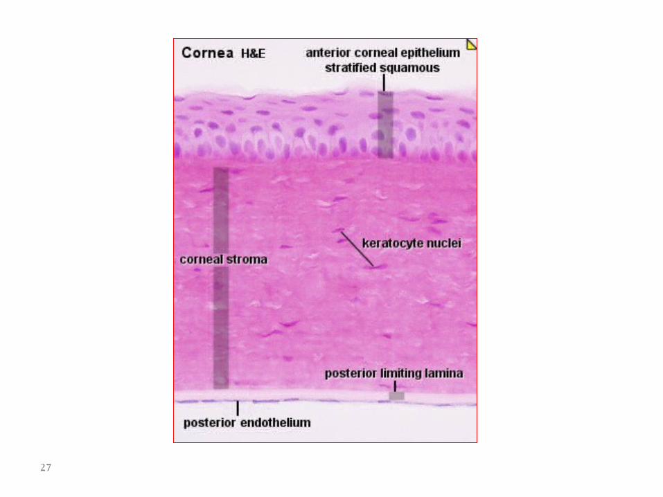
- Cornea - Anterior continuation of the sclera, covering the anterior one-fifth of the eye -Transparent and avascular; transparency results from the ordered arrangement of its collagen fibers and low state of tissue hydration. - Convex curvature aids in focusing light (refraction). - Layers (anterior to posterior) 1- Corneal epithelium. Covers the anterior surface of the cornea; composed of a moist, stratified squamous epithelium that is continuous with the bulbar conjunctiva at the limbus 2- Bowman’s membrane. Acellular collagenous layer beneath the corneal epithelium 3 - Stroma. Multiple layers of parallel collagen fibers constitute the majority of the cornea. The collagen fibers in each layer are arranged at about right angles to adjacent layers. The highly ordered arrangement of these fibers contributes to the transparency of the cornea. 4- Descemet’s membrane. Thickened basal lamina of the corneal endothelium 5- Corneal endothelium. Simple squamous epithelium covering the posterior surface of the cornea; regulates the hydration state of the stroma
26
27

- Corneo - scleral junction (limbus) - Transition zone between the cornea and the sclera - Bowman’s membrane ends and the corneal epithelium thickens at this junction. -Trabecular meshwork. Irregular channels in the stroma that are lined by endothelium. Drains the aqueous humor from the anterior chamber to maintain proper intraocular pressure. The channels of the trabecular meshwork merge to form the canal of Schlemm, a ring-like sinus that encircles the limbus and drains into the venous system.
28
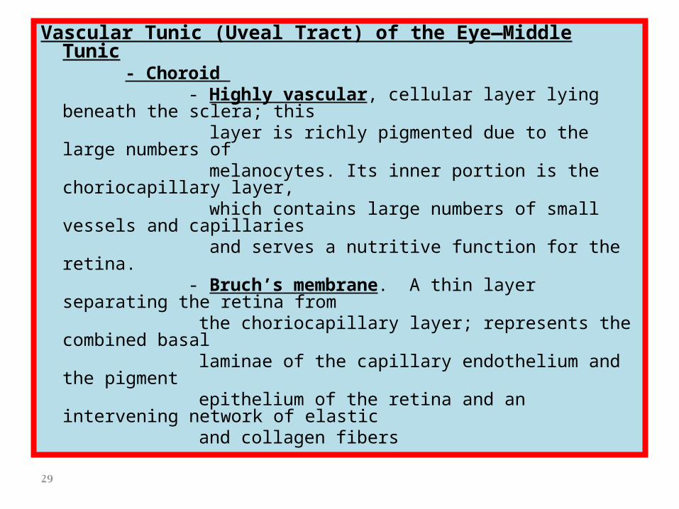
Vascular Tunic (Uveal Tract) of the Eye—Middle Tunic - Choroid - Highly vascular, cellular layer lying beneath the sclera; this layer is richly pigmented due to the large numbers of melanocytes. Its inner portion is the choriocapillary layer, which contains large numbers of small vessels and capillaries and serves a nutritive function for the retina. - Bruch’s membrane. A thin layer separating the retina from the choriocapillary layer; represents the combined basal laminae of the capillary endothelium and the pigment epithelium of the retina and an intervening network of elastic and collagen fibers
29

- Ciliary body - Anterior expansion of the choroid forming a ring that encircles the lens; appears triangular in cross-section - Composed of a core of connective tissue and muscle; lined on its vitreal surface by two layers of columnar cells, an inner pigmented epithelium and an outer layer of nonpigmented cells. This layer, the nonsensory retina, represents the attenuated anterior part of the sensory layer of the retina. - Ciliary processes - Ridge-like extensions from the ciliary body - Zonule fibers. Emerge from between the processes and attach to the lens capsule - The aqueous humor is produced by the epithelium of the ciliary processes. - Ciliary muscles. Smooth muscle fibers that insert on the sclera and ciliary body; contraction of circularly arranged fibers releases tension on the zonule fibers, allowing the lens to assume a more spherical shape, thus providing for focusing on near objects (accommodation). Contraction of radially oriented smooth muscle fibers results in flattening of the lens, thus providing for focusing on far objects.
30

31

32

- Iris - Disc-shaped structure that arises from the anterior margin of the ciliary body; separates anterior and posterior chambers and par- tially covers the lens - Composed of loose connective tissue that is covered on its anterior surface by an incomplete layer of pigment cells and fibroblasts. Its posterior surface is covered by a double layer of pigmented epithelial cells. - Pupil. Central opening in the iris, the diameter is regulated by contraction of two sets of intrinsic smooth muscle in the iris. - Dilator pupillae muscle. Derived from the more anterior, pigmented epithelial layer; consists of radially oriented cells whose contraction widens the aperture of the pupil - Constrictor pupillae muscle. Consists of circularly oriented smooth muscle fibers surrounding the pupil; contraction of these fibers decreases the diameter of the pupil.
33

- Retina—Inner Tunic - Inner-most of the three layers, forming a cup-shaped structure. The posterior portion is photosensitive and extends forward to the ciliary body, terminating as the ora serrata. The nonphotosensitive anterior portion is reduced in thickness and number of layers and forms the posterior lining of the ciliary body and the posterior lining of the iris. - The photosensitive portion contains the photoreceptors, which transduce light into nervous impulses, and neurons, which perform the initial integration of the visual signals. - Overview of retinal cytoarchitecture - Basic plan of the retina consists of a three-cell pathway - Rods and cones. Photoreceptors that transduce light energy into neural activity and
form the photoreceptor layer; their nuclei are located in the outer nuclear layer. - Bipolar cells. Synapse with rods and cones; nuclei are located in the inner nuclear layer. - Ganglion cells. Synapse with bipolar cells; cell bodies are located in the ganglion cell layer; axons from these cells form the optic nerve fiber layer as they pass toward the optic disc, head of the optic nerve. - Regions of synaptic integration - Outer plexiform layer. Location of synapses of rods and cones with bipolar cells - Inner plexiform layer. Location of synapses of bipolar cells and ganglion cells
34

35

-Layers of the retina–from outer to inner - Composed of 10 layers. The naming of the layers is based
on their position relative to the path of the neural conduction, not the path of light.
- Pigment epithelium - Cytoplasm contains numerous melanin granules to
absorb light and reduce reflection - Columnar epithelial cells with apical microvilli whose bases are adherent to Bruch’s membrane in the choroid - Cells posses a cylindrical sheath that surrounds the apical tips of the photoreceptors; these sheaths aid in phagocytosis and digestion of membranous discs shed by the photoreceptors
36

- Photoreceptor layer - Composed of rods and cones - Rods are sensitive to low light intensity, outnumber cones and are located throughout the retina - Cones are less numerous than rods, sensitive to high intensity light and respond to color. Cones provide greater visual acuity and are concentrated in the fovea centralis. - Outer segment. Contains flattened, membranous discs that contain the visual pigments rhodopsin (rods) and iodopsins (cones). - Inner segment. Separated from the outer segment by a constriction, contains the major synthetic and energy-producing organelles. - External limiting membrane. Not a true membrane; formed by adherent
junctions of Mueller cells, modified astrocytes, with the photoreceptors - Outer nuclear layer. Location of the nuclei of rods and cones - Outer plexiform layer. Region of synaptic contacts between photoreceptor
axons and bipolar cell dendrites - Inner nuclear layer. Location of cell bodies of bipolar cells. Also present are
additional neurons, amacrine and horizontal cells. Inner plexiform layer. Location of synaptic contacts between bipolar cell axons and ganglion cell dendrites.
37

- Ganglion cell layer. Location of cell bodies of ganglion cells - Optic nerve fiber layer. Collections of unmyelinated ganglion cell axons that pass toward the optic disc, the head of the optic nerve, where they exit to form the optic nerve (cranial nerve II). - Internal limiting membrane. Formed by the basal portions of
Mueller cells - Fovea centralis. Region of the retina providing greatest visual
acuity, consists entirely of cones; other retinal layers are displaced centri - fugally to allow for an unimpeded path for the light to reach the photoreceptors. - Optic disc (“blind spot”). Region composed only of axons from retinal ganglion cells as they pass through the sclera to form the
optic nerve
38

-Lens - Biconcave, transparent, and elastic - Suspended by radially oriented zonule fibers that extend from the ciliay body to insert into the lens capsule - Structure of the lens - Lens capsule. A thickened basal lamina, produced by the subcap - sular epithelium, surrounds the entire lens. - Subcapsular epithelium. Simple cuboidal epithelium, present only on the anterior surface of the lens; apical surfaces of the cells are directed toward the center of the lens. - Lens fibers. Derived from cells of the subcapsular epithelium pri - marily in the equatorial region of the lens; lens fibers are highly differentiated cells that lose their organelles and become filled with crystallin proteins. - Contraction of the ciliary muscle releases tension on the zonule fibers, allowing the lens to assume a more spherical shape which provides for focusing on near objects (accommodation).
39

40

41