E-ISSN: JPP 2018; 7(6): 1425-1440 Phytochemical profiling ... · ~ 1425 ~ Journal of Pharmacognosy...
16
~ 1425 ~ Journal of Pharmacognosy and Phytochemistry 2018; 7(6): 1425-1440 E-ISSN: 2278-4136 P-ISSN: 2349-8234 JPP 2018; 7(6): 1425-1440 Received: 17-09-2018 Accepted: 18-10-2018 Arun Kashivishwanath Shettar Department of Bioinformatics and Biotechnology, Akkamahadevi Women’s University, Vijayapura, Karnataka, India Shivkumar B Madagi Department of Bioinformatics and Biotechnology, Akkamahadevi Women’s University, Vijayapura, Karnataka, India Joy H Hoskeri Department of Bioinformatics and Biotechnology, Akkamahadevi Women’s University, Vijayapura, Karnataka, India Ankala Basappa Vedamurthy PG, Department of Studies in Biotechnology and Microbiology, Karnatak University, Dharwad, Karnataka, India Correspondence Arun Kashivishwanath Shettar Department of Bioinformatics and Biotechnology, Akkamahadevi Women’s University, Vijayapura, Karnataka, India Phytochemical profiling, in-vitro antioxidant and anti-inflammatory activities of Hopea ponga, Kandelia candel, Vitex leucoxylon and Rhizophora apiculata Arun Kashivishwanath Shettar, Shivkumar B Madagi, Joy H Hoskeri and Ankala Basappa Vedamurthy Abstract Phytochemical investigation and evaluation of in vitro antioxidant and anti-inflammatory activities of H. ponga, K. candel, V. leucoxylon and R.apiculata. Plant extraction was carried out by soxhlet extraction method with increasing polarity of solvents viz., methanol, ethanol and water. Qualitative phytochemical analysis was performed to investigate the phytochemical content. Antioxidant potential of the plant extracts were analyzed by four different in vitro antioxidant assays viz., ferric ion reducing antioxidant power, phosphomolybdenum, hydrogen peroxide scavenging assay and DPPH assay and anti- inflammatory activity was carried out by using protein denaturation in-vitro bioassay. Phytochemical analysis of all the four experimental plants revealed that each solvent extracts contained broad spectrum of secondary metabolites including phenolics, flavonoids, tannins and glycosides. Aqueous extracts of V. leucoxylon and R. apiculata exhibited the highest phenolic content; methanolic extract of K. candel exhibited highest flavonoids content and therefore possessed significant antioxidant capacity. In vitro anti-inflammatory assay revealed that methanol extract of K. candel showed highest anti-inflammatory activity, when compared to all other extracts whereas K. candel ethanol extract showed least activity. The present study revealed that different solvent extracts of H. ponga, K. candel, V. leucoxylon and R. apiculata leaves contain broad spectrum of bioactive compounds. Results confirm that aqueous extract of V. leucoxylon and R. apiculata showed significant antioxidant activity and methanol extract of K. candel showed high anti-inflammatory activity. Further study requires purification, characterization and structural elucidation of phytochemicals from these extracts that may help in the development of new drug formulations against inflammation. Keywords: Phytochemicals; H2O2 assay; 2,2-diphenyl-1-picrylhydrazyl 1. Introduction Worldwide more than 30,000 plant species have been used for medicinal purposes (Haripriya et al., 2010) [19] . India is a home to thousands of potential medicinal plant species; it is ranked sixth among 12 mega biodiversity countries of the world (Kiruba & Jeeva, 2010; Anpin Raja et al., 2010; Mahesh et al., 2010; Meena et al., 2009; Prakash et al., 2008) [21, 5, 27, 31, 39] . Medicinal plants are backbone of Indian traditional system of medicine. Some of the ethno- medicines have been incorporated in the organized system of medicine, however much larger number of ethno-medicines has remained untouched especially in western ghat region of Karnataka (Bhat satyanarayana, 2004) [7] . Plants are the rich sources of secondary metabolites such as alkaloids, phenols, flavonoids, tannins, saponins, glycosides, terpenoids etc. that posses a wide array of biological properties including antibacterial, antifungal, antioxidant and anticancer (De-Fatima et al.,2006) [14] . Reactive oxygen species (ROS) have been causative agents for many diseases from malaria to hemorrhagic shock to AIDS. Oxidative stress due to free radicals causes various forms of tissue damage and inflammation. Oxidative stress also plays an important role in the development of several degenerative changes in cells and tissues which ultimately leads to several degenerative disorders (Alho & Leinonen, 1999) [4] . Antioxidants are the chemicals which can prevent or slow the oxidative damage of cells. They act as free radical scavengers and hence prevent and repair damage caused due to the free radicals. Recently natural products as antioxidants are gaining high importance due to their high potential in health promotion by preventing the pathogenesis of many diseases including diabetes, cancer and cardiovascular diseases. Plant are rich source of antioxidant phytochemicals, several plants are reported as natural sources for antioxidants viz., phenols, flavonoids etc. Uptake of these antioxidants in dietary supplements will reduce many ailments including the heart diseases (Lekameera et al., 2008; Zadak et al., 2009) [24, 65] .
Transcript of E-ISSN: JPP 2018; 7(6): 1425-1440 Phytochemical profiling ... · ~ 1425 ~ Journal of Pharmacognosy...
~ 1425 ~
Journal of Pharmacognosy and Phytochemistry 2018; 7(6): 1425-1440
E-ISSN: 2278-4136 P-ISSN: 2349-8234 JPP 2018; 7(6): 1425-1440 Received: 17-09-2018 Accepted: 18-10-2018
Arun Kashivishwanath Shettar Department of Bioinformatics and Biotechnology, Akkamahadevi Women’s University, Vijayapura, Karnataka, India Shivkumar B Madagi Department of Bioinformatics and Biotechnology, Akkamahadevi Women’s University, Vijayapura, Karnataka, India Joy H Hoskeri Department of Bioinformatics and Biotechnology, Akkamahadevi Women’s University, Vijayapura, Karnataka, India Ankala Basappa Vedamurthy PG, Department of Studies in Biotechnology and Microbiology, Karnatak University, Dharwad, Karnataka, India Correspondence Arun Kashivishwanath Shettar Department of Bioinformatics and Biotechnology, Akkamahadevi Women’s University, Vijayapura, Karnataka, India
Phytochemical profiling, in-vitro antioxidant and
anti-inflammatory activities of Hopea ponga, Kandelia candel, Vitex leucoxylon and Rhizophora
apiculata
Arun Kashivishwanath Shettar, Shivkumar B Madagi, Joy H Hoskeri and Ankala Basappa Vedamurthy Abstract Phytochemical investigation and evaluation of in vitro antioxidant and anti-inflammatory activities of H. ponga, K. candel, V. leucoxylon and R.apiculata. Plant extraction was carried out by soxhlet extraction method with increasing polarity of solvents viz., methanol, ethanol and water. Qualitative phytochemical analysis was performed to investigate the phytochemical content. Antioxidant potential of the plant extracts were analyzed by four different in vitro antioxidant assays viz., ferric ion reducing antioxidant power, phosphomolybdenum, hydrogen peroxide scavenging assay and DPPH assay and anti-inflammatory activity was carried out by using protein denaturation in-vitro bioassay. Phytochemical analysis of all the four experimental plants revealed that each solvent extracts contained broad spectrum of secondary metabolites including phenolics, flavonoids, tannins and glycosides. Aqueous extracts of V. leucoxylon and R. apiculata exhibited the highest phenolic content; methanolic extract of K. candel exhibited highest flavonoids content and therefore possessed significant antioxidant capacity. In vitro anti-inflammatory assay revealed that methanol extract of K. candel showed highest anti-inflammatory activity, when compared to all other extracts whereas K. candel ethanol extract showed least activity. The present study revealed that different solvent extracts of H. ponga, K. candel, V. leucoxylon and R. apiculata leaves contain broad spectrum of bioactive compounds. Results confirm that aqueous extract of V. leucoxylon and R. apiculata showed significant antioxidant activity and methanol extract of K. candel showed high anti-inflammatory activity. Further study requires purification, characterization and structural elucidation of phytochemicals from these extracts that may help in the development of new drug formulations against inflammation. Keywords: Phytochemicals; H2O2 assay; 2,2-diphenyl-1-picrylhydrazyl 1. Introduction Worldwide more than 30,000 plant species have been used for medicinal purposes (Haripriya et al., 2010) [19]. India is a home to thousands of potential medicinal plant species; it is ranked sixth among 12 mega biodiversity countries of the world (Kiruba & Jeeva, 2010; Anpin Raja et al., 2010; Mahesh et al., 2010; Meena et al., 2009; Prakash et al., 2008) [21, 5, 27, 31, 39]. Medicinal plants are backbone of Indian traditional system of medicine. Some of the ethno-medicines have been incorporated in the organized system of medicine, however much larger number of ethno-medicines has remained untouched especially in western ghat region of Karnataka (Bhat satyanarayana, 2004) [7]. Plants are the rich sources of secondary metabolites such as alkaloids, phenols, flavonoids, tannins, saponins, glycosides, terpenoids etc. that posses a wide array of biological properties including antibacterial, antifungal, antioxidant and anticancer (De-Fatima et al.,2006) [14]. Reactive oxygen species (ROS) have been causative agents for many diseases from malaria to hemorrhagic shock to AIDS. Oxidative stress due to free radicals causes various forms of tissue damage and inflammation. Oxidative stress also plays an important role in the development of several degenerative changes in cells and tissues which ultimately leads to several degenerative disorders (Alho & Leinonen, 1999) [4]. Antioxidants are the chemicals which can prevent or slow the oxidative damage of cells. They act as free radical scavengers and hence prevent and repair damage caused due to the free radicals. Recently natural products as antioxidants are gaining high importance due to their high potential in health promotion by preventing the pathogenesis of many diseases including diabetes, cancer and cardiovascular diseases. Plant are rich source of antioxidant phytochemicals, several plants are reported as natural sources for antioxidants viz., phenols, flavonoids etc. Uptake of these antioxidants in dietary supplements will reduce many ailments including the heart diseases (Lekameera et al., 2008; Zadak et al., 2009) [24, 65].

~ 1426 ~
Journal of Pharmacognosy and Phytochemistry Inflammation is a prominent phenotype of various diseases such as rheumatoid arthritis, atherosclerosis and asthma, although inflammation is primarily a protective response against pathogens, toxins and allergens (Gil, 2002) [17]. There are many synthetic drugs are available to treat inflammation but they have disadvantages because of their detrimental side effects on the gastrointestinal tract, kidneys and on the cardiovascular system and reappearance of symptoms after discontinuation (Srinivasan et al., 2001; Alexandrina, 2010) [55, 3]. Since from ancient times inflammatory disorders and related diseases have been treated with plant or plant derived formulations, because of their specific action and less side effects (Krishnaswamy, 2008; Marc et al., 2008) [23, 29]. In the present study, Hopea ponga, Kandelia candel, Vitex leucoxylon and Rhizophora apiculata plants were selected for phytochemical investigation, in vitro antioxidant and anti-inflammatory studies. Hopea ponga is an endemic tree belonging to Dipterocapaceae family found in tropical ever green forest of western India and it is widely distributed along the Western Ghat of Karnataka (Shiddamallayya et al., 2008) [51]. H. ponga is categorized as an endangered tree species under the International Union for Conversation of Nature Red List of threatened species. This plant was reported to be used as traditional medicine in the treatment of piles and snake bite (Muralikrishnan & Chandrashekar, 1997) [34]. Bark of Hopea ponga is known to have high content of tannin and acts as astringent (Shivaprasad et al., 1999) [52]. Methanolic extract of seed wings of Hopea ponga exhibits antioxidant and antibacterial activity (Sukesh et al., 2011) [57]. Literature survey indicates that there are only few pharmacological studies are reported on this plant, revealing the scope for our venture in unmasking its anti-inflammatory property. Kandelia candel is the mangrove tree belonging to Rhizophoraceae family which is distributed along the western region of India. K. candel whole plant is reported to have antidiabetic activity (Rollet, 1981; Saxena, 1975) [46, 48]. In fact rhizophoraceae species are known to have pharmacological activities. Methanolic extract of K. candel is used as anti-hyperglycemic agent in India (Tiwari et al, 2008) and bark, flowers and leaves were reported to have antiviral and antimicrobial properties (Thangam & Kathiresan, 1997; Williams, 1999) [59, 63]. Vitex leucoxylon is commonly known as five leaved chaste tree and belongs to the verbenaceae family. It is small to large deciduous tree, growing up to 20 m in height. It is widely distributed along the Western Ghats of India. The leaves of V. leucoxylon are reported to have medicinal properties like relieving headache, fever and catarrh (Chanda, 1982) [11]. Reports indicate that aqueous and ethanolic extracts of V. leucoxylon leaves possess antipsychotic, antidepressant, analgesic, anti-inflammatory, anti-parkinsonian and antimicrobial activities (Makwana et al., 1994; Sarma et al., 1990) [28, 47]. Even the root and bark of V. leucoxylon are reported to use as astringent and febrifuge (Meena et al., 2010) [30]. Many hepatoprotective agents were isolated from leaves and bark of V. leucoxylon which includes β-sitosterol, vitexin, isovitexin and aucubin (Rao, 1997). Rhizophora apiculata is the tree species of mangrove tree belonging to rhizophoraceae family. In Malaysia, the leaves of R. apiculata are assayed as antibreast cancer (Nurhanan et al., 2008) [35]. Studies on HPLC investigation of R. apiculata have shown the presence of catechin monomer, a antioxidant flavonoid (Rahim et al., 2008) [41]. This plant is reported to possess anti-inflammatory and anti-tumor properties and is also used to regulate the antioxidant enzymes in biological system (Vinod & Guruvayoorappan, 2014) [62]. Presence of
tannins is reported in the bark of R. apiculata, which is known to possess antibacterial and antiviral properties (Sukardjo, 1987; Afidah et al., 2007; Motsei et al., 2003) [56, 1, 33]. Bark of R. apiculata is used as a traditional medicine in the treatment of diarrhoea and wounds (School of Thai Traditional Medicine, 1981; Traditional medicine association, 1980). In Malaysia, pyroligneous acid from R. apiculata species have been used as sterilizing agent, deodorizer, fertilizer, antimicrobial agent and growth promoting agent (Loo et al., 2006) [26]. Alkaline extract from leaf of R. apiculata reported to inhibit the HIV replication and HIV induced cytopathic effects. Some other studies have confirmed the antiviral property of R. apiculata extracts, which may be due to presence of anti-polysaccharide in the extracts that acts as an antiviral agent (Kirtikar et al., 1935) [35]. However these four above mentioned plants have not been subjected for investigation for their antioxidant and anti-inflammatory activities. With this background, the present study was undertaken to evaluate the antioxidant and anti-inflammatory properties of Hopea ponga, Kandelia candel, Vitex leucoxylon & Rhizophora apiculata plants collected from Western Ghats region of Karnataka, India. 2. Materials and methods 2.1 Plant Collection Leaves of H. ponga, K. candel, V. leucoxylon and R. apiculata collected from Anashi forest range of Western Ghats, Uttar Kannada District, Karnataka, India during the period of May, 2017. The leaves were identified and authenticated by Dr. Kotresha K, Dept of Botany, Karnatak Science College, Dharwad; Karnataka by referring to the voucher specimen deposited in the Dept of Botany, Karnatak Science College, Dharwad, Karnataka (Voucher specimen No 002, 003, 004 and 005). Fresh plant leaves material was collected and washed under running tap water, shade dried and then homogenized to coarsely powder. The powder was stored in airtight containers at -20 °C for further use for crude solvent extraction. 2.2 Crude Extraction Coarsely powdered dried leaves of H. ponga, K. candel, V. leucoxylon and R. apiculata [100g each] were subjected to successive solvent extraction using soxhlet apparatus separately. The extraction of each plant leaves material was done with different solvents in their increasing order of polarity which includes methanol, ethanol and distilled water. Each time the plant material was dried and later extracted with next high polar solvents (following the strategy of extraction in series of increasing the solvent polarity). All extracts were concentrated in Buchi rotoevaporator, followed by removal of traces of solvent by using desiccator. 2.3 Phytochemical analysis The crude extracts (methanol, ethanol and distilled water extracts) of H. ponga, K. candel, V. leucoxylon and R. apiculata plants was qualitatively tested for the presence of different phytochemical constituents namely alkaloids, flavonoids, glycosides, phenols, lignin’s, saponins, sterols, tannins, anthraquinone and reducing sugar by following the standard procedure reported by Deepti et al., (2012) [13] (Deepti et al., 2012) [13]. 2.4 Estimation of total phenolic content The total phenolic content of the H. ponga, K. candel, V. leucoxylon and R. apiculata leaf extract was estimated by

~ 1427 ~
Journal of Pharmacognosy and Phytochemistry using Folin-Ciocalteu method of Singleton et al., (1999) [53] (Singleton et al., 1999) [53] with slight modification. Gallic acid was used as the reference standard. A volume of 0.5 ml of plant extract was mixed with 2 ml of the Folin-ciocalteu reagent (10 fold) and was neutralized with 4 ml of sodium carbonate solution (8% w/v). The reaction mixture was incubated at room temperature for 30 min for color development. The absorbance of the resulting color was measured at 765 nm using UV-VIS spectrophotometer. The total phenolic contents were estimated from the linear equation of standard curve prepared with gallic acid. The content of total phenolic compounds expressed as mg/g gallic acid equivalent. 2.5 Estimation of flavonoids content The flavonoids content in the plant extracts was estimated according Chang et al., (2002) [12] (Chang et al., 2002) [12] with quercetin as reference standard. It is an aluminium chloride colorimetric method in which each extract (0.5 ml) separately mixed with 1.5 ml of methanol, 0.1 ml of 10% aluminium chloride, 0.1 ml of 1M potassium acetate and 2.8 ml of distilled water. The reaction mixture was kept at room temperature for 30 min; the absorbance of the reaction mixture was measured at 415 nm using a UV-VIS Spectrophotometer. The value of optical density was used to calculate the flavonoids content present in the sample and the calibration curve was plotted by using quercetin solutions at concentrations 12.5 to 100 µg/ml in methanol. 2.6 Determination of antioxidant activity by using in-vitro methods A) Ferric ion reducing antioxidant power assay (FRAP) Ferric ions reducing power was measured according to the method of Oyaizu (1986) [36] with a slight modification (Oyaizu, 1986) [36]. Methanol, ethanol and aqueous extracts of H. ponga, V. leucoxylon, R. apiculata and K. candel in different concentrations ranging from 100 µl to 500 µl were mixed with 2.5 ml of 20 mM phosphate buffer and 2.5 ml 1%, w/v potassium ferricyanide, and then the mixture was incubated at 50 °C for 30 min. After incubation, 2.5 ml of 10%, w/v trichloroacetic acid and 0.5 ml 0.1%, w/v ferric chloride were added to the mixture, which was incubated for 10 min. Finally, the absorbance was measured at 700 nm using a UV-VIS Spectrophotometer. Ascorbic acid was used as reference standard. All samples were assayed in triplicates. B) Phosphomolybdenum Assay (PM) Total antioxidant activity was estimated by Phosphomolybdenum assay using standard procedure of Prieto et al., (1999) [40] (Prieto et al., 1999) [40]. Methanol, ethanol and aqueous extracts of H. ponga, V. leucoxylon, R. apiculata and K. candel in different concentrations ranging from 100 µl to 500 µl were added to each test tube individually containing 3 ml of distilled water and 1 ml of Molybdate reagent solution. These tubes were incubated at 95 oC for 90 min. After incubation, these tubes were normalized to room temperature for 20-30 min and the absorbance of the reaction mixture was measured at 695 nm using a UV-VIS Spectrophotometer. Ascorbic acid was used as reference standard. All samples were assayed in triplicates. C) Hydrogen peroxide scavenging assay: The antioxidant activity of methanol, ethanol and aqueous extracts of H. ponga, V. leucoxylon, R. apiculata and K. candel was evaluated with ascorbic acid as a standard based
on their ability to scavenge the hydrogen peroxide (Gulcin et al., 2002) [18]. 0.6 ml of 4mM H2O2 solution in phosphate buffer (pH-7.4) was added to 0.5 ml of known concentration of standard ascorbic acid and to tubes containing different concentrations ranging from 100 µl to 500 µl of plant extracts in phosphate buffer (pH-7.4). Absorbance of the solution was measured at 230 nm after 10 min against the blank solution containing phosphate buffer without hydrogen peroxide. Control was prepared by replacing the sample or standard with phosphate buffer. All samples were assayed in triplicates. The percentage of inhibition was calculated by using formula method. Percentage of inhibition% = Ac-At/Ac x 100 D) DPPH free radical-scavenging ability assay Radical scavenging activities of methanol, ethanol and aqueous extracts of H. ponga, V. leucoxylon, R. apiculata and K. candel were determined using the DPPH radical as a reagent, according to the methods of Rice-Evans et al., (1997) [47] (Rice-Evans et al., 1997) [47]. 100 µL of a DPPH radical solution in ethanol (60 µM) was mixed with 100 µL of sample solution in ethanol (different concentrations, w/v). The mixture was incubated for 30 min in the dark at room temperature and then absorbance was measured at 517 nm using a UV-VIS Spectrophotometer. Ascorbic acid was used as a reference standard. The DPPH scavenging activity of each sample was calculated using the following equation: % inhibition= Ac-At/Ac x 100 Where, Ac is the absorbance of the control reaction (100 µL of ethanol with 100 µL of the DPPH solution), and At is the absorbance of the test sample. The experiment was done in triplicate. The IC50 value was calculated for all the samples. Lower absorbance of the reaction mixture indicated higher free radical activity. E) Evaluation of in-vitro anti-inflammatory activity Anti-inflammatory activity of methanol, ethanol and aqueous extracts of H. ponga, V. leucoxylon, R. apiculata and K. candel was evaluated by protein denaturation method as described by Padmanabhan et al., (2012) [37] with slight modifications (Padmanabhan et al., 2012) [37]. Diclofenac sodium, a powerful non-steroidal anti- inflammatory drug was used as a standard drug. The reaction mixture consisting of 2 mL of different concentrations of methanol and aqueous extract of H. ponga, V. leucoxylon and R. apiculata and methanol, ethanol and aqueous extracts of K. candel (500 µg/mL) with standard Diclofenac sodium (500 µg/mL) and 2.8 mL of phosphate buffered saline (pH 6.4) was mixed with 2 mL of egg albumin (from fresh hen’s egg) and incubated at (27±1) °C for 15 min. Denaturation was induced by keeping the reaction mixture at 70 °C in a water bath for 10 min. After cooling, the absorbance was measured at 660 nm by using double distilled water as blank. Each experiment was done in triplicate. The percentage inhibition of protein denaturation was calculated by using the following formula:
% inhibition= At-Ac/Ac x 100 Where, At =absorbance of test sample; Ac=absorbance of control.
2.7 Statistical analysis All experiments were performed in triplicates (n=3) and the data are presented as the mean ± standard deviation. Differences between the means of the individual groups were analyzed using the analysis of variance procedure of SPSS

~ 1428 ~
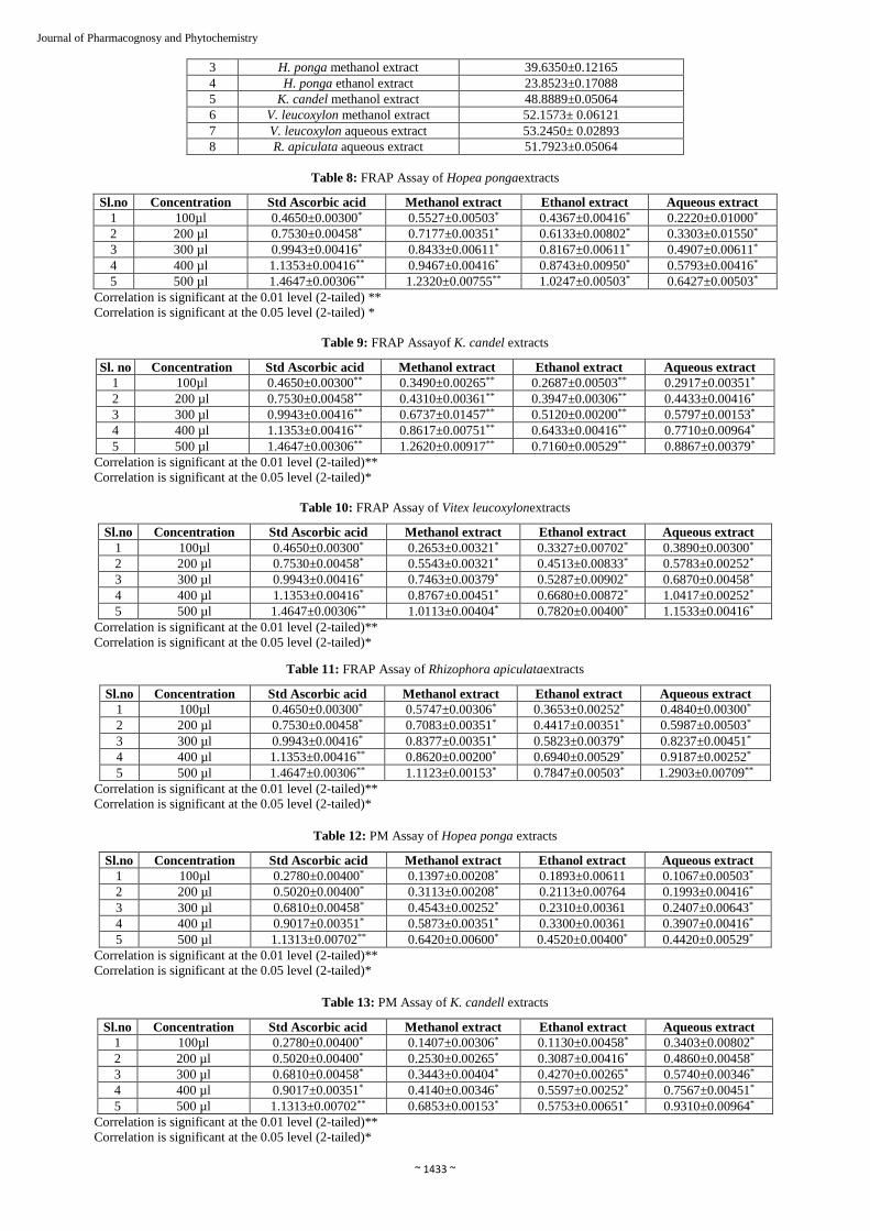
Journal of Pharmacognosy and Phytochemistry software 20 Version (IBM). The significance of differences was defined at the p< 0.05 and p< 0.01 level. 3. Results 3.1 Phytochemical Analysis In the present study, the qualitative phytochemical analysis of methanol, ethanol and aqueous extracts of H. ponga, K. candel, V. leucoxylon and R. apiculata plants was performed by following the standard procedure of various different biochemical tests. In case of H. ponga out of three different extracts, phenols, flavonoids and tannins were present in methanol extract, ethanol extract and aqueous extract, glycosides and sterols were detected in all solvent extracts (Table. 1). In case of K. candel, alkaloids were detected in methanol and aqueous extracts; whereas methanol, ethanol and aqueous extracts shows the presence of phenols, flavonoids and tannins, among all the three different extracts, presence of lignin was detected in only ethanol extract; glycosides were detected in ethanol and aqueous extracts (Table. 2). In case of V. leucoxylon, among all the three different extracts, phenols and flavonoids in methanol and aqueous extracts; tannins in ethanol and aqueous extracts; glycosides in methanol and ethanol extracts; whereas saponins was absent in all extracts (Table. 3). R. apiculata extracts shows variation in all phytochemical contents, among all the three extracts; phenol and flavonoids were detected in methanol and aqueous extracts; tannins were only detected in ethanol extract; glycosides were detected in methanol and ethanol extracts; saponins was absent in all solvent extracts (Table.4). 3.2 Total yield of Crude extract Phytochemical screening of H. ponga, K. candel, V. leucoxylon and R. apiculata plants shows the variation in phytochemical content and all extracts exhibited as good source of secondary metabolites. The total yield of crude extracts from H. ponga, K. candel, V. leucoxylon and R. apiculata leaves by using the solvents viz. methanol, ethanol and aqueous were calculated and shown in Table. 5. 3.3 Total Phenol content In the present study, total phenolic content of different extracts of H. ponga, K. candel, V. leucoxylon and R. apiculata leaves was determined by the FCR method (Singleton et al., 1999) [53] and expressed as gallic acid equivalents (GAE) per gram of plant extracts. Methanol extract of K. candel exhibited highest amount of phenolic content among the extracts i.e 55.91±0.0540 mg/g of gallic acid equivalent. Variation was observed in total phenolic content in each extracts (Table. 6). 3.4 Total Flavonoids content In the present study, total flavonoids content of different extracts of H. ponga, K. candel, V. leucoxylon and R. apiculata leaves was determined by aluminium chloride colorimetric method (Chang et al., 2002) [12] and expressed as quercetin equivalents (QE) per gram of plant extracts. Aqueous extract of V. leucoxylon exhibited highest amount of flavonoids content (53.24 ± 0.02893 mg/g) among the all extracts of quercetin equivalent. Variation was observed in total flavonoids content in each extracts (Table. 7). 3.5 FRAP Assay In the present study, different concentrations of methanol, thanol and aqueous extracts of H. ponga, V. leucoxylon, R.
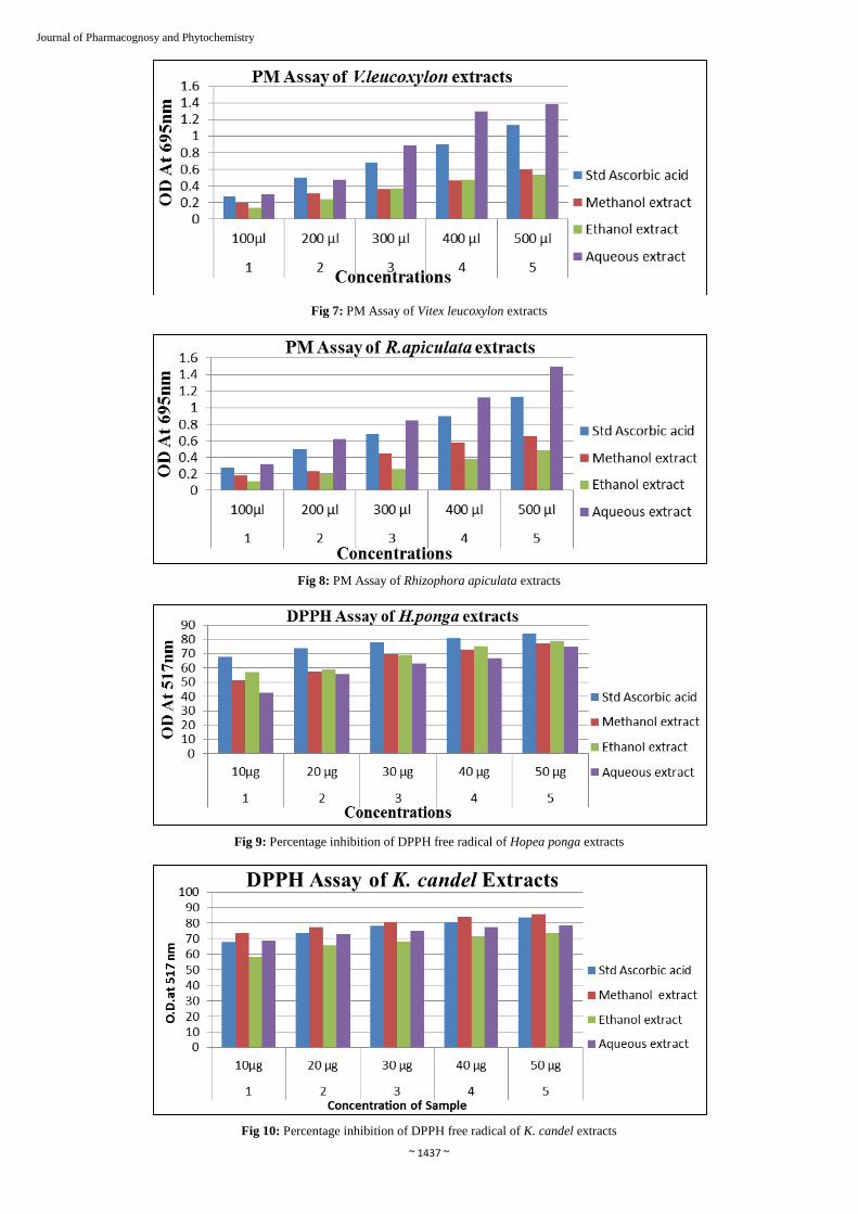
apiculata and K. candel were subjected to FRAP assay along with ascorbic acid as a standard. Absorbance of the blue colored mixture was measured at 700 nm. The FRAP values were determined as optical density readings. Higher optical density indicates the higher reducing power. In case of H. ponga methanol extract of known concentration (500µl) showed good antioxidant activity with absorbance values i.e 1.2320±0.00755, which was less than the absorbance value of standard ascorbic acid i.e. 1.4647±0.00306, results are shown in Table. 8. In case of K. candel, among methanol, ethanol and aqueous extracts, methanol extract exhibited higher activity among other extracts with absorbance value 1.2620±0.00917 (Table. 9). In FRAP assay of V. leucoxylon methanol, ethanol and aqueous extracts performed along with ascorbic acid as standard. Among these three extracts aqueous extract, aqueous extract showed higher activity than other extracts with absorbance value (1.1533±0.00416) (Table. 10). In case of R. apiculata, aqueous extract exhibited significant activity than methanol and ethanol extracts (Table. 11). 3.6 Phosphomolybdenum Assay (PM) PM assay was used to evaluate total antioxidant capacity of plant extracts along with standard. The antioxidant activity depends on the reducing power of extracts in reduction of phosphate-Mo (VI) at acidic pH. In the present study different concentrations of methanol, ethanol and aqueous extracts of H. ponga, V. leucoxylon, R. apiculata and K. candel along with the ascorbic acid as a standard were used for PM assay. In case of H. ponga methanol, ethanol and aqueous extracts showed a very less activity when compared with standard ascorbic acid value (1.1313±0.00702) (Table. 12.). PM assay of methanol, ethanol and aqueous extracts of K. candel reveals that among three extracts aqueous extract exhibited highest activity than methanol and ethanol extracts i.e. 0.9310±0.00964 which was near to standard ascorbic value i.e. 1.1313±0.00702 (Table. 13.). In case of V. leucoxylon among methanol, ethanol and aqueous extracts, aqueous extract showed highest activity than methanol extract and standard ascorbic acid i.e. 1.3863±0.00252 (Table. 14.). In case of R. apiculata, methanol, ethanol and aqueous extracts were analyzed for antioxidant activity along with ascorbic acid as a standard. The results showed that aqueous extract showed to be having highest activity (1.4923±0.00252) than both methanol extract and standard (Table. 15.). 3.7 Hydrogen peroxide scavenging assay In hydrogen peroxide radical scavenging assay known concentration (100µg) of methanol, ethanol and aqueous extracts of H. ponga, V. leucoxylon, R. apiculata and K. candel were subjected along with ascorbic acid as a standard. The results revealed that aqueous extract of V. leucoxylon showed highest scavenging activity than standard and among all extracts (80.8667±0.15535). The values of remaining extracts and standard are tabulated in Table. 16. 3.8 DPPH free radical-scavenging ability assay In the present study, the different concentrations of methanol, ethanol and aqueous extracts of H. ponga, V. leucoxylon, R. apiculata and K. candel were evaluate for their free radical scavenging property by DPPH free radical scavenging assay. The antioxidant capacity of the extracts was compared with ascorbic acid as standard antioxidant. In case of H. ponga methanol, ethanol and aqueous extracts shown good scavenging activity but inhibition percentage was less than standard ascorbic acid value (83.9100±0.35000). The results

~ 1429 ~
Journal of Pharmacognosy and Phytochemistry are shown in Table. 17. The DPPH assay of methanol, ethanol and aqueous extracts of K. candel shows that methanol extract exhibited significant scavenging activity than ethanol, aqueous extracts and standard (86.0100±0.30806). The scavenging activity is represented as percentage of inhibition (%) in Table. 18. In case of V. leucoxylon among three extracts, methanol and aqueous extracts showed promising antioxidant activity with percentage of inhibition 81.0733±0.17786 and 83.0200±0.24021 respectively (Table.19). In case of R. sapiculata among methanol, ethanol and aqueous extract, aqueous extract showed higher antioxidant activity than methanol extract (83.5700±0.12124) (Table. 20). 3.9 In-vitro anti-inflammatory assay Anti-inflammatory study of known concentrations (100µg) of methanol, ethanol and aqueous extracts of H. ponga, V. leucoxylon, R. apiculata and K. candel were subjected for anti-inflammatory activity through protein denaturation assay. The in-vitro anti-inflammatory activity of the extracts was comparable to the Diclofenac sodium, a reference drug. Significant difference was observed among all extracts in the denaturation of protein. The results revealed that methanol extract of K. candel exhibited significant anti-inflammatory activity with percentage of inhibition 96.9133% than standard drug and all the extracts. Whereas standard drug Diclofenac sodium showed 94.2467% of inhibition of protein. The results are tabulated in Table. 21. 4. Discussion Plant resources are used as raw material for different purposes with various applications, they not only provide basis needs of life but also they are the valuable source of phytochemicals. Plant derived natural products had great promise for the discovery and development of new drugs. Preliminary screening tests are useful in the detection of different secondary metabolites in the plant crude extracts. Extraction with different solvents with increasing polarity influences the phytoconstituents present in the plants. Hence, in the present study different solvents (methanol, ethanol and aqueous) were used for extraction. In the present study, the phytochemical analysis of H. ponga, K. candel, V. leucoxylon and R. apiculata showed the presence of broad spectrum of bioactive compounds such as alkaloids, saponins, tannins, flavonoids and phenols in commonly. These bioactive compounds are reported to have different pharmacological activities like such as anticancer, anti-proliferative, antimicrobial, antioxidant and anti- tuberculosis activities (Sreeram et al., 2005; Supayarg et al., 2004) [54, 58]. Several studies show that alkaloids, glycosides are known to possess antimicrobial properties (Balandrin & Klocke, 1988) [6]. In the present study, methanol extract of H. ponga; methanol, ethanol and aqueous extracts of K. candel; showed the presence of alkaloids. Saponins are considered as key ingredient to produce inhibitory effect on inflammation, they also used as dietary supplements and nutraceuticals. Different physical and biological properties of saponins made them useful drugs such as antimicrobial, anti- inflammatory and hemolytic agent (George et al., 2002; Xu et al., 1996) [16, 64]. In the present investigation, methanol extract of H. ponga and V. leucoxylon only showed the presence of saponins, where as it was absent in remaining plant extracts. Many tannin drugs are being used as astringent. They are also used as healing agent, anti-inflammatory agent, and anti-toed and also against burns and piles. Tannins are considered to be possessing medicinal properties like antiviral, antibacterial,
anti-parasitic, anticancer, antiulcer and antioxidant agents. It also investigated that this was able to inhibit HIV replication (Lv et al., 2004; Akiyama et al., 2001; Kolodziej & Kiderlen, 2005) [26, 2, 25]. In the present study, methanol and ethanol extracts of H. ponga and V. leucoxylon; methanol, ethanol and aqueous extracts of K. candel showed the presence of tannins, whereas in case of R. apiculata it was found to be absent in all extracts. Many literatures and extensive investigation indicates that the plant phenolics are the major primary compounds which are acting as free radical scavengers. Even the flavonoids are considered to be diverse and broad group of natural components which possess broad spectrum of biological activities including antioxidant activity (Blois, 1958) [9]. In the present study, methanol and ethanol extracts of H. ponga; methanol, ethanol and aqueous extracts of K. candel; methanol and aqueous extracts of V. leucoxylon and R. apiculata showed the presence of phenols. On consideration with flavonoids methanol extract of H. ponga; methanol, ethanol and aqueous extracts of K. candel; methanol and aqueous extracts of V. leucoxylon and R. apiculata showed the presence of flavonoids. In the present study, the total phenolic content and flavonoids content was also calculated. Methanol extract of K. candel exhibited highest amount of phenolic content among all the extracts (55.91±0.0540 mg/g) of gallic acid equivalent and aqueous extract of V. leucoxylon exhibited highest amount of flavonoids content among all the extracts (53.24±0.02893 mg/g) of quercetin equivalent. Phenolic compounds and flavonoids can play the role of antioxidant through different mechanisms including terminating free radicals, reducing the oxygen concentration, transforming primary products of oxidation into non oxidant molecules and acts as metal chelators (Shahidi & Naczk, 2004) [50]. Free radicals are the chemical species which contains one or more unpaired electrons. These are highly unstable and attain stability cause damage to other molecules by extracting electrons from them. These free radicals enhances the abnormal uncontrolled oxidation reaction in the body which leads to the failure of antioxidant defense mechanism and causes damage to the cell structures which increases the risk factors for many diseases such as Alzheimer’s disease, Parkinson disease, cardiovascular disorders, liver disease, inflammation and cancer (Rajkapoor et al., 2010) [42]. In the antioxidant defense system, enzymatic antioxidant such as superoxide dismutase (SO), catalase (CAT), gluatathaione peroxidase (CPx) and non-enzymatic plant derived antioxidants such as caretenoids, ascorbic acid, phenol, flavonoids etc., are having antioxidant capacity acts as scavengers in the living system. Risk of the chronic disease and its progression can be achieved by increasing natural antioxidant defense or supplementing with the proven antioxidants (Rajkumar et al., 2010) [43]. Antioxidant mechanisms in biological tissues are extremely complex and by the only one method it is difficult to decide the antioxidant capacity of crude extracts (Carocho & Ferreira, 2013) [10]. A large number of in-vitro methods are available to evaluate the antioxidant activity of pure compound or extracts. Hence in the present study, four in-vitro assays viz., FRAP, PM, H2O2 and DPPH assay are employed. FRAP assay includes use of ferricyanide and ferric ions as chromogenic oxidants. It gives an immediate result of a large range of individual antioxidants in dose dependent manner and intensity of color directly proportional to the reducing power of antioxidants. The color of the reaction mixture which measured at 700nm and higher the absorbance

~ 1430 ~
Journal of Pharmacognosy and Phytochemistry indicates higher antioxidant activity. In the present study methanol extract of K. candel and aqueous extract of R. apiculata exhibited higher activity over all extracts with absorbance 1.2620±0.00917 and 1.2903±0.00709 respectively. PM assay is a quantitative method used to evaluate redox reaction by antioxidant, oxidants with the involvement of ligand molybdenum. It involves the longer incubation period at higher temperature which influences the auto-oxidation reaction in the mixture. It directly estimates the reducing potent of the extracts. The reaction mixture forms green Phosphomolybdenum colored complex at acidic pH which is measured at 695nm. The reduction activity of extracts and standard drug was increased with increasing in concentrations. In the present study, aqueous extract of V. leucoxylon and R. apiculata possessed higher activity ((1.3863 and 1.4923 respectively) which was higher than the standard and remaining extracts. H2O2 assay is one of the common methods used to investigate the antioxidant capacity of the extracts. Hydrogen peroxide is very inactive but in some cases it causes adverse effects to cell by releasing hydroxyl radicals in the cells and become toxic to the cells. H2O2 can be removed by antioxidants such as phenols, polyphenols and flavonoids (Esmaeili & Sonboli, 2010) [15] and protects mammalian cells from damage induced by hydrogen peroxide. In this assay, scavenging activity of extracts was detected and compared with ascorbic acid which is taken as reference standard. In H2O2 assay strong oxidizing agent which inactivate certain enzymes directly and also react
with metal ions like Fe+2, Cu+2 and leads to the toxic effects (Bhatia et al., 2011) [8]. In the present study, aqueous extract of V. leucoxylon exhibited highest scavenging activity with the 80.8667% percentage of inhibition on comparing with standard and remaining plants extracts. DPPH assay is most widely accepted method for evaluating antioxidant activity of many plant based drugs and crude extracts (Philips et al., 2010) [38]. It is based on the reduction of methanolic solution of colored free radical DPPH by free radical scavengers. Concentration of free radical scavengers is proportional to scavenging DPPH with the absorbance at 517 nm. The reducing potential is measured by decreasing in absorbance by the action extracts. In the present study, aqueous extract of V. leucoxylon and R. apiculata showed higher antioxidant activity with 83.0200 and 83.5700 percentage of inhibition which was comparable to inhibition percentage of standard (83.9100). Inflammation is a common reaction of living tissues towards infection and injuries (Mohamed et al., 2011) [32]. Steroidal anti-inflammatory agents are available but they induce damage to the lymphocytes and causes severe side effects, so the anti-inflammatory agents from natural sources like plants getting more importance and they are more promising agent with less side effect. In the present study, in-vitro protein denaturation method was used in which methanol extract of K. candel showed to be exhibit highest anti-inflammatory activity than the standard drug and all extracts with 96.9133 percentage inhibition of the protein denaturation.
Table 1: Phytochemical constituents present in the H. pongaleaves
Tests Methanol extract Ethanol extract Aqueous extract A] Alkaloids
Iodine - - - Waganer’s + - -
Dragendroff’s + - - B] Flavonoids
Pew’s Test ++ + - Shinoda Test ++ ++ - NaoH Test ++ ++ -
C] Glycosides K-K Test + ++ ++
Glycoside Test + ++ ++ Conc. H2SO4 Test + ++ ++
Molish Test + ++ + D] Phenols
Ellagic acid Test + + - Phenols Test + + -
E] Lignin Lignin Test - - - Lobat Test - - -
F] Saponins Foam Test + - -
Haemolysis Test ++ - - G] Sterols
L-B Test + + + Salkowsk Test + + +
H] Tannins Gelatin Test + + -
Lead acetate Test ++ ++ - I] Anthraquinone
Bomtrager’s Test - - - J] Phlobatanin - - - K] Oils and Fats
Filter paper Test ++ ++ ++ Saponification Test + - ++
-: Absent; +: Moderate; ++: High presence.

~ 1431 ~
Journal of Pharmacognosy and Phytochemistry
Table 2: Phytochemical constituents present in the K. candelleaves
Tests Methanol extract Ethanol extract Aqueous extract A] Alkaloids
Iodine - - + Waganer’s + - +
Dragendroff’s + - + B] Flavonoids
Pew’s Test ++ - - Shinoda Test ++ - - NaoH Test ++ - -
C] Glycosides K-K Test - + ++
Glycoside Test - + ++ Conc. H2SO4 Test - + ++
Molish Test - + + D] Phenols
Ellagic acid Test ++ ++ + Phenols Test ++ ++ +
E] Lignin Lignin Test - + - Lobat Test - + -
F] Saponins Foam Test - - -
Haemolysis Test - - + G] Sterols
L-B Test + + + Salkowsk Test + + +
H] Tannins Gelatin Test + + +
Lead acetate Test ++ ++ + I] Anthraquinone
Bomtrager’s Test - - - J] Phlobatanin - - - K] Oils and Fats
Filter paper Test + - + Saponification Test + - ++
-: Absent; +: Moderate; ++: High presence.
Table 3: Phytochemical constituents present in the V. leucoxylon leaves
Tests Methanol extract Ethanol extract Aqueous extract A] Alkaloids
Iodine - - - Waganer’s + + -
Dragendroff’s + + - B] Flavonoids
Pew’s Test ++ + + Shinoda Test ++ - ++ NaoH Test ++ + +
C] Glycosides K-K Test + + -
Glycoside Test + + - Conc. H2SO4 Test + + -
Molish Test + + - D] Phenols
Ellagic acid Test ++ ++ - Phenols Test - - -
E] Lignin Lignin Test - - - Lobat Test + - -
F] Saponins Foam Test ++ - -
Haemolysis Test ++ - - G] Sterols
L-B Test - - - Salkowsk Test - + -
H] Tannins Gelatin Test + + -

~ 1432 ~
Journal of Pharmacognosy and Phytochemistry Lead acetate Test + ++ -
I] Anthraquinone Bomtrager’s Test - - -
J] Phlobatanin - - - K] Oils and Fats
Filter paper Test - + + Saponification Test + - ++
-: Absent; +: Moderate; ++: High presence.
Table 4: Phytochemical constituents present in the R. apiculataleaves
TESTS Methanol extract Ethanol extract Aqueous extract A] Alkaloids
Iodine - - - Waganer’s + - -
Dragendroff’s - - - B] Flavonoids
Pew’s Test - - ++ Shinoda Test - - + NaoH Test - - ++
C] Glycosides K-K Test + + -
Glycoside Test + + - Conc. H2SO4 Test + + +
Molish Test + + - D] Phenols
Ellagic acid Test ++ ++ ++ Phenols Test ++ ++ ++
E] Lignin Lignin Test - - - Lobat Test - - -
F] Saponins Foam Test - - -
Haemolysis Test - - + G] Sterols
L-B Test + + ++ Salkowsk Test - - +
H] Tannins Gelatin Test - + -
Lead acetate Test - ++ - I] Anthraquinone
Bomtrager’s Test - - - J] Phlobatanin - - - K] Oils and Fats
Filter paper Test - ++ + Saponification Test + - -
-: Absent; +: Moderate; ++: High presence.
Table 5: Total yield of different solvents extract of experimental plants
Sl. No. Sample Total yield 1. H. ponga methanol extract 4% 2. H. ponga ethanol extract 2.9% 3. H. ponga aqueous extract 5% 4. K. candel methanol extract 5.7% 5. K. candel ethanol extract 4% 6. K. candel aqueous extract 3.5% 7. V. leucoxylon methanol extract 5.7% 8. V. leucoxylon ethanol extract 6.5% 9. V. leucoxylon aqueous extract 7.5% 10. R. apiculata methanol extract 5.4% 11. R. apiculata ethanol extract 5% 12. R. apiculata aqueous extract 8%
Table 6: Total Phenol content in different solvent extracts of experimental plants
S. No. Sample Total phenolic content (mg) 1 X. americana methanol extract 88.30 ± 0.14 2 X. americana aqueous extract 91.40 ± 0.14 3 H. ponga methanol extract 29.5495±0.15368 4 H. ponga ethanol extract 34.0630±0.06800 5 K. candel methanol extract 55.9189±0. 05405 6 K. candel ethanol extract 14.4144±0.10927 7 K. candel aqueous extract 31.5495±0.10922 8 V. leucoxylon methanol extract 40.2252± 0.06799 9 V. leucoxylon aqueous extract 44.1801± 0.08691
10 R. apiculata chloroform extract 20.1171±0.09492 11 R. apiculata methanol extract 40.1171±0.10922 12 R. apiculata ethanol extract 22.1171±0.08254 13 R. apiculata aqueous extract 44.9549±0.05630
Table 7: Total flavonoids content in different solvent extracts of experimental plants
Sl. No. Sample Total flavonoids content (mg) 1 X. americana methanol extract 38.20 ± 0.12 2 X. americana aqueous extract 61.40 ± 0.173
~ 1433 ~
Journal of Pharmacognosy and Phytochemistry 3 H. ponga methanol extract 39.6350±0.12165 4 H. ponga ethanol extract 23.8523±0.17088 5 K. candel methanol extract 48.8889±0.05064 6 V. leucoxylon methanol extract 52.1573± 0.06121 7 V. leucoxylon aqueous extract 53.2450± 0.02893 8 R. apiculata aqueous extract 51.7923±0.05064
Table 8: FRAP Assay of Hopea pongaextracts
Sl.no Concentration Std Ascorbic acid Methanol extract Ethanol extract Aqueous extract 1 100µl 0.4650±0.00300* 0.5527±0.00503* 0.4367±0.00416* 0.2220±0.01000*
2 200 µl 0.7530±0.00458* 0.7177±0.00351* 0.6133±0.00802* 0.3303±0.01550*
3 300 µl 0.9943±0.00416* 0.8433±0.00611* 0.8167±0.00611* 0.4907±0.00611*
4 400 µl 1.1353±0.00416** 0.9467±0.00416* 0.8743±0.00950* 0.5793±0.00416*
5 500 µl 1.4647±0.00306** 1.2320±0.00755** 1.0247±0.00503* 0.6427±0.00503*
Correlation is significant at the 0.01 level (2-tailed) ** Correlation is significant at the 0.05 level (2-tailed) *
Table 9: FRAP Assayof K. candel extracts
Sl. no Concentration Std Ascorbic acid Methanol extract Ethanol extract Aqueous extract 1 100µl 0.4650±0.00300** 0.3490±0.00265** 0.2687±0.00503** 0.2917±0.00351*
2 200 µl 0.7530±0.00458** 0.4310±0.00361** 0.3947±0.00306** 0.4433±0.00416*
3 300 µl 0.9943±0.00416** 0.6737±0.01457** 0.5120±0.00200** 0.5797±0.00153*
4 400 µl 1.1353±0.00416** 0.8617±0.00751** 0.6433±0.00416** 0.7710±0.00964*
5 500 µl 1.4647±0.00306** 1.2620±0.00917** 0.7160±0.00529** 0.8867±0.00379*
Correlation is significant at the 0.01 level (2-tailed)** Correlation is significant at the 0.05 level (2-tailed)*
Table 10: FRAP Assay of Vitex leucoxylonextracts
Sl.no Concentration Std Ascorbic acid Methanol extract Ethanol extract Aqueous extract 1 100µl 0.4650±0.00300* 0.2653±0.00321* 0.3327±0.00702* 0.3890±0.00300*
2 200 µl 0.7530±0.00458* 0.5543±0.00321* 0.4513±0.00833* 0.5783±0.00252*
3 300 µl 0.9943±0.00416* 0.7463±0.00379* 0.5287±0.00902* 0.6870±0.00458*
4 400 µl 1.1353±0.00416* 0.8767±0.00451* 0.6680±0.00872* 1.0417±0.00252*
5 500 µl 1.4647±0.00306** 1.0113±0.00404* 0.7820±0.00400* 1.1533±0.00416*
Correlation is significant at the 0.01 level (2-tailed)** Correlation is significant at the 0.05 level (2-tailed)*
Table 11: FRAP Assay of Rhizophora apiculataextracts
Sl.no Concentration Std Ascorbic acid Methanol extract Ethanol extract Aqueous extract 1 100µl 0.4650±0.00300* 0.5747±0.00306* 0.3653±0.00252* 0.4840±0.00300*
2 200 µl 0.7530±0.00458* 0.7083±0.00351* 0.4417±0.00351* 0.5987±0.00503*
3 300 µl 0.9943±0.00416* 0.8377±0.00351* 0.5823±0.00379* 0.8237±0.00451*
4 400 µl 1.1353±0.00416** 0.8620±0.00200* 0.6940±0.00529* 0.9187±0.00252*
5 500 µl 1.4647±0.00306** 1.1123±0.00153* 0.7847±0.00503* 1.2903±0.00709**
Correlation is significant at the 0.01 level (2-tailed)** Correlation is significant at the 0.05 level (2-tailed)*
Table 12: PM Assay of Hopea ponga extracts
Sl.no Concentration Std Ascorbic acid Methanol extract Ethanol extract Aqueous extract 1 100µl 0.2780±0.00400* 0.1397±0.00208* 0.1893±0.00611 0.1067±0.00503*
2 200 µl 0.5020±0.00400* 0.3113±0.00208* 0.2113±0.00764 0.1993±0.00416*
3 300 µl 0.6810±0.00458* 0.4543±0.00252* 0.2310±0.00361 0.2407±0.00643*
4 400 µl 0.9017±0.00351* 0.5873±0.00351* 0.3300±0.00361 0.3907±0.00416*
5 500 µl 1.1313±0.00702** 0.6420±0.00600* 0.4520±0.00400* 0.4420±0.00529*
Correlation is significant at the 0.01 level (2-tailed)** Correlation is significant at the 0.05 level (2-tailed)*
Table 13: PM Assay of K. candell extracts
Sl.no Concentration Std Ascorbic acid Methanol extract Ethanol extract Aqueous extract 1 100µl 0.2780±0.00400* 0.1407±0.00306* 0.1130±0.00458* 0.3403±0.00802*
2 200 µl 0.5020±0.00400* 0.2530±0.00265* 0.3087±0.00416* 0.4860±0.00458*
3 300 µl 0.6810±0.00458* 0.3443±0.00404* 0.4270±0.00265* 0.5740±0.00346*
4 400 µl 0.9017±0.00351* 0.4140±0.00346* 0.5597±0.00252* 0.7567±0.00451*
5 500 µl 1.1313±0.00702** 0.6853±0.00153* 0.5753±0.00651* 0.9310±0.00964*
Correlation is significant at the 0.01 level (2-tailed)** Correlation is significant at the 0.05 level (2-tailed)*

~ 1434 ~
Journal of Pharmacognosy and Phytochemistry Table 14: PM Assay of Vitex leucoxylonextracts
Sl.no Concentration Std Ascorbic acid Methanol extract Ethanol extract Aqueous extract 1 100µl 0.2780±0.00400* 0.1960±0.00200 0.1333±0.00611 0.2983±0.00252*
2 200 µl 0.5020±0.00400* 0.3073±0.00306 0.2420±0.00800 0.4723±0.00153*
3 300 µl 0.6810±0.00458* 0.3647±0.00306* 0.3680±0.00400 0.8940±0.00200*
4 400 µl 0.9017±0.00351* 0.4660±0.00200* 0.4700±0.00346* 1.2933±0.00231**
5 500 µl 1.1313±0.00702** 0.5947±0.00306* 0.5380±0.00529* 1.3863±0.00252**
Correlation is significant at the 0.01 level (2-tailed)** Correlation is significant at the 0.05 level (2-tailed)*
Table 15: PM Assayof Rhizophora apiculataextracts
Sl.no Concentration Std Ascorbic acid Methanol extract Ethanol extract Aqueous extract 1 100µl 0.2780±0.00400 0.1830±0.00557 0.1073±0.00503 0.3197±0.00512
2 200 µl 0.5020±0.00400* 0.2343±0.00252 0.1953±0.00306 0.6250±0.00265*
3 300 µl 0.6810±0.00458* 0.4463±0.00208* 0.2623±0.00379 0.8433±0.00321*
4 400 µl 0.9017±0.00351* 0.5770±0.00265* 0.3807±0.00306 1.1257±0.00153**
5 500 µl 1.1313±0.00702** 0.6537±0.00153* 0.4853±0.00416* 1.4923±0.00252**
Correlation is significant at the 0.01 level (2-tailed)** Correlation is significant at the 0.05 level (2-tailed)*
Table 16: H2O2 Assay of different solvent extracts of experimental plants
Sl. No. Concentration Treatment % Inhibition 1 100 µg Standard 74.4633± 0.13051 2 100 µg H. ponga methanol extract 64.8267±0.07506 3 100 µg H. ponga ethanol extract 58.2333±0.11504 4 100 µg H. ponga aqueous extract 54.4444±0.21297 4 100 µg K. candel methanol extract 70.8167±0. 07506 5 100 µg K. candel ethanol extract 35.3167±0.10017 6 100 µg K. candel aqueous extract 39.6467±0. 11240 7 100 µg V. leucoxylon methanol extract 65.1000± 0.09539 8 100 µg V. leucoxylon ethanol extract 54.3321± 0.15592 9 100 µg V. leucoxylon aqueous extract 80.8667± 0.15535
10 100 µg R. apiculata methanol extract 69.4700±0.13454 11 100 µg R. apiculata ethanol extract 58.3770±0.18849 12 100 µg R. apiculata aqueous extract 73.2267±0.11676
Table 17: Anti-inflammatory Assay of different solvent extracts of experimental plants
Sl. No. Concentration Treatment % Inhibition 1 100 µg Standard 94.2467±1.90148 2 100 µg H. ponga methanol extract 69.5133±1.41394 3 100 µg H. ponga ethanol extract 20.1000±1.63000 4 100 µg H. ponga aqueous extract 31.1594±1.66032 5 100 µg K. candel methanol extract 96.9133±1.66064 6 100 µg K. candel ethanol extract 12.3133±2.79185 7 100 µg K. candel aqueous extract 54.5233±3.53856 8 100 µg V. leucoxylon methanol extract 34.7767±2.17500 9 100 µg V. leucoxylon ethanol extract 16.4855±1.13139
10 100 µg V. leucoxylon aqueous extract 54.8867±1.95809 11 100 µg R. apiculata methanol extract 27.3500±1.90638 12 100 µg R. apiculata ethanol extract 21.9202±1.90866 13 100 µg R. apiculata aqueous extract 77.8933±1.66064
Table 18: Percentage inhibition of DPPH free radical ofHopea ponga extracts
Sl.no Concentration Std Ascorbic acid Methanol extract Ethanol extract Aqueous extract 1 10µg 67.7867±0.17898* 51.6300±0.35000 57.0633±0.52596 42.7350±0.81862
2 20 µg 73.6533±0.23502* 57.4500±0.35000* 58.9300±0.41146* 55.6332±0.48524*
3 30 µg 78.2000±0.30806** 69.8900±0.29206* 69.4233±0.44287* 62.9370±0.23310*
4 40 µg 80.8000±0.35679** 73.1100±0.35679** 75.3667±0.24420** 66.9386±0.52558**
5 50 µg 83.9100±0.35000** 77.2700±0.35000** 78.9000±0.42297** 74.8251±0.46620**
Correlation is significant at the 0.01 level (2-tailed)** Correlation is significant at the 0.05 level (2-tailed)*
Table 19: Percentage inhibition of DPPH free radical ofK. candel extracts
Sl.no Concentration Std Ascorbic acid Methanol extract Ethanol extract Aqueous extract 1 10µg 67.7867±0.17898* 73.8867±0.46501** 58.2300±0.29206* 68.7200±0.41146*
2 20 µg 73.6533±0.23502** 77.3067±0.35572** 65.6500±0.35679* 72.8533±0.32347**

~ 1435 ~
Journal of Pharmacognosy and Phytochemistry 3 30 µg 78.2000±0.30806** 80.7100±0.19975** 68.2700±0.15100* 74.8967±0.48840**
4 40 µg 80.8000±0.35679** 84.0200±0.73980** 71.6367±0.24420* 77.4233±0.17898**
5 50 µg 83.9100±0.35000** 86.0100±0.30806** 73.6267±0.56889** 78.7833±0.23502**
Correlation is significant at the 0.01 level (2-tailed)** Correlation is significant at the 0.05 level (2-tailed)*
Table 20: Percentage inhibition of DPPH free radical of V. leucoxylon extracts
Sl.no Concentration Std Ascorbic acid Methanol extract Ethanol extract Aqueous extract 1 10µg 67.7867±0.17898* 57.8800±0.35679 46.0761±0.58662 68.2167±0.35572*
2 20 µg 73.6533±0.23502* 67.2033±0.35572* 59.4794±0.59808 70.1167±0.37434**
3 30 µg 78.2000±0.30806** 72.5700±0.29206** 60.5112±0.48524 75.9067±0.27135**
4 40 µg 80.8000±0.35679** 78.3567±0.40624** 72.4941±0.46620* 80.3333±0.24420**
5 50 µg 83.9100±0.35000** 81.0733±0.17786** 78.0108±0.48524** 83.0200±0.24021**
Correlation is significant at the 0.01 level (2-tailed)** Correlation is significant at the 0.05 level (2-tailed)*
Table 21: Percentage inhibition of DPPH free radical ofRhizophora apiculata extracts
Sl.no Concentration Std Ascorbic acid Methanol extract Ethanol extract Aqueous extract 1 10µg 67.7867±0.17898* 61.4167±0.53463* 39.8601±0.46620 66.8533±0.40624*
2 20 µg 73.6533±0.23502* 65.7300±0.47000* 48.7179±0.23308 71.5200±0.41146*
3 30 µg 78.2000±0.30806** 70.6633±0.37434* 58.2750±0.69930 78.0067±0.48439**
4 40 µg 80.8000±0.35679** 74.9400±0.35000** 66.4335±o.46620* 81.2700±0.35679**
5 50 µg 83.9100±0.35000** 78.1200±0.43936** 74.6697±0.37465** 83.5700±0.12124**
Correlation is significant at the 0.01 level (2-tailed)** Correlation is significant at the 0.05 level (2-tailed)*
Fig 1: FRAP assay of Hopea ponga extracts
Fig 2: FRAP assay of K. candel extracts

~ 1436 ~
Journal of Pharmacognosy and Phytochemistry
Fig 3: FRAP assay of Vitex leucoxylon extracts
Fig 4: FRAP assay of Rhizophora apiculata extracts
Fig 5: PM Assay of Hopea ponga extracts
Fig 6: PM assay of K. candel extracts
~ 1437 ~
Journal of Pharmacognosy and Phytochemistry
Fig 7: PM Assay of Vitex leucoxylon extracts
Fig 8: PM Assay of Rhizophora apiculata extracts
Fig 9: Percentage inhibition of DPPH free radical of Hopea ponga extracts
Fig 10: Percentage inhibition of DPPH free radical of K. candel extracts

~ 1438 ~
Journal of Pharmacognosy and Phytochemistry
Fig 11: Percentage inhibition of DPPH free radical of V. leucoxylon extracts
Fig 12: Percentage inhibition of DPPH free radical of Rhizophora apiculata extracts
5. Conclusion Phytochemical analysis of H. ponga, K. candel, V. leucoxylon and R. apiculata plants showed that each extract contains broad spectrum of bioactive compounds that included alkaloids, glycosides, saponins, tannins, phenols and flavonoids. These bioactive compounds already reported to have several medicinal properties which include anticancer, anti-proliferative, antimicrobial, antioxidant, anti-tuberculosis, antimicrobial, anti-inflammatory, hemolytic agent and antiviral. The present study also showed high phenolics and flavonoids content in methanol extract of K. candel and aqueous extract of V. leucoxylon respectively. In context of antioxidant assays almost all selected extracts showed moderate activity but aqueous extract of V. leucoxylon and R. apiculata proven to be having significant antioxidant activity over all extracts. In case of anti-inflammatory activity, methanol extract of K. candel showed significant noticeable anti-inflammatory activity on comparing with standard drug and remaining extracts. Statistically the antioxidant assays were observed significant difference (P<0.01 and P<0.05) in each extracts. These differences in the activity may be due the variation in the chemical composition of extracts. Thus the present study concludes that aqueous extract of V. leucoxylon and R. apiculata and methanol extract of K. candel exhibited higher antioxidant and anti-inflammatory activities respectively. However further studies are needed to find out the structural of bioactive compounds and investigate mechanisms of antioxidant and anti-inflammatory activities of the bioactive compounds.
6. Conflict of Interest We wish to confirm that there are no known conflicts of interest associated with this publication 7. References 1. Afidah AR, Rocca E, Steinmetz J, Kassim MJ, Adnan R,
Ibrahim MS. Corros Sci. 2007; 49:402-417. 2. Akiyama H, Fujii K, Yamasaki O, Oono T, Iwatsuki K.
Antibacterial action of several tannins against Staphylococcus aureus. J Antimicrob Chemother. 2001; 48(4):487-491.
3. Alexandrina L. Dumitrescu. ‘‘Antibiotics and Antiseptics in Periodontal Therapy’’. Berlin/Heidlburg, Springer verlang, 2010.
4. Alho H, Leinonen J. Total antioxidant activity measured by chemi luminescence methods. Methods in Enzymology. 1999; 299:3.
5. Anpin Raja RD, Prakash JW, Jeeva S. Antibacterial activity of some medicinal plants used by Kani tribe, southern Western Ghats, Tamilnadu, India. In: Trivedi PC, Editor. Ethnic Tribes and Medicinal Plans. Pointer Publishers, Jaipur, 2010, 28-45.
6. Balandrin MJ, Klocke JA. Medicinal, aromatic and industrial materials from plants. Bajaj Springer-Verlag, Berlin, Heidelberg, 1988, 1-36.
7. Bhat satyanarayana. Janapada vaidyadalli sasya vaividhya (Kannada). Bangalore MRL Infotech service, 2004.
8. Bhatia L, Bishnoi H, Chauhan P, Kinja K, Shailesh S. In vitro comparative antioxidant activity of ethanolic

~ 1439 ~
Journal of Pharmacognosy and Phytochemistry extracts of Glycosmis pentaphylla and Bauhinia variegate. Recent Res Sci. Technol. 2011; 3(7):1-3.
9. Blois MS. Antioxidant determination by the use of a stable free radical. Nat. 1958; 181:1199-1200.
10. Carocho M, Ferreira ICFR. A review on antioxidants, prooxidants and related controversy: Natural and synthetic compounds, screening and analysis methodologies and future perspectives. Food and Chemical Toxicology. 2013; 51:15-25.
11. Chanda YR. The wealth of India: A dictionary of Indian Raw materials and Industrial products; Publication and Information Directorate, CSIR, New Delhi, 1982, 520-521.
12. Chang C, Yang M, Wen H, Chern J. Estimation of total flavonoids content in propolis by two complementary colorimetric methods. Journal of Food Drug Analysis. 2002; 10:178-182.
13. Deepti K, Umadevi V, Vijayalakshmi G, Vinod Polarao B. Antimicrobial activity and phytochemical analysis of Morinda tinctoria Roxb. Leaf extracts. Asian Pac J Trop Biomed. 2(Suppl 3). 2012, S1440-2.
14. De-Fatima A, Modolo LV, Conegero LS, Pilli RA, Ferreira CV, Kohn LK, et al. Lactones and their derivatives:h biological activities, mechanisms of action and potential leads for drug design. Curr Med Chem. 2006; 13:3371-3384.
15. Esmaeili MA, Sonboli A. Antioxidant, free radical scavenging activities of Salvia brachyantha and its protective effect against oxidative cardiac cell injury. Food Chem Toxicol. 2010; 48:846-853.
16. George F, Zohar Kerem, Harinder PSM. Klaus Becker., 2002. The biological action of saponins in animal systems: a review. Brit J Nutr. 2010; 88(6):587-605.
17. Gil Ã. Polyunsaturated fatty acids and inflammatory diseases. Biomedicine and Pharmacotherapy. 2002; 56:388-96.
18. Gulcin I, Buyukokuroglu ME, Oktay M, Kufrevioglu O, I. On the in vitro antioxidant properties of melatonin. J Pineal Res. 2002; 33:167-71.
19. Haripriya D, Selvan N, Jeyakumar N, Periasamy RS, Johnson M, Irudayaraj V. The effect of extracts of Selaginella involvens and Selaginella in aequalifolia leaves on poultry pathogens. Asian Pacific Journal of Tropical Medicine. 2010; 3(9):678-681.
20. Kirtikar KR, Basu BD. Indian Medical Plants. Lalit Mohan Basu, Allahabad, India, 1935, 2793.
21. Kiruba S, Jeeva S. Flowering of bamboos in two biodiversity hotspots of India. The Indian Forester. 2010; 136(1):137-140.
22. Kolodziej H, Kiderlen AF. Antileishmanial activity and immune modulatory effects of tannins and related compounds on Leishmania parasitised RAW 264.7 cells. Phytochemistry. 2005; 66(17):2056-2071.
23. Krishnaswamy K. Traditional Indian spices and their health significance. Asia Pacific Journal of Clinical Nutrition. 2008; 17:265-68.
24. Lekameera R, Vijayabasker P, Somasundaram ST. Evaluating antioxidant property of brown algae Colpomenia sinusa (Deb. Ex. Sol). Afri J Food Sci. 2008; 2:126-130.
25. Loo AY, Jain K, Darah I. Antioxidant and radical scavenging activities of the pyroligneous acid from a mangrove plant, Rhizophora apiculata. Food Chem. 2006; 104:300-307.
26. Lv L, Liu SW, Jiang SB, Wu SG. Tannin inhibits HIV-1 entry by targeting gp41. Acta Pharmacology Sin. 2004; 25(2):213-218.
27. Mahesh M, Binisha GR, Brinitha BR, Vinaya VG, Jeeva S. Pteridophyte flora of Kanyakumari wildlife sanctuary. In: National Seminar on Conservation and Management of Wetlands in an Era of Climate Change, Organized by Department of Botany, Nesamony Memorial Christian College, Marthandam, Tamilnadu, India, 2010.
28. Makwana HG, Ravishankar B, Shukla VJ, Vijayan NP, Sasikala CK, Saraswathy VN, et al. General pharmacology of Vitex leucoxylon linn leaves. Indian J. Physiol. Pharmacol. 1994; 38:95-100.
29. Marc EB, Nelly A, Annick DD, Frederic D. Plants used as remedies anti rheumatic and antineuralgic in the traditional medicine of Lebanon Journal of Ethno pharmacology. 2008; 1203:15-34.
30. Meena AK, Uttam Singh Yadav AK, Singh B, Rao MM. International Journal of Pharmaceutical and Clinical Research. 2010; 2(1):01-09.
31. Meena R, Santhana Kumar G, Asir Selin Kumar R. Ethnomedicinal shrubs of Marunduvalmalai, western Ghats, Tamilnadu, India. J Basic Applied Bio. 2009; 3(1&2):67-70.
32. Mohamed STK, Azeem AK, Dilip C, Sankar C, Prasanth NV. Duraisami R. Anti-inflammatory activity of the leaf extacts of Gendarussa vulgaris Nees. Asian Pac J Trop Biomed. 2011; 1(2):147-149.
33. Motsei ML, Lindsey KL, Van Staden J, Jager AK. J Ethnopharmacol. 2003; 86:235-241.
34. Muralikrishnan H, Chandrashekar KR. Regeneration of hopea ponga: influence of wing loading and viability of seeds. J Trop for Sci. 1997; 10:58-65.
35. Nurhanan MY, Asiah O, Mohd Ilham MA, Siti Syarifah MM, Norhayati I, Lili Sahira H. Anti-proliferative activities of 32 Malaysian plant species in breast cancer cell lines. J Trop For Sci. 2008; 20:77-81.
36. Oyaizu M. Studies on products of browning reaction: antioxidant activities of products of browning reaction prepared from glucosamine. Jpn J Nutr. 1986; 44:307-15.
37. Padmanabhan P, Jangle SN. Evaluation of in-vitro anti-inflammatory activity of herbal preparation, a combination of four herbal plants. Int J Basic Appl Med Sci. 2012; 2(1):109-16.
38. Philips A, Philips S, Arul V, Padmakeerthiga B, Renju V, Santha S, et al. Free radical scavenging activity of leaf extracts of Indigofera aspalathoides - An in vitro analysis. J Pharm Sci Res. 2010; 2:322-328.
39. Prakash JW, Anpin Raja RD, Asbin Anderson N, Christhudhas Williams B, Regini GS, Bensar K, et al. Ethnomedicinal plants used by Kani tribes of Agasthiyamalai biosphere reserve, Southern Western Ghats. Indian Journal of Traditional Knowledge. 2008; 7(3):410-413.
40. Prieto P, Pineda M, Aguilar M. Spectrophotometric quantitation of antioxidant capacity through the formation of a phosphomolybdenum complex: specific application to the determination of vitamin E. Anal Biochem. 1999; 269:337-41.
41. Rahim AA, Rocca E, Steinmetz J, Kassim MJ, Ibrahim MS, Osman H. Antioxidant activities of mangrove Rhizophora apiculata bark extracts. Food Chem. 2008; 107:200-7.

~ 1440 ~
Journal of Pharmacognosy and Phytochemistry 42. Rajkapoor B, Burkan ZE, Senthilkumar R. Oxidants and
human diseases: role of antioxidant medicinal plants-a review. Pharmacologyonline. 2010; 1:1117-1131.
43. Rajkumar V, Guha G, Kumar RA, Mathew L. Evaluation of antioxidant activities of Bergenia ciliate rhizome. Rec Nat Prod. 2010; 4:38-48.
44. Rao RVK, Satyanarayana T, Jena R. Phytochemical studies on Vitex leucoxylon L. Indian Drugs. 1997; 34:50-51.
45. Rice-Evans C, Miller N, Paganga G. Antioxidant properties of phenolic compounds. Trends Plant Sci. 1997; 2:152-9.
46. Rollet B. Bibliography on mangrove research. London: UNESCO Paris Pub Information Retrieval Ltd. 1981; 1600-1975, 479.
47. Sarma SP, Aithal KS, Srinivasan KK, Udupa AL, Kumar V, Kulkarni DR, Rajagopal PK. Anti-inflammatory and wound healing activities of the crude alcoholic extract and flavonoids of Vitex leucoxylon. Fitoterapia. 1990; 61:263-265.
48. Saxena H. A survey of the plants of Orissa (India) for tannins, saponins, flavonoids and alkaloids. Lloydia. 1975; 38:346-51.
49. School of Thai Traditional Medicine. Principle of Thai traditional Pharmacy. Bangkok: Prachettuphon wimolmungkalaramrajworawihorn Temple Press, 1981.
50. Shahidi F, Naczk M. Phenolic in Food and Nutraceutical. CRC Press, BocaRaton, FL, 2004, 1-558.
51. Shiddamallayya N, Azra Y, Gopakumar K. Medico botanical survey Parvatha kkuke subramanaya Mangalore karnatak. Indian J Tradi Med. 2008; 9(1):96-9.
52. Shivaprasad PV, Vasanthraj BK, Chandrashekar KR. Dipterocarps of the Western Ghats of Karnataka. Indian J Forest. 1999; 9:201-6.
53. Singleton VL, Orthofer R, Lamuela-Raventos RM. Analysis of total phenols and other oxidation substrates and antioxidants by means of Folin-Ciocalteu reagent. Methods Enzymol. 1999; 299:152-78.
54. Sreeram NP, Lynn S, Susanne HH, Yantra N, Yajun Z, Muralieedharan GN, et al. In vitro proliferative, apoptic and antioxidant activities of plinicalgin, eliagic acid and a total pomegranate tannin extract are enhanced in combination with other polyphenols found in pomegranate juice. J Nutri Biochem. 2005; 16:360-367.
55. Srinivasan K, Muruganandan S, Lal J, Chandra S, Tandan SK, Ravi Prakash V. Evaluation of anti-inflammatory activity of Pongamia pinnata in rats. J. Ethnopharmacol. 2001; 78:151-157.
56. Sukardjo S. Forest Ecol Manag. 1987; 20:233-252. 57. Sukesh Syed Hidayath, Haneef M, Arunkumar K,
Chandrashekar KR. Phytochemical evaluation, antioxidant and antibacterial activity of seed wings of Hopea ponga (Dennst). Mabberly Int J Pharm Pharm Sci. 2011; 4(8):2593-5.
58. Supayarg V, Amornat L, Wapper J, Trachade S, Souwalak PP, Thanornjit S. Effective medicinal plants against enterohaemorrhagic Escherichia coli 0157:47. J Ethnopharm. 2004; 94:49-54.
59. Thangam TS, Kathiresan K. Mosquito larvicidal activity of mangrove plant extracts and synergistic activity of Rhizophora apiculata with pyrethrum against Culex quinquefasciatus. Int J Pharma. 1997; 35:1-3.
60. Tiwari P, Rahuja N, Kumar R, Lakshmi V, Srivastava M, N, Agarwal SC, et al. Search for antihyperglycemic
activity in few marine flora and fauna. Indian J Sci. Technol. 2008; 1(5):1-5.
61. Traditional medicine association (Mahathaad Temple). A text book of Thai traditional pharmacy. Bangkok: Pithukaksorn Press, 1980.
62. Vinod P, Guruvayoorappan C. Anti-inflammatory and Anti-tumor Activity of the Marine Mangrove Rhizophora apiculata. J. Immunotoxicol. 2014; 9:341-452.
63. Williams LA. Rhizophora mangle (Rhizophoraceae) triterpenoids with insecticidal activity. Naturwissenschaften. 1999; 86:450-2.
64. Xu R, Zhao W, Xu J, Shao B, Qin G. Studies on bioactive saponins from Chinese medicinal plants. Adv Exp Med &Biol. 1996; 404:371-382.
65. Zadak Z, Hyspler R, Ticha A, Hronek M, Fikrowa P, Rathouska J, et al. Antioxidants and vitamins in clinical conditions. Physiol Res. 2009; 58:513-517.