DYSFUNCTIONAL BREATHING - CORE
300
DYSFUNCTIONAL BREATHING: ITS PARAMETERS, MEASUREMENT AND RELEVANCE A thesis submitted in fulfilment of the requirements for the degree of Doctor of Philosophy Rosalba Courtney D.O. School of Health Sciences Science, Engineering and Technology Portfolio RMIT University February 2011
Transcript of DYSFUNCTIONAL BREATHING - CORE
1
DYSFUNCTIONAL BREATHING: ITS PARAMETERS, MEASUREMENT AND
RELEVANCE
A thesis submitted in fulfilment of the requirements for the degree of
Doctor of Philosophy
Rosalba Courtney D.O.
School of Health Sciences Science, Engineering and Technology Portfolio
RMIT University
February 2011

2
DECLARATION BY CANDIDATE
Except where due acknowledgment is made the work is that of the
candidate alone.
The work has not been submitted previously, in whole or in part for any
other academic award.
The content of the thesis is work that has been carried out since the
official commencement date of the approved research project.
Any editorial work, paid or unpaid, carried out by a third party is
acknowledged.
Rosalba Courtney
22nd February 2011
3
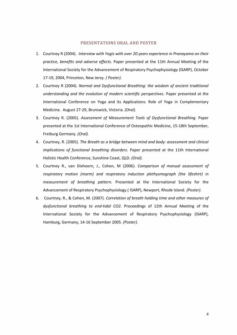
LIST OF PUBLICATIONS ARISING FROM THIS THESIS
PEER REVIEWED PUBLICATIONS
1. Courtney R (2005). Breathing Therapies. Journal of Complementary Medicine, 4(2), 81-85.
2. Courtney R (2008). Strengths, weaknesses and possiblities of the Buteyko Method.
Biofeedback, 36(2), 59-63.
3. Courtney R, Cohen M (2008). Investigating the claims of Konstantin Buteyko M.D., PhD: The
relationship of Breath Holding Time to end tidal CO2 and other measures of dysfunctional
breathing. Journal of Alternative and Complementary Medicine, 14(2),115-123.
4. Courtney R, Dixhoorn Jv, Cohen M (2008). Evaluation of breathing pattern: comparison of a
Manual Assessment of Respiratory Motion (MARM) and respiratory induction
plethysmography. Applied Psychophysiology and Biofeedback, 33, 91-100.
5. Courtney R, Greenwood KM (2009). Preliminary investigation of a measure of dysfunctional
breathing symptoms: the Self Evaluation of Breathing Questionnaire (SEBQ). International
Journal of Osteopathic Medicine, 12, 121-127.
6. Courtney R,Reece J (2009). Comparison of the Manual Assessment of Respiratory Motion
(MARM) and the Hi Lo breathing assessment determining a simulated breathing pattern.
International Journal of Osteopathic Medicine, 12, 86-91.
7. Courtney R (2009). Functions and dysfunctions of breathing and their relationship to
breathing therapy. International Journal of Osteopathic Medicine,12, 78-85.
8. Courtney R, Greenwood K (2011). "Relationships between measures of dysfunctional
breathing in a population with concerns about their breathing." Journal of Bodywork and
Movement Therapies 15(1): 24-34.
9. Courtney R, Greenwood KM., Dixhoorn Jv., Anthonnison E. (2011) “'Medically unexplained
dyspnea partly mediated by dysfunctional (thoracic dominant) breathing pattern' Journal of
Asthma, 48(30):259-65
10. Courtney R., Cohen M., Dixhoorn, Jv., Greenwood K. (2011) “Relationship of spontaneous
breathing pattern and ability to achieve “coherence” of heart rate variability”. Alternative
Therapies in Health and Medicine, 17(3).
.
4
PRESENTATIONS ORAL AND POSTER
1. Courtney R (2004). Interview with Yogis with over 20 years experience in Pranayama on their
practice, benefits and adverse effects. Paper presented at the 11th Annual Meeting of the
International Society for the Advancement of Respiratory Psychophysiology (ISARP), October
17-19, 2004, Princeton, New Jersy. ( Poster).
2. Courtney R (2004). Normal and Dysfunctional Breathing: the wisdom of ancient traditional
understanding and the evolution of modern scientific perspectives. Paper presented at the
International Conference on Yoga and its Applications: Role of Yoga in Complementary
Medicine. August 27-29, Brunswick, Victoria. (Oral).
3. Courtney R. (2005). Assessment of Measurement Tools of Dysfunctional Breathing. Paper
presented at the 1st International Conference of Osteopathic Medicine, 15-18th September,
Freiburg Germany. (Oral).
4. Courtney, R. (2005). The Breath as a bridge between mind and body: assessment and clinical
implications of functional breathing disorders. Paper presented at the 11th International
Holistic Health Conference, Sunshine Coast, QLD. (Oral).
5. Courtney R., van Dixhoorn, J., Cohen, M (2006). Comparison of manual assessment of
respiratory motion (marm) and respiratory induction plethysmograph (the lifeshirt) in
measurement of breathing pattern. Presented at the International Society for the
Advancement of Respiratory Psychophysiology.( ISARP), Newport, Rhode Island. (Poster).
6. Courtney, R., & Cohen, M. (2007). Correlation of breath holding time and other measures of
dysfunctional breathing to end-tidal CO2. Proceedings of 12th Annual Meeting of the
International Society for the Advancement of Respiratory Psychophysiology (ISARP),
Hamburg, Germany, 14-16 September 2005. (Poster).

5
ACKNOWLEDGEMENTS
The long journey of completing this thesis was sustained by a fascination with
breathing therapy that came initially from observing some of my patients whose
health seemed to be completely transformed as a result of the diligent practice
of breathing therapy. I would like to thank these people who without exception
showed great dedication and a firm commitment in their efforts to help
themselves. From them came the first breath of inspiration.
I am grateful to all the researchers, clinicians and practitioners of
breathing therapy who have gone before me and developed the large body of
ideas and knowledge that this thesis rests on. I am particularly grateful to the
members of the International Society for the Advancement of Respiratory
Psychophysiolgy (ISARP). The high quality ISARP meetings showed me how
science is applied to this field of breathing therapy. I feel very fortunate to have
met scientists of the highest caliber at these meetings including Dr Jan van
Dixhoorn who became a collaborator on a number of projects and manuscripts
included in this thesis. Dr van Dixhoorn and Els Anthonissen also provided the
raw data for final study in this thesis, which he and Els had collected over
several years from their patients at the Center for Breathing Therapy in
Amersfoort, the Netherlands.
Practitioners of the Buteyko Breathing Method were also very helpful to
me during this research project providing information on this technique and its
theoretical underpinnings.
To the editors and reviewers of the journals to which I submitted the
various manuscripts I feel deep gratitude. Their editorial comments and
suggestions always resulted in a better manuscript and their input slowly trained
me in the craft of scientific writing.
Of course I could not have done it without help from my supervisors,
Professors Marc Cohen and Ken Greenwood and Dr Ray Myers who provided
excellent guidance, critical appraisal and support during my candidature. They
were unfailingly patient and supportive with my early attempts at writing, and
without the slightest show of annoyance. The clarity of their thinking lit the way
out of several murky holes during the course of this thesis.

6
Prof Marc Cohen and Ken Greenwood were both co-authors on several of
the published manuscripts. Dr John Reece provided statistical support and was
the co-author on one of the published manuscripts. Dr Cliff da Costa also
provided statistical advice.
The Australian Osteopathic Association provided financial assistance
through the National Fund for Research into Osteopathy. These funds provided
much needed assistance for research costs and were greatly appreciated.
Thanks to Dr Ray Myers who allocated funds for the capnometer used in this
project despite the other pressing funding needs of the RMIT Osteopathic
department.
I am very grateful to all the research volunteers who patiently co-
operated in the various research projects and generously gave their time. Also to
the medical and allied health pracititioner‟s who referred people to the research
projects. The Student Osteopathic Medicine Association assisted by advertising
for research participants for some of research studies. The Osteopathic schools
at Victoria University and at RMIT University and the Australian Osteopathic
Association provided rooms and administrative support for some of the research
projects. Kylie Spencer and Phillip Loft did a fantastic job as examiners in the
first MARM and LifeShirt study.
Last but not least, my love and gratitude to family and friends who gave
support and encouragement. Many friends and colleagues gave me places to
stay and good company when I travelled to Melbourne. Thanks especially to
Laura and Martin Sykes in this regard. My sons Aubrey and Cael and my
husband Michael, my nearest and dearest, apart from sweetening my life also
helped in practical ways. Aubrey by finding difficult-to-find articles in the stacks
of the medical library as Sydney University despite being busy with his own
university studies. Cael by helping me in the clinic and at home with so many
tasks and Michael, who proved that he is the world‟s kindest and most
committed husband by actually reading and editing my work.

7
TABLE OF CONTENTS
DECLARATION BY CANDIDATE ......................................................................................................................... 2
LIST OF PUBLICATIONS ARISING FROM THIS THESIS .......................................................................... 3
ACKNOWLEDGEMENTS ........................................................................................................................................... 5
TABLE OF CONTENTS ............................................................................................................................................... 7
INDEX OF TABLES AND FIGURES ................................................................................................................... 14
LIST OF ABBREVIATIONS .................................................................................................................................... 18
THESIS ABSTRACT ................................................................................................................................................... 20
CHAPTER 1-INTRODUCTION .............................................................................................................................. 22
Background ..................................................................................................................................... 22
The Objectives of this Thesis ...................................................................................................... 23
Evolution of the Research Questions in the Experimental Portion of the Thesis .......... 24
Organisation of Chapters ............................................................................................................. 27
CHAPTER 2- THE FUNCTIONS OF BREATHING ....................................................................................... 29
Introduction .................................................................................................................................... 29
Primary Functions of Breathing ................................................................................................ 31
The Respiratory Pump ................................................................................................................. 31
Maintenance of levels of O2, CO2 and pH ............................................................................. 32
Special Functions of Breathing ................................................................................................... 33
Postural and Motor Control ........................................................................................................ 33
Speech and Vocalisation ............................................................................................................. 33
Psychophysiological Functions of Breathing ....................................................................... 34
Breathing, Oscillations and Homeostasis .............................................................................. 36
A Bridge Between Mind and Body: The Somatic Perspective ....................................... 37
Esoteric and Psychospiritual Functions of Breathing ........................................................ 38
Salutongenic and Therapeutic Breathing ............................................................................... 39

8
Conclusion ....................................................................................................................................... 42
CHAPTER 3: HYPERVENTILATION DISORDERS ..................................................................................... 43
Introduction .................................................................................................................................... 43
Definitions ........................................................................................................................................ 43
Hyperventilation ............................................................................................................................ 43
Chronic Hyperventilation ........................................................................................................... 44
Intermittent Hyperventilation .................................................................................................... 44
Hyperventilation Syndrome ....................................................................................................... 44
History of Hyperventilation Syndrome .................................................................................... 45
Doubts about the role of Hypocapnia in HVS ...................................................................... 47
The Hyperventilation Provocation Test and acute Hypocapnia in HVS...................... 47
Prevalence of Hyperventilation Disorders ............................................................................... 48
Etiology of Hyperventilation Disorders .................................................................................... 49
Behavioral and Psychological Causes .................................................................................... 49
Organic Causes .............................................................................................................................. 50
Perpetuating Factors in the Etiology of HV .......................................................................... 52
Signs and Symptoms ..................................................................................................................... 55
Pathophysiology ............................................................................................................................. 56
Investigation and Differential Diagnosis .................................................................................. 60
Measuring CO2 Levels ................................................................................................................. 60
Hyperventilation Provocation Test .......................................................................................... 61
Breath Holding Time in Hyperventilation Disorders ......................................................... 62
Buteyko’s formula ......................................................................................................................... 62
Symptom Questionnaires ............................................................................................................ 63
Interventions for Hyperventilation Disorders ........................................................................ 64
Buteyko Breathing Technique ................................................................................................... 64
Conclusions...................................................................................................................................... 65

9
CHAPTER 4 - THE FUNCTIONS AND DYSFUNCTIONS OF BREATHING PATTERN ............. 67
Introduction .................................................................................................................................... 67
Normal Breathing .......................................................................................................................... 67
The Chest Wall ............................................................................................................................... 68
The Muscles of Respiration ....................................................................................................... 69
Timing and Volume Components of Normal Breathing ................................................... 74
Regularity of Normal Breathing ............................................................................................... 75
Dysfunctional Patterns of Breathing ......................................................................................... 76
Dysfunctions of breathing co-ordination ............................................................................... 76
Abnormalities of Timing and Volume .................................................................................... 78
Incidence of Breathing Pattern Disorders ............................................................................... 79
Etiology of dysfunctional breathing patterns .......................................................................... 81
Biomechanical Factors................................................................................................................. 81
Behavioral and Psychological Factors .................................................................................... 84
Organic Disease ............................................................................................................................. 85
Consequences of Breathing Pattern Dysfunctions ................................................................. 86
Hyperventilation and Hypoventilation ................................................................................... 87
Haemodynamics and Lymphatic System ............................................................................... 88
Posture and Motor Control ......................................................................................................... 89
Musculo-Skeletal Pain ................................................................................................................. 90
Other .................................................................................................................................................. 90
Evaluation of Breathing Pattern ................................................................................................ 90
Concluding Remarks .................................................................................................................... 91
CHAPTER 5- DYSFUNCTIONAL BREATHING ........................................................................................... 92
Introduction .................................................................................................................................... 92
Use of the term ‘Dysfunctional Breathing’ .............................................................................. 92
Assessment of Dysfunctional Breathing ................................................................................... 93

10
Relationship of Dysfunctional Breathing to Hyperventilation and Breathing Pattern
Disorders .......................................................................................................................................... 93
Psychophysiological Aspects of Dysfunctional Breathing .................................................... 94
Breathing, Homeostasis and Hyperarousal ........................................................................... 94
Perception, Dyspnea and Control of Breathing: Factors in Self Regulation .............. 96
Inaccurate Perception of Breathing.......................................................................................... 96
Pleasantness and Unpleasantness of Breathing Sensations .............................................. 97
Flexibiilty and Responsiveness of Breathing ........................................................................... 98
Dysfunctional Breathing and Breathing Therapies ............................................................... 99
Concluding remarks ................................................................................................................... 100
OVERVIEW SECTION ONE- MEASURING BREATHING PATTERN102
CHAPTER 6 - .............................................................................................................................................................. 103
EVALUATION OF BREATHING PATTERN: COMPARISON OF A MANUAL ASSESSMENT
OF RESPIRATORY MOTION (MARM) AND RESPIRATORY INDUCTION
PLETHYSMOGRAPHY .......................................................................................................................................... 103
Abstract: ....................................................................................................................................... 103
Introduction: ................................................................................................................................ 104
Method........................................................................................................................................... 108
Results............................................................................................................................................ 112
Discussion ..................................................................................................................................... 121
Conclusion: ................................................................................................................................... 125
CHAPTER 7- ............................................................................................................................................................... 126
COMPARISON OF THE MANUAL ASSESSMENT OF RESPIRATORY MOTION (MARM)
AND THE HI LO BREATHING ASSESSMENT IN DETERMINING A SIMULATED
BREATHING PATTERN. ...................................................................................................................................... 126
Abstract ......................................................................................................................................... 126
Introduction ................................................................................................................................. 128
Method........................................................................................................................................... 130
Results............................................................................................................................................ 133
11
Discussion ..................................................................................................................................... 138
Conclusions................................................................................................................................... 141
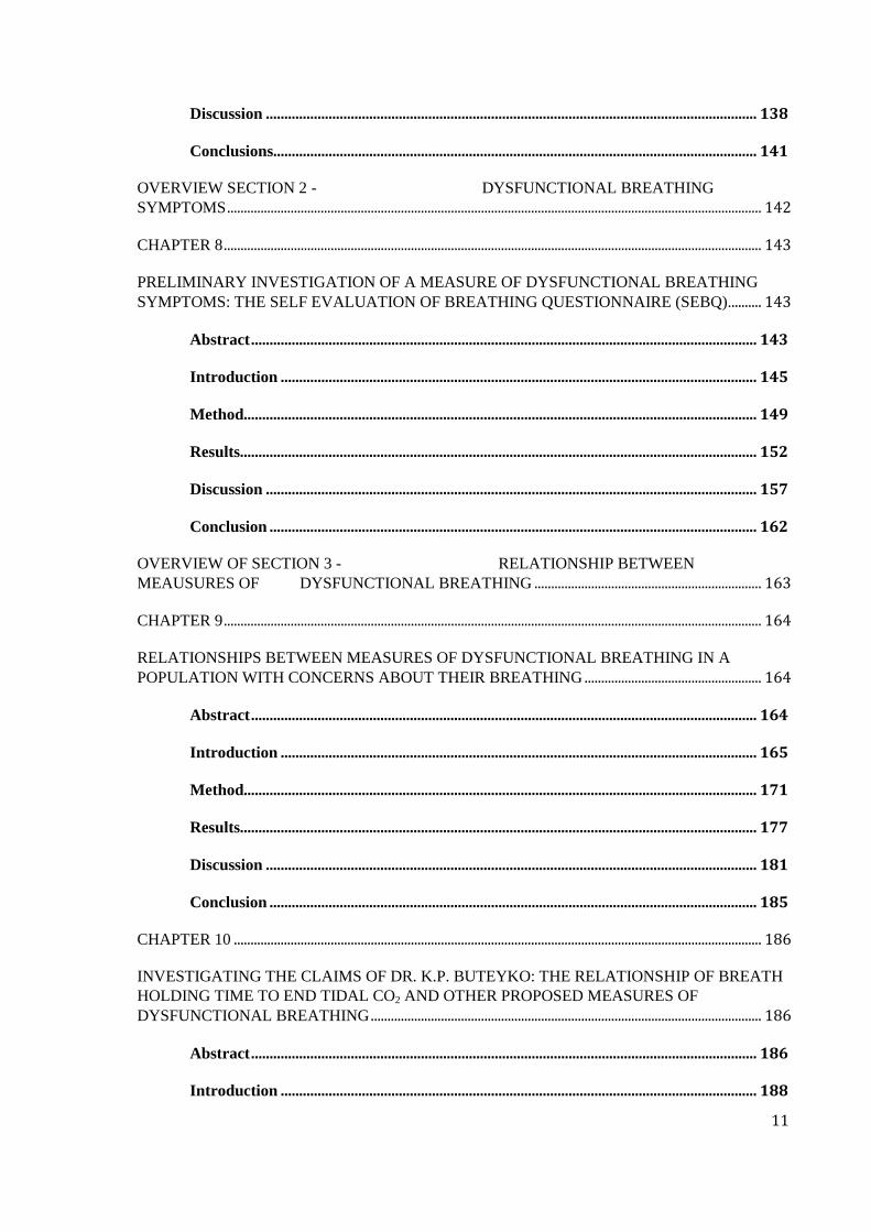
OVERVIEW SECTION 2 - DYSFUNCTIONAL BREATHING
SYMPTOMS ................................................................................................................................................................ 142
CHAPTER 8 ................................................................................................................................................................. 143
PRELIMINARY INVESTIGATION OF A MEASURE OF DYSFUNCTIONAL BREATHING
SYMPTOMS: THE SELF EVALUATION OF BREATHING QUESTIONNAIRE (SEBQ).......... 143
Abstract ......................................................................................................................................... 143
Introduction ................................................................................................................................. 145
Method........................................................................................................................................... 149
Results............................................................................................................................................ 152
Discussion ..................................................................................................................................... 157
Conclusion .................................................................................................................................... 162
OVERVIEW OF SECTION 3 - RELATIONSHIP BETWEEN
MEAUSURES OF DYSFUNCTIONAL BREATHING .................................................................... 163
CHAPTER 9 ................................................................................................................................................................. 164
RELATIONSHIPS BETWEEN MEASURES OF DYSFUNCTIONAL BREATHING IN A
POPULATION WITH CONCERNS ABOUT THEIR BREATHING ..................................................... 164
Abstract ......................................................................................................................................... 164
Introduction ................................................................................................................................. 165
Method........................................................................................................................................... 171
Results............................................................................................................................................ 177
Discussion ..................................................................................................................................... 181
Conclusion .................................................................................................................................... 185
CHAPTER 10 .............................................................................................................................................................. 186
INVESTIGATING THE CLAIMS OF DR. K.P. BUTEYKO: THE RELATIONSHIP OF BREATH
HOLDING TIME TO END TIDAL CO2 AND OTHER PROPOSED MEASURES OF
DYSFUNCTIONAL BREATHING ..................................................................................................................... 186
Abstract ......................................................................................................................................... 186
Introduction ................................................................................................................................. 188

12
Methods ......................................................................................................................................... 190
Results. .......................................................................................................................................... 194
Discussion ..................................................................................................................................... 200
Conclusion .................................................................................................................................... 202
OVERVIEW OF SECTION 4 - MEASURES OF DYSFUNCTIONAL
BREATHING AND BREATHING THERAPY ............................................................... 204
CHAPTER 11 .............................................................................................................................................................. 205
RELATIONSHIP BETWEEN DYSFUNCTIONAL BREATHING PATTERNS AND ABILITY TO
ACHIEVE TARGET HEART RATE VARIABILITY WITH FEATURES OF “COHERENCE”
DURING BIOFEEDBACK ..................................................................................................................................... 205
CHAPTER 12 .............................................................................................................................................................. 206
MEDICALLY UNEXPLAINED DYSPNEA: PARTLY MODERATED BY DYSFUNCTIONAL
(THORACIC DOMINANT) BREATHING PATTERN ............................................................................... 206
Abstract ......................................................................................................................................... 206
Introduction ................................................................................................................................. 208
Method........................................................................................................................................... 211
Results............................................................................................................................................ 213
Discussion ..................................................................................................................................... 220
Conclusion .................................................................................................................................... 223
CHAPTER 13 .............................................................................................................................................................. 224
DISCUSSION .............................................................................................................................................................. 224
Introduction ................................................................................................................................. 224
Aims and Objectives................................................................................................................... 224
Principal Findings....................................................................................................................... 224
Assessment of Dysfunctional Breathing and its measures ............................................. 225
Symptom Questionnaires .......................................................................................................... 227
Comprehensive Assessment of Dysfunctional Breathing ............................................... 232
Definining Dysfunctional Breathing ..................................................................................... 232
Possible Mechanisms of Breathing Therapy ...................................................................... 236

13
Implications for Breathing Therapies .................................................................................... 240
Limitations.................................................................................................................................... 241
Future Research .......................................................................................................................... 242
Final Conclusions ........................................................................................................................ 243
References: ................................................................................................................................... 245
APPENDICES .............................................................................................................................. 284
14
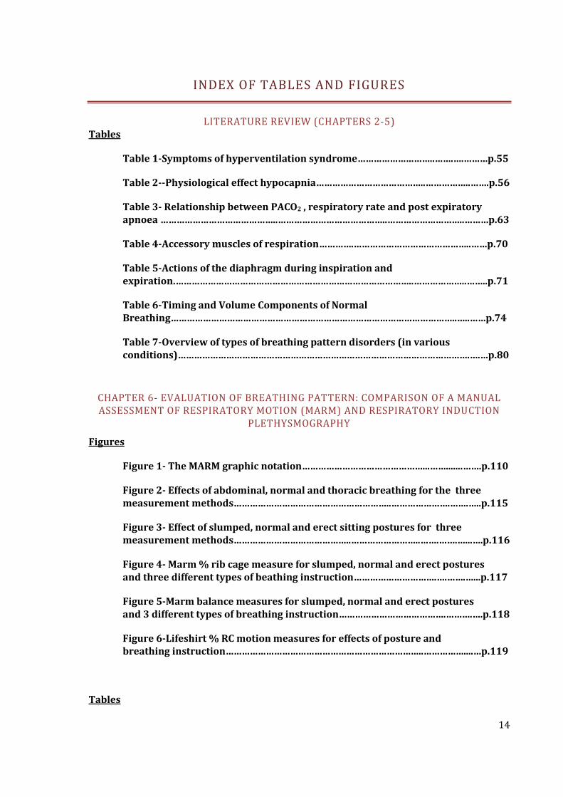
INDEX OF TABLES AND FIGURES
LITERATURE REVIEW (CHAPTERS 2-5) Tables
Table 1-Symptoms of hyperventilation syndrome………………………..…….….………p.55
Table 2--Physiological effect hypocapnia…………………………………..……………..…….p.56
Table 3- Relationship between PACO2 , respiratory rate and post expiratory apnoea ……………………………………..…………………………………..………………………..………p.63
Table 4-Accessory muscles of respiration………….……………………………………..……p.70
Table 5-Actions of the diaphragm during inspiration and expiration.……………………………………………………………………………..………………..……..p.71
Table 6-Timing and Volume Components of Normal Breathing……………………………………………………………………………………………..…..……p.74
Table 7-Overview of types of breathing pattern disorders (in various conditions)……………………………………………………………………………………………….….…p.80
CHAPTER 6- EVALUATION OF BREATHING PATTERN: COMPARISON OF A MANUAL ASSESSMENT OF RESPIRATORY MOTION (MARM) AND RESPIRATORY INDUCTION
PLETHYSMOGRAPHY
Figures
Figure 1- The MARM graphic notation………………………………………...…….......…….p.110
Figure 2- Effects of abdominal, normal and thoracic breathing for the three measurement methods…………………………………………………..………………….…….…..p.115
Figure 3- Effect of slumped, normal and erect sitting postures for three measurement methods……………………………………..……………………..…….…….…...….p.116
Figure 4- Marm % rib cage measure for slumped, normal and erect postures and three different types of beathing instruction………………………….….…….…...p.117
Figure 5-Marm balance measures for slumped, normal and erect postures and 3 different types of breathing instruction………………………………….……….….p.118
Figure 6-Lifeshirt % RC motion measures for effects of posture and breathing instruction………………………………………………………………..……………....…p.119
Tables
15
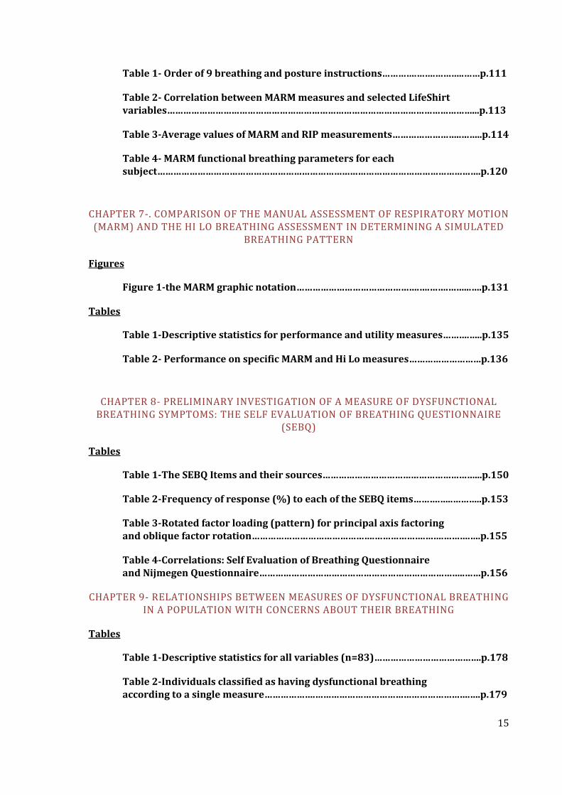
Table 1- Order of 9 breathing and posture instructions………….…….………..……p.111
Table 2- Correlation between MARM measures and selected LifeShirt variables……………………………………………………………………………………………………...p.113
Table 3-Average values of MARM and RIP measurements……………………..……..p.114
Table 4- MARM functional breathing parameters for each subject………………………………………………………………………………………………………….p.120
CHAPTER 7-. COMPARISON OF THE MANUAL ASSESSMENT OF RESPIRATORY MOTION (MARM) AND THE HI LO BREATHING ASSESSMENT IN DETERMINING A SIMULATED
BREATHING PATTERN
Figures
Figure 1-the MARM graphic notation……………………………………….….…….……...….p.131
Tables
Table 1-Descriptive statistics for performance and utility measures……….…..p.135
Table 2- Performance on specific MARM and Hi Lo measures………………………p.136
CHAPTER 8- PRELIMINARY INVESTIGATION OF A MEASURE OF DYSFUNCTIONAL BREATHING SYMPTOMS: THE SELF EVALUATION OF BREATHING QUESTIONNAIRE
(SEBQ)
Tables
Table 1-The SEBQ Items and their sources…………………………………………………...p.150
Table 2-Frequency of response (%) to each of the SEBQ items……….…..………..p.153
Table 3-Rotated factor loading (pattern) for principal axis factoring and oblique factor rotation……………………………………….……………….…….……….….p.155
Table 4-Correlations: Self Evaluation of Breathing Questionnaire and Nijmegen Questionnaire…………………………………………………………………..……p.156
CHAPTER 9- RELATIONSHIPS BETWEEN MEASURES OF DYSFUNCTIONAL BREATHING IN A POPULATION WITH CONCERNS ABOUT THEIR BREATHING
Tables
Table 1-Descriptive statistics for all variables (n=83)………………………………….p.178
Table 2-Individuals classified as having dysfunctional breathing according to a single measure……………….………………………………………………….….p.179
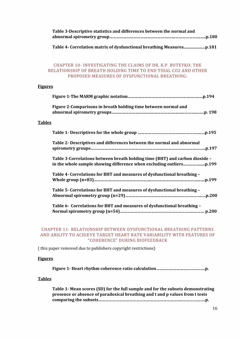
16
Table 3-Descriptive statistics and differences between the normal and abnormal spirometry group……………………………………………..…..………………….…..p.180
Table 4- Correlation matrix of dysfunctional breathing Measures.....................p.181
CHAPTER 10- INVESTIGATING THE CLAIMS OF DR. K.P. BUTEYKO: THE RELATIONSHIP OF BREATH HOLDING TIME TO END TIDAL CO2 AND OTHER
PROPOSED MEASURES OF DYSFUNCTIONAL BREATHING:
Figures
Figure 1-The MARM graphic notation……………………………………………….…….….p.194
Figure 2-Comparisons in breath holding time between normal and abnormal spirometry groups………………………….…………………….……..………....…..p. 198
Tables
Table 1- Descriptives for the whole group ………………………………………………..…p.195
Table 2- Descriptives and differences between the normal and abnormal spirometry groups………………………………………………………..……………………….……..p.197
Table 3-Correlations between breath holding time (BHT) and carbon dioxide – in the whole sample showing difference when excluding outliers……..……......p.199
Table 4- Correlations for BHT and measures of dysfunctional breathing – Whole group (n=83)…………….……………………………..……………………….…………...….p.199
Table 5- Correlations for BHT and measures of dysfunctional breathing –Abnormal spirometry group (n=29)…………….……………………………………..……..…p.200
Table 6- Correlations for BHT and measures of dysfunctional breathing – Normal spirometry group (n=54)………………………………………………………..……… p.200
CHAPTER 11- RELATIONSHIP BETWEEN DYSFUNCTIONAL BREATHING PATTERNS AND ABILITY TO ACHIEVE TARGET HEART RATE VARIABILITY WITH FEATURES OF
“COHERENCE” DURING BIOFEEDBACK
( this paper removed due to publishers copyright restrictions)
Figures
Figure 1- Heart rhythm coherence ratio calculation…………………………………….p.
Tables
Table 1- Mean scores (SD) for the full sample and for the subsets demonstrating presence or absence of paradoxical breathing and t and p values from t tests comparing the subsets………………………………………………………………..…………….….p.
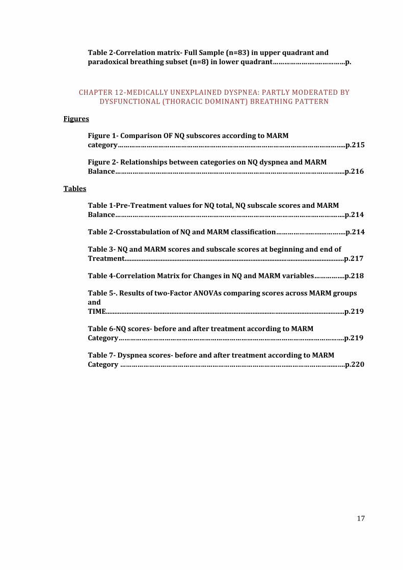
17
Table 2-Correlation matrix- Full Sample (n=83) in upper quadrant and paradoxical breathing subset (n=8) in lower quadrant………………….….…………p.
CHAPTER 12-MEDICALLY UNEXPLAINED DYSPNEA: PARTLY MODERATED BY DYSFUNCTIONAL (THORACIC DOMINANT) BREATHING PATTERN
Figures
Figure 1- Comparison OF NQ subscores according to MARM category………………………………………………………………………………………………………..p.215
Figure 2- Relationships between categories on NQ dyspnea and MARM Balance………………………………………………………………………………………………………...p.216
Tables
Table 1-Pre-Treatment values for NQ total, NQ subscale scores and MARM Balance…………………………………………………………………………………………….……….….p.214
Table 2-Crosstabulation of NQ and MARM classification………………..…....……….p.214
Table 3- NQ and MARM scores and subscale scores at beginning and end of Treatment...............................................................................................................................p.217
Table 4-Correlation Matrix for Changes in NQ and MARM variables…………….p.218
Table 5-. Results of two-Factor ANOVAs comparing scores across MARM groups and TIME..........................................................................................................................................p.219
Table 6-NQ scores- before and after treatment according to MARM Category………………………………………………….……………………………………..…………….p.219
Table 7- Dyspnea scores- before and after treatment according to MARM Category ……………………………………………………………………………...…………………...….p.220
18
LIST OF ABBREVIATIONS
BBT Buteyko Breathing Technique
BHT Breath holding time
BHT-DD Breath holding time till first desire to breathe
BHT-IRM Breath holding time till involuntary muscle motion
CO2 Carbon Dioxide
COPD Chronic Obstructive Pulmonary Disease
CP Control Pause
CSF Cerebral spinal fluid
DB Dysfunctional breathing
ETCO2 End Tidal CO2
FEV1 Forced expiratory volume in 1 second
FVC Forced vital capacity
HGYB How Good is Your Breathing Test
HRV Heart Rate Variability
HVPT Hyperventilation Provocation Test
HVS Hyperventilation Syndrome
LRCA Lower Rib Cage Abdomen
MARM Manual Assessment of Respiratory Motion
MARM%R Manual Assessment of Respiratory Motion percentage of rib cage motion
M%RC Mean % of rib cage contribution to tidal volume as measured by RIP.
MP Maximum Pause
NQ Nijmegen Questionnaire
OEP Optoelectronic Plethysmography
PaCO2 Partial pressure of arterial carbon dioxide
PACO2 Partial pressure of alveolar carbon dioxide
PO2 Partial pressure of oxygen
QCT Quick Coherence Technique
19
RIP Respiratory Induction Plethysmography
SEBQ Self Evaluation of Breathing Questionnaire
SPO2 Oxygen saturation of hemoglobin
TE Time taken for exhalation
TI Time taken for inhalation
TI/Ttot Fractional inspiratory time
Ttot Duration of whole cycle of breath
URC Upper rib cage
Vmin Minute volume
VT Tidal Volume
VT/TI Mean inspiratory flow
WBB Whole Body Breathing

Abstract
20
THESIS ABSTRACT
Background- The commonly held idea that „proper‟ breathing is important for
health is generally accompanied by the view that incorrect or „dysfunctional‟
breathing has adverse effects on health. It has been proposed that correction of
dysfunctional breathing through breathing therapy reduces symptoms and
improves the health of patients with conditions such as asthma, anxiety,
speech disorders, chronic muscular skeletal dysfunction and medically
unexplained physical symptoms. However, investigation of the impact of
dysfunctional breathing and breathing therapies is hampered by the fact that
dysfunctional breathing is not well defined and that there are few validated
measures or standardized protocols to measure it.
Aims - The four main objectives of the experimental portion of thesis are:
To further develop and validate some clinical tools for assessment of
dysfunctional breathing;
To explore and evaluate current methods for assessing dysfunctional
breathing particularly those used by breathing therapists;
To investigate the relationships between measures of dysfunctional
breathing with a view to understanding the possible definitions and
dimensions of dysfunctional breathing; and
To explore possible mechanisms of breathing therapy.
Format- A series of seven studies were untaken. These papers, which are all
published (or in press) manuscripts, form the experimental portion of this thesis.
The seven studies presented in the empirical section of this thesis are grouped in
four sub-sections- The first of these contains two studies on the topic of
„Measuring Breathing Pattern‟, the second contains one study on the topic
„Evaluating Dysfunctional Breathing Symptoms‟, the third include two studies
that investigate „Relationships between Measures of Breathing Functionality‟ and
the final sub-section contains two studies that explores „Measures of
Dysfunctional Breathing and Breathing Therapy‟.
Methods-Various non-experimental and quasi-experimental study designs were
used. The studies reported in Chapters 6 and 7 tested inter-examiner reliability
and accuracy of manual breathing assessment tools. Chaper 8 involved
Abstract
21
administration of a survey with subsequent exploratory factor analysis of the
survey items. Chapters 9, 10, 11 and were primarily correlational studies.
Chaper 12 was a post-hoc analysis of clinical data. The participants in the study
reported in Chapter 6 were 10 Yoga teachers or breathing therapists and 3
Osteopaths as examiners. The participants in the study reported in Chapter 7
were 56 Osteopaths and Osteopathic students. The 83 participants in the studies
reported in Chapters 8,9,10 and 11 were either healthy or suffered from mild
medical conditions. The 62 participants in the study reported in Chapter 12 were
consecutive patients receiving treatment with a method of breathing and
relaxation therapy called Whole Body Breathing.
Results/Discussion - Manual techniques for evaluating breathing pattern
appear to be useful and reasonably accurate for evaluating thoracic dominant
breathing and paradoxical breathing. The Self Evaluation of Breathing
Questionnaire appears to have two dimensions that might be related to
biochemical and biomechanical aspects of breathing dysfunction. Poor
relationships between different categories of measures of dysfunctional
breathing suggest that dysfunctional breathing has several dimensions.
Breathing pattern appears to moderate dyspnea and altering breathing pattern
may be one mechanism of breathing therapy.
Conclusion - While dysfunctional breathing cannot be strictly defined at
present, for practical purposes dysfunctional breathing is probably best
characterized as multi-dimensional. Dysfunctional breathing can occur in at least
three dimensions: biochemical, breathing pattern and breathing related
symptoms and these might not co-exist. Comprehensive measurement of
dysfunctional breathing should include measures that evaluate all these
dimensions. The therapeutic mechanisms of breathing theory are likely to be
complex and include psychological, biomechanical and physiological parameters.
However, dysfunctional patterns of breathing are one factor that influences
patients‟ response to breathing therapy. Also correction of dysfunctional
breathing patterns is one likely therapeutic pathway of breathing therapy,
particularly for patients with dyspnea.

Introduction
22
CHAPTER 1-INTRODUCTION
BACKGROUND
This thesis explores dysfunctional breathing, its measurement, possible
definitions and its relationship to breathing therapy.
Many diverse groups, including respiratory psychophysiologists, physical
therapists, somatic or mind/body therapists and practitioners of Yoga, Qi Gong
or specialized breathing therapies like the Buteyko Breathing Technique share
the belief that breathing can be salutogenic and therapeutic. To various
degrees, they hold the view that the optimization of breathing is a means to
improving health of the mind and body. A corollary to this view is that
dysfunctional breathing is both a cause and consequence of poor health.
Various types of breathing therapy have been used, with apparent
success, to treat conditions such as asthma, anxiety, speech disorders, chronic
musculoskelatal dysfunction and medically unexplained physical symptoms.
However, research into the use of breathing therapies in these conditions is
hampered by the fact that their mode of action is not well understood.
One proposed mechanism of breathing therapies is that they correct
dysfunctional breathing; yet dysfunctional breathing does not have a precise
definition. The term can refer to hyperventilation, breathing pattern disorders
or medically unexplained dyspnea but in a broad sense the term can also be
taken to imply a disturbance of any function of breathing.
Dysfunctional breathing is evaluated using of a range of measures, such
as levels of oxygen and carbon dioxide, breathing pattern, breath holding time
and symptom questionnaires. These measures can be seen to be representative
of biochemical, biomechanical, psychological and interoceptive parameters. The
relationships between these various parameters are not well understood. It is
not clear if these measures represent distinct or related aspects of breathing
functionality, and it is also not clear which measures should be used in a
comprehensive evaluation of dysfunctional breathing or in clinical assessments.
Introduction
23
Improvements in dysfunctional breathing measures, such as breath
holding time, breathing pattern, carbon dioxide measures and symptom
questionnaires, have, at different times, been reported to be associated with
positive therapeutic responses in patients practicing different types of breathing
therapies. Therefore, a better understanding of these measures may be the key
to developing understanding of the mechanisms of breathing therapies
Finally, some measures of dysfunctional breathing, including the Manual
Assessment of Respiratory Motion (MARM) and the Hi Lo breathing assessment,
appear promising, but have not been adequately validated. Also, symptoms
proposed to indicate the presence of dysfunctional breathing might provide the
basis for a dysfunctional breathing questionnaire. This thesis begins by further
developing and evaluating the MARM and the Hi Lo and then describes the
development and factor analysis of a new questionnaire, the Self Evaluation of
Breathing Questionnaire (SEBQ) for evaluating dysfunctional breathing
symptoms.
THE OBJECTIVES OF THIS THESIS
The overall objectives of this thesis are to explore possible definitions of
dysfunctional breathing and gain a better understanding of the role of
dysfunctional breathing in the efficacy of breathing therapies. These objectives,
which are the focus of the experimental portion of this thesis, are approached
through an exploration of the measures of dysfunctional breathing, particularly
those used by proponents of breathing therapies.
The main objectives of the experimental portion of thesis are:
To further develop and validate some clinical tools for assessment of
dysfunctional breathing;
To explore and evaluate current methods for assessing dysfunctional
breathing particularly those used by breathing therapists;
To investigate the relationships between measures of dysfunctional
breathing with a view to understanding the possible definitions and
dimensions of dysfunctional breathing; and
To explore possible mechanisms of breathing therapy.
Introduction
24
EVOLUTION OF THE RESEARCH QUESTIONS IN THE EXPERIMENTAL PORTION OF THE THESIS
The specific research questions addressed in the individual studies that make
up the experimental portion of this thesis evolved over the course of this
research. All the papers in the experimental portion of the thesis have either
been published or are in press. The order in which these studies are presented
is not the same order in which they were performed or in the order that
manuscripts were published. This section of the introduction describes the
progression of the studies, and the evolution of the research questions
addressed in this thesis to assist the reader in understanding how they are
linked.
One of the initial objectives of this thesis was to investigate the
assumption of a breathing therapy called the Buteyko Breathing Technique
(BBT) that hypocapnia was the most important aspect of dysfunctional
breathing and that the therapeutic mechanism of the BBT was dependent on
reducing hyperventilation and normalising CO2 levels in the blood and in the
lungs.
Dr. Buteyko, originator of the BBT, developed a formula for assessing
CO2 levels from breathing holding time and assumed that improvements in
patient‟s breath holding time indicated improved CO2 status. The first study
conducted in this thesis tested Dr. Buteyko‟s formula for calculating CO2 from
breath holding time. It also looked at how breath holding time and CO2 levels
correlated with other measures of dysfunctional breathing. This study resulted
in a published study, presented in Chapter 10 titled “Investigating the claims of
Dr. K.P. Buteyko: The relationship of breath holding time to end tidal CO2 and
other proposed measures of dysfunctional breathing”.
Further analysis of the data gathered in this study was used in three
other studies. The first of these, presented in Chapter 9, has been published
and is titled “Relationships between Measures of Dysfunctional Breathing in a
Population with Concerns about Their Breathing”. Given that screening tools for
dysfunctional breathing include measures that might represent distinct aspects
of breathing functionality (e.g. biochemical, biomechanical, psychological or
Introduction
25
physiological), a primary aim of this study was to further understand the
dimensions of dysfunctional breathing and begin to explore what range of
assessments might be necessary for a comprehensive evaluation of breathing.
The second study from this same data set, presented in Chapter 11, is in
press and is titled “Relationship between dysfunctional breathing patterns and
ability to achieve target heart rate variability with features of “coherence”
during biofeedback”. This study was based on the hypothesis that breathing
patterns commonly associated with dysfunctional breathing (i.e. thoracic
breathing and paradoxical breathing) would adversely affect cardiorespiratory
interactions and the amplitude and frequency of heart rate variability.
The third study from this data set, presented in Chapter 8, describes the
development and structural analysis of a questionnaire, the Self Evaluation of
Breathing Questionnaire (SEBQ). This questionnaire was compiled from various
sources and contains items proposed to identify individuals with dysfunctional
breathing. The need for this type of questionnaire arose because the validated
questionnaire that researchers used to identify dysfunctional breathing, the
Nijmegen Questionnaire, was devised to measure hyperventilation syndrome
rather than the broader definition of dysfunctional breathing sought in this
thesis. Therefore a new questionnaire, the SEBQ, was developed and its
structure analyzed. The published manuscript of this study is titled “Preliminary
investigation of a measure of dysfunctional breathing symptoms: The Self
Evaluation of Breathing Questionnaire (SEBQ)”. The main aim of the structural
(factor) analysis of the SEBQ was to explore whether the items of this
questionnaire represented distinct and separate dimensions of dysfunctional
breathing.
Results of these studies and the literature indicated that breathing
pattern was an important aspect of dysfunctional breathing, yet much of the
literature only seemed to define breathing pattern in vague terms. The manual
palpation techniques used in the studies undertaken in this thesis, the Manual
Assessment of Respiratory Motion (MARM) and the Hi Lo appeared promising,
but needed further validation studies to determine their rigour and
Introduction
26
reproducibility. This led to the two studies that investigated the utility and
validity of these manual techniques for measuring breathing pattern.
The first of these published studies, validating the MARM, presented in
Chapter 6, is titled “Evaluation of Breathing Pattern: Comparison of a Manual
Assessment of Respiratory Motion (MARM) And Respiratory Induction
Plethysmography”. The main aims of this study were firstly to compare the
MARM to a gold standard instrument for measuring breathing pattern, the
Respiratory Induction Plethysmograph, and, secondly to determine the level of
agreement achieved by different examiners.
The second study assessing the MARM and Hi Lo, presented in Chapter
7, is titled, “Comparison of the Manual Assessment of Respiratory Motion
(MARM) and the Hi Lo Breathing Assessment in determining a simulated
breathing pattern”. In the previous study, a high level of inter-examiner
reliability was found for the MARM. Given that this previous study had used
very experienced practitioners, it was not clear whether less experienced
practitioners would be able to use these techniques with a similar degree of
accuracy. Therefore, in this second study, the MARM and another technique
called the Hi Lo were taught, in a short two hour training session, to two
groups. One group was made up of experienced manual therapists
(Osteopaths), the other group were students of Osteopathy. The main aim of
this study was to gauge whether a two hour training period was sufficient to
learn these techniques and whether accurate performance in the use of these
techniques was affected by the general level of experience in manual therapy.
The final study, presented in Chapter 12, evaluated the therapeutic
mechanism of an established breathing therapy, called “Whole Body Breathing”.
This study explored the relationships between breathing symptom and
breathing pattern with the aim of determining whether breathing pattern
moderated dysfunctional breathing symptoms. This study, titled “Medically
unexplained dyspnea: partly moderated by dysfunctional (thoracic dominant)
breathing pattern”, has been accepted for publication and is in press.
Introduction
27
The order in which the studies are presented in this thesis is described in
the following section, which also outlines the content of the literature review
chapters.
ORGANISATION OF CHAPTERS
THIS FIRST (CURRENT) CHAPTER IS THE INTRODUCTION
Chapter 1 - Introduction
CHAPTERS 2 - 5 ARE THE LITERATURE REVIEW CHAPTERS
Chapter 2 - The Functions of Breathing explores ideas about the functions
of breathing as a means of understanding the possible parameters of
dysfunctional breathing.
Chapter 3 - Hyperventilation Disorders provides an overview of the
literature on hyperventilation disorders. It examines the changing perspectives
on the role of hyperventilation in dysfunctional breathing.
Chapter 4 - The Functions and Dysfunctions of Breathing Pattern
provides an overview of the functional anatomy of breathing. It also considers
the role of breathing patterns disorders in dysfunctional breathing.
Chapter 5 - Dysfunctional Breathing discusses current usage of the term,
dysfunctional breathing, and the various measures used to assess it. This
chapter also identifies possible parameters of dysfunctional breathing based on
proposed functions of breathing.
CHAPTERS 5 - 12 ARE THE EXPERIMENTAL PORTION OF THIS THESIS
SECTION ONE - CONTAINS TWO STUDIES EXAMINING MANUAL TECHNIQUES FOR EVALUATING
BREATHING PATTERN
Chapter 6 - „Evaluation of breathing pattern: Comparison of a manual
assessment of respiratory motion (MARM) and respiratory induction
plethysmography‟.
Introduction
28
Chapter 7 - „Comparison of the manual assessment of respiratory motion
(MARM) and the Hi Lo breathing assessment in determining a simulated
breathing pattern‟.
SECTION TWO- CONTAINS ONE STUDY WHICH DESCRIBES THE DEVELOPMENT AND
STRUCTURAL ANALYSIS OF A DYSFUNCTIONAL BREATHING QUESTIONNAIRE
Chapter 8 - „Preliminary investigation of a measure of dysfunctional breathing
symptoms: The Self Evaluation of Breathing Questionnaire (SEBQ)‟.
SECTION 3-CONTAINS TWO STUDIES, FROM THE SAME DATA SET, THAT EXPLORE THE
RELATIONSHIPS BETWEEN MEASURES OF DYSFUNCTIONAL BREATHING
Chapter 9 - „Relationships between measures of dysfunctional breathing in a
population with concerns about their breathing‟.
Chapter 10 - ‘Investigating the claims of Dr. K.P. Buteyko: The relationship of
breath holding time to end tidal CO2 and other proposed measures of
dysfunctional breathing‟.
SECTION 4- CONTAINS TWO STUDIES THAT EXPLORE THE RELATIONSHIPS BETWEEN
DYSFUNCTIONAL BREATHING AND BREATHING THERAPY
Chapter 11 - ‘Relationship between dysfunctional breathing patterns and
ability to achieve target heart rate variability with features of “coherence” during
biofeedback‟.
Chapter 12 - „Medically unexplained dyspnea: partly moderated by
dysfunctional (thoracic dominant) breathing pattern‟.
THE FINAL CHAPTER CONTAINS THE THESIS DISCUSSION AND CONCLUSIONS
Chapter 13 - Discussion

The Functions of Breathing
29
CHAPTER 2- THE FUNCTIONS OF BREATHING
INTRODUCTION
To answer the question „what is dysfunctional or abnormal breathing?‟ one can
first enquire „what is normal, functional or optimal breathing?‟ This section of
the literature review explores these questions from the scientific literature and
also from the perspective of some breathing therapies and various religious and
healing traditions, some of which have not been scientifically evaluated but are
of interest because they provide a broad context for later chapters.
Optimal breathing is often described as something that occurs in the
perfectly healthy person who is in a state of physical rest and emotional ease.
The following quote gives this description of the breathing pattern of such a
person:
“During quiet rest a normal man breathes with effortless ease; he is not aware of his breathing or
of a sensation of breathlessness (or dyspnea). His respiratory frequency is usually below 16
breaths per minute and if he is of average size, the tidal volume is less than 600ml” p 599 (Bass
and Gardner 1985).
The Indian yoga literature describes optimal breathing in a person with
peak physical health and emotional ease as slow, regular, even, diaphragmatic
and nasal (Ramacharaka 1904; Swami Rama 1976; Sovik 2000). Taoist yoga
adherents describe perfect breathing as inaudible and “so smooth that the fine
hairs within the nostrils remain motionless” p136 (Blofeld 1978).
It has been argued that equating optimal or functional breathing with an
ideal of breathing found in a healthy, calm and resting person has limitations,
because the average person living in the „real world‟ does not live in a state of
continual physical rest or constant emotional calm. A person‟s breathing, to be
functional, must adapt appropriately to changes in their external environment
and their internal metabolic and emotional conditions. Particular breathing
patterns that appear less than optimal may actually be an appropriate response
to a particular circumstance and individuals may suffer from diseases or other

The Functions of Breathing
30
conditions that benefit from breathing adaptations that are in conflict with
ideals of optimal breathing (Dixhoorn van 2007). Elsa Gindler, the originator of
the Gymnastik movement of the early 1900‟s, from which many prominent
European and American breathing therapies developed, says
“if we observe successful people we can often see that they display a wonderful flexibility in
reacting, in constantly changing from activity to rest. They have flexible breathing or functional
breathing” p9 (Johnson 1995).
Breathing functionality has often been characterized in terms of how well
normal levels of CO2 are maintained (McLaughlin 2009). One breathing therapy,
the Buteyko Breathing Technique, equates optimal breathing and superior
physical health and endurance with the presence of carbon dioxide levels well
above established textbook norms (Buteyko 1990; Stark and Stark 2002).
Breathing functionality is also judged on the basis of the biomechanical
functions of the muscular and skeletal components of the respiratory pump (De
Troyer and Estenne 1988) and other aspects of breathing pattern and
behaviour (Chaitow 2002). Breathing therapists often note breathing patterns
and particular patterns are considered to be more optimal or functional than
others based on their biomechanical efficiency although there is some
argument about how to best characterise an ideal breathing pattern (Kaminoff
2006).
These various ways of viewing functional breathing provide some insight
but do not give a complete picture. In the interests of having a more rounded
view, this thesis starts with the premise that functional breathing is breathing
that performs and adapts its various functions to quickly and appropriately
meet the changing needs of the individual. These needs may be related to
changes in physical, metabolic or psychophysiological conditions.
This leads to the next question explored in this literature review: „What
are the functions of breathing?‟
The primary visible function of breathing involves the biomechanical
action of the respiratory pump, which moves air into and out of the lungs. The
other primary function, related to the first, is biochemical and involves
The Functions of Breathing
31
maintenance of correct arterial Oxygen (O2) and Carbon Dioxide (CO2) levels
and the regulation of the body‟s pH. However, breathing has other functions,
referred to here as secondary functions. For example, breathing affects motor
control and postural stability and plays several roles in physiological and
psychological regulation. Breathing can influence homeostatic functions in other
systems including the autonomic nervous system, the circulatory system, the
vestibular system, pH regulation and metabolism. Breathing is also thought to
have important functions in self-regulation of stress and emotion. It is also
worth taking note that most of the world‟s major religions believe that
breathing has psycho-spiritual functions. These secondary functions are to a
varying extent dependent on the efficiency and functionality of the primary
biomechanical and biochemical functions of breathing, the ability to accurately
perceive breathing sensations, and the efficiency and resilience of breathing
control mechanisms.
PRIMARY FUNCTIONS OF BREATHING
THE RESPIRATORY PUMP
The biomechanical actions of the respiratory pump play a vital role in
influencing the movement of air into and out of the lungs and also influence
pressure and volume fluctuations within the circulatory and lymphatic systems.
The approximate 21,000 breaths taken per day by the average person are
dependent on the actions of the respiratory muscles on the chest wall. Changes
in chest wall dimensions result in pressure changes within the intrapleural and
alveolar spaces in relationship to atmospheric pressure and as a result, air
moves either into or out of the lungs. Before inspiration, intrapleural pressure is
–5 cm H2O and alveolar pressure is 0 cm H2O. During inspiration, alveolar
pressure and intrapleural pressure fall becoming lower than atmospheric
pressure. This establishes a driving pressure and air moves into the lungs
inflating them. On expiration alveolar pressure becomes positive and
intrapleural pressure rises creating movement of air out of the lungs (West
1985).

The Functions of Breathing
32
In addition to the movement of air, the actions of the respiratory pump
affect other systems including the cardiovascular system, the lymphatic system
and the muscular-skeletal system. A functional respiratory pump requires the
properly co-ordinated action of the primary muscles of respiration such as the
diaphragm, scalenes, intercostals and also the secondary muscles of
respiration. An optimally functioning respiratory pump therefore requires
normal length, strength, resting tone and responsiveness of respiratory
muscles.
Functions and dysfunctions of the biomechanics of breathing are
discussed in Chapter 4 of this thesis.
MAINTENANCE OF LEVELS OF O2, CO2 AND PH
Breathing is a key contributor to the biochemical state of the internal milieu. By
exchanging CO2 for O2, breathing controls the fundamental gaseous fuels of
life‟s energy which move between the arterial and venous blood to the alveoli
through the process of diffusion. By altering ventilation to regulate CO2,
breathing also maintains pH in the narrow range needed for health (West 1985;
Lumb 2000).
The health and integrity of the lung tissue and the circulatory system is
fundamental to the maintenance of normal levels of the respiratory gases which
are disturbed by disruptions in breathing or its regulation. This thesis concerns
itself with functional breathing disturbances resulting primarily from
disturbances in regulation of respiration rather than pathology of the heart and
lungs.
Functions and dysfunctions of the biochemical aspects of breathing are
discussed further in Chapter 3 of this thesis.

The Functions of Breathing
33
SPECIAL FUNCTIONS OF BREATHING
POSTURAL AND MOTOR CONTROL
Breathing impacts muscular and skeletal function because the habitual use of
breathing muscles during respiration affects how these muscles are used for
non-breathing movement and postural support.
There are close links between breathing and musculoskeletal function.
Particular movements tend to be tied to specific phases of respiration. For
example, during extension of the spine one tends to inhale and during flexion
one tends to exhale, while lifting a heavy object often leads to the breath being
held in, the Valsalva maneuver and standing on the toes leads to contraction of
the diaphragm (Lewitt 1980).
Many muscles of respiration have key postural functions and are
important for motor control and stability of the spine, neck and shoulder girdle.
Even primary respiratory muscles are important for postural support and
movement. The diaphragm is now known to have postural as well as
respiratory functions (Lewitt 1980; Hodges, Butler et al. 1997; Hodges and
Gandevia 2000). The intercostal muscles are involved in trunk rotation, in
addition to their role in producing rib cage motion for ventilation (De Troyer,
Kelly et al. 1985). In the lower body, the abdominal muscles are considered to
be primarily postural muscles that also have a respiratory function (Lewitt
1980). In the upper body, muscles such as the trapezius, sternomastoid and
pectoral muscles whose primary function is to move the neck and shoulder
girdle, become involved in breathing when respiration or posture is faulty or
when the ventilatory needs of the body increase (Lewitt 1980; Kapreli 2008).
SPEECH AND VOCALISATION
The ability to regulate the motor control of breathing muscles for speech and
vocalization is linked to the efficiency of the biomechanics of breathing and
integration of neuronal control mechanisms of speech and breathing (Gandevia,
Butler et al. 2002). Human speech and emotional expression, requires fine
control of the muscles of breathing beyond that required for normal ventilation

The Functions of Breathing
34
(Bunn and Mead 1971). This fine motor control, integrated with cognitive
factors, is needed to vary normal quiet breathing and enable the production of
long phrases punctuated with quick inspirations placed appropriately to give
proper linguistic meaning and to control pitch and intonation (MacLarnon and
Hewitt 1999).
Stress, emotion and cognitive process can simultaneously affect
breathing and speech mechanisms producing dysfunctional muscle tension in
these two related systems (Chapell 1994). Paradoxical vocal cord dysfunction, a
condition often misdiagnosed as asthma, which often results from severe
psychosocial and intense emotion stress, is associated with respiratory difficulty
and tense patterns of breathing (Leo and Konakanchi 1999). In conditions such
as muscle tension dysphonia, speech muscles contract inappropriately and are
incorrectly coupled with inspiration rather than expiration (Vertigan, Gibson et
al. 2006). In muscle tension dystonia, breathing is generally inadequate to
support proper voice production and most patients show signs of tension in the
neck muscles which also function as accessory muscles of respiration (Altman,
Atkinson et al. 2005). Attention to correct breathing and relaxation can greatly
assist these speech dysfunctions (Lee and Son 2005).
PSYCHOPHYSIOLOGICAL FUNCTIONS OF BREATHING
From the large number of popular media references to the use of breathing for
relaxation and stress reduction, it seems widely accepted that attention to, and
modification of, breathing produces relaxation and a sense of control in times of
stress. Some popular book titles about this subject include “Breathe for Life;
De-stress and Enhance Your Fitness” (Gabriel 2000), “Natural Stress and
Anxiety Relief; How to Use the Johnson Breathing Technique” (Johnson 2008),
“The Breath Connection; How to Reduce Psychosomatic Stress-Related
Disorders with Easy-To-Do Breathing Exercises” (Fried 1990). In fact, Umezawa
(2001) found that breathing modification is the most common self-regulation
strategy for relaxation and stress management (Umezawa 2001).
Research indicates that breathing regulation is effective in changing one‟s
emotional state and response to stress. It has been reported that voluntary

The Functions of Breathing
35
breath modulation accounts for 40% of the variance in positive feelings such as
joy and negative feelings such as fear, sadness and anger (Philippot, Gaëtane
et al. 2002). Breathing has been shown to calm both mind and body, increase
resilience in stressful situations and dampen levels of psychological and
physiological arousal (McCaul, Solomons et al. 1979). Numerous studies have
also shown that conscious control of breathing improves anxiety, depression
and panic disorder (Ley 1999; Ley and Timmons 1999; Brown and Gerbarg
2005).
The routes whereby breathing regulates physiological, cognitive and
emotional states associated with stress are not entirely understood, but several
psychological and physiological mechanisms have been suggested. It has been
proposed that attention to the breath absorbs the mind, distracting it from
negative self talk (Chapell 1994). Another reason proposed for the tension
reducing effects of breath modulation is that it increases the sense of control or
indirectly decreases tension by influencing complex whole body systems
(Dixhoorn van 2007). Focused attention to breathing, particularly when
combined with its modulation, is proposed to quieten the regions in the cortex
involved with anticipation, planning and worry, synchronize brain wave activity
in ways similar to meditation, and regulate dysfunctions in the limbic system
brought about by chronic stress (Brown and Gerbarg 2005). Pleasurable
somatic sensations such as those that can be experienced through slow relaxed
breathing are proposed to signal to the brain that homeostasis is being well
maintained. These pleasurable sensations activate pleasure networks in the
cortex, limbic system and autonomic nervous system that are stress reducing
and generally salutogenic (Esch 2004).
Many studies attribute the psychophysiological effects of breathing
modification to the fact that it regulates the functions of the autonomic nervous
system. Breathing influences sympatho-vagal balance and can produce short-
term amplification of parasympathetic activity during a stressful task (Nogawa,
Yamakoshi et al. 2007). Regular practice of slow controlled breathing has also
been shown to increase basal parasympathetic activity and reduce sympathetic
activity (Pal, Velkumary et al. 2004). The increased activity of the

The Functions of Breathing
36
parasympathetic nervous system encouraged by certain types of controlled
breathing promotes homeostasis and assists recovery and restoration of
function in body systems disturbed by stress (Recordati and Bellini 2004). The
sympathetic dampening and parasympathetic promoting effects of breathing on
autonomic nervous system function have been seen in patients with chronic
obstructive pulmonary diseases (Raupach, Bahr et al. 2008), essential
hypertension (Kaushik, Kaushik et al. 2006) and other diseases (Brown and
Gerbarg 2005). Dynamic breathing practices, which actually stimulate the
activity of the sympathetic nervous system, by increasing respiratory rate, and
alternating changing rhythms, may also be useful in re-setting neuro-endocrine
components of the stress response when they are used in combination with
calming practices (Telles 1992; Brown and Gerbarg 2005).
BREATHING, OSCILLATIONS AND HOMEOSTASIS
Oscillations or fluctuations of activity within systems are vital to homeostasis
and regulation because they increase adaptability and coordinated interaction
of systems to changes in environmental conditions (Giardino, Lehrer et al.
2000). Breathing functions as an oscillating system that also interacts with, and
is a key influence on, other oscillating systems. It influences the feedback
mechanisms that maintain homeostasis in systems such as the baroreflex
system and the cardiovascular system through its ability to affect the
fluctuations in pressure and autonomic nervous function that drive these
feedback mechanisms (Daly 1986; Bernardi 2001).
Breathing can be consciously manipulated to entrain other oscillations
and increase physiological regulation. When breathing frequency is slowed to
between 4 and 6 breaths per minute (0.06-0.1Hz), oscillations in blood
pressure, heart rate and autonomic nervous system tend to synchronize at this
frequency and be amplified due to resonance effects between these systems
(Lehrer, Vaschillo et al. 2000; Vaschillo and Lehrer 2002; Song and Lehrer
2003). Specific frequencies of breathing can improve coupling between body
systems and result in improved physiological efficiency (Yasuma and Hyano
2004). These resonance effects between cardio respiratory oscillations and
autonomic function, produced by breathing rates of around 0.1 Hz, have

The Functions of Breathing
37
implications for health as evidenced by the fact that regular practice assists
people with a range of conditions including asthma, COPD, depression,
hypertension and irritable bowel syndrome (Gevirtz 1999; Giardino, Chan et al.
2004; Lehrer, Vaschillo et al. 2004; Karavidas, Lehrer et al. 2007).
Similar to the oscillations in other systems, breathing rhythms can
become disturbed in disease or as a result of psychological stress (Wilhelm
2001) yet, unlike other oscillations, breathing can be brought under conscious
control and thus provides an avenue for physiological self-regulation.
A BRIDGE BETWEEN MIND AND BODY: THE SOMATIC PERSPECTIVE
Breathing is highly responsive to, and reflects levels of, physiological and
psychological arousal and metabolic activity (Dixhoorn van 2007). The breath is
also often referred to as a bridge, connector or channel between the body and
mind because of the inter-relationship between emotions, mental processes,
patterns of body tension and breathing (Johnson 1995; Ley 1999; Mijares
2009). Van Dixhoorn (2007) considers that the breath functions as an indicator
of psychological states, presumably in ways that may not always be realised.
There is a tradition of breathing therapy, which has as an underlying tenant
that increased self-awareness allows better self-regulation with awareness of
breathing being considered particularly important. Western modern somatic
psychotherapies such as Reichian Therapy and Middendorf Breathing Therapy
describe the breath as being a conduit to an expanded self-awareness which is
beyond normal day-to-day awareness and which allows a person to access
suppressed feelings, the body‟s innate wisdom and the subconscious mind
(Middendorf 1990; Johnson 1995; Mijares 2009). It is also believed that
becoming aware of, and fully sensing, the natural breath connects a person
with the deeper levels of their psyche and thus supports physical and
psychological integration. Middendorf describes the process as holding on the
“guide rope of the breath until the clarity of consciousness can develop out of
the unconscious” p 78 (Johnson 1995).
The Functions of Breathing
38
ESOTERIC AND PSYCHOSPIRITUAL FUNCTIONS OF BREATHING
Many of the world‟s major religions include practices that utilise the breath for
spiritual purposes. They claim that the breath is a means to control the mind
and body, and a pathway to spiritual attainment. This section reviews some of
the views about the psychospiritual functions of breathing. No attempt is made
to evaluate these from a scientific perspective, as little research exists.
However these esoteric views on the functions of breathing are of interest
because they illustrate long held beliefs that presumably reflect human
experience over many cultures over a long period of time.
In Middle Eastern religions, including Sufism and the mystical branches
of the Jewish religion, breathing practices are combined with movement,
intonation of sacred syllables and phrases and particular types of visualization
(Mijares 2009). The Sufis claim that the supreme value of breath is its ability to
connect one with divine consciousness.
Sufi master Hazrat Inayat Khan has been quoted as saying:
“Once a man has touched the depths of his own being by the help of the breath, then it becomes
easy for him to become at one with all that exists on heaven and earth” p .151(Mijares 2009).
The Upanishads, ancient Hindu scriptures that constitute the core
teachings of Vedanta and form the basis of modern Yoga, refer to the use of
breath control and breath attention as means to refine consciousness and
achieve states of bliss and transcendence (Feuerstein 2008). In Yoga, breath
control is claimed to be the key to gaining the mental control needed for
spiritual development. Patanjali, who is often credited with originating much of
what has now become modern Yoga, states that:
“the restriction of the fluctuations of consciousness is achieved through expulsion and retention of
the breath, according to the yogic rules” Yoga sutras of Patanjali , p219. (Feuerstein 2008)
One of the most enduring books on yoga breathing practices “Science of
Breath” by Yogi Ramacharaka (1904) says that:
“Man’s mental power, happiness, self-control, clear-sightedness, morals and even his spiritual
growth may be increased by an understanding of the science of breath” p 9(Ramacharaka 1904).
The Functions of Breathing
39
In Buddhism, regular and extensive practice of mindful attention to the
breath is a core practice for spiritual development. The Annapanasati, Buddha‟s
teachings on the mindfulness of breathing, describes the importance of
breathing practices for the development of the mindful states needed for the
attainment of self-knowledge, wisdom and ultimately enlightenment:
“When mindfulness of breathing is developed and cultivated, it is of great fruit and great benefit.
It fulfills the four foundations of mindfulness. When the four foundations of mindfulness are
developed and cultivated, they fulfill the seven enlightenment factors. When the seven
enlightenment factors are developed and cultivated, they fulfill true knowledge and deliverance”
p.943 (Nanamoli 2001)
In a number of cultures, the breath is equated to spirit or vital energy
and breathing practices are credited with the ability to enhance and control the
movement of this vital energy or life force. This is proposed to be made evident
by the fact that the word for breath and the word for life force or vitality are
one and the same in many languages, e.g. in India the term prana is used, in
China the equivalent term Qi is used, in Tibet the term is lung , in Islam it is
baraka and in Hebrew, ruach. Kalahari Bushmen use the term num and the
Lakota Sioux use the term neyatoneyah (Lewis 1997). The ancient Greeks also
appear to have made connection between the function of the diaphragm and
thus the breath, with the mind. The Greek word phren means mind and we still
refer to the nerve which innervates the diaphragm as the phrenic nerve.
SALUTONGENIC AND THERAPEUTIC BREATHING
There is a long tradition of using breathing as a salutogenic tool to enhance
wellbeing and as a therapeutic tool to treat disease. In Indian and Chinese
Taoist yoga, the salutogenic and therapeutic potency of breath control, are
explained in terms of its ability to influence life force (Prana and Qi) and also to
calm the nervous system and reduce stress (Ramacharaka 1904; Swami Rama
1976; Nagendra 1999; Sovik 2000; Janke 2002). In Chinese Taoist yoga it is
believed that a person who practices breathing control is able to prevent
disease and prolong life (Blofeld 1978; Zi 1994; Lewis 1997). In a classic text

The Functions of Breathing
40
of Chinese Medicine from the Han Dynasty (200 BCE-220BCE),‟ The Yellow
Emperors Classic Book of Medicine‟, it is stated:
“ Health, well-being, and long life can only be achieved by remaining centered with one’s spirit,
guarding against squandering one’s Qi, using breath and movement to maintain the free flow of
Qi and blood, aligning with the natural forces of the seasons, and cultivating the tranquil heart
and mind” P 75, Janke (2002).
A complete list of diseases that Indian and Taoists yogis claim to be
improved with regular breathing practices (often combined with movement)
would be very long and is not attempted here. However, some common
examples include; bronchial asthma, diabetes mellitus, hypertension, ischemic
heart disease, ophthalmic disorders, irritable bowel syndrome, colitis, anxiety
neurosis, depression, tension headache, migraine, arthritis, back pain and
epilepsy (Foundation 1996; Sancier 1996; Nagendra 1999). Research studies
on Indian yoga breath control or pranayama have indicated that pranayama
may be of benefit in the following conditions, insomnia (Khalsa 2004), heart
disease(Spicuzza 2000; Shannahoff-Khalsa, Sramek et al. 2004), asthma
(Murthy 1983; Nagarathna 1985; Singh 1990; Vedanthan 1998), non insulin
dependent diabetes (Monro R 1992), epilepsy, obsessive compulsive disorder
and depression(Janakiramaiah 1998; Murthy 1998; Janakiramaiah 1999;
Vedamurthachar, Janakiramaiah et al. 2006).
There are also a number of more recently developed western breathing
therapies, which are used to treat a wide range of disorders. There is a growing
body of scientific evidence for the effectiveness of some of these breathing
therapies in a range of diseases including asthma, heart disease, anxiety and
depression(Brown and Gerbarg 2005; Dixhoorn van 2007; Karavidas, Lehrer et
al. 2007; Meuret, Wilhelm et al. 2008; Bruton and Thomas 2011).
In a systematic review of breathing techniques for asthma, Ernst
reported that at least 30% of the 3837 respondents interviewed used breathing
techniques to relieve their asthma symptoms (Ernst 2000). The most recent
Cochrane review on breathing therapies for asthma identified a number of
possibly helpful therapies including the Buteyko Breathing Technique (BBT)
(Holloway 2003). There have been at least five published clinical trials on BBT
The Functions of Breathing
41
asthma (Bowler 1998; Opat, Cohen et al. 2000; Cooper, Oborne et al. 2003;
McHugh, Aicheson et al. 2003; Abramson, Borg et al. 2004; Slader, Reddel et
al. 2006; Cowie, Underwood et al. 2008). These clinical trials indicate that
people learning the Buteyko Method are able to substantially reduce medication
with no deterioration in their lung function or asthma control, although no
studies have demonstrated objective changes in lung function. The quality of
evidence for the Buteyko Method according to an Australian Department of
Health report is stronger than that for any other complementary medicine
treatment of asthma (Marks, Kotsirilos et al. 2005). Recent studies indicate
that several other types of breathing therapies also help asthma symptoms and
lung function, for example, resonant frequency breathing biofeedback (Lehrer,
Vaschillo et al. 2004), capnometry biofeedback (Ritz, Meuret et al. 2009) and
breathing rehabilitation(Holloway and West 2007; Thomas, McKinley et al.
2009).
Breathing therapy has also been found to be very helpful for
cardiovascular disease. In a study comparing patients who received standard
cardiac rehabilitation with those receiving additional training in breathing
therapy after myocardial infarction (MI), it was found that the breathing
therapy group had about a 30% decrease in cardiac events at 5 year follow-up
(van Dixhoorn and Duivenvooden 1999). Another study showed that exercise
training in patients with MI was not always successful in preventing future
cardiac events, however the risk of treatment failure was reduced by half when
relaxation and breathing training was added to exercise training (Dixhoorn,
Duivenvoorden et al. 1989). Other breathing therapy based on yoga breathing
was also found to improve hemodynamics and various cardiorespiratory risk
factors in cardiac patients (Murthy 1998; Shannahoff-Khalsa, Sramek et al.
2004).
The effectiveness of breathing therapies in psychological conditions and
chronic stress has also been shown in several studies. In major depression,
both resonant frequency biofeedback and modern yoga derived breathing
techniques appear to be effective (Janakiramaiah 1998; Murthy 1998;
Karavidas, Lehrer et al. 2007). People with anxiety and panic disorder also
The Functions of Breathing
42
show beneficial response to capnometry and other breathing therapy protocols
(Tweedale, Rowbottom et al. 1994; Abu-Hijleh, Habbal et al. 1995; Meuret,
Wilhelm et al. 2008).
This sampling of the literature on breathing therapies, while not
exhaustive, strongly suggests that breathing may be able to function as a
therapeutic or salutogenic aid, yet despite many positive research findings,
breathing therapies are not widely utilised. One reason for this may be that
there is insufficient understanding of the mechanisms underlying breathing
therapies. Certain theories about the mechanisms of breathing therapies
propose that they helpful because they correct some aspect of dysfunctional
breathing.
CONCLUSION
One of the aims of this thesis is to elucidate the nature of dysfunctional
breathing. The task has been approached by broadly and inclusively examining
the functions of breathing. It can be concluded that the functions of breathing
are proposed to go beyond moving air into and out to the lungs for the
purposes of maintaining blood gas levels. Breathing is proposed to interact
with, and influence, other physiological systems. It is also proposed to have
therapeutic, homeostatic, regulatory, psychophysiological and spiritual
functions. Understanding the broad functions of breathing and the nature of
dysfunctional breathing may help to elucidate the mechanisms of breathing
therapies.

Hyperventilation Disorders
43
CHAPTER 3: HYPERVENTILATION DISORDERS
INTRODUCTION
One of the most important functions of breathing is the maintenance of normal
blood levels of oxygen (O2) and carbon dioxide (CO2). It is well known that
depletion of oxygen (hypoxia) and accumulation of CO2 (hypercapnia) can occur
when the lungs are diseased or circulation is impaired. However, in the absence
of severe pathology of the lungs and/or circulatory system, CO2 depletion is
more common than its accumulation (Fried 1987; Lumb 2000). Low CO2 levels
can develop quickly because of the very high solubility of CO2, and lack of
feedback mechanisms for its retention (Levitsky 1995). The ease with which
CO2 is excreted means that sustained increases in ventilatory drive resulting
from stress, disease or disorders of respiratory control can result in significant
depletion of CO2 (Heistad, Wheeler et al. 1972; Fried 1987; Fried 1993).
The breathing dysfunction that results in CO2 depletion or hypocapnia is
called hyperventilation. Hyperventilation disorders discussed in this chapter
include chronic hyperventilation, intermittent hyperventilation and
hyperventilation syndrome.
DEFINITIONS
HYPERVENTILATION
Hyperventilation is defined as breathing in excess of metabolic demands, which
results in alveolar and arterial hypocapnia (Comroe 1974; Mines 1986; Hlasta
and Berger 1996). The relationship between alveolar ventilation and arterial
CO2 levels is shown in the alveolar ventilation equation. This equation shows an
inverse relationship between PaCO2 and ventilation so that as ventilation
increases PaCO2 decreases (Berne and Levy 1996).

Hyperventilation Disorders
44
VA (L/min) X PaCO2 (mm Hg) = VCO2 (ml/min x K).
(VA)=alveolar ventilation
(PaCO2)= arterial PCO2
(VCO2) = carbon dioxide production.
The unit conversion constant K, is 0.863 mmHg X L/ml.)
Transient increases in minute ventilation that produce hypocapnia are
normal responses to increased psychological or physiological arousal and, in the
majority of cases, the increased ventilation is adequately compensated by
subsequent decreases in respiratory drive so that homeostasis is maintained.
However, a hyperventilation disorder exists when this response is exaggerated
or persistently results in sustained hypocapnia or frequent intermittent
hypocapnia.
CHRONIC HYPERVENTILATION
Individuals with chronic hyperventilation appear to have a low set point for CO2
resulting in persistently low CO2 levels. While CO2 levels may normalize during
sleep, they return to low levels soon after waking and supplemental CO2
inhalation does not return their CO2 levels to normal (Gardner 1986; Jack,
Rossiter et al. 2003).
INTERMITTENT HYPERVENTILATION
In some individuals there may be normal, or only slightly reduced, levels of CO2
at rest and the exaggerated tendency to hyperventilate is only demonstrated in
response to, or in anticipation of, psychological or physiological challenge such
as stress or physical exercise (Gardner 1986; Jack, Rossiter et al. 2003). These
individuals have some similarities to the sub-category of panic disorder patients
with hyperventilation tendencies (Ley 1992). However, it has been proposed
that they differ from panic disorder patients in that they are able to tolerate
supplemental CO2 (Jack, Rossiter et al. 2003).
HYPERVENTILATION SYNDROME
The definition of Hyperventilation Syndrome (HVS) has changed in the last 20
years as a result of controversy about the relationship of hypocapnia with the
large number of symptoms that had over time come to be associated with this

Hyperventilation Disorders
45
syndrome. The definition agreed upon by attendees of the 4th International
Symposium on Psychophysiology in Southampton in 1984 is as follows:
“Hyperventilation syndrome is a syndrome characterized by a variety of somatic symptoms
induced by a physiologically inappropriate hyperventilation and usually produced in whole or in
part by voluntary hyperventilation” p287, (Howell 1990).
The definition of the HVS, the role of hypocapnia in producing symptoms
historically associated with the HVS and the very existence of the HVS itself
came to be questioned over the next decade (Gardner 1995). By 1997, it
became clear that the definition proposed in Southampton in 1984 was
insufficient. A new definition was proposed at the 3rd International Conference
of Respiratory Psychophysiology in Nijmegen, The Netherlands. This definition
continues to link medically unexplained physical symptoms with dysregulated
breathing but decreases the emphasis on the importance of hypocapnia as an
etiological factor:
Hyperventilation Syndrome is “ a dysregulation of ventilation, causing hypocapnia, in the absence
of somatic causes for hyperventilation, with symptoms not necessarily linked to hypocapnia” p73
(Molema and Folgering 1997).
Despite this more flexible definition, HVS remains a controversial
diagnosis. The following brief review of the history of the Hyperventilation
Syndrome is presented to provide a context to understanding the controversies
surrounding the Hyperventilation Syndrome.
HISTORY OF HYPERVENTILATION SYNDROME
Before the 1980s, Hyperventilation Syndrome (HVS) was a generally accepted
diagnosis for patients with anxiety, disturbed breathing and unexplained
“psychosomatic” symptoms and was argued to be common, yet infrequently
recognized (Lum 1975; Lum 1976; Magarian 1982). Since that time the
existence of a defined HVS has been increasingly questioned. After the late
1980‟s scientists began to increasingly voice doubts about the specific role of
CO2 in the symptoms of HVS and the gold standard diagnostic test for HVS, the

Hyperventilation Disorders
46
Hyperventilation Provocation Test (HVPT) (Garssen 1992; Hornsveld and
Garssen 1996).
While some researchers tried to develop new models that did not entirely
dismiss hypocapnia in the pathophysiology of HVS (Hornsveld and Garssen
1996; Howell 1997; Folgering 1999; Wilhelm, Gertivz et al. 2001) others
argued that HVS probably did not exist (Hornsveld 1997). In subsequent years,
HVS became a controversial diagnosis; researchers tended to avoid it and
clinicians struggled to find new names to describe patients with the
combination of breathing abnormalities and unexplained symptoms (Hornsveld
1997). In recent years, researchers have begun to reconsider the importance of
hyperventilation (Warburton and Jack 2006), and in the field of respiratory
psychophysiology there has been a renewal of research interest in the role of
hypocapnia in symptom production. This recent work tends to support a
moderating role for CO2 in asthma and panic disorder (Howell 1990; Meuret
and Ritz 2010).
Since the beginning of the nineteenth century it has been known that
central and peripheral neurovascular symptoms such as numbness, dizziness,
muscle hypertonicity and tingling sensations could be brought on by voluntary
forced ventilation (Bass 1989) which produces hypocapnia and respiratory
alkalosis (Haldane 1908). The idea of a hyperventilation syndrome, which
included these classic symptoms of hypocapnia and additional symptoms such
as chest pain, palpitations, breathlessness, disorientation and anxiety, began
after Kerr reported his use of the hyperventilation challenge to elicit these
complaints in 35 patients whose symptoms were otherwise unexplained (Kerr
1937) (Chaitow 2002). The symptoms of one of the main diagnostic labels
which preceded HVS, the “effort syndrome”, were subsequently attributed to
hypocapnia and respiratory alkalosis (Soley and Shock 1938). The range of
symptoms attributed to HVS eventually came to encompass a large number of
symptoms of central and peripheral neurovascular, muscular, respiratory,
cardiac, gastrointestinal origin (Soley 1938; Lewis 1953; Magarian 1982).
Typical symptoms of HVS are shown in Table 1.
Hyperventilation Disorders
47
DOUBTS ABOUT THE ROLE OF HYPOCAPNIA IN HVS
CHRONIC HYPOCAPNIA IN HVS
One reason that the existence of HVS as a distinct syndrome began to be
questioned is that patients diagnosed with HVS did not consistently show
chronic CO2 deficit. Researchers found evidence of chronic hypocapnia in a
proportion of individuals with HVS but this finding was not universal (Gardner
1995). By the mid 1990‟s Gardner, in a review of HVS, described the
uncertainty about the boundaries of HVS and doubts about its existence largely
because of the lack of correlation between symptoms and pCO2. He reported
that patients could have low CO2 but no symptoms, while other people could
have relatively normal CO2 levels but still exhibit the symptoms of HVS
(Gardner 1995). Howell measured PCO2 levels in 31 patients with
disproportionate breathlessness and other symptoms of hyperventilation and
found that they had mostly normal levels of CO2 (Howell 1990). Han, in
comparing 399 symptomatic hyperventilators with 347 normals, found no
difference in End Tidal CO2 (ETCO2) (Han, Stegen et al. 1996). In a major
review of hyperventilation, Hardonk and Beumer reported that ETCO2 levels
were not significantly different in symptomatic or normal controls when
measured in the laboratory (Hardonk and Beumer 1979).
THE HYPERVENTILATION PROVOCATION TEST AND ACUTE HYPOCAPNIA IN HVS
The production of symptoms during voluntary hyperventilation sometimes
known as the Hyperventilation Provocation Test (HVPT) has been a cornerstone
of the diagnosis of HVS and its validity was assumed, but not rigorously tested
(Magarian 1982) (Vansteenkiste 1991). Some studies found a relationship
between hyperventilation and the onset of recognized HVS symptoms (Soley
1938; Hardonk and Beumer 1979; Huey and West 1983). However, other
studies showed a poor relationship or showed that that factors such as mental
stress were equally capable of producing the same symptoms (Garssen 1992;
Spinhoven, Onstein et al. 1993; Hornsveld and Garssen 1996).
The validity of the HVPT was challenged most strongly by the studies of
Hornsveld and Garssen (1996) who showed that a positive response to the
Hyperventilation Disorders
48
HVPT was not dependant on CO2, because their research participants also
experienced symptoms when breathing a placebo isocapnic gas mixture
(Hornsveld, Garsson et al. 1996). In one study 115 patients believed to have
HVS were given the HVPT, 74% of their subjects were positive on the test and
reported the onset of their symptoms after hyperventilation (Hornsveld,
Garsson et al. 1996). However, 65% of these responders were also positive on
the placebo test, during which CO2 levels were kept stable through manual
titration. A second stage of this study, involving transcutaneous monitoring of
CO2 levels of patients in their daily lives, showed that patients suffering from
attacks of the HVS symptoms suffered only a very slight drop in CO2 levels at
the onset of their symptoms and this usually followed rather than preceded the
onset of symptoms (Hornsveld 1997). As this study, and others (Hornsveld
1997), found that neither chronic nor acute deficiency of CO2 could be
experimentally linked to HVS these authors recommended that the term
hyperventilation syndrome be discontinued (Hornsveld 1997). In recognition of
the fact that causes of breathing-related symptoms are unclear and often
associated with psychological disturbance, researchers proposed that the term
hyperventilation syndrome be replaced with behavioral breathlessness (Howell
1990) or unexplained breathing disorder (UBD) or chronic symptomatic
hyperventilation (Gardner 1995; Howell 1997). Since this research, the term
hyperventilation syndrome is used infrequently and the terms dysfunctional
breathing (Thomas, McKinley et al. 2001) and breathing pattern disorder
(Chaitow 2002) have become more common (Dixhoorn 1997; Chaitow 2002;
Stanton, Vaughn et al. 2008).
PREVALENCE OF HYPERVENTILATION DISORDERS
Reports on the prevalence of HVS, in times before this syndrome became
questioned, estimated that between 5-10% of the general population suffer
with this condition (Brown 1953; Folgering 1999). These estimations were often
made on the basis of symptom checklists and on patients‟ recognition of these
symptoms during the Hyperventilation Provocation Test (HVPT) rather than on
CO2 measurement. Given the current doubt about specificity of the
characteristic HVS symptoms to hypocapnia and the questionable validity of the

Hyperventilation Disorders
49
HVPT, the reported prevalence estimates need to be interpreted with caution
(Hornsveld 1997).
However, there is reported increased prevalence of hyperventilation and
corresponding hypocapnia in individuals with specific medical and psychological
conditions including panic disorder and anxiety (Cowley 1987; Wilhelm, Gerlach
et al. 2001), depression (Damas Mora, Grant et al. 1976), chronic pain
(Wilhelm, Gertivz et al. 2001), functional cardiac disorders (Wilhelm, Gertivz et
al. 2001), chronic fatigue syndrome (Bazelmans, Bleijenberg et al. 1997;
Bogaerts, Hubin et al. 2007), fibromyalgia (Naschitz, Mussafia-Priselac et al.
2006), asthma (McFadden Jr 1968; Demeter and Cordasco 1986; Tobin 1989;
Osbourne 2000; Bruton and Holgate 2005) and autism (Galletti, Brinciotti et al.
1989).
ETIOLOGY OF HYPERVENTILATION DISORDERS
Hyperventilation has been attributed to psychological, physiological and organic
causes. In a review of 150 cases of individuals with hyperventilation, Lewis
attributed 98 to psychological causes, 47 to mixed psychogenic and organic
origin and 5 to purely organic origin (Lewis 1953).
BEHAVIORAL AND PSYCHOLOGICAL CAUSES
Hyperventilation disorders are linked to psychological states such as anxiety,
which tend to produce physiological hyperarousal, but not necessarily to any
formal psychiatric diagnosis with the possible exception of panic disorder
(Gardner 1995). It is well known that strong emotions, particularly when they
result in sustained increases in sympathetic nervous system tone, increase
respiratory drive and promote hyperventilation (Heistad, Wheeler et al. 1972;
Wientjes 1992). Specific emotions and behavioural traits that have been linked
at various times to hyperventilation disorders include fear, insecurity, anxiety,
resentment, anger, repressed guilt, perfectionism, frustration, and grief (Kerr
1937; Friedman 1944; Lum 1976; Magarian 1982; Bass and Gardner 1985;
Howell 1990).
Hyperventilation Disorders
50
Panic disorder, a condition with complex psychophysiological causes,
increases the predisposition to hyperventilation in many suffers (Hegel and
Ferguson 1997; Wilhelm, Gertivz et al. 2001). Some studies have shown that
panic disorder patients generally have more respiratory symptoms and more
hypocapnia than other types of anxiety disorder patients (Hegel and Ferguson
1997). Results of other studies suggest that this is not always the case
(Garssen 1992). This may be due to the fact that relationships between
hyperventilation and aggravation of panic disorder symptoms exist only in the
"hyperventilation subtype" of panic disorder where respiratory symptoms
predominate (Ley 1992; Hegel and Ferguson 1997).
There are a number of theories that attempt to explain how the
mechanisms underlying panic disorder cause hyperventilation. One of these
theories, Klein‟s Suffocation False Alarm Theory proposes that individuals with
panic disorder have abnormal sensitivity to CO2 and are unable to tolerate
even slight elevations in CO2, which trigger feelings of dyspnea and
hyperventilation (Klein 1993). A more recent theory suggests that the
disturbances in respiratory control which exist in many panic disorder patients
are just one manifestation of a more general homeostatic dysregulation trigged
by abnormal sensitivity and imbalances in neurotransmitters in the brain‟s fear
network, which is proposed to be composed of the hippocampus, the medial
prefrontal cortex, the amgydala and its brain stem projections (Nardi, Friere et
al. 2009).
ORGANIC CAUSES
A large number of organic diseases have been observed to predispose
individuals to hyperventilate (Lewis 1953; Lum 1976; Magarian 1982; Fried
1987). A summary is given by Hardonk and Beumer (Hardonk and Beumer
1979) and by Folgering (Folgering 1999). Some of the main organic causes of
hyperventilation are discussed below.
Hyperventilation Disorders
51
RESPIRATORY DISEASE
It has often been observed that hyperventilation occurs as a consequence of
chronic respiratory diseases, for example, asthma (Herxheimer 1946;
McFadden Jr 1968; Clark and Gibson 1980; Tobin 1989; Osbourne 2000;
Bruton and Holgate 2005), COPD, chronic bronchitis and fibrosing alveolitis
(Gardner 1995; Laffey and Kavanagh 2002).
The tendency to hyperventilation in asthmatics and in some individuals
with COPD, so called pink puffers, can be a direct consequence of broncho-
constriction. Acute increases in flow resistive loads, due to broncho-
constriction, stimulate sensory receptors in the airway and appear to be the
cause of increased ventilatory drive in asthma (Kelsen, Fleeger et al. 1979). In
more chronic airway obstruction, as seen in long term suffers of COPD,
endogenous opioids appear to play a part in moderating this response
(Scardella, Parisi et al. 1986).
Individuals with COPD differ in their response to the chronic stress of
dyspnea and airway obstruction, with some individuals adapting more than
others to the chronic airway obstruction and chronic dyspnea. Endogenous
opioids inhibit the response of ventilatory drive and dsypnea perception to a
range of nociceptive respiratory stimuli (Santiago and Edelman 1985). In
patients with COPD blocking of endogenous opioids by the opioid inhibiting drug
Naloxone results in increased ventilatory drive(Santiago, Sheft et al. 1984). In
chronic airway obstruction, some individuals become habituated to the these
loads and ventilatory response becomes blunted because of the increased
activity of the opioid pathways in these individuals(Santiago, Remolina et al.
1981).
Undiagnosed, mild asthma is thought to be the main cause of some cases
of chronic hyperventilation, resulting in classic hyperventilation symptoms of
paraesthesiae and tetany which resolve when the lung inflammation is treated
(Gardner 1992; Osbourne 2000). Buteyko (1990), in a unique twist, proposed
the reverse etiology, that hyperventilation causes asthma and that the
bronchoconstriction of asthma is a useful defense mechanism produced by the
body to correct hyperventilation. In Buteyko‟s view, correction of
Hyperventilation Disorders
52
hyperventilation and restoration of normocapnia is not only a means for
improving the symptoms of asthma, but also removes the cause of asthma
(Stalmatski 1999). This theory has not been adequately tested and while
having some truth is probably overly simplistic.
CARDIOVASCULAR DISEASE
Hyperventilation has been found to have a strong relationship to functional
cardiac disorders (Grossman 1983; Gilbert 1999; Wilhelm, Gertivz et al. 2001).
However, organic heart disease can also increase some patient‟s predisposition
to hyperventilation. Heart failure, pulmonary hypertension and aortic valve
disease can all produce hyperventilation (Gardner 1995).
Individuals with functional cardiac disorders and symptoms such as chest
pain and sinus tachycardia have been found to have significantly higher levels
of hypocapnia than normal controls (Wilhelm 2001) with improvement
occurring after breathing therapy (DeGuire, Gervitz et al. 1996).
OTHER DISEASES
Increased hyperventilation tendencies have been observed in patients with
chronic fatigue syndrome (Saish, Deale et al. 1984; Rosen, King et al. 1990;
Naschitz, Mussafia-Priselac et al. 2006), chronic pain (Wilhelm, Gertivz et al.
2001), fibromyalgia (Naschitz, Mussafia-Priselac et al. 2006), epilepsy (Fried
1993), depression (Damas Mora, Grant et al. 1976) and migraine (Fried 1993).
PERPETUATING FACTORS IN THE ETIOLOGY OF HV
It has been proposed that, once HV is triggered due to psychological,
physiological or organic causes, another set of cognitive, conditioning and
physiological factors and breathing behaviors perpetuate it (Clark and Gibson
1980; Margraf 1993; Gardner 1995).
FEAR AND MISATTRIBUTION OF SYMPTOMS
Apprehension of the symptoms arising from hyperventilation is thought to be
one of the factors that perpetuate hyperventilation (Lewis 1953; Ley 1989).
This model proposes that people interpret symptoms such as dizziness, chest
pain or dyspnea as indications of serious disease and therefore increase their
Hyperventilation Disorders
53
hyperventilation as a result of their fear and feelings of impending doom. This
model might not be valid for all symptoms or in all cases of hyperventilation,
but does appear to apply to sensations of dyspnea in asthmatics (Carr, Lehrer
et al. 1992).
CONDITIONING AND HABIT
The tendency to hyperventilation, like other abnormal breathing behaviours,
can become conditioned (Gallego and Perruchet 1991; Jack, Rossiter et al.
2003). Conditioning has been proposed to be one of the more important factors
that perpetuates hyperventilation (Ley 1999; Jack, Rossiter et al. 2003).
Hyperventilatory respiratory responses to stressful stimuli which are
anticipatory to physical threat or metabolic needs, rather than responsive to
present metabolic needs, are to some extent normal (Suess 1980; Homma and
Masaoka 2008). However, they are stronger in anxious individuals (Masaoka
and Homma 1997) and also subject to conditioning, thus increasing the
tendency for susceptible individuals, such as those with anxiety, to
hyperventilate in response to a widening number of situations (Gallego and
Perruchet 1991).
It has also been proposed that particular breathing habits such as
frequent sighing and rapid speech patterns, which involve gasping intake of
breathing and thoracic breathing, may all perpetuate hyperventilation
tendencies in susceptible individuals (Lum 1976; Magarian 1982; Gardner
1995).
ABNORMALITIES OF RESPIRATORY CONTROL
Individuals with hyperventilation disorders have demonstrable abnormalities of
breathing control that tend to produce and sustain abnormally elevated
ventilatory drive (Hardonk and Beumer 1979; Gardner 1986; Jack, Rossiter et
al. 2003). One peculiarity that has been noted is the tendency for ventilation to
increase in response to hypocapnia rather than the normal decrease (Folgering
and Colla 1978). Folgering and Colla (1978) propose that these abnormalities of
CO2 regulation are evidence of normal negative feedback responses being
turned into abnormal positive feedback responses. In chronic hyperventilation,
the set point of CO2 appears to be fixed at abnormally low levels so that
Hyperventilation Disorders
54
restoration of CO2 levels to normal, through breathing supplemental CO2 or
during sleep, is not sustained on waking or on return to breathing ambient air
(Gardner 1986; Jack, Rossiter et al. 2003).
This increased ventilatory drive in hyperventilation disorders have been
proposed to be due to change in sensitivity of chemoreceptors (Heistad,
Wheeler et al. 1972) but there is evidence to suggest that behavioural
mechanisms originating in the central nervous system have a stronger impact
on determining ventilation in hyperventilation disorders than abnormalities in
chemoreflex control (Hormbrey, Jacobi et al. 1988; Jack, Rossiter et al. 2003).

Hyperventilation Disorders
55
SIGNS AND SYMPTOMS
A large number of symptoms have been associated with hyperventilation. A
typical list is shown in Table 1:
TABLE 1-SYMPTOMS OF HYPERVENTILATION SYNDROME1
Central Neurovascular Faintness, dizziness, unsteadiness, impairment of concentration and
memory, feelings of unreality and, infrequently, complete loss of
consciousness.
Peripheral Neurovascular Numbness, tingling and coldness of the distal extremities, especially
the hands, and often involving the face and perioral regions.
Muscular Variable, usually diffuse, muscular spasm and myalgia, coarse
tremors, twitching movements and, uncommonly, carpopedal spasm
with generalized tetany.
Respiratory Shortness of breath, tightness in or about the chest and, less
impressive, but equally significant, sighing respirations and excessive
yawning.
Cardiac Palpitations, tachycardia, “skipped beats” and atypical chest pain.
The chest pain is usually described as momentary, sharp twinges
about or lateral to the nipple, or as a more persistent, dull, aching
pressure over the lower anterior thorax.
Gastrointestinal Marked mouth dryness, dysphagia (often suggestive of globus
hystericus) and distressing abdominal bloating, belching and
flatulence.
Psychic Variable degrees of overt anxiety, tension and apprehension except
in certain hysterical subjects, who display inappropriate pseudo
calmness.
General Easy fatigability, diffuse weakness, insomnia and chronic exhaustion.
1 From Lewis (1953, p.918)
BREATHING PATTERN
A number of abnormal breathing patterns have been observed in association
with hyperventilation disorders. Most commonly described are thoracic
breathing, increased sighing, irregular and unstable breathing, altered ratios
between inhalation and exhalation, and gasping (Lum 1975; Magarian 1982;
Carr, Lehrer et al. 1992; Fried 1993).
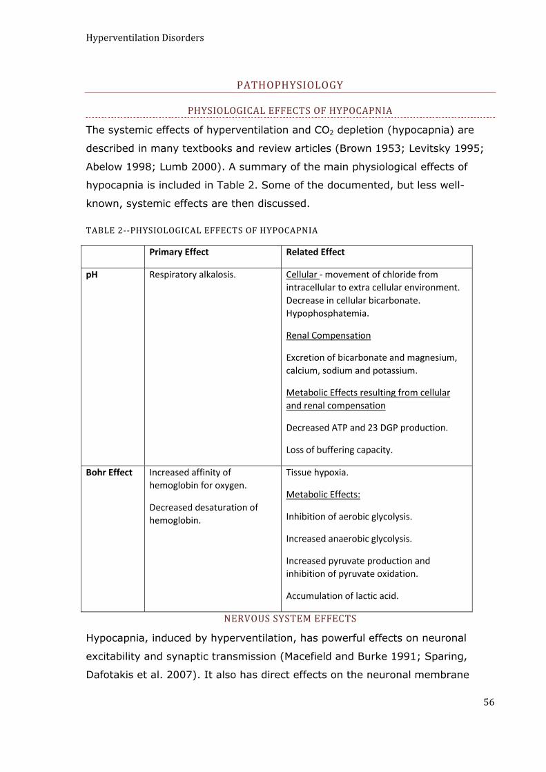
Hyperventilation Disorders
56
PATHOPHYSIOLOGY
PHYSIOLOGICAL EFFECTS OF HYPOCAPNIA
The systemic effects of hyperventilation and CO2 depletion (hypocapnia) are
described in many textbooks and review articles (Brown 1953; Levitsky 1995;
Abelow 1998; Lumb 2000). A summary of the main physiological effects of
hypocapnia is included in Table 2. Some of the documented, but less well-
known, systemic effects are then discussed.
TABLE 2--PHYSIOLOGICAL EFFECTS OF HYPOCAPNIA
Primary Effect Related Effect
pH Respiratory alkalosis.
Cellular - movement of chloride from
intracellular to extra cellular environment.
Decrease in cellular bicarbonate.
Hypophosphatemia.
Renal Compensation
Excretion of bicarbonate and magnesium,
calcium, sodium and potassium.
Metabolic Effects resulting from cellular
and renal compensation
Decreased ATP and 23 DGP production.
Loss of buffering capacity.
Bohr Effect Increased affinity of
hemoglobin for oxygen.
Decreased desaturation of
hemoglobin.
Tissue hypoxia.
Metabolic Effects:
Inhibition of aerobic glycolysis.
Increased anaerobic glycolysis.
Increased pyruvate production and
inhibition of pyruvate oxidation.
Accumulation of lactic acid.
NERVOUS SYSTEM EFFECTS
Hypocapnia, induced by hyperventilation, has powerful effects on neuronal
excitability and synaptic transmission (Macefield and Burke 1991; Sparing,
Dafotakis et al. 2007). It also has direct effects on the neuronal membrane

Hyperventilation Disorders
57
(Balestrino and Somjen 1988) and on production of neurotransmitters.
Glutamine, a precursor of the neurotransmitters glutamic acid and gamma
amino butyric acid, varies linearly with cerebral spinal fluid bicarbonate and
hydrogen ion concentration (Hoop, Shih et al. 1983). Hypocapnia stimulates
glutamate secretion into the synaptic cleft and activates the N-methyl-D-
aspartate receptor (Vannucci, Brucklacher et al. 1997). Gamma activity
increases during hypocapnia as a result of enhanced GABAergic transmission
(Jensen, Hari et al. 2002).
THE RESPIRATORY SYSTEM
In the lungs hypocapnia can contribute to bronchoconstriction, decreased lung
compliance and lung damage (Sterling 1968; McFadden Jr, Stearns et al. 1977;
O'Cain, Hensley et al. 1979; Kolbe, Kleeherger et al. 1987; Van Den Elshout
1991; Lindeman, Croxton et al. 1998; Laffey 2003; Bruton and Holgate 2005).
The mechanisms of lung injury by hypocapnia, which are ameliorated by
supplemental CO2, include increased pulmonary capillary permeability,
parenchymal injury and decreased surfactant (Laffey 2003).
CARDIOVASCULAR EFFECTS
Blood pressure, myocardial contractility and cardiac blood flow can also be
adversely affected by hypocapnia (Laffey and Kavanagh 1999; Ahrens,
Schallom et al. 2001; Laffey and Kavanagh 2002). Cardiac output and stroke
volume fall in response to hypocapnic alkalosis (Zwillich, Pierson et al. 1976).
Coronary artery spasm is a well known response to hyperventilation (Magarian
and Mazur 1991). These effects on the heart can contribute to clinically
relevant coronary pathology and to functional cardiac syndrome(Grossman
1983; DeGuire, Gertvitz et al. 1992; Wilhelm, Gertivz et al. 2001)
HYPERVENTILATION IN THE CRITICALLY ILL
Hypocapnia, hyperventilation and respiratory alkalosis are common findings in
intensive care patients. A systematic analysis of PaCO2 levels and mortality
indicated a direct and linear correlation between PaCO2 levels and
mortality.(Mazzara, Ayres et al. 1974) The authors of this study found that
when 114 patients in an intensive care unit were categorized into four groups
on the basis of carbon dioxide, the patients with the lowest PaCO2 had the
Hyperventilation Disorders
58
highest mortality. Patients (n=25) with PaCO2 of 15mm Hg or less had a
greater than 88% mortality rate, patients (n=35) with a PaCO2 of 20 to
25mmHg had a 77% mortality rate, patients (n=33) with a PaCO2 of 25 to
30mmHg had a 73% mortality rate, patients (n=21) with a PaCO2 of 35 to 45
mm Hg had a mortality of 29% (Mazzara, Ayres et al. 1974).
SPECIFICITY OF HYPERVENTILATION SYMPTOMS TO HYPOCAPNIA
Many symptoms attributed to hyperventilation disorders are not easily
explained by hypocapnia alone. There is often the interplay of psychological
factors and perceptual-cognitive processes (Saltzman 1963; Folgering 1999;
Ley 1999). Several studies have shown that, while the tendency to
hyperventilate in response to stress occurs in most people, the development of
symptoms in association with hypocapnia is variable and tends to occur in
individuals who are predisposed in some way (Saltzman 1963; Suess 1980;
Bass and Gardner 1985). The symptoms of hyperventilation disorders seem to
be strongly influenced by psychological factors and conditions such as increased
adrenergic tone (Folgering 1999). Fear and catastrophic interpretation of the
symptoms of hyperventilation is proposed to be an important factor in
amplifying symptoms (Lewis 1953; Bass and Gardner 1985; Folgering 1999).
Also, catastrophic interpretation of symptoms, in addition to increasing the
person‟s anxiety and the distress associated with the symptom, further disturbs
breathing creating a vicious cycle (Lum 1976).
Some symptoms attributed to hyperventilation disorders have a stronger
relationship to CO2 levels than others. Somatic symptoms, particularly
symptoms such as dizziness, numbness and tingling of extremities and tetany,
have a clearer relationship to hypocapnia, while psychological symptoms such
as tension and anxiety are more strongly related to psychological factors
(Wientjes and Grossman 1994). Uncomfortable respiratory sensations related
to dyspnea, such as feeling unable to breathe deeply, suffocation and air
hunger, do not appear to be strongly related to hypocapnia (Hormbrey, Jacobi
et al. 1988; Gardner 1992; Hornsveld and Garssen 1996). Howell proposes that
dyspnea symptoms may be more related to neuromuscular influences than to
biochemical factors (Howell 1997).

Hyperventilation Disorders
59
SENSITISATION AND CONDITIONING IN BREATHING AND SYMPTOM PERCEPTION
In a review of HVS, Folgering (1999) argues that the poor relationship between
symptoms of hyperventilation and hypocapnia may be a result of classical
conditioning (Folgering 1999). In this model, which accounts for the persistence
of symptoms in the absence of hypocapnia, it is proposed that symptoms are
initially triggered by hypocapnia, but become learned over time and are
subsequently triggered by situations, behaviours and emotions that were
previously related to hypocapnia. Recent research has shown that this is the
case with dizziness, a common reaction to hyperventilation, which can be
initially produced in response to hypocapnia and then conditioned to arise in
response to particular odours in the absence of hypocapnia (Bresseleers, Van
Diest et al. 2010).
The inconsistent relationship between hypocapnia and symptoms of
hyperventilation syndrome may also be due to sensitisation to hypocapnia,
which leads to an exaggerated response to fluctuations in CO2. Howell (1990)
found that individuals vary widely in their reactions to the effects of hypocapnia
and that individuals with rapid onset of symptoms in response to hypocapnia
had the largest numbers of classic hyperventilation symptoms (Howell 1990).
More recent studies have shown that sensitisation to CO2 happens more readily
in anxious individuals and that sensations of air hunger occur at lower CO2
levels in these cases (Wan, Van Diest et al. 2008).
BREATHING PATTERN AS A PATHOPHYSIOLOGICAL FACTOR
The abnormalities in breathing pattern that have been observed in individuals
with hyperventilation disorders have been proposed to be a source of some
symptoms traditionally associated with hyperventilation, but which do not
appear to correlate with hypocapnia (Hornsveld 1997). Air hunger, or inability
to take a satisfying breath, is a common symptom in hyperventilation disorders
(Magarian 1982; Gardner 1995). This symptom can occur as a result of
hyperinflation of the lungs (O'Donnell 2006) and is associated with poor
respiratory muscle co-ordination and other dysfunctions related to the
biomechanics of breathing. This is discussed in greater detail in Chapter 4.
Hyperventilation Disorders
60
Thoracic breathing is one breathing pattern that has been found to
correlate with hyperventilation symptoms in asthmatics and individuals with
panic disorder that may contribute to at least some of these symptoms (Carr,
Lehrer et al. 1992).
INVESTIGATION AND DIFFERENTIAL DIAGNOSIS
MEASURING CO2 LEVELS
CAPNOGRAPHY
Carbon dioxide levels can be effectively assessed in most cases with
capnography, a process of continuously recording the level of CO2 in expired air
by infrared spectroscopy. The percentage of CO2 at the end of expiration, called
End Tidal CO2 (ETCO2), gives a close approximation of the alveolar (PACO2)
concentration which in turn reflect arterial levels of CO2 (PaCO2)(Corbo, Bijur et
al. 2005; Donald and Paterson 2006; Nagler and Krauss 2008).
TRANSCUTANEOUS MONITORING OF CO2
Another method for continuous noninvasive measurement of PaCO2 is via
transcutaneous monitoring, which is done via application of an infrared
transducer applied to the skin. Transcutaneous CO2 pressure has been reported
to accurately reflect arterial CO2 pressure (PaCO2) (Greenspan, Block et al.
1981). However, this method of CO2 assessment for hyperventilation disorders
has been criticised because rapid changes in CO2, which tend to occur in these
disorders, are not accurately reflected in this measurement technique due to
the time delay inherent in this technology (Meuret and Ritz 2010).
RESTING CO2
Reported levels of alveolar CO2 considered to indicate hyperventilation vary
according to the source. Respiratory physiology texts have stated that levels of
PaCO2 less than 37 mmHg indicate physiological hyperventilation (Comroe
1974). Many individuals can be found to have CO2 below this level, but are not
obviously symptomatic. Gardner found that mean resting levels of CO2 in
normal subjects was 36.4 mmHg with 2 standard deviations below this being
32.2mm Hg. He states that 30 is the lower limit of normal (Gardner 1986). At
Hyperventilation Disorders
61
the other extreme, Buteyko (1990) claimed that levels below 38.5 mmHg
should be considered to indicate hyperventilation. The basis on which Buteyko
made this claim is not clear due to difficulty in accessing studies published in
the former Soviet Union. Buteyko believed that ideal ventilation levels as found
in optimally healthy people would result in levels around 45 mmHg and that
this was associated with a breath holding time of 60 seconds (Buteyko 1990).
STRESS TESTING OF CO2
In many patients with signs and symptoms suggestive of hyperventilation
disorders, there is no evidence of decreased baseline end-tidal CO2 levels, and
abnormalities of CO2 are only uncovered with some type of stress provocation
or in response to exercise. There may be a fall of CO2 in anticipation of
measurement (Vansteenkiste, Rochette et al. 1991) or in anticipation of
exercise (Jack, Rossiter et al. 2004). Sustained decreases of CO2 levels in
response to exercise can unmask hyperventilation tendencies in individuals who
at rest may have only slightly reduced CO2 levels (Jack, Rossiter et al. 2004).
Recollection of stressful events or emotionally charged memories, “the think
test”, has also in the past been used at part of a clinical protocol to unmask
hyperventilation tendencies (Nixon and Freeman 1988).
HYPERVENTILATION PROVOCATION TEST
The Hyperventilation Provocation Test (HVPT) has, until recent years, been a
cornerstone of the diagnostic criteria for Hyperventilation Syndrome. During the
HVPT the patient hyperventilates for a period of time that can vary from as
little as 20 deep breaths (Howell 1990), to a more often recommended 3
minutes (Hardonk and Beumer 1979) (Vansteenkiste 1991). HVPT has been
used as a diagnostic tool with and without confirmation of its effects on alveolar
CO2 levels (Han, Stegen et al. 1996; Howell 1997).
When the HVPT is used without concurrent measurement of CO2, the
examiner relies on the patient‟s reporting of symptom recognition. As discussed
previously, this has been found to be invalid as a means of diagnosing
hyperventilation disorders and is thought to give false positive results
(Hornsveld, Garsson et al. 1996). The HVPT can also give false negative results
Hyperventilation Disorders
62
when patients reassured by the presence of the experimenter and the relative
safety and predictability of experimental conditions when compared to real life
do not experience their usual breathing related symptoms (Bass and Gardner
1985).
One use of the HVPT that is still considered valid is to measure the rate
of recovery of CO2 after voluntary hyperventilation (Warburton and Jack 2006).
Slow recovery of CO2 after the HVPT can also identify individuals with
abnormalities of respiratory control that sustain hyperventilation tendencies.
Recovery of CO2 to pretest levels in normal individuals occurs within 1 to 3
minutes (Hardonk and Beumer 1979), but in hyperventilation disorders
recovery is delayed. It is suggested that this delayed recovery due to
impairment of the normal negative feedback mechanisms which generally
inhibit ventilation as hypocapnia increases (Hardonk and Beumer 1979;
Folgering 1999; Warburton and Jack 2006).
BREATH HOLDING TIME IN HYPERVENTILATION DISORDERS
Breath holding time has often been observed to be abnormally short in
hyperventilation disorders (Gardner 1995; Eke and McNally 1996; Jack, Darke
et al. 1998; Warburton and Jack 2006). In one study by Jack and colleagues,
breath holding time in individuals with idiopathic hyperventilation was
significantly reduced to a mean of 20 seconds versus controls who had a mean
of over 60 seconds (Jack, Rossiter et al. 2003).
BUTEYKO’S FORMULA
Extensive studies were reported to be undertaken in the former Soviet Union by
Dr. K. P Buteyko on the correlation between breathing holding time and CO2.
He patented a formula in the former Soviet Union for using breath holding time
as a measure of PaCO2. Practitioners of the Buteyko Breathing Technique (BBT)
use breath-holding time as an indirect measure of arterial CO2 levels and
consider the Control Pause (CP= time of post expiratory breath hold) as a
measure of alveolar CO2 concentration according to the formula:
PaCO2 % = 3.5% +.05 MP
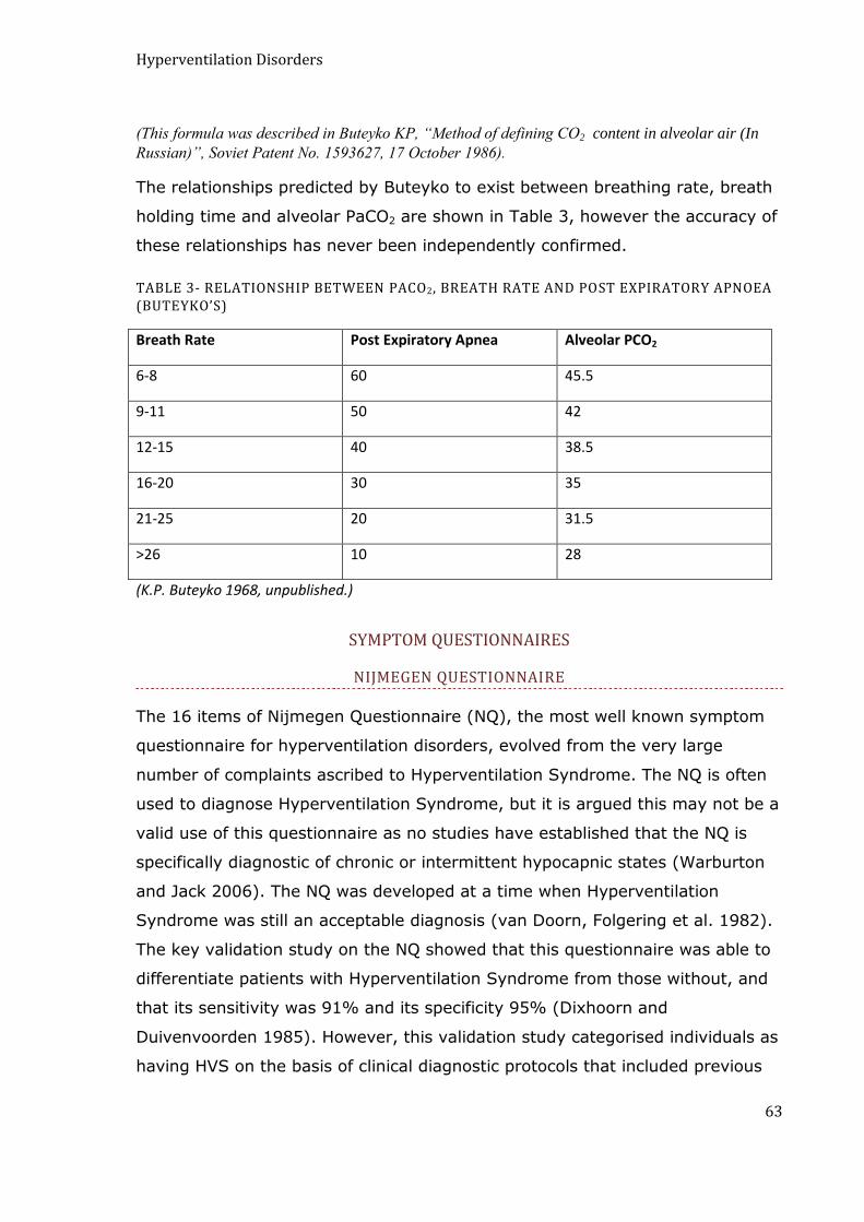
Hyperventilation Disorders
63
(This formula was described in Buteyko KP, “Method of defining CO2 content in alveolar air (In
Russian)”, Soviet Patent No. 1593627, 17 October 1986).
The relationships predicted by Buteyko to exist between breathing rate, breath
holding time and alveolar PaCO2 are shown in Table 3, however the accuracy of
these relationships has never been independently confirmed.
TABLE 3- RELATIONSHIP BETWEEN PACO2, BREATH RATE AND POST EXPIRATORY APNOEA (BUTEYKO’S)
Breath Rate Post Expiratory Apnea Alveolar PCO2
6-8 60 45.5
9-11 50 42
12-15 40 38.5
16-20 30 35
21-25 20 31.5
>26 10 28
(K.P. Buteyko 1968, unpublished.)
SYMPTOM QUESTIONNAIRES
NIJMEGEN QUESTIONNAIRE
The 16 items of Nijmegen Questionnaire (NQ), the most well known symptom
questionnaire for hyperventilation disorders, evolved from the very large
number of complaints ascribed to Hyperventilation Syndrome. The NQ is often
used to diagnose Hyperventilation Syndrome, but it is argued this may not be a
valid use of this questionnaire as no studies have established that the NQ is
specifically diagnostic of chronic or intermittent hypocapnic states (Warburton
and Jack 2006). The NQ was developed at a time when Hyperventilation
Syndrome was still an acceptable diagnosis (van Doorn, Folgering et al. 1982).
The key validation study on the NQ showed that this questionnaire was able to
differentiate patients with Hyperventilation Syndrome from those without, and
that its sensitivity was 91% and its specificity 95% (Dixhoorn and
Duivenvoorden 1985). However, this validation study categorised individuals as
having HVS on the basis of clinical diagnostic protocols that included previous
Hyperventilation Disorders
64
diagnosis by practitioner (partly on the basis of presence of HVS symptoms),
presence of disordered breathing (rapid, thoracic, irregular, frequent sighing),
and reproduction of symptoms during the HVPT, a test which is no longer
considered a valid tool for confirmation of hyperventilation disorders (Hornsveld
and Garssen 1996). Another validation study by Vansteenkist and colleagues,
found an 86% correlation between high scores on the NQ (> 24) and
recognition of unexplained symptoms of daily life during the HVPT
(Vansteenkiste 1991). However, in this study the symptoms that were most
commonly reported during the HVPT were breathlessness, breathing restriction,
tension, palpitations and chest pain. These symptoms are not strongly
associated with hypocapnia and could be due to other factors including
disturbances of the biomechanical breathing dysfunction (Hormbrey, Jacobi et
al. 1988; Gardner 1992; Hornsveld 1996; Howell 1997).
INTERVENTIONS FOR HYPERVENTILATION DISORDERS
Successful approaches to correcting hyperventilation include various
combinations of psychotherapy, relaxation training, breathing re-education and
biofeedback (Lum 1975; van Doorn, Folgering et al. 1982; Grossman 1984; Ley
and Timmons 1999). There has been vigorous debate as to whether the
successful outcomes from these therapies are due to improved CO2 levels or to
relaxation effects and psychological factors (Garssen 1992; Garssen, de Ruiter
et al. 1993; Ley 1993; Wilhelm, Gertivz et al. 2001). While a number of studies
have shown improvements in CO2 regulation after therapy (van Doorn,
Folgering et al. 1982; Grossman 1984) and provided evidence for a moderation
of symptoms by CO2 (Meuret, Wilhelm et al. 2008; Ritz, Meuret et al. 2009),
other studies indicate that symptom improvement is not necessarily correlated
with improved CO2 status (Garssen 1992)
BUTEYKO BREATHING TECHNIQUE
Hyperventilation Disorders
65
The Buteyko Breathing Technique (BBT) specifically aims to raise CO2 levels
through breathing retraining with very little focus on psychological factors. BBT
is most well known as a treatment for asthma, but has been used to treat a
large number of conditions believed by Buteyko to be related to
hyperventilation. The BBT has been tested in a number of clinical trials, which
have all tended to show therapeutic benefit (Bowler 1998; Opat, Cohen et al.
2000; Cooper, Oborne et al. 2003; McGowan 2003; McHugh, Aicheson et al.
2003; Abramson, Borg et al. 2004; McHugh, Duncan et al. 2006; Slader,
Reddel et al. 2006; Cowie, Underwood et al. 2008). As with other therapies for
hyperventilation, it is not clear if BBT treatment effects are due to changes in
CO2 status (Al-Delaimy 2001). It is argued that effects of the BBT are due to
non-specific effects, for example sense of control over asthma symptoms but
this has not been rigorously tested (Slader, Reddel et al. 2006).
CONCLUSIONS
Hyperventilation is an important aspect of dysfunctional breathing that has
established physiological effects and negative health consequences. Individuals
are predisposed to hyperventilate for psychological and physiological reasons
and in response to diseases of the respiratory, cardiovascular and nervous
systems.
Hyperventilation Syndrome is no longer considered a valid diagnosis
because of the complex relationship between symptoms and acute and chronic
hypocapnia. The previous gold standard for diagnosis of hyperventilation
disorders, symptom recognition during the Hyperventilation Provocation Test
may not be a valid way of establishing the relationship of symptoms to CO2
levels although it is a useful test for uncovering abnormalities of CO2 control.
Assessment of CO2 through measurement of end tidal or transdermal CO2 are
valid and well established ways of directly establishing presence of hypocapnia.
In the clinical setting, presence of hyperventilation is sometimes inferred
indirectly. Symptoms questionnaires, such as the Nijmegen Questionnaire,
Breath Holding Time and presence of breathing pattern abnormalities such as
rapid breathing, thoracic breathing and irregular breathing are sometimes
Hyperventilation Disorders
66
assumed to indicate presence of hyperventilation. However, a clear relationship
between CO2 and these measures has not been established. The question of the
relationship of these measures to chronic hypocapnia is addressed in this thesis
in two separate chapters. Chapter 10 looks specifically at Buteyko‟s claims
about the relationship of breath holding time to CO2 and Chapter 9 examines
the relationships between various measures of dysfunctional breathing,
including CO2 measures derived from capnography, the Nijmegen
Questionnaire, breathing pattern and breath holding time.
Functions and Dysfunctions of Breathing Pattern
67
CHAPTER 4 - THE FUNCTIONS AND DYSFUNCTIONS OF
BREATHING PATTERN
INTRODUCTION
The efficiency with which the mechanical act of breathing is performed is an
important aspect of breathing functionality. Efficient breathing patterns depend
on the co-ordination of many breathing muscles, appropriate timing and
volume adjustments and a balance between stability and responsiveness in
order to respond to changes in the internal and external environment and to
maintain homeostasis.
Prior to the 1940‟s, the literature on functional breathing disorders paid
greater attention to describing breathing pattern abnormalities than on
measuring and reporting on a patient‟s carbon dioxide status (Maytum 1934;
Christie 1935). In more recent times breathing pattern disturbances do not
appear to have been studied as closely as hyperventilation disorders. However,
dysfunctional or „tense‟ breathing patterns have often been observed in patients
believed to have Hyperventilation Syndrome and it has been proposed that
dysfunctions of respiratory muscles and the respiratory pump might be the
source of some symptoms, such as dyspnea and chest wall pain. These
symptoms have been incorrectly thought to arise from hypocapnia (Hormbrey,
Jacobi et al. 1988; Wolf 1994; Hornsveld 1997), and it is suggested that
inefficient breathing patterns which become habitual might play a role in
perpetuating hyperventilation disorders (Lum 1976; Gardner 1995).
NORMAL BREATHING
Components of breathing pattern that have been studied in normal individuals
include relative timing and volumes of inspiration and expiration, breathing
movement and co-ordination patterns (Tobin, Tejvir et al. 1983; De Troyer and
Estenne 1988), also the role of sighing and irregularity in regulation of normal
breathing (Vlemincx, Taelman et al. 2010).

Functions and Dysfunctions of Breathing Pattern
68
The following section is a brief review of some key aspects of the
functional anatomy of breathing to introduce terminology and concepts that are
used in later discussions in this chapter and subsequent chapters.
THE CHEST WALL
The chest wall is comprised of two distinct compartments, separated by the
diaphragm. The upper compartment is often referred to as the rib cage and the
lower as the abdomen (Konno and Mead 1966). Because the lower
compartment includes the lower six ribs, the terms rib cage and abdomen are
potentially confusing, and these two compartments are more accurately
referred to as upper rib cage (URC) and lower rib cage/abdomen (LRCA). The
term thoracic breathing generally refers to breathing that is evidenced by
greater upper rib cage motion and the terms abdominal breathing and
diaphragmatic breathing refers to breathing that involves motion of the lower
rib cage and abdomen (Lewis 1997; Kaminoff 2006). These two rib cage
compartments both move in three planes: vertical, lateral and
anterior/posterior; however their movements differ because of differences in
their bony and muscular articulations (De Troyer and Estenne 1988).
UPPER RIB CAGE (URC)
The upper ribs tend to move with the sternum, as a unit, rather than displaying
individual movement. The predominant motion of the upper ribs, described as
being like a pump handle, is in the anterior/posterior and in the vertical plane.
There is little lateral expansion of the upper ribs during respiration. During
quiet breathing the primary muscle action is from the intercostal muscles with
some contribution from the scalenes (De Troyer and Estenne 1988). When
ventilatory needs increase, accessory muscle use increases. Accessory muscles
of respiration which move the upper rib cage have other movement functions,
on the neck and upper limb, which must be integrated with respiration
(Kapandji 1974).

Functions and Dysfunctions of Breathing Pattern
69
LOWER RIB CAGE/ ABDOMEN (LRCA)
The lower six ribs have a greater ability to move independently when compared
to the upper six ribs. The motion of the lower ribs is often described as bucket
handle motion, moving cephalad and widening during inspiration and moving
caudad and narrowing during exhalation. The intercostals, the diaphragm and
abdominal muscles all attach to the lower ribs and produce the characteristic
respiratory motion of the LRCA (De Troyer and Estenne 1988).
BALANCE OF CHEST WALL MOTION DURING NORMAL RESPIRATION
In normal breathing the contribution of URC and LRCA motion is relatively equal
and the compartments move synchronously, increasing their dimensions during
inspiration and decreasing their dimensions during exhalation (Tobin, Tejvir et
al. 1983).
There is some normal variability in the balance of URC to LRC motion,
influenced by age and gender. Older people tend to use more LRCA motion
during breathing (also called abdominal breathing) than younger people
(Sharp, Goldberg et al. 1975; Faithfull, Jones et al. 1979; Verschakelen and
Demedts 1995) and females use more URC motion during breathing (also called
thoracic breathing) than males (Sharp 1983; Verschakelen and Demedts 1995).
There is also variability in the balance and synchrony of chest wall
motion in response to changes in posture, ventilatory drive, activity levels, and
rate and volume of respiration. Upright posture, large volume of inspiratory
effort, rapid respiration and increased activity tend to produce greater URC
motion (Sharp, Goldberg et al. 1975; De Troyer and Estenne 1988).
Despite the findings that particular diseases (Tobin, Tejvir et al. 1983),
increased ventilatory drive and activity levels are associated with functional
imbalances in rib cage motion(Sharp, Goldberg et al. 1975; De Troyer and
Estenne 1988) , it is generally observed that the breathing of normal healthy
individuals at rest is characterized by balanced motion of the upper and lower
compartments of breathing (Chaitow 2002, Pryor and Prasad 2002).
THE MUSCLES OF RESPIRATION
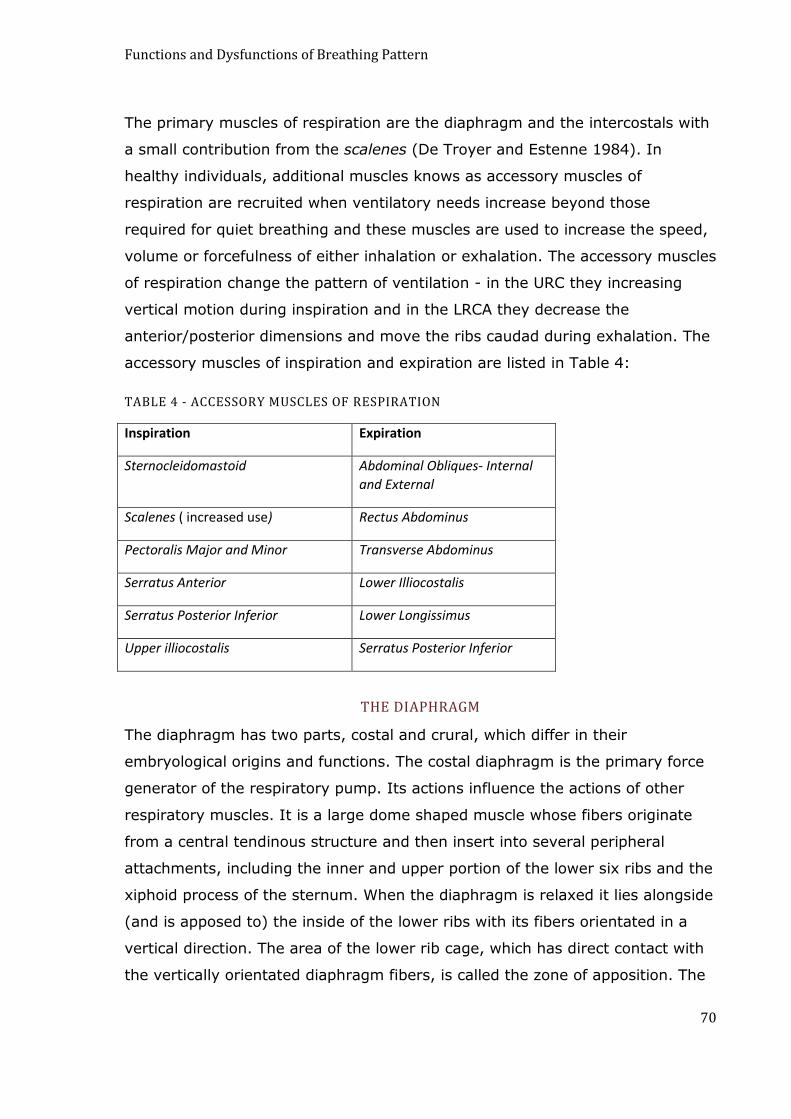
Functions and Dysfunctions of Breathing Pattern
70
The primary muscles of respiration are the diaphragm and the intercostals with
a small contribution from the scalenes (De Troyer and Estenne 1984). In
healthy individuals, additional muscles knows as accessory muscles of
respiration are recruited when ventilatory needs increase beyond those
required for quiet breathing and these muscles are used to increase the speed,
volume or forcefulness of either inhalation or exhalation. The accessory muscles
of respiration change the pattern of ventilation - in the URC they increasing
vertical motion during inspiration and in the LRCA they decrease the
anterior/posterior dimensions and move the ribs caudad during exhalation. The
accessory muscles of inspiration and expiration are listed in Table 4:
TABLE 4 - ACCESSORY MUSCLES OF RESPIRATION
Inspiration Expiration
Sternocleidomastoid Abdominal Obliques- Internal
and External
Scalenes ( increased use) Rectus Abdominus
Pectoralis Major and Minor Transverse Abdominus
Serratus Anterior Lower Illiocostalis
Serratus Posterior Inferior Lower Longissimus
Upper illiocostalis Serratus Posterior Inferior
THE DIAPHRAGM
The diaphragm has two parts, costal and crural, which differ in their
embryological origins and functions. The costal diaphragm is the primary force
generator of the respiratory pump. Its actions influence the actions of other
respiratory muscles. It is a large dome shaped muscle whose fibers originate
from a central tendinous structure and then insert into several peripheral
attachments, including the inner and upper portion of the lower six ribs and the
xiphoid process of the sternum. When the diaphragm is relaxed it lies alongside
(and is apposed to) the inside of the lower ribs with its fibers orientated in a
vertical direction. The area of the lower rib cage, which has direct contact with
the vertically orientated diaphragm fibers, is called the zone of apposition. The

Functions and Dysfunctions of Breathing Pattern
71
curvature of the diaphragm, its length and the size of the zone of apposition
are maximised when the diaphragm is relaxed at the end of exhalation. This
zone of apposition represents about 30% of the total surface area of the rib
cage at the end of a normal exhalation (at functional residual capacity) (De
Troyer and Estenne 1988). The size of the zone of apposition, the length and
curvature of the diaphragm influence its power and efficiency.
ACTIONS OF THE COSTAL DIAPHRAGM ON THE RIB CAGE AND ABDOMEN
During an effective exhalation the diaphragm relaxes and lengthens and the
zone of apposition is maximized. During inspiration the diaphragm shortens and
descends towards the abdominal cavity, increasing intra-abdominal pressure. It
lifts and widens the lower rib cage and also displaces the abdominal wall
anteriorly. The actions of diaphragm are summarized in table 5:
TABLE 5 - ACTIONS OF THE DIAPHRAGM DURING INSPIRATION AND EXPIRATION
Length Shape Zone of
Apposition
Actions on Rib
Cage
Effects on
intra-
abdominal
pressure
Inspiration Shortens
(contracts)
flattens decreases Lift and widen
lower 6 ribs (
to horizontal
orientation)
increases
Expiration Lengthens
(relaxes)
domes increases Depress and
narrow lower
6 ribs (to
vertical
orientation)
decreases
ABDOMINAL MUSCLES
The muscles that form the anterior and lateral abdominal wall (rectus
abdominus, external oblique, internal oblique and transverse abdominus) play
several important roles in respiration.
ACTIONS OF ABDOMINAL MUSCLES
The abdominal muscles are accessory muscles of expiration, which become
activated during forced expiration; however, they also play a role in inhalation
Functions and Dysfunctions of Breathing Pattern
72
because of their interaction with the diaphragm. Weakness of the abdominal
muscles affects both inspiratory and expiratory power.
Their contraction during exhalation decreases the dimensions of the
chest wall by pulling the sternum and ribs downward and decreasing the
anterior and posterior diameter of the rib cage. It also increases abdominal
pressure, helping to push the diaphragm upwards maximizing its curvature and
resting length in preparation for inspiration.
Abdominal contraction during inspiration creates a mechanical and
energy advantage for the diaphragm and rib cage motion. It increases the
curvature and length of the diaphragm maximizing its power and efficiency
because of the well known length/tension rule of muscles to increase power
with increased length (De Troyer and Estenne 1988; Society 2002; Finucane,
Panizza et al. 2005). Abdominal contraction during inspiration causes the end-
expiratory position of the diaphragm and the end-expiratory volume of the
abdomen to be smaller than during relaxation. This action of the diaphragm
promotes lengthening and prevents excessive shortening of the diaphragm,
increasing its ability to generate pressure throughout the inspiratory phase. It
also helps the lower rib cage to expand laterally during inspiration.
CO-ORDINATION OF THE DIAPHRAGM AND ABDOMINAL MUSCLES DURING RESPIRATION
Abdominal displacements are often used as approximations of diaphragm
displacements; however this is an oversimplification as diaphragm contraction
can produce motion that is localized at the lateral rib cage as well as at the
abdomen (Konno and Mead 1966; Loring, Mead et al. 1985). Individuals with
normal respiratory muscle control are capable of voluntarily modifying their
breathing to emphasise either abdominal protrusion or lateral rib cage
expansion during inspiration. If abdominal tone is maintained during inhalation,
the increase in abdominal pressure resulting from diaphragm descent is
directed towards lifting and widening the lower six ribs in an inspiratory
direction. During inspiration, if abdominal tone is relaxed, or if abdominal
protrusion is exaggerated, lateral rib cage motion will be inhibited (Konno and
Mead 1966; Loring, Mead et al. 1985; De Troyer and Estenne 1988).

Functions and Dysfunctions of Breathing Pattern
73
There is substantial argument about whether „diaphragmatic breathing‟ is
most efficiently performed by holding the abdomen firm and emphasizing
lateral rib cage motion or by fully protruding the abdomen during inspiration
(Kaminoff 2006). There is no absolute answer to this question because the
relationship between lateral rib cage and abdominal motion to efficiency of
diaphragm action is dependent on a range of circumstances. In most healthy
individuals, the relative amount of rib cage to abdominal motion resulting from
diaphragm contraction naturally varies on a continuum in response to changes
in posture, muscle tone, activity level, respiratory drive, volume and timing (De
Troyer 1983).
During relaxation, and in the supine position, there is a natural tendency
to increased forward displacement of the abdomen during inspiration (De
Troyer 1983; Verschakelen and Demedts 1995). This relaxed configuration of
thoraco-abdominal motion is also evident in sitting and standing if the person
has low abdominal tone or if they voluntarily relax the abdomen (Verschakelen
and Demedts 1995). On standing and upright sitting posture and during
physical activity, there is a natural tendency for the resting muscle tone of the
abdominal muscles to increase with localisation of breathing motion to the
lateral rib cage in the lower rib cage. This is referred to as the active thoraco-
abdominal configuration of respiratory muscle activity (Sharp, Goldberg et al.
1975; De Troyer 1983; Verschakelen and Demedts 1995). In the active
configuration, there is less forward abdominal displacement and greater lateral
rib cage motion (produced by the diaphragm) and vertical motion of the rib
cage (produced by the accessory muscles of respiration)(De Troyer 1983; De
Troyer 1997).
The decreased amount of abdominal motion during sitting and standing
is proposed to be a normal and functional response that increases the efficiency
of the diaphragm by preventing the diaphragm from shortening (descending)
excessively during inspiration (Sharp, Goldberg et al. 1975; De Troyer 1983).
Preservation of diaphragm length and curvature enables stronger contraction
and greater pressure from the diaphragm to be exerted during inspiration (De
Troyer 1983). It may also increase postural support from the diaphragm and
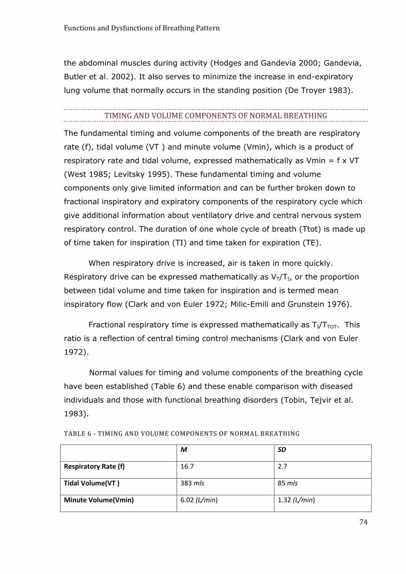
Functions and Dysfunctions of Breathing Pattern
74
the abdominal muscles during activity (Hodges and Gandevia 2000; Gandevia,
Butler et al. 2002). It also serves to minimize the increase in end-expiratory
lung volume that normally occurs in the standing position (De Troyer 1983).
TIMING AND VOLUME COMPONENTS OF NORMAL BREATHING
The fundamental timing and volume components of the breath are respiratory
rate (f), tidal volume (VT ) and minute volume (Vmin), which is a product of
respiratory rate and tidal volume, expressed mathematically as Vmin = f x VT
(West 1985; Levitsky 1995). These fundamental timing and volume
components only give limited information and can be further broken down to
fractional inspiratory and expiratory components of the respiratory cycle which
give additional information about ventilatory drive and central nervous system
respiratory control. The duration of one whole cycle of breath (Ttot) is made up
of time taken for inspiration (TI) and time taken for expiration (TE).
When respiratory drive is increased, air is taken in more quickly.
Respiratory drive can be expressed mathematically as VT/TI, or the proportion
between tidal volume and time taken for inspiration and is termed mean
inspiratory flow (Clark and von Euler 1972; Milic-Emili and Grunstein 1976).
Fractional respiratory time is expressed mathematically as TI/TTOT. This
ratio is a reflection of central timing control mechanisms (Clark and von Euler
1972).
Normal values for timing and volume components of the breathing cycle
have been established (Table 6) and these enable comparison with diseased
individuals and those with functional breathing disorders (Tobin, Tejvir et al.
1983).
TABLE 6 - TIMING AND VOLUME COMPONENTS OF NORMAL BREATHING
M SD
Respiratory Rate (f) 16.7 2.7
Tidal Volume(VT ) 383 mls 85 mls
Minute Volume(Vmin) 6.02 (L/min) 1.32 (L/min)

Functions and Dysfunctions of Breathing Pattern
75
Time taken for inspiration (TI) 1.60 s .30 s
Mean inspiratory flow (VT/TI) .424 mls/sec .032 mls/sec
Fractional inspiratory time (TI/TTOT) .249 .54
From Tobin (1983), (values for young people, mean age 29)
RELATIONSHIP OF TIMING AND VOLUME TO BREATHING PATTERN IN NORMAL INDIVIDUALS
Rapid respiratory maneuvers, particularly rapid shallow respirations are carried
out using the active thoraco-abdominal configuration and the accessory
muscles of respiration. Slower movements are carried out with greater
contribution from abdominal motions (i.e. the relaxed configuration) (Sharp,
Goldberg et al. 1975). The velocity of rib cage motion during inspiration is
greater than that for the abdomen, so therefore the rib cage tends to be
recruited when respiratory is rate increased or during inspiratory maneuvers
requiring speed (Sharp, Goldberg et al. 1975; Verschakelen and Demedts
1995).
In normal healthy people, small increases in breathing volume tend to
involve greater abdominal motion than smaller volumes (Sharp, Goldberg et al.
1975). As volumes increase beyond those found in normal tidal breathing,
abdominal motion gradually decreases and thoracic motion predominates
(Sharp, Goldberg et al. 1975; Verschakelen and Demedts 1995). The decrease
in abdominal motion during very large volumes of breathing will be functional
when it assists in preserving a mechanically advantageous length and radius of
curvature of the diaphragm.
In cases of increased respiratory drive where (VT/TI) is increased (Tobin,
Tejvir et al. 1983), one would expect to see the active thoraco/abdominal
configuration with activation of accessory muscles of respiration. This has been
found to be the case in individuals with asthma and COPD (Muller, Bryan et al.
1981; Marchand and Decramer 2000).
REGULARITY OF NORMAL BREATHING
It is generally accepted that normal breathing is not completely regular and is
characterised by a balance between stability and variability (Baldwin, Suki et al.

Functions and Dysfunctions of Breathing Pattern
76
2004). Normal breathing patterns include appropriate changes of rate, volume
and respiratory muscle use and respiratory behaviors such sighing and yawing
(Leiner and Abrahamowitz 1958). Variability in normal breathing is evidence of
its responsiveness and sensitivity to changes in environment, activity levels,
emotional arousal and metabolic activity (Wientjes 1992; Boiten 1998).
Variability of breathing and respiratory behaviors, such as sighing and yawning
are important for the state of dynamic equilibrium needed to promote optimal
functioning of respiratory control feedback loops (Aljadeff, Molho et al. 1993;
Baldwin, Suki et al. 2004; Qureshi, Khalik et al. 2009; Vlemincx, Van Diest et
al. 2009). Sighing is proposed to assist in repartitioning and normalizing lung
volumes (Aljadeff, Molho et al. 1993) and to act as a resetter mechanism,
promoting structured variability in breathing pattern in situations where
variability is abnormally reduced or excessively chaotic (Vlemincx, Van Diest et
al. 2009).
DYSFUNCTIONAL PATTERNS OF BREATHING
Dysfunctional breathing patterns include dysfunctions of breathing co-
ordination (Lewis 1953; Lum 1975; Folgering 1999), abnormalities of volume
and timing (Wientjes 1992) and excessive or reduced variability of breathing
(Loveridge, West et al. 1984; Sody, Kiderman et al. 2008). Mouth breathing is
also an abnormal breathing habit that adversely affects airway and respiratory
muscle function (Lee, Choi et al. 2007; Correa and Berzin 2008; Hallani,
Wheatley et al. 2008; Yi, Jardim et al. 2008; Kim, Choi et al. 2010).
DYSFUNCTIONS OF BREATHING CO-ORDINATION
In its normal relaxed state the two compartments of the chest wall, the URC
and the LRCA, create an effective respiratory pump which moves in response to
co-ordinated actions created by the diaphragm, scalenes, intercostals,
abdominals and accessory muscles of respiration (Konno and Mead 1966; De
Troyer and Estenne 1988). However, under particular circumstances this co-
ordinated action is lost and breathing pattern becomes dysfunctional.
The 2 main dysfunctions of breathing co-ordination are:

Functions and Dysfunctions of Breathing Pattern
77
1. Thoracic dominance in breathing; and
2. Asynchronous and paradoxical breathing.
THORACIC BREATHING
The term thoracic breathing is the common term used to refer to URC breathing
(Chaitow 2002). In this type of breathing upper rib cage motion, produced by
the accessory muscles of respiration, dominates over lower rib cage and
abdominal motion. Thoracic breathing, when present as a habitual mode of
breathing and persisting during quiet breathing, can generally be considered as
dysfunctional (Lewitt 1980). However its presence can be normal and functional
in certain situations, for example when there are sudden increases in
ventilatory need, during rapid inspiratory maneuvers and during increased
physical activity (Sharp, Goldberg et al. 1975; De Troyer 1983; De Troyer and
Estenne 1984; Verschakelen and Demedts 1995). In these cases, increased
upper thoracic movement relative to abdominal motion can be evidence of the
normal active thoraco-abdominal configuration described by De Troyer (1984),
which often helps to preserve diaphragm length during increased activity,
postural change, increased ventilatory need and on sudden increases in
ventilation.
In popular opinion, decreased abdominal motion, relative to upper
thoracic motion confirms poor diaphragm action (Lewis 2004 (accessed
15/10/2010); Kaminoff 2006). While this can be the case, particularly in the
case of more pronounced respiratory muscle dysfunction (Fitting and Grassino
1987; De Troyer and Estenne 1988; Hruska 1997), it is not always so and
abdominal motion may not have a clear relationship to diaphragm activity
(Gilbert and Auchincloss 1981). Maintenance of abdominal tone during
inspiration can improve diaphragm efficiency and strength, particularly if there
is adequate elevation and lateral expansion of the lower 6 ribs (De Troyer and
Estenne 1988).
ASYNCHRONOUS AND PARADOXICAL BREATHING
In normal breathing, the upper rib cage and lower rib cage/abdomen
compartments increase their dimensions during inspiration in synchrony. If the
Functions and Dysfunctions of Breathing Pattern
78
dimensions of either of these compartments decrease during inspiration, it is
referred to as asynchronous or paradoxical breathing (Jubran and Tobin 1992;
Hammer and Newth 2009).
Thoraco-abdominal asynchrony is generally considered to be
dysfunctional because of its association with respiratory distress and with
weakness and hypertonicity of respiratory muscles (Wolfson, Strohl et al. 1983;
Fitting and Grassino 1987; Jubran and Tobin 1992; Hammer and Newth 2009).
However, paradoxical inward motion of the abdomen during inspiration is not
always dysfunctional. In fact inward abdominal motion during inspiration can be
a normal and functional response to increased lung volume, physical activity,
rapid respiratory maneuvers or standing posture, assisting in the maintenance
of diaphragm length and curvature (De Troyer 1983; Wolfson, Strohl et al.
1983). Paradoxical breathing is likely to be dysfunctional when it is not
adequately compensated by lateral motion of the rib cage and the lower rib
cage narrows instead of widening during inspiration (De Troyer 1983). It is also
dysfunctional when it is inappropriately present, for example during rest and at
times when ventilatory needs are not elevated.
ABNORMALITIES OF TIMING AND VOLUME
Tobin (1983) compared timing and volume parameters between normal
individuals and those with anxiety, asthma, COPD and restrictive lung diseases.
The general trend across all non-normal individuals studied showed increased
volume and rates of breathing, increased minute volume, increased respiratory
drive (VT/TI), more rapid inspiration (T1), with a decrease in the duty cycle
(TI/TTOT) (Tobin, Tejvir et al. 1983).
RESPIRATORY INSTABILITY
Excessive respiratory instability with large variations of tidal volumes, frequent
respiratory pauses and increased frequency of sighing and/or gasping breaths
has often been reported in individuals identified as having breathing
dysfunction (Maytum 1934; Magarian 1982; Howell 1990; Butani and O'Connell
1997; Caldirola 2004). While some irregularity of breathing appears to be
normal, exaggerated respiratory instability is probably a sign that respiratory

Functions and Dysfunctions of Breathing Pattern
79
control mechanisms are struggling to maintain homeostasis in the face of
increased allostatic load and increased demands on the nervous and respiratory
systems (Baldwin, Suki et al. 2004).
Extent of breathing irregularity has been used to characterise the level of
entropy or disorder in the respiratory system (Caldirola 2004). It has been
proposed that unstructured irregularity in breathing, where patterns of
irregularity are random, is a more reliable indicator of disordered breathing
than standard static measures of respiratory rate, CO2 and minute volume. This
is because in many situations the mean values of these respiratory parameters
are normal and evidence of disorder of homeostatic respiratory control is only
indicated by the presence of excessive and random irregularities of breathing
(Perna, Caldirola et al. 2004).
Decreased variability of breathing, which often exists in individuals with
chronic obstructive and restrictive lung diseases (Loveridge, West et al. 1984;
Brack, Jubran et al. 2002) and chronic anxiety (Van Diest, Thayer et al. 2006),
is also dysfunctional. The lack of flexibility and responsiveness of breathing
results in impaired respiratory control and inhibits appropriate matching of
ventilation to conditions of rest and activity (Loveridge, West et al. 1984;
Brack, Jubran et al 2002).
MOUTH BREATHING
Mouth breathing affects the biomechanics of upper airway muscles (Tafil-Klawe,
2003; Lee, 2007) and diaphragm action (Yi, Jardim et al. 2008). It adversely
affects upper airway collapse in sleep apnea (Kim, Choi et al. 2010) and lung
function in asthmatics (Petruson and Theman 1996; Hallani, Wheatley et al.
2008).
INCIDENCE OF BREATHING PATTERN DISORDERS
Breathing pattern disorders and respiratory muscle dysfunction have been
reported in individuals with various medical and psychological conditions. A
selective overview of these is given in Table7:

Functions and Dysfunctions of Breathing Pattern
80
TABLE 7 - OVERVIEW OF TYPES OF BREATHING PATTERN DISORDERS REPORTED IN VARIOUS CONDITIONS
Condition Types of Breathing Pattern
Disorder
Reference
Hyperventilation and
Medically Unexplained
Symptoms
Thoracic Breathing
Irregularity of breathing
(tidal volume and respiratory
pauses)
(Lum 1976; Magarian 1982; Han
1996).
Anxiety Disorders
including Panic
Disorder
Thoracic breathing
Increased sighing
Irregularity of breathing
(tidal volume, respiratory
pauses)
(Wilhelm 2001; Wilhelm, Gertivz
et al. 2001; Caldirola 2004;
Blechert, Michael et al. 2007)
Musculo-skeletal Pain Increased sighing
Poor co-ordination
Thoracic Breathing
Paradoxical Breathing
(Baker 1934; Lewitt 1980;
Gardner 1995; Schleifer 2002;
Perri and Halford 2004; Kapreli,
Vourazanis et al. 2009)
Speech Disorders Poor breathing co-ordination (Altman, Atkinson et al. 2005;
Vertigan, Gibson et al. 2006)
Asthma Increased sighing
Irregularity of breathing
Poor respiratory muscle co-
ordination.
Hypertonic inspiratory
muscles
Thoracic breathing
Mouth Breathing
Altered rate, timing and
volume relationships
(Derenne, Macklem et al. 1978;
Schiff 1980; Muller, Bryan et al.
1981; Tobin, Tejvir et al. 1983;
Hormbrey, Jacobi et al. 1988;
Perrin, Perrin et al. 1993;
Fokkema, Maarsingh et al. 2006;
Prys-Picard, Kellet et al. 2006;
Hagman, Janson et al. 2008;
Hallani, Wheatley et al. 2008)
COPD Thoracic breathing
Rapid shallow breathing
Paradoxical breathing
Altered rate, timing and
volume relationships
Decreased variability of
breathing.
(Tobin, Tejvir et al. 1983;
Loveridge, West et al. 1984;
Sackner and Gonzalez 1984;
Gilmartin and Gibson 1986;
Ferrari, Goti et al. 1997)
Heart Disease Diaphragm dysfunction
Respiratory muscle
weakness
(Ambrosino, Opasich et al. 1994;
Witt, Borges et al. 1997;
Caruana, Petrie et al. 2001)
References refer to various combinations of the breathing patterns listed.
Functions and Dysfunctions of Breathing Pattern
81
ETIOLOGY OF DYSFUNCTIONAL BREATHING PATTERNS
The respiratory pump can become distorted and inefficient due to
biomechanical disturbances such as compromised strength, co-ordination and
efficiency of key respiratory muscles and in response to physiological and
psychological factors and to some organic diseases which increase the
demands or decrease the efficiency of the respiratory pump (Derenne, Macklem
et al. 1978; De Troyer and Estenne 1988; Teixeira, Cherin et al. 2005).
BIOMECHANICAL FACTORS
DIAPHRAGM DYSFUNCTION
The diaphragm is a key driver of the respiratory pump and its function affects
the function of other respiratory muscles. Weakness and hypertonicity of the
diaphragm can contribute to the development of dysfunctional breathing
patterns. The resting length of the diaphragm, its curvature and the size of the
zone of apposition, achieved at the end of exhalation, are all important
determinants of the power and efficiency of the diaphragm during inspiration
(Konno and Mead 1966; De Troyer and Estenne 1988). As with all other
muscles, the power and effectiveness of the diaphragm is maximised with
greater length and decreased if this muscle is attempting to operate in a
shortened hypertonic form (De Troyer and Estenne 1988; Society 2002;
Finucane, Panizza et al. 2005). Inefficiency and weakness of the diaphragm
leads to the increased recruitment of accessory muscles of respiration and
contributes to thoracic and paradoxical breathing (Hruska 1997).
The diaphragm also has important proprioceptive functions that are
involved in respiratory control feedback loops. It has a large number of afferent
fibers that have functional significance in perception of respiratory sensations.
Reduced length of the diaphragm (due to tonic contraction) appears to alter
respiratory sensations in ways that contribute to increased respiratory drive
(Frazier and Revelette 1991; Zhang and Davenport 2003).
ABDOMINAL WEAKNESS
Abdominal muscle weakness can aggravate diaphragm dysfunction (McConnell
1928; Hruska 1997). Both tonic and phasic contraction of abdominal muscles
Functions and Dysfunctions of Breathing Pattern
82
assist the function of the diaphragm during inspiration and expiration and can
to some extent compensate for diaphragm dysfunction (De Troyer 1983;
Cassart, Pettiaux et al. 1997; Finucane, Panizza et al. 2005). Contraction of the
abdomen during inspiration prevents the diaphragm from shortening
excessively during standing posture and during rapid and large volume
respiratory maneuvers (Sharp, Goldberg et al. 1975; De Troyer 1983). During
expiration, contraction of the abdomen assists diaphragm doming, increasing
its length and curvature in preparation for effective contraction during
inspiration. People with dysfunctional diaphragms, such as many individuals
with COPD, can become more breathless and develop more abnormal breathing
if the abdomen is too relaxed (Cahalin, Braga et al. 2002). Increasing
abdominal tone artificially using abdominal binding can assist these individuals
(Pryor and Prasad 2002).
HYPERINFLATION AND LUNG VOLUMES
Hyperinflation of the lungs decreases the efficiency, strength and power of
several respiratory muscles, particularly the diaphragm and intercostal muscles,
and alters the breathing pattern. In hyperinflation, the diaphragm becomes
shorter and loses its curvature, as it is forced to take a lower resting position in
the thorax (Wolfson, Strohl et al. 1983; Celli 1989). This shortening of the
diaphragm fibers decreases the power and efficiency of the diaphragm, due to
the laws of length, tension relationships which apply to all contracting muscles
(Finucane, Panizza et al. 2005).
Higher lung volume results in less of the diaphragm being apposed to chest
wall. This decreases the ability of the diaphragm to move the lower rib cage in a
lateral and vertical direction (De Troyer and Estenne 1988). At functional
residual capacity the zone of apposition can take up to 1/3 to 1/4 of chest wall.
At total lung capacity, the zone of apposition can be reduced to 0 percent
(Hruska 1997). With reduction of the zone of apposition, the diaphragm fibers
attached to the lower six ribs, become orientated transversely rather than
vertically. A transverse orientation of these fibers inhibits their ability to lift and
widen the lower six ribs. Instead, there is a tendency for the diaphragm to pull
the lower lateral ribcage inward, decreasing the transverse diameter of the lower

Functions and Dysfunctions of Breathing Pattern
83
rib cage during inhalation (Zocchi, Garzaniti et al. 1987; De Troyer and Estenne
1988; Celli 1989). When the lungs are at 30% of inspiratory capacity, the costal
diaphragm no longer expands the rib cage, and above 30% inspiratory capacity
the costal diaphragm has a rib cage deflating action (Wolfson, Strohl et al.
1983). At enlarged volumes there is also a tendency for the motion of the
abdomen to reverse its timing so that inspiration is accompanied by inward
motion of the abdomen and expiration is accompanied by outward motion of the
abdomen (Wolfson, Strohl et al. 1983).
Reducing lung volumes can improve the function of the diaphragm. This
is clearly demonstrated in COPD patients who experience reductions in lung
volume after undertaking surgery to remove sections of their lungs. Lung
reduction surgery has been shown in several studies to improve
neuromechanical coupling of the diaphragm and individuals who receive this
surgery consistently report increased exercise tolerance and reduced symptoms
of dyspnea (Laghi, Jubran et al. 1998; Lahrmann, Wild et al. 1999; Cassart,
Hamacher et al. 2001).
The actions of the internal and external intercostal muscles are also
affected by lung volume. At low lung volumes, stimulation of these muscles will
result in a net motion of rib elevation. At high lung volumes, the combined
action of the internal and external intercostal muscles causes rib descent (De
Troyer, Kelly et al. 1985). During hyperinflation, the ribs shift from their normal
oblique position to a more horizontal position, impairing inspiratory action of rib
cage muscles and making the rib cage stiff and difficult to expand (Tobin
1988). Restrictions of the rib cage then further inhibit diaphragm function
(Hussain, Rabinovitch et al. 1985).
EFFECT OF INCREASED RESPIRATORY DRIVE ON BREATHING PATTERN
Elevated respiratory drive, when persistent, tends to produce chronic
hypertonicity of respiratory muscles and this can contribute to abnormal
breathing pattern (Hruska 1997; Binazzi, Lanini et al. 2004). Upper chest
(thoracic) breathing and asynchronous breathing can be the result of increased
respiratory drive (Sharp, Goldberg et al. 1975; De Troyer and Estenne 1984;
Verschakelen and Demedts 1995). Persistent increases in inspiratory muscle

Functions and Dysfunctions of Breathing Pattern
84
tone, not balanced by relaxation and rest of these muscles can overload
respiratory muscles and contribute to fatigue (Gorini, Ginanni et al. 1990;
Rochester 1993; Ambrosino, Opasich et al. 1994). Hypertonic respiratory
muscles, particularly the diaphragm and intercostals, are at a mechanical
disadvantage and therefore functionally weak, inefficient and more easily
fatigued (Rochester 1993; O'Donnell, Banzett et al. 2007).
BEHAVIORAL AND PSYCHOLOGICAL FACTORS
Psychological and emotional states influence respiratory control and respiratory
rhythm generation (Manning, Shea et al. 1992; Wientjes 1992; Boiten 1998;
Homma and Masaoka 2008). Inputs from higher centers in the cortex and
limbic structures can disrupt respiratory timing and rhythm, normally regulated
by the brain stem pacemaker and pre-Botzinger complex neural networks
(Perna, Caldirola et al. 2004). These disruptions are often the result of
emotional and psychological states, such as fear and anxiety, which are known
to influence breathing patterns independently of central and peripheral
chemoreceptor input (Boiten 1998; Jack, Rossiter et al. 2003). Persistent
activation of fear networks in the hippocampus, cortex, also the amgydala and
its projections and dysfunctions in serotonergic, opioid and GABA
neurotransmitter systems are proposed to be one mechanism of breathing
disruption in individuals with panic disorder (Perna, Caldirola et al. 2004; Nardi,
Freire et al. 2009). Abnormal respiratory inputs from the lungs and respiratory
muscles can further aggravate this derangement (Whipp 1987; Frazier and
Revelette 1991; Binazzi, Lanini et al. 2004).
Hyperarousal, brought on by mental and emotional processes contributes
to allostatic load and affects the capacity of the body to maintain its stability
and response to change (Sterling and Eyer 1988). Breathing is particularly
sensitive to states of hyperarousal, during which signs of increased respiratory
drive are evidence of the body‟s readiness for action. Anticipation of coming
physical and emotional events, that contain some element of threat, has the
distinct effect of increasing respiratory rate, reducing time of exhalation and
changing respiratory pattern (Masaoka and Homma 2001; Homma and
Masaoka 2008). Specific effects on the diaphragm are also seen. Fluoroscopic

Functions and Dysfunctions of Breathing Pattern
85
studies show that in situations of emotional stress, the diaphragm shows signs
of hypertonicity becoming flattened and immobile (Faulkner 1941; Wolf 1994) .
Individuals with psychological disorders can exhibit both inflexibility of
breathing and excessive breathing irregularity. Both panic disorder and anxiety
can be associated with irregularity (Han, Stegen et al. 1996; Wilhelm, Gertivz
et al. 2001; Caldirola 2004). However, trait anxiety is specifically characterised
by a reduced flexibility and responsiveness of breathing that is proposed to be
associated with the wider pervasive reduction in behavioural and
psychophysiological responsiveness and adaptability found in these individuals
(Van Diest, Thayer et al. 2006).
Inappropriate breathing patterns may be the result of conditioning. It is
known that breathing pattern can be conditioned by fear, anxiety and air
hunger (Martin, Powell et al. 1980; Gallego and Perruchet 1991; Wan, Van
Diest et al. 2008). It has been proposed that breathing habits such as thoracic
breathing and excessive sighing, which are appropriate in particular
circumstances, are dysfunctional when they persist as conditioned breathing
habits that are inflexible and inappropriate (Lum 1976; Gardner 1995; Ley
1999).
ORGANIC DISEASE
CARDIOVASCULAR AND RESPIRATORY DISEASE
Disease processes in the respiratory and cardiovascular systems such as
asthma, COPD and heart failure can increase ventilatory needs, stimulate
respiratory drive and alter respiratory control with resulting alterations in
breathing pattern (Perrin, Perrin et al. 1993; Caruana, Petrie et al. 2001;
Hagman, Janson et al. 2008). These breathing patterns, such as rapid shallow
breathing, thoracic breathing and paradoxical breathing can be functional
adaptations to respiratory and cardiovascular pathology but might also
contribute to increased physical symptoms (Gallego, Benammou et al. 1997;
Mandak and McConnell 1998; Cahalin, Braga et al. 2002; Bernardi, Hilz et al.
2003; O'Donnell, Banzett et al. 2007). Breathing pattern adaptations to organic
disease are presumably more likely to become dysfunctional when complicated
Functions and Dysfunctions of Breathing Pattern
86
by changes in breathing behavior and perception brought about by
psychological and behavioral factors (Butani and O'Connell 1997).
CONSEQUENCES OF BREATHING PATTERN DYSFUNCTIONS
BREATHING SYMPTOMS
DYSPNEA
The term dyspnea describes the sensation of respiratory distress or discomfort.
Dyspnea varies not only in intensity but also in the qualities of the sensation
which vary according to pathophysiological mechanisms (Simon, Schwartzstein
et al. 1989; Elliot, Adams et al. 1991; Mahler, Harver et al. 1996; Harver,
Mahler et al. 2000; Scano, Stendardi et al. 2005; Lougheed, Fisher et al.
2006). The ability of muscles to respond appropriately, and in proportion to
central motor command is a key factor in determining extent of dyspnea in
particular the sense of “unsatisfied respiration”(O'Donnell, Banzett et al. 2007).
Inefficient breathing patterns, shortened and hypertonic respiratory muscles,
rigidity of the chest wall and respiratory muscle weakness increase the amount
of respiratory muscular effort required to move the chest wall (Killian and Jones
1988). Because these factors inhibit the ability of the respiratory muscles to
respond appropriately to central respiratory motor command they may
contribute to dyspnea (Altose 1992).
Cambell and Howard originally proposed the theory of muscle length-
tension inappropriateness as the major cause of dyspnea (Campbell and Howell
1963). Research since that time has shown that dyspnea is a more complex
phenomenon than defined by this theory as it is reliant on multiple sensory
inputs and affective components with interacting relationships (Manning and
Schwartzstein 1995; De Peuter, Van Diest et al. 2004; O'Donnell, Banzett et al.
2007). However, the extent of afferent re-efferent dissociation or disparity
between ventilation achieved and ventilation commanded is still accepted as
central to the quality and intensity of dyspnea particularly the sense of
“unsatisfied respiration” (Manning and Schwartzstein 1995).
Breathing pattern changes are associated with dyspnea as both a cause
and a consequence. Dyspnea tends to increase recruitment of the accessory

Functions and Dysfunctions of Breathing Pattern
87
respiratory muscles rather than the diaphragm and therefore increases thoracic
breathing and a tendency to asynchronous breathing (Breslin, Garoutte et al.
1990). Dysfunctions of breathing pattern are also proposed to result in
disproportionate breathlessness in asthmatics (Prys-Picard, Kellet et al. 2006;
Hagman, Janson et al. 2008). In COPD, rapid shallow and thoracic breathing is
correlated with dyspnea ratings (Ferrari, Goti et al. 1997). Voluntary changing
of breathing pattern from thoracic to abdominal reduces respiratory drive in
patients with COPD (Sackner, Gonzalez et al. 1984) and in some COPD patients
helps to reduce dyspnea (Cahalin, Braga et al. 2002).Hyperinflation and the
resulting changes in respiratory muscle function, which act as important
influences on breathing pattern, are well documented contributors to specific
qualities of dyspnea, particularly the sense of unsatisfied respiration (Marchand
and Decramer 2000; Lougheed, Fisher et al. 2006; O'Donnell 2006; O'Donnell,
Banzett et al. 2007).
Breathing patterns such as slow abdominal breathing or techniques that
reduced lung hyperinflation might also reduce dyspnea symptoms because of
beneficial influence on lung function. Slow abdominal breathing, at the rate of
4-7 breaths per minute, decreases respiratory resistance in asthmatics (Lehrer,
Vaschillo et al. 2004) and improves ventilation to perfusion ratios, maximizing
gas exchange (Vascillo, Vaschillo et al. 2004; Yasuma and Hyano 2004).
Therapeutic breathing patterns might also include maintaining low lung
volumes or emphasising relaxed exhalation and they may assist in maximising
diaphragm length and curvature, relaxing accessory and intercostal muscles
thus improving neuromechanical coupling of respiratory motor effort to central
command and reducing dyspnea (O'Donnell, Banzett et al. 2007).
HYPERVENTILATION AND HYPOVENTILATION
Hyperventilation and abnormal breathing patterns often co-exist, however the
exact relationship between breathing pattern and hyperventilation is not
entirely clear, as breathing pattern disorders may exist without hypocapnia
(Hormbrey, Jacobi et al. 1988). Abnormal breathing pattern and respiratory
muscle dysfunctions may reinforce and perpetuate hyperventilation tendencies
in susceptible individuals (Gardner 1995). Resolution of respiratory,
Functions and Dysfunctions of Breathing Pattern
88
psychological and somatic symptoms after breathing training is sometimes
associated with changes in breathing pattern and not in changes in carbon
dioxide levels (Han 1996).
Hypercapnia can also be a consequence of inspiratory muscle weakness and
consequent inefficient breathing patterns. In patients with COPD, rapid and
shallow breathing is linked to inefficiency of breathing and to severity of
breathlessness (Gorini, Misuri et al. 1996).
HAEMODYNAMICS AND LYMPHATIC SYSTEM
A functional respiratory pump creates rhythmic pressure fluctuations between
the thorax and the abdomen that are important for the movement of body
fluids such as blood and lymph. Normal pressure development during the
respiratory cycle is characterised by a decrease in intra-thoracic pressure
during inhalation and an increase in intra-abdominal pressure during expiration
(Dornhorst, Howard et al. 1952; DeBoer, Karemaker et al. 1987). Paradoxical
motion of the abdomen and dysfunction of the diaphragm alters normal
pressure relationships between the thoracic and abdominal compartment during
inspiration and expiration. In paradoxical motion of the abdomen during
inspiration, abdominal pressures can decrease rather than increase during
inspiration (Wolfson, Strohl et al. 1983). This probably affects the amplitude of
rhythmic fluctuations of blood volume, which activate homeostatic reflexes
involved in maximizing optimal cardiorespiratory interaction and regulating
blood pressure (Bernardi, Porta et al. 2001; Yasuma and Hyano 2004).
Respiration is one of the important extrinsic influences on lymphatic flow,
interacting with intrinsic motility of lymph vessels and organs to either enhance
or dampen their activity (Gashev 2002). The pressure differentials arising as a
result of activity of the respiratory pump creates fluctuations in central venous
pressure which directly affect lymph drainage from the lungs and abdomen
(Lattuada and Hedenstierna 2006; Hedenstierna and Lattuada 2008) and
affects transmural pressure in lymphatic vessels, which modulates the function
of intrinsic pacemakers in the lymphatic system. Recent research also shows
that the lymphatic vessels in the diaphragm itself form a specialised system for
Functions and Dysfunctions of Breathing Pattern
89
draining fluid from the peritoneal cavity and returning it to the vascular system
(Abu-Hijleh, Habbal et al. 1995). This suggests that breathing patterns that
alter normal respiratory pressure dynamics have a detrimental effect on the
function of the cardiovascular and lymphatic systems.
POSTURE AND MOTOR CONTROL
The respiratory muscles of the lower rib cage, particularly the diaphragm and
abdominal muscles, also have movement, postural and other functions that can
be compromised when respiratory drive or ventilatory needs increase (McGill,
Sharratt et al. 1995; Gandevia, Butler et al. 2002). The respiratory functions of
these muscles need to be integrated with their many other functions, such as
swallowing, speech, valsalva maneuvers, spinal stabilization and movement of
the trunk and limbs. This need for integration of these separate functions
places considerable demands on the mechanism of motor control (Rassler and
Kohl 2000; Gandevia, Butler et al. 2002). Balance mechanisms, involving the
vestibular system and proprioceptive mechanisms in the lower limbs, are also
intricately involved with respiration and dysfunctions of balance and respiration
often co-exist (Sakellari, Bronstein et al. 1997; Yates, Billig et al. 2002;
Janssens, Brumagne et al. 2010). Respiratory muscle overload, such as occurs
when ventilatory needs are high or when breathing pattern is dysfunctional,
adversely affects postural balance, proprioceptive function of the lower limbs
(Sakellari, Bronstein et al. 1997; Janssens, Brumagne et al. 2010) and neuro-
muscular co-ordination needed for prevention of spinal and other types of
muscular-skeletal injury (McGill, Sharratt et al. 1995; Gandevia, Butler et al.
2002)
In the upper body, breathing dysfunction is known to affect head and
neck posture and muscle co-ordination of the upper limbs. A tendency to carry
the head forward of the body with anterior rotation of the cranial base is a
postural change commonly associated with breathing difficulty. Forward head
posture is a well-known response to obstructed breathing and is common in
children with chronic nasal allergy and mouth breathing (Cuccia, Lotti et al.
2008; Chaves, De Andrade et al. 2010). Forward head posture can also indicate
the presence of a short flat diaphragm and weak abdominals due to the fact
Functions and Dysfunctions of Breathing Pattern
90
that positioning the head in front of the body tends to increase the resting
length of diaphragm (Hruska 1997; Pryor and Prasad 2002). This head posture,
while facilitating breathing, has several adverse effects on the biomechanics of
the head, neck and jaw and is associated with temporo-mandibular joint
syndrome, neck pain and headache (Hruska 1997). Mouth breathing is a key
factor in the development of narrow face, crowded teeth and defective jaw
alignment (Bresolin 1983).
MUSCULO-SKELETAL PAIN
Dysfunctional breathing patterns have been associated with musculo-skeletal
pain in various regions, such as the chest wall (Wheatley 1975), neck and
shoulder region (Perri and Halford 2004; Kapreli, Vourazanis et al. 2009), and
lower back (Chaitow 2004). Breathing disorders were found in one study to be
greater predictors of lower back pain than either obesity or inactivity (Smith,
Russell et al. 2006). Shoulder problems may also result because hypertonic
trapezius muscles contract during the initial stages of shoulder movement
rather than towards the end. This altered pattern of scapular-humeral motion is
associated with shoulder pain and rotator cuff dysfunction (Pryor and Prasad
2002).
OTHER
Breathing pattern dysfunctions are also involved in pelvic floor dysfunction and
incontinence (Hodges, Sapsford et al. 2007; Hung, Hsiao et al. 2010) and also
in disorders of speech and vocalization (Altman, Atkinson et al. 2005; Vertigan,
Gibson et al. 2006).
EVALUATION OF BREATHING PATTERN
In the research environment assessment of breathing pattern is often
undertaken using magnetometry (Konno and Mead 1966) and respiratory
induction plethysmography (Wilhelm and Roth 2003) and in recent years with
optoelectronic plethysmography (Aliverti, Dellaca et al. 2000). However, in the
clinical environment, observation and palpation are the main methods of
Functions and Dysfunctions of Breathing Pattern
91
assessment (Chaitow 2002; Pryor and Prasad 2002; Perri 2007). Few of these
techniques have been validated.
CONCLUDING REMARKS
Breathing pattern abnormalities represent neuromuscular and biomechanical
aspect of dysfunctional breathing. There are various types of breathing pattern
disorders including disorders of respiratory muscle co-ordination, timing,
stability and responsiveness. Breathing pattern disorders are reported in the
similar circumstances and populations as hyperventilation disorders but it is
possible that they exist even when carbon dioxide levels are normal. Breathing
pattern disorders many contribute to problems of speech, posture, motor
control and balance. They may aggravate symptoms of dyspnea and muscular
skeletal pain. Habitual presence of breathing pattern dysfunctions, such as
thoracic and paradoxical breathing, may impair the function of homeostatic
reflexes originating from oscillating hemodynamic fluctuations. Despite their
potential importance disorders of breathing pattern are often overlooked, this
may be due to a lack of validated clinical tools that can measure and quantify
disturbances in breathing pattern.
Dysfunctional Breathing
92
CHAPTER 5- DYSFUNCTIONAL BREATHING
INTRODUCTION
Dysfunctional breathing is not a precise term and has no rigorous definition
(Morgan 2002). In this thesis, dysfunctional breathing is broadly viewed as
breathing that results in disturbance or inefficiency of the primary or secondary
functions of breathing. The two primary functions of breathing, as discussed in
most textbooks on respiratory physiology (Lumb 2000{Levitsky, 1995 #190)},
are biomechanical and biochemical as seen in:
1) the actions of the respiratory pump; and
2) exchange of respiratory gases- O2, CO2 and maintenance of the body‟s
pH so that these remain within the normal parameters needed for health.
The secondary functions of breathing include its role in posture, motor
control, and speech production. In addition, there are psychophysiological
functions of breathing related to its role in self-regulation of stress and arousal.
USE OF THE TERM ‘DYSFUNCTIONAL BREATHING ’
The term „dysfunctional breathing‟ (DB) has been used increasingly over the
last decade, particularly by those involved in breathing therapies (Thomas
2003; Hagman, Janson et al. 2008; Stanton, Vaughn et al. 2008). Its use has
increased after doubts about the presence of hypocapnia in patients with the
hyperventilation syndrome made the use of this term unacceptable (Dixhoorn
1997; Hornsveld 1997). At times it seems that the term dysfunctional breathing
has been used as a proxy for the term hyperventilation syndrome, given that
the main criteria for diagnosing DB has been the Nijmegen Questionnaire
(Thomas, McKinley et al. 2001; Thomas, McKinley et al. 2005), a questionnaire
that was originally validated for hyperventilation (Dixhoorn and Duivenvoorden
1985). However, at other times the term dysfunctional breathing has been used
to refer exclusively to breathing pattern dysfunction (Prys-Picard and Niven
2008) or when unusual and disordered breathing patterns exist in combination
Dysfunctional Breathing
93
with medically unexplained symptoms and anxiety (Hagman, Janson et al.
2008; Stanton, Vaughn et al. 2008).
ASSESSMENT OF DYSFUNCTIONAL BREATHING
The assessment of dysfunctional breathing is not standardised and there are no
agreed clinical protocols for evaluating dysfunctional breathing. Assessments of
dysfunctional breathing may include a range of measures, for example,
questionnaires including the Nijmegen Questionnaire (Thomas, McKinley et al.
2005) and other questionnaires that evaluate breathing symptoms and
behaviours (White 2005; Hagman, Janson et al. 2008), assessment of
breathing pattern using respiratory induction plethysmography (Prys-Picard and
Niven 2008) and other (unvalidated) measures of breathing pattern (Perri and
Halford 2004; Hagman, Janson et al. 2008), also capnometry (McLaughlin
2009) and breath holding time (Buteyko 1990). The relationship between these
measures and the extent to which they overlap is not established. The nature
of these relationships, however, may help to further clarify definitions of
dysfunctional breathing and assist in the development of clinical protocols for
evaluating dysfunctional breathing.
RELATIONSHIP OF DYSFUNCTIONAL BREATHING TO HYPERVENTILATION AND BREATHING PATTERN DISORDERS
Hyperventilation is clearly a type of dysfunctional breathing (Dixhoorn 1997;
Warburton and Jack 2006; Stanton, Vaughn et al. 2008). However, it is not
possible, on the basis of current research, to evaluate the extent of hypocapnia
in patients described as having dysfunctional breathing because most studies
have not systematically evaluated carbon dioxide levels (Thomas, McKinley et
al. 2001; Thomas 2003; Thomas, McKinley et al. 2005; Hagman, Janson et al.
2008; Prys-Picard and Niven 2008; Stanton, Vaughn et al. 2008; Thomas,
McKinley et al. 2009).
Various types of breathing pattern disturbances may be considered to be
forms of dysfunctional breathing. The breathing patterns and behaviours
described in patients categorised as having dysfunctional breathing at different

Dysfunctional Breathing
94
times have included excessive sighing (Perrin, Perrin et al. 1993; Prys-Picard,
Kellet et al. 2006; Hagman, Janson et al. 2008), thoracic breathing, increased
breathing frequency (Hagman, Janson et al. 2008), paradoxical breathing and
paradoxical vocal cord dysfunction (Warburton and Jack 2006). Some of these
breathing pattern disorders influence the way breathing contributes to its
secondary functions in posture, motor control, speech and vocalization.
PSYCHOPHYSIOLOGICAL ASPECTS OF DYSFUNCTIONAL BREATHING
Chapter 2 of this thesis reviewed the secondary functions of breathing and how
breathing involves promoting physiological states that support homeostasis and
self–regulation of arousal levels, stress and mood.
BREATHING, HOMEOSTASIS AND HYPERAROUSAL
“The biological purpose of the aroused state is to allow an organism to ‘cope’ physiologically,
behaviorally and emotionally with specific environmental demands. Arousal must be followed by
a period of relaxation for homeostasis to be restored” (Sterling and Eyer 1988).
The aroused state is generally associated with increased ventilatory drive
which may lead to hypocapnia and breathing pattern disorders. Dysfunctional
breathing may therefore result from inappropriately sustained arousal (Boiten
1998). Breathing disturbances, therefore, may be indicative of a deeper core
dysfunction i.e. inability to achieve physical and psychological states of rest,
and functional breathing, needed for recovery of homeostasis. In the early
literature on battle fatigued soldiers, which formed the basis for later enquiry
into dysfunctional breathing, mental and physical exhaustion resulting from
prolonged hyperarousal were considered to be the cause of breathing and
cardiovascular symptoms (Da Costa 1871; Lewis 1918). Lewis, in his reports of
battle fatigued soldiers, catalogues a typical range of breathing dysfunctions
found in these men, including hyperpnoea, depletion of the alkaline buffering
system (presumably as a compensation for hyperventilation), gross reduction in
breath holding time and upper thoracic breathing pattern (Lewis 1918; Nixon
1995). Lewis called the condition he observed in the soldiers “effort syndrome”
and stressed that it was not a disease, but a homeostatic disorder, where the
Dysfunctional Breathing
95
ability to make and sustain effort was reduced by homeostatic failure. His
treatment for this soldiers included proper restoration of sleep, alternating
activity and rest and breathing instruction (Nixon 1995).
Disruption of autonomic nervous system function is a key feature of
homeostatic dysregulation (Recordati and Bellini 2004). The modulation of
physiological states of activity and rest needed for regulation of homeostasis
requires fluctuating levels of activity in the sympathetic and parasympathetic
branches of the autonomic nervous system. In prolonged hyperarousal,
parasympathetic withdrawal occurs, resulting in dominance of the sympathetic
nervous system (Thayer and Lane 2000). This is evidenced by increased heart
rate and reduced heart rate variability (Berntson, Cacioppo et al. 1993).
Functional breathing modulates arousal, balances autonomic nervous
system function and promotes homeostasis (Lehrer, Vaschillo et al. 2000;
Bernardi 2001). It can be argued that dysfunctional breathing is breathing that
does not perform these autonomic nervous system and homeostatic regulating
functions. Habitual presence of thoracic breathing, paradoxical breathing,
hyperventilation or excessively irregular breathing might all indicate a general
dysfunction of a person‟s ability to alternate the arousal state with periods of
rest. It is also possible that presence of these types of breathing might in some
way contribute to perpetuating states of hyperarousal not balanced with periods
of rest.
Some laboratory research has shown that increased rate and volume of
breathing tends to promote hyperarousal. In one study, healthy subjects were
asked to breathe at three different rates and five volumes. As respiration rates
and respiratory volumes increased there was a clear, precipitous decrease of
parasympathetic nervous system tone that reduced with each increase in
breath rate (up to 66% reduction with most rapid breathing) (Meersman,
Reisman et al. 1995).
Dysfunctional Breathing
96
PERCEPTION, DYSPNEA AND CONTROL OF BREATHING: FACTORS IN SELF REGULATION
Three factors necessary for breathing to be effectively used for self regulation
include:
1. Accurate perception;
2. The ability to experience breathing sensations as pleasant; and
3. Flexibility and responsiveness of breathing.
INACCURATE PERCEPTION OF BREATHING
Poor perception of whether one‟s breathing is normal or obstructed and lack of
sensitivity or hypersensitivity to sensations arising from breathing modulations
are aspects of breathing dysfunction (Klein, Walders et al. 2004). Breathing
sensations can be overperceived or underperceived with inaccurate perceptions
of breathing sensations being caused by factors that disrupt other aspects of
respiratory control. These causative factors include altered chemosensitivity,
hyperinflation of the lungs (Lougheed 2007), psychological factors such as
anxiety and panic, and personality traits such as defensiveness and tendencies
to attention deficit (Gibson 1995; Lehrer, Feldman et al. 2002).
Inaccurate perception of breathing may have implications for the use of
breathing as a tool for self-regulation and for conditions associated with
dysfunctional breathing such as asthma. As previously discussed in Chapter 2,
it has been suggested that individuals who fully, and presumably accurately,
sense their breathing are more able to use this as a focus for self–awareness
and self-regulation. It is possible that inaccurate perception of breathing
sensations is also associated with a more general reduction of interoceptive
accuracy, which interferes with overall mind–body self-regulation capability, but
also has specific impacts in particular disease states. In the case of asthma,
inaccurate perception of breathing sensations can also contribute to morbidity,
reduced quality of life and, in the case of underperception, to fatal and near
fatal consequences (Gibson 1995; Magadle, Berar-Yanay et al. 2002).

Dysfunctional Breathing
97
Attention to breathing sensations is an important part of meditation and
relaxation techniques that aim to improve psychological, spiritual and physical
health (Mijares 2009). Therefore, lack of breathing awareness and inaccurate
perception of breathing sensations may have implications for breathing and
meditation strategies that aim to increase mind-body connection by focus on
the sensations of the breath (Johnson 1995).
PLEASANTNESS AND UNPLEASANTNESS OF BREATHING SENSATIONS
The presence of disproportional and exaggerated dyspnea, which cannot be
adequately explained, is often taken as an indicator of the presence of
dysfunctional breathing (Prys-Picard, Kellet et al. 2006; Hagman, Janson et al.
2008). Unexplained dyspnea, which contains both sensory and affective
components, is thought to be related to anxiety and other psychogenic factors
(Howell 1990; Han, Zhu et al. 2004). In conditions such as asthma, the extent
of dyspnea symptoms is aggravated by anxiety (Deshmukh, Toelle et al. 2008).
The affective component (pleasantness or unpleasantness) of dyspnea is
particularly vulnerable to the effect of emotions, increasing with negative
emotions and decreasing with positive emotions (von Leupoldt, Mertz et al.
2006). Social and demographic factors which have been shown to increase the
incidence of dyspnea include low income, unemployment, living alone and being
of the female gender (Currow 2009). The factors suggest a stress related
component to dyspnea.
Frequent experience of breathing as unpleasant or frightening may
reinforce dysfunctions of breathing and also have an adverse influence on
psychophysiological self-regulation and the function of affective neural circuitry
(Rosenkranz and Davidson 2009). There are a number of theories proposing
that physiological sensations contribute to the development of emotions or
affective states (James 1994; Craig 2002; Rosenkranz and Davidson 2009).
Recent research has shown an anatomical basis for dyspnea, primarily involving
limbic system structures and a central autonomic network that provides links
between physiological sensations, homeostatic regulation and affective states
(Craig 2002; Craig 2009; Rosenkranz and Davidson 2009). Dyspnea is

Dysfunctional Breathing
98
considered to be one of the strongest fear-inducing sensations (Ley 1989). It is
proposed that the unpleasant and threatening affective components of
dyspnea, which are known to activate these the limbic and autonomic networks
(von Leupoldt, Sommer et al. 2008), produce feelings of anxiety and other
types of negative affect and increase defensive efferent homeostatic responses,
for example bronchoconstriction or other types of muscle tension (Rosenkranz
and Davidson 2009). If one accepts that negative emotional states increase the
unpleasant affective components of dyspnea (von Leupoldt, Mertz et al. 2006;
Schon, Dahme et al. 2008) and fear and catastrophic interpretations associated
with dyspnea increase anxiety and negative emotional states (Ley 1989), then
it is clear that exaggerated sensations of dyspnea, and its unpleasantness, are
important aspects of functional breathing disorders.
The ability to experience, and produce, pleasurable sensations during
breathing exercises may enhance a person‟s ability to use breathing for
relaxation and self-regulation. Pleasurable sensations activate some of the
same limbic and paralimbic substrates as unpleasant sensations. However
pleasurable sensations, which are generally associated with the satisfaction of
homeostatic needs, have relaxing and salutogenic effects that differ markedly
from those associated with unpleasant sensations (Esch 2004). Several
breathing therapies pay particular attention to directing sensory awareness and
mental attention to the pleasant nature of breathing (Johnson 1995; Dixhoorn
van 2007). Focus on the pleasant aspects of breathing sensations is
emphasized in Buddhist meditation techniques that instruct the practitioner to
pay attention to the “beautiful breath” (Brahm 206). While there has been little
research on this topic, there is some suggestion that the extent to which a
person experiences pleasantness of respiratory sensation during breathing
modulations affects its usefulness as a tool for self-regulation (Conrad 2007). It
is also possible to hypothesize that, when sensations associated with breathing
are unpleasant or fear inducing, there is impairment in a person‟s ability and
willingness to use modulation of breathing to reduce arousal.
FLEXIBIILTY AND RESPONSIVENESS OF BREATHING
Dysfunctional Breathing
99
A key aspect of breathing functionality is its appropriateness and ability to
adapt quickly to changing emotional and environmental conditions. This
adaptability and responsiveness is influenced by a combination of psychological,
physiological, neurological (Thayer and Lane 2000) and muscular factors. It
seems reasonable to assume that individuals who are unable to modify
breathing patterns are inhibited in their abilities to apply breathing strategies
for self-regulation. Lack of flexibility and responsiveness of breathing may also
reduce an individual‟s sense of control and contribute to the negative affective
components of dyspnea.
DYSFUNCTIONAL BREATHING AND BREATHING THERAPIES
The idea that a spectrum of breathing functionality exists, from optimal to
dysfunctional, forms the basis for most breathing therapies. Subsequently, the
implicit aim of most breathing retraining protocols is to correct dysfunctional
breathing and optimize breathing functionality. The methodological differences
between various approaches to breathing therapy are, to a large extent,
influenced by how they conceptualize the construct of „dysfunctional breathing‟.
This also influences which aspects of breathing functionality are evaluated and
subsequently targeted for rehabilitation by practitioners of these therapies. For
example, the Buteyko Breathing Technique targets hypocapnia as the most
important aspect of breathing functionality (Stalmatski 1999). Yoga breathing
therapists generally target breathing pattern (Sovik 2000). The Papworth
Method of breathing and relaxation training was originally devised as a
treatment for hyperventilation syndrome by physiotherapists working with Dr.
Claud Lum at the Papworth Hospital in Cambridge (Lum 1976), and current
usage of this method for dysfunctional breathing targets hyperinflation,
hyperventilation, oral breathing and rapid and thoracic breathing (Lum 1976;
Holloway and West 2007).
It is difficult to compare the merits and determine the appropriate use of
different breathing retraining methods because there has not been adequate
research testing the assumptions about the nature of dysfunctional breathing
and the various means of assessing it. For example, the proponents of the
Dysfunctional Breathing
100
Buteyko Breathing Technique focus only on the hypocapnia aspects of
dysfunctional breathing and the breathing protocols used in the Buteyko
Breathing Technique have as their sole aim the normalization of hypocapnia
(Stalmatski 1999; Stark and Stark 2002). The Buteyko Breathing Technique
evaluates changes in breath holding time as an indirect means of evaluating
carbon dioxide, with the assumption that any changes of carbon dioxide are
due to improved carbon dioxide status. Yet most studies on the Buteyko
Breathing Technique have not investigated the effects of this breathing therapy
on carbon dioxide status, breath holding time or the relationships between
these two aspects of breathing functionality.
A series of studies by Thomas (2001, 2003, 2005, 2009) evaluated
dysfunctional breathing and its response to treatment on the basis of the
Nijmegen Questionnaire (NQ) and reported that standard physiotherapist-
taught breathing therapy improved responses to this questionnaire as well as
improving dysfunctional breathing. However, since the hyperventilation
syndrome has been invalidated, due to lack of consistent relationship of its
classic symptoms with either acute or chronic hypocapnia (Hornsveld 1997), it
is not entirely clear which aspect of homeostatic or breathing dysfunction are
actually measured by the NQ.
CONCLUDING REMARKS
Dysfunctional breathing is not clearly defined, even though the term is in
common usage. A working definition is given in this chapter for dysfunctional
breathing, which loosely defines dysfunctional breathing as breathing that does
not efficiently perform its primary or secondary functions. Hyperventilation,
breathing pattern disorders, hyperarousal, excessive dyspnea, inaccurate
perceptions of breathing sensation and lack of flexibility and responsiveness of
breathing may all contribute to dysfunctional breathing.
Dysfunctional breathing is an important consideration in breathing
therapies and there are many different views on breathing dysfunction and its
measurement. Measures used to evaluate DB include breath holding time,
breathing pattern assessment, symptom questionnaires and capnometry, yet
Dysfunctional Breathing
101
these measures are not well understood and a number of commonly-used
measures require further development and testing.
Further investigation of these measures, particularly of their
relationships, will help to elucidate the nature of dysfunctional breathing and
indicate the extent to which biochemical, biochemical and symptomatic aspects
dysfunctional breathing represent linked or separate dimension.
Understanding the relationships between these measures will also help to
indicate the range of measures needed to perform a comprehensive evaluation
of dysfunctional breathing and assist in the understanding of the mechanisms
of breathing therapies.

Section One Overview
102
OVERVIEW SECTION ONE-
MEASURING BREATHING PATTERN
The following section contains two published studies that investigate
manual techniques for evaluating breathing pattern.
Chapter 6- „Evaluation Of Breathing Pattern: Comparison Of A Manual
Assessment Of Respiratory Motion (MARM) And Respiratory Induction
Plethysmography‟.
Chapter 7- „Comparison of the Manual Assessment of Respiratory Motion
(MARM) and the Hi Lo breathing Assessment in determining a simulated
breathing pattern‟.

Evaluation of Breathing Pattern; Comparison of MARM and RIP
103
CHAPTER 6 -
EVALUATION OF BREATHING PATTERN: COMPARISON OF A
MANUAL ASSESSMENT OF RESPIRATORY MOTION (MARM) AND
RESPIRATORY INDUCTION PLETHYSMOGRAPHY
ABSTRACT:
Background - Altered breathing pattern is an aspect of dysfunctional breathing
but few standardized techniques exist to evaluate it. This study investigates a
technique for evaluating and quantifying breathing pattern, called the Manual
Assessment of Respiratory Motion (MARM) and compares it to measures
performed with Respiratory Induction Plethysmography (RIP).
Method - Twelve subjects altered their breathing pattern and posture while
two examiners assessed their breathing using the MARM. Simultaneous
measurements with RIP were taken. Inter-examiner agreement and agreement
between MARM and RIP were assessed. The ability of the measurement
methods to differentiate between diverse breathing and postural patterns was
compared.
Results - High levels of agreement between examiners were found for
measures of upper rib cage motion relative to motion of the lower rib
cage/abdomen with the MARM. Interexaminer agreement was not evident when
measuring lung volume. The measures of upper rib cage dominance during
breathing correlated with similar measures obtained from RIP. Both RIP and
MARM measures methods were able to differentiate between abdominal and
thoracic breathing patterns, but only MARM was able to differentiate between
breathing changes occurring as result of slumped versus erect sitting posture.
Conclusion - This study suggests that the MARM is a reliable clinical tool for
assessing breathing pattern.
Evaluation of Breathing Pattern; Comparison of MARM and RIP
104
INTRODUCTION:
The aim of this study is to determine the utility of a technique called the Manual
Assessment of Respiratory Motion (MARM) to assess and quantify breathing
pattern, in particular the distribution of breathing motion between the upper
and lower parts of the rib cage and abdomen under various conditions. It is a
manual technique that once acquired is practical, quick and inexpensive. Its
utility is assessed on the basis of the inter-rater reliability and its ability to
differentiate between clearly different breathing patterns.
Non-invasive estimation of breathing movement has been used to derive
several respiratory parameters, including time components like breathing
frequency, inhalation time, exhalation time and exhalation pauses, as well as
volume components like tidal volume and pattern of recruitment of respiratory
muscles (Society 2002). Measures of breathing pattern usually involve the
assessment of displacement and movement of the two main functional
compartments of the body involved in breathing i.e. the upper rib cage and
lower rib cage/abdomen.
In the research setting the two main types of instrumentation used to
evaluate breathing pattern are Respiratory Induction Plethysmography (RIP)
and Magnetometry (Society 2002), whilst in the clinical environment, the
cheaper and less time consuming methods of observation and palpation are the
mainstay of breathing pattern assessment(Clanton and Diaz 1995). Clinical
techniques for evaluating muscle and rib cage movement and recruitment
patterns frequently involve manual palpation and visual assessment (Chaitow
2002; Pryor and Prasad 2002), however, these procedures have not been
standardised or validated.
The Manual Assessment of Respiratory Movement (MARM)
The MARM is a palpatory procedure based on the examiners
interpretation and estimation of motion perceived by their hands at the
posterior and lateral lower rib cage. The examiner using the MARM can gauge
various aspects of breathing such as rate and regularity, but its particular utility
Evaluation of Breathing Pattern; Comparison of MARM and RIP
105
is for assessing breathing pattern and the relative distribution of breathing
motion between the upper rib cage and lower rib cage and abdomen.
The MARM also takes into account the form of the spinal column, whose
extended or flexed form constitutes a third degree of freedom of breathing
movement (Smith and Mead 1986). Extension of the spinal column increases
the distance between the pubic symphysis and xiphoid process elevates the
ribcage, facilitating upward motion of the sternum/upper thorax (pump-handle
motion) as well as abdominal expansion. Thus, it facilitates inhalation in a
vertical direction („length breathing‟). By contrast, a slumped posture inhibits
the vertical movement of inhalation, increases pressure of abdominal contents
to increase diaphragm length and promotes lateral expansion and sideways
elevation of the lower ribs or bucket-handle movement. Thus, it facilitates
inhalation in a horizontal direction („width breathing‟). The MARM is able to
differentiate between these breathing patterns and assess asymmetry between
the two sides of the body. In case of scoliosis or sideways distortion of the
spinal column there is a marked difference in breathing movement between the
left and right sides of the body and this can be registered clearly by the
examiner‟s two hands. Such asymmetry adds even more degrees of freedom of
breathing movement that are unlikely to be adequately assessed by RIP.
An assumption of the MARM procedure is that breathing is a global
movement of expansion (inhalation) and contraction (exhalation) of the body.
From the manual assessment of motion at the lower ribs the examiner
constructs a mental picture of global breathing motion, represented by an
upper line and a lower line, originating from the centre of a circle or ellipse,
together creating a slice in a pie chart, which represents the area of expansion
(see figure 1). Specific features of the global change in form that can be
estimated are: the degree that the sternum and upper thorax are lifted
upwards, the degree that the lower ribs lift and expand sideways and the
degree that diaphragmatic descent expands the abdomen outwards. The
predominance of motion in either the upper rib cage/sternum or the lower rib
cage/abdomen determines the direction of the global change with inhalation, as
Evaluation of Breathing Pattern; Comparison of MARM and RIP
106
either predominantly in an upward or downward direction and the shape
change as either elongation or widening.
Individuals may differ in their breathing response to postural change. For
example, when the spine is extended, inspiration may result in a general
increase in breathing motion with greater involvement of both upper thorax and
abdomen or result in upward elevation of the chest with little increase or
paradoxical decrease in abdominal motion.
The two lines of the MARM are a simplified way of describing the global
form of inhalation. The recent technology of opto-electronic plethysmography
(OEP) shares the same assumption of the MARM and uses between 40 and 80
markers on the body that can be followed by several cameras (Aliverti, Dellaca
et al. 2000). From these recordings the form and volume of the „sphere‟ is
calculated, or mathematically recreated, which corresponds accurately with
actual breathing movement. The procedure is much like the creation of
animation movies. From OEP research it appears that there are many degrees
of freedom in respiratory movement (or form changes), all resulting from more
or less successful adaptations of breathing to different circumstances (Aliverti,
Cala et al. 1997; Aliverti, Ghidoli et al. 2003; Aliverti, Kayser et al. 2007).
With the subject intentionally breathing in different ways, the examiner
can use the MARM to test breathing functionality. The procedure is derived from
the practice of breathing therapy, which aims to test and increase the
functional adaptability or flexibility of breathing (Dixhoorn van 2007). For
instance, the subject can be asked to breathe normally and more deeply, to
breathe with emphasis on upper thoracic or more abdominal inhalation, to
breathe in an upright or easy sitting posture. The range of breathing patterns
produced suggests that functional breathing involves flexibility and a range of
breathing patterns. This may be operationalised in the MARM as the largest
distance between the highest upper line and the lowest lower line across
several maneuvers (see figure 1)
The diaphragmatic, abdominal and rib cage muscles all have optimum
length tension relationships and co-ordination patterns that make breathing

Evaluation of Breathing Pattern; Comparison of MARM and RIP
107
most efficient when all muscle groups are equally involved (De Troyer and
Estenne 1988). This suggests that „optimal‟ breathing occurs when there is an
even distribution of breathing effort between the two main functional
compartments of the body involved in breathing i.e. upper rib cage and lower
rib cage/ abdomen. Such a balance also provides the greatest flexibility of the
respiratory system to respond to any internal or external alterations to
respiratory drive. Efficient breathing therefore occurs when „percent rib cage‟ is
around 50 and „balance‟ (upper half minus lower half) is minimal. An uneven
breathing distribution without good reason may be considered to be
unnecessary, effortful and dysfunctional. The distribution of breathing effort can
be measured using RIP by determining the % rib cage motion and can be
assessed using the MARM by deriving measures of 'balance' and % rib cage
motion, which indicate the relative contributions of upper and lower half of the
body.
The MARM procedure was first developed and applied in a follow-up
study of breathing and relaxation therapy with cardiac patients in the 1980s. It
appeared that two years after breathing therapy the MARM still showed
differences between experimental and control patients (Dixhoorn 1994). The
experimental group showed more involvement of the lower half of the body and
better balance, both at rest and during deep breathing. Later preliminary tests
of inter-examiner reliability indicated that the MARM has potential as a clinical
and research tool for evaluating breathing pattern (Dixhoorn 2004) and that
further investigations are warranted.
In this study experienced „breathers‟ were used to test the ability of the
MARM to differentiate between nine different breathing patterns and postures.
The study also assessed the validity and inter-examiner reliability of the MARM
by comparing the measures made with the MARM to measures made
simultaneously with RIP (Vivometrics Lifeshirt system).
The hypotheses were:
1. Different examiners will make similar assessments when using the MARM
on the same subject breathing consistently.
Evaluation of Breathing Pattern; Comparison of MARM and RIP
108
2. There will be a significant correlation between MARM and RIP
measurements of „percent ribcage‟.
3. RIP measures of „percent ribcage‟ and MARM measure of „balance‟ are
able to differentiate between (a) voluntary thoracic and abdominal
breathing, and (b) breathing with an extended and slumped spinal
column.
4. Experienced breathers have „percent ribcage‟ values of about 50,
„balance‟ values approaching zero and a large total range of MARM across
the different procedures.
The study was undertaken at RMIT University, Biomedical Sciences Laboratory
in Melbourne Australia. Ethics approval was received from RMIT University
ethics committee and all subjects gave written consent.
METHOD
Examiners
The tests were done by three experienced osteopaths all of who had
several years of clinical experience in manual therapy. One of them (RC), the
principle investigator, was personally trained in MARM by Van Dixhoorn while
the other examiners all had two hours of instruction and practice using the
MARM during which time subjects altered their posture and breathing pattern
with each examiner being given appropriate feedback about their palpation
technique and findings. RC did the MARM on all subjects and her data were
used for comparison with results using the Lifeshirt and with the other
examiners.
The MARM
The examiners received the following instructions on how to perform and
record the MARM:
‘Sit behind the subject and place both your hands on the lower lateral rib cage so that your whole
hand rests firmly and comfortably and does not restrict breathing motion. Your thumbs should be
approximately parallel to the spine, pointing vertically and your hand comfortably open with

Evaluation of Breathing Pattern; Comparison of MARM and RIP
109
fingers spread so that the little finger approaches a horizontal orientation. Note that the 4th and
5th finger reach below the lower ribs and so they can feel abdominal expansion. You will make an
assessment of the extent of overall vertical motion that your hands feel relative to the overall
lateral motion. Also decide if the motion is predominantly upper rib cage, lower rib
cage/abdomen or relatively balanced. Use this information to determine the relative distance
from the horizontal line of the upper and lower lines of the MARM diagram. The upper line will be
further from the horizontal and closer to the top if there is more vertical and upper rib cage
motion. The lower line will be further from the horizontal and closer to the bottom if there is
more lateral and lower rib cage/abdomen motion. Finally get a sense of the overall magnitude
and freedom of rib cage motion. Place lines further apart to represent greater overall motion and
closer for less motion.’ For photograph of examiners hand position see Figure 1
Examiners were required to draw two lines to form a „pie chart‟ for each
event. The MARM graphic notation, which was drawn by the examiner, can be
seen in Figure 2. The MARM variables were calculated by measuring angles
determined from the two lines drawn by examiners, based on their palpatory
impressions, with the top taken to be 180 degrees and the bottom at 0
degrees. An upper line (A) represents the “highest point of inhalation” and is
made by the examiners perception and estimation of the relative contribution of
the upper rib cage particularly the extent of vertical motion of the sternum and

Evaluation of Breathing Pattern; Comparison of MARM and RIP
110
upper rib cage. The lower line (B) represents the “lowest part of inhalation” and
this corresponds to examiners perception and estimation of the relative
contribution of lower rib motion and abdominal motion particularly the extent of
lateral expansion. With more thoracic breathing the upper line A is placed
higher and when breathing is more abdominal with greater lateral expansion of
the lower rib cage the lower line B is placed lower.
The three MARM measurement variables are:
1) Volume = angle formed between upper line and lower line (area AB).
2) Balance = difference between angle made by horizontal axis (C) and
upper line (B) and horizontal and lower line (AC-CB).
3) Percent rib cage motion= area above horizontal /total area between
upper line and lower line x 100 (AC/AB x 100).
Subjects
Subjects were 12 “experienced breathers”, who were yoga or breathing
therapy teachers and included 7 females and 5 males aged between 25-65
years (average 37 years). Subjects were requested to manipulate their
breathing pattern and keep the pattern for some minutes, to allow for each
measurement. They were taken through a trial run of breathing and posture
requirements to confirm their ability to comply with instructions.
A
C
B
FIGURE 2- THE MARM GRAPHIC NOTATION

Evaluation of Breathing Pattern; Comparison of MARM and RIP
111
Procedures- Breathing and Posture Instructions
While wearing the Lifeshirt, the subjects were instructed to follow a
sequence of nine different posture and breathing combinations. These
instructions were displayed on a computer screen and explained verbally in the
same order to each person (Table 1). They were asked to keep the same
breathing and posture pattern until a digital timer signaled the time to stop
after 3 minutes. During each 3-minute interval examiners performed the MARM
procedure and recorded their findings without consultation with each other or
the subject. The onset of each breathing period was recorded on a handheld
electronic diary that is part of the LifeShirt™ system. This enabled subsequent
identification and analysis of data for each separate period.
TABLE 1 - ORDER OF 9 BREATHING AND POSTURE INSTRUCTIONS
1. Breathe normally - sit in
your normal posture.
4. Breathe normally - sit in
slumped posture.
7. Breathe abdominally - sit
in slumped posture.
2. Breathe thoracially - sit in
your normal posture.
5. Breathe normally - sit in
erect posture.
8. Breathe thoracically - sit in
erect posture.
3. Breathe abdominally - sit
in your normal posture.
6. Breathe thoracically - sit in
slumped posture.
9. Breathe abdominally - sit
in erect posture.
Data Collection
Respiratory Induction Plethysmography (RIP)
The LifeShirt™ (Vivometrics, Inc. California, USA) was the RIP device
used to record electronic data. After measurement of chest dimensions, the
subjects were asked to put on the correct size LifeShirt vest to ensure that
there was correct body contact with the RIP bands. The motion detecting RIP
bands embedded in the LifeShirt vest surround the circumference of the body
at the thoracic region under the axilla and around the abdomen. Three ECG
Evaluation of Breathing Pattern; Comparison of MARM and RIP
112
electrodes were also attached to the chest wall. Calibration was performed at
the start of each session using a fixed volume calibration bag.
RIP measures were recorded on the Lifeshirt flash card and later
downloaded into the Vivologic software (Vivometrics, Inc. California, USA) and
data exported for analysis in SPSS.
The Lifeshirt uses RIP along with ECG and other sensors to measure a
large number of cardiorespiratory variables, most of which are not comparable
to MARM measures. RIP variables with expected correspondence to MARM
measures were: Percentage Rib Cage motion (%RC) and Mean Phase Relation
of Total Breath (MPRTB). For exploratory purposes we also analyzed: Mean
Inspiratory Flow (Vtti), Peak Inspiratory Flow (PifVt), Peak Inspiratory Flow of
Rib Cage (PifRC), Peak Inspiratory Flow of Abdomen (PifAB), Ventilation/ Peak
Inspiratory Flow Ratio (Ve Pif), Inspiratory Tidal Volume (ViVol).
Data Analysis
Pearson‟s correlation coefficient and intra-class correlations were
calculated to check for agreement between examiners and between Lifeshirt and
the MARM. To test the ability of the MARM and RIP to differentiate between the
9 different breathing patterns and postures we performed a within-subject
analysis of variance for these jointly, and then individually, for each
measurement method.
RESULTS
We were able to extract artifact free raw data of each of the 9 events for at
least 2 minutes.
Agreement Between Examiners Using MARM Measures
Pearson‟s correlation coefficients indicated that examiners using the
MARM were in good agreement with each other for the MARM balance measure,
r =.851, p=.01 and MARM % rib cage motion, r =.844, p=.01. There was no
statistically significant agreement between examiners on the MARM volume
measure, r =.134.

Evaluation of Breathing Pattern; Comparison of MARM and RIP
113
Intra-class correlation coefficients calculated for MARM measures using
two way random effects model and absolute agreement definition suggest that
examiners showed agreement for MARM balance, ICC=.850, p=.0001, CI
(0.788, 0.895) and for MARM percent rib cage motion, ICC=.844, p=.0001, CI
(0.780, 0.891).
Agreement Between MARM and RIP (LifeShirt Measures).
The values for Pearson‟s correlations coefficient between MARM and RIP
measures are shown in Table 2. There was a high and statistically significant
correlation between the two measures of „percent rib cage‟, r=. 597, p=. 01,
while the RIP „percent rib cage‟ correlated equally strongly with MARM balance,
r=. 591, p=. 01, but much less with MARM volume, r=0.21, p<0.05.
As for the other RIP variables, there was a small correlation between RIP
mean phase relation of the total breath (MPRTB) and the MARM %RC measure
and a „Balance‟ between URC and LRCA measure, implying that as rib cage
involvement increased there was a tendency for breathing to become more
asynchronous. Also, there were positive correlations between peak inspiratory
flow and the two principle MARM measures. Inspiratory flow resulting from rib
cage expansion correlated positively with MARM %RC and Balance, whereas
inspiratory flow resulting from abdominal expansion correlated negatively with
them. Thus, the MARM‟s assessment of thoracic or abdominal breathing
movement confirmed the degree of estimated airflow achieved by thorax or
abdomen.
TABLE 2 - CORRELATION BETWEEN MARM MEASURES AND SELECTED LIFESHIRT VARIABLES
MARM%RC MARM Balance MARM Volume
LS%RC .597** .59** .21*
MPRTB .202* .20* .081
VTti .063 .074 -.051
PifVt .027 .039 -.037
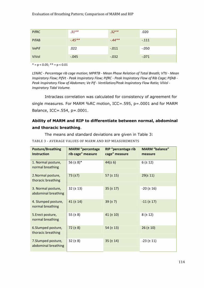
Evaluation of Breathing Pattern; Comparison of MARM and RIP
114
PifRC .31** .32** .020
PifAB -.45** -.44** -.111
VePif .022 -.011 -.050
ViVol -.045 -.032 -.071
* = p < 0.05; ** = p < 0.01
LS%RC - Percentage rib cage motion; MPRTB - Mean Phase Relation of Total Breath; VTti - Mean
Inspiratory Flow; PifVt - Peak Inspiratory Flow; PifRC - Peak Inspiratory Flow of Rib Cage; PifAB -
Peak Inspiratory Flow of Abdomen; Ve Pif - Ventilation/Peak Inspiratory Flow Ratio; ViVol -
Inspiratory Tidal Volume.
Intraclass correlation was calculated for consistency of agreement for
single measures. For MARM %RC motion, ICC=.595, p=.0001 and for MARM
Balance, ICC=.554, p=.0001.
Ability of MARM and RIP to differentiate between normal, abdominal
and thoracic breathing.
The means and standard deviations are given in Table 3:
TABLE 3 - AVERAGE VALUES OF MARM AND RIP MEASUREMENTS
Posture/Breathing
Instruction
MARM “percentage
rib cage” measure
RIP “percentage rib
cage” measure
MARM “balance”
measure
1. Normal posture,
normal breathing
56 (± 8)* 44(± 6) 6 (± 12)
2.Normal posture,
thoracic breathing
73 (±7) 57 (± 15) 29(± 11)
3. Normal posture,
abdominal breathing
32 (± 13) 35 (± 17) -20 (± 16)
4. Slumped posture,
normal breathing
41 (± 14) 39 (± 7) -11 (± 17)
5.Erect posture,
normal breathing
55 (± 8) 41 (± 10) 8 (± 12)
6.Slumped posture,
thoracic breathing
72 (± 8) 54 (± 13) 26 (± 10)
7.Slumped posture,
abdominal breathing
32 (± 8) 35 (± 14) -23 (± 11)

Evaluation of Breathing Pattern; Comparison of MARM and RIP
115
8.Erect posture,
thoracic breathing
76 (± 7) 52 (± 12) 32 (± 8)
9.Erect posture,
abdominal breathing
30 (± 9) 31 (± 22) -25 (± 10)
* mean and standard deviation
A within-subject analysis of variance using 3 factors (breathing, posture
and measurement method) showed significant differences between normal,
abdominal and thoracic breathing across all 9 events (F (2,16)=78.6, p=. 0001,
eta squared .908). As can be seen in Figure 2, for all 3 types of measurement
methods, instructions to breathe thoracically in the 3 postures (normal, erect
and slumped) resulted in more rib cage involvement than instructions to
breathe normally or breath abdominally. Similarly, instructions to breathe
abdominally in all 3 postures resulted in lesser rib cage involvement that that
seen in normal or thoracic breathing.
FIGURE 1 - EFFECTS OF ABDOMINAL, NORMAL AND THORACIC BREATHING FOR THE 3 MEASUREMENT METHODS-(10 SUBJECTS)
Method 1 = MARM percentage rib cage, Method 2 = RIP percentage rib cage, Method 3 = MARM balance.

Evaluation of Breathing Pattern; Comparison of MARM and RIP
116
Separate analysis of the 3 measurement methods using within-subject
analysis of variance with 2 factors (breathing and posture) showed that each of
the measurement methods was able to detect the voluntary breathing changes.
For the MARM percentage rib cage measure (F(2,22)= 191.2, p=. 0001, partial
eta squared .946) and for the MARM balance measure (F(2,22)=189.4,
p=.0001, partial eta squared .945) the ability to differentiate between
breathing patterns was very high. For the RIP percentage rib cage measure
(F(2,16) =12.89, p=. 0001, partial eta squared=. 617), however, the ability
was less. This suggests that both the MARM and RIP are able to differentiate
between breathing patterns, with the MARM‟s being markedly better.
MARM and RIP differentiation of effects of postural change on
breathing
The within-subject analysis of variance using 3 factors (breathing,
posture and measurement method) showed that no overall significant
difference resulted from changes in posture (F(4, 32)=2.8, p=. 091, partial eta
squared .258). Investigation by analysis of variance of the individual
measurement methods showed that the MARM measure of % rib cage motion
was able to detect differences in breathing that resulted from changes in
posture (F(2,22)=6.29, p=. 007, eta squared= .364) and the MARM measure of
balance was also able to detect these differences (F(2,22)=189.4, p=. 006, eta
squared=.371). Figure 3 shows the differences in breathing measures brought
about by changes in posture, for the 3 measurement methods.
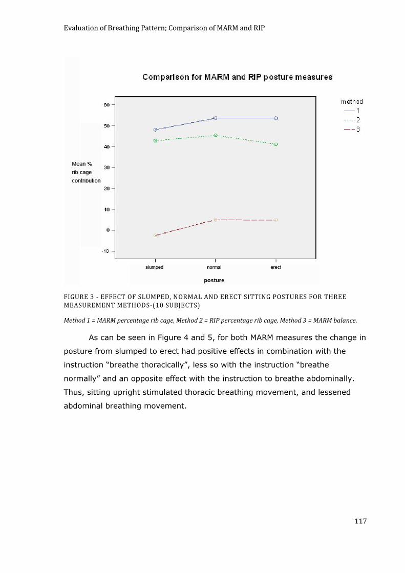
Evaluation of Breathing Pattern; Comparison of MARM and RIP
117
FIGURE 3 - EFFECT OF SLUMPED, NORMAL AND ERECT SITTING POSTURES FOR THREE MEASUREMENT METHODS-(10 SUBJECTS)
Method 1 = MARM percentage rib cage, Method 2 = RIP percentage rib cage, Method 3 = MARM balance.
As can be seen in Figure 4 and 5, for both MARM measures the change in
posture from slumped to erect had positive effects in combination with the
instruction “breathe thoracically”, less so with the instruction “breathe
normally” and an opposite effect with the instruction to breathe abdominally.
Thus, sitting upright stimulated thoracic breathing movement, and lessened
abdominal breathing movement.
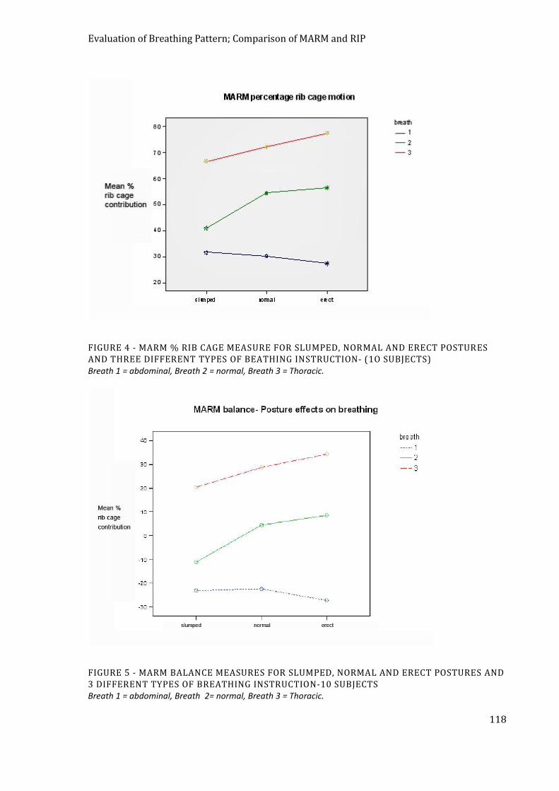
Evaluation of Breathing Pattern; Comparison of MARM and RIP
118
FIGURE 4 - MARM % RIB CAGE MEASURE FOR SLUMPED, NORMAL AND ERECT POSTURES AND THREE DIFFERENT TYPES OF BEATHING INSTRUCTION- (1O SUBJECTS) Breath 1 = abdominal, Breath 2 = normal, Breath 3 = Thoracic.
FIGURE 5 - MARM BALANCE MEASURES FOR SLUMPED, NORMAL AND ERECT POSTURES AND 3 DIFFERENT TYPES OF BREATHING INSTRUCTION-10 SUBJECTS Breath 1 = abdominal, Breath 2= normal, Breath 3 = Thoracic.
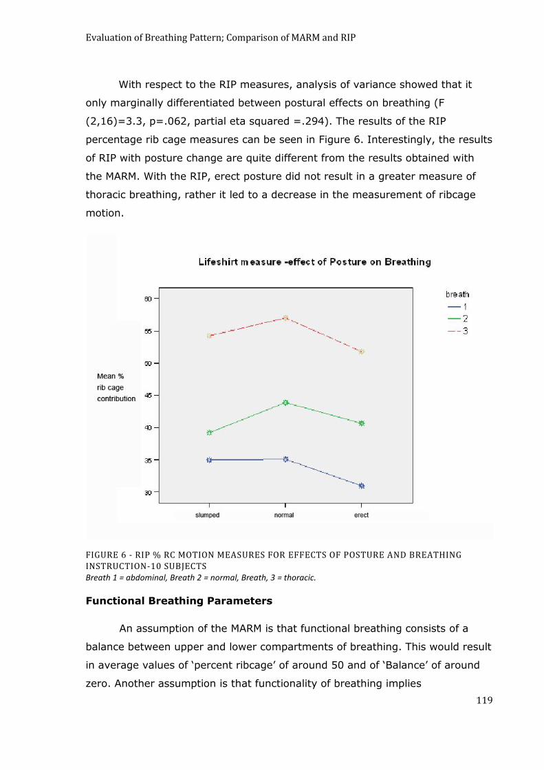
Evaluation of Breathing Pattern; Comparison of MARM and RIP
119
With respect to the RIP measures, analysis of variance showed that it
only marginally differentiated between postural effects on breathing (F
(2,16)=3.3, p=.062, partial eta squared =.294). The results of the RIP
percentage rib cage measures can be seen in Figure 6. Interestingly, the results
of RIP with posture change are quite different from the results obtained with
the MARM. With the RIP, erect posture did not result in a greater measure of
thoracic breathing, rather it led to a decrease in the measurement of ribcage
motion.
FIGURE 6 - RIP % RC MOTION MEASURES FOR EFFECTS OF POSTURE AND BREATHING INSTRUCTION-10 SUBJECTS
Breath 1 = abdominal, Breath 2 = normal, Breath, 3 = thoracic.
Functional Breathing Parameters
An assumption of the MARM is that functional breathing consists of a
balance between upper and lower compartments of breathing. This would result
in average values of „percent ribcage‟ of around 50 and of „Balance‟ of around
zero. Another assumption is that functionality of breathing implies
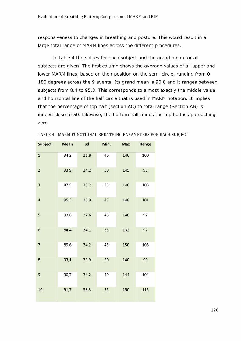
Evaluation of Breathing Pattern; Comparison of MARM and RIP
120
responsiveness to changes in breathing and posture. This would result in a
large total range of MARM lines across the different procedures.
In table 4 the values for each subject and the grand mean for all
subjects are given. The first column shows the average values of all upper and
lower MARM lines, based on their position on the semi-circle, ranging from 0-
180 degrees across the 9 events. Its grand mean is 90.8 and it ranges between
subjects from 8.4 to 95.3. This corresponds to almost exactly the middle value
and horizontal line of the half circle that is used in MARM notation. It implies
that the percentage of top half (section AC) to total range (Section AB) is
indeed close to 50. Likewise, the bottom half minus the top half is approaching
zero.
TABLE 4 - MARM FUNCTIONAL BREATHING PARAMETERS FOR EACH SUBJECT
Subject Mean sd Min. Max Range
1 94,2 31,8 40 140 100
2 93,9 34,2 50 145 95
3 87,5 35,2 35 140 105
4 95,3 35,9 47 148 101
5 93,6 32,6 48 140 92
6 84,4 34,1 35 132 97
7 89,6 34,2 45 150 105
8 93,1 33,9 50 140 90
9 90,7 34,2 40 144 104
10 91,7 38,3 35 150 115

Evaluation of Breathing Pattern; Comparison of MARM and RIP
121
11 85,3 35 35 140 105
12 89,9 32,7 45 140 95
mean 90,77 34,34 42,08 142,42 100,33
sd 3,56 1,70 6,11 5,21 7,00
As to the range of MARM values, Table 4 shows the lowest lower line
(minimum) and highest upper line (maximum) and the distance between them
(range). The average range between lowest and highest MARM line is about
100. In all subjects the maximal range is 90 or larger, which is more than half
of the total range of a half circle of 180 degrees.
DISCUSSION
The good agreement between examiners and between the MARM and
comparable RIP measures along with the MARM‟s ability to differentiate clearly
divergent patterns of breathing and posture suggest that the MARM is a useful
and reliable tool for the assessment of breathing pattern with good inter-rater
reliability. This confirms previous results (Dixhoorn 2004; Dixhoorn 2004)
It appears that the MARM is a global assessment in the double meaning
of global: general and spherical. The MARM provides a general indication of
distribution of breathing pattern in its three dimensional form and was better
able to distinguish between thoracic and abdominal breathing than RIP.
All four specific hypotheses relating to the utility of MARM were
confirmed. Its ability to distinguish between more thoracic and more abdominal
breathing was even greater than RIP.
Given the fact that the subjects were „experienced breathers‟, practiced
in breath control, it was expected that their MARM values would be close to
optimal. The results confirmed that under resting and normal conditions the
values of MARM, which theoretically range between 0 and 180, have an
average of the almost exact middle value of 90. This implies a percent ribcage
Evaluation of Breathing Pattern; Comparison of MARM and RIP
122
of about 50 and an even distribution of breathing between the two main
compartments, which is expressed in the measure of „balance‟ approaching
zero.
As a measure of functionality MARM can be used to test flexibility of
breathing pattern by assessing the response to sufficiently divergent postural
and respiratory instructions and by determining the maximum difference
between upper and lower lines. The present data suggest that the maximum
difference between upper and lower lines of MARM across several instructions
should be at least 90. In theory, upward breathing moves fully vertically to lift
the sternum and downward breathing moves fully vertically to press on the
pelvic floor. The assessor‟s hands however are placed at the middle of the body
and this limits the information acquired. The values of around 100 therefore
seem to represent the limits of the range that can be assessed by the MARM.
The variation between subjects is remarkably small for all parameters. This
indicates that subjects may be taken as a sample of truly experienced
breathers who are able to modify their breathing pattern as far as is feasible
without creating undue effort.
More studies are needed to establish optimal cutoff scores by comparing
the outcome to untrained and less experienced subjects as well as to patients
with breathing or other difficulties. Re-analysis of one data set from a previous
unpublished study showed that 12 subjects performing 3 different breathing
events had comparable average values.
One option for future studies assessing breathing functionality is to have
subjects bend sideways, in order to imitate a scoliotic C-curve and test the
adaptability of the ribcage to these posture changes. A strong characteristic of
the MARM is its ability to distinguish differences between the left and right side
of the chest. In case of even slight scoliosis, which is quite common, there may
be marked differences between the two sides that remain unnoticed by
traditional instrumental recordings. Such distortions may give rise to both
disturbance of breathing movement as well as a sense of dyspnea. Currently,
only Opto-electronic plethysmography gives an accurate image of the exact
Evaluation of Breathing Pattern; Comparison of MARM and RIP
123
shape of the breathing movement in all its variations (Aliverti, Dellaca et al.
2000).
Several limitations of this study should be noted. One is the low
reliability of the absolute distance between the two lines. We called it „volume‟
but it may be more accurate to call it „area‟. The exact place and distance of the
two lines on the half circle appears to depend on the assessor‟s personal
preference. There was no inter-rater agreement on the distance between the
lines and it correlated only to a small degree with the RIP measures. Given the
high agreement between assessors across the nine events on the other MARM
measures, however, it seems probable that the assessor‟s preference of the
placement of the lines is stable. Thus, it is likely that in clinical practice the
clinician may compare his assessment on one occasion with his assessment at
another occasion. This remains to be tested in a future study. Possibly, more
intensive training is necessary including specific focus on the placement of the
lines to increase reliability of „area‟ assessment.
Another potential limitation is the requirements to perform the MARM
correctly. In applying the MARM, one forms a mental picture of the general
change in shape of the body with in- and ex-halation. It requires sensitive
hands as well as imagination. The assessors were all trained and experienced
osteopaths who were clearly able to perform the MARM correctly. It is not
sufficient to simply put one‟s hands on another person‟s body and record any
movement that one notices locally. The touch should be clear and firm but not
intrusive or constrictive or in any way inhibit free breathing movement. The
hands need to follow respiratory motion and the assessor should try to picture
the origin and direction of the locally experienced movement. Possibly the
MARM is particularly useful for clinicians who are experienced in touching other
subject‟s bodies in a sensitive and perceptive way. The first two authors who
are experienced practitioners have now taught the MARM to many subjects,
with good practical success. However it remains to be seen how less
experienced examiners are able to perform.
A limitation of the design of this study is the possibility of observer bias,
because of the fixed order of the events and the possibility of visual information
Evaluation of Breathing Pattern; Comparison of MARM and RIP
124
to establish posture. In future studies the order of events could be random.
However, there is a natural sequence in difficulty of the events, which should
not be ignored. In this study the examiners were not aware of any expected
changes in breathing from posture. However they may have responded to
visual cues, the assessor may have expected to feel more thoracic breathing
when the subject was seen straightening up and the spine was extended. The
experience of the author most familiar with this technique (JvD) is that the
upper line of MARM can indeed be expected to rise in extended posture and
examiners may have quickly discovered this. In future studies it may be
advisable to use a random order of events, undisclosed to the assessor. Further
blinding could also be achieved by randomly allocating the types of breathing
and posture instructions given to the “breathers”. Still, the act of extending the
spine will always be noticeable by the hands on the back extension on the
lower line is open, however, and cannot be firmly predicted. When the distance
between symphysis pubis and sternum is increased there is also more space for
abdominal expansion. Some subjects may use this space and increase
abdominal expansion whereas others may predominantly lift the chest and
show decreased abdominal expansion. Thus, it is important for the assessor to
be as neutral as possible and observe the actual movements perceived by the
hands, and not try to guess the breathing pattern.
The study used a relatively small number of subjects and compared
results of only two examiners; this was another limitation of this study.
The correlation between the MARM and the RIP measures at 0.60 was
not very high for a reliability assessment. We believe this is because RIP only
measures lateral expansion while the MARM also measured vertical motion. It is
interesting that RIP measurement did not respond to spinal extension as MARM
did. In fact, RIP showed a decrease of ribcage motion while the MARM showed
an increase. This may reflect the fact that when the ribcage is lifted upwards
„pump-handle‟ or vertical motion of the rib cage dominates and there is a loss
of some of its sideways expansion or „bucket-handle‟ motion. MARM is
apparently able to register the real upward motion, whereas RIP is limited to
purely sideways expansion.

Evaluation of Breathing Pattern; Comparison of MARM and RIP
125
In future studies the MARM may be used to clarify the concept of
„dysfunctional breathing‟. This concept refers on the one hand to functional
respiratory complaints, like disproportionate breathlessness and can be
measured for instance by Nijmegen Questionnaire for hyperventilation
complaints (Thomas, McKinley et al. 2001). On the other hand it refers to
disturbances in the biological function of breathing, without real physical
causes. Signs include asynchrony of breathing movement between thorax and
abdomen, predominantly upper-thoracic breathing, frequent or deep sighs,
mouth breathing, exaggerated use of auxiliary respiratory muscles (Chaitow
2002). It is still unclear to what degree the two definitions overlap. Functional
respiratory complaints may be caused by uneven distribution of breathing
movement, but also by other causes, like stress or anxiety (Morgan 2002). The
MARM can be used to measure breathing movement and help to elucidate its
role in the etiology of respiratory complaints. More specifically, as a tool in
breathing therapy, the MARM is useful to quantify the effect of breathing
therapy on the quality of respiratory movement. If such effects are related to
improvements in complaints, it may be argued that they were due to
disturbances in breathing movement.
CONCLUSION:
The MARM appears to be a valid and reliable clinical and research tool for
assessing breathing movement with good inter-examiner and a greater ability
to distinguish vertical ribcage motion that RIP. Further studies to confirm its
clinical utility are warranted.
This is a copy of an article published in Applied Psychophysiology © [2008] [copyright Springer Verlag;
Applied Psychophysiology and Biofeedback is available online at: http://www.springerlink.com
Reference-. Courtney R, Dixhoorn Jv, Cohen M (2008). Evaluation of breathing pattern: comparison of a Manual Assessment of Respiratory Motion (MARM) and respiratory induction plethysmography. Applied Psychophysiology and Biofeedback, 33, 91-100
Overview Section 2
126
CHAPTER 7-
COMPARISON OF THE MANUAL ASSESSMENT OF RESPIRATORY
MOTION (MARM) AND THE HI LO BREATHING ASSESSMENT IN
DETERMINING A SIMULATED BREATHING PATTERN.
ABSTRACT
Aims: The aim of this study was to assess and compare the validity and
potential utility of two manual breathing assessment procedures: the Manual
Assessment of Respiratory Motion (MARM) and the Hi Lo Breathing Assessment.
A secondary aim was to gauge the influence of the practitioner‟s general level
of manual therapy experience on their ability to learn and perform these
assessment techniques.
Method: 56 osteopaths and osteopathic students were taught the MARM and
the Hi Lo Breathing Assessment and trained to simulate breathing patterns. The
participants, acting alternatively as breathers and examiners, then attempted
to accurately determine whether the breathing patterns simulated by their
partner were predominately abdominal, thoracic or, in the case of the Hi Lo,
paradoxical. Participants were surveyed on their confidence in the use of each
technique, their perceived ease in using each technique, and their intended
future use of the techniques. Student and practitioner ability to detect
simulated breathing patterns were compared for the MARM and Hi Lo.
Results: Overall scores for correctly determining breathing patterns were not
significantly different for the MARM or the Hi Lo, and there was no notable
moderation of this effect according to experience, with both practitioners and
students demonstrating a high level of performance on both techniques. There
were some differences in accuracy of performance across different breathing
styles, with Hi Lo assessment of paradoxical breathing being more difficult to
identify correctly. Ease of learning was similar for MARM and Hi Lo but
confidence in using the techniques, and intended future use were higher for the

Overview Section 2
127
MARM. There were some significant relationships between these utility
measures and performance, particularly on the MARM.
Conclusions: This study supports the validity of the Hi Lo and builds on
previous work to strengthen the evidence for the validity of the MARM.
Responses to the survey indicate that, overall, participants preferred the MARM
to the Hi Lo. This study is a preliminary investigation of these techniques.
Future studies to test the validity of these techniques should be performed in a
clinical setting on individuals with actual rather than simulated breathing
pattern disturbances.

Overview Section 2
128
INTRODUCTION
Breathing pattern disturbance and abnormal function of the respiratory muscles
has been proposed to contribute to symptoms such as dyspnea (Breslin,
Garoutte et al. 1990; Wolf 1994), neck and shoulder girdle pain (Perri and
Halford 2004), and temporomandibular joint disorders (Hruska 1997). It has
also been argued that a person‟s habitual breathing patterns may influence
posture and spinal stability, and it has been proposed that correct breathing is
the foundation for the correction of dysfunctional movement and postural
patterns (Lewitt 1980; Perri 2007). It is difficult to evaluate the impact of
breathing pattern on symptoms, movement and postural patterns on the basis
of previous studies because the characteristics of correct or dysfunctional
breathing pattern are not clearly defined and the measurement techniques used
to evaluate breathing pattern have not been standardized or validated.
Nevertheless in the clinical environment, breathing pattern is often
assessed by observation and palpation and several palpatory techniques for
assessment of breathing pattern have been described in manual therapy texts
and other publications (Clanton and Diaz 1995; Hillegass and Sadowsky 2001;
Chaitow 2002; Pryor and Prasad 2002; Perri and Halford 2004; Perri 2007) with
techniques differing in the hand placement of the operator, and interpretation
and recording of palpatory findings.
In a previous study we investigated one technique for evaluating and
quantifying breathing pattern, the Manual Assessment of Respiratory Motion
(MARM), and compared it with measures performed with Respiratory Induction
Plethysmography (RIP), an established standard for measuring breathing
pattern (Wilhelm and Roth 2003). We also tested inter-examiner agreement
when two examiners used this technique to differentiate between different
breathing and postural patterns. High levels of agreement between examiners
were found with two MARM measures that reflected balance of thoracic to
abdominal breathing, ric =.85, p < .001. Examiners‟ MARM measures also
correlated with similar measures obtained from RIP, r =.59, p < .01. Both RIP
and MARM methods were able to differentiate between abdominal and thoracic
Overview Section 2
129
breathing patterns, but only MARM was able to differentiate between breathing
changes occurring as an incidental result of postural change. It was concluded
that the MARM was a reliable clinical tool for assessing breathing patterns and
demonstrated better sensitivity to more dimensions of rib cage motion than RIP
(Courtney, van Dixhoorn et al. 2008).
The MARM procedure was first developed and applied in a follow-up
study of breathing and relaxation therapy with cardiac patients in the 1980s. It
appeared that two years after breathing therapy the MARM still showed
differences between experimental and control patients (Dixhoorn 1994). It is
similar to other breathing assessment techniques that are based on the
examiner‟s interpretation and estimation of the motion of their hands when
placed at the posterior and lateral lower rib cage. However, the MARM is of
particular interest as a clinical and research tool because it includes a system of
notation that allows the examiner to derive numerical values for two variables
related to relative distribution of breathing motion and another numerical
variable for area of breathing involvement. The examiner can also gauge, rate
and record their general impressions of breathing regularity, rib cage stiffness
and symmetry of breathing.
The Hi Lo can be used to assess the motion of the upper rib cage and
lower rib cage/abdomen and determine aspects of breathing such as rate,
rhythm, relative motion and phase relation of upper and lower breathing
compartments (Chaitow 2002). The Hi Lo assesses breathing from the motion
observed at the front of the body while the MARM assessment is made with the
examiner hands at the back on the mid thoracic and lateral lower rib cage and
waist. The Hi Lo findings are reported as qualitative descriptions or as
dichotomous variables in comparison with the MARM, which assigns numerical
values. To our knowledge, no studies have examined the relationships in scores
obtained between these two assessment methods, or compared their accuracy.
In the previous validation study of the MARM, the observed high levels of
inter-examiner agreement and agreement between the MARM and RIP may
have been due to the fact that the examiners were all experienced osteopaths
(Courtney, van Dixhoorn et al. 2008). It is unknown to what extent

Overview Section 2
130
performance on various breathing assessments, such as the MARM and the Hi
Lo, is moderated by the experience of the administrator; for example, do
experienced practitioners and students differ in the accuracy of their
assessments derived from these techniques?
The main aim of this study was to examine the relationship between
therapists‟ performance in the use of the MARM and Hi Lo by assessing the
sensitivity and consistency of these techniques when used to assess simulated
breathing patterns. Another aim was to gauge whether accurate performance in
the use of these techniques was dependant on the examiners‟ general levels of
experience in manual therapy. This was done by comparing results achieved by
experienced osteopaths with those of osteopathic students. Finally,
relationships between performance on the MARM and Hi Lo, and participants‟
views on their confidence in and perceived ease of use of each technique and
their intention to use each technique in the future were also considered.
METHOD
Volunteer examiners who were either osteopathic students (n = 27) or
practicing osteopaths (n = 29) who attended a two-hour training class that
utilized a structured training format. In the training session, participants were
taught how to do the simulated breathing techniques and the MARM and Hi Lo
breathing assessment techniques.
Participants were paired, with one acting alternatively as „examiner‟ and
one as „breather‟. Pre-screening of breathing ability was used to exclude people
who were clearly unable to correctly modify their breathing pattern.
The breather was instructed to alter their breathing pattern three times
according to randomly selected written instructions, firstly while the examiner
performed the MARM and subsequently while the examiner performed the Hi
Lo. In the case of the Hi Lo, breathing instructions were various random
combinations of thoracic, abdominal or paradoxical breathing. In the case of
the MARM the breathing instructions were various random combinations of
thoracic or abdominal breathing but not paradoxical breathing. The examiner,
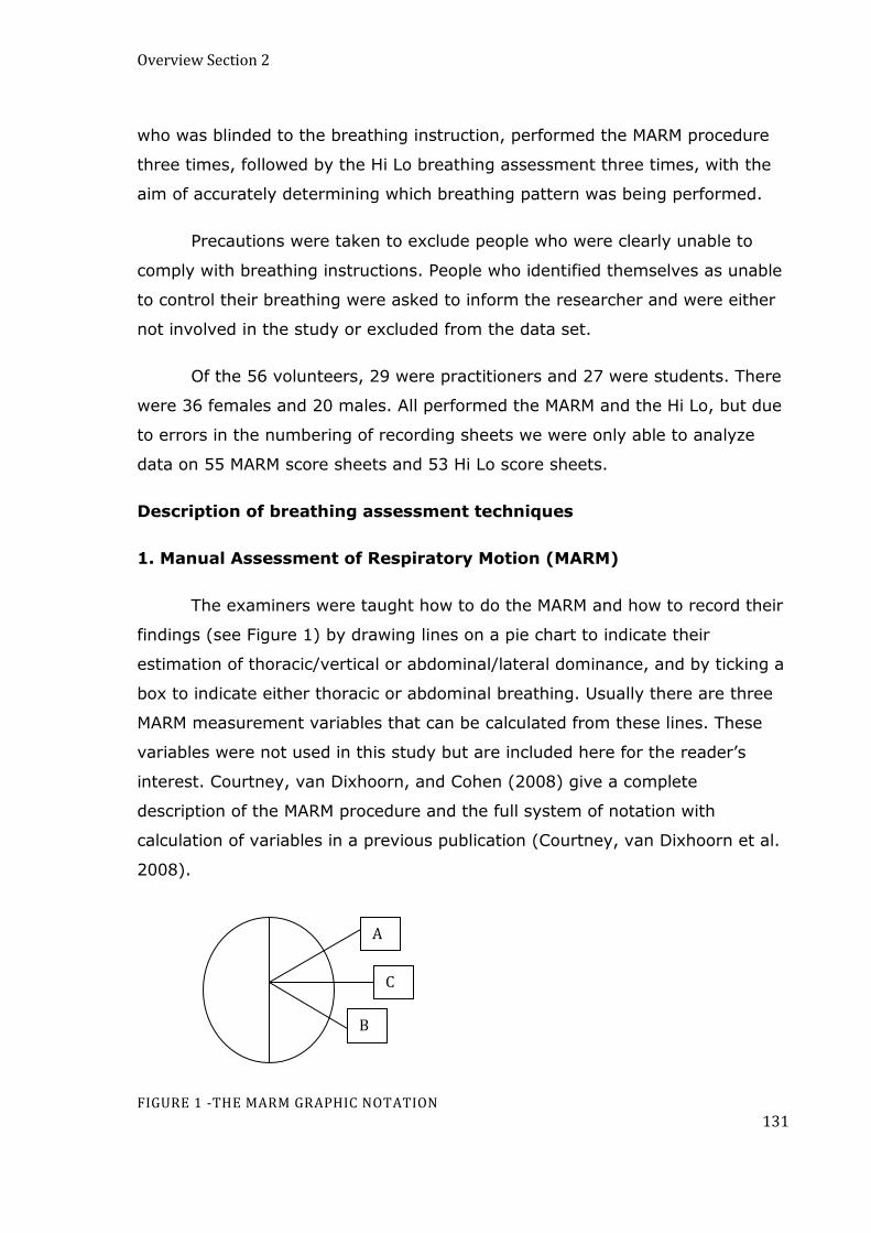
Overview Section 2
131
who was blinded to the breathing instruction, performed the MARM procedure
three times, followed by the Hi Lo breathing assessment three times, with the
aim of accurately determining which breathing pattern was being performed.
Precautions were taken to exclude people who were clearly unable to
comply with breathing instructions. People who identified themselves as unable
to control their breathing were asked to inform the researcher and were either
not involved in the study or excluded from the data set.
Of the 56 volunteers, 29 were practitioners and 27 were students. There
were 36 females and 20 males. All performed the MARM and the Hi Lo, but due
to errors in the numbering of recording sheets we were only able to analyze
data on 55 MARM score sheets and 53 Hi Lo score sheets.
Description of breathing assessment techniques
1. Manual Assessment of Respiratory Motion (MARM)
The examiners were taught how to do the MARM and how to record their
findings (see Figure 1) by drawing lines on a pie chart to indicate their
estimation of thoracic/vertical or abdominal/lateral dominance, and by ticking a
box to indicate either thoracic or abdominal breathing. Usually there are three
MARM measurement variables that can be calculated from these lines. These
variables were not used in this study but are included here for the reader‟s
interest. Courtney, van Dixhoorn, and Cohen (2008) give a complete
description of the MARM procedure and the full system of notation with
calculation of variables in a previous publication (Courtney, van Dixhoorn et al.
2008).
FIGURE 1 -THE MARM GRAPHIC NOTATION
A
C
B

Overview Section 2
132
Variables Calculated From MARM Graphic Notation
Variable Description Calculation
Area of Breathing Angle formed between
upper line and lower line. Angle A B
Balance
Difference between angle
made by horizontal axis (C)
and upper line (A) and
horizontal line (C) and lower
line (B).
AC - CB
Percent rib cage motion
Area above horizontal/ total
area between upper line and
lower line x 100.
AC/AB X 100
The following is the description given to examiners for how to
perform the MARM:
“The examiner sits behind the subject and places their hands on the lower lateral rib cage. The
hands rest firmly but do not direct or restrict breathing motion. The hands are comfortably open
with fingers spread so that the little finger approaches a horizontal orientation and the thumbs
are approximately vertical. The examiner’s lower fingers are below the lower ribs to feel
abdominal expansion. The examiner makes an assessment of the overall vertical motion relative
to the overall lateral motion. Simultaneously they evaluate to what extent the motion is
predominantly upper rib cage, lower rib cage/abdomen, or in balance. The examiner then draws
two lines. An upper line (A) represents the degree of vertical and upper thoracic motion and the
lower line (B) represents the degree of lower ribs and abdominal motion. The horizontal line (C)
represents the thoraco-lumbar junction.”
2. Hi Lo Breathing Assessment
The examiners received the following instructions for how to perform and
record the Hi Lo:
“Stand at the front and slightly to the side of the person. Place one hand on their sternum and one
hand on their upper abdomen. Determine whether thoracic or abdominal motion is dominant
during breathing and to what extent this is so. Also check for paradoxical breathing by seeing if
the abdomen moves in a direction opposite to the thorax during breathing; this is evident during
Overview Section 2
133
inhalation if the abdomen moves toward the spine, and during exhalation if the abdomen moves
in an outward direction.”
The extent of thoracic or abdominal breathing was rated using a score
between 1 and 3. To avoid confusion and to allow direct comparison with MARM
scores, examiners were also asked to tick a box indicating if they considered
the breathing pattern to be predominantly abdominal or thoracic. Assessment
of paradoxical breathing was also recorded by ticking a box.
3. Survey regarding utility of MARM and Hi Lo
Participants were given a questionnaire about their experience of
learning and using the MARM and Hi Lo and their self-perceived ability to
conduct the breathing techniques. The questions were as follows:
1. I found the (MARM or Hi Lo) easy to use.
2. I felt confident using the (MARM or Hi Lo).
3. I intend to use the (MARM or Hi Lo) in the future.
4. I was able to do the breathing techniques.
The responses to these questions were made by circling one of the following:1
= strongly agree, 2 = agree, 3 = unsure, 4 = disagree, 5 = strongly disagree.
RESULTS
Data preparation and coding
All data were entered into a single SPSS spreadsheet for analysis.
Examiners‟ performance on both the MARM and Hi Lo were recorded
dichotomously as either correct or incorrect for both thoracic and abdominal
breathing, and, in the case of the Hi Lo only, for paradoxical breathing. Given
the method of test administration, as outlined above, this meant that there
were unequal numbers of tests of thoracic and abdominal breathing across the
two methods. The scores for the three testing sessions were totalled to obtain
an overall score out of three for both methods, although it should be noted that
this total score of three could be made up of any combination of abdominal,
Overview Section 2
134
thoracic, and, in the case of the Hi Lo, paradoxical breathing. The data relating
to ease, confidence and intention toward future use of the two techniques was
recorded in line with the five-point Likert scale described above. Because of the
wording of the scale, a low score on these questions indicated a positive
response.
Comparison of accuracy between the MARM and Hi Lo
Descriptive results for the MARM and Hi Lo total scores and the utility
measures are presented in Table 1. The mean total correct score on the MARM
across both practitioners and students was slightly lower than the mean total
correct score on the Hi Lo, but a Wilcoxon non-parametric signed ranks test
found this difference non-significant. Further, a non-parametric correlation
found no significant relationship between the two sets of scores. Non-
parametric analyses were used for all significance testing because of the ordinal
nature of the data.
These differences were not moderated by the experience of the
administrator. Although the differences between the MARM and Hi Lo were
slightly higher for the practitioners than the students, no significant differences
were found between the scores of the two techniques for each group
separately, and no significant correlations were found between performance on
the two techniques for each group separately. These analyses used the same
statistical methods as those outlined above. Further, a Mann-Whitney U test
found no significant difference between practitioners and students in overall
performance on either technique.
It is possible that these statistical outcomes were affected by a
pronounced ceiling affect in the data. Given a maximum score of 3 for each
technique, the mean values indicate nearly perfect performance for many
participants. The data reflects this, with 50% (27/54) of participants scoring 3
on both measures. This proportion of perfect performance was higher for
practitioners (16/26, 62%) than students, (11/28, 39%).

Overview Section 2
135
TABLE 1 - DESCRIPTIVE STATISTICS FOR PERFORMANCE AND UTILITY MEASURES
Students
(n = 29)
Practitioners
(n = 27)
Total
(n = 56)
Measure
M SD M SD M SD
MARM – Total Correct 2.46 0.64 2.54 0.99 2.48 0.82
Hi Lo – Total Correct 2.61 0.50 2.77 0.51 2.69 0.51
MARM – Ease of Use 1.36 0.73 1.41 0.97 1.38 0.85
MARM – Confidence in Use 1.57 0.84 1.67 1.07 1.62 0.95
MARM – Intention Future Use 1.61 0.69 1.63 1.01 1.62 0.85
Hi Lo – Ease of Use 1.46 0.64 1.52 0.70 1.49 0.66
Hi Lo – Confidence in Use 2.04 0.79 2.19 0.88 2.11 0.83
Hi Lo – Intention Future Use 2.29 0.81 2.52 1.05 2.40 0.94
There were, however, some notable differences in accuracy across the
different breathing styles (i.e. thoracic, abdominal, & paradoxical). Table 2
presents a detailed breakdown on performance for both practitioners and
students on each technique for the different breathing styles. A Cochrane‟s Q
test found no significant differences in performance across thoracic and
abdominal breathing styles for the MARM for either the sample as a whole or
for practitioners and students separately; however, a significant difference
across the three breathing styles of the Hi Lo (thoracic, abdominal &
paradoxical) was observed for the sample as a whole, Q(2) = 10.36, p = .006,
with performance on paradoxical breathing (66% correct) inferior to both
abdominal breathing (94% correct) and thoracic breathing (96% correct). This
result was moderated by experience, with no significant difference found for
practitioners, but a significant difference found for students, Q(2) = 6.25, p =
.044; again, paradoxical breathing was found to be less easy to identify than
either thoracic or abdominal breathing.
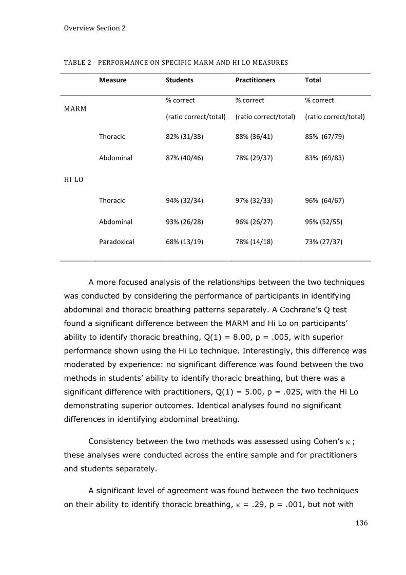
Overview Section 2
136
TABLE 2 - PERFORMANCE ON SPECIFIC MARM AND HI LO MEASURES
Measure Students Practitioners Total
MARM % correct
(ratio correct/total)
% correct
(ratio correct/total)
% correct
(ratio correct/total)
Thoracic 82% (31/38) 88% (36/41) 85% (67/79)
Abdominal 87% (40/46) 78% (29/37) 83% (69/83)
HI LO
Thoracic 94% (32/34) 97% (32/33) 96% (64/67)
Abdominal 93% (26/28) 96% (26/27) 95% (52/55)
Paradoxical 68% (13/19) 78% (14/18) 73% (27/37)
A more focused analysis of the relationships between the two techniques
was conducted by considering the performance of participants in identifying
abdominal and thoracic breathing patterns separately. A Cochrane‟s Q test
found a significant difference between the MARM and Hi Lo on participants‟
ability to identify thoracic breathing, Q(1) = 8.00, p = .005, with superior
performance shown using the Hi Lo technique. Interestingly, this difference was
moderated by experience: no significant difference was found between the two
methods in students‟ ability to identify thoracic breathing, but there was a
significant difference with practitioners, Q(1) = 5.00, p = .025, with the Hi Lo
demonstrating superior outcomes. Identical analyses found no significant
differences in identifying abdominal breathing.
Consistency between the two methods was assessed using Cohen‟s ;
these analyses were conducted across the entire sample and for practitioners
and students separately.
A significant level of agreement was found between the two techniques
on their ability to identify thoracic breathing, = .29, p = .001, but not with
Overview Section 2
137
abdominal breathing. The agreement observed on the thoracic breathing scores
was not moderated by experience, with significant agreement being observed
for students, = .53, p = .001, and practitioners ( could not be calculated for
this group because there were no incorrect assessments using the Hi Lo).
Despite the lack of overall agreement between the two methods on their ability
to correctly identify abdominal breathing, there was some moderation
according to experience, with a significant difference being observed for
practitioners, = .29, p = .037, but not for students.
Simulated breathing
In response to the statement “I found the breathing techniques easy to
do”, only 3.8% of participants disagreed or strongly disagreed with this
statement, although 14.8% of participants indicated that they were unsure.
This figure most likely reflects uncertainty rather than an inability or true
difficulty in performing the thoracic, abdominal and paradoxical breathing
maneuvers.
Utility measures
Across both practitioner groups, there were no significant differences
between the MARM and the Hi Lo with regard to perceived ease in using the
technique. A Wilcoxon signed ranks test did, however, find significant overall
differences between the two methods in terms of intended future use, z = 4.73,
p < .001, and confidence in using, z = 3.15, p = .002. In both instances, the
more positive impressions were in favor of the MARM. This difference was not
notably moderated by experience, with a similar pattern of significant results
emerging for both students and practitioners on both confidence and intention
toward future use. Further, Mann-Whitney U tests revealed no significant
overall differences between students and practitioners on any of these
variables.
An interesting pattern of results emerged when ease, confidence and
intention toward future use were related to performance on the two techniques.
Non-parametric correlations revealed no significant relationships between any

Overview Section 2
138
of the utility measures and performance on the Hi Lo; however, performance on
the MARM was significantly correlated with its perceived ease of use, r = .31, p
= .018, perceived confidence in use of the technique, r = .28, p = .028, and
intention to use the technique in the future, r = .43, p = .001. In each
instance, high scores on the utility measure (i.e. reflecting higher perceived
ease, more confidence, and a more positive intention toward future use) were
related to more accurate performance with the technique. Further, these
relationships were clearly moderated by experience. For students, intention to
use the MARM in the future revealed a strong positive relationship with
performance on the method, r = .49, p = .006, which was not evident in the
practitioners; however, significant positive relationships were found for the
practitioners between perceived ease of using the MARM and performance on
the MARM, r = .51, p = .007, and between confidence in using the MARM and
performance, r = .53, p = .004.
Additional qualitative written comments and feedback from 6 participants
suggested that the hand positioning of the MARM was more effective because it
gave a good sense of upper rib cage movement and the balance of upper rib
cage to lower rib cage abdomen motion without intruding on the patients‟
breathing. Comments also indicated participants favored the system of notion
used by the MARM and that hand placement on the front of the body with the
Hi Lo tended to be more intrusive and was more likely to influence the patient
to artificially alter their breathing pattern in response to the examiner‟s hands.
DISCUSSION
This study indicates that less experienced practitioners with only a small
amount of practice and training can use the MARM and Hi Lo with similar levels
of accuracy to experienced practitioners. There were only minor differences
observed between students and practitioner‟s performance for example
students were better able to identify abdominal breathing using the MARM and
in turn practitioners were better able to identify paradoxical breathing using the
Hi Lo. More practitioners than students achieved a perfect score. However,

Overview Section 2
139
overall both students and practitioners achieved similar levels of accuracy for
most simulated breathing patterns.
Paradoxical breathing, performed by the “breather” drawing in the
abdomen during inhalation was found to be the most difficult simulated
breathing pattern for examiners using the Hi Lo to determine. It is difficult in a
simulation to reproduce the actual muscle activity that occurs in real life
paradoxical breathing. In individuals with natural paradoxical breathing there is
usually heightened respiratory drive resulting in tonic activity of the accessory
muscles of inspiration. This produces a strong vertical and upper chest motion
during inspiration, which can be accompanied by some dynamic hyperinflation
of the lungs. In this scenario the diaphragm tends to become short and flat and
inefficient at producing lateral expansion of the lower rib cage during inhalation
(Loring, Mead et al. 1985; De Troyer and Estenne 1988; Celli 1989; Hruska
1997).
The MARM evaluates expansion of the lower lateral rib cage and in some
situations this may be a more reliable indicator of diaphragm efficiency than the
Hi Lo observation of anterior abdominal displacement. With decreased force of
diaphragm , decreased lateral basal expanasion is usually accompanied by a
compensatory increase in assessor muscle use and, therefore, an increase in
vertical and thoracic motion during inspiration. The fact that these two motions
are simultaneously monitored by the MARM is a strength of this technique.
MARM performance in both groups was related to how easy the examiner
found this technique and their confidence in using it. Performance on the Hi Lo
did not relate to any of the utility measures i.e. ease, confidence, or future use.
This indicates that individuals who found the MARM difficult and were not
confident in using it did not perform as well. This suggests that some further
training in MARM was needed by some individual to improve ease and
confidence and it is reasonable to speculate that this would have improved
performance of the MARM in these individuals.
Preference for the MARM was shown in 2 ways; survey results showed
that both students and practitioners were more confident with the MARM and

Overview Section 2
140
more likely to use the MARM in the future. Invited comments showed that
participants preferred the hand position and system of notation of the MARM.
They found the hand position less intrusive, and felt that breathers were less
likely to change their breathing in response to the examiner‟s hands when the
examiner was sitting behind and placing their hands on the lower ribs only.
However, most examiners indicated an intention to use both techniques in the
future. Student‟s intention to use the techniques in the future was related to
their performance on the MARM and Hi Lo. Interestingly practitioner‟s intention
to use the techniques was not related to their individual performance but rather
to their evaluation of the utility of the technique itself based on other factors.
There are other functions and ways of using the MARM and Hi Lo not
tested in this study. For instance the MARM can be used to assess and note the
examiner‟s impressions of the stiffness of rib cage and general freedom of
breathing motion. MARM can also be used for assessing symmetry of breathing,
which can be compromised by scoliosis and also by unilateral diaphragm
dysfunction (Hruska 1997). Non-uniform distribution of pressure distorts the rib
cage and markedly stiffens it. In normal situations lung volume can increase up
to 70% of maximum workload without stiffening the rib cage, however when
there is non-uniform distribution of breathing this distorts and stiffens the rib
cage (Kenyon, Cala et al. 1997). The Hi Lo and the MARM are best used in
various body positions and with different breathing instructions to fully evaluate
the flexibility, and thus functionality, of breathing.
There are several limitations to this study, some of which were self-
imposed due to the need to maintain uniformity of teaching and testing. In
designing the study we decided on a two-hour limit to teach both techniques
and tried to maintain consistency across all four teaching sessions despite
seeing that a longer training period with more individualized feedback could
have been beneficial in some cases. While we did ask individuals to exclude
themselves in the pre-test period if they felt they could not modify their
breathing pattern as instructed, it was not possible with the current design to
check that each breather was simulating each breathing technique correctly in
each case. This makes interpretation of the accuracy scores more difficult

Overview Section 2
141
because lack of accuracy could have been due to the breather rather than the
examiner.
CONCLUSIONS
Both the MARM and the Hi Lo appear to accurately assess thoraco-abdominal
relationships in breathing when used by both experienced clinicians and
osteopathic students. Both practitioner and students have slightly more positive
impressions of the MARM. As each technique has its own strengths and
limitations the evaluation of dysfunctional breathing may best be performed
using a combination of both techniques. It needs to be recognised that
breathing pattern assessment may be more difficult in real life clinical situations
where altered thoraco-abdominal breathing patterns occur in response to
altered neuro-muscular function, organ pathology and respiratory control
mechanisms. Therefore, future studies to validate these techniques should be
performed in a clinical setting on individuals with actual rather than simulated
breathing pattern disturbances.
This is a copy of an article published in the International Journal of Osteopathic Medicine © [2009] [copyright
Elsevier.]; International Journal of Osteopathic Medicine is available online at: http://www.elsevier.com
Reference- Courtney R,Reece J (2009). Comparison of the Manual Assessment of Respiratory Motion
(MARM) and the Hi Lo breathing assessment determining a simulated breathing pattern. International Journal
of Osteopathic Medicine, 12, 86-91
Overview Section 2
142
OVERVIEW SECTION 2 -
DYSFUNCTIONAL BREATHING SYMPTOMS
This section contains one published study which describes the
development and Factor Analysis of a dysfunctional breathing questionnaire
called the Self Evaluation of Breathing Questionnaire (SEBQ).
Chapter 8- ‘Preliminary investigation of a measure of dysfunctional
breathing symptoms: The Self Evaluation of Breathing Questionnaire (SEBQ)‟

Self Evaluation of Breathing Questionnaire
143
CHAPTER 8
PRELIMINARY INVESTIGATION OF A MEASURE OF
DYSFUNCTIONAL BREATHING SYMPTOMS: THE SELF
EVALUATION OF BREATHING QUESTIONNAIRE (SEBQ).
ABSTRACT
Introduction: Dysfunctional Breathing (DB) can be defined by the presence of
unexplained breathing symptoms. However, validated questionnaires to
comprehensively evaluate all dimensions of breathing symptoms proposed to
be associated with DB are not available. This paper discusses the development
and exploration of the dimensionality of a preliminary questionnaire, the Self
Evaluation of Breathing Questionnaire, whose items were derived from a
popular Internet questionnaire for evaluating breathing functionality and
breathing symptoms proposed in the scientific literature to be discriminative for
DB.
Method: The 17-item SEBQ was administered to 83 adults. Exploratory factor
analysis was performed and correlations calculated between the SEBQ and the
Nijmegen Questionnaire (NQ), which is a validated questionnaire for
hyperventilation syndrome.
Results/Discussion: Two dimensions were found in the SEBQ. One dimension
named “lack of air” appears to reflect sensations of air hunger that may relate
more to chemoreceptor aspects of breathing sensation. The other dimension
named “perception of inappropriate or restricted breathing” appears to reflect
sensations and observations about the work of breathing and may relate more
to the biomechanical aspects of breathing sensation. The correlations between
the SEBQ and the NQ were .6 for the 17-item SEBQ and .3 for the final 12-item
SEBQ, which contained the strongest items of the two dimensions.
Conclusion: Breathing symptoms associated with dysfunctional breathing
arising from predominately biomechanical aspects of breathing might be
distinguishable from symptoms arising from factors reflecting chemoreceptor

Self Evaluation of Breathing Questionnaire
144
input. The two dimensions of the SEBQ may represent related but distinct
aspects of dysfunctional breathing symptoms that appear different to those
assessed by the Nijmegen Questionnaire. The SEBQ, if further developed, may
be a useful clinical assessment tool that could more discriminatively evaluate
the separate dimensions of breathing symptoms and their response to
treatments that aim to improve the functionality of breathing.
Self Evaluation of Breathing Questionnaire
145
INTRODUCTION
Despite there being no precise definition, it has been proposed that
dysfunctional breathing (DB) exists when there are a number of respiratory
complaints in otherwise healthy individuals or in individuals with only mild
medical conditions (Howell 1990). Furthermore, aspects of breathing
functionality may exacerbate symptoms in various diseases such as asthma and
cardiovascular disease (Grossman 1983; Bernardi and Gabutti 2001; Thomas,
McKinley et al. 2001). Statements about the prevalence of dysfunctional
breathing are difficult to interpret due to the vague nature of this condition and
it would be useful to have some way of monitoring breathing symptoms
thought to be related to DB (Lum 1975; Fried 1993; Thomas, McKinley et al.
2001).
Practitioners of breathing therapy currently assess breathing functionality
on the basis of self-evaluation of breathing behavior and breathing symptoms
(Hendricks 1995; Stalmatski 1999; White 2005). Of the few instruments which
inquire comprehensively into breathing complaints, there is only one validated
questionnaire, the Nijmegen Questionnaire (NQ), which has been used in the
scientific literature to identify dysfunctional breathing (Dixhoorn and
Duivenvoorden 1985; Thomas, McKinley et al. 2001). However, the NQ has
only been validated for the hyperventilation syndrome, which may be just one
aspect of dysfunctional breathing (Morgan, 2002; van Dixhoorn, 1985; 1997).
The 15 items of the NQ include 11 items that inquire about psychological,
neurological and cardiovascular symptoms commonly found in the
hyperventilation syndrome with only 4 questions obviously relating to breathing
sensations. These are: 1. Faster and deeper breathing; 2. Short of breath; 3.
Constricted chest; and, 4. Unable to breathe deeply. It is possible that this
small number of questions on respiratory symptoms in the NQ might not
inquire sufficiently into all dimensions of respiratory symptoms. The diagnosis
of Hyperventilation Syndrome, which the Nijmegen Questionnaire was originally
designed to investigate, has been questioned in recent years and a boarder
definition of dysfunctional breathing is being sought. Thus,, the purpose of the
SEBQ was to explore the nature of unexplained symptoms that are commonly
Self Evaluation of Breathing Questionnaire
146
reported in people with dysfunctional breathing that may or may not be part of
Hyperventilation Syndrome and are not extensively investigated by the NQ.
A literature search of MEDLINE, Science Direct, Cochrane Library and
reference lists of original and review articles to find relevant instruments
showed a paucity of validated instruments for investigating dysfunctional
breathing. It showed that most validated questionnaires, other than the
Nijmegen Questionnaire, that investigate breathing complaints have been
designed for, and tested on, individuals with cardiorespiratory disease or other
chronic illness rather than for individuals with dysfunctional breathing or
unexplained breathing disorders. Questionnaires like the St. Georges
Questionnaire (Jones, Quirk et al. 1992), the Chronic Respiratory
Questionnaire (Wijkstra, TenVergert et al. 1994), and the UCSD Shortness of
Breath Questionnaire (Eakin, Resnikoff et al. 1998) assess symptoms,
situational measures of dyspnea and impact of symptoms on daily life as a way
of monitoring severity of disease states (Meek 2004).
There is a body of literature developed over 20 years on the language of
dyspnea, showing that dyspnea has many dimensions and that these various
dimensions arise from different pathophysiological mechanisms (Simon 1989,
Simon 1990, Mahler 1996, Han 2008).It has also been shown that medically
unexplained dyspnea, which is considered to be part of the clinical presentation
of dysfunctional breathing, can be differentiated on the basis of particular
qualities of dyspnea (Han 2008).The Breathing Descriptor tool is the only
validated questionnaire which identifies the various dimensions of respiratory
discomfort, but to our knowledge has not been extensively used as a tool for
evaluating symptoms in individuals who may have dysfunctional breathing
(Mahler, Harver et al. 1996; Harver, Mahler et al. 2000).
The set of items which were devised for this study and named the Self
Evaluation of Breathing Questionnaire (SEBQ) were derived from published
studies by Burton (Burton 1993) and Howell (Howell 1990) that proposed
symptoms discriminative for dysfunctional breathing, from other literature in
the field (Fried 1993; Wilhelm, Gertivz et al. 2001), discussion with colleagues,
relevant clinical experience and from a public domain internet questionnaire
Self Evaluation of Breathing Questionnaire
147
titled ”How Good is your Breathing Test, Take our Free Breathing Test and See”
(HGYB) (White 2005). No item of the SEBQ was taken directly from any single
source, most items were included because they were suggested by several
sources and appeared plausible from our own clinical experience or after
discussion with colleagues. The published sources of the major influences for
each item of the SEBQ are given in Table 1.
The five very similar symptoms described by Howell and Burton as being
discriminative for DB were based on findings from separate studies by each of
these authors. In the first study Howell observed the following characteristics
of unexplained breathing symptoms in individuals thought to have dysfunctional
breathing:
1. Breathlessness occurred at rest;
2. Breathlessness was poorly correlated with level of fitness or physical exertion;
3. Breathlessness was associated with other body symptoms suggestive of
hyperventilation; and
4. A sense of difficulty taking a breath in.
A majority of the patients suffering from these symptoms also experienced
increased anxiety, depression or chronic unresolved stress (Howell 1990).
Burton, in a subsequent study of patients presenting to general practice with
functional symptoms associated with many breathing complaints reported the
following seemingly related symptoms:
1. Dyspnea at rest;
2. Dyspnea on exertion;
3. Dyspnea occurring with onset of other physical symptoms; and
4. Difficulty taking a deep breath.
In addition Burton found that patients reported experiencing dyspnea when
anxious (Burton 1993).
SEBQ items that were based on the symptoms described by Howell and
Burton include:
1. “I get breathless even when resting”,
Self Evaluation of Breathing Questionnaire
148
2. “I get easily breathless on physical exertion out of proportion to my fitness”,
3. “I get breathless in association with my physical symptoms”,
4. “I can‟t take a deep or satisfying breath”, and
5. “I get breathless when I am anxious.”
Two questions of the SEBQ relate to difficulty in co-coordinating breathing
with speech with resulting fatigue and breathlessness associated with speech.
This is believed by Fried to be one of the important signs of dysfunctional
breathing (Fried 1987; Fried 1993). In the SEBQ, this was investigated with
two items: “I get short of breath when speaking or reading out loud”; and, “I
have trouble co-ordinating my breathing when I am speaking”.
It is frequently described in the literature that excessive irregularity of
breathing is a common finding in dysfunctional breathing and that this is mostly
in the form of frequent sighing, yawning and gasping for breath (Han, Stegen
et al. 1996; Wilhelm 2001). In the SEBQ this symptom is investigated with the
item “I sigh, yawn or gasp”.
A further nine SEBQ items were partially derived from the HYBG (White
2005). In several instances, a number of questions from the HGYB that seemed
overlapping were consolidated into a single SEBQ question. In other instances
HGYB questions were rephrased for clarity of meaning and to make them
consistent with the language used in the SEBQ. The HGYB questionnaire, while
unvalidated, unstructured and containing questionable claims about normal
breathing was useful because it contained common assumptions about the type
of symptoms that characterise people with either “good” or “bad” breathing.
While many items from the HGYB were not considered relevant or plausible the
HGYB questions that were used to inform the SEBQ items were chosen because
clinical experience and discussion with colleagues suggested that they did seem
to reflect the symptoms seen in dysfunctional breathing.
The aim was to explore what dimensions existed in this list of questions.
To further explore the items of the SEBQ we compared its dimensions to the
NQ, an established questionnaire for hyperventilation, to see if the SEBQ
assessed different qualities of breathing symptoms to those investigated by the

Self Evaluation of Breathing Questionnaire
149
NQ. In this study we administered an initial version of the SEBQ to 83
individuals, performed exploratory factor analysis (EFA) to identify the
dimensions underlying responses to this questionnaire and then tested for
presence of correlations with the NQ.
METHOD
Participants
The 83 individuals who participated in this study were 29 males and 54
females, whose average age was 49 years. They were either healthy or
suffered from mild medical conditions, including mild asthma. 29 of these
subjects were found to have abnormal spirometry defined by either FEV1 or
FVC >15% below predicted.
Participants were recruited from general practices and complementary
medicine clinics in Sydney, Australia. Flyers and brochures were placed in
waiting rooms and practitioners received a letter about the study. The
participants did not have their breathing assessed by practitioners prior to the
study and did not necessarily receive a diagnosis of dysfunctional breathing.
These individuals who volunteered or were recommended to this study
generally were motivated to participate in this study because they had concerns
about breathing complaints or interest in knowing if they had correct breathing.
This sample is therefore representative of the type of individual who while
seeing a practitioner for unrelated complaints might have secondary concerns
about breathing and also be likely to self-administer breathing tests such as the
Internet “”How good is your breathing test”.
Materials
The SEBQ (Table 1) contains 17 items. Responses to these items were on
a 4-point Likert scale:
(0) never/not true at all;
(1) occasionally/a bit true;
(2) frequently-mostly true; and
(3) very frequently/very true.
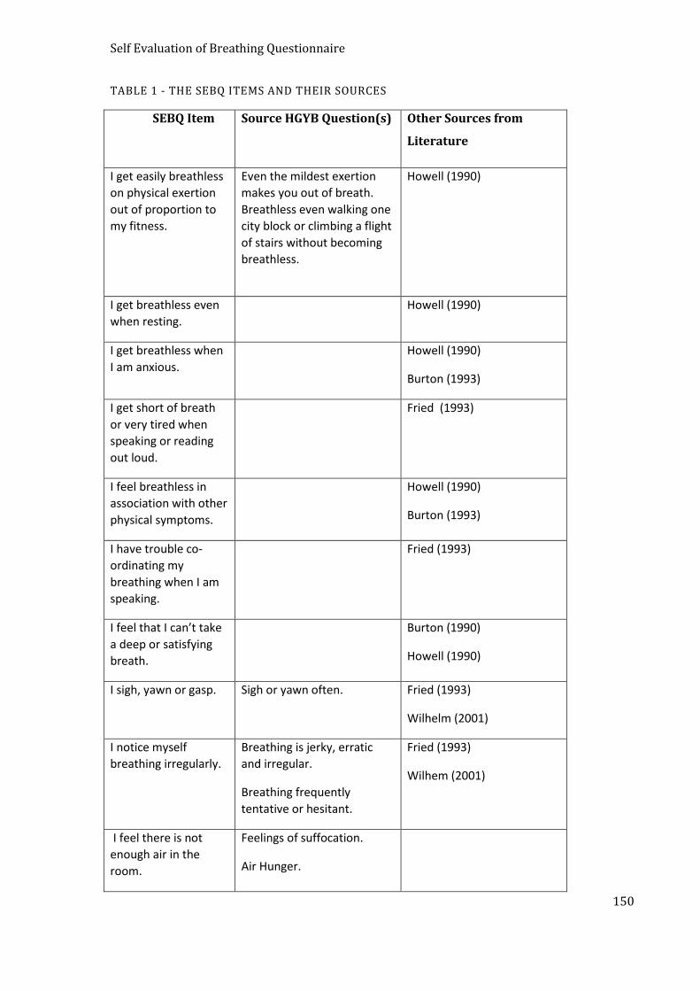
Self Evaluation of Breathing Questionnaire
150
TABLE 1 - THE SEBQ ITEMS AND THEIR SOURCES
SEBQ Item Source HGYB Question(s) Other Sources from
Literature
I get easily breathless
on physical exertion
out of proportion to
my fitness.
Even the mildest exertion
makes you out of breath.
Breathless even walking one
city block or climbing a flight
of stairs without becoming
breathless.
Howell (1990)
I get breathless even
when resting.
Howell (1990)
I get breathless when
I am anxious.
Howell (1990)
Burton (1993)
I get short of breath
or very tired when
speaking or reading
out loud.
Fried (1993)
I feel breathless in
association with other
physical symptoms.
Howell (1990)
Burton (1993)
I have trouble co-
ordinating my
breathing when I am
speaking.
Fried (1993)
I feel that I can’t take
a deep or satisfying
breath.
Burton (1990)
Howell (1990)
I sigh, yawn or gasp. Sigh or yawn often. Fried (1993)
Wilhelm (2001)
I notice myself
breathing irregularly.
Breathing is jerky, erratic
and irregular.
Breathing frequently
tentative or hesitant.
Fried (1993)
Wilhem (2001)
I feel there is not
enough air in the
room.
Feelings of suffocation.
Air Hunger.

Self Evaluation of Breathing Questionnaire
151
I can’t catch my
breath.
Can’t catch your breath.
Shortness of breath
My breathing feels
stuck or restricted.
Breathing feels stuck.
Breathing is labored or
restricted.
Deep breathing curtailed.
I feel that my rib cage
is tight and can’t
expand.
Chest is large and stiff.
Tightness, soreness or
pressure in the chest or
below breast bone.
My clothing often
feels too tight or
uncomfortable
around my chest.
Chest feels large and stiff,
feeling like band around
chest.
Feel bump, lump below
breastbone when you try to
take a deep breath.
I find myself holding
my breath at various
times.
Often catch yourself not
breathing during waking
hours.
Hold your breath a lot.
I notice myself
breathing shallowly
using my upper chest
and shoulders.
Do you raise your collar
bones?
Do you raise your shoulders?
Do your neck muscles bulge
out?
I notice myself
breathing quickly.
Individuals are asked to
count and record their
breaths per minute.
Self Evaluation of Breathing Questionnaire
152
The Nijmegen Questionnaire (NQ) is a 15 item questionnaire whose
items are based on complaints ascribed to the Hyperventilation Syndrome
(Dixhoorn and Duivenvoorden 1985).The NQ contains items relating to
respiratory, psychological, cardiovascular, neurological and gastrointestinal
complaints. Responses to the items are reported on a 5-point ordinal scale
ranging from (1) never to (5) very frequently.
Procedure
The study received approval from the RMIT University Ethics Committee.
Participants were informed that this was a study investigating dysfunctional
breathing and the ways of measuring it. The study was conducted over a 12
month period.
Statistical Analysis.
As the aim was to identify underlying constructs, the SEBQ items were
analyzed using principal axis factoring. We used several guidelines and a series
of factor analyses (FA) before making our final decision on number of factors to
retain (Ford, MacCallum et al. 1986). Our final decision was guided by the fit of
the hypothetical factors produced by the FA to conceptual and theoretical
considerations. We used orthogonal rotation for all trial factor analysis and
tested both orthogonal and oblique rotations in the final factor analysis to
evaluate which of the two gave superior simple structure. Once the likely
number of factors was decided, we removed variables with loadings
substantially below .6. The decision was based on a rule of thumb
recommendation that for a sample size of 85, the smallest meaningful factor
loading was .60 (Gardner 2007).
RESULTS
The distribution of responses for each of the items of the SEBQ are shown in
Table 2. Most items showed a range of response on the scale from (0) -
never/not true at all to (3) - very frequently/very true. SEBQ scores for the
sum of the original items ranged from 0 to 30 with a mean of 13.4 (SD=8.2).
SEBQ scores for the final 12 retained items ranged from 0 to 24 with a mean of
9.8 (SD=6.5) (Table 2 shows frequency of responses for each of SEBQ items).

Self Evaluation of Breathing Questionnaire
153
Scores for the Nijmegen Questionnaire ranged from 0 to 51, with the mean
score being 17.5 (SD=9.7).
TABLE 2 - FREQUENCY OF RESPONSE (%) TO EACH OF THE SEBQ ITEMS
Order Item 0 1 2 3
1 I get easily breathless out of proportion to my fitness.
16.9 50.6 20.0 10.6
2 I notice myself breathing shallowly. 17.6 49.4 21.2 9.4
3 I get short of breath reading and talking.
53.1 24.7 13.6 8.6
4 I sigh yawn or gasp.* 12.9 56.5 21.2 7.1
5 I feel I cannot get a deep or satisfying breath.
34.1 42.4 15.3 5.9
6 I notice myself breathing irregularly. 42.4 36.5 12.9 4.7
7 My breathing feels stuck or restricted. 44.7 34.1 15.3 3.5
8 My ribcage feels tight and can’t expand. 48.2 35.3 10.6 3.5
9 I notice myself breathing quickly. 35.3 49.4 9.4 3.5
10 My clothing feels tight and uncomfortable around my chest.
72.9 11.8 9.4 3.5
11 I get breathless when I am anxious. 41.0 38.6 18.1 2.4
12 I find myself holding my breath. 31.8 45.9 17.6 2.4
13 I feel breathless in association with other physical symptoms.
48.2 40.0 5.9 2.4
14 I have trouble co-ordinating my breathing when I am speaking.
56.6 34.1 5.9 2.4
15 I can’t catch my breath. 57.6 29.4 9.4 1.2
16 I feel that the air is stuffy, as if not enough air in the room.
44.7 43.5 8.4 1.2
17 I get breathless even when I am resting. 64.7 29.4 2.4 0.0
Likert Scale: 0.= Never or not true at all, 1.= occasionally, a bit true, 2 = frequently,mostly true,3 =
very frequently – very true. *Items in italics deleted in final 12 item version of SEBQ.
Data were screened for multivariate outliers by calculating Mahalanobis‟
distances. Four possible outliers had Mahalanobis‟ distances slightly higher than
the cut-off point indicated by chi-squared distribution tables. Visual inspection

Self Evaluation of Breathing Questionnaire
154
of the data gathered from these cases showed no irregularities and due to the
low ratio of participants to variables it was decided to retain these cases.
There were a substantial number of correlations between variables. The
Kaiser-Myer-Olkin measure of sampling adequacy produced a score of .84 and
Bartlett‟s test of sphericity was significant (p<.0001) indicating that the
correlations found in the data set were suitable for principal axis factoring. Anti-
image correlations and covariance matrices were calculated to estimate
measurement of sampling adequacy (MSA). No variables showed MSA‟s below
.6 indicating that all variables were suitable for inclusion in this analysis. Low
communalities (.259) were seen for only one item “my clothing feels tight” and
this item was later removed as it also was shown to have low factor loading
when the final factor solution was achieved.
The initial solution extracted five factors with eigen values greater than 1
and explained 70% of the variance. However, the scree plot did not support the
extraction of 5 factors. Both 4 and 5 factor solutions were rejected, as it was
evident after orthogonal rotation that these solutions were not interpretable
due to the large number of split loadings. A 2- factor and possibly a 3- factor
solution appeared more likely from visual inspection of the scree plot. The
factor analysis was then re-run with specifications for a 3- factor and then a 2-
factor solution followed by orthogonal rotation of factors. A 3- factor solution
explained 56% of the variance and the orthogonally rotated factor matrix
showed at least seven split loadings and was not interpretable. A 2- factor
solution explained 58% of the variance and the orthogonally rotated factor
matrix showed only two items (“can‟t catch my breath” and “trouble co-
ordinating breathing when speaking”) with loadings split between the 2-factors.
Factor 1 explained 45% of the variance and Factor 2 explained 13% of the
variance.
A satisfactory result was seen when oblique factor rotation (direct
oblimin) was applied to the 2- factor solution as a simple structure (where
items load predominantly on just one factor) was evident. The factor correlation
matrix showed a correlation of .63 between factors indicating that the factors
are related.
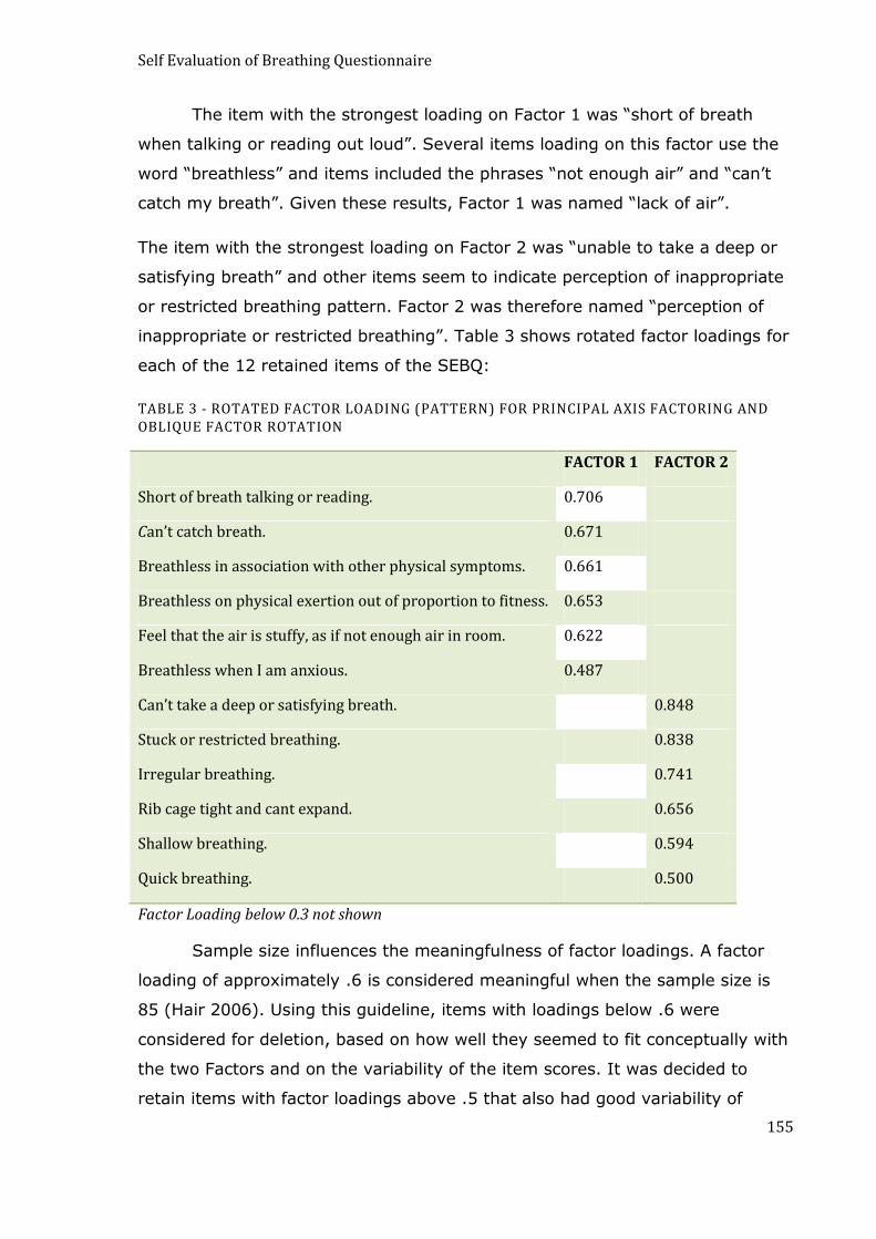
Self Evaluation of Breathing Questionnaire
155
The item with the strongest loading on Factor 1 was “short of breath
when talking or reading out loud”. Several items loading on this factor use the
word “breathless” and items included the phrases “not enough air” and “can‟t
catch my breath”. Given these results, Factor 1 was named “lack of air”.
The item with the strongest loading on Factor 2 was “unable to take a deep or
satisfying breath” and other items seem to indicate perception of inappropriate
or restricted breathing pattern. Factor 2 was therefore named “perception of
inappropriate or restricted breathing”. Table 3 shows rotated factor loadings for
each of the 12 retained items of the SEBQ:
TABLE 3 - ROTATED FACTOR LOADING (PATTERN) FOR PRINCIPAL AXIS FACTORING AND OBLIQUE FACTOR ROTATION
FACTOR 1 FACTOR 2
Short of breath talking or reading. 0.706
Can’t catch breath. 0.671
Breathless in association with other physical symptoms. 0.661
Breathless on physical exertion out of proportion to fitness. 0.653
Feel that the air is stuffy, as if not enough air in room. 0.622
Breathless when I am anxious. 0.487
Can’t take a deep or satisfying breath. 0.848
Stuck or restricted breathing. 0.838
Irregular breathing. 0.741
Rib cage tight and cant expand. 0.656
Shallow breathing. 0.594
Quick breathing. 0.500
Factor Loading below 0.3 not shown
Sample size influences the meaningfulness of factor loadings. A factor
loading of approximately .6 is considered meaningful when the sample size is
85 (Hair 2006). Using this guideline, items with loadings below .6 were
considered for deletion, based on how well they seemed to fit conceptually with
the two Factors and on the variability of the item scores. It was decided to
retain items with factor loadings above .5 that also had good variability of
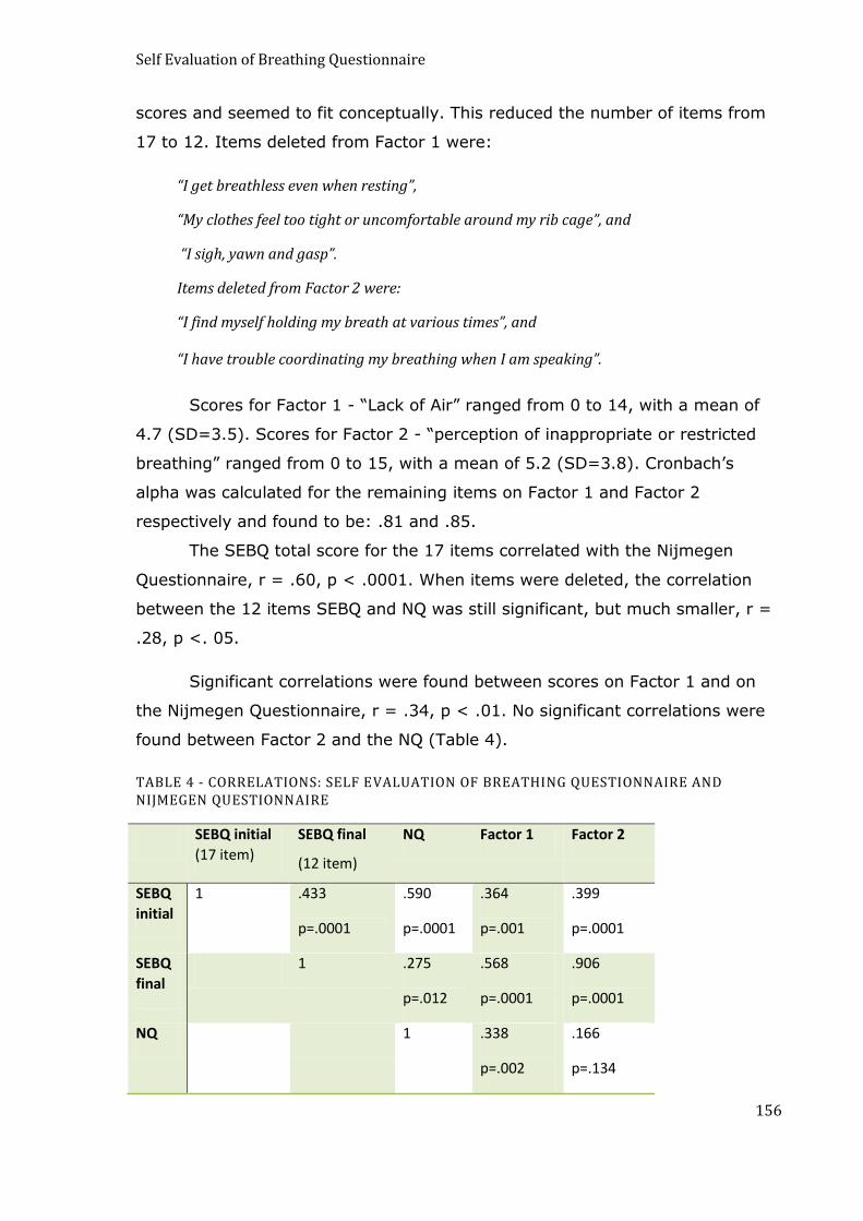
Self Evaluation of Breathing Questionnaire
156
scores and seemed to fit conceptually. This reduced the number of items from
17 to 12. Items deleted from Factor 1 were:
“I get breathless even when resting”,
“My clothes feel too tight or uncomfortable around my rib cage”, and
“I sigh, yawn and gasp”.
Items deleted from Factor 2 were:
“I find myself holding my breath at various times”, and
“I have trouble coordinating my breathing when I am speaking”.
Scores for Factor 1 - “Lack of Air” ranged from 0 to 14, with a mean of
4.7 (SD=3.5). Scores for Factor 2 - “perception of inappropriate or restricted
breathing” ranged from 0 to 15, with a mean of 5.2 (SD=3.8). Cronbach‟s
alpha was calculated for the remaining items on Factor 1 and Factor 2
respectively and found to be: .81 and .85.
The SEBQ total score for the 17 items correlated with the Nijmegen
Questionnaire, r = .60, p < .0001. When items were deleted, the correlation
between the 12 items SEBQ and NQ was still significant, but much smaller, r =
.28, p <. 05.
Significant correlations were found between scores on Factor 1 and on
the Nijmegen Questionnaire, r = .34, p < .01. No significant correlations were
found between Factor 2 and the NQ (Table 4).
TABLE 4 - CORRELATIONS: SELF EVALUATION OF BREATHING QUESTIONNAIRE AND NIJMEGEN QUESTIONNAIRE
SEBQ initial
(17 item)
SEBQ final
(12 item)
NQ Factor 1 Factor 2
SEBQ
initial
1 .433
p=.0001
.590
p=.0001
.364
p=.001
.399
p=.0001
SEBQ
final
1 .275
p=.012
.568
p=.0001
.906
p=.0001
NQ 1 .338
p=.002
.166
p=.134
Self Evaluation of Breathing Questionnaire
157
DISCUSSION
This study was a preliminary exploration of the SEBQ, a compilation of
questions suggested by various sources to represent symptoms associated with
DB. Two separate but correlated dimensions of breathing sensation were
identified with the SEBQ. These may correlate with either biomechanical or
chemoreceptor factors. Factor 1 was named “lack of air” because it contained
phrases similar to those used by individuals whose chemoreceptors were
challenged by hypoxia or hypercapnia (Banzett, Lansing et al. 1990; Lansing
2000). All items using the word breathless loaded on this factor, as did phrases
such as “short of breath and “not enough air” and “can‟t catch my breath”. This
was differentiated from Factor 2 which we called “perception of inappropriate or
restricted breathing” and which appeared to relate more to biomechanical
factors. The highest loading items were “I cannot take a deep and satisfying
breath” and “My breathing feels stuck or restricted”. These items describe a
sense of restricted or otherwise unsatisfied respiration. Other items such as
“notice myself breathing quickly”, “notice myself breathing shallowly using my
chest and upper shoulders” and “notice myself breathing irregularly” are related
to awareness of the inappropriateness of some aspect of the mechanical work
of breathing.
The idea that biomechanical and/or chemoreceptor inputs contribute to DB
is consistent with the literature that suggests that sensations akin to “lack of
air” and “perception of inappropriate or restricted breathing” may arise
predominately from different receptors in the respiratory system and may
involve somewhat different physiological mechanisms. Previous research
suggests that different phrases are consistently used to describe the sensations
associated with particular tasks and that symptom descriptors vary according to
whether the afferent signals to the cortex arise predominately from
chemoreceptors, chest wall biomechanical receptors or receptors associated
with the lungs (Simon, Schwartzstein et al. 1989). Furthermore, subsequent
studies showed that individuals with particular diseases could be differentiated
according to the phrases they used which suggests that different qualities of
breathing discomfort may reflect underlying pathological and physiological
processes (Simon and Schwartzstein 1990; Elliot, Adams et al. 1991).
Self Evaluation of Breathing Questionnaire
158
Descriptive phrases for different qualities of breathing sensation have been
found in different studies to form from 8 to 12 different clusters (Simon,
Schwartzstein et al. 1989; Simon and Schwartzstein 1990; Elliot, Adams et al.
1991; Mahler, Harver et al. 1996). Through multidimensional scaling, Harver et
al.(2000) found that underlying these there appear to be three major
categories of symptoms (Harver, Mahler et al. 2000). Two of these appear to
be related to sensations and perceptions related to the biomechanical work of
breathing and the third to sensations arising from chemical drivers of
breathing. The two biomechanical factors, which were called “difficulty
breathing and phase of respiration” and “depth and frequency of breathing”,
contain many descriptors similar to items in SEBQ Factor 2, “perception of
inappropriate or restricted breathing”. Given a larger sample size or a sample
with more severe dysfunctional breathing we may also have found two
biomechanical factors. It‟s also likely that the SEBQ does not contain items that
properly represent the two biochemical dimensions of breathing symptoms and
future versions of the SEBQ should contain more items from the Breathing
Descriptor Tool. The category described by Harver as “perceived need and
urge to breathe” and also as “air hunger” appears to be similar to the SEBQ
Factor 1 “lack of air”.
Many items in SEBQ Factor 2 “perception of inappropriate or restricted
breathing” are recognisably similar to sensations documented in situations
where the work of breathing is made more difficult. For example, chest wall
strapping of healthy volunteers sufficient to restrict normal respiratory motion
resulted in the sensations of “unsatisfied respiration” and “inspiratory
difficulty” (O'Donnell, Hong et al. 2000). “Unsatisfied respiration” and a sense
of difficulty taking a deep and satisfying breath is a common symptom in
patients with impaired function of the respiratory muscles and rib cage due to
hyperinflation of the lungs (Lougheed, Fisher et al. 2006). In hyperinflation, the
rib cage stiffens and respiratory muscles, in particular the diaphragm and
accessory muscles of respiration, shorten which decreases their ability to
respond adequately to efferent command from the motor cortex (Wolfson,
Strohl et al. 1983; De Troyer and Estenne 1988; Finucane, Panizza et al. 2005;
O'Donnell 2006). The description of the breath as shallow is common in
patients with neuromuscular and chest wall disorders and this sensation is
Self Evaluation of Breathing Questionnaire
159
presumed to arise from receptors in the chest wall and muscles of breathing
and from efferent-reafferent dissociation (Simon and Schwartzstein 1990). All
of these sensations (shallow respiration, difficulty in taking a deep or satisfying
breath and feelings of restricted breathing) may arise when there is dissociation
between efferent motor command and reafferent ventilatory motor response
and when the motor output of the respiratory system is not able to match the
expectations generated in the sensory cortex by corollary discharge from the
motor cortex (Beach and Schwatrzstein 2006). These sensations are the
qualities described by the items in the SEBQ Factor 2.
Items associated with the SEBQ Factor 1 “lack of air” may indicate a state
of respiratory awareness associated with afferent input from chemoreceptor
activity. The sensory quality of “air hunger” or “urge to breathe” is most
strongly linked to changes in blood gases such as carbon dioxide or changes in
the respiratory drive and arises predominately from afferent input from central
and peripheral chemoreceptors (Lansing 2000). Research has shown that the
sensations of “air hunger” or “urge to breathe” are distinguishable from
breathing sensations related to the work or effort of breathing (Banzett,
Lansing et al. 1990; Lansing 2000).
Another possible way of interpreting the two factors identified in the SEBQ
is that they are two different aspects of interoception. Items in Factor 1 “lack or
air” are predominately about feeling or sensing. The items in Factor 2
“perception of inappropriate or restricted breathing” are predominately about
observing or noticing. It has been proposed that sensory experiences such as
breathing discomfort and pain have at least 3 components:
1. A sensory or discriminative component;
2. A cognitive or evaluative component; and
3. An affective or motivational component (Harver, Mahler et al. 2000).
Items on the two factors might be describing different aspects of the sensory
and cognitive experience of breathing discomfort. As both over- and under-
perception of breathing sensations can be problematic, particularly in
individuals with respiratory diseases, it may be useful to monitor how sensory
Self Evaluation of Breathing Questionnaire
160
or feeling aspects of breathing sensations are rated in comparison to evaluative
and noticing descriptions of breathing sensations (Klein, Walders et al. 2004).
Dysfunctional breathing has in the past been most associated with
hyperventilation, a condition which has a narrow biochemical definition and is
dependent on the finding of hypocapnia (Comroe 1974). However,
biomechanical aspects of breathing also influence its functionality. Low carbon
dioxide was once presumed to be the main cause of hyperventilation
symptoms, but this has been shown not to be the case and breathing
symptoms are often unexplained (Hornsveld 1997). People with unexplained
breathing symptoms, and even those presumed to have hyperventilation
syndrome, often show normal levels of carbon dioxide but abnormalities of
breathing pattern are common (Han, Stegen et al. 1996; Wilhelm, Gertivz et
al. 2001). The function of the respiratory muscles is a cause of respiratory
discomfort and could contribute to specific characteristics of the sensation of
dyspnea (Killian and Jones 1988).
Relationship of SEBQ with NQ
The correlation between the SEBQ score and NQ was substantially
reduced when the SEBQ items with low factor loadings were deleted. This
indicates that the NQ does not strongly represent the two factors found in the
SEBQ and the SEBQ could not be relied upon to detect hyperventilation
syndrome. The SEBQ Factor 1 component “lack of air” showed a small
correlation with the NQ, however there was no correlation between Factor 2
“perception of inappropriate and restricted breathing” and the NQ.
Interestingly the four breathing items in the Nijmegen Questionnaire, found by
van Dixhoorn and Duivenvoorden to have with high loadings (> .55) on the
breathing component, conceptually seem to range over both dimensions of the
SEBQ. NQ items “constricted chest”, “accelerated or deepened breathing” and
“unable to breathe deeply” are similar to items in SEBQ Factor 2 “perception of
inappropriate or restricted breathing”, and NQ item “shortness of breath”
appears similar to SEBQ Factor 1 “lack of air” items. However, the finding that
SEBQ Factor 2 did not correlate with the NQ suggests that the NQ differs in its
representation of sensations arising from possible biomechanical breathing
dysfunctions.
Self Evaluation of Breathing Questionnaire
161
Deleted Items
Certain items deleted due to low factor loadings in this study should still be
considered for inclusion in future versions of the SEBQ. The item “I feel
breathless even while resting” was reported by Howell as being a discriminative
symptom in patients with unexplained breathlessness, due to the finding that
this symptom was much more prevalent in patients whose breathing symptoms
were linked with functional or psychogenic factors rather than cardiorespiratory
pathology. In this present study the response to this item showed the lowest
variability of response with 65% of responders reporting never experiencing
this, 29% reporting it sometimes and only 2% experiencing this symptom
frequently. This could be due to the fact that the sample was somewhat diverse
and was not adequately representative of individuals with severe unexplained
breathing symptoms. Other deleted items such as “I sigh, yawn and gasp” and
“I find myself holding my breath” load weakly on Factors 1 and 2 respectively,
but show a good variability in response and could contribute useful information
about breathing irregularity which is a well-known aspect of disordered
breathing (Abelson, Weg et al. 2001; Wilhelm 2001).
Future Items for SEBQ
The SEBQ may benefit from additional items such as those found in the
Breathing Descriptor Tool. Suggested items from the Breathing Descriptor Tool
that might not be well represented in the SEBQ are:
“Breath does not go in all the way”,
“Breath not does go out all the way”,
“Breathing is heavy”,
“I feel that I am breathing more”,
“My breathing requires work”, and
“My breathing requires effort”.
It is also suggested that any future questionnaire inquires about the
prevalence of mouth breathing during the day, at night and during exercise.
Limitations
This study would have benefited from increased number of participants.
The study sample may not have adequately represented individuals with

Self Evaluation of Breathing Questionnaire
162
dysfunctional breathing. These factors and omission of items such as those
found in the Breathing Descriptor Tool may prevent the identification of some
dimensions of breathing symptoms.
Recommendations for Future Research
This study should be viewed as a preliminary exploration of the SEBQ.
Further development of the items of the SEBQ and further testing on the
specific populations for which it is intended is required to validate this
questionnaire. In particular the SEBQ should be tested on sample populations
with a diagnosis of dysfunction breathing, with unexplained breathing
complaints and with cardiorespiratory disease and the findings between these
groups and normals should be compared.
It is recommended that the questionnaire be further developed to clarify
wording of questions and that additional items be included so that all
dimensions of breathing symptoms and important breathing behaviours are
represented. A comprehensive breathing questionnaire would assist in
quantifying and monitoring the effects of treatments such as breathing therapy,
manual therapy, relaxation training or biofeedback on respiratory symptoms
and respiratory behaviours in individuals with and without cardiorespiratory
disease. Future versions of the SEBQ should be scrutinized by experts in the
field and investigated for test-retest reliability.
CONCLUSION
The two dimensions of the SEBQ may represent strongly related, but distinct,
aspects of dysfunctional breathing symptoms representing biochemical and
biomechanical mechanisms. The clinical assessment of these two dimensions
may prove useful for more specific diagnosis and treatment of dysfunctional
breathing. Future work is required to develop the SEBQ and evaluate its
performance in terms of discriminating between different patient categories and
normal individuals.
This is a copy of an article published in the International Journal of Osteopathic Medicine © [2009] [copyright
Elsevier.]; International Journal of Osteopathic Medicine is available online at: http://www.elsevier.com
Reference-Courtney R, Greenwood KM (2009). Preliminary investigation of a measure of dysfunctional breathing symptoms: the Self Evaluation of Breathing Questionnaire (SEBQ). International Journal of Osteopathic Medicine, 12, 121-127
Self Evaluation of Breathing Questionnaire
163
OVERVIEW OF SECTION 3 -
RELATIONSHIP BETWEEN MEAUSURES OF
DYSFUNCTIONAL BREATHING
This section contains two published studies which investigate the
relationships between measures of dysfunctional breathing.
Chapter 9 - „Relationships between measures of dysfunctional breathing
in a population with concerns about their breathing‟.
Chapter 10 - ‘Investigating the claims of Dr. K.P. Buteyko: The
Relationship of Breath Holding Time to end tidal CO2 and other proposed
measures of dysfunctional breathing‟.
Relationships Between Measures of Dysfunctional Breathing
164
CHAPTER 9
RELATIONSHIPS BETWEEN MEASURES OF DYSFUNCTIONAL
BREATHING IN A POPULATION WITH CONCERNS ABOUT THEIR
BREATHING
ABSTRACT
Background: Dysfunctional breathing (DB) is implicated in physical and
psychological health; however evaluation is hampered by lack of rigorous
definition and clearly defined measures. Screening tools for DB include
biochemical measures such as end-tidal CO2, biomechanical measures such
assessments of breathing pattern, breathing symptom questionnaires and tests
of breathing function such as breath holding time.
Aim: This study investigates whether screening tools for dysfunctional
breathing measure distinct or associated aspects of breathing functionality.
Method: 84 self-referred or practitioner-referred individuals with concerns
about their breathing were assessed using screening tools proposed to identify
DB. Correlations between these measures were determined.
Results: Significant correlations where found within categories of measures
however correlations between variables in different categories were generally
not significant. No measures were found to correlate with carbon dioxide levels.
Conclusion: DB cannot be simply defined. For practical purposes DB is
probably best characterized as a multidimensional construct with at least three
dimensions, biochemical, biomechanical and breathing symptoms.
Comprehensive evaluation of breathing dysfunction should include measures of
breathing symptoms, breathing pattern, resting CO2 and also include functional
measures such a breath holding time and response of breathing to physical and
psychological challenges.

Relationships Between Measures of Dysfunctional Breathing
165
INTRODUCTION
Dysfunctional Breathing (DB) is commonly used to describe disturbances in
breathing functionality that impact on health (Dixhoorn 1997; Morgan 2002 ;
Dixhoorn 2004; Thomas, McKinley et al. 2005; Prys-Picard and Niven 2008;
Stanton, Vaughn et al. 2008). The definition of DB however is unclear and no
gold standards exist to define it. Dysfunctional breathing includes
hyperventilation or breathing in excess of metabolic needs but also refers to
breathing pattern abnormalities, poor breathing control and presence of
breathing symptoms (Dixhoorn 1997; Morgan 2002; Warburton and Jack
2006). Scientists have until recently focused their attention on hyperventilation,
which is defined as breathing in excess of metabolic requirements that results
in depletion of carbon dioxide (Comroe 1974). However the importance of
hyperventilation and hypocapnia in producing the symptoms associated with DB
is disputed (Hornsveld, Garsson et al. 1996; Hornsveld 1997). It has been
proposed that DB symptoms may arise as a result of non biochemical breathing
dysfunctions or have neurological causes (Howell 1997). A broader definition of
dysfunctional breathing, which considers the multiple functions of breathing,
may be a more useful way to characterize DB and to determine its prevalence
and impact (Dixhoorn 1997).
Maintenance of normal levels of blood gases such as carbon dioxide is an
important, if not the key function of breathing; however breathing has other
important functions. Breathing functions in posture and motor control (Lewitt
1980; McGill, Sharratt et al. 1995; Hodges, Heijnen et al. 2001). It is a key
influence on oscillating rhythms that are important for homeostasis, autonomic
nervous system regulation and efficient interaction between body systems
(Giardino, Lehrer et al. 2000; Bernardi 2001; Song and Lehrer 2003). Normal
and precisely controlled breathing is also important for voice production and
regulation of speech (MacLarnon and Hewitt 1999; Sivasankar and Erickson
2009).
Biomechanical, neurological and psychological aspects of breathing are
not always tightly linked to biochemical parameters and their other
relationships are complex and not adequately understood (Han, Stegen et al.

Relationships Between Measures of Dysfunctional Breathing
166
1996). Attempts to tie the symptoms associated with dysfunctional breathing to
only the biochemical dimension i.e. hyperventilation and hypocapnia have not
been successful (Burton 1993; Hornsveld 1997). Physical and psychological
causes of breathing dysfunction are often interwoven and can be difficult to
separate, however, DB is thought to contribute to additional symptoms not
adequately explained by the main presenting complaint (Han, Zhu et al. 2004).
Research has shown that symptoms associated with dysfunctional breathing are
strongly influenced by anxiety and other emotional states and in some cases
the psychological influences are primary (Wientjes and Grossman 1994; Han,
Zhu et al. 2004). Other symptoms, particularly various qualities of dyspnea
have been linked to breathing pattern abnormalities and poor neuromechanical
coupling during breathing (O'Donnell 2006; Prys-Picard and Niven 2008).
Muscular skeletal dysfunctions, in addition to speech and voice problems
appear to be predominately linked with dysfunctions of breathing pattern and
neural control of respiration rather than to the body‟s carbon dioxide status
(McGill, Sharratt et al. 1995; Gandevia, Butler et al. 2002).
The accumulation of studies showing the presence of breathing
disturbances in highly symptomatic patients and the results of research
showing that patients with a range of symptoms and medical conditions
improve after breathing therapy (Lum 1975; Grossman 1984; Tweedale,
Rowbottom et al. 1994; Han 1996; Meuret, Rosenfield et al. 2009) lends weight
to the importance of assessment and optimization of breathing functionality in
patient care. However, evaluation of dysfunctional breathing is currently
hampered by lack of clear measurement guidelines. Measures used by
practitioners as screening tools to identify dysfunctional breathing include
biochemical measures such as end-tidal CO2 (Hardonk and Beumer 1979;
McLaughlin 2009), biomechanical measures such as assessments of breathing
pattern (Prys-Picard, Kellett et al. 2004), breathing symptom questionnaires
(Thomas, McKinley et al. 2005; Courtney and Greenwood 2009) and tests of
breathing function such as breath holding time (Courtney and Cohen 2008).

Relationships Between Measures of Dysfunctional Breathing
167
Symptom questionnaires used to evaluate DB
The Nijmegen Questionnaire (NQ) is the most commonly used
questionnaire to identify DB. The 16 item NQ was originally devised to test for
HVS and includes 4 questions on respiratory symptoms and another 12 items
on peripheral and central neurovascular or general tension related symptoms
(Dixhoorn and Duivenvoorden 1985). A questionnaire called the Self Evaluation
of Breathing Questionnaire (SEBQ) has also been devised to specifically assess
respiratory symptoms and breathing behaviours reported to be associated with
DB. The SEBQ includes a larger number of respiratory items than the NQ and
can differentiate two distinct dimensions of breathing discomfort - “lack of air”,
which is probably related to chemoreceptor derived sensations and “perception
of inappropriate or restricted breathing”, which is probably related to the
biomechanics of breathing and breathing perception (Courtney and Greenwood
2009). Normal values for the SEBQ have not been established as yet and this
questionnaire is only useful at present for assessing change in breathing
symptoms within individuals after treatment. However, it does have potential
as a screening tool for DB once further studies are done to validate this
instrument. Normal values for the NQ in European studies are generally around
a sum score of 10 (Han, Stegen et al. 1996; Han, Stegen et al. 1998; Thomas,
McKinley et al. 2005) whereas in China values are lower and average around 5
(Han, Zhu et al. 2004). In categorizing individuals as having DB, cut-offs of
both 20 and 22 have been found useful (van Doorn, Folgering et al. 1982;
Dixhoorn and Duivenvoorden 1985; Dixhoorn and Hoefman 1985).
Clinical measures of dysfunctional breathing patterns
Clinicians usually assess breathing patterns using observation and
palpation and historically have used a range of techniques, most of which have
not been validated (Pryor and Prasad 2002; Perri 2007). One component of
breathing pattern that is considered dysfunctional, is chronic thoracic dominant
breathing at rest. Recently a technique called the Manual Assessment of
Respiratory Motion (MARM), which can quantify the extent of thoracic dominant
breathing as well as other aspects of breathing pattern, has been found to have
high levels of inter-examiner reliability and to agree with measures made

Relationships Between Measures of Dysfunctional Breathing
168
simultaneously with Respiratory Induction Plethysmography (Courtney, van
Dixhoorn et al. 2008). Normal healthy individuals appear to have balanced
breathing with relatively equal motion of upper rib cage to lower rib cage
abdominal motion. Perfectly balanced breathing gives a MARM value of 0.
Normal values for the MARM in this study of 12 yoga teachers and breathing
therapy practitioners were around 6. MARM values above 30 can be considered
dysfunctional, as they are at least 2 standard deviations above the mean values
found in normal healthy individuals (Courtney, van Dixhoorn et al. 2008).
Another aspect of breathing pattern considered dysfunctional is the presence of
paradoxical or asynchronous breathing (Prys-Picard, Kellett et al. 2004). In
paradoxical breathing the belly is drawn in and lower rib cage narrows rather
than expands during inhalation. Practitioners generally assess the presence of
paradoxical breathing simply by asking the patient to breathing in gently,
slightly deeply and into the belly while they observe the respiratory phase
relationship of chest and belly motion. If the belly moves inward, decreasing its
dimensions during inhalation, the breathing is considered to be paradoxical.
This simple observation by the practitioner of chest and belly motion sometimes
called the Hi Lo breathing assessment has been found to be reasonably
accurate for determining different types of simulated breathing patterns
including paradoxical breathing (Courtney and Reece 2009).
Carbon dioxide levels and DB
Persistent low levels of resting carbon dioxide might be expected in
individuals with dysfunctional breathing as evidenced by chronic persistent
hyperventilation. However there is considerable argument about what
parameters constitute normal values of resting CO2, and the usefulness of
resting CO2 as a means of identifying individuals with hyperventilation
tendencies, because the tendency to symptom producing hyperventilation can
be intermittent rather than chronic and only become apparent in response to
physical or psychological challenge testing (Hardonk and Beumer 1979;
Warburton and Jack 2006). Some older texts state that levels of carbon dioxide
below 37mm Hg indicate hyperventilation (Comroe 1974) and more recent
texts place normal CO2 levels as above 35mmHg (Levitsky 1995). Gardener

Relationships Between Measures of Dysfunctional Breathing
169
found that many individuals had CO2 levels chronically below 35 mmHg with no
apparent symptoms until levels were taken below 30mmHg. In fact he found
that mean levels of CO2 in healthy individuals were around 36.2 mmHg with 2
standard deviations below this level being 32.2 (Gardner 1995). Regardless of
arguments over CO2 cut-offs, it can be concluded that persistently low CO2 and
low CO2 in response to challenge testing is an aspect of dysfunctional breathing
worthy of measurement, particularly as end-tidal CO2 which fairly accurately
represents arterial CO2, can be easily measured with modern capnometry
equipment (McLaughlin 2009).
Breath holding time and dysfunctional breathing
Breath holding ability is an aspect of breathing functionality that is
commonly disturbed in individuals with tendencies to hyperventilation and to
dysfunctional breathing (Jack, Darke et al. 1998; Warburton and Jack 2006).
Breath holding time in individuals with chronic idiopathic hyperventilation has
been reported to be only 20 seconds, when held at the end of inhalation, in
comparison to normal individuals whose breath holding time is around 60
seconds when performed according to the same instructions (Jack, Rossiter et
al. 2004). Breath holding time differs markedly depending on how it is
performed, being affected by whether the hold occurs after inhalation or
exhalation and by the size of the breath taken at the beginning of the breath
hold (Mithoefer 1965). One breath holding time protocol, which uses a
somewhat standardized procedure and is used for evaluating and monitoring
dysfunctional breathing, is the Buteyko Method technique of the Control Pause.
The Control Pause is a post expiratory breath hold and is performed with two
slight variations. In one variation the breath is held until the first urge to
breathe and in another variation until the first involuntary motion of the
respiratory muscles (Courtney and Cohen 2008). Control Pause levels of below
20 seconds are proposed to indicate the presence of dysfunctional breathing
and to correlate with low resting carbon dioxide levels (Stalmatski 1999; Stark
and Stark 2002).
To date there has been little research into systematically investigating
the relationships between biochemical, biomechanical and symptomatic
Relationships Between Measures of Dysfunctional Breathing
170
measures of dysfunctional breathing commonly used by clinicians and
therapists to evaluate their patients. The small amount of research that does
exists tends to suggest that biochemical, symptomatic and breathing pattern
aspects of breathing dysfunction do not necessarily co-exist in individuals
suspected of having some type of DB. For example disturbances in breathing
pattern are not always associated with chronically decreased baseline levels of
CO2 (Han, Stegen et al. 1996; Pine, Coplan et al. 1998; Caldirola 2004) and
changes in breathing pattern and symptoms after breathing therapy may not
be accompanied by changes in CO2 (Han 1996). Symptoms believed to be
characteristic of dysfunctional breathing may only be mildly related to chronic
CO2 levels or even acute changes in CO2 (Burton 1993; Hornsveld 1997). It has
been previously reported that Buteyko‟s Control Pause does not correlate with
resting CO2 levels (Courtney and Cohen 2008). These observations could imply
that breathing pattern, symptoms and carbon dioxide levels reflect distinct
aspects of breathing functionality and that dysfunctional breathing might best
be characterised as a complex condition with multiple dimensions.
While a combination of measurement tools are sometimes used to
evaluate dysfunctional breathing and establish its prevalence in particular
populations (Stanton, Vaughn et al. 2008), researchers and practitioners of
different breathing therapies may for historical or convenience reasons evaluate
only one aspect of breathing function. A number of studies have determined
prevalence of DB on the basis of symptom questionnaires alone (Thomas,
McKinley et al. 2001; Humphriss, Baguley et al. 2004; Thomas, McKinley et al.
2005) or have emphasised the measurement of carbon dioxide levels
(McLaughlin 2009) or breathing pattern (Perri and Halford 2004). If the
measurement in one dimension of breathing functionality does not prove to be
highly correlated with the measure in another dimension, this may result in
incorrect assumptions about prevalence. Research into the relationships
between measures of DB will help to clarify what range of measures are needed
for comprehensive evaluation of DB. This type of research would help to
determine what minimum requirements are needed for comprehensive
evaluation of the various aspects of breathing dysfunction in the clinical

Relationships Between Measures of Dysfunctional Breathing
171
environment. It would also assist an understanding of how best to characterise
DB.
The aims of this study are:
1. To compare the prevalence of DB on the basis of a range of measures,
and
2. To evaluate the relationships and correlations between various measures
of DB.
Given the difficulties and lack of consensus on gold standard definitions of
DB, we have chosen a pragmatic study design and have applied a range of
clinically used measures of DB to a population with concerns about their
breathing rather than attempting to test a population fitting any particular
definition of DB. We believe this approach has greater external validity and
applicability for practitioners as it more closely mimics what occurs in “real life”
clinical situations.
METHOD
Participants
Participants were recruited from general practices and complementary
medicine clinics in Sydney, Australia. Flyers and brochures were placed in
waiting rooms and practitioners received a letter about the study that was
described as an investigation into the measurement of “incorrect” and
dysfunctional breathing and that it presented an opportunity for individuals to
have their breathing assessed.
This attracted individuals with a range of mild medical conditions who had
concerns or curiosity about their breathing or who wished to improve their
breathing. They had not been comprehensively assessed for presence of
dysfunctional breathing, however most subjects came to the study because
they were referred or self referred for investigation of dysfunctional breathing.
As people generally self refer, or are referred for breathing therapies to
improve a range of health problems as well as for health optimisation, this

Relationships Between Measures of Dysfunctional Breathing
172
sample represent the types of individuals who might use breathing therapies or
go to see a therapist for assessment of their breathing.
The 83 individuals who participated in this study were 29 males and 54
females, whose average age was 49 years. They were either healthy or
suffered from mild medical conditions including mild asthma. Twenty-nine of
these subjects were found to have abnormal spirometry defined by either
Forced Expiratory Volume at 1 second (FEV1) or Forced Vital Capacity (FVC)
>15% below predicted. Descriptive statistics and mean values for the various
measures used can be seen in Table 1. Of the 83 participants, 63 (75.9%) had
dysfunctional breathing according to at least one measure if the most
conservative cut-off for this measure is applied. Number of participants with DB
according to each of the individual measures is shown in Table 2.
Most of the participants had not undertaken any breathing training, with
the exception of one female who had trained in the Buteyko Breathing
Technique. Some participants practiced yoga but the exact numbers were not
recorded.
Measures
Spirometry
Spirometry was performed using a laptop-based spirometer (Spirocard,
QRS Diagnostics, Plymouth, MN). The variables used were Forced Expiratory
Volume in 1 second (FEV1) and Forced Vital Capacity (FVC). Individuals with
FEV1 or FVC <15% below predicted were classified as having abnormal
spirometry.
Oxygen and end-tidal carbon dioxide measurement
End-tidal carbon dioxide (ETCO2) levels were sampled with a two-
pronged nasal canula, and readings were taken with a combined oxymeter and
capnometer (BCI, Capnocheck, Waukesha, WI). The equipment was calibrated
and checked for accuracy with a known gas mixture. ETCO2, along with O2
saturation (SPO2), respiratory rate, and heart rate, were measured
continuously for about 25 minutes while the person filled out various
Relationships Between Measures of Dysfunctional Breathing
173
questionnaires including several that were not related to this study but were
used for distraction purposes. The filling out of questionnaire was used to
distract participants from excessive attention on their breathing, a source of
error that tends to alter breathing rate, volume and pattern. They were advised
not to speak and to breath nasally at all times. After excluding data from the
first 2 minutes to allow for the subject settling in, the average ETCO2 was
calculated and used in determining correlation coefficients with other variables.
In classifying individuals as having DB based on low resting ETCO2, two cut-offs
were used- 35 mmHg and 32 mm Hg.
The Manual Assessment of Respiratory Motion
The Manual Assessment of Respiratory Motion (MARM) is a clinical tool
used to assess breathing pattern that has been shown to have clinical utility
and validity (Courtney, van Dixhoorn et al. 2008; Courtney and Reece 2009).
This palpatory technique permits the examiner to assess the relative
contribution of upper thoracic to lower thoracic and abdominal compartments
during breathing and calculate quantitative measures of the balance between
these two compartments in breathing. A number of variables can be derived
from the MARM procedure, and two of these were used in this study. The first
of these is % rib cage motion (MARM %RC) and the second is MARM balance.
The examiner using the MARM places their open hands over the subject‟s back
at the region of the lower four to six ribs. The examiner‟s thumbs are about 1
inch from the spine and oriented vertically. The examiner‟s hands are spread so
that the lower three fingers are oriented in a transverse direction. This hand
placement makes it possible for the examiner to feel lateral and vertical motion
of the rib cage and assess relative contribution from the upper rib cage and the
lower rib cage/abdomen. The examiner draws a diagram with an upper line to
represent extent of the upper rib cage and vertical motion and a lower line to
represent extent of lower rib cage/abdomen motion. Calculations are then
made for thoracic diaphragm “balance” and % rib cage motion (Courtney, van
Dixhoorn et al. 2008). Mean measures for the MARM balance measure were
found in this previous study to be around 6 (+-12). Cut-offs of 30 were used to
classify individuals with DB on the basis of the MARM balance measure. For the
Relationships Between Measures of Dysfunctional Breathing
174
MARM % RC measure, where normals had mean levels of 56 (+-8) a cut-off of
70 was used.
The Hi Lo Breathing Assessment
This technique involves simple observation by the practitioner of chest
and belly motion. The Hi Lo breathing assessment has been found to be
reasonably accurate for determining various types of breathing patterns
including simulated paradoxical breathing (Courtney and Reece 2009). In this
study patients were asked to slowly and a little bit deeply “breathe into the
belly” while the Hi Lo was used to assess presence of paradoxical breathing.
During the Hi Lo, the examiners hands were placed on the anterior
central upper chest and clavicular area (Hi) and the anterior upper abdomen
(Lo). From this hand position the examiner determined whether abdominal
motion was “paradoxical” i.e. whether it moved inward towards the spine,
during inspiration despite the patient‟s attempts to breathe into and expand
their belly.
Breath Holding Time Tests
Due to different views on the exact procedure for the Buteyko Control
Pause, two breath holding tests were performed (Courtney and Cohen 2008).
In the first test, participants held their breath until they experienced a definite
sensation of discomfort or recognizable difficulty in holding the breath (BHT-
DD). The second test involved the time until the first involuntary movement of
the respiratory muscles (BHT-IRM). Participants were instructed to sit quietly
and breathe normally. They were then asked to breathe gently and at the end
of a normal exhalation to pinch their noses and hold the breath. Of these 2
breath holding procedures, the BHT-IRM is likely to be the most reproducible
and physiologically stable because involuntary motion of the respiratory
muscles has been found to be a more a consistent measure of breaking point of
breath holding than subjective sensation of the urge to breath (Lin 1974).
Measurement was done with a stopwatch that measured to 0.01 of a second.
This number was rounded to 0.1 of a second. All breath-holding procedures
were repeated three times. As the procedure did not require complex learning

Relationships Between Measures of Dysfunctional Breathing
175
and all participants were able to master this procedure easily, the mean rather
than the best result was used in calculating correlations. Prolongation of breath
holding time, which can occur when subjects are asked to hold their breath to
maximal breaking point (Heath 1968), did not occur with either of breath
holding procedures used in this study. We presume this was because subjects
did not hold their breath “as long as they could” and instead followed
instructions to only hold until the first muscular impulse (BHT-IRM) or
intensification of dyspnea (BHT-DD).
Cut-offs of 20 and 30 seconds were used to classify people as having
dysfunctional breathing for both these 2 breath hold protocols. These cut-offs
were based on Buteyko Method claims that BHT less than 30 seconds indicated
mild dysfunctional breathing (and correlated with resting ETCO2 of 36 mmHg)
and less than 20 indicated more severe dysfunctional breathing (and correlated
with resting ETCO2 of approximately 32 mm Hg) (Buteyko 1990; Novozhilov
2010).
The following series of questionnaires were administered and
measurements taken:
The Nijmegen Questionnaire
The 17 item Nijmegen Questionnaire (NQ) is a checklist of symptoms
initially believed to reflect hyperventilation syndrome (HVS). It was first
developed by van Doorn and colleagues who demonstrated that the test-retest
reliability was valid (r=.0.87) (van Doorn, Folgering et al. 1982). The NQ has
been demonstrated as able to identify patients (identified by clinicians on the
basis of symptoms and observation of breathing behaviours) as suffering from
HVS (Dixhoorn and Duivenvoorden 1985). Subsequent studies have also shown
that the symptoms of the NQ are reproducible by voluntary hyperventilation
(Vansteenkiste 1991). In recent years, studies have used the NQ to identify DB
as well as hyperventilation syndrome (Thomas, McKinley et al. 2001; Thomas,
McKinley et al. 2005). According to van Dixhoorn the mean score for the
healthy population is around 11.0 (SD 7.6) (Dixhoorn 2008). Other studies
have found mean NQ scores vary around 5-10 in healthy individuals without DB

Relationships Between Measures of Dysfunctional Breathing
176
(Han 1996; Han, Stegen et al. 1998; Han, Zhu et al. 2004; Thomas, McKinley
et al. 2005). Values for the Nijmegen Questionnaire greater than 23 are
commonly used to signify DB (Dixhoorn and Duivenvoorden 1985). However in
one study in a physiotherapy practice comparing patients with a clear musculo-
skeletal diagnosis with those with dysfunctional breathing, a cut off score of 20
proved adequate to classify 88% of patients (Dixhoorn and Hoefman 1985).
The Self Evaluation of Breathing Questionnaire (SEBQ)
This questionnaire was compiled from various sources. Its items were
derived after considering symptoms proposed by Burton, Howell, Fried and
other literature to be discriminative for DB, discussion with colleagues, relevant
clinical experience of the author and from a public domain Internet
questionnaire titled „„How Good is your Breathing Test, Take our Free Breathing
Test and See‟‟ (HGYB) (White 2005). No item of the SEBQ was taken directly
from any single source, most items were included because they were suggested
by several sources and appeared plausible (Courtney and Greenwood 2009).
Procedure
The Human Research Ethics Committee of RMIT University approved the
study. All data collection was completed within a single two-hour visit. The
participants were given the series of questionnaires to fill out while attached to
a capnometer and pulse oxymeter. While the participants were either reading
or filling out the questionnaires the following respiratory parameters were
measured, end tidal CO2, oxygen saturation, respiratory rate and heart rate.
These respiratory parameters were measured over approximately 25 minutes.
A single examiner (RC) then assessed breathing pattern by performing the
Manual Assessment of Respiratory Motion (MARM) (Courtney, van Dixhoorn et
al. 2008). Following assessment of breathing pattern, breath-holding times
were tested. The final procedure performed was spirometry with participants
asked to perform three forced respiratory maneuvers and the best result used
to minimise effects of technique error.

Relationships Between Measures of Dysfunctional Breathing
177
RESULTS
As can be seen in Table 1, the mean levels for all dysfunctional breathing
measures is not particularly high for this group, however, means for the NQ
(18) and the MARM (19) are higher and more dysfunctional than those found
in studies of normal individuals. In individuals with normal breathing, mean
values for sum scores of the NQ are around 10 (Han 1996; Han, Stegen et al.
1998; Thomas, McKinley et al. 2001) and for the MARM are around 6
(Courtney, van Dixhoorn et al. 2008).
TABLE 1 - DESCRIPTIVE STATISTICS FOR ALL VARIABLES (N=83)
M SD Min Max
FEV1 (% predicted) 93 13 58 121
Av. SPO2 (% Hb Sat) 96 2 91 100
Av. ETCO2 (mmHg) 38 4 26 48
Respiration rate 16 4 7 26
MARM (%RC) 73 27 33 178
MARM Balance 19 18 -20 75
Av. BHT-DD (s) 26 12 11 68
Av. BHT-IRM (s) 30 12 13 72
Nijmegen Questionnaire
18 10 0 51
SEBQ 13 8 0 32
The majority of individuals fit criteria of having dysfunctional breathing
on the basis of at least one proposed DB measure. Of the 83 participants, 63
(75.9%) had dysfunctional breathing according to at least one measure using
the most conservative cut-off criteria for measures such as the NQ, BHT and
ETCO2 that had two possible cut-offs. Using the less stringent cut-offs for the
NQ (>20) and the two types of BHT (<30), 66 of the 83 participants (79.5%)
might have been classified by some practitioners as having dysfunctional
breathing. This is illustrated in Table 2.
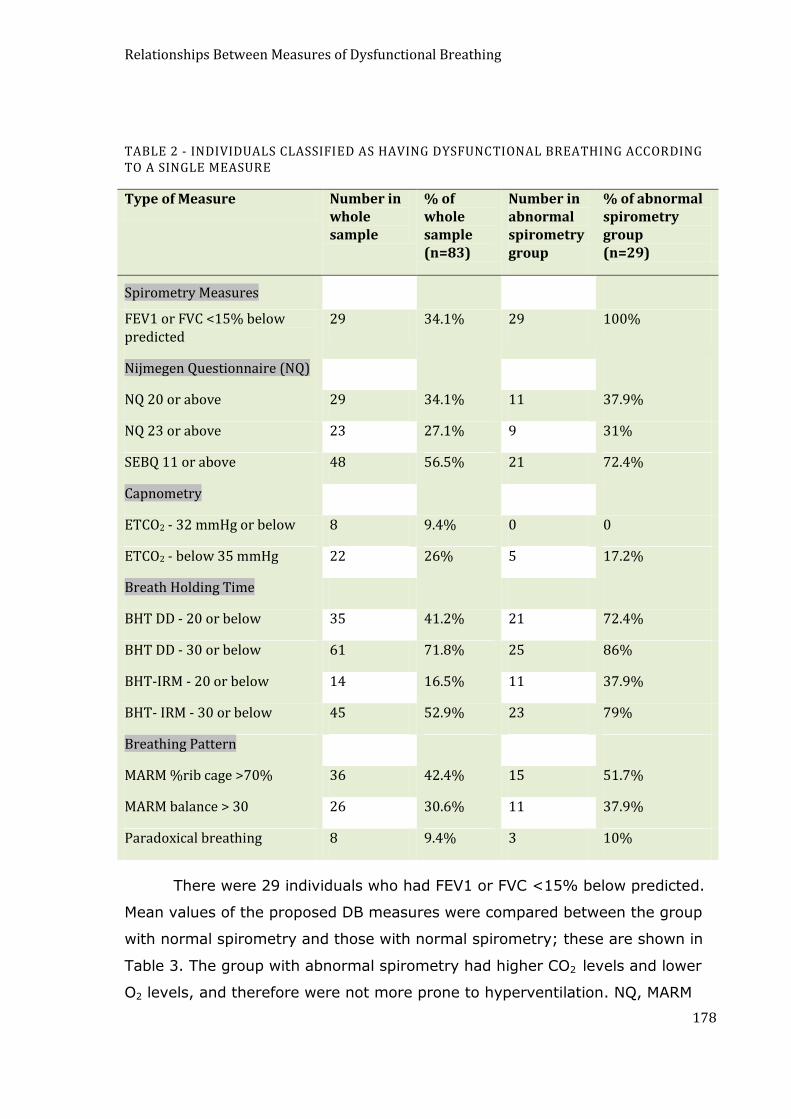
Relationships Between Measures of Dysfunctional Breathing
178
TABLE 2 - INDIVIDUALS CLASSIFIED AS HAVING DYSFUNCTIONAL BREATHING ACCORDING TO A SINGLE MEASURE
Type of Measure Number in whole sample
% of whole sample (n=83)
Number in abnormal spirometry group
% of abnormal spirometry group (n=29)
Spirometry Measures
FEV1 or FVC <15% below predicted
29 34.1% 29 100%
Nijmegen Questionnaire (NQ)
NQ 20 or above 29 34.1% 11 37.9%
NQ 23 or above 23 27.1% 9 31%
SEBQ 11 or above 48 56.5% 21 72.4%
Capnometry
ETCO2 - 32 mmHg or below 8 9.4% 0 0
ETCO2 - below 35 mmHg 22 26% 5 17.2%
Breath Holding Time
BHT DD - 20 or below 35 41.2% 21 72.4%
BHT DD - 30 or below 61 71.8% 25 86%
BHT-IRM - 20 or below 14 16.5% 11 37.9%
BHT- IRM - 30 or below 45 52.9% 23 79%
Breathing Pattern
MARM %rib cage >70% 36 42.4% 15 51.7%
MARM balance > 30 26 30.6% 11 37.9%
Paradoxical breathing 8 9.4% 3 10%
There were 29 individuals who had FEV1 or FVC <15% below predicted.
Mean values of the proposed DB measures were compared between the group
with normal spirometry and those with normal spirometry; these are shown in
Table 3. The group with abnormal spirometry had higher CO2 levels and lower
O2 levels, and therefore were not more prone to hyperventilation. NQ, MARM

Relationships Between Measures of Dysfunctional Breathing
179
values and respiratory rates were approximately equal in normal and abnormal
spirometry groups. The only increased signs of DB in the abnormal spirometry
group were shorter BHT and increased SEBQ scores.
TABLE 3 - DESCRIPTIVE STATISTICS AND DIFFERENCES BETWEEN THE NORMAL AND ABNORMAL SPIROMETRY GROUPS
Normal spirometry (n=54)
Abnormal spirometry (n=29)
Mean Difference
P Values
Males 17 12
Females 37 17
Average Age (Yrs)
49(+-13) 48 (+- 14)
FEV1 (% pred) 99(+- 10) 82(+- 11) 17 .0001
Av. ETCO2 (mmHg)
37(+- 4) 39(+- 4) 2 .01
Av. BHT-DD (s) 28(+-12 ) 20(+-8 ) 7.8 .0001
Av. BHT-IRM (s) 33(+- 11) 24(+- 10) 9 .0001
Av. SPO2 (%Hb Sat)
96(+- 2) 95(+- 2) 1 .04
Resp. Rate 16(+-4 ) 17(+- 4) 1 .79
NQ 17(+- 9) 19(+-11 ) 2 .35
MARM (%RC) 70(+- 21) 77(+-35 ) 7 .29
MARM Balance 19(+-16) (19+-18) .9 .82
SEBQ - total 12(+-7) 16(+-8) 4.5 .008
Correlations between measures are shown in Table 4. As can be seen,
significant correlations where found within the following categories of
measures:
1. Symptom questionnaires;
2. Breath holding times; and
3. Breathing pattern measures.

Relationships Between Measures of Dysfunctional Breathing
180
Scores for the two questionnaires, the NQ and the SEBQ, were strongly
correlated. The two types of breath holding, BHT-DD and BHT-IRM, were also
strongly correlated. The two MARM variables, MARM balance and % RC, were
also correlated.
TABLE 4 - CORRELATION MATRIX OF DYSFUNCTIONAL BREATHING MEASURES
Biochemical measures
Biomechanical measures Breath holding time
Symptom question
PO2 ETCO2 RR MARM %RC
MARM bal.
BHT-DD
BHT-IRM NQ SEBQ
FEV1 .10
p=.38
-.08
p=.47
-.09
p=.44
-.19
p=.09
-.08
p=.43
.31
p=.004
.31
p=.005**
-.133
p=.23
-.264
p=.02
PO2 1 -.0.23
p=.84
-.10
p=.10
-.14
p=.22
-.12
p=.26
.24
p=.03
.186
p=.09
.11
p=.26
.02
p=.80
ETCO2 1 -.14
p=.20
-.02
p=.82
-.136
p=.221
-.241
p=.03
-.198
p=.07
-.12
p=.27
.01
p=.96
RR 1 .09
p=.40
.07
p=.55
-.18
p=.11
-.16
p=.14
-.20
p=.08
-.14
p=.22
MARM%RC 1 .82
p=.0001
-.090
p=.42
-.25
p=.02
-.04
p=.70
.11
p=.31
MARM bal. 1 -.07
p=.54
-.204
p=.06
.01
p=.90
.14
p=.20
BHT-DD 1 .84
p=.0001
-.20
p=.07
-.17
p=.12
BHT-IRM 1 -.18
p=.10
-.20
p=.07
NQ 1 .75
p=.0001

Relationships Between Measures of Dysfunctional Breathing
181
Correlations between variables in different categories were generally not
significant. The only significant correlation were between BHT-IRM and MARM
% rib cage motion. And also FEV1 levels and SEBQ sum scores were correlated.
Carbon dioxide, measured in this study by end tidal CO2, did not correlate
significantly with symptom questionnaires, breathing pattern, PO2 or FEV1. A
statistically significant, but weak, correlation was found with one type of
breathing holding - BHT-DD. It should be noted that this correlation was
negative, opposite to the expected direction.
There was a correlation between BHT-DD and SpO2 levels.
The SEBQ was negatively correlated with FEV1. FEV1 also correlated with
both types of breath holding time.
DISCUSSION
Strict definitions of DB are not possible at present, but for practical purposes it
is probably most useful not to think of DB as a single entity limited to the
biochemical dimension of breathing functionality (as in Hyperventilation
Syndrome) but to consider breathing symptoms and breathing pattern as
potentially separate aspects of DB which need be measured in their own right.
This study found that significant correlations exist only within categories of
breathing measures, but not between categories. The two symptom
questionnaires investigated in this study, the NQ and the SEBQ were found to
correlate, as did the two measures of breath holding time and the two similar
MARM measures of breathing pattern. Biochemical measures, oxygen and
carbon dioxide, were not related. Therefore the clinician who wishes to do a
comprehensive evaluation of breathing functionality should consider using a
range of measurement tools, including breathing symptom questionnaires,
breathing pattern evaluation and CO2 measurement.
In this sample of people, which on average had only mild dysfunctional
breathing, there was very little relationship between the categories of
measures. The only significant correlation between categories, was between
one type of breath holding BHT-IRM, where the breath was held until the first
Relationships Between Measures of Dysfunctional Breathing
182
involuntary motion of the respiratory muscles, and the extent of the thoracic
dominant breathing pattern, represented as percentage of upper rib cage
contribution to breathing motion (MARM % rib cage). No measures were found
to correlate with end-tidal CO2. This indicates that individuals with dysfunctions
in one aspect of breathing functionality do not necessarily have dysfunctions in
other aspects, particularly if the breathing dysfunction is not severe.
In the group with abnormal spirometry it is possible that parenchymal
disease influenced their CO2 levels. Poor transfer of blood gases across
damaged lung tissue, particularly in individuals with COPD could mean that
even in the case of alveolar hyperventilation, CO2 levels appeared normal.
However, given the fact that individuals with severe lung disease were excluded
from this study this is unlikely.
The negative correlation between FEV1 and the SEBQ indicates that patients
with poor lung function were more likely to experience subjective sensations of
breathing difficulty.
The most well recognised form of dysfunctional breathing is
hyperventilation which is strictly defined by a biochemical definition i.e.
breathing in excess of metabolic requirements so that a depletion of carbon
dioxide occurs(Comroe 1974). Despite its biochemical criteria, hyperventilation
is sometimes presumed to exist on the basis of symptoms, on findings of
abnormal breathing patterns or length of breath holding time (Lum 1976;
Dixhoorn and Duivenvoorden 1985; Stark and Stark 2002). This study suggests
that dysfunctions of breathing pattern, shortened breath holding time and DB
symptoms can exist without chronic hypocapnia. This is consistent with
previously published studies that have also found that baseline CO2 does not
always relate to symptoms of dysfunctional breathing (Folgering and Colla
1978; Vansteenkiste 1991). However, there is literature that shows that
individuals with high levels of hyperventilation type symptoms do have
depressed levels of CO2 when compared to symptom free controls (Gardner
1986). So while it cannot be presumed that there is no association between
symptoms of DB and resting CO2, it should be recognised that the relationship
is complex and can be influenced by other moderating factors such as anxiety.
Relationships Between Measures of Dysfunctional Breathing
183
The complex relationship beween hypocapnia and HV was demonstrated
by Wintjes and Grossman who showed that in a group of 83 healthy individuals
CO2 contributed only 4% of the variance in HV symptoms (Wientjes and
Grossman 1994). The current finding that dysfunctional breathing pattern is not
necessarily associated with low resting CO2 levels is consistent with studies that
have also found that symptomatic individuals with disturbed breathing pattern
often have normal levels of CO2 (Han, Stegen et al. 1996). Again it cannot be
presumed that there is no association between breathing pattern and CO2
levels, as many clinicians and researchers have observed abnormal breathing
pattern in individuals with hyperventilation (Lum 1976). However, it can be
concluded that the level of resting CO2 cannot be presumed from presence of
symptoms commonly thought to be associated with dysfunctional breathing,
from breath holding time or from breathing pattern. The clinician can only be
certain of CO2 levels if these are specifically measured. It should also be noted
that measurement of resting CO2 might not identify individuals who
hyperventilate in response to psychological stress or physical exercise or whose
CO2 regulating capacitates are compromised. This is done variously through
capnometry combined with exercise or psychological challenge or the
Hyperventilation Provocation Test (Hardonk and Beumer 1979; Warburton and
Jack 2006).
In this study the MARM measures reflecting thoracic dominance in
breathing show a small degree of correlation with breath holding till first desire
to breathe (BHT-DD). It seems reasonable to hypothesise that this may be
because both these measures reflect respiratory drive, with increased
respiratory drive increasing the extent of thoracic breathing and decreasing
breath-holding time. It is also possible that thoracic breathing patterns
themselves affect perception of dyspnea sensations related to the breaking
point of breath holding.
This study did not show an association between general symptoms of
dysfunctional breathing, as measured by the SEBQ or the NQ and breathing
pattern. These findings are unexpected and not consistent with clinician‟s
observations that individuals with high levels of symptoms associated with DB

Relationships Between Measures of Dysfunctional Breathing
184
tend to have breathing pattern disturbances (Lum 1976; Howell 1997). And
that specific qualities of breathing discomfort or dyspnea are affected by
patterns of respiratory muscle use (Simon and Schwartzstein 1990; Altose
1992; O'Donnell 2006). The lack of relationship in this current study may be
due to the fact that the sample used tended to represent individuals with only
mild signs of dysfunctional breathing. Correlations not evident in this sample
with mild DB might be stronger in individuals with more severe DB. Also
relationships that do exist between breathing pattern and symptoms may be
non-linear.
Another factor contributing to the poor correlation found between
symptoms and actual physiological and biomechanical aspects of breathing,
such as end-tidal CO2 breathing pattern and FEV1 might be the individuality of
cognitive processes involved in recognizing, attending to, and then evaluating
physical symptoms. Certain individuals show persistent tendencies to over-
perceive or under-perceive symptoms related to breathing function (Teeter and
Bleeker 1998; Klein, Walders et al. 2004). Thus, while the findings of this
study suggest that symptoms do not necessarily correlate with biochemical or
biomechanical or breath holding measures across individuals, they do not
diminish the possibility or importance of relationships between symptoms and
breathing functions within individuals.
Limitations of this study
Not all ways of measuring DB were assessed in this study. For example
measurement of carbon dioxide at rest, as was undertaken during this study,
will only reveal chronic persistent hyperventilation and may not be adequate for
revealing which individuals are prone to intermittent hyperventilation in
response to physical or psychological stress (Hardonk and Beumer 1979).
The interpretation of these results is limited to their use as general
screening tools, as the patient population used in this study was not
representative of individuals with pronounced breathing dysfunction.
Relationships not evident in this population might be evident in a sample more
representative of individuals with hypocapnia, markedly disturbed breathing
Relationships Between Measures of Dysfunctional Breathing
185
pattern or abnormal Nijmegen Questionnaire scores. Representation from
patients in disease categories thought to be affected by dysfunctional breathing
is needed to make more robust assumptions.
Future Research
Further Research is required to investigate the presence of more
complex relationships between the various dimensions of breathing
dysfunction. Research should also target individuals with stronger evidence of
breathing dysfunction or with specific ailments.
CONCLUSION
For practical purposes DB is probably best characterised as multi-dimensional.
DB can occur in at least 3 dimensions: biochemical, breathing pattern and
breathing related symptoms, and these might not co-exist. Screening for DB
with measures representing only one of these dimensions may not lead to
realistic estimations of the prevalence and impact of the various types of
breathing dysfunctions. Comprehensive evaluation of breathing dysfunction
should include measures of breathing symptoms, breathing pattern, resting CO2
and also include functional measures such a breath holding time and response
of breathing to physical and psychological challenges.
This is a copy of an article published in the Journal of Bodywork and Movement Therapies © [2011] copyright
Elsevier.; Journal of Bodywork and Movement Therapies is available online at: http://www.elsevier.com.
Ref: Courtney R, Greenwood K (2011). "Relationships between measures of dysfunctional breathing in a
population with concerns about their breathing." Journal of Bodywork and Movement Therapies 15(1): 24-34.

Investigating the Claims of Dr. Buteyko: Breathing Holding Time and CO2
186
CHAPTER 10
INVESTIGATING THE CLAIMS OF DR. K.P. BUTEYKO: THE
RELATIONSHIP OF BREATH HOLDING TIME TO END TIDAL CO2
AND OTHER PROPOSED MEASURES OF DYSFUNCTIONAL
BREATHING
ABSTRACT
Objectives: Dr K.P. Buteyko claimed that Breath Holding Time (BHT) can be
used to detect chronic hyperventilation and that BHT predicts alveolar CO2
(PACO2) according to his patented mathematical formula. The Buteyko
Breathing Technique (BBT) is believed to correct chronic hyperventilation as
evidenced by increased BHT. In this study, we test Buteyko‟s claims and
explore the relationship between BHT and ETCO2 as well as measures of
dysfunctional breathing (DB) including the Nijmegen questionnaire (NQ), a self
evaluation of breathing questionnaire (SEBQ) and thoracic dominant breathing
pattern.
Subjects: 83 adults healthy or suspected of having dysfunctional breathing, 29
with abnormal spirometry readings, 54 with normal spirometry
Outcomes: BHT, performed according to BBT protocols, was measured along
with ETCO2 and other measures of DB including the Nijmegen Questionnaire,
and the Manual Assessment of Respiratory Motion (MARM), a palpatory
technique for measuring thoraco-abdominal balance during breathing.
Correlations between measures of DB were made in the whole sample and also
in subgroups with normal or abnormal spirometry. DB measures were
compared in normal and abnormal spirometry groups.
Results: The results revealed a negative correlation between BHT and ETCO2
(r=.-241, p<0.05), directly opposite to Buteyko‟s claims. BHT was significantly
shorter in people with abnormal spirometry (FEV1 or FVC < 15% below
predicted) with no difference in ETCO2 levels between the abnormal and normal

Investigating the Claims of Dr. Buteyko: Breathing Holding Time and CO2
187
spirometry groups. In the abnormal spirometry group, lower BHT was found to
correlate with a thoracic dominant breathing pattern, (r= -.408, p< .028).
Conclusion: Although BHT does not predict resting ETCO2; it does correlate
with breathing pattern in subjects with abnormal spirometry. It is proposed that
altered breathing pattern could contribute to breathing symptoms such as
dyspnea and that breathing therapies such as BBT might influence symptoms
by improving the efficiency of the biomechanics of breathing.
Investigating the Claims of Dr. Buteyko: Breathing Holding Time and CO2
188
INTRODUCTION
Dysfunctional breathing (DB) and breath holding time (BHT) and the
Buteyko Breathing Technique (BBT)
For many years clinicians and researchers have reported that abnormal
or Dysfunction Breathing (DB) influences health and patient‟s symptoms (Kerr
1937; Magarian 1982; Bass 1989). DB is not clearly defined and diagnosis is
made on the basis of symptoms (Dixhoorn and Duivenvoorden 1985; Thomas,
McKinley et al. 2001), breathing pattern (Chaitow 2002; Dixhoorn 2004) and
presence of hyperventilation and hypocapnia (Magarian 1982; Bass 1989).
Practitioners of the Buteyko Breathing Technique (BBT) use Breath Holding
Time (BHT) as a way of indicating presence of DB and believe that length of
BHT equates to extent of hyperventilation and degree of resulting hypocapnia
(Stalmatski 1999). The current medical literature supports a broader definition
of DB than one limited to hyperventilation and hypocapnia (Howell 1997;
Morgan 2002) because DB related symptoms and abnormal breathing patterns
can exist even when carbon dioxide levels are normal (Burton 1993; Hornsveld
1997).
Recent discussions on DB are beginning to consider the impact of non-
biochemical aspects of respiration (Howell 1997; Dixhoorn 2004; Bruton and
Thomas 2006) such as efficiency of breathing pattern. The work of breathing is
most efficient when a co-ordinated contribution from the diaphragm, abdominal
muscles and rib cage muscles results in balanced motion between the upper rib
cage and the lower rib cage and abdomen (De Troyer and Estenne 1988).
Uneven distribution of chest wall motion with dominance of upper rib cage or
thoracic motion may be due to normal and transient situations such as
emotional arousal or increased respiratory effort and load as well as due to
abnormalities of lung function (Sharp, Goldberg et al. 1975; Tobin, Tejvir et al.
1983). Unevenness of motion of the chest wall where the upper rib cage
movement dominates and lower rib cage expansion is impaired can indicate
biomechanically induced DB that results in hyperinflation and contributes to
breathing symptoms such as dyspnea (Tobin 1988; O'Donnell 2006).
Investigating the Claims of Dr. Buteyko: Breathing Holding Time and CO2
189
BHT is an indicator of a person‟s ventilatory response to biochemical,
biomechanical and psychological factors (Mithoefer 1965; Rassovsky, Kushner
et al. 2000; Delapille, Verin et al. 2001) and it seems reasonable to suggest
that abnormally shortened BHT may indicate abnormalities in respiratory
control that result in DB (Lin 1974; Lumb 2000). The idea that BHT is a simple
test of DB has been suggested by various sources, including proponents of the
BBT (Jack, Darke et al. 1998).
The BBT is a breathing therapy that is used to treat at least 99 different
symptoms and conditions (www.buteyko.ru/eng/bolenz.shtml) including
hypertension, diabetes, sleep apnea, anxiety, depression, insomnia and
epilepsy (Stark and Stark 2002) which practitioners claim are related to DB.
Asthma is the condition most people associate with BBT and several clinical
trials have indicated that the BBT helps symptom control in asthma, along with
a reduction in medication use and improved quality of life (Bowler 1998; Opat,
Cohen et al. 2000; Cooper, Oborne et al. 2003; McHugh, Aicheson et al. 2003;
Slader, Reddel et al. 2006). BBT practitioners believe that the reduction of
symptoms and improved health due to the BBT (Stalmatski 1999; Stark and
Stark 2002) are a result of increased CO2 levels and longer BHT (Stalmatski
1999; Stark and Stark 2002), however studies to date do not support this
(Bowler 1998).
Measurement of BHT is an essential part of the BBT and is integrated into
its theoretical basis as well as to how the technique is practiced. Dr. Konstantin
Buteyko originator of the BBT reported that his protocol for measuring breath
holding could accurately predict carbon dioxide levels and detect chronic
hyperventilation in both healthy and sick individuals (Buteyko 1990). He
patented the following formula for calculating PCO2 from BHT:
PACO2%= 3.5 +. 05xBHT (Buteyko 1986)
This formula has never been subjected to rigorous scrutiny.
In this study, we aimed to test the claims of Dr. Buteyko regarding the
correlation between BHT and End Tidal CO2 (ETCO2) in the general population.

Investigating the Claims of Dr. Buteyko: Breathing Holding Time and CO2
190
We also aimed to explore the relationship between BHT and DB in this same
population, by examining the correlation between BHT and thoracic dominant
breathing pattern as determined by the MARM, as well as the presence of
symptom clusters frequently associated with DB as determined by the
Nijmegen Questionnaire (NQ) and the Self Evaluation of Breathing
Questionnaire (SEBQ).
Since it has been proposed that people with asthma and chronic
respiratory diseases are more likely to have DB (Thomas, McKinley et al. 2001)
we were interested to see if these proposed measures of DB and their
relationships were influenced by lung function status.
METHODS
The Human Research Ethics Committee of RMIT University approved this study.
Subjects:
Subjects from the general population were recruited from general
practices and complementary medicine clinics in Sydney, Australia.
The study was performed on 83 volunteers, aged 18 or over who were
either healthy or suffered from mild medical conditions, which did not restrict
their capacity to work or significantly affect their lifestyle. Of the 83 subjects,
29 had FEV1 or FVC at least 15% lower than predicted for age, gender and race
and were classified as having abnormal spirometry.
Sample Size
We chose our sample size on the basis of a power analysis, which
estimated that to detect a significant correlation of 0.3 with 80% power and
significance level of .05 would require 85 subjects.
The 83 subjects were analyzed as a whole and also divided into 2 groups,
an abnormal spirometry group of 29 subjects and a normal spirometry group of
54 subjects, for purposes of comparison.
Breath Holding Protocol:

Investigating the Claims of Dr. Buteyko: Breathing Holding Time and CO2
191
The breath hold test used by BBT practitioners to calculate PACO2 is
generally referred to as the Control Pause (CP) although the somewhat
confusing term Maximum Pause (MP) is also used. CP and MP are both
measured after normal exhalation at functional residual capacity (Stark and
Stark 2002) (Stalmatski 1999). BBT practitioners were surveyed via the
Buteyko Practitioners Support Network website about the precise instructions
used to determine correct BHT. This survey revealed that the most common
instruction for determining the CP was to hold the breath until the point of
definite discomfort or the first sensation of difficulty was experienced. An
alternative protocol is to hold the breath until the first Involuntary Respiratory
Movement (IRM). This has been described as the physiological breaking point of
breath holding (Lin 1974). Private communications with Dr. Buteyko‟s
successor, Dr. Andrey Novolizov and his son Dr. Vladimir Buteyko confirmed
that to determine the CP the breath should be held till IRM. In this study both
ways of determining BHT were tested; breath holding till first difficulty or first
desire to breathe (BHT-DD) and until first involuntary motion of the respiratory
muscles (BHT-IRM).
Participants were instructed to sit quietly and breathe normally. They
were then asked to breathe gently in and out and at the end of a normal
exhalation to pinch their nose and hold the breath. Measurement was done with
a stopwatch which measured to 0.01 of a second. This number was rounded
down to 0.1 of a second. All breath holding procedures were repeated three
times and the mean used in calculating correlations.
End Tidal Carbon Dioxide (ETCO2) Measurement:
ETCO2 levels were sampled with a two pronged nasal canula and
readings taken with a capnometer (BCI, Capnocheck, USA, 2001). The
equipment was calibrated and checked for accuracy with a known gas mixture.
ETCO2, along with O2 saturation (SPO2), respiratory rate and heart rate, was
measured for about 25 minutes while the person filled out various
questionnaires. After excluding data from the first 2 minutes to allow for the
subject settling in, the average ETCO2 was calculated and used in determining
correlation co-efficients with other variables.
Investigating the Claims of Dr. Buteyko: Breathing Holding Time and CO2
192
Other Measurements
Spirometry was performed using a laptop-based spirometer (QRS
Diagnostics, Spirocard, USA, 2003).
Breathing Questionnaires:
Questionnaires used to detect the possible presence of DB symptoms
included the Nijmegen questionnaire (NQ) and the Self Evaluation of Breathing
Questionnaire (SEBQ). The items on the Nijmegen Questionnaire (NQ) are an
abbreviated list of the most common symptoms of DB and this questionnaire
has been validated as an accurate tool for identifying patients likely to suffer
from DB (Dixhoorn and Duivenvoorden 1985). Our version of the SEBQ is
derived from an abbreviation of a questionnaire available on the internet which
claimed to be able to predict whether the responder had breathing dysfunction
(White 2005) with emphasis on symptoms which the scientific literature
suggested were common in DB. (Howell 1990; Burton 1993). We performed
Factor Analysis of the SEBQ and found 2 dimensions of symptoms that we
named „breathlessness‟ and „self perception of breathing abnormality‟.
Other Questionnaires:
The subjects were given the Hospital Anxiety and Depression Inventory,
(HAD) to assess anxiety and depression.
The Manual Assessment of Respiratory Motion (MARM)
Clinical evaluation of breathing pattern is usually performed by
observation and palpation (Clanton and Diaz 1995). In this study we used a
breathing pattern assessment technique called the Manual Assessment of
Respiratory Motion (MARM) to assess 'balance' between upper lower chest wall
movements. The MARM has been demonstrated to have good inter-examiner
reliability (Dixhoorn 2004) and be consistent with measures performed using
Respiratory Induction Plethysmography (Courtney, van Dixhoorn et al. 2006).
This palpatory technique permits the examiner to assesses the relative
contribution of thoracic and diaphragmatic action during breathing and

Investigating the Claims of Dr. Buteyko: Breathing Holding Time and CO2
193
calculate a quantitative measure of thoracic dominance in breathing which we
called % rib cage motion (Dixhoorn 2004). The examiner using the MARM
places their open hands over the subject‟s back at the region of the lower 4-6
ribs. The examiner‟s thumbs are about 1 inch from the spine and orientated
vertically. The examiner‟s hands are spread so that the lower 3 fingers are
orientated in a transverse direction. This hand placement makes it possible for
the examiner to feel lateral and vertical motion of the rib cage and assess
relative contribution from the upper rib cage and the lower rib cage/ abdomen.
The examiner draws a diagram with an upper line to represent extent of the
upper rib cage and vertical motion and a lower line to represent extent of lower
rib cage/abdomen motion. Calculations are then made for thoracic diaphragm
“balance” and % rib cage motion. (See figure 1)
FIGURE 1 - THE MARM GRAPHIC NOTATION
Operator draws Line A to represent upper rib cage and vertical motion and Line B for lower rib cage and lateral motion. MARM measures are Balance = angle AC-angle CB, % Rib Cage Motion = % of angle AC/angleAB.
Statistical Analysis:
The data was entered, checked for any abnormalities or errors and then
analysed using SPSS (ref and place). Pearson's correlation coefficient was used
to test for association between the 2 measures of BHT: BHT-DD and BHT-IRM,
and all measures of DB. Students T test was used to detect differences between
Line A- upper line representing
upper rib cage and vertical motion
Line C- representing horizontal line and mid-point between upper and lower rib cage motion
Line B- lower line representing lower rib cage and lateral motion
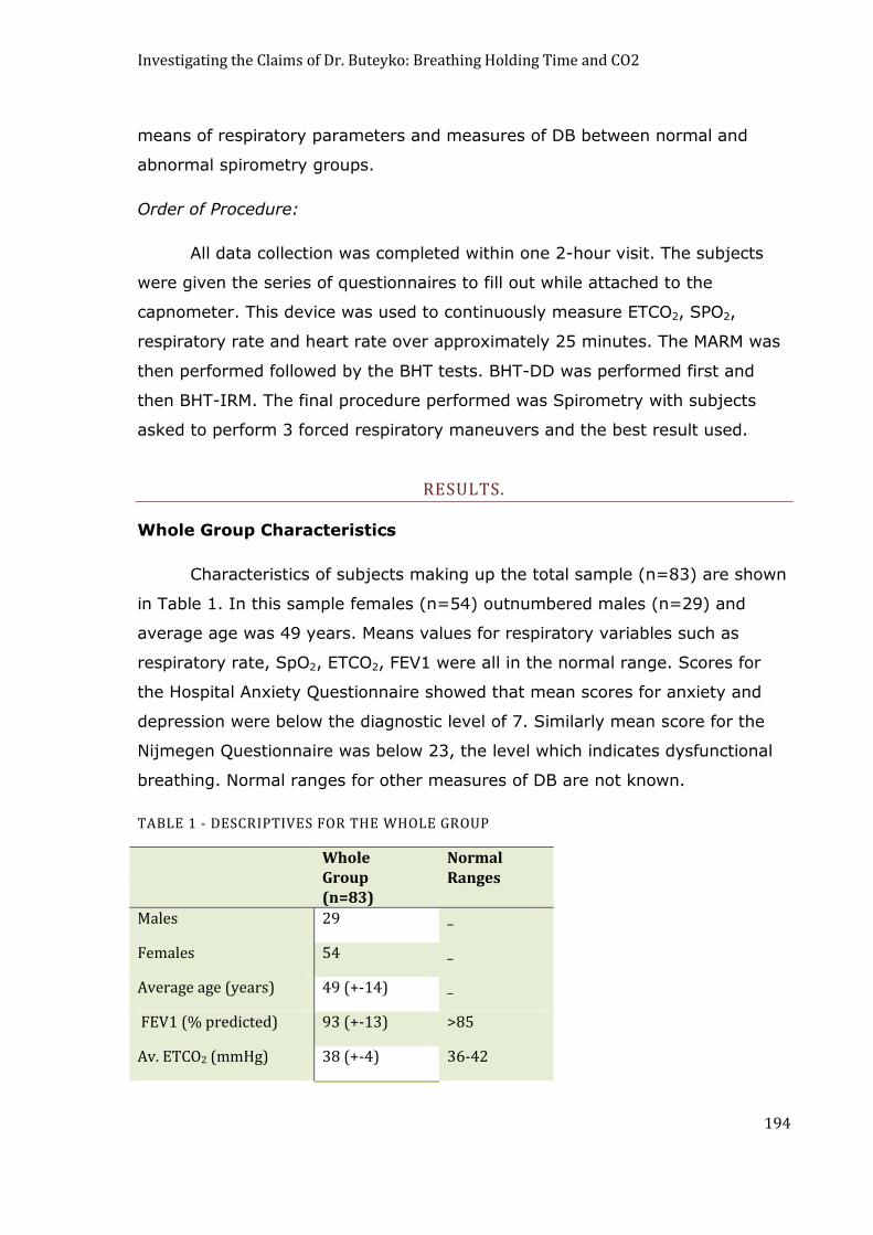
Investigating the Claims of Dr. Buteyko: Breathing Holding Time and CO2
194
means of respiratory parameters and measures of DB between normal and
abnormal spirometry groups.
Order of Procedure:
All data collection was completed within one 2-hour visit. The subjects
were given the series of questionnaires to fill out while attached to the
capnometer. This device was used to continuously measure ETCO2, SPO2,
respiratory rate and heart rate over approximately 25 minutes. The MARM was
then performed followed by the BHT tests. BHT-DD was performed first and
then BHT-IRM. The final procedure performed was Spirometry with subjects
asked to perform 3 forced respiratory maneuvers and the best result used.
RESULTS.
Whole Group Characteristics
Characteristics of subjects making up the total sample (n=83) are shown
in Table 1. In this sample females (n=54) outnumbered males (n=29) and
average age was 49 years. Means values for respiratory variables such as
respiratory rate, SpO2, ETCO2, FEV1 were all in the normal range. Scores for
the Hospital Anxiety Questionnaire showed that mean scores for anxiety and
depression were below the diagnostic level of 7. Similarly mean score for the
Nijmegen Questionnaire was below 23, the level which indicates dysfunctional
breathing. Normal ranges for other measures of DB are not known.
TABLE 1 - DESCRIPTIVES FOR THE WHOLE GROUP
Whole Group (n=83)
Normal Ranges
Males 29 _
Females 54 _
Average age (years) 49 (+-14) _
FEV1 (% predicted) 93 (+-13) >85
Av. ETCO2 (mmHg) 38 (+-4) 36-42

Investigating the Claims of Dr. Buteyko: Breathing Holding Time and CO2
195
Av. BHT-DD (s) 26 (+-12) _
Av. BHT-IRM (s) 30 (+-12) _
Av. SPO2 (% Hb Sat) 96 (+-2) >94
Resp. rate 16 (+-4) <16
Nijmegen Questionnaire 18 (+-10) <23
MARM (%RC) 73 (+-27) _
MARM Bal. 19 (+-18) _
Anxiety (HAD) 6 (+-3) <7
Depression (HAD) 3 (+-3) <7
SEBQ - total 13 (+-8) _
SEBQ -dyspnea 4 (+-3) _
SEBQ - abnormal br. sensation
4 (+-3) _
KEY TO ABBREVIATED VARIABLES IN TABLE 1 AND 2:
FEV1% predicted - Forced Expiratory Volume at one second as a percentage of that predicted for age, race and gender. Av.ETCO2 - average end tidal CO2 from 25 minutes collection time. Av. BHT-DD - average Breath Holding Time until first definite desire to breath. Av. BHT-IRM - average Breath Holding Time until first involuntary motion of the respiratory muscles. Av. SpO2 - average percentage saturation of Hemoglobin over 25 minutes. Resp. rate - respirations per minute MARM (%RC) - % contribution of chest or rib cage motion to breathing. MARM bal - Difference between total chest wall and upper rib cage motion. Anxiety (HAD) - Anxiety score on Hospital and Anxiety and Depression Scale Depression (HAD) - Depression score on Hospital and Anxiety Scale SEBQ - total - total score of SEBQ SEBQ – dyspnea - Self-evaluation of breathing questionnaire - dyspnea score SEBQ - abnormal br - Self-evaluation of breathing questionnaire - perception of breathing abnormality score.
Comparisons between the whole, normal and abnormal spirometry
groups.
Table 2 compares values for breathing parameters and DB measures
between subjects with normal and abnormal spirometry i.e. FEV1 or FVC less
than 15% below predicted.

Investigating the Claims of Dr. Buteyko: Breathing Holding Time and CO2
196
Subjects in the abnormal spirometry group had a mean difference in %
predicted FEV1 of 17% (p=0.0001). Table 2 shows that the abnormal
Spirometry groups had ETCO2 levels, 2mm Hg higher (p=0.01) and SpO2 levels
1% lower (p=0.04). They also scored 4.5 points higher on the SEBQ
(p=0.008). People with abnormal spiromety readings had shorter BHT for both
types of breath holding (illustrated in Figure 2) and a greater % of rib cage
contribution to breathing as measured by the MARM. The difference in BHT-DD
was 7.8 seconds (p=0.0001) and 9 seconds for BHT-IRM (p=0.0001) however
the 7% difference in MARM % rib cage was not statistically significant
(p=0.29).
TABLE 2 - DESCRIPTIVES AND DIFFERENCES BETWEEN THE NORMAL AND ABNORMAL SPIROMETRY GROUPS
Normal
spirometry
(n=54)
Abnormal
spirometry
(n=29)
Mean
Difference
Normal &
Abnormal
spirometry
P Values
Males 17 12
Females 37 17
Average
Age (years)
49(+-13) 48 (+- 14)
FEV1 (% predicted) 99(+- 10) 82(+- 11) 17 .0001
Av ETCO2 (mmHg) 37(+- 4) 39(+- 4) 2 .01
Av BHT-DD (s) 28(+-12 ) 20(+-8 ) 7.8 .0001
Av. BHT-IRM (s) 33(+- 11) 24(+- 10) 9 .0001
Av. SPO2 (%Hb Sat) 96(+- 2) 95(+- 2) 1 .04
Resp. Rate 16(+-4 ) 17(+- 4) 1 .79
Nijmegen Questionnaire
17(+- 9) 19(+-11 ) 2 .35
MARM (%RC) 70(+- 21) 77(+-35 ) 7 .29
MARM Bal. 19(+-16) (19+-18) .9 .82

Investigating the Claims of Dr. Buteyko: Breathing Holding Time and CO2
197
Anxiety (HAD) 6(+- 3) 6(+-3 ) .2 .79
Depression (HAD) 3(+-3 ) 3(+-3 ) .2 .75
SEBQ-total 12(+-7) 16(+-8) 4.5 .008
SEBQ-dyspnea 4(+-3 ) 4(+-3 ) .3 .62
SEBQ-abnormal br. sensation
4(+-3 ) 5(+- 4) .6 .36
0
10
20
30
40
Time in
Seconds
BHT-DD BHT-IRM
Breath Holding
Procedure
Figure 2- Comparisons in Breath Hold
Time between Normal and Abnormal
Spirometry Groups
NormalSpirometry
AbnormalSpirometry
Bar Graph comparing the two types of breath holding in people with normal spirometry and abnormal spirometry (FEV1 or FVC 15% < predicted) 1. Breath holding at functional residual capacity till first feelings of definite discomfort (BHT-DD) 2. Breath holding at functional residual capacity until first involuntary motion of the respiratory muscles (BHT-IRM).
Correlations between BHT, ETCO2 and other measures of DB were
performed separately on the whole group n=83, the abnormal spirometry group
(n=29) and the normal spirometry group (n=54).
Correlations between BHT, ETCO2 and other measures of DB in the
whole group
Correlations between BHT and ETCO2 and other measures of
dysfunctional breathing were performed in the whole group (n=83). There was
a statistically significant negative correlation for ETCO2 and BHT-DD (r=-0.241,
p=0.028). This correlation was strongly influenced by 2 cases that had very
long breath holding times and very low ETCO2 levels. When these 2 cases were
excluded, no statistically significant correlations were evident (See Table 3).

Investigating the Claims of Dr. Buteyko: Breathing Holding Time and CO2
198
TABLE 3 - CORRELATIONS BETWEEN BREATH HOLDING TIME (BHT) AND CARBON DIOXIDE – IN THE WHOLE SAMPLE SHOWING DIFFERENCE WHEN EXCLUDING OUTLIERS.
BHT-DD
All cases
(n=83)
BHT-DD
Excluding 2
outliers
(n=81)
BHT-IRM
All cases
(n=83)
BHT-IRM
Excluding 2
outliers
(n=81)
PCO2 Pearsons
Correlation
-.241 -.099 -.198 -.040
Sig. (2-tailed) .028 .376 .073 .720
It was decided to keep these 2 cases in the data because there did not
appear to be any actual measurement error to justify excluding them. As can
be seen in Table 4 when the whole group was analyzed there was also a small
correlation between SpO2 and BHT-DD (r=0.238, p =0.031). However, there
was no correlation between BHT and any of the other measures of DB.
TABLE 4 - CORRELATIONS FOR BHT AND MEASURES OF DYSFUNCTIONAL BREATHING – WHOLE GROUP (N=83)
SpO2 RR NQ MARM SEBQ ETCO2
BHT-DD Pearsons
Correlation
.238*
-.178
-.198
-.068
-.174
-.241*
Sig. (2 tailed) .031 .107 .073 .539 .116 .028
BHT-IRM Pearsons
Correlation
.186
-.165
-.185
-.204
-.201
-.198
Sig. (2 tailed) .093 .137 .095 .064 .069 .073
*Statistically significant correlations for BHT-DD with ETCO2 and SpO2
Correlations between BHT, ETCO2 and other measures of DB- abnormal
spirometry group
A thoracic dominant breathing pattern determined by the MARM measure
of % rib cage motion was found to correlate with BHT in the abnormal
spiromety group. In this group a significant correlation (r=-0.408, p=0.02)
existed between the MARM % rib cage motion and BHT-IRM. There was no
correlation between the MARM % rib cage motion and BHT-DD (r=0.163,
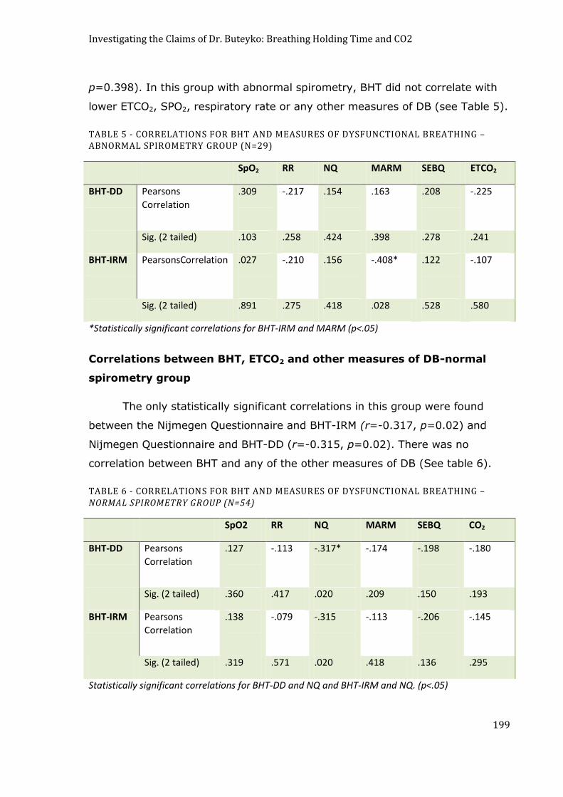
Investigating the Claims of Dr. Buteyko: Breathing Holding Time and CO2
199
p=0.398). In this group with abnormal spirometry, BHT did not correlate with
lower ETCO2, SPO2, respiratory rate or any other measures of DB (see Table 5).
TABLE 5 - CORRELATIONS FOR BHT AND MEASURES OF DYSFUNCTIONAL BREATHING –ABNORMAL SPIROMETRY GROUP (N=29)
SpO2 RR NQ MARM SEBQ ETCO2
BHT-DD Pearsons
Correlation
.309
-.217
.154
.163
.208
-.225
Sig. (2 tailed) .103 .258 .424 .398 .278 .241
BHT-IRM PearsonsCorrelation .027
-.210
.156
-.408*
.122
-.107
Sig. (2 tailed) .891 .275 .418 .028 .528 .580
*Statistically significant correlations for BHT-IRM and MARM (p<.05)
Correlations between BHT, ETCO2 and other measures of DB-normal
spirometry group
The only statistically significant correlations in this group were found
between the Nijmegen Questionnaire and BHT-IRM (r=-0.317, p=0.02) and
Nijmegen Questionnaire and BHT-DD (r=-0.315, p=0.02). There was no
correlation between BHT and any of the other measures of DB (See table 6).
TABLE 6 - CORRELATIONS FOR BHT AND MEASURES OF DYSFUNCTIONAL BREATHING – NORMAL SPIROMETRY GROUP (N=54)
SpO2 RR NQ MARM SEBQ CO2
BHT-DD Pearsons
Correlation
.127
-.113
-.317*
-.174
-.198
-.180
Sig. (2 tailed) .360 .417 .020 .209 .150 .193
BHT-IRM Pearsons
Correlation
.138
-.079
-.315
-.113
-.206
-.145
Sig. (2 tailed) .319 .571 .020 .418 .136 .295
Statistically significant correlations for BHT-DD and NQ and BHT-IRM and NQ. (p<.05)

Investigating the Claims of Dr. Buteyko: Breathing Holding Time and CO2
200
DISCUSSION
Relationship between BHT and ETCO2
Buteyko‟s claim that BHT can be used to determine hypocapnia and
chronic hyperventilation in the general population was not supported in this
study.
The statical significance of the negative correlation between BHT and
ETCO2 which is opposite to the positive correlation claimed by Buteyko, was
dependant on two extreme cases who had both previously undertaken
breathing training, one using yoga and the other using the BBT. The finding of
long BHT with low ETCO2 in these 2 cases was completely opposite to what
would have been predicted by Buteyko‟s formula but in keeping with results of
at least one other study which found an inverse relationship between BHT and
alveolar CO2 tension. (Stanley, Cunningham et al. 1975). It is possible that
breath holding ability may have been increased by breathing training in these
individuals and that breathing training may have led to a blunted
chemoreceptor response to CO2.
Because patients with symptomatic hyperventilation or DB are more
likely to have instability of breathing rather than chronic hypocapnia (Folgering
and Colla 1978; Han, Stegen et al. 1996) a relationship between low BHT and
intermittent hypocapnia remains a possibility.
Validity of breath holding time procedure
Our study found that average BHT for both types of breath holding
protocols, BHT-DD and BHT-IRM was well below the 60 second goal of Buteyko
practitioners (Stark and Stark 2002) but consistent with three separate studies
that reported average breath holding at functional residual capacity to be 31
seconds. (Mithoefer 1965). This value is very close to the average BHT-IRM
that we found to be 30 seconds. A maximal breath hold may have given a
higher value closer to Buteyko‟s ideal however we found that there was a very
inconsistent response when people were asked to hold their breath as long as
they possibly could. This instruction sometimes led to BHT that was even

Investigating the Claims of Dr. Buteyko: Breathing Holding Time and CO2
201
shorter than BHT-DD perhaps due to fearful anticipation of the uncomfortable
sensations of prolonged breath holding. We decided to use BHT-IRM in
preference to simple instruction to “hold as long as you possibly can” as our
maximal BHT. It seemed that this was a more objective or physiological
endpoint to breath holding (Lin 1974). That this was a true test of Buteyko‟s
patent was confirmed by communications with Buteyko‟s Russian successors
(Private communications 9/04/05).
Relationship between BHT and thoracic breathing pattern
The group of subjects in this study with abnormal spirometry were found
to have shorter BHT than the normal spirometry group. The type of breath
holding performed to first involuntary motion of respiratory muscles i.e. BHT-
IRM was found to correspond to thoracic breathing pattern in this group but not
to ETCO2.
It has been shown that in people with abnormal lung function, breathing
pattern can reflect increases in respiratory centre drive without causing
changes in minute ventilation or carbon dioxide levels (Chadha, Schneider et al.
1984). Breathing pattern shows a tendency to become more thoracic when
stimulated by a variety of factors that increase respiratory centre drive such as
anxiety, breathing obstruction or increased respiratory load (Sharp, Goldberg et
al. 1975; Tobin, Tejvir et al. 1983; Verschakelen and Demedts 1995). Factors
that stimulate respiratory centre drive could also shorten BHT because of
heightened response to sensations of dyspnea. Whether this correlation
indicates any type of causal relationship between thoracic breathing pattern
and length of BHT needs to be elucidated by future studies.
BHT and symptom perception in asthma
Abnormal spirometry findings do not give a complete indication of lung
function and the findings of this study need to be repeated on diagnosed
asthmatics. We speculate that the relationship we found in the abnormal
spirometry group between short BHT and thoracic dominant breathing exists in
asthmatics. Our speculation is supported by reports of other studies which
report that asthmatics and others with chronic airway obstruction exhibit

Investigating the Claims of Dr. Buteyko: Breathing Holding Time and CO2
202
profound changes in respiratory muscle function that decrease the efficiency of
the biomechanics of breathing (Tobin 1988; O'Donnell 2006). Increased
respiratory drive and tonic contraction of inspiratory muscles, hyperinflation of
the lungs and the resulting poor diaphragm function leads to poor co-ordination
of rib cage to diaphragm muscles and a thoracic dominant breathing pattern.
The presence of hyperinflation in particular contributes to breathing symptoms
such as dyspnea and respiratory sensations such as inability to take a satisfying
breath (Brennan, Morris et al. 1983; Altose 1992; Mannning and Schwartzstein
1995; O'Donnell 2006).
Considering that our study showed that shorter BHT correlates with a
more thoracic dominant breathing pattern, one can assume that the
lengthening of BHT which is reported to occur after BBT training (Stalmatski
1999; Stark and Stark 2002) could be accompanied by a more balanced and
efficient breathing pattern.
A limitation of this study is that insufficient numbers of subjects in the
abnormal spirometry sub-group may not have allowed us to detect significant
correlations that existed in this group. Future studies should be conducted with
a larger sample size to test our findings.
Another limitation of this study may have been the inclusion of subjects
who had previous Buteyko or Yoga training as in this study the two subjects
with previous breathing training had a disproportionately large influence on our
finding of a negative correlation between BHT and CO2.
Future studies are needed to clarify whether BBT training actually does
result in an improved breathing pattern and whether this improved breathing
pattern is a mechanism of symptom improvement in asthma.
CONCLUSION
BHT does not show a positive correlation with ETCO2 as predicted by the
Buteyko formula; however BHT does correlate with the degree of thoracic
dominance in breathing pattern.
Investigating the Claims of Dr. Buteyko: Breathing Holding Time and CO2
203
A thoracic dominant breathing pattern and poor neuromechanical
coupling of respiratory muscles due to hyperinflation might influence symptom
perception in asthmatics. It remains to be established whether breathing
pattern is responsive to breathing training and whether this is one of the
therapeutic mechanisms of breathing therapies such as BBT or Yoga.
This chapter is a copy of an article published in the Journal of Alternative and Complementary Medicine ©
[2008] [copyright Mary Ann Liebert, Inc.]; Journal of Alternative and Complementary Medicine is available
online at: http://www.liebertonline.com.
Reference; Courtney R, Cohen M (2008). Investigating the claims of Konstantin Buteyko M.D., PhD: The relationship of Breath Holding Time to end tidal CO2 and other measures of dysfunctional breathing. Journal of Alternative and Complementary Medicine, 14(2),115-123
Overview of Section 4
204
OVERVIEW OF SECTION 4 -
MEASURES OF DYSFUNCTIONAL BREATHING
AND BREATHING THERAPY
This section contains two studies that are currently in press. These two
studies explore the relationships between dysfunctional breathing and response
to breathing therapy.
Chapter 11 - Relationship between dysfunctional breathing patterns and
ability to achieve target heart rate variability with features of “coherence”
during biofeedback.
Chapter 12 - Medically unexplained dyspnea: partly moderated by a
dysfunctional (thoracic dominant) breathing pattern.
Dyspnea and Dysfunctional Breathing
205
CHAPTER 11
RELATIONSHIP BETWEEN DYSFUNCTIONAL BREATHING
PATTERNS AND ABILITY TO ACHIEVE TARGET HEART RATE
VARIABILITY WITH FEATURES OF “COHERENCE” DURING
BIOFEEDBACK
This chapter contains an article published in Alternative Therapies in Health and
Medicine © [2011] [copyright InnoVision Health Media.]; Alternative Therapies in
Health and Medicine is available online at: http://www.alternative-therapies.com.
Paper has been removed due to publisher’s copyright restrictions
Reference: Courtney R., Cohen M., Dixhoorn, Jv., Greenwood K. (2011) “Relationship of
spontaneous breathing pattern and ability to achieve “coherence” of heart rate variability”.
Alternative Therapies in Health and Medicine, 17(3)
Dyspnea and Dysfunctional Breathing
206
CHAPTER 12
MEDICALLY UNEXPLAINED DYSPNEA: PARTLY MODERATED BY
DYSFUNCTIONAL (THORACIC DOMINANT) BREATHING
PATTERN
ABSTRACT
Dysfunctional breathing (DB) may contribute to disproportionate dyspnea and
other medically unexplained symptoms. The extent of dysfunctional breathing is
often evaluated using the Nijmegen Questionnaire (NQ) or by the presence of
abnormal breathing patterns. The NQ was originally devised to evaluate one
form of dysfunctional breathing - hyperventilation syndrome. However, the
symptoms identified by the NQ are not primarily due to hypocapnia and may be
due to other causes including breathing pattern dysfunction. The relationships
between breathing pattern abnormalities and NQ symptoms have not been
investigated. This study investigates these relationships.
Method: 62 patients with medically unexplained complaints, that seemed to be
associated with tension and breathing dysfunction, were referred, or self-
referred, for breathing and relaxation therapy. Dysfunctional breathing
symptoms and breathing patterns were assessed at the beginning and end of
treatments using the NQ for assessment of DB symptoms, and the Manual
Assessment of Respiratory Motion (MARM) to quantify the extent of thoracic
dominant breathing. Subscales for the NQ were created in four categories-
tension, central neurovascular, peripheral neurovascular and dyspnea.
Relationships between the NQ (sum scores and subscales) and the MARM were
explored.
Results: Mean NQ scores were elevated and mean MARM values for thoracic
breathing were also elevated. There was a small correlation pre-treatment
between MARM and NQ (r=0.26, p<0.05), but classification of subjects as
normal/abnormal on both measurements agreed in 74% (p < 0.001) of
patients. From the sub scores of NQ only the respiratory or „dyspnea‟ items
Dyspnea and Dysfunctional Breathing
207
correlated with the MARM values. Dyspnea was only elevated for subjects with
abnormal MARM. After treatment, both MARM and NQ returned to normal
values (p< 0.0001). Changes in NQ were largest for subjects with abnormal
MARM pre-treatment. There was a large interaction between the change in the
NQ sub score dyspnea and initial MARM values (p<0.001).
Conclusion: Symptoms associated with dysfunctional breathing, particularly
dyspnea, are related to thoracic dominant breathing in patients with medically
unexplained complaints. Breathing therapy, using the Whole Body Breathing
method, can effectively restore normal breathing pattern and reduce
dysfunctional breathing symptoms. Medically unexplained dyspnea is partly
moderated by dysfunctional breathing pattern.

Dyspnea and Dysfunctional Breathing
208
INTRODUCTION
Dysfunctional Breathing (DB) is thought to exist in patients with asthma
although it is not well defined (Morgan 2002). DB is proposed to have several
dimensions related to biomechanical, biochemical and other psychophysiological
functions of breathing (Courtney, Greenwood et al. 2011). In the clinical and
research setting patients have been categorised as having DB on the basis of
symptoms scores from the Nijmegen Questionnaire (NQ) (Thomas, McKinley et
al. 2005) and also on the basis of abnormalities of breathing pattern (Prys-
Picard and Niven 2008).
The causes of dysfunctional breathing symptoms measured by the
NQ are not well understood. Originally NQ symptoms were thought to be
primarily due to hyperventilation and hypocapnia but recent findings have
shown that this is not the case for all symptoms (Hornsveld 1997). Central and
peripheral neurovascular symptoms, such as tingling and numbness can be
linked to abnormalities of CO2 levels (Hornsveld and Garssen 1996) however,
respiratory complaints appear to have a stronger relationship to breathing
pattern (Han, Schepers et al. 2000; Prys-Picard, Kellet et al. 2006).
The NQ contains three subscales of symptoms that relate primarily to
either respiratory, peripheral neurovascular or central neurovascular
dimensions and a fourth dimension which contains symptoms commonly
associated with tension (Dixhoorn and Duivenvoorden 1985). These four
symptom dimensions may arise from different pathophysiological and/or
psychological causes.
Alterations in breathing pattern are often reported in individuals with
functional breathing disorders (Lum 1975; Howell 1997). Shallow upper chest
or thoracic dominant breathing is one type of breathing pattern that has been
observed in people with DB. Lum describes this breathing as “heaving of the
upper sternum and lack of lateral costal expansion” (Lum 1975). Thoracic
dominant breathing is a normal response to certain conditions- change to more
upright posture, sudden increases in the ventilation needs, rapid inspiration and
increased respiratory drive (Sharp, Goldberg et al. 1975; De Troyer and
Dyspnea and Dysfunctional Breathing
209
Estenne 1988). However, normal healthy individuals tend to have “balanced”
breathing at rest with relatively equal contributions from upper chest/thoracic
and lower chest/abdominal compartments (Courtney, van Dixhoorn et al.
2008). In patients without somatic disease, the presence of habitual upper
chest/thoracic breathing is considered dysfunctional. This type of breathing
pattern is commonly found in patients with respiratory and cardiac disease and
is thought to be due to increased respiratory drive and subsequent increased
neural output to inspiratory muscles, which can become shortened and
hypertonic (Schiff 1980; Muller, Bryan et al. 1981; Loveridge, West et al.
1984). Upper chest breathing might sometimes be helpful in patients with lung
or heart disease but might also disadvantage the operating conditions of
respiratory muscles, contribute to hyperinflation and increase patient‟s
perception of respiratory difficulty and dyspnea (Killian and Jones 1988;
Lougheed, Fisher et al. 2006).
In asthmatics DB in the form of hyperventilation or breathing pattern
disorders may contribute to dyspnea and other symptoms. Asthmatics have
been found to have higher scores on the Nijmegen Questionnaire (NQ) than
non-asthmatics (Thomas, McKinley et al. 2005). Breathing retraining in
asthmatics has been found to reduce NQ scores and to improve asthma control
(Holloway and West 2007; Cowie, Underwood et al. 2008; Thomas, McKinley et
al. 2009). The reason for the reduced symptoms in asthmatics after breathing
therapy is not clear. It proposed that breathing techniques might improve
asthma because they increase carbon dioxide levels, which may be low due to
the hyperventilation tendencies that exist in some asthmatics (Osbourne
2000;Bruton 2005). Some breathing therapies such as the Buteyko Breathing
Technique (BBT) and Capnometry assisted biofeedback specifically aim to raise
CO2 levels as a means to improving asthma (Cowie, Underwood et al. 2008;
Ritz, Meuret et al. 2009 (Bowler 1998 ;Cooper, 2003). There is some evidence
that normalization of CO2 levels by capnometry is beneficial in asthma (Ritz,
Meuret et al. 2009) although it is unlikely that this is the sole mode of action of
BBT (Al-Delaimy 2001; Courtney and Cohen 2008). Dysfunctional patterns of
breathing have also been proposed to contribute to exaggerated dyspnea in
asthmatics (Lougheed 2007) and the presence of abnormal breathing patterns
Dyspnea and Dysfunctional Breathing
210
are thought to provide one possible means of identifying asthma patients who
might benefit from breathing therapy (Prys-Picard and Niven 2008).
Instrumentation for measuring breathing pattern includes Respiratory
Induction Plethysmography (RIP) and more recently Opto Electronic
Plethysmography (OEP), however clinicians often use observation and palpation
(Aliverti, Ghidoli et al. 2003; Wilhelm and Roth 2003). One recently validated
clinical tool is the Manual Assessment of Respiratory Motion (MARM) (Courtney,
van Dixhoorn et al. 2008). The MARM has high inter-examiner reliability and
was found to be able to evaluate more dimensions of rib cage motion than RIP.
This is because the MARM includes the vertical motion of the upper rib cage in
the calculation of thoracic dominance variables. The RIP, by contrast, only
considers the lateral expansion of the upper thoracic relative to the lower
thoracic/ abdominal compartments.
To our knowledge no studies have systematically explored the
relationship between NQ and breathing pattern. In addition, while it has been
shown that breathing therapy, which reduces patient‟s symptoms, may also
improve breathing pattern (Han 1996 th), the specific effects on thoracic
breathing have not been investigated. These considerations have led to the
questions addressed in this study:
What types of relationships exist between breathing pattern
(MARM) and dysfunctional breathing symptoms (NQ) in patients
prior to treatment?
Do breathing pattern and the dysfunctional breathing symptoms
respond similarly to treatment?
Does breathing pattern abnormality (thoracic dominance) affect
the response of NQ scores to breathing therapy?
Dyspnea and Dysfunctional Breathing
211
METHOD
Participants
The participants were consecutive patients receiving treatment with a
method of breathing and relaxation therapy called Whole Body Breathing (WBB)
in the private practice of one of the authors (EA) from Jan 2008 to May 2009.
There were 62 patients - 16 men and 46 women, aged on average 39 (± 16)
years and treated for 5.9 (± 1.5) individual sessions of one hour. Patients were
referred by medical practitioners, psychologists or self-referred. They suffered
from a range of complaints, all classified as „functional‟ or „medically
unexplained‟. Patient data used in this study was conditional on the patient‟s
consent that their data, in anonymous form, could be used for research
purposes.
Materials/ Measures
The Manual Assessment of Respiratory Motion
The Manual Assessment of Respiratory Motion (MARM) was used to
quantify the extent of thoracic dominance in breathing. The MARM procedure,
which has been described previously (Courtney, van Dixhoorn et al. 2008),
allows the examiner to record a graphic representation of their impression of
the direction and relative dominance of upper rib cage motion to lower rib
cage/abdomen motion. The MARM balance measure has been demonstrated to
have good inter-examiner reliability and to be consistent with measures
performed using respiratory induction plethysmography (RIP) (Courtney, van
Dixhoorn et al. 2008). In this previous study, by Courtney and van Dixhoorn,
balance values were on average 6 ±12 and in another study of 16 normal
individuals values were similar at 1.6 ±15 (unpublished data). Thus, as a
rather strict criterion for „thoracic dominance‟, we choose a value of MARM
balance of 30 as a cut-off as this represents a value more than 2 standard
deviations from the theoretical optimal value of zero and from the mean found
in normal individuals.
Dyspnea and Dysfunctional Breathing
212
Nijmegen Questionnaire
The Nijmegen Questionnaire (NQ) was originally developed as a
symptom checklist to identify persons with hyperventilation syndrome
(Dixhoorn and Duivenvoorden 1985). It is a 16 item questionnaire asking about
the frequency of incidence of complaints and indicated on a five point ordinal
scale:
never = 0,
rarely =1,
seldom=2,
often =3, and
very often=4.
Test-retest reliability of NQ is excellent (r = .87; p < .01). A cut-off
score of 23 is commonly used as a criterion of abnormal values. This is based
on the original validation study that differentiated patients with a positive from
those with a negative hyperventilation provocation test (Doorn, Folgering et al.
1983). However, since the provocation test is no longer a valid gold standard,
we use a lower value of 20 as the cut-off criterion as a result of subsequent
studies which showed that this score was best able to differentiate
hyperventilation patients form normals (Dixhoorn 2008) (Dixhoorn & Courtney,
Nijmegen Questionnaire revisited – submitted for publication).
The 16 items correspond to the classical hyperventilation symptoms of
dyspnoea, dizziness and paresthaesias, in addition to symptoms of tension and
anxiety. On the basis of principal components analysis (Dixhoorn and
Duivenvoorden 1985), as well as the actual content, we calculated four
subscores. We were particularly interested in the four respiratory items and
grouped them in one subscore „dyspnea‟ (range 0 – 16). The criterion value of
abnormality for this subscore was calculated as 4/16 of the value of 20, which
is equal to a cut-off value of 5. The items reflecting tingling, stiffness and cold
extremities were grouped in the subscore „peripheral‟. The items reflecting
blurred vision and dizziness were grouped in the subscore „central‟. The items
referring to anxiety, tension, palpitations and chest pain were grouped in the
subscore, labeled „tension‟.
Dyspnea and Dysfunctional Breathing
213
The Intervention: Whole Body Breathing
Whole Body Breathing (WBB) is a breathing and relaxation protocol.
It is individualised so that modifications to the protocol are made according to
the patient‟s response. It includes a repertoire of approximately 50 instructions
using movement, attention and breathing, manual techniques, and talking
about the patient‟s experiences (Dixhoorn van 2007). Central to the therapy is
training in self-regulation of tension. The number of treatments in this study
ranged from 4 to 11 treatments. Practitioners trained in Whole Body Breathing
undertake a 3 year training course in which they are trained in this
comprehensive therapy and its implementation. A comprehensive discussion of
this therapy is beyond the scope of this paper. For a more complete overview of
this intervention the reader is referred to the chapter titled. Whole-Body
Breathing: a systems perspective on respiratory retraining, pages 291-332 in
Principles and Practice of Stress Management, edited by Lehrer, Woolfolk and
Sime (Dixhoorn van 2007)
Procedure
Data were collected and entered directly into a computer database.
Patients were classified by their main complaint into one of several categories
of functional complaints (including: tension, hyperventilation complaints,
burnout, chronic fatigue, headache, insomnia, anxiety/panic/phobia,
depression, low back pain, chronic neck/arm/shoulder pain (CANS),
fibromyalgia, whiplash, chronic pain, functional breathing problems). The NQ
was completed and the MARM was performed. After the session NQ sum score
was calculated and MARM values were measured and entered into the
database. This procedure was repeated at the 4th or 5th session and at the end
of treatment. At a later stage of the data collection period NQ subscale scores
were calculated and entered.
RESULTS
MARM and NQ Pre-Treatment
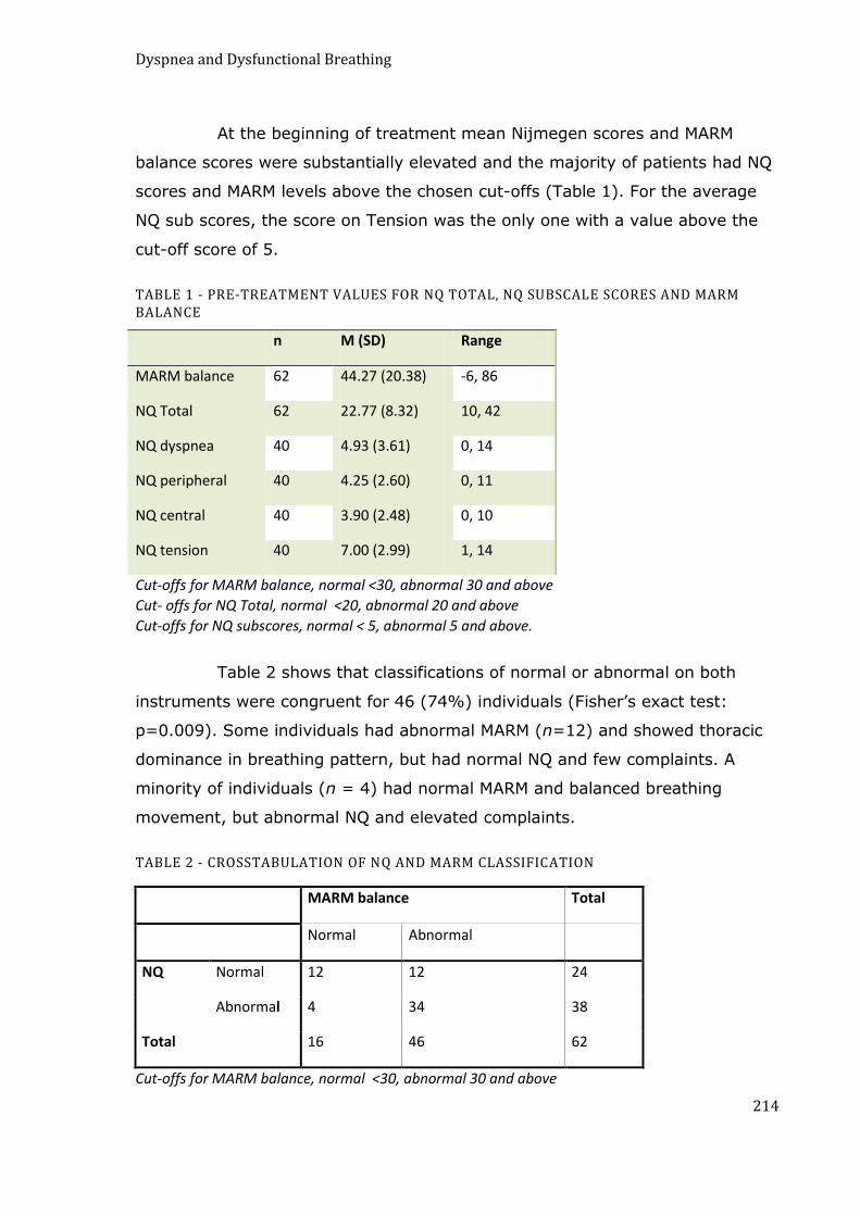
Dyspnea and Dysfunctional Breathing
214
At the beginning of treatment mean Nijmegen scores and MARM
balance scores were substantially elevated and the majority of patients had NQ
scores and MARM levels above the chosen cut-offs (Table 1). For the average
NQ sub scores, the score on Tension was the only one with a value above the
cut-off score of 5.
TABLE 1 - PRE-TREATMENT VALUES FOR NQ TOTAL, NQ SUBSCALE SCORES AND MARM BALANCE
n M (SD) Range
MARM balance 62 44.27 (20.38) -6, 86
NQ Total 62 22.77 (8.32) 10, 42
NQ dyspnea 40 4.93 (3.61) 0, 14
NQ peripheral 40 4.25 (2.60) 0, 11
NQ central 40 3.90 (2.48) 0, 10
NQ tension 40 7.00 (2.99) 1, 14
Cut-offs for MARM balance, normal <30, abnormal 30 and above
Cut- offs for NQ Total, normal <20, abnormal 20 and above
Cut-offs for NQ subscores, normal < 5, abnormal 5 and above.
Table 2 shows that classifications of normal or abnormal on both
instruments were congruent for 46 (74%) individuals (Fisher‟s exact test:
p=0.009). Some individuals had abnormal MARM (n=12) and showed thoracic
dominance in breathing pattern, but had normal NQ and few complaints. A
minority of individuals (n = 4) had normal MARM and balanced breathing
movement, but abnormal NQ and elevated complaints.
TABLE 2 - CROSSTABULATION OF NQ AND MARM CLASSIFICATION
MARM balance Total
Normal Abnormal
NQ Normal 12 12 24
Abnormal 4 34 38
Total 16 46 62
Cut-offs for MARM balance, normal <30, abnormal 30 and above
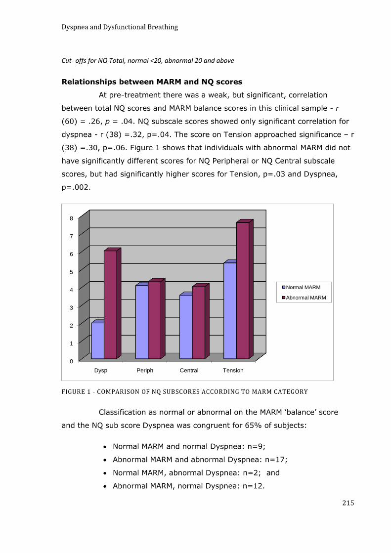
Dyspnea and Dysfunctional Breathing
215
Cut- offs for NQ Total, normal <20, abnormal 20 and above
Relationships between MARM and NQ scores
At pre-treatment there was a weak, but significant, correlation
between total NQ scores and MARM balance scores in this clinical sample - r
(60) = .26, p = .04. NQ subscale scores showed only significant correlation for
dyspnea - r (38) =.32, p=.04. The score on Tension approached significance – r
(38) =.30, p=.06. Figure 1 shows that individuals with abnormal MARM did not
have significantly different scores for NQ Peripheral or NQ Central subscale
scores, but had significantly higher scores for Tension, p=.03 and Dyspnea,
p=.002.
FIGURE 1 - COMPARISON OF NQ SUBSCORES ACCORDING TO MARM CATEGORY
Classification as normal or abnormal on the MARM „balance‟ score
and the NQ sub score Dyspnea was congruent for 65% of subjects:
Normal MARM and normal Dyspnea: n=9;
Abnormal MARM and abnormal Dyspnea: n=17;
Normal MARM, abnormal Dyspnea: n=2; and
Abnormal MARM, normal Dyspnea: n=12.
0
1
2
3
4
5
6
7
8
Dysp Periph Central Tension
Normal MARM
Abnormal MARM

Dyspnea and Dysfunctional Breathing
216
The association between MARM and dyspnea classifications was
statistically significant (Fisher exact test, p <0.03). It is remarkable that only
two subjects with normal MARM had high dyspnea scores. In the scatterplot
(Figure 2) the upper left quadrant is practically empty, implying that all
subjects with high NQ dyspnea scores breathe thoracically. By contrast, a
thoracic dominant breathing pattern can be present without the presence of
dyspnea symptoms (lower right quadrant).
60 30 0
Marm balance
12.5
10
7.5
5
2.5
0
NQ
d
ys
pn
oea
Figure 2 Relationships between categories on NQ dyspnoea and MARM Balance.
Normalization of MARM and NQ after treatment
The response of MARM and NQ to breathing and relaxation therapy
was positive (table 3): average MARM and NQ scores (both total and sub
scores) greatly improved after treatment by WBB (p < .0001) and moved into
the normal zone.
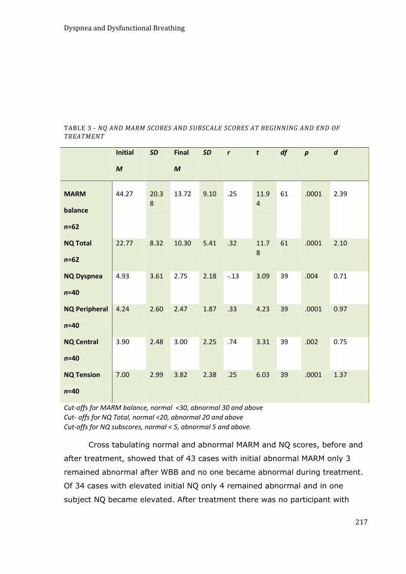
Dyspnea and Dysfunctional Breathing
217
TABLE 3 - NQ AND MARM SCORES AND SUBSCALE SCORES AT BEGINNING AND END OF TREATMENT
Initial
M
SD Final
M
SD r t df p d
MARM
balance
n=62
44.27 20.3
8
13.72 9.10 .25 11.9
4
61 .0001 2.39
NQ Total
n=62
22.77 8.32 10.30 5.41 .32 11.7
8
61 .0001 2.10
NQ Dyspnea
n=40
4.93 3.61 2.75 2.18 -.13 3.09 39 .004 0.71
NQ Peripheral
n=40
4.24 2.60 2.47 1.87 .33 4.23 39 .0001 0.97
NQ Central
n=40
3.90 2.48 3.00 2.25 .74 3.31 39 .002 0.75
NQ Tension
n=40
7.00 2.99 3.82 2.38 .25 6.03 39 .0001 1.37
Cut-offs for MARM balance, normal <30, abnormal 30 and above
Cut- offs for NQ Total, normal <20, abnormal 20 and above
Cut-offs for NQ subscores, normal < 5, abnormal 5 and above.
Cross tabulating normal and abnormal MARM and NQ scores, before and
after treatment, showed that of 43 cases with initial abnormal MARM only 3
remained abnormal after WBB and no one became abnormal during treatment.
Of 34 cases with elevated initial NQ only 4 remained abnormal and in one
subject NQ became elevated. After treatment there was no participant with

Dyspnea and Dysfunctional Breathing
218
both elevated NQ and thoracic dominance. Thus, treatment was effective in
normalizing dysfunctional breathing.
The nature of the response to treatment was further studied by
calculating correlations between the changes in the measurements. Changes in
MARM balance were only correlated with changes in NQ dyspnea but not with
changes in NQ total or other NQ subscale scores (Table 4). However, changes
in NQ dyspnea also correlated with changes in NQ tension and NQ total,
implying that dyspnea was also related to tension and possibly to other factors.
TABLE 4 - CORRELATION MATRIX FOR CHANGES IN NQ AND MARM VARIABLES
Change
Dyspnea
Change
Peripheral
Change
Central
Change
Tension
Change
MARM
Balance
Change NQ
.54
p<.0001
.66
p<.0001
.12
p=.44
.64
p<.0001
.10
p=.44
Change
Dyspnea
.18
p=.274
-.20
p=.212
.39
p=.01
.32
p=.05
Change
Peripheral
-.02
p=.89
.36
p=.02
-.14
p=.40
Change
Central
.10
p=.533
-.03
p=.858
Change
Tension
.11
p=.492
The final question was whether thoracic dominance moderates the
effect of treatment on complaints. A two factor, mixed design ANOVA was
conducted to compare NQ total scores between those with normal and
abnormal MARM (between subject‟s factor) prior to therapy, at the beginning
and at the end of therapy (within subjects factor). The ANOVA yielded three F
values that examined the main effect for the MARM group, the main effect for
TIME, and the INTERACTION between the MARM group and TIME. The results
are shown in Table 5:
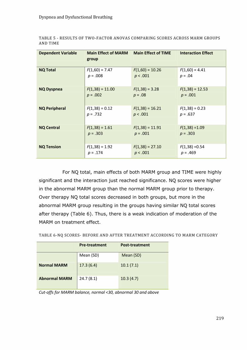
Dyspnea and Dysfunctional Breathing
219
TABLE 5 - RESULTS OF TWO-FACTOR ANOVAS COMPARING SCORES ACROSS MARM GROUPS AND TIME
Dependent Variable Main Effect of MARM
group
Main Effect of TIME Interaction Effect
NQ Total F(1,60) = 7.47
p = .008
F(1,60) = 10.26
p < .001
F(1,60) = 4.41
p = .04
NQ Dyspnea F(1,38) = 11.00
p = .002
F(1,38) = 3.28
p = .08
F(1,38) = 12.53
p = .001
NQ Peripheral F(1,38) = 0.12
p = .732
F(1,38) = 16.21
p < .001
F(1,38) = 0.23
p = .637
NQ Central F(1,38) = 1.61
p = .303
F(1,38) = 11.91
p = .001
F(1,38) =1.09
p = .303
NQ Tension F(1,38) = 1.92
p = .174
F(1,38) = 27.10
p < .001
F(1,38) =0.54
p = .469
For NQ total, main effects of both MARM group and TIME were highly
significant and the interaction just reached significance. NQ scores were higher
in the abnormal MARM group than the normal MARM group prior to therapy.
Over therapy NQ total scores decreased in both groups, but more in the
abnormal MARM group resulting in the groups having similar NQ total scores
after therapy (Table 6). Thus, there is a weak indication of moderation of the
MARM on treatment effect.
TABLE 6-NQ SCORES- BEFORE AND AFTER TREATMENT ACCORDING TO MARM CATEGORY
Pre-treatment Post-treatment
Mean (SD) Mean (SD)
Normal MARM 17.3 (6.4) 10.1 (7.1)
Abnormal MARM 24.7 (8.1) 10.3 (4.7)
Cut-offs for MARM balance, normal <30, abnormal 30 and above

Dyspnea and Dysfunctional Breathing
220
The same analysis was done for the NQ Dyspnea score. A significant
main effect for MARM group was found, as was a highly significant interaction.
For this variable, the groups were different prior to therapy with elevated
average scores in the abnormal MARM group and normal scores in the normal
MARM group. In the abnormal group, scores decreased after therapy, whereas
a small increase was found in the normal MARM group, resulting in similar
values for both groups after therapy (Table 7). Thus, there is a clear indication
of MARM moderating the treatment effect in dyspnea.
TABLE 7 - DYSPNEA SCORES- BEFORE AND AFTER TREATMENT ACCORDING TO MARM CATEGORY
Pre-treatment Post-treatment
Mean (SD) Mean (SD)
Normal MARM 2.0 (2.1) 3.5 (3.2)
Abnormal MARM 6.0 (3.4) 2.4 (1.6)
Cut-offs for MARM balance, normal <30, abnormal 30 and above
For the remaining NQ subscale scores, the only significant effect was a
main effect of time. For these variables, the normal and abnormal MARM
groups had similar mean scores prior to and after therapy. Scores in both
groups decreased over therapy. MARM did not mediate treatment effect at all.
DISCUSSION
This study shows that while there is only a weak linear relationship between
hyperventilation symptoms and breathing pattern, classifications of
dysfunctional breathing made on the basis of normal or abnormal NQ and
MARM scores agree in 74% of patients. The majority of patients with elevated
Nijmegen Questionnaire scores also have a thoracic dominant breathing
pattern. Only a minimal number of patients with high NQ scores had a
„balanced‟ or evenly distributed (thoracic to abdominal) breathing pattern. This
supports clinical observation that individuals with „hyperventilation‟ symptoms
tend to have disturbed (upper thoracic) breathing pattern (Lum 1975). Thoracic
dominant breathing pattern seems to be particularly related to the group of
Dyspnea and Dysfunctional Breathing
221
respiratory items of the NQ. Only participants with abnormal MARM have
elevated scores on dyspnea.
The second major finding of the study is that breathing and
relaxation therapy, by the method of Whole Body Breathing (WBB), reduced
dysfunctional breathing symptoms and improved breathing pattern. Both MARM
and NQ returned to normal values. After therapy no subject was classified as
having dysfunctional breathing on the basis of these two variables. However, it
appeared that the breathing pattern only moderated dyspnea items and not
other NQ subscales. By implication, treatment effect on all other NQ subscales
was not due to the pattern of breathing movement but to other psychological or
physiological processes. These processes, which are also targeted by Whole
Body Breathing (WBB), include psychophysiological self-regulatory processes
such as mental or physical relaxation, body awareness and cognitive
restructuring (Giardino, Chan et al. 2004).
The single largest reduction of symptoms occurred in NQ subscale
“tension” which includes items relating to tension, anxiety, heart palpitations
and chest pain. Reduction in these symptoms was not mediated by breathing
pattern and it may have been due to other respiratory variables like CO2 level
(Meuret, Rosenfield et al. 2009) or to various physical and psychological effects
of relaxation (Ost and Breitholtz 2000; Conrad and Roth 2007).
To our knowledge, this is the first study that documents the
importance of thoracic dominant breathing pattern for the experience of
medically unexplained dyspnea. It needs to be emphasized however, that the
association is found in patients without a somatic cause for dyspnea. The
relationship of breathing pattern to dyspnea perception may be different in
patients with respiratory or cardiovascular disease.
In patients with COPD and the presence of rapid and shallow pattern
of breathing, a mode of breathing that is also usually thoracic dominant, were
found to correspond with extent of dyspnea (Ferrari, Goti et al. 1997). However
the authors of this study suggest that this mode of breathing is an adaptive
response that might actually assist in reducing excessive respiratory drive.

Dyspnea and Dysfunctional Breathing
222
Preliminary data on 33 patients with COPD showed they had thoracic dominant
breathing and a higher score on MARM balance (27.8 ± 42.4) than normal
individuals; however, there was no relationship with grading of disease severity
(Gold1-4) (Dixhoorn 2009).
There are no MARM data yet on patients with asthma or
cardiovascular disease. However there is some evidence that abnormal
breathing patterns occur in individuals with these diseases. In asthmatic
patients, exaggerated dyspnea and unexplained complaints have been
attributed to „dysfunctional breathing‟, (Thomas, McKinley et al. 2001; Prys-
Picard, Kellet et al. 2006; Hagman, Janson et al. 2008; Prys-Picard and Niven
2008; Thomas, McKinley et al. 2009) although only a few of these studies
include data on breathing pattern (Prys-Picard, Kellet et al. 2006). In asthma,
even mild broncho constriction has been found to produce shortening and
hypertonicity of inspiratory muscles and these muscle changes would tend to
favor thoracic dominant breathing (Muller, Bryan et al. 1981; Lougheed, Fisher
et al. 2006). Patients with heart failure often complain of dyspnea and this has
been attributed in part to „inefficient pattern of rapid shallow respiration‟
(Mandak and McConnell 1998). Assessment of MARM within these populations
will be necessary to establish values of MARM balance, both before and after
treatment. Possibly MARM may be helpful to establish the pathway of therapy
effect for various treatments, including medication, biofeedback or exercise
training (Giardino, Chan et al. 2004). Few studies on breathing therapies for
asthma have investigated their effects on breathing pattern and this may prove
useful in the refinement of the application and development of these types of
therapies.
Implementation of MARM in the clinical setting may be hampered by
the need for training and the time required for the complete protocol. Test-
retest reliability of the therapist in the present study was not determined, but
the very high correlation between pre test MARM and change in MARM indicates
that reliability would be satisfactory. This issue will be important however in
future studies that employ MARM.

Dyspnea and Dysfunctional Breathing
223
An important limitation of the study is the absence of any longer
term follow-up to assess whether the changes in breathing pattern and
complaints are stable and sustained. Previous studies using the MARM in a
two-year follow-up of myocardial infarction patients showed a stable effect of
treatment and prompted later validation studies (Dixhoorn 1994).
CONCLUSION
The majority of individuals with a large number of dysfunctional breathing
symptoms are also likely to have a thoracic dominant breathing pattern.
Breathing pattern and symptoms can normalize after breathing therapy. The
extent of thoracic dominant breathing seems an important moderator for
dyspnea symptoms and an important component of treatment.
This is a copy of an article published in the Journal of Asthma © 2011 copyright; Journal of
Asthma is available online at:http://informahealthcare.com.

Discussion
224
CHAPTER 13
DISCUSSION
INTRODUCTION
Discussions of the each of the seven studies conducted have been included in
the empirical chapters of this thesis. This final chapter seeks to draw together
and highlight the principal findings of these studies and discuss their
implications.
AIMS AND OBJECTIVES
As will be recalled from Chapter 1 (p.23 ), the overall aims of this thesis were:
To further develop and validate some clinical tools for assessment of
dysfunctional breathing;
To explore and evaluate current methods for assessing dysfunctional
breathing particularly those used by breathing therapists;
To investigate the relationships between measures of dysfunctional
breathing with a view to understanding the possible definitions and
dimensions of dysfunctional breathing; and
To explore possible mechanisms of breathing therapy.
PRINCIPAL FINDINGS
The principal findings of the seven studies will be discussed in relationship to
how they have contributed to knowledge on:
1. the measures of dysfunctional breathing and their role in its
assessment;
2. the possible definitions and dimensions of dysfunctional breathing;
and,
3. the possible mechanisms of breathing therapy.
Discussion
225
ASSESSMENT OF DYSFUNCTIONAL BREATHING AND ITS MEASURES
BREATHING PATTERN
Dysfunctional breathing measures that were either developed or investigated in
detail in this thesis include two measures for evaluating breathing pattern - the
MARM and the Hi Lo.
Clinicians often use observation and palpation and techniques similar to
the MARM and Hi Lo to assess breathing pattern rather than instrumentation,
such as respiratory induction plethysmograph, which is the most common
instrument used in the research setting (Pryor and Prasad 2002).
Results of the study reported in Chapter 6 indicate that the MARM is a
useful and reliable tool, with high inter-rater reliability (r =.851, p=.01) for
assessment of one aspect of breathing pattern - thoracic dominant breathing.
This research identified a number of advantages of the MARM over respiratory
induction plethysmography in that the MARM is relatively easy to learn,
inexpensive, applicable to clinical environments and unlike RIP, which only
evaluates rib cage motion in the lateral and anterior/posterior planes, the
examiner using the MARM is able to perceive vertical motion of the rib cage
during inspiration. This vertical motion, produced by the accessory muscles of
inspiration, is an important component of thoracic dominant breathing and
inclusion of this third (vertical) plane of motion is necessary for accurate
representation of thoracic dominant breathing. Like the MARM, the Hi Lo
breathing assessment is also easy to learn and reasonably accurate in detecting
simulated breathing patterns.
In comparing the MARM and the Hi Lo it appears that both techniques
have their particular strengths. A strength of the MARM is that it provides a
means to measure and assign numerical values, in contrast to the Hi Lo, which
only reports dichotomous variables. Also, the MARM, by evaluating the lateral
motion of the lower rib cage rather than the anterior protrusion of the
abdomen, may give a better representation of diaphragm action, while the Hi
Lo appears to be better for assessing paradoxical breathing and asynchrony of

Discussion
226
breathing. The evaluation of dysfunctional breathing pattern is probably best
performed using a combination of both techniques.
The MARM and Hi Lo are useful for the clinical environment because they
are cheap and accessible. In the research environment, instrumentation such
as respiratory induction plethysmography is preferable due to greater precision
of measurement. However, limitations were identified in the ability of
respiratory induction plethysmography to evaluate the vertical dimension of rib
cage motion. This vertical motion of the upper rib cage, produced by the
accessory muscles of respiration, is an important aspect of thoracic breathing.
The inability of respiratory induction plethysmography to measure this aspect of
thoracic breathing may significantly impair its ability to evaluate thoracic
dominant breathing. Opto electronic plethysmography which measures more
dimensions of breathing gives a more complete representation of breathing
pattern and is superior to respiratory induction plethysmography in this regard.
Breathing pattern is often not measured in a systematic manner in the
clinical environment and most research into dysfunctional breathing has not
included detailed investigations of breathing pattern. This may have resulted in
this aspect of dysfunctional breathing being overlooked.
The treatment of conditions, such as disorders of continence (Hung,
Hsiao et al. 2010), speech (Altman, Atkinson et al. 2005), lower back and neck
pain (Smith, Russell et al. 2006; Perri 2007; Correa and Berzin 2008; Kapreli,
Vourazanis et al. 2009) and dysfunctions of posture, balance and motor control
(Lewitt 1980; Gandevia, Butler et al. 2002), which are believed to be
aggravated by dysfunctional breathing patterns would benefit from careful
monitoring and systematic measurement of breathing pattern to monitor
patient‟s response to treatment. Given that the MARM and Hi Lo appear to be
easily learnt, practitioners with differing backgrounds could probably be trained
to identify dysfunctional breathing patterns using these techniques.
Research into the role of breathing pattern dysfunction in these types of
conditions and others could include measures such as the MARM and Hi Lo in

Discussion
227
addition to instrumentation such as respiratory induction plethysmography and
opto electronic plethysmography.
SYMPTOM QUESTIONNAIRES
The SEBQ was constructed because an extensive search of the literature
showed that there were no validated questionnaires for assessing dysfunctional
breathing. The Nijmegen Questionnaire, sometimes used for identifying
dysfunctional breathing, was designed to measure hyperventilation syndrome,
which is just one aspect of dysfunctional breathing.
The SEBQ contains many more items specific to breathing than the NQ and
factor analysis of these SEBQ items identified the presence of two separate, but
correlated, dimensions. These two dimensions appear to represent biochemical
and biomechanical aspects of dyspnea. The first of these dimensions named
„lack of air‟ contains items very similar to those identified by other researchers
as “air hunger” or “urge to breathe”. These verbal descriptors are believed to
be associated with afferent input from chemoreceptor activity (Banzett, Lansing
et al. 1990; Lansing 2000).
The second dimension, which contained items that appear to relate to
biomechanical factors, was labeled “perception of inappropriate or restricted
breathing”. The items loading highest on this dimension such as “I cannot take
a deep and satisfying breath” and “My breathing feels stuck or restricted”
convey a sense of restricted or otherwise unsatisfied respiration. These items
are recognisably similar to sensations documented in situations where the work
of breathing is made more difficult, such as when the chest wall is strapped
(O'Donnell, Hong et al. 2000) and are reported by patients with impaired
function of the respiratory muscles and rib cage (Lougheed, Fisher et al. 2006,
Simon and Schwartzstein 1990). The types of sensations identified are believed
to arise, as least in part, from receptors in the chest wall and muscles of
breathing (Simon and Schwartzstein 1990) and also represent the qualities of
dyspnea that arise when the motor output of respiratory system is not able to
match the expectations generated in the sensory cortex by corresponding
discharges from the motor cortex (Beach and Schwatrzstein 2006).
Discussion
228
Another possible way of interpreting the two dimensions identified in the
SEBQ is that they are two different aspects of interoception with the first being
related to the sensory or feeling aspect of interoception and the second being
related to the cognitive or evaluative aspects of interoception. These two
dimensions of the SEBQ may thus be representative of perceptual as well as
biochemical and biomechanical aspects of dysfunctional breathing. However,
this needs to be clarified in future studies.
The SEBQ has potential as a tool for evaluating dysfunctional breathing,
but needs further development and testing. The two dimensions of the SEBQ
may represent strongly related, but distinct, aspects of dysfunctional breathing
symptoms, representing biochemical and biomechanical mechanisms, and also
sensory and cognitive aspects of interoception. The clinical assessment of these
two dimensions may prove useful for more specific diagnosis and treatment of
dysfunctional breathing.
It has been suggested that the weak association between dysfunctional
breathing (hyperventilation syndrome) symptoms and hypocapnia may be
partly explained by the fact that these arise from other causes such as tense
breathing patterns (Hornsveld 1997). The presence of a separate biomechanical
and evaluative dimension to dysfunctional breathing symptoms in the SEBQ
tends to support this assertion.
To evaluate dysfunctional breathing symptoms, there may be value in
administering both the SEBQ and the NQ.
There are several problems associated with the NQ. Firstly, the
hyperventilation syndrome, which it was designed to measure, is no longer
considered to be a valid diagnosis. So the question arises, what aspects of
breathing dysfunction does the NQ measure? The fact that improvements in
breathing pattern were only associated with improvements in the NQ subscale
“dyspnea” suggests that only this part of NQ questionnaire is closely linked with
breathing pattern, while other NQ subscales may be more closely linked to
psychological or biochemical aspects of breathing.
Discussion
229
Also, because the NQ focuses on the hyperventilation syndrome, it
includes many items that are traditionally identified with the pathophysiology of
hypocapnia (e.g. dizziness, parestheisas, blurred vision, stiffness in
extremities) and only four items that specifically refer to respiratory sensations
(e.g. faster deeper breathing, inability to breathe deeply, shortness of breath
and tight feelings in the chest).
Overall the NQ was found to correlate with the SEBQ total score and with
Factor 1 of the SEBQ - “Lack of Air”. However, the NQ does not correlate with
Factor 2 of the SEBQ - “Perception of inappropriate or restricted breathing”.
This suggests that the NQ may not adequately encompass items associated
with the biomechanical aspects of breathing dysfunction or from the
cognitive/evaluative aspects of interoception. Administration of the SEBQ in
addition to the NQ allows monitoring of a larger number of symptoms, which
may assist in ongoing patient evaluation.
BREATH HOLDING TIME
Breath holding time (BHT) has been found to be shorter in individuals with
hyperventilation disorders (Jack, Darke et al. 1998 {Warburton 2006). Dr KP
Buteyko, originator of the Buteyko breathing technique which is predominately
used for the treatment of asthma, patented a formula for determining CO2 on
the basis of breath holding time.
In the study reported in Chapter 10 of this thesis, it was found
that breath holding time did not correlate with end tidal CO2 as predicted by the
formula patented by Dr. K.P. Buteyko. In fact, the results revealed a slight
negative correlation between BHT and ETCO2, (r= -.241) directly opposite to
Buteyko‟s claims. This negative relationship does not appear to be an artefact
as an inverse relationship between breath holding time and alveolar CO2
tension as has been reported by researchers in the past (Godfrey and Campbell
1969), and it seems reasonable to assume that in individuals with relatively
normal CO2 thresholds, the lower the starting CO2 the longer it will take for CO2
to accumulate to the level where intensity of dyspnea results in the termination
of breath holding.

Discussion
230
Breath holding time has a long history as a test of chemosensitivity and
the findings in Chapter 10 do not disprove an association between
chemoreceptor thresholds and breath holding time but they do indicate that
mechanisms other than chemosensitivty are involved (Stanley, Cunningham et
al. 1975). Studies in the past have shown that individuals with greater
chemosensitivity to CO2 will, for a given level of starting CO2 tension, have a
shorter breath holding time (Stanley, Cunningham et al. 1975). However more
recent studies have shown that individuals with chronic and intermittent
hypocapnia and short breath holding time do not generally have altered
chemosensitivity. These contradictory findings between older and more recent
studies suggests that mechanisms in addition to altered chemoreceptor
thresholds, including behavioural and mechanical factors contribute to short
breath holding time in hyperventilation and in other types of dysfunctional
breathing (Jack, Rossiter et al. 2003).
The finding that breath holding time does not correlate with ETCO2,
according to Buteyko‟s formula, has implications for the Buteyko Breathing
Technique. The therapeutic rationale of the Buteyko Breathing Technique is tied
to beliefs about the importance of hypocapnia and beliefs that effectiveness of
this technique is dependent on normalisation of PACO2. This finding seriously
questions the validity of this rationale because the main outcome measure used
by Buteyko practitioners to identify normalisation of CO2 levels in patients is
increased length of breath holding time.
In this study, breath holding time was found to be linked with breathing
pattern rather than with CO2 levels, raising the possibility that this might be a
more significant mechanism of breathing therapies than correction of
hypocapnia. Length of breath holding time is considered to be an important
index of dyspnea threshold (Nishino 2009). In the study reported in Chapter 10
of this thesis, breath holding time was found to be significantly lower in
individuals with abnormal spirometry, and, in these individuals breath holding
time was found to correlate with thoracic dominant breathing pattern (r=.408,
p =.028). This association between the presence of thoracic dominant
breathing pattern and shortened breath holding time suggests an increased
Discussion
231
tendency to dyspnea in those with thoracic breathing. The detection of thoracic
breathing may therefore be a more useful measure for Buteyko and other
therapists than BHT and may provide insight into people who are more likely to
respond to Buteyko-type therapies.
Despite the fact that breath holding time does not appear to accurately
reflect carbon dioxide status, it is still a useful tool for assessing breathing.
Breath holding induces dyspneic sensations and the time taken till breath
holding first produces dyspnea sensations can indicate the threshold of
dyspneic sensation, while final breaking point of breath holding can indicate
maximum tolerance of dyspnea (Nishino 2009). The length of breath holding
time at functional residual capacity, for the initial period of no sensation and
until the person feels the first desire to breathe is similar to the Buteyko
Control Pause (Buteyko 1990). This measure has been shown to be a relatively
stable measure that is less susceptible to behavioral influence than the period
of maximum tolerance to breath holding (Nishino, Sugimori et al. 1996) which
is similar to the Buteyko Maximum Pause (Stalmatski 1999). Breath holding
time has also been proposed to be a useful measure of perceptual inaccuracy in
patients with near fatal asthma (Nannini, Zaietta et al. 2007). It is possible that
improvements in breath holding time after breathing therapy such as BBT, not
associated with improved lung function, might indicate a blunting of dyspnea
perception. Increased breath holding time after BBT and other breathing
therapy might also be associated with improvements in breathing pattern.
These issues need to be explored in future research.
In conclusion, despite the fact that breath holding time tends to be lower
in individuals with established hyperventilation disorders, breath holding time
does not correlate precisely with resting carbon dioxide in all individuals. This is
probably due to the fact that factors other than carbon dioxide, including
breathing pattern, presence of respiratory disease and psychological factors are
of importance.
CAPNOGRAPHY
It appears that hyperventilation can only be evaluated by measurement of
carbon dioxide, yet dysfunctions of breathing pattern, shortened breath holding

Discussion
232
time and dysfunctional breathing symptoms can exist without chronic
hypocapnia. Therefore, hyperventilation cannot be diagnosed on the basis of
abnormal breathing patterns, length of breath holding time or symptoms.
Acute, chronic or intermittent hyperventilation can only be diagnosed by
direct measurement of arterial CO2, either transdermally or from extracted
blood or indirectly through measurement of end tidal CO2 using a capnograph.
Relying on resting CO2 might not identify individuals with frequently recurrent,
intermittent hyperventilation that is indicative of poor regulation of CO2
(Warburton and Jack 2006). In these individuals, the problems of CO2 may only
become evident when capnometry is performed to measure the CO2 response to
physical or psychological challenge. These individuals, once induced to
hyperventilate, after an exercise stress test or some form of psychological
stress, tend to have very slow recovery of CO2 to pre-challenge levels (Hardonk
and Beumer 1979; Warburton and Jack 2006).
COMPREHENSIVE ASSESSMENT OF DYSFUNCTIONAL BREATHING
Screening for dysfunctional breathing with measures representing only one of
its dimensions may not lead to realistic estimations of the true prevalence and
impact of the various types of breathing dysfunctions.
Comprehensive evaluation of breathing dysfunction should include
measures of breathing symptoms, breathing pattern, resting CO2 and also
include functional measures such a breath holding time and response of
breathing to physical and psychological challenges.
DEFININING DYSFUNCTIONAL BREATHING
DYSFUNCTIONAL BREATHING IS MULTIDIMENSIONAL
In the literature review section of this thesis (Chapter 5), dysfunctional
breathing is proposed to exist when any of the functions of breathing are
impaired. Given all of the possible functions of breathing, this definition is
rather broad. Subsequently, a working definition of dysfunctional breathing was
derived from the study reported in Chapter 9, which concluded that
dysfunctional breathing cannot be simply defined and that for practical
Discussion
233
purposes it is probably best characterized as a multidimensional construct with
at least three dimensions: biochemical, biomechanical, and breathing
symptoms. This conclusion was reached because, while significant correlations
were found within biochemical, biomechanical and symptom categories of
dysfunctional breathing measures, no correlations were found between
categories.
Despite controversies about the hyperventilation syndrome,
hyperventilation is still the most well recognized physiological aspect of
dysfunctional breathing. Other aspects of dysfunctional breathing such as
breathing pattern are often ignored. In the study reported in Chapter 9, the
lack of correlations between breathing pattern and symptomatic measures with
ETCO2 supports the view that dysfunctional breathing is not limited to
hyperventilation and that it includes biomechanical and symptomatic aspects as
well. This has implications for future research into dysfunctional breathing
which should include measurements of all these various aspects.
The working definition of dysfunctional breathing proposed in this thesis
is in keeping with a substantial body of research which also reports findings
that biochemical, symptomatic and breathing pattern aspects of breathing
dysfunction do not necessarily co-exist in individuals suspected of having
dysfunctional breathing (Burton 1993; Han 1996;Hornsveld 1997). However, it
cannot be presumed that there is no association between dysfunctional
breathing symptoms, breathing pattern, breath holding time and resting CO2, as
it has been observed on repeated occasions that these do sometimes co-exist
(Lum 1976; Gardner 1986; Howell 1997) even though the relationships are
complex.
In patients with more severe breathing dysfunction, relationships
between categories of measures are more likely to exist and these individuals
are also more likely to have disturbances in several aspects of breathing
functionality. In a subsequent study, reported in Chapter 13, which investigated
patients with more severe breathing disturbance than that those in Chapter 9,
there was evidence of stronger relationships between categories of breathing
dysfunction. A small linear correlation between breathing pattern and

Discussion
234
dysfunctional breathing symptoms (as measured by the NQ) could be seen in
these patients (r=0.26, p<0.05). It also found that classification as normal or
dysfunctional on the basis of breathing pattern, as measured by the MARM and
on the basis of the NQ, agreed in 74% (p < 0.001) of patients.
The view that biochemical and biomechanical aspects of dysfunctional
breathing are distinct was also suggested from the factor analysis of the SEBQ
which showed separate loadings for items similar to symptoms known to arise
from biomechanical aspects of breathing and symptoms known to arise from
altered chemoreceptor inputs. As previously discussed in this thesis, the two
factors of the SEBQ may also indicate separate dimensions for the sensory or
feeling aspects of interoception and the cognitive or evaluative aspects.
PYSCHOLOGICAL INFLUENCES ON DYSFUNCTIONAL BREATHING AND ITS SYMPTOMS
It is clear from the literature that psychological factors play an important role in
producing dysfunctional breathing and also in influencing symptom perception.
Psychological factors need to be considered in explaining some of the
more difficult to interpret findings in this thesis. For example, the poor
correlations between the SEBQ scores and other measures of dysfunctional
breathing were unexpected. This lack of correlation might have been due to the
study participants who, as a group, only had mild dysfunctional breathing. It
could also be partly due to the influence of social, cognitive and emotional
factors on interoception and symptom reporting which confound these
relationships between subjective and objective measures (Lougheed
2007;Bogaerts, Van Eylen et al. 2010).
Accurate perception and normal sensory awareness of breathing
contribute to normal control of breathing and are probably important for the
effective use of breathing for therapeutic and salutogenic purposes. Poor
perception of asthma symptoms can contribute to increased morbidity and
mortality (Magadle, Berar-Yanay et al. 2002) while poor perception of breathing
related sensations and lack of breathing awareness, as well as over-perception
or excessive awareness of physiological sensations associated with breathing,
are possible aspects of breathing dysfunction. If the two factors of the SEBQ
Discussion
235
are interpreted as relating to the sensory and cognitive aspects of
interoception, very high scores or very low scores that do not correspond to
objective findings might, in some individuals, indicate a disturbance in
interoception.
BREATHING PATTERN AS AN ASPECT OF DYSFUNCTIONAL BREATHING
Functional breathing is „balanced‟ between the upper rib cage and the lower rib
cage/abdomen when a person is at rest. It is also flexible and responsive to
changes in posture and can be modulated at will to become either thoracic or
abdominal. These conclusions were reached as a result of the first MARM study
(Chapter 6) based on the data gathered from the breathing patterns of yoga
practitioners and breathing therapy teachers who participated in this study. In
the study in Chapter 12, it was found that after breathing therapy, breathing
pattern became more „balanced‟ and less thoracic. This more balanced
breathing pattern was associated with decreased symptoms scores in general
and decreased dyspnea in particular.
A predominately thoracic breathing pattern at rest is likely to be
dysfunctional, particularly if it is fixed and unresponsive to changes in posture
or attempts at voluntary modulation. Under the same circumstances,
paradoxical breathing can also be considered dysfunctional. The finding in
Chapter 11 that thoracic breathing and paradoxical breathing were associated
with decreased „coherence‟ suggests that these types of breathing are less
physiologically efficient and the presence of these types of breathing pattern
may inhibit homeostasis. HRV patterns similar to those described indicating
„coherence‟ are believed to reflect resonance effects between the respiratory,
cardiovascular and autonomic nervous systems (Lehrer, Vaschillo et al. 2000;
Vaschillo and Lehrer 2002). In this study, thoracic dominant breathing was
associated with decreased ability to produce „coherence‟ of HRV (r=-.463, p =
.0001) and individuals with paradoxical breathing had the lowest HRV
coherence (t(8) = 10.7, p = .001) with the negative relationship between
coherence of HRV and thoracic breathing being strongest in this group (r = -
.768, p = .03).
Discussion
236
It is evident from the studies performed and the above discussions that
dysfunctional breathing cannot be defined precisely at this time, and that for
practical purposes dysfunctional breathing is probably best characterized as a
multidimensional construct with at least three dimensions: biochemical,
biomechanical and breathing symptoms. Other dimensions may include
sensory, cognitive and psychological aspects with all these aspects being
potentially important in different populations.
FUNCTIONAL OPTIMAL BREATHING- WHAT IS IT?
If dysfunctional breathing cannot be adequately defined perhaps optimal
breathing can be defined as breathing that provides the greatest efficiency,
balance and flexibility of response in any particular situation and that optimally
fulfills all its functions.
POSSIBLE MECHANISMS OF BREATHING THERAPY
PHYSIOLOGICAL-BIOCHEMICAL AND NEUROMECHANICAL
Normalisation of carbon dioxide levels is unlikely to be the only mechanism of
breathing therapies such as the BBT. Normalisation of breathing pattern is
likely to be another possible mechanism. The lack of correlation between
Butyeko‟s breath holding time formula and carbon dioxide levels throws serious
doubt onto Buteyko‟s claim that clinical improvements from people learning
these techniques are only due to changes in CO2. Several studies have tested
CO2 levels after BBT (Bowler 1998) and other breathing therapies and have
found that despite signs of clinical improvement, mean CO2 levels have not
increased (Holloway and West 2007; Thomas, McKinley et al. 2009). However,
it cannot be assumed that there is no relationship between clinical
improvements from breathing therapies and carbon dioxide.
There is evidence that hypocapnia has clear pathogenic effects, and
normalisation of carbon dioxide has been shown to be protective in disease
states (Laffey and Kavanagh 2002;Laffey 2003), Studies that have looked at
individual changes in CO2 have found that increased levels of carbon dioxide
occurring as a result of breathing therapy does, to some extent, explain the
reduced number of symptoms in asthmatics (Ritz, Meuret et al. 2009) and
Discussion
237
sufferers of panic disorder (Meuret, Rosenfield et al. 2009). However, a
substantial amount of research has also shown that this is not a simple
relationship (Hormbrey 1988;Hornsveld 1997) and that other mechanisms
involving, for example, psychological and biomechanical factors also play a role.
The finding that breath holding time correlates with extent of thoracic dominant
breathing pattern also suggests that breathing pattern may influence the
outcomes of breathing therapy.
The increased breath holding time reported by Buteyko practitioners in
individuals learning the BBT are proposed to be associated with many different
types of health improvements including reduced dyspnea (Stalmatski 1999). Breath
holding effectively produces the dyspneic sensation, and increases in the length of a
person‟s breath holding time after breathing training imply that their threshold to
dyspneic sensation has increased (Pryor and Prasad 2002). Furthermore, a
correlation between breath holding time and breathing pattern suggests an
association also exist between dyspnea and breathing pattern and thus increased
breath holding time after BBT might be associated with a change in dyspnea
thresholds and/or more „balanced‟ breathing.
The literature suggests that breathing pattern changes are both a cause and
effect of dyspnea. It has often been reported that individuals with asthma and other
respiratory diseases have an increased tendency to breathing pattern disorders
such as thoracic breathing and paradoxical breathing (Muller, Bryan et al. 1981;
Tobin 1983; Loveridge, West et al. 1984). But the literature also suggests that
breathing pattern disturbances can be a cause of dyspnea, particularly dyspnea that
is unexplained or disproportionate to the amount of pathology present (Lougheed,
Fisher et al. 2006; O'Donnell 2006; Prys-Picard, Kellet et al. 2006).
The conclusion of the final study reported in this thesis in Chapter 12,
“Medically unexplained dyspnea: Partly moderated by dysfunctional (thoracic
dominant) breathing pattern”, was that breathing pattern partially moderates
dyspnea. In this study it was found that, before treatment, dyspnea scores
were only elevated in patients with thoracic dominant breathing. After
Discussion
238
treatment, NQ dyspnea scores and breathing pattern as measured by the
MARM normalised. Improvement in NQ dyspnea scores was largest for subjects
with abnormal MARM measurements pre-treatment and reduction in dyspnea
scores were strongly related to the extent of improvement in breathing pattern.
Several authors have proposed that there is an association between
abnormal function of the respiratory muscles and the extent of respiratory drive
and dyspnea (Sackner 1984;Sharp 1985; Breslin, Garoutte et al. 1990; Wolf
1994). Breathing therapies conventionally used by physical therapists have
often targeted breathing pattern dysfunctions (Pryor and Prasad 2002). The
inconsistent success of the more mechanistic breathing therapies, which
narrowly target breathing pattern rather than broader breathing functions,
suggests that they need refinement and might benefit from a more holistic
approach (Cahalin, Braga et al. 2002). Therapies that consider mind-body
interactions and factors such as relaxation appear to be more successful than
those that do not (Conrad 2007;Holloway 2007).
Breathing techniques that reduce hyperinflation would tend to improve
breathing pattern and reduce dyspnea (O'Donnell 2006). Different types of
breathing techniques could potentially reduce hyperinflation (e.g. slow abdominal
breathing can reduce respiratory drive and indirectly effect hyperinflation (Sackner,
Gonzalez et al. 1984), and training in effective exhalation techniques may be
another route (Ferrari, Goti et al. 1997; Nield, Soo Hoo et al. 2007), as could the
low volume breathing used by the BBT. It is not possible on the basis of the studies
in this thesis to do anything other than speculate that reduced volume breathing
might improve hyperinflation, but, if this is the case, it might act in similar ways to
lung volume reduction surgery which is known to decrease ventilatory drive,
improve neuromechanical coupling of respiratory muscles and significantly improve
dyspnea (Laghi, Jubran et al. 1998; Hida 1999; Lahrmann, Wild et al. 1999;
Cassart, Hamacher et al. 2001).

Discussion
239
PSYCHOLOGICAL
Breathing techniques often produce psychophysiological and relaxation effects
even if these are not actively pursued. A key breathing instruction of BBT is to
“gradually reduce your breathing by relaxing the muscle of respiration until
you feel a light lack of air” (personal communication, Dr. Buteyko, 1993). The
fact that patients are then instructed to do this as frequently as possible
during the day is likely, in those who practice this technique, to induce a more
relaxed, mindful and embodied state. Also, the person who is focusing on
breathing during the day is distracted from rumination and other types of
negative cognitions. The effect is likely to be similar to mindfulness-based
stress reduction techniques (Salmon, Sephton et al. 2004).
Another aspect of the BBT is the person actively pursues the “lack of
air” sensation; this may decrease fear of dyspnea and increase sense of
control. Given that experiential avoidance and fear of bodily sensations is
likely to increase the discomfort associated with dyspnea (Hayes, Wilson et al.
1996; Tull, Rodman et al. 2008), willingness to accept and even actively
pursue dyspnea-like sensations might create a positive emotional affect that
mitigates the sensory and affective components of dyspnea (von Leupoldt,
Mertz et al. 2006). Willingness to experience the uncomfortable and fear-
inducing sensation of dyspnea perhaps changes one‟s relationship to this
sensation in a way that increases one‟s sense of control and reduces anxiety
(Rosenkranz and Davidson 2009). Decreased fear of dyspnea may also
increase perceptual accuracy, as it has been shown that the greater a person‟s
fear of bodily sensations, the lower their self-awareness (Petersen and Ritz
2010).
Relaxation effects of breathing therapies might be an important aspect of
their clinical effectiveness (Dixhoorn van 2007; Lahmann, Nickel et al. 2009).
Lehrer (2010) has pointed out that, in the case of asthma, relaxation effects
are not sufficient to explain the positive response to breathing therapies and
has proposed that biomechanical factors (i.e. bronchodilation from deep slow
breathing) may play a key role (Lehrer 2010). Interestingly, research exists to
show that both large volume (Duggan, Chan et al. 1990) and low volume

Discussion
240
(Douglas, Drummond et al. 1981) breathing can reduce bronchoconstriction,
although the bronchodilating effect of increased lung volumes related to lung
stretching, when a large breath is taken, termed pulmonary hysteresis, is much
better known (Nadel and Tierney 1961). The effects of pulmonary hysteresis
are blunted in asthmatics and individuals with COPD and are inhibited by
corticosteroids, which can lead one to speculate that the therapeutic potency of
taking large volume or deep breaths in people with severe chronic respiratory
disease may be limited (Fairshter 1985; Pellegrino, Sterk et al. 1998), but still
a factor worth considering. One beneficial effect of low volume breathing might
be to reduce hyperinflation.
Thus, it appears likely that symptoms associated with dysfunctional
breathing, particularly dyspnea, are related to thoracic dominant breathing in
patients with medically unexplained complaints and that breathing therapy can
effectively restore normal breathing pattern and reduce dysfunctional breathing
symptoms.
IMPLICATIONS FOR BREATHING THERAPIES
Breathing therapies, particularly those that target dysfunctional breathing, may
improve their success by evaluating the various aspects of dysfunctional
breathing reflected in the different categories of measures explored in this
thesis.
Comprehensive evaluation with these measures is one means for
monitoring the effects of different types of breathing instructions on specific
types of breathing dysfunction. The obvious benefit of this is that the breathing
therapy can be tailored and the practice refined for each individual. Lack of
response of a particular dysfunctional breathing parameter might suggest the
need for refinement of technique or indicate the need for a change in approach.
Closer attention to measurement of breathing pattern may improve the
effectiveness of breathing therapies. In the study reported in Chapter 11
“Relationship between dysfunctional breathing patterns and ability to achieve
target heart rate variability with features of “coherence” during biofeedback”, it
Discussion
241
was found that resting breathing pattern influences a person‟s ability to achieve
the heart rate variability targets that are associated with positive response to
HRV biofeedback. Patients not responding to this type of therapy may be held
back by the fact that their habitual thoracic or paradoxical breathing pattern is
dysfunctional and correction and restitution of normal balanced breathing may
improve therapy outcomes.
Monitoring of breathing pattern may also improve the outcomes of
breathing therapies that aim to improve dyspnea. This is relevant because
breathing therapy is commonly used in the treatment of chronic respiratory
diseases such as asthma and chronic obstructive pulmonary disease.
LIMITATIONS
Each study presented in this thesis has its limitations and these have been
mentioned in the relevant chapters describing these studies and will not be
repeated here.
There are a number of general limitations regarding how the seven
studies were able to satisfy the three main objectives stated in the introduction
relating to definitions of dysfunctional breathing, its measurement and its role
in breathing therapy. Given the scope and breadth of these questions, it can be
appreciated that these could only be partially answered by the limited number
of studies undertaken in this thesis.
The nature of the sample used in the study reported in Chapter 9 means
that the practical working definition given for dysfunctional breathing in this
chapter applies only to individuals with mild dysfunctional breathing. In
individuals with more severe signs of breathing dysfunction relationships
between measures appear to be stronger indicating that the findings in Chapter
9 may be different when breathing dysfunction is severe.
It is proposed that dysfunctional breathing exists when there is
impairment of any of the functions of breathing. Some of these functions of
breathing might include its role in posture and motor control, speech and
vocalization and use as a tool for psychophysiological self-regulation and

Discussion
242
salutogenesis. This thesis has not investigated these aspects of breathing
functionality, nor explored their relationship to the biochemical, biomechanical
and symptomatic measures that were investigated.
Only limited conclusions can be reached on the mechanisms of breathing
therapies. These mechanisms are likely to be complex and to involve the
interaction of psychological and physiological factors not explored in this thesis.
FUTURE RESEARCH
Further studies to confirm the validity and clinical utility of the MARM and Hi Lo
are warranted. These should be conducted in populations with real rather than
simulated breathing pattern dysfunction.
It is recommended that the SEBQ questionnaire be further developed to
clarify wording of questions and that additional items be included so that all
dimensions of breathing symptoms and important breathing behaviors are
represented. Future work is required to evaluate the performance of the SEBQ
in terms of discriminating between different patient categories and normal
individuals.
Future investigations into the mechanisms of breathing therapies are
necessary and these would benefit from inclusion of the biochemical,
biomechanical and symptomatic measures of dysfunctional breathing to assess
pre-treatment status and as outcome measures. Assessing changes in these
measures and the relationships of these changes to improvement in disease
status, psychometric measures and quality of life would help to further
elucidate whether the therapeutic pathways of breathing therapy are purely
psychosomatic or involve physiological factors. As discussed previously, the
mechanisms of breathing therapy are likely to be complex, but research to
identify these mechanisms would assist in the further development of breathing
therapies and also would help to improve their effectiveness.
Further research is needed to identify which patients most benefit from
breathing therapy, whether on the basis of medical or psychological complaint
or type of breathing dysfunction. Given the large numbers of breathing
Discussion
243
therapies and large array of breathing techniques it is often difficult to choose
which breathing techniques might be most suitable for a particular individual. It
is likely that some breathing therapy will be more effective than others for
particular types of breathing dysfunctions, for particular personality types or in
particular disease conditions.
Future research might also explore the philosophical and spiritual aspects
of breathing identified in the literature review of this thesis. And whether it is
possible to facilitate optimal breathing in otherwise healthy individuals and
thereby circumvent disease as postulated by yogis and other mystics? It would
be interesting to investigate how psychological therapies or perhaps
psychospiritual practices might be combined with breathing practices to
facilitate optimal breathing.
FINAL CONCLUSIONS
This thesis has helped to clarify the characteristics of dysfunctional breathing
and identified that it has at least three dimensions - biochemical, biomechanical
and symptomatic. This highlights the need to include a range of measures that
represent these various dimensions for a thorough assessment of dysfunctional
breathing.
The presence of different aspects of dysfunctional breathing identified in
this thesis - hyperventilation, breathing pattern disorders and breathing related
symptoms- can all potentially have adverse effects on health and quality of life.
However, dysfunctional breathing can easily be overlooked if a range of
measures of breathing functionality are not applied. The availability of simple
and accessible tools to measure the different types of dysfunctional breathing
should help to facilitate this. Some simple tools that have been developed or
explored in this thesis include the MARM, the Hi Lo breathing assessment, the
SEBQ and breath holding time. All these measures are simple to use and
accessible.
All of the dimensions of dysfunctional breathing identified in this thesis
are potentially important for understanding the mechanisms of breathing
Discussion
244
therapy. At the time of completing this thesis there appears to be a chasm
between the beliefs of practitioners of the Buteyko method about the
mechanisms of breathing therapies and the evidence to support these beliefs.
This chasm will prevent the widespread acceptance of the Buteyko method
craved by its practitioners.
Lack of general understanding of the mechanisms of breathing therapy
inhibits the refinement and development of this therapeutic approach generally.
Understanding of the mechanisms for possible therapeutic benefits of breathing
therapies is an essential step in the development of best practice protocols for
this type of therapy.
There is also the likehood that without an exploration of both
physiological and psychological mechanisms, breathing therapy may be
undervalued and insufficiently developed.
It has been stated that anecdotal reports and personal testimonials of
the success of breathing therapy tend to overstate their effectiveness and this
has led to a predjudice in the scientific community against these types of
therapies (Bruton and Holgate 2005). As evidence for the effectiveness of
breathing therapy accumulates it is becoming clearer that further investigation
of their mechanisms is necessary (Bruton and Thomas 2011).
Lack of understanding of what are probably complex physiological and
psychological mechanisms underlying these therapies may result in positive
treatment outcomes being inappropriately dismissed as being due only to
placebo effects. If this was the case and breathing therapies were consequently
condemned to linger forever as fringe therapies, this would be an unfortunate
loss.
245
REFERENCES:
Abelow B. (1998). Understanding acid-base. Baltimore, Williams and Wilkins.
Abelson J L., Weg J G, Nesse R, Curtis M. (2001). "Persistent respiratory irregularity
in patients with panic disorder." Biological Psychiatry 49: 588-595.
Abramson, M, Borg B, Doran C , Giolando F , Hartley F, Jack S (2004). "A
randomised controlled trial of the Buteyko method for asthma." Int. J
Immunorehabil 6: 244.
Abu-Hijleh M , Habbal O, Mogattash, S T. (1995). "The role of the diaphragm in
lymphatic absorption from the periotoneal cavity." Journal of Anatomy 186(Pt. 3):
453-467.
Ahrens T , Schallom L, Bettorf K, Ellner S (2001). "End-tidal carbon dioxide
measurements as a prognostic indicator of outcome in cardiac arrest." American
Journal of Critical Care 10(6): 391.
Al-Delaimy W K, Hay, S M,Gain K R , Jones D T , Crane J. (2001). "The effects of
carbon dioxide on exercise-induced asthma: an unlikely explanation for the effects
of Buteyko breathing training." Med J Aust 174: 72-74.
Aliverti A , Cala S J, Duranti R, Ferrigno G, Kenyon C, Pedotti A. (1997). "Human
respiratory muscle actions and control during exercise." J Appl Physiol 83(4): 1256-
1269.
Aliverti A, Dellaca R, Dellaca R, Pelosi P,Chiumello D, Pedotti A,Gattinoni L.
(2000). "Optoelectronic plethysmography in intensive care patients." Am J Respir
Crit Care 161(5): 1546-1552.
Aliverti A, Ghidoli G, Dellaca R, Pedotti A, Macklem P. (2003). "Chest Wall
kinematic determinants of diaphragm length by optoelectronic plethysmography
and ultrasonography." J Appl Physiol 94(2): 621-630.
Aliverti A, Kayser B, Macklem P. (2007). "A human model of the pathophysiology
of chronic obstructive pulmonary disease." Respirology 12(4): 478-485.
Aljadeff G, Molho M, Katz I, Benzaray S, Vemini Z, Shiner R,. (1993). "Pattern of
lung volumes in patients with sighing breathing." Thorax 48: 809-811.
Altman K W, Atkinson C, Lazarus C. (2005). "Current and emerging concepts in
muscle tension dysphonia: a 30 month review." J Voice 19(2): 261-267.
246
Altose M. (1992). "Respiratory Muscles and Dyspnea." Seminars in Respiratory
Medicine 13(1): 1-6.
Ambrosino N, Opasich C, Crotti P ,Cobelli F, Tavazzi L, Rampulla C. (1994).
"Breathing pattern, ventilatory drive and respiratory muscle strength in patients
with chronic heart failure." European Repiratory Journal 7: 17-22.
Angelone A and Coulter N A. (1964). "Respiratory sinus arrhythmia: a frequency
dependant phenomenon." J Appl Physiol 19(3): 479-482.
Ashutosh K, Gilbert R, Auchincloss J H. (1975). "Asynchronous breathing
movements in patients with obstructive pulmonary disease." Chest 67(5).
Baker D M. (1934). "Sighing respiration as a symptom." Lancet 1: 174.
Baldwin DN, Suki B, Pillow J J, Roiha H A , Minocchieri S, Frey U. (2004). "Effects of
sighs on breathing memory and dynamics in healthy infants." J Appl Physiol 97(5):
1830-1839.
Balestrino M. and Somjen G G.(1988). "Concentration of carbon dioxide, interstitial
pH and synaptic transmission in hippocampal formation of the rat." Journal of
Physiology 396: 247 -266.
Banzett R B, Lansing R W, O'Donnell D, Wamboldt. (1990). "'Air hunger' from
increased PCO2 persists after complete neuromuscular block in humans."
Repiration Physiology 81(1): 1-17.
Bass C. (1989). "The hyperventilation syndrome." Respiratory Disease in Practice 41:
13-16.
Bass C and Gardner J (1985). "Respiratory and psychiatric abnormalities in chronic
symptomatic hyperventilation." British Medical Journal 290: 1387-1390.
Bass C and Gardner W(1985). "Emotional influence on breathing & breathlessness."
Journal of Psychomatic Research 29(6): 599-609.
Bazelmans E, Bleijenberg G, Vercoulen JH, van der Meer J W, Folgering, H. (1997).
"The chronic fatigue syndrome and hyperventilation." J Psychosomatic Research
43(4): 371-377.
Beach D and Schwatrzstein R M. (2006). The genesis of breathlessness. Dyspnoea in
Advanced Disease: a Guide to Clinical Management. S. Booth and D. Dudgeon.
Oxford, Oxford University Press.
Bellofiore S F, Ricciardolo L M, Ciancio N, Sapienza M A ,Patane A, Mistretta A, Di
Maria G U (1996). "Changes in respiratory drive account for the magnitude of
dyspnea during bronchoconstriction in asthma." Eur Respir J 9: 1155-1159.

247
Bernardi L. (2001). "Modulatory effects of respiration." Autonomic Neuroscience
90(1-2): 47-56.
Bernardi L and Gabutti A (2001). "Slow breathing reduces chemoreflex response to
hypoxia and hypercapnia and increases baroreflex sensitivity." Journal of
Hypertension 19: 2221-2228.
Bernardi L, Hilz M, Stemper B (2003). "Respiratory and cerebrovascular responses
to hypoxia and hypercapnia in familial dystonia." Am J Respir Crit Care 167(141-
149).
Bernardi L, Porta C, Gabutti A, Spicuzza L, Sleight, P. (2001). "Modulatory effects
of respiration." Autonomic Neuroscience 90(1-2): 47-56.
Bernardi L,Wdowczyk-Szulc J, Valenti C. (2000). "Effects of controlled breathing,
mental activity and mental stress with or without verbalisation on heart rate
variability." Journal of the American College of Cardiology 35(6): 1462-1469.
Berne R, Levy M. (1996). Principles of Physiology. New York, Mosby.
Berntson G, Cacioppo J, Quigly K (1993). "Respiratory sinus arrhythmia:autonomic
origins, physiological mechanisms and psychophysiological implications."
Psychophysiology 30: 183-196.
Binazzi B , Lanini B , Scano G. (2004). "Assessing respiratory drive and central
motor pathways in humans: clinical implications." Lung 182(2): 91-100.
Blechert J, Michael T, Grossman, P. Lajtman M, Wilhelm FH. (2007). "Autonomic
and respiratory characteristics of posttraumatic stress disorder and panic disorder."
Psychosomatic Medicine 69(9): 935-943.
Blofeld J. (1978). Taoism: the road to immortality. Boulder, Shambala.
Bogaerts K, Hubin M , van Diest, I, De Peuter S,Van Houdenhove B,Van Wambeke
P, Crombez G, van den Bergh, O. (2007). "Hyperventilation in patients with chronic
fatigue syndrome: the role of coping strategies." Behav Res Ther 11: 2679-2690.
Bogaerts K , Van Eylen L , Li W, Bresseleers J, Van Diest I,De Peuter S, Stans L,
Decramer M, Van den Bergh, O. (2010). "Distorted symptom perception in patients
with medically unexplained symptoms." Journal of Abnormal Psychology 119(1):
226-234.
Boiten F A. (1998). "The effects of emotional behavior on components of the
respiratory cycle." Biological Psychology 49: 29-51.
248
Bowler S D, Green A, Mitchell A. (1998). "Buteyko breathing technique in asthma: a
blinded randomised controlled trial." Medical Journal of Australia 169(7): 575-578.
Brack T, Jubran A , Tobin M J. (2002). "Dyspnea and decreased variability of
breathing in patients with restrictive lung disease." Am J Respir Crit Care Med
165(9): 1260-1264.
Brahm A, Mindfulness, Bliss and Beyond. Boston, Wisdom Publications.
Brennan N J, Morris A J , Green M. (1983). "Thoracoabdominal mechanics during
tidal breathing in normal subjects and in emphysemas and fibrosing alveolitis."
Thorax 38: 62-66.
Breslin E H, Garoutte B C, Kohlman-Carrieri V,Celli B, (1990). "Correlations
between dyspnea, diaphragm and sternomastoid recruitment during inspiratory
resistance breathing in normal subjects." Chest 98(2): 298-303.
Bresolin D. (1983). "Mouth breathing in allergic children: Its relationship to
dentofacial development." American Journal of Orthodontics and Dentofacial
Orthopedics.
Bresseleers J, Van Diest I , De Peuter S, Verhamme P,Van den Bergh O. (2010).
"Feeling Lightheaded: The Role of Cerebral Blood Flow." Psychosomatic Medicine
DOI: 10.1097/PSY.0b01e3181e68e94.
Brown E. B. (1953). "Physiological effects of hyperventilation." Physiological
Reviews 33(4): 445-471.
Brown R and Gerbarg P (2005). "Sudarshan Kriya Yogic breathing in the treatment
of stress, anxiety, and depression. Part II--clinical applications and guidelines." J
Alternative and Complementary Medicine 11(4): 711-717.
Brown R and Gerbarg P (2005). "Sudarshan Kriya yogic breathing in the treatment
of stress, anxiety, and depression: part 1-neurophysiological model." J Alternative
and Complementary Medicine 11(1): 189-201.
Bruton A and Holgate S (2005). "Hypocapnia and Asthma: A mechanism for
breathing retraining?" Chest 127(5): 1808-1818.
Bruton A. and Thomas M (2006). "Breathing therapies and bronchodilator use in
asthma." Thorax 61: 643-644.
Bruton A and Thomas M (2011). "The role of breathing training in asthma
managment." Curr Opin Allergy Clin Immunol 11(1): 53-57.

249
Bunn J C and Mead J (1971). "Control of ventilation during speech." Journal of
Applied Psychology 31(6): 870 - 872.
Burton C D (1993). "Hyperventilation in patients with recurrent functional
symptoms." British Journal of General Practice 43: 422 - 425.
Butani L and O'Connell E J (1997). "Functional respiratory disorders." Ann Allergy
Asthma and Immunology 79(2): 91-99.
Buteyko K. P (1986). Method of defining CO2 content in alveolar air. Patent. Soviet
Union, Dr. K.P. Buteyko. No. 1593627.
Buteyko K P (1990). Experience of Application in Medical Practice. Moscow, Patriot.
(Out of print, copy available from author of this thesis)
Cahalin L , Braga M, Matsuo Y, Hernandez, P. (2002). "Efficacy of diaphragmatic
breathing in persons with chronic obstructive pulmonary disease: A review of the
literature." Journal of Cardiopulmonary Rehabilitation. 22(1): 7-21.
Caldirola D. (2004). "Approximate entropy of respiratory patterns." Am J Psychiatry
161(79-87): 79-87.
Campbell E J and Howell J B (1963). "The sensation of breathlessness." British
Medical Bulletin 19(1): 36-40.
Carr R E, Lehrer P, Hochron S M. (1992). "Panic symptoms in asthma and panic
disorder: A preliminary test of the dyspnea-fear theory." Behaviour Research and
Therapy 30(3): 251-261.
Caruana L, Petrie M C, McMurry J J, MacFarlane N G (2001). "Altered diaphragm
position and function in patients with chronic heart failure." European Journal of
Heart Failure 3(2): 183-187.
Cassart M, Hamacher J, Verbandt Y, Wildermuth S, Ritscher D, Russi E,de
Francquen P, Capello M, Weder W, Esenne M (2001). "Effects of lung volume
reduction surgery for emphysema on diaphragm dimensions and configuration."
Am J Respir Crit Care Med 163: 1171-1175.
Cassart M ,Pettiaux N, Genevois P, Paivav M, Esenne M. (1997). "Effect of chronic
hyperinflation on diaphragm length and surface area." Am. J Respiratory Care
Medicine 156: 504-508.
Celli B. (1989). "Clinical and Physiological Evaluation of Respiratory Muscle
Function." Clinics in Chest Medicine 10(2).
250
Chadha T, Schneider A, Birch S, Jenouri G, Sackner M. (1984). "Breathing pattern
during induced bronchoconstriction." J of Appl Physiol 56: 1053-1059.
Chaitow L. (2004). "Breathing pattern disorders, motor control and low back pain."
Journal of Osteopathic Medicine 7(1): 33-40.
Chaitow L, BradleyD ,GilbertC (2002). Multidisciplinary approaches to breathing
pattern disorders. Edinburgh, London, New York, Philadelphia, St. Louis, Sydney,
Toronto, Churchill Livingston.
Chapell, M. S. (1994). "Inner speech and respiration: toward a possible mechanism
of stress reduction." Percept Mot Skills 79(2): 803-811.
Chaves T C, De Andrade E, Silva T.S,Monteiro S A,Watanabe P C, Oliveir A S,
Grossi D B. (2010). "Craniocervical posture and hyoid bone position in children
with mild and moderate asthma and mouth breathing." Int J Pediatr
Otorhinolaryngol 74(9): 1021-1027.
Childre D, McCraty R, Atkinson M. (2007). Method and apparatus for facilitating
physiological coherence and autonomic balance. United States of America,
Quantum Intec, Inc.
Christie R V. (1935). "Some types of respiration in the neuroses." Q J Med 16: 427.
Clanton T. and T. Diaz (1995). "Clinical assessment of the respiratory muscles."
Physical Therapy 75(11): 983-999.
Clark F J and C von Euler (1972). "On the regulation of the depth and rate of
breathing." J Physiol 222: 267-295.
Clark P. S. and J R Gibson (1980). "Asthma, hyperventilation and emotion."
Australian Family Physician 9: 715-719.
Cohen H D.,Goodenough D R, et al. (2007). "The effects of stress on components of
the respiration cycle." Psychophysiology 12(4): 377-380.
Comroe J H (1974). Physiology of Respiration. Chicago, Year Book Medical
Publishers.
Conrad A. (2007). "Psychophysiological effects of breathing instructions for stress
management." Appl Psychophysiology and Biofeedback 32(2): 89-98.
Conrad A. and W. T. Roth (2007). "Muscle relaxation therapy for anxiety
disorders:it works but how?" J Anxiety Disorders 21(3): 243-264.

251
Cooper S, Oborne J, Harrison T, Tattersfield A. (2003). "Effect of two breathing
exercises (Buteyko and pranayama) in asthma: a randomised controlled trial."
Thorax 58: 674 - 679.
Corbo J , Bijur P , Lahn M , Gallagher E J. (2005). "Concordance between
capnography and arterial blood gas measurement of carbon dioxide in acute
asthma." Annals of Emergency Medicine 46(4): 323-327.
Correa E C and F. Berzin (2008). "Mouth breathing syndrome: Cervical muscles
recruitment during nasal inspiration before and after respiratory and postural
exercises on Swiss Ball." International Journal of Pediatric Otorhinology 72(9): 1335-
1343.
Courtney R and Cohen M (2008). "Investigating the claims of Konstantin Buteyko
M.D., PhD: The relationship of breath holding time to end tidal CO2 and other
proposed measures of dysfunctional breathing." Journal of Alternative and
Complementary Medicine 14(2): 115-123.
Courtney R , Greenwood K , Cohen M(2011). "Relationships between measures of
dysfunctional breathing in a population with concerns about their breathing."
Journal of Bodywork and Movement Therapies 15(1): 24-34.
Courtney R and Greenwood K M (2009). "Preliminary investigation of a measure of
dysfunctional breathing symptoms: the Self Evaluation of Breathing Questionnaire
(SEBQ)." International Journal of Osteopathic Medicine 12: 121-127.
Courtney R and Reece J (2009) J. "Comparison of the Manual Assessment of
Respiratory Motion (MARM) and the Hi Lo Breathing Assessment in determining a
simulated breathing pattern." International Journal of Osteopathic Medicine 12: 86-
91.
Courtney R, van Dixhoorn J, Cohen M. (2008). "Evaluation of breathing pattern:
Comparison of a manual assessment of respiratory motion (MARM) and
respiratory induction plethysmography." Applied Psychophysiology and
Biofeedback 33: 91-100.
Cowie R , Underwood M F, Reader P G. (2008). "A randomised controlled trial of
the Buteyko technique as an adjunct to conventional management of asthma."
Respiratory Medicine 102(5): 726-732.
Cowley D. S. (1987). "Hyperventilation and Panic Disorder." American Journal of
Medicine 83: 923-929.
Craig A D. (2002). "How do you fee? Interoception: the sense of the physiolgocial
condition of the body." Nat Rev Neurosci. 3(8): 655-666.

252
Craig A D. (2009). "How do you feel, now? The anterior insula and human
awareness." Nat Rev Neurosci. 10(1): 59-70.
Cuccia A M, Lotti M, Carradona D (2008). "Oral breathing and head posture."
Angle Orthod. 78(1): 77-82.
Culbert T. (2004). A Practitioners Guide-Applications of the Freeze-Framer
Interactive Learning System. H. Martin and R. McCraty. Boulder Creek,
California, HeartMath.
Da Costa J M (1871). "On irritable heart: a clinical study or a form of functional
cardiac disorder and its consequences." American Journal of Medicine 61: 17-51.
Daly M (1986). Interactions between respiration and circulation. Handbook of
Physiology: The respiratory system. N. S. Cherniack and J. G. Widdcombe.
Bethesda, MD, American Physiological Society. Part 2: Section 3: 529-594.
Damas Mora L, Grant L, Kenyon P, Patel M K, Jenner F A. (1976). "Respiratory
ventilation and carbon dioxide levels in syndromes of depression." British Journal
of Psychiatry 129: 457-464.
Davies L. (2002). "A non-invasive measure of baroreflex sensitivity without blood
pressure measurement." American Heart Journal 143: 441-447.
De Peuter S, Van Diest I, Lemaigre V,.Verleden G, Demedts M.G, van den Bergh O.
(2004). "Dyspnea: the role of psychological processes." Clinical Pyschology Review
24: 557-581.
De Troyer A. (1983). "Mechanical role of the abdominal muscles in relation to
posture." Respiration Physiology 53: 341-353.
De Troyer A. (1997). Mechanics of the chest wall muscles. Neural Control of the
Respiratory Muscles. A. Miller, A. Bianchi and B. Bishop. New York, CRC Press: 59-
73.
De Troyer A and Estenne M (1984). "Coordination between rib cage muscles and
diaphragm during quiet breathing in humans." J of Appl Physiol 57: 899-906.
De Troyer A and Estenne M (1988). "Functional anatomy of the respiratory
muscles." Clinics in Chest Medicine 9(2): 175-193.
De Troyer A, Kelly S, Macklem P T. (1985). "Mechanics of intercostal space and
actions of external and internal intercostal muscles." J Clin Invest 75: 850.
253
DeBoer R W, Karemaker J M, Strakee J. (1987). "Hemodynamic fluctuations and
baroreflex sensitivity in humans: a beat to beat model." American Journal of
Physiology Heart Circulation Physiology 253: 680-689.
DeGuire S, Gertvitz R, Kawahara Y. (1992). "Hyperventilation syndrome and the
assessment of treatment for functional cardiac syndromes." American Journal of
Cardiology 70: 673-677.
DeGuire S , Gervitz R, Hawkinson D (1996). "Breathing retraining: A three-year
follow-up study of treatment for hyperventilation syndrome and associated
functional cardiac syndromes." Biofeedback and Self Regulation 21(2): 191-198.
Delapille P , Verin E, Tourney-Chollet,Pasquis, P. (2001). "Breath-holding time:
effects of non-chemical factors in divers and non-divers." Eur Respir J 442: 588-594.
Demeter S L and Cordasco M D (1986). "Hyperventilation syndrome and asthma."
The American Journal of Medicine 81: 989-994.
Derenne J P , Macklem P, Tourney-Chollet, Pasquis, P. (1978). "The respiratory
muscles: mechanics, control and pathophysiology. ( Part 1)." American Review of
Respiratory Disease 118: 119-133.
Deshmukh V M, Toelle B G, Usherwood T, O'Grady B, Jenkins C.R. (2008). "The
association of comorbid anxiety and depression with asthma-related quality of life
and symptom perception in adults." Respirology 13(5): 695-702.
Dixhoorn J. (1998). "Cardiorespiratory effects of breathing and relaxation
instruction in myocardial infarction patients." Biol. Psychol 49(1-2): 123-135.
Dixhoorn J. (2004). "A method for assessment of one dimension of dysfunctional
breathing: distribution of breathing movement." Biological Psychology 67: 415-416.
Dixhoorn J. (2009). De adembeweging gekwantificeerd: 'notatie adembereik' of
MARM. Amersfoort, AOS.
Dixhoorn J and Duivenvoorden H (1985). "Efficacy of Nijmegen questionnaire in
recognition of the hyperventilation syndrome." Journal of Psychosomatic Research
29(2): 199-205.
Dixhoorn J and Duivenvoorde H (1999). "Effect of relaxation therapy on cardiac
events after myocardial infarction: a 5-year follow up study." J Cardiopulmonary
Rehabilitation 19(3): 178-185.
Dixhoorn J , Duivenvoorden H, Staal HA, Pool, J (1989). "Physical training and
relaxation therapy in cardiac rehabilitation assessed through a composite criterion
for training outcome." Am Heart J 118(3): 545-552.
254
Dixhoorn J and Hoefman J D (1985). "Hyperventilatieklachten in de
fysiotherapiepraktijk." Ned T Fysiotherapie 95(7/8): 167-171.
Dixhoorn J Jv. (2008). De Nijmeegse vragenlijst in de evaluatie van adem- en
ontspanningstherapie.(p. 77-86). Adem- en ontspanningstherapie aan de tand
gevoeld. J. J. v. Dixhoorn, Dixhoorn-Verhoeven, I.P.M., Uwland, N. Amersfoort:
Centrum AOT.
Dixhoorn J v. (1994). Two year follow up of breathing pattern in cardiac patients.
Proceedings 25th Annual meeting, Wheat Ridge, CO, USA, Association for Applied
Psychophysiology and Biofeedback.
Dixhoorn J v. (1997). "Hyperventilation and Dysfunctional Breathing." Biol. Psychol
46: 90-91.
Dixhoorn J v. (2004). "A method for assessment of one dimension of dysfunctional
breathing:distribution of breathing movement." Biological Psychology 67: 415-416.
Dixhoorn van J. (2007). Whole-Body breathing: a systems perspective on respiratory
retraining. Principles and practice of stress management. P. M. Lehrer, R. L.
Woolfolk and W. E. Sime. New York, Guilford Press: 291-332.
Donald M and Paterson B (2006). "End tidal carbon dioxide monitoring in
prehospital and retrieval medicine: a review." Emerg Med J 23(9): 728-730.
Doorn P, Folgering H,Colla P. (1983). "Een vragenlijst voor
hyperventilatieklachten." De Psycholoog 18: 573-577.
Dornhorst A C , Howard P, Leathart G L. (1952). "Respiratory variations in blood
pressure." Circulation 6: 553-558.
Douglas N J , Drummond G B , . (1981). "Breathing at low lung volumes and chest
strapping: a comparison of lung mechanics." J. Applied. Physiol. 50(3): 650-657.
Duggan C J , Chan J, Whealan A J, Berend N. (1990). "Bronchodilatation induced by
deep breaths in relation to transpulmonary pressure and lung volume." Thorax
45(12): 930-934.
Eakin E G, Resnikoff P M , Prewitt L M, Kaplan R M. (1998). "Validation of a new
dyspnea measure: the UCSD Shortness of Breath Questionnaire. University of
California, San Diego." Chest 113: 619-624.
Eckberg D (1983). "Human sinus arrhythmia as an index of vagal cardiac outflow."
Journal of Applied Physiology 54(4): 961-966.
255
Eke M and McNally R (1996). "Anxiety sensitivity, suffocation fear, trait anxiety,
and breath holding duration as predictors of response to carbon dioxide challenge."
Behaviour Research and Therapy 34(8): 603-607.
Elliot M W , Adams L, Cockcroft A, Macrae K D, Murphy K, Guz A. (1991). "The
language of breathlessness: use of verbal descriptors by patients with
cardiorespiratory disease." Am Rev Respiratory Diseases 144: 826-832.
Ernst E. (2000). "Breathing techniques-adjunctive treatment modalities for asthma?
A systematic review." Eur Respir J 15: 969-972.
Esch T. (2004). "The neruobiology of pleasure, reward processes, addiction adn
their health implications." Neuroendocrinology Letters 25: 235-251.
Fairshter R D (1985). "Airway hysteresis in normal subjects and individuals with
chronic airflow obstruction." J Appl Physiol 58(5): 1505-1510.
Faithfull D, Jones J G ,Jordan C. (1979). "Measurement of the relative contribution of
rib cage and abdomen/diaphragm to tidal breathing in man." British Journal
Anaesthesia 51: 391.
Faulkner W B. (1941). "The effect of the emotions upon diaphragm function:
observations in 5 patients." Psychosomatic Medicine 3(2): 187-189.
Ferrari K , Goti P, Duranti R,Landelli I, Misure G,.Mancini M, Rosi E, Scano G.
(1997). "Breathlessness and control of breathing in patients with COPD." Monaldi
Arch Chest Dis. 52(1): 18-23.
Feuerstein G. (2008). The Yoga Tradition; its history, literature, philosophy and
practice. Arizona, Hohm.
Finucane K , Panizza J,Singh B. (2005). "Efficiency of the normal human diaphragm
with hyperinflation." J of Appl Physiol 99: 1402-1411.
Fitting J W and Grassino A E (1987). "Diagnosis of diaphragm dysfunction." Clinics
in Chest Medicine 8(1): 91-103.
Fokkema D S., Maarsingh E J,Vann Eykern L A, van Aalderen W M C. (2006).
"Different breathing patterns in healthy and asthmatic children: Responses to an
arithmetic task." Respiratory Medicine 100(1): 148-156.
Folgering H (1999). "The pathophysiology of hyperventilation syndrome." Monaldi
Arch Chest Dis. 54(4): 365-372.

256
Folgering H and Colla P (1978). "Some anomalies in the control of PACO2 in
patients with a hyperventilation syndrome." Bull. Europ. Physiopath. Resp. 14: 503-
512.
Ford K, MacCallum R C ,Tait M. (1986). "The application of exploratory factor
analysis in applied psychology: A critical review and analysis." Personnel
Psychology 39: 291-314.
Foundation, The Art of Living. (1996). Healing Breath, Breathing Health. Bangalore,
Vyajtu Vikas Kendra, The Art of Living Foundation.
Frazier D T and Revelette W R (1991). "Role of phrenic nerve afferents in the control
of breathing." J Appl Physiol 70(2): 491-496.
Freyschuss U and Melcher A (1975). "Sinus arrhythmia in man: influence of tidal
volume and oesophageal pressure." Scand J Clin. Lab. Invest. 35: 487-496.
Fried R. (1987). The Hyperventilation Syndrome. Baltimore, London, John Hopkins
University Press.
Fried R. (1990). The Breath Connection; How to Reduce Psychosomatic and Stress-
Related Disorders with Easy-To-Do Breathing Exercises. New York, Plenum Press.
Fried R. (1993). The Psychology and Physiology of Breathing. New York, Plenum.
Friedman (1944). "Studies concerning the aetiology and pathogenesis of
neurocirculatory asthenia." War Medicine 6: 221-227.
Gabriel S. (2000). Breathe for Life; De-stress and Enhance Your Fitness. Melbourne,
Hardie Grant Books.
Gallego J , Benammou S ,Vardon G, Chambille B, Denjean A, Lorina H. (1997).
"Influence of thoracoabdominal pattern of breathing on respiratory resistance."
Respiration Physiology 108(2): 143-152.
Gallego J and Perruchet P (1991). "Classical conditioning of the ventilatory response
in humans." Journal of Applied Physiology 70: 676-682.
Galletti F, Brinciotti M,Trassati G, Pelliccia A. (1989). "Hyperventilation in the
awake state in children with autistic traits." Arch Dis Child 64(6): 1039-1048.
Gandevia S C, Butler J E , Hodges P W, Taylor J A. (2002). "Balancing acts:
respiratory sensations, motor control and human posture." Clin. Exp Pharmacol
Physiol 29(1-2): 118-121.

257
Gardner J. (2007). Factor analysis and principal components analysis. ACSPRI
Summer Program , Australian National University. Brisbane, University of
Queensland: 52.
Gardner W. (1992). "Hyperventilation Syndromes." Respiratory Medicine 86: 273-
275.
Gardner W. (1992). "Recurrent hyperventilation tetany due to mild asthma."
Respiratory Medicine 86: 349-351.
Gardner W. (1995). "The pathophysiology of hyperventilation disorders." Chest 109:
516-533.
Gardner W , Meah M S , Bass C. (1986). "Controlled study of respiratory responses
during prolonged measurement in patients with chronic hyperventilation." The
Lancet 2: 826-830.
Garssen B , de Ruiter C ,Van Dyke R, Hornsveld H K. (1993). "Will hyperventilation
survive? A rejoinder to Ley ( 1993)." Clinical Psychology Review 13(7): 683-690.
Garssen B , De Ruiter C , Van Dyck R. (1992). "Breathing retraining: A rational
placebo." Clinical Pyschology Review 12: 141-153.
Gashev A. (2002). "Physiological aspects of lymphatic contractile functions: current
perspectives." Ann N Y Acad Sci 979: 178-187.
Gevirtz R. (1999). "Resonance frequency training to restore autonomic homeostasis
for treatment of psychophysiological disorders." Biofeedback 4: 7-9.
Giardino N D , Chan L,Borson S. (2004). "Combined heart rate variability and pulse
oximetry biofeedback for chronic obstructive pulmonary disease: a feasibility
study." Applied Psychophysiology and Biofeedback 29: 121-133.
Giardino N D, Lehrer P, Edelberg R.(2000). The role of oscillations in self-
regulation. Stress and Health. K. a. McGuigan, Harwood.
Gibson G J (1995). "Perception, personality, and respiratory control in life-
threatening asthma." Thorax 50(Suppl 1): S2-4.
Gilbert C (1999). "Breathing and the cardiovascular system." Journal of Bodywork
and Movement Therapies 3(4): 215-224.
Gilbert R and Auchincloss J H (1981). "Relationship of rib cage and abdomen
motion to diaphragm function during quiet breathing." Chest 80: 607-612.
258
Gilmartin J J and Gibson G J (1986). "Mechanism of paradoxical rib cage motion in
patients with COPD." Am Rev Respiratory Diseases 134: 683-687.
Godfrey S and Campbell E J. (1969). "Mechanical and chemical control of breath
holding." Q J Exp Physiol Cogn Med Sci. 54(2): 117-128.
Gorini M , Ginanni R , et al. (1990). "Inspiratory muscle strength and respiratory
drive in patients with rheumatoid arthritis." Am Rev Respiratory Diseases 142(2):
289-294.
Gorini M , Misuri G, Corrado A, Duranti R, Iandelli I, De Paola E, Scano G.(1996).
"Breathing pattern and carbon dioxide retention in severe chronic obstructive
pulmonary disease." Thorax 51(7): 677-683.
Greenspan G H, Block A J ,Haldeman L W, Lindsey S, Martin C S. (1981).
"Transcutaneous noninvasive monitoring of carbon dioxide tension." Chest 80(4):
442-446.
Grossman P. (1983). "Respiration, stress and cardiovascular function."
Psychophysiology 20(3): 284-300.
Grossman P., De Swart J C D & Defares P B (1984). "A controlled study of a
breathing therapy for treatment of hyperventilation syndrome." Journal of
Pyschomatic Research 29(1): 49-58.
Grossman P. and Taylor W. (2007). "Toward understanding respiratory sinus
arrhythmia: Relations to cardiac vagal tone, evolution and biobehavioral functions."
Biological Psychology 74: 263-285.
Hagman C , Janson C ,Emptner M.. (2008). "A comparison between patients with
dysfunctional breathing and patients with asthma." Clin Respir J. 2(2): 86-91.
Hair J F (2006). Multivariate data analysis, Pearson Prentice Hall.
Haldane J S, Poulton E P. (1908). "The effects of want of oxygen on respiration."
Journal of Physiology 37: 390-407.
Hallani M , Wheatley J R, et al. (2008). "Enforced mouth breathing decreases lung
function in mild asthmatics." Respirology 13(4): 553-558.
Hallani M, Wheatley J R, Amis T C (2008). "Initiating oral breathing in response to
nasal loading: asthmatics versus healthy subjects." Eur Respir J 31(4): 800-806.
Hammer J and Newth C J (2009). "Assessment of thoraco-abdominal asynchrony."
Pediatric Respiratory Reviews 10(2): 75-80.
259
Han J, Schepers R, Stegen K,Van ven Bergh O ,Van de Woestijne K.P. (2000).
"Psychosomatic symptoms and breathing pattern." Journal of Psychomatic Research
49(5): 319-333.
Han J, Stegen K, Simkens K, Caubergh M, Schepers R, Van den Bergh O, Clement J,
Van de Woestjine K P.. (1996). "Unsteadiness of breathing in patients with
hyperventilation syndrome & anxiety disorders." Euro Respiratory journal 10: 167-
176.
Han J, Zhu Y, Li S, Luo D, Hu Z. (2004). "Medically Unexplained dyspnea,
psychological characteristics and role of breathing therapy." Chinese Medical
Journal 117(1): 6-13.
Han J N, Stegen K, Schepers R, van den Bergh O, Van de Woestijine K P.(1998).
"Subjective symptoms and breathing pattern at rest and following hyperventilation
in anxiety and somatoform disorders." Journal of Psychosomatic Research 45(6):
519-532.
Han J , Stegen K , De Valack C , Clement J , Van de Woestjine K P (1996). "Influence
of breathing therapy on complaints, anxiety and breathing pattern in patients with
hyperventilation syndrome and anxiety disorders." Journal of Psychomatic
Research 41(No 5): 481 - 493.
Han, J. N., Y. Zhu, et al. (2008). "The language of medically unexplained dyspnea."
Chest 133(4): 961-968.
Hardonk H J and Beumer H M (1979). Hyperventilation Syndrome. Handbook of
Clinical Neurology: Neurological manifestations of systemic disease. P. J. Vinken
and G. W. Bruyn. Amsterdam, North Holand Biomedical Press. 38, part 1.
Harver A, Mahler D, Schwartzstein M, Baird J. (2000). "Descriptors of
breathlessness in healthy individuals: Distinct and separate constructs." Chest 118:
679-690.
Hayes S C , Wilson K G ,Gifford E V , Strosahl K. (1996). "Experimental avoidance
and behavioral disorders: a functional dimensional approach to diagnosis and
treatment." J Consult Clin Psychol 64(6): 1152-1568.
Heath (1968). "An increase in breath holding time appearing after breath holding."
Respiration Physiology 108: 101-115.
Hedenstierna G. and Lattuada M (2008). "Lymphatics and lymph in acute lung
injury." Current Opinion in Critical Care 14(1): 31-36.
260
Hegel M T and Ferguson R J (1997). "Psychophysiological assessment of respiratory
function in panic disorder: evidence of a hyperventilation subtype." Psychosomatic
Medicine 59(3): 224-230.
Heistad D R , Wheeler, Mark A L , Schmid P G, Abboud F M. (1972). "Effects of
adrenergic stimulation on ventilation in man." J Clin Invest 51(6): 1469-1475.
Hendricks G. (1995). Conscious Breathing: Breathwork for health, stress release,
and personal mastery. New York, Bantam Books.
Herxheimer H. (1946). "Hyperventilation Asthma." The Lancet 6386: 83-87.
Hida W. (1999). "Role of ventilatory drive in asthma and chronic obstructive
pulmonary disease." Curr Opin Pulm Med. 5(6): 339-343.
Hillegass E. and Sadowsky H (2001). Cardiopulmonary Physical Therapy. London,
W.B Saunders Company.
Hirsch J A and B. Bishop B (1981). "Respiratory sinus arrhythmia in humans: how
breathing pattern modulates heart rate." Am J Physiology ( Heart Circ. Physiol. 241:
H620-H629.
Hlasta M P and Berger A J (1996). Physiology of Respiration. Oxford, Oxford
University Press.
Hodges P , Butler J ,Mackenzie D K, Gandevia S C. (1997). "Contraction of the
human diaphragm during rapid postural adjustments." Journal of Physiology
505(Pt. 2): 539-548.
Hodges P and Gandevia S C (2000). "Activation of the human diaphragm during a
repetitive postural task." Journal of Physiology 522(1): 165-175.
Hodges P , Heijnen I, Gandevia S C. (2001). "Postural activity of the diaphragm is
reduced in humans when respiratory demand increases." Journal of Physiology
537(3): 999-1008.
Hodges P , Sapsford W R. Penget L H. (2007). "Postural and respiratory functions of
the pelvic floor muscles." Neurourol Urodyn 26(3): 362-371.
Holloway E. (2003). "Breathing exercises for asthma." The Cochrane Library(1).
Holloway E and West R J (2007). "Integrated breathing and relaxation training
(Papworth Method) for adults with asthma in primary care: a randomised
controlled trial." Thorax 62(12): 1039-1042.

261
Homma I and Masaoka Y (2008). "Breathing rhythms and emotions." Exp Physiol
93(9): 1011-1021.
Hoop B , Shih V E, Kazemi H. (1983). "Relationship between central nervous system
hydrogen ion regulation and amino acid metabolism in hypercapnia." American
Review of Respiratory Disease 128(1): 45-49.
Hormbrey J , Jacobi M , Patil P, Saunders K. (1988). "CO2 response and pattern of
breathing in patients with symptomatic hyperventilation, compared to asthmatic
and normal subjects." Euro Respir J 1: 846-852.
Hornsveld H and Garssen B (1996). "The low specificity of the Hyperventilation
Test." Journal of Psychosomatic Research 41(5): 435-449.
Hornsveld H , Garsson B , Fieldeldij Dop M J, Van Spiegel P I, De Haes J. (1996).
"Double-bind placebo-controlled study of the hyperventilation provocation test and
the validity of the hyperventilation syndrome." The lancet 348: 154-158.
Hornsveld H K, Garsson B. (1996). "Hyperventilation Syndrome." The Netherlands
Journal of Medicine 50: 13-20.
Hornsveld H K, Garsson B. (1997). "Hyperventilation syndrome: an elegant but
scientifically untenable concept." Neth J Med 50: 13-20.
Howell J. (1990). "Behavioural breathlessness." Thorax 45: 287-292.
Howell J. (1997). "The hyperventilation syndrome: a syndrome under threat?"
Thorax 52: 530-534.
Hruska R. (1997). "Influences of dysfunctional respiratory mechanics on orofacial
pain." Dental Clinics of North America 41(2): 211-227.
Huey S R and West S G (1983). "Hyperventilation: Its relation to symptom
experience and to anxiety." Journal of Abnormal Psychology 92: 422-432.
Humphriss R L , Baguley D M ,Andersson G, Wagstaff S. (2004). "Hyperventilation
in the vestibular clinic: use of the Nijmegen Questionnaire." Clin Otolaryngol Allied
Sci 29(3): 232-237.
Hung H C, Hsiao S M , Chih S Y , Lin H H , Tsaua J Y. ( 2010) An alternative
intervention for urinary incontinence: retraining diaphragmatic, deep abdominal
and pelvic floor muscle coordinated function." Manual Therapy 15(3): 273-279.

262
Hussain S, Rabinovitch B, Macklem P, Pardy R.(1985). “Effects of separate rib cage
and abdominal restriction on exercise performance in normal humans." J of Appl
Physiol 58(6): 2020-2026.
Innes J A , De Cort S C , Kox W, Guz A. (1993). "Within-breath modulation of left
ventricular function during normal breathing and positive pressure ventilation in
man." Journal of Physiology 460: 487-502.
Jack S, Darke K,Oats K, Pearson M, Warburton C. (1998). "Breath hold time in
idiopathic hyperventilators." Eur Respir J 12(28): 412S.
Jack S, Rossiter H, Warburton C, Whipp B J. (2003). "Behavioral infuences and
physiological indices of ventilatory control in subjects with idiopathic
hyperventilation." Behavioral Modification 27(5): 637-652d.
Jack S, Rossiter H B ,Pearson M G. (2004). "Ventilatory responses to inhaled carbon
dioxide, hypoxia, and exercise in idiopathic hyperventilation." Am J Respiratory
and Critical Care Medicine 170: 118-125.
Jack S , Rossiter H B ,Warburton C, Whipp B J. (2003). "Relationship between
behavioral influences and ventilatory control mechanisms in patients with
idiopathic hyperventilation." Behavior Modification 27: 637-652.
James W. (1994). "The physical basis of emotion." Psychological Review 101(2): 205-
210.
Janakiramaiah N , Gangadhar B N, Naga Venkatesha Murthy P. J., Harish, M. G.,
Subbakrishna, D K., Vedamurthachar A. (1998). "Therapeutic Efficacy of Sudarshan
Kriya Yoga (SKY) in Dysthmic Disorder." Nimhans Journal(January): 21-28.
Janakiramaiah N, Gangadhar B N, Naga Venkatesha, Murthy P J, Harish M. G.,
Subbakrishna D. K., Vedamurthachar A. (1999). "Antidepressant efficacy of
Sudarshan Kriya Yoga (SKY) in melaancholia: a randomized compartison with
electroconvulsive therapy (ECT) and imipramine." Journal of Affective Disorders
57: 255-259.
Janke R. (2002). The Healing Promise of Qi. New York, McGraw Hill.
Janssens L , Brumagne S, Polspoel K, Toosters T, McConnell A. (2010). "The effect of
inspiratory muscles fatigue on postural control in people with and without
recurrent low back pain." Spine 35(10): 1088-1094.
Jensen O , Hari R, Kaila K. (2002). "Visually evoked gamma responses in the human
brain are enhanced during voluntary hyperventilation." NeuroImage 15(3): 575-586.
263
Johnson D. H. (1995). Breath, Bone and Gesture. San Francisco, North Atlantic
Books.
Johnson H. (2008). Natural Stress and Anxiety Relief; How to Use the Johnson
Breathing Technique. Melbourne, Wilkinson.
Jones P, Quirk F, Chloe M, Baveystock M, Littlejohns P. (1992). "A self-complete
measure of health status for chronic airflow limitation." Am Rev Respiratory
Diseases 145: 1321-1327.
Jubran A and Tobin M (1992). "The effect of hyperinflation on rib cage-abdominal
motion." Am Rev Respiratory Diseases 146: 1378-1382.
Kelsen, S. G., B. Fleeger, et al. (1979). "The respiratory neuromuscular response to
hypoxia hypercapnia and obstruction to airlfow in asthma." Am Rev Respir Dis
120(3): 517-527.
Kaminoff L. (2006). "What yoga therapists should know about the anatomy of
breathing." International Journal of Yoga Therapy 16: 67-77.
Kapandji I. A. (1974). The Physiology of the Joints; Volume 3. The Trunk and
Vertebral Column. New York, Churchill Livingston.
Kapreli E. (2008). "Neck pain causes respiratory dysfunction." Medical Hypotheses
70(5): 1009-1013.
Kapreli E , Vourazanis E, (2009). "Respiratory dysfunction in chronic neck pain
patients. A pilot study." Cephalagia 29(7): 701-710.
Karavidas M K, Lehrer P, Vaschillo E, Vaschillo B, Marin B. (2007). "Preliminary
results of an open-label study of heart rate variability biofeedback for the treatment
of major depression." Applied Psychophysiology and Biofeedback 32: 19-30.
Kaushik R M, Kaushik R, Mahajan S K , Rajesh V (2006). "Effects of mental
relaxation and slow breathing in essential hypertension." Complement Ther Med.
14(2): 120-126.
Kenyon C S , Cala S J, Yan A, Aliverti A, Durati, R, Pedotti A, Macklem P. (1997).
"Rib cage mechanics during quiet breathing and exercise in humans." J of Appl
Physiol 83(4): 1242-1255.
Kerr W, Dalton J, Gliebe P, (1937). "Some physical phenomena associated with the
anxiety states and their relation to hyperventilation." Annals of Internal Medicine
11: 961-969.
264
Khalsa S (2004). "Treatment of insommnia with yoga: a preliminary study with
sleep-wake diaries." Applied Psychophysiology and Biofeedback 29(4): 269-278.
Killian K and Jones N (1988). "Respiratory muscles and dyspnea." Clinics in Chest
Medicine 9(2): 237-248.
Kim E , Choi J , Kim K W, Kim T H ,Lee S H ,Lee H M, Shin C, Lee K Y ,Lee S H,
(2010). "The impacts of open-mouth breathing on upper airway space in obstructive
sleep apnea: 3-D MDCT analysis." Eur Arch Otorhinology DOI: 10.1007/s00405-010-
1397-
Klein D F, (1993). "False suffocation alarms, spontaneous panics, and related
conditions. An integrative hypothesis." Archives of General Psychiatry 50(4): 306-
317.
Klein R B, Walders N, McQuaid E. (2004). "The asthma risk grid: clinical
interpretation of symptom perception." Allergy and Asthma Proc. 25(1): 1-6.
Kolbe J, Kleeherger S (1987). "Hypocapnia-induced constriction of the canine
peripheral airways exhibits tachyphylaxis." Journal of Applied Physiology 63: 497-
504.
Konno K, and Mead J. (1966). "Measurement of separate volume changes of rib cage
and abdomen during breathing." J of Appl Physiol 22(3): 407-422.
Laffey J. (2003). "Carbon dioxide attentuates pulmonary impariment resulting from
hyperventilation." Critical Care Medicine 31(11): 2634-2640.
Laffey J G and Kavanagh B P (1999). "Carbon dioxide and the critically ill; too little
of a good thing?" The Lancet 354: 1283-1286.
Laffey J G and Kavanagh B P (2002). "Hypocapnia." N Engl J of Med 347(1): 43-54.
Laghi F , Jubran A , Topeli, Fahey A, Garrity P, Arcidi E,.de Pinto J, Edwards D,
Tobin M. (1998). "Effect of lung volume reduction surgery on neuromechanical
coupling of the diaphragm." Am J Respir Crit Care Med 157: 475-483.
Lahmann C, Nickel M, Schuster T, Sauer N, Ronel J,Noll-Hussong M, Tritt, K,
Nowak D, Rohricht F, Loew T. (2009). "Functional relaxation and guided imagery
as complementary therapy in asthma: A randomized controlled clinical trial."
Psychotherapy and Psychosomatics 78: 233-239.
Lahrmann H, Wild M , Wanke T,Tschernko E, Wisser W, Klepetko W,Zwick H.
Chest 116(6): 1593-1600.
265
Lamine S B, Calabrese P , Perrault H,Dinh T P,Eberhard A, Benchetrit G. (2004).
"Individual differences in respiratory sinus arrhythmia." Am J Physiol Heart Circ
Physiol 286: H2305-H2313.
Lansing R W (2000). "The perception of respiratory work and effort can be
independent of the perception of air hunger." Am J Respir Crit Care Med 162: 1690-
1696.
Lattuada M and Hedenstierna G (2006). "Abdominal lymph flow in an endotoxin
sepsis model: influence of spontaneous breathing and mechanical ventilation."
Critical Care Medicine 34(11): 2788-2792.
Lee E K and Son Y I (2005). "Muscle tension dysphonia in children: voice
characteristics and outcome of voice therapy." Int J Pediatr Otorhinolaryngol 69(7):
911-917.
Lee S H , Choi J H, Shin C, Lee H M, Kwon S Y, Lee S H. (2007). "How does open-
mouth breathing influence upper airway anatomy." Laryngoscope 117(6): 1102-
1106.
Lehrer P. (2010). Slow breathing therapies for asthma: Do they work? Why?
Proceedings of the 17th Annual Meeting of the International Society for the
Advancement of Respiratory Psychophysiology (ISARP), Biological Psychology
2011; 87:168-180.
Lehrer P, Feldman J, Giardino N D, Song S H, Schmaling K. (2002). "Psychological
aspects of asthma." Journal of Consulting and Clinical Psychology 70(3): 691-711.
Lehrer P , Vaschillo E, Vaschillo B. (2000). "Resonant frequency biofeedback
training to increase cardiac variability: rational and manual for training." Journal of
Applied Psychophysiology and Biofeedback 25(3): 177-190.
Lehrer P , Vaschillo E, Vaschillo B, Scardella A, Siddique M, Habib R. (2004).
"Biofeedback treatment for asthma." Chest 126: 352-361.
Leiner G C and Abrahamowitz S (1958). "The symptom of sighing: physiologic and
pathologic observations." Chest 34: 60-72.
Leo R J and Konakanchi R (1999). "Psychogenic respiratory distress: A case of
paradoxical vocal cord dysfunction and literature review." Prim Care Companion J
Clin Psychiatry 1(2): 39-46.
Levitsky M G (1995). Pulmonary Physiology. New York, McGraw Hill.
Lewis (1997). The Tao of Natural Breathing. San Francisco, Mountain Wind.
266
Lewis B. I. (1953). "The Hyperventilation Syndrome." Annals of Internal Medicine
38: 918-927.
Lewis D. (2004 (accessed 15/10/2010)). "http://www.authentic-
breathing.com/how_the_diaphragm_works.htm."
Lewis T. (1908). "Studies of the relationship between respiration and blood
pressure." J Physiol ( Part 2) 37: 233.
Lewis T. (1918). The soldiers heart and the effort syndrome. London, Shaw and
Sons.
Lewitt K. (1980). "Relationship of faulty respiration to posture, with clinical
implications." Journal of the American Osteopathic Association 79(8): 525-528.
Ley R. (1989). "Dyspneic-fear and catastrophic cognitions in hyperventilatory panic
attacks." Behaviour Research and Therapy 27(5): 549-554.
Ley R. (1992). "The many faces of Pan: psychological and physiological differences
among three types of panic attacks." Behav Res Ther 30(4): 347-357.
Ley R. (1993). "Breathing retraining in the treatment of hyperventilatory complaints
and panic disorder: a reply to Garssen, de Ruiter, and van Dyck." Clinical
Pyschology Review 13(5): 393-408.
Ley R (1999). "The modification of breathing behavior. Pavlovian and operant
control in emotion and cognition." Behav Modif 23(3): 441-479.
Ley R and B. Timmons (1999). "A survey of studies reporting the outcome of
breathing retraining." Biol. Psychol 43(3): 242-243.
Lin Y C, Lally D A, Moore T O, Hong S K (1974). "Physiological and conventional
breath holding breaking points." J of Appl Physiol 37(September): 291 - 296.
Lindeman K S, Croxton T L , Hirschman C A. (1998). "Hypocapnia induced
contraction of procine airway smooth muscle." Eur Respir J 12: 1046-1052.
Loring S H, Mead J , Griscome N T. (1985). "Dependence of diaphragm length on
lung volume and thoracoabdominal configuration." J of Appl Physiol 59: 1961-1970.
Lougheed D, Fisher T, O’Donnell. (2006). "Dynamic hyperinflation during
bronchoconstriction in asthma: implications for symptom perception." Chest 130:
1072-1081.
Lougheed M D (2007). "Variability in asthma: symptom perception, care and
outcomes." Can J Physiol Pharmacol. 85(1): 149-154.
267
Loveridge B, West P, Anthonisen N R, Keyger M H. (1984). "Breathing pattern in
patients with chronic obstructive pulmonary disease." American Review of
Respiratory Disease 130: 730-733.
Lum L. (1976). The syndrome of habitual chronic hyperventilation. Modern Trends
in Psychosomatic Medicine. O. HIll. London, Butterworth. 3: 196-229.
Lum L C (1975). "Hyperventilation: The tip and the iceberg." Journal of
Psychomatic Research 19: 375-383.
Lum, L C (1976). "Modern trends in psychosomatic medicine." 3: 196-229.
Lumb A B. (2000). Applied Respiratory Physiology. Oxford, Butterworth
Heinemann.
Lumb A B (2000). Chapter 5: Control of Breathing. Nunn's Respiratory Physiology:
5th Edition. Oxford, Butterworth-Heinemann: 105-106.
Macefield G and Burke D (1991). "Paresthesias and tetany induced by voluntary
hyperventilation: increased excitability of human cutaneous and motor axons."
Brain 114: 527-540.
MacLarnon A M and Hewitt G P (1999). "The evolution of human speech: the role
of enhanced breathing control." Am J Phys Anthropol. 109(3): 341-363.
Magadle R, Berar-Yanay N, Weiner P. (2002). "The risk of hospitalization and near-
fatal and fatal asthma in relation to the perception of dyspnea." Chest 212(2): 329-
333.
Magarian G. (1982). "Hyperventilation Syndromes: Infrequently recognized
common expressions of anxiety and stress." Medicine 61(4): 219 - 236.
Magarian G and Mazur D.(1991). "The hyperventilation challenge test. Another
means of identifying coronary vasospasm in patients with angina-like chest pain."
Chest 99(1): 199-204.
Mahler D, Harver A , Lentine T. (1996). "Descriptors of breathlessness in
cardiorespiratory diseases." Am J Respir Crit Care Med 154: 1357-1363.
Mandak J and McConnell T R (1998). "Pulmonary manifestations of chronic heart
failure." Journal of Cardiopulmonary Rehabilitation. 18: 89-93.
Manning H and Schwartzstein R (1995). "Pathophysiology of dyspnea." New
England Journal of Medicine 333(1547-1553).
268
Manning H , Shea S A , Schwartzstein, M.Lansing, R.W.Brown, R.Banzett, R.B.
(1992). "Reduced tidal volume increases "air hunger" at fixed PCO2 in ventilated
quadriplegics." Respiratory Physiology: 9019-9030.
Mannning H and Schwartzstein R (1995). "Pathophysiology of dyspnea." New
England Journal of Medicine 333(23): 1547-1551.
Marchand E. and Decramer M (2000). "Respiratory muscle function and drive in
chronic obstructive pulmonary disease." Clin Chest Med. 21(4): 679-692.
Margraf J. (1993). "Hyperventilation and panic disorder: a psychophysiological
connection." Advances in Behaviour Research and Therapy 15(1): 49-74.
Marks G , Kotsirilos V, Cohen M, Lutrell C, .Massie J, McGuire T, Mullins R,.Slader
C. (2005). Asthma and Complementary Therapies: a guide for health professionals.
Canberra, National Asthma Council Australia & Australian Department of Health
and Ageing.
Martin J G, Powell E , Shore S. (1980). "The role of respiratory muscles in the
hyperinflation of bronchial asthma." American Review of Respiratory Disease
121(13): 441-447.
Masaoka Y. and Homma I (1997). "Anxiety and respiratory patterns: Their
relationship during mental stress and physical load." International Journal of
Psychophysiology 27: 153 - 159.
Masaoka Y and Homma I (2001). "The effects of anticipatory anxiety on breathing
and metabolism in humans." Respiration Physiology 128(2): 171-177.
Maytum C K (1934). "Abnormal respiration of functional origin." Proceedings of
Staff Meetings of Mayo Clinic 9: 308-311.
Mazzara J T, Ayres S M ,Grace W.J. (1974). "Extreme hypocapnia in the critically ill
patient." The American Journal of Medicine 56: 450 -456.
McCaul K D, Solomons S , Holmes D S. (1979). "Effects of paced respiration and
expectations on physiological responses to threat." Journal of Personality and Social
Psychology 37: 564-571.
McConnell C. (1928). "The diaphragm." Journal of the American Osteopathic
Association 31: 87-91.
McCraty R. (2006). "The Coherent Heart: Heart-Brain Interactions,
Psychophysiological Coherence, and the Emergence of System-Wide Order."
Institute of Hearth Math, Boulder Creek, California
269
McFadden Jr. E R, Lyons H A, (1968). "Arterial blood gas tension in asthma." The
New England Journal of Medicine 278(19).
McFadden Jr. E R, Stearns D R , Ingram R H, Leith D E. (1977). "Relative
contributions of hypocarbia and hyperpnea as mechanisms in post exercise
asthma." J Appl Physiol 42(1): 22-27.
McGill S , Sharratt M ,Sequin J P. (1995). "Loads on spinal tissues during
simultaneous lifting and ventilatory challenge." Ergononomics 38(9): 1772-1792.
McGowan J. (2003). Health Education in Asthma Management-Does the Buteyko
Institute Method Make a Difference. Glasgow, Acorn Nursing Agency.
McHugh P , Aicheson F , et al. (2003). "Buteyko Breathing Technique for asthma: an
effective intervention." The New Zealand Medical Journal 116(1187): U710.
McHugh P, Duncan B ,Houghton F. (2006). "Buteyko breathing technique and
asthma in children: a case series." The New Zealand Medical Journal 119(1234):
U1988.
McLaughlin L. (2009). "Breathing evaluation and retraining in manual therapy."
Journal of Bodywork and Movement Therapies 13: 276-282.
Meek P M (2004). "Measurement of dyspnea in chronic obstructive pulmonary
disease: what is the tool telling you?" Chronic Respiratory Diseases 1: 29-37.
Meersman R, Reisman S, Daum M, Zorowirz R, Leifer M, Findley T. (1995).
"Influence of respiration on metabolic, hemodynamic, psychometric and R-R
interval power spectral parameters." American Journal Physiology Heart
Circulation Physiology 38: H1437-H1440.
Meuret A and Ritz T (2010). "Hyperventilation in panic disorder and
asthma:Empirical evidence and clinical strategies." International Journal of
Psychophysiology 78(1):68-79.
Meuret A, Rosenfield D, Hofmann SG, Suvak M K ,Roth W T. (2009). "Changes in
respiration mediate changes in fear of bodily sensation in panic disorder." Journal
of Psychiatric Research 43(6): 634-641.
Meuret A , Wilhelm F, Ritz T, Roth W. (2008). "Feedback of end-tidal pCO2 as a
therapeutic approach for panic disorder." J Psychiatric Research 42(7): 560-568.
Middendorf I. (1990). The Perceptible Breath. Panderborn, Junfermann-Verlag.
Mijares S G (2009). The Revelation of the Breath: a tribute to its wisdom, power and
beauty. Albany, State University of New York Press.
270
Milic-Emili J. and Grunstein M M (1976). "Drive and timing components of
ventilation." Chest 70: 131-133.
Mines A H (1986). Respiratory Physiology. New York, Raven Press.
Mithoefer J (1965). Breath Holding. Handbook of Physiology, Respiration. H. Rahn
and Fenn. Washington D.C., American Physiological Society. 2, section 3, chapter
38: 1011-1026.
Molema J and Folgering H (1997). "Abstracts of 3rd International Society for the
Advancement of Respiratory Psychophysiology (ISARP) Congress." Biological
Psychology 46: 73-97.
Monro R P, Coumar , Nagarathna R, Dandona P. (1992). "Yoga therapy for NIDDM;
a controlled trial." Journal of Complementary Medicine 6(2): 66-68.
Morgan M. (2002). "Dysfunctional Breathing Asthma: Is it common, identifiable and
correctable." Thorax 57(Suppl II): ii31-ii35.
Muller J , Bryan A ,Zamel N. (1981). "Tonic inspiratory muscle activity as a cause of
hyperinflation in asthma." J of Appl Physiol 50: 279-282.
Murthy K R, Sahaj B K, Silaramaraju P. (1983). "Effect of pranayama (rechaka,
puraka & khumbaka) on bronchial asthma-an open study." Lung India 5: 187-191.
Murthy P J, Janakiramaiah N, Gangadhar B N, Subbakrishna D K (1998). "P300
amplitude & antidepressant response to Sudarshan Kriya Yoga (SKY)." Journal of
Affective Disorders 50: 45-48.
Nadel J A and Tierney D F (1961). "Effect of a previous deep inspiration on airway
resistance in man." J Appl Physiol 16: 717-719.
Nagarathna R N. (1985). "Yoga for Bronchial Asthma: a controlled study." British
Medical Journal 291: 1077-1079.
Nagendra H R (1999). Pranayama, the art and science. Bangalore, Vivekananda
Kendra Yoga.
Nagler J and Krauss B (2008). "Capnography: a valuable tool for airway
management." Emerg Med Clin North Am. 26(4): 881-897.
Nanamoli B, Ed. (2001). The Middle Length Discourses of the Buddha- A
Translation of the Majjhima Nikaya. Massachusetts, Wisdom Publications.

271
Nannini L J, Zaietta G A , Guerrera J A, Varela J A,Fernandez OM , Flores D.M.
(2007). "Breath-holding test in subjects with near-fatal asthma. A new index for
dyspnea perception."
Nardi A E, Freire R C , Zin W A. (2009). "Panic disorder and control of breathing."
Respiratory Physiology and Neurobiology 30 (167): 133-143.
Naschitz J E , Mussafia-Priselac R, et al. (2006). "Patterns of hypocapnia on tilt in
patients with fibromyalgia, chronic fatigue syndrome, nonspecific dizziness, and
neurally mediated syncope." Am J Med Sci 331(6): 295-303.
Nield M A, Soo Hoo G W, Roper J M, Santiago S. (2007). "Efficacy of pursed-lips
breathing: a breathing pattern retraining strategy for dyspnea reduction." J
Cardiopulmonary Rehabilitation 27(4): 237-244.
Nishino T. (2009). "Pathophysiolgy of dyspnea evaluated by breath-holding test:
studies of furosemide treatment." Respiratory Physiology and Neurobiology 167:
20-25.
Nishino T, Sugimori K , Ishikawa T. (1996). "Changes in the period of no respiratory
sensation and total breath-holding time in sucessive breath holding trials." Clin Sci
91: 755-761.
Nixon P G (1995). "An appraisal of Thomas Lewiss effort syndrome." QJM: 741-747.
Nixon P G and Freeman L J (1988). "The think test: A further technique to elicit
hyperventilation." Journal of the Royal Society of Medicine 81: 277-279.
Nogawa M, Yamakoshi T , Ikarashi A,.Tanaka S,Yamakoshi K. (2007). "Assessment
of slow-breathing relaxation technique in acute stressful tasks using a multipurpose
non-invasive beat-by-beat cardiovascular monitoring system." Conf Proc IEEE Eng
Med Biol Soc. 2007: 5323-5325.
Novozhilov, A. (2010). "Buteyko Control Pause." From
http://knol.google.com/k/buteyko-control-pause-cp#.
O'Cain C, Hensley M J , McFadden E A, Ingram R H. (1979). "Pattern and
mechanism of airway response to hypocapnia in normal subjects." J Appl Physiol
47(1): 8-12.
O'Donnell D, Banzett R B , Carrieri-Kholman V,.Casaburi R, Davenport
P,.Gandevia S C, Gelb AF, Mahler D,.Webb K.(2007). "Pathophysiology of dyspnea
in chronic obstructive pulmonary disease." Proc Am Thorac Society 4: 145-168.

272
O'Donnell D E (2006). "Hyperinflation, dyspnea, and exercise intolerance in chronic
obstructive pulmonary disease." Proceedings Of The American Thoracic Society
3(2): 180-184.
O'Donnell D E, Hong H, Webb K. (2000). "Respiratory sensation during chest wall
restriction and dead space loading in exercising men." Journal of Applied
Physiology 88(5): 1859-1869.
Opat A., Cohen M , Baily M J. (2000). "A clinical trial of the Buteyko breathing
technique in asthma as taught by a video." Journal of Asthma 37: 557-564.
Osbourne C. (2000). "Hyperventilation and asymptomatic chronic asthma." Thorax
55: 1016-1022.
Ost L G and Breitholtz E (2000). "Applied relaxation vs. cognitive therapy in the
treatment of generalized anxiety disorder." Behav Res Ther 38: 777-790.
Pal G , Velkumary S , Madanmohan. (2004). "Effect of short-term practice of
breathing exercises on autonomic functions in normal human volunteers." Indian J
Med Research 120(2): 115-122.
Pellegrino R, Sterk P J, Sont J K, Brusasco V. (1998). "Assessing the effect of deep
inhalation on airway calibre: a novel approach to lung function in bronchial asthma
and COPD." Eur Respir J 12(5): 1219-1227.
Perna G, Caldirola D , Bellodi L. (2004). "Panic disorder: from respiration to the
homeostatic brain." Acta Neuropsychiatrica 16: 57-67.
Perri M A. (2007). Rehabilitation of Breathing Pattern Disorders. Rehabilitation of
the Spine: A Practitioners Manual. C. Leibenson. Baltimore, Lippincot, Williams
and Wilkins.: 369-387.
Perri M A. and Halford E (2004). "Pain and faulty breathing: a pilot study." Journal
of Bodywork and Movement Therapies 8(4): 297-306.
Perrin P V, Perrin R J , Rooklin A R. (1993). "When a sigh is just a sigh and not
asthma." Ann Allergy 71(5): 478-480.
Petersen S. and T. Ritz (2010). "The role of fearful beliefs in the relationship between
situational self-awareness and report of breathing-related sensations." Br Journal
Health Psychology abstract pub med.
Petruson B and K Theman (1996). "Reduced nocturnal asthma by improving nasal
breathing." Acta Otolaryngol 116(3): 490-492.
273
Philippot P , Gaëtane C, Blairy S. (2002). "Respiratory feedback in the generation of
emotion." Cognition and Emotion 16: 605-607.
Pine D S., Coplan J D, Papp L.A, Klein R G, Martinez J M, Kovalenko, Tancer N,
Moreau D, Dummit III E.S,.Shaffer D, Klein D F,Gorma J M. (1998). "Ventilatory
Physiology of Children and Adolescents With Anxiety Disorders." Arch Gen Psych
55(2): 123-129.
Pryor J A and Prasad S A (2002). Physiotherapy for Respiratory and Cardiac
Problems. Edinburg, Churchill Livingstone.
Prys-Picard C , Kellet F, Niven R M. (2006). "Disproportionate breathlessness
associated with deep sighing breathing in a patient presenting with difficult-to-
treat asthma." Chest 130: 1723-1725.
Prys-Picard C, Kellett F, Niven R M. (2004). "Respiratory inductance
plethysmography for the diagnosis of dysfunctional breathing in patients with
severe asthma." Journal of Allergy and Clinical Immunology -Program and
Abstracts of papers presented during Scientific Sessions - AAAAI 60th Annual
Meeting 113(2, Supplement 1): S270.
Prys-Picard C, and Niven R M (2008). "Dysfunctional breathing in patients with
asthma." Thorax 63(6): 568.
Qureshi M, Khalik M, Kwiatkowski K, Alvaro R.E. (2009). "Morphology of sighs
and their role in the control of breathing in preterm infants, term infants and
adults." Neonatology 96(1): 43-49.
Ramacharaka Y. (1904). Science of Breath: The Oriental Breathing Philosophy.
Chicago, Yogi Publication Society.
Rassler B. and Kohl J (2000). "Coordination-related changes in the rhythms of
breathing and walking in humans." Eur J Appl Physiol 82(4): 280-288.
Rassovsky Y, Abrams K, Kushner M G . (2000). "Psychological and physiological
predictors of response to carbon dioxide challenge in individuals with panic
disorder." Journal of Abnormal Psychology 109(4): 616-623.
Raupach T, Bahr F, Herrman P ,Luethje L, Heusser K,Hasenfuss G, Bernardi L,
Andreas S.. (2008). "Slow breathing reduces sympathoexcitation in COPD." Eur
Respir J 32(2): 387-392.
Recordati G, and Bellini T G (2004). "A definition of internal constancy and
homeostasis in the context of non-equilibrium thermodynamics." Exp Physiol 89:
27-38.
274
Rickard J (1985). "On the detection theory of autocoherence." Information Theory,
IEEE Transaction on 31(1): 80-90.
Ritz T and Dahme B (2006). "Implementation and interpretation of respiratory sinus
arrhythmia measures in psychosomatic medicine: practice against better evidence."
Psychosomatic Medicine 68: 617-627.
Ritz T, Meuret A, Wilhelm F, Roth W. (2009). "Changes in pCO2 symptoms, and
lung function of asthma patients during capnometry-assisted breathing training.".
Rochester D F. (1993). "Respiratory muscles and ventilatory failure." Am J Med Sci
305(6): 394-402.
Rosen S, King C, Wilkinson J C, Nixon P. (1990). "Is Chronic Fatigue Syndrome
synonymous with effort syndrome." Journal of Royal Society of Medicine 83: 761-
764.
Rosenkranz M A and Davidson R J (2009). "Affective neural circuitry and mind-
body influences in asthma." Neuroimage 47(3): 972-980.
Sackner M and Gonzalez H (1984). "Assessment of asynchronous and paradoxic
motion between rib cage and abdomen in normal subjects and in patients with
chronic obstructive pulmonary disease." American Review of Respiratory Disease
130: 588-593.
Sackner M A, Gonzalez H F , Jenouri G, Rodriguez M. (1984). "Effects of abdominal
and thoracic breathing on breathing pattern components in normal subjects and in
patients with chronic obstructive pulmonary disease." Am Rev Respiratory
Diseases 130(4): 548-547.
Saish S G, Deale A, Gardner W N, Wesseley S. (1984). "Hyperventilation and
chronic fatigue syndrome." Q J Med 87(1): 63-67.
Sakellari V, Bronstein A, Corna S, Hammon CA, Jones S , Wolsley C.J (1997). "The
effects of hyperventilation on postural control mechanisms." Brain 120 (Pt 9): 1659-
1673.
Salmon P, Sephton S, Weissbecker I, Hoover K,Ulmer C, Studts JL. (2004).
"Mindfulness meditation in clinical practice." Cognitive and Behavioral Practice 11:
434-446.
Saltzman H , Heyman A , Seiker H (1963). "Correlation of clinical and physiological
manifestations of sustained hyperventilation." New England Journal of Medicine
June: 1431-1435.
275
Sancier K M (1996). "Medical Applications of Qi Gong." Alternative Therapies 2(1):
40-47.
Santiago, T. V., Remolina C, Scoles, V, Edelman, N.H. (1981). "Endorphins and the
control of breathing.Ability of naloxone to restore flow-resistive load compensation
in chronic obstructive pulmonary disease." N Engl J Med 304(20): 1190-1195.
Santiago, T. V., Sheft S.A, Khan, A.V., Edelman,N.H. (1984). "Effect of naloxone on
the respiratory responses to hypoxia in chronic obstructive pulmonary disease.".
Santiago, T. V. and Edelman N.H. (1985). "Opioids and breathing." J Appl
Physiology 59(6): 1675-1685.
Scano G, Stendardi L, Grazzini M. (2005). "Understanding dyspnea by its
language." Eur Respir J 25: 380-385.
Scardella, A. T.,Parisi R.A. Phair, d.K., Santiago, T.V., Edelman N.H.. (1986). "The
role of endogenous opioids in the ventilatory response to acute flow-resistive
loads." Am Rev Respir Dis 133(1): 26-31.
Schiff M. (1980). "Control of breathing in asthma." Clin Chest Med. 1(1): 85-89.
Schleifer L. (2002). "A hyperventilation theory of job stress." American Journal of
Industrial Medicine 41: 420-432.
Schon D, Dahme B, Leupoldt A V. (2008). "Associations between the perception of
dyspnea, pain and negative affect." Psychophysiology 45(6): 1064-1067.
Shannahoff-Khalsa D, Sramek Kennel M D, Jamieson S W. (2004). "Hemodynamic
observations on a yogic breathing technique claimed to help eliminate and prevent
heart attacks: a pilot study." J Alternative and Complementary Medicine 10(5): 757-
766.
Sharp J. T. (1983). "The respiratory muscles in emphysema." Clinics in Chest
Medicine 4: 421-432.
Sharp J T (1985). "Therapeutic considerations in respiratory muscle function." Chest
88(2 Suppl): 118S-123S.
Sharp J T , GoldbergN B, Druz W S, Danon J. (1975). "Relative contributions of rib
cage and abdomen to breathing in normal subjects." Journal of Applied Physiology
39(4): 608-618.
Shea S A (1996). "Behavioural and arousal-related influences on breathing in
humans." Experimental Physiology 81: 1-26.

276
Siepmann M, Aykac V, Unterdorfer J, Petrowski K , Mueck-Weymann M. (2008). "A
pilot study on the effects of heart rate variability biofeedback in patients with
depression and in healthy subjects." Applied Psychophysiology and Biofeedback
33(4): 195-201.
Simon P and Schwartzstein M (1990). "Distinguishable types of dyspnea in patients
with shortness of breath." Am Rev Respiratory Diseases 142: 1009-1014.
Simon P, Schwartzstein R, Weiss J, Lavine K, Fencl V, Tenghtsoonian M,
Weinburger S. (1989). "Distinguishable sensations of breathlessness induced in
normal volunteers." Am Rev Respiratory Diseases 140: 1021-1027.
Singh V , Wisniewski A , Britton J , Tattersfield A. (1990). "Effect of yoga breathing
exercises (pranayama) on airway reactivity in subjects with asthma." The Lancet
335: 1381-1383.
Sivasankar M and Erickson E (2009). "Short-duration accelerated breathing
challenges affect phonation." Laryngoscope 119(8): 1658-1663.
Slader C, Reddel H K, Spencer L M, Belousova E G, Armour C L, Bosnic-Anticevic
S Z, Thien F C, Jenkins C R.(2006). "Double blind randomised controlled trial of two
different breathing techniques in the management of asthma." Thorax 61: 651-656.
Smith J C and Mead J (1986). "Three degree of freedom description of movement of
the chest wall." J of Appl Physiol 60(2): 928-934.
Smith M, Russell A, Hodges P W. (2006). "Disorders of breathing and continence
have a stronger association with back pain than obesity and physical activity." Aust
J Physiotherapy 52(1): 11-16.
Society, American Thoracic Society and European Respiratory Society. (2002).
"ATS/ERS statement on respiratory muscle testing." Am J Respir Crit Care Med 166:
518-624.
Sody A N, Kiderman A, Biton A, Furst A. (2008). "Sigh syndrome: is it a sign of
trouble." J Fam Pract. 57(1): E1-5.
Soley M H, Shock N W (1938). "The aetiology of effort syndrome." American
Journal of Medical Science 196: 840.
Song S H and Lehrer P (2003). "The effects of specific respiratory rates on heart rate
and heart rate variability." Applied Psychophysiology and Biofeedback 28(1): 13-23.
Sovik R. (2000). "The Science of breathing-the yogic view." Progress in Brain
Research 122: 491-505.

277
Sparing R, Dafotakis M, Buelte D, Meister I G ,Noth G. (2007). "Excitability of
human motor and visual cortex before, during, and after hyperventilation." J Appl
Physiol 102(1): 406-411.
Speck D F and Revelette W R (1987). "Attenuation of phrenic motor discharge by
phrenic afferents." J Appl Physiol 62(3): 941-945.
Spicuzza L, Gabutti A, Porta C, Montano N, Bernardi L. (2000). "Yoga and
chemoreflex response to hypoxia and hypercapnia." The Lancet 356(9240): 1495-
1496.
Spinhoven P H , Onstein E J ,Sterk P J,Le Haen-Versteijnen D. (1993). "Discordance
between symptom and physiological criteria for the hyperventilation syndrome."
Journal of Psychomatic Research 37(3): 281-289.
Stalmatski A. (1999). Freedom from Asthma: Buteyko's revolutionary treatment.
London, Kyle Cathie Ltd.
Stanley N N, Cunningham E L , Altose M, Kelsen R S, Levine B, Cherniak L. (1975).
"Evaluation of breath holding in hypercapnia as a simple clinical test of respiratory
chemosensitivity." Thorax 30: 337-343.
Stanton A E, Vaughn P ,Carter R, Bucknell C E. (2008). "An observational
investigation of dysfunctional breathing and breathing control therapy in a
problem asthma clinic." J Asthma 45(9): 758-765.
Stark J and Stark R (2002). The Carbon Dioxide Syndrome. Coorparoo, Buteyko On
Line Ltd.
Sterling G M (1968). "The mechanism of bronchoconstriction due to hypocapnia in
man." Clin. Sci. 34: 277-285.
Sterling P and Eyer J. (1988). Allostasis: A new paradigm to explain arousal
pathology. Handbook of Life Stress, Cognition and Health. S. Fisher and J. Reason.
New York, John Wiley and Sons: 629-649.
Suess W M, Alexander AB, Smith D D , Sweeney H W , Marion R J (1980). "The
effects of psychological stress on respiration: A preliminary study of anxiety and
hyperventilation." Psychophysiology 17(6): 535 - 540.
Swami Rama, Ballentine R, Hymes A. (1976). Science of Breath: A practical guide.
Honesdale, PA, The Himalayan Institute Press.
278
Tafil-Klawe, M. and J. J. Klawe (2003). "Role of breathing in genioglosus muscle
response to hypoxia in older and younger subjects." J Physiol Pharmacol 54 Suppl
1: 48-54.
Taha B H, Simon P M, Dempsey J A, Skatrud J B, Olber C (1995). "Respiratory sinus
arrhythmia in humans: an obligatory role for vagal feedback from the lungs." J
Appl Physiol 78(2): 638-645.
Task Force, European Society of Cardiology and North American Society of Racing
and Electrophysiolgy (1996). "Heart Rate Variability: Standards of Measurement,
Physiological Interpretation and Clinical Use." Circulation 93: 1043-1065.
Teeter, J. G. and Bleeker E R. (1998). "Relationship between ariway obstruction and
respiratory symptoms in adult asthmatics." Chest 113: 272-277.
Teixeira A, Cherin P, Demoule A,Levy-Soussan M, Straus C,Verin E,. Zelter
M,Derenne JP, Herson S, Similowski T. (2005). "Diaphragmatic dysfunction in
patients with idiopathic inflammatory myopathies." Neuromuscular Disorders
15(1): 32-39.
Telles S, Desiraju T. (1992). "Heart rate alterations in different types of
pranayamas." Indian Journal of Physiol Pharmacol 36(4): 287-281.
Thayer J F and Lane R D (2000). "A model of neruovisceral integration in emotion
regulation and dysregulation." Journal of Affective Disorders 61: 201-216.
Thomas M, McKinley R, Mellor S, Watkin G, Holloway E, Scullion J, Shaw DE,
Wardlaw A, Pavrod I. (2009). "Breathing exercises for asthma: a randomised
controlled trial." Thorax 64(1): 55-61.
Thomas M, McKinley R K , Freeman E, Foy C. (2001). "Prevalence of dysfunctional
breathing in patients treated for asthma in primary care: cross sectional survey."
BMJ 322: 1098-1100.
Thomas M, McKinley R K, Freeman E, Foy C, Price D. (2005). "The presence of
dysfunctional breathing in adults with and without asthma." Prim Care Respir J.
14(2): 78-82.
Thomas M, McKinley R K, Freeman E, Foy C, Prodger P, Price D, (2003). "Breathing
retraining for dysfunctional breathing in asthma; a randomised controlled trial."
Thorax 58: 110-115.
Tillier A, McCraty R, Atkinson M. (1996). "Cardiac coherence: A new, noninvasive
measure of autonomic nervous system order." Alternative Therapies in Health and
Medicine 2(1): 52-65.

279
Tobin M (1988). "Respiratory Muscles in Disease." Clinics in Chest Medicine 9(2):
263-286.
Tobin M, Tejvir S, Chadha MD , Jenouri G, Birch S, Hacik B, Gazeroglu B.S, Sackner
M. (1983). "Breathing patterns 2. Diseased subjects." Chest 84: 286-294.
Tobin M, Tejvir S, Chadha MD , Jenouri G, Birch S, Hacik B, Gazeroglu B.S, Sackner
M.. (1983). "Breathing patterns 1. Normal subjects." Chest 84: 202-205.
Tobin M, Yang K, Upson D. (1989). "Breathing pattern in asthma." Chest: editorials
95(1).
Tull M T, Rodman S A, Roemer L. (2008). "An examination of the fear of bodily
sensations and body hypervigilance as predictors of emotion regulation difficulties
among individuals with a recent history of uncued panic attacks." J Anxiety
Disorders 22(4): 750-760.
Tweedale P , Rowbottom M I, McHardy G I. (1994). "Breathing Training:effect on
anxiety and depression scores in behavioural breathlessness." Journal of
Psychomatic Research 38(1): 11-21.
Umezawa, A. (2001). Facilitation and inhibition of breathing during changes in
emotion in Respiration and Emotion. Eds. Haruki Y, Homma I, Umezawa A and
Masaoka Y. Tokyo, Springer Verlag: 139-148.
Van Den Elshout F, Van Herwaarden C , Folgering H. (1991). "Effects of
hypercapnia and hypocapnia on respiratory resistance in normal and asthmatic
subjects." Thorax 46: 28-32.
van Diest I, Janssens T,. (2008). "Affective modulation of inspiratory motor drive."
Psychophysiology 46(1): 12-16.
Van Diest I, Thayer J F, Bogaerts K, Fannes S, Davenport P, Van den Bergh O.
(2006). "Anxiety and respiratory variability." Physiol Behav. 89(2): 189-195.
van Dixhoorn J and Duivenvooden H J.(1999). "Effects of relaxation therapy on
cardiac events after mycardial infarction: a 5-year follow-up study." J Cardiopulm
Rehabil. 19(3): 178-185.
van Doorn P, Folgering H,Colla P. (1982). "Control of end-tidal CO2 in the
hyperventilation syndrome: effects of biofeedback and breathing instructions
compared." Bulletin European Physiolpathology and Respiratory 18(6): 829-836.

280
Vannucci R , Brucklacher R, Vannucci S J. (1997). "Effect of carbon dioxide on
cerebral metabolism during hypoxia-ischemia in the immature rat." Pediatric
Research 42(1): 24-29.
Vansteenkiste J, Rochette F, Demedts M. (1991). "Evaluation of the clinical
usefulness of capnography curves during a hyperventilation provocation test in the
diagnosis of hyperventilation syndrome." Acta Clin Belg 46(3): 142-149.
Vansteenkiste J, Rochette F, Demedts M. (1991). "Diagnostic tests of
hyperventilation syndrome." Euro Respiratory journal 4: 393-399.
Vaschillo E. and Lehrer P (2002). "Heart rate variability biofeedback as a method for
assessing baroreflex function: a preliminary study of resonance in the
cardiovascular system." Applied Psychophysiology and Biofeedback 27(1): 1-27.
Vascillo E., Vaschillo B , Lehrer P (2004). "Heartbeat synchronises with respiratory
rhythm only under specific circumstances." Chest 126(4): 1385-1386.
Vedamurthachar A, Janakiramaiah N, Hegde J M, Shetty T K, Subbakrishna D K,
Sureshbabu S V, Gangadhar B N. (2006). "Antidepressant efficacy and hormonal
effects of Sudarshana Kriya Yoga (SKY) in alcohol dependent individuals." J Affect
Disord 94(1-3): 249-253.
Vedanthan P, Lakshmyya N , Kesavalu B , Murthy M ,. (1998). "Clinical Study of
Yoga Techniques in University Students with Asthma: A control study." Allergy
and Asthma Proc. 19(1): 3-9.
Verschakelen J A and Demedts M G (1995). "Normal thoracoabdominal
motions:influence of sex, age, posture, and breath size." Am J Respir Crit Care Med
151: 399-405.
Vertigan A E , Gibson P G, Theodorus D G, Winkworth A L ,Borgas T, Reid C
(2006). "Involuntary glottal closure during inspiration in muscle tension
dysphonia." Laryngoscope 116(4): 643-649.
Vlemincx E , Taelman J ,De Peuter S, van den Bergh O. (2010). "Sigh rate and
respiratory variablity during mental load and sustained attention."
Psychophysiology DOI: 10.1111/j.1469-8986.2010.01043.x.
Vlemincx E , Van Diest I , De Peuter S, Bresseleers J, Bogaerts K, Fannes S,. Li W,
van den Bergh O. (2009). "Why do you sigh? Sigh rate during induced stress and
relief." Psychophysiology 46(5): 1005-1013.

281
Vlemincx E , Van Diest I, Lehrer P, Aubert A E, van den Bergh O. (2009).
"Respiratory variability preceding and following sighs: A resetter hypothesis."Biol
Psychol 84 (1):82-87.
von Leupoldt A, Mertz C, Kegat S, Burmester S, Dahm B, Kegar S, Baumann H J,
Klose H, Dahm B, Buchel C. (2006). "The impact of emotions on the sensory and
affective dimension of perceived dyspnea." Psychophysiology 43(4): 382-386.
von Leupoldt A, Sommer T , Kegar S, Baumann H J, Klose H, Dahme B, Buchel C.
(2008). "The unpleasantness of perceived dyspnea is processed in the anterior insula
and amygdala." Am J Respir Crit Care Med 177(9): 1026-1032.
Walker G T (1932). "World weather." Memoires of Royal Meteorological. Society.
4(36): 53-84.
Wan L , Van Diest I, De Peuter S, Bogaerts K, Oyen N, Hombroux N, Van de
Woestijne K P, Gallego J, Van den Bergh O. (2008). "Repeated experiences of air
hunger and ventilatory behavior in response to hypercapnia in the standardized
rebreathing test: effects of anxiety." Biol Psychol. 77(2): 223-232.
Warburton C and Jack S (2006). "Can you diagnose hyperventilation." Chronic
Respiratory Diseases 3: 113-115.
West J. (1985). Respiratory Physiology. London, Williams and Wilkins.
Wheatley C E (1975). "Hyperventilation syndrome: a frequenct cause of chest pain."
Chest 68(2): 195-199.
Whipp B J (1987). The control of breathing in man. Manchester, Manchester
University Press.
White M (2005). Free Breathing Test: How good is your breathing.
www.breathing.com.
Wientjes C (1992). "Respiration in psychophysiology: methods and applications."
Biological Psychology 34: 179-203.
Wientjes C and Grossman P (1994). "Overactivity of the psyche or the soma?
Interdividual associations between psychosomatic symptoms, anxiety, heart rate
and end-tidal partial carbon dioxide pressure." Psychosomatic Medicine 56: 533-
540.
Wijkstra P J, TenVergert E M , Van Alterna R , Otten V, Postma D S, Kraan J, Koeter
G H (1994). "Reliability and validity of the chronic respiratory questionnaire
(CRQ)." Thorax 49: 465-467.

282
Wilhelm F, Gerlach A, Roth W T. (2001). "Slow recovery from voluntary
hyperventilation in panic disorder." Psychosomatic Medicine 63(4): 638-649.
Wilhelm F H , Gertivz R, Roth W (2001). "Respiratory dysregulation in anxiety,
functional, cardiac, and pain disorders: Assessment, phenomenology, and
treatment." Behavioral Modification 25: 513-545.
Wilhelm F H and Roth W (2003). "The Lifeshirt; an advanced system for
ambulatory measurement of respiratory and cardiac function." Behavior
Modification 27(5): 671-691.
Wilhelm F H, Trabert W, Roth W (2001). "Characteristics of sighing in panic
disorder." Society of Biological Psychiatry 49: 606-614.
Witt C, Borges C, Haake H, Reindi I, Kleber F X,Bauman G, (1997). "Respiratory
muscle weakness and normal ventilatory drive in dilative cardiomyopathy."
European Heart Journal 18: 1322-1328.
Wolf S. (1994). "Diaphragmatic spasm: A neglected cause of dyspnea and chest
pain." Integrated Physiological and Behavioral Sciences 29(1): 74-76.
Wolfson D, Strohl K, DiMarco A, Altose M. (1983). "Effects of an increase in end-
expiratory volume on the pattern of thoracoabdominal movement." Respiration
Physiology 53: 273-283.
www.buteyko.ru/eng/bolenz.shtml. Retrieved accessed 1st August 2007.
Yang C, Kuo T, Chan S. (1996). "Auto and cross spectral analysis of cardiovascular
fluctuations during pentobarbital anesthesis in the rat." Am J Physiol 270(2 Pt 2):
H575-582.
Yasuma F. and Hyano J (2004). "Respiratory Sinuse Arrhythmia. Why does
heartbeat syncronize with respiratory rhythm." Chest 125: 683-690.
Yates B J , Billig I, Cotter L A, Mori R L, Card J P. (2002). "Role of the vestibular
system in regulating respiratory muscle activity during movement." Clin Exp
Pharmacol Physiol 29(1-2): 112-117.
Yi L C , Jardim J R , Inoue D P, Pignatari S S. (2008). "The relationship between
excursions of the diaphragm and curvatures of the spinal column in mouth
breathing children." J Pediatr (Rio J) 84(2): 171-177.
Zhang W and Davenport P (2003). "Activation of thalamic ventroposteriolateral
neurons by phrenic nerve afferents in cats and rats." J Appl Physiol 94(1): 220-226.
Zi N. (1994). The art of Breathing. Glendale, California, Vivi Company.
283
Zocchi L, Garzaniti N , Newman S, Macklem P. (1987). "Effect of hyperinflation and
equalization of abdominal pressure on diaphragmatic action." J of Appl
Physiol 62: 1655-1664.
Zwillich C , Pierson D , Creagh E M, Weil J V. (1976). "Effects of hypocapnia and
hypocapnic alkalosis on cardiovascular function." Journal of Applied
Physiology 40(3): 333-337

284
APPENDICES
QUESTIONNAIRES
Self Evaluation of Breathing Questionnaire (Version 1)
Self Evaluation of Breathing Questionnaire (Version 2)
Self Evaluation of Breathing Questinnaire (Version 3)
The Nijmegen Questionnaire
PLAIN LANGUAGE STATEMENTS
Measures of Dysfunctional Breathing
MARM Study 1-Inter-Examiner Reliability and Comparison with Lifeshirt
MARM Study 2-Comparison of MARM and Hi Lo in Osteopaths and Students
Breathing Therapy-Effects on MARM, NQ and Dyspnea
285
QUESTIONNAIRES
The Questionnaires presented in this appendix include
The Self Evaluation of Breathing Questionnaire (SEBQ) – Version 1, 2 and 3
SEBQ Version 1 – was the original questionnaire administer to research participants
SEBQ Version 2- was the questionnaire attached as an appendix to the published manuscript. . It includes additional items recommended in discussion of this manuscript.
Courtney, R., & Greenwood, K. M. (2009). Preliminary Investigation of a measure of dysfunctional breathing symptoms: the Self Evaluation of Breathing Questionnaire (SEBQ). International Journal of Osteopathic Medicine, 12, 121-127
SEB Q Version 3-is the latest version currently being used by practitioners and scheduled to be published in a book. It includes the same items as SEBQ version 2 but some wording of items is changed.
The Nijmegen Questionnaire

286
SELF EVALUATION OF BREATHING QUESTIONNAIRE- (VERSION 1)
Never or not true at all
Occasionally a bit true
Frequently mostly true
Very frequently – very true
I get easily breathless on physical exertion
out of proportion to my fitness
0 1 2 3
I get breathless even when resting 0 1 2 3 I get breathless when I am anxious 0 1 2 3 I get short of breath or very tired when
reading out loud or talking a lot
0 1 2 3
I feel breathlessness in association with
other physical symptoms
0 1 2 3
I feel that the air is stuffy, as if there is not
enough air in the room.
0 1 2 3
I feel I cannot get a deep or satisfying breath 0 1 2 3 I can’t catch my breath 0 1 2 3 My breathing feels stuck, restricted 0 1 2 3 I Feel that my ribcage is tight and can’t
expand.
0 1 2 3
My clothing often feels too tight or
uncomfortable around my chest.
0 1 2 3
I sigh, yawn or gasp. 0 1 2 3 I find myself holding my breath at various
times
0 1 2 3
I notice myself breathing shallowly using my
upper chest and shoulders.
0 1 2 3
I notice myself breathing quickly. 0 1 2 3 I notice myself mouth breathing 0 1 2 3 I have trouble co-ordinating my breathing
when I am speaking
0 1 2 3
I notice myself breathing irregularly. 0 1 2 3

287
THE SELF EVALUATION OF BREATHING QUESTIONNAIRE (VERSION 2)
Scoring this questionnaire: (0) never/not true at all; (1) occasionally/a bit true; (2)
frequently-mostly true; and, (3) very frequently/very true
1 I get easily breathless out of proportion to my
fitness
0 1 2 3
2 I notice myself breathing shallowly 0 1 2 3
3 I get short of breath reading and talking 0 1 2 3
4 I notice myself sighing 0 1 2 3
5 I noticing myself yawning 0 1 2 3
6 I feel I cannot get a deep or satisfying breath 0 1 2 3
7 I notice that I am breathing irregularly 0 1 2 3
8 My breathing feels stuck or restricted 0 1 2 3
9 My ribcage feels tight and cant expand 0 1 2 3
10 I notice that I am breathing quickly 0 1 2 3
11 I get breathless when I am anxious 0 1 2 3
12 I find myself holding my breath 0 1 2 3
13 I feel breathless in association with other physical
symptoms
0 1 2 3
14 I have trouble coordinating my breathing when I
am speaking
0 1 2 3
15 I cant catch my breath 0 1 2 3
16 I feel that the air is stuffy, as if not enough air in
the room
0 1 2 3
17 I get breathless even when I am resting 0 1 2 3
18 My breath feels like it does not go in all the way 0 1 2 3
19 My breath feels like it does not go out all the way 0 1 2 3
20 My breathing is heavy 0 1 2 3
21 I feel that I am breathing more 0 1 2 3
22 My breathing requires work 0 1 2 3
23 My breathing requires effort 0 1 2 3
24 I find myself breathing through my mouth during
the day
0 1 2 3
25 I breathe through my mouth at night while I am
asleep
0 1 2 3

288
THE SELF EVALUATION OF BREATHING QUESTIONNAIRE (VERSION 3)
Scoring this questionnaire: (0) never/not true at all; (1) occasionally/a bit true; (2) frequently-mostly
true; and, (3) very frequently/very true
1 I get easily breathless out of proportion to my
fitness
0 1 2 3
2 I notice myself breathing shallowly 0 1 2 3
3 I get short of breath reading and talking 0 1 2 3
4 I notice myself sighing 0 1 2 3
5 I noticing myself yawning 0 1 2 3
6 I feel I cannot get a deep or satisfying breath 0 1 2 3
7 I notice that I am breathing irregularly 0 1 2 3
8 My breathing feels stuck or restricted 0 1 2 3
9 My ribcage feels tight and cant expand 0 1 2 3
10 I notice myself breathing quickly 0 1 2 3
11 I get breathless when I’m anxious 0 1 2 3
12 I find myself holding my breath 0 1 2 3
13 I feel breathless in association with other physical
symptoms
0 1 2 3
14 I have trouble coordinating my breathing when I
am speaking
0 1 2 3
15 I can’t catch my breath 0 1 2 3
16 I feel that the air is stuffy, as if not enough air in
the room
0 1 2 3
17 I get breathless even when I am resting 0 1 2 3
18 My breath feels like it does not go in all the way 0 1 2 3
19 My breath feels like it does not go out all the way 0 1 2 3
20 My breathing is heavy 0 1 2 3
21 I feel that I am breathing more 0 1 2 3
22 My breathing requires work 0 1 2 3
23 My breathing requires effort 0 1 2 3
24 I find myself breathing through my mouth during
the day
0 1 2 3
25 I breathe through my mouth at night while I sleep 0 1 2 3
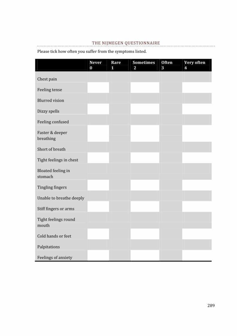
289
THE NIJMEGEN QUESTIONNAIRE
Please tick how often you suffer from the symptoms listed.
Never 0
Rare 1
Sometimes 2
Often 3
Very often 4
Chest pain
Feeling tense
Blurred vision
Dizzy spells
Feeling confused
Faster & deeper
breathing
Short of breath
Tight feelings in chest
Bloated feeling in
stomach
Tingling fingers
Unable to breathe deeply
Stiff fingers or arms
Tight feelings round
mouth
Cold hands or feet
Palpitations
Feelings of anxiety

290
PLAIN LANGUAGE STATEMENTS
2.1 Measures of Dysfunctional Breathing
2.2 MARM Study 1- Inter-examiner Reliability and Comparison with RIP
2.3 MARM Study 2- Comparison of MARM and Hi Lo in Osteopaths and Students
2.4 Breathing Therapy- Effects on MARM, NQ and Dyspnea

291
MEASURES OF DYSFUNCTIONAL BREATHING
PATIENT EXPLANATORY STATEMENT
Research Project-ASSESSMENT OF MEASUREMENT TOOLS USED TO IDENTIFY DYSFUNCTIONAL
BREATHING
Investigators: Professor Marc Cohen, Dr. Ray Myers, Rosalba Courtney.
Dear Volunteer,
Thank you for considering volunteering for this project. This purpose of this statement is to ensure that you have a clear understanding of this project and what it is trying to achieve. Also this statement will tell you what is required of you and what to expect from us.
This study is being undertaken by Rosalba Courtney, through the School of Life Sciences at RMIT University, Melbourne, Australia. The primary supervisor of this project is Professor Marc Cohen ,the second supervisor is Dr. Ray Myers.
THE PROJECT EXPLAINED.
This project hopes to contribute to the understanding of some of the measurement tools used to determine dysfunctional breathing so that clinicians can more confidently decide when dysfunctional breathing may be a contributing factor to a person’s health problems.
Many research studies over the last 50 years have indicated that between 5-10% of the healthy population has “dysfunctional breathing”. The figure is much higher in people suffering from anxiety (up to 80%) or conditions such as asthma (up to 30%). Some people consider that having normal breathing is very important in the maintenance of optimal health; however dysfunctional breathing is often overlooked by clinicians.
Definition of Dysfunctional Breathing:
Dysfunctional breathing is breathing that contributes to the aggravation of physical or psychological symptoms.
Dysfunctional breathing includes hyperventilation, a condition where people breathe more than their body’s need, this results in acute or chronic deficiency of carbon dioxide in the blood. Dysfunctional breathing also includes other types of breathing pattern disorders which may not result in carbon dioxide depletion.
In this project we will apply some measurements that have been used for identifying dysfunctional breathing and see how well they correlate with carbon dioxide levels and how the various measures correlate with each other.
Measures of Dysfunctional Breathing we are hoping to further understand:
Carbon dioxide levels in exhaled air.
This tells us about the carbon dioxide levels in the blood. When we are overbreathing or hyperventilating, carbon dioxide levels will be lower than normal. Exactly what levels definitely indicate hyperventilation are controversial and vary according to individual factors.

292
Breath HoldingTime.
It has been shown by many investigators that dysfunctional breathing and hyperventilation are associated with shortened breath holding ability.
One of the important things this project hopes to clarify is the claim made by Russian physician Dr. Buteyko that a person’s breath holding time correlates precisely with their carbon dioxide levels.
The Nijmegen Questionnaire
This questionnaire has been shown to have a strong ability to predict people whose symptoms are related to hyperventilation syndrome or dysfunctional breathing. This research project hopes to see how test response on this questionnaire correlate with carbon dioxide levels. We also wish to see if there is a correlation between test scores on this questionnaire and breath holding time.
Variability of the Heart Rate
The heart rate usually responds to changes in breathing and to whether a person is inhaling or exhaling. In people with dysfunctional breathing the heart rate may not be as responsive to changes in breathing. Information about heart rate variability may be useful as part of the overall picture of dysfunctional breathing. The investigators will see how this correlates with the other measures of dysfunctional breathing.
The pattern of muscle use in breathing.
Unusual use of the muscles of breathing is one aspect of dysfunctional breathing.
The electrical resistance of points on your fingers and toes
This may indicate the amount of activity in your nervous system, a factor that may be reflected in your breathing.
OVERVIEW OF WHAT YOU WILL BE ASKED TO DO IF YOU PARTICIPATE IN THIS PROJECT
You will only be required to attend for one session of assessment. The time anticipated for the tests is 60 to 90 minutes.
After giving us some basic health details
You will undertake a lung function test with a spirometer. This involves you blowing hard into a tube attached to a computer based spirometry program. The purpose of this is to see what your lung capacity is and to determine if there may be some undetected lung problem.
As you fill out other questionnaires ** we will measure.
The amount of carbon dioxide in the air you exhale during normal breathing.-This is done with a device called a capnometer. A small plastic tube will be placed in the entrance to each nostril and then hooked over your ears.
The movement of your respiratory muscles.-This is measured with 2 tension sensing band that you will wear on your upper and lower chest.
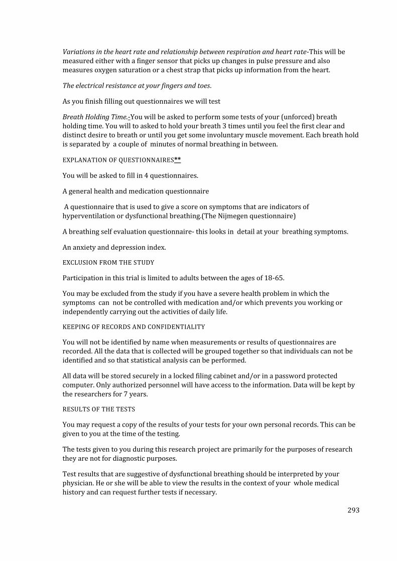
293
Variations in the heart rate and relationship between respiration and heart rate-This will be measured either with a finger sensor that picks up changes in pulse pressure and also measures oxygen saturation or a chest strap that picks up information from the heart.
The electrical resistance at your fingers and toes.
As you finish filling out questionnaires we will test
Breath Holding Time.-You will be asked to perform some tests of your (unforced) breath holding time. You will to asked to hold your breath 3 times until you feel the first clear and distinct desire to breath or until you get some involuntary muscle movement. Each breath hold is separated by a couple of minutes of normal breathing in between.
EXPLANATION OF QUESTIONNAIRES**
You will be asked to fill in 4 questionnaires.
A general health and medication questionnaire
A questionnaire that is used to give a score on symptoms that are indicators of hyperventilation or dysfunctional breathing.(The Nijmegen questionnaire)
A breathing self evaluation questionnaire- this looks in detail at your breathing symptoms.
An anxiety and depression index.
EXCLUSION FROM THE STUDY
Participation in this trial is limited to adults between the ages of 18-65.
You may be excluded from the study if you have a severe health problem in which the symptoms can not be controlled with medication and/or which prevents you working or independently carrying out the activities of daily life.
KEEPING OF RECORDS AND CONFIDENTIALITY
You will not be identified by name when measurements or results of questionnaires are recorded. All the data that is collected will be grouped together so that individuals can not be identified and so that statistical analysis can be performed.
All data will be stored securely in a locked filing cabinet and/or in a password protected computer. Only authorized personnel will have access to the information. Data will be kept by the researchers for 7 years.
RESULTS OF THE TESTS
You may request a copy of the results of your tests for your own personal records. This can be given to you at the time of the testing.
The tests given to you during this research project are primarily for the purposes of research they are not for diagnostic purposes.
Test results that are suggestive of dysfunctional breathing should be interpreted by your physician. He or she will be able to view the results in the context of your whole medical history and can request further tests if necessary.

294
RESULTS OF THE RESEARCH
The results will be used to write reports of the research, this may be published in medical or other journal or magazines. The results may also be presented at conferences. You may access the results of the research by contacting the researchers. You will not be personally identified in any dissemination of the results of the research.
WITHDRAWAL FROM PROJECT
You can withdraw at any time, if you so choose and are under no obligation to complete any of the tests or questionnaires.
QUESTIONS ABOUT THE RESEARCH
If you have any questions at any stage about this research project or your involvement you can contact Rosalba Courtney on 02-99183460 or 0414918819
ADVERSE EVENTS OR COMPLAINTS
The measurements are safe and non invasive, however in the unlikely event that adverse events do occur you will be referred to your local doctor. I am also required to notify the RM IT University Human Research Ethics Committee.
Thank you for taking the time to read this form.
Professor Marc Cohen Dr. Ray Myers Rosalba Courtney
(Supervisor) (Supervisor) (Principal Investigator)
Any complaints about your participation in this study may be directed to The Secretary of Human Research Ethics Committee, RMIT, 124 La Trobe St. Melbourne Vic. 3000. Telephone 99251745
295
MARM STUDY 1- INTER-EXAMINER RELIABILITY AND COMPARISON WITH LIFESHIRT
PATIENT EXPLANATORY STATEMENT
Research Project- COMPARISON OF A MANUAL ASSESSMENT OF RESPIRATORY MOTION (MARM)
WITH AMBULATORY RESPIRATORY PLETHYSMOGRAPH MEASUREMENTS USING THE “LIFE
SHIRT”
Investigators- Professor Marc Cohen, Dr. Ray Myers, Rosalba Courtney
Dear Participant
You are invited to participate in a research project being conducted by RMIT University. This information sheet describes the project in straight forward language or “plain English”. Please read this sheet carefully and be confident that you understand its contents before deciding whether to participate. If you have any questions about the project, please ask any of the investigators.
WHO IS INVOLVED IN THE RESEARCH PROJECT? WHY IS IT BEING CONDUCTED?
This research is being undertaken by Rosalba Courtney through the division of Osteopathy, School of Health Sciences, RMIT University. The supervisors of this project are Professor Marc Cohen and Dr. Ray Myers.
WHY YOU HAVE BEEN APPROACHED?
This research requires the participation of individuals who have an understanding of how to modify their breathing pattern. We have approached yoga teachers, singing teachers and breathing therapy teachers and others who are used to working with their breath.
WHAT IS THE PROJECT ABOUT? WHAT ARE THE QUESTIONS BEING ADDRESSED?
Alteration in the normal balance of thoracic to abdominal motion during breathing is one aspect of dysfunctional breathing. We are testing the reliability of a hands on technique used to measure the balance between thoracic and abdominal motion during breathing. This technique is called the Manual Assessment of Respiratory Motion or MARM. We are trying to see how well this technique is able to pick up subtle changes in breathing. We will test the accuracy of this technique by comparing results from two examiners using the MARRM and with measurements made by a device, the Vivometrics lifeshirt which also measures and records changes in breathing pattern. We are planning to test 10 people.
IF I AGREE TO PARTICIPATE WHAT WILL I BE REQUIRED TO DO?
You will be asked to wear the lifeshirt while the breathing tests are being done. This may be for a period of up to 1 hour. During this time 2 separate examiners will measure the movements of your chest and abdomen during breathing, by placing their hands on your back over the middle and lower rib cage. You will be asked to breathe normally and then to make a series of simple alterations to your posture and breathing while the two examiners test your breathing with the MARM and record their measures.
WHAT ARE THE RISKS OR DISADVANTAGES ASSOCIATED WITH PARTICIPATION?

296
There are no perceived risks associated with participation in this project. You will only be asked to make ordinary adjustments in your breathing and posture similar to what you would make in your day to day life.The lifshirt does not in any way alter the functions of your body. It measures changes in heart rate and breathing by electrical sensors placed on the outside of your body in the manner of a heart rate monitor.
WHAT ARE THE BENEFITS ASSOCIATED WITH PARTICIPATION?
Your participation will contribute to the furthering of knowledge of dysfunctional breathing patterns and their assessment. You may also personally gain some knowledge about breathing patterns and measurement of dysfunctional breathing from participating in this research.
WHAT WILL HAPPEN TO THE INFORMATION I PROVIDE?
All information gathered from your participation will form part of pooled data and you will not be identified personally. Any information given by you will be kept confidential and will be disclosed only if 1) It is to protect you or others from harm 2) a court order is produced or 3) you provide the researchers with written permission.
Results from MARM and Lifeshirt measurements will be combined with other participants results then analysed and findings written up as part of a research thesis for RMIT University. Results may also be presented at conferences and written up for publication in research papers.
Data will be kept in a password protected computer and in a locked filing cabinet. On completion of the study the data will be kept at RMIT for a period of 5 years before being destroyed.
WHAT ARE MY RIGHTS AS A PARTICIPANT?
You have the rights to withdraw your participation at any time without prejudice. You can also ask to have your unprocessed data withdrawn and destroyed provided that it can be reliably identified, and provided that doing so does not increase any risk for you. You also have the right to have any questions answered at any time.
WHOM SHOULD I CONTACT IF I HAVE ANY QUESTIONS?
If you have any questions please contact Rosalba Courtney 02-99183460, [email protected] or Professor Marc Cohen 03-99257440, [email protected]
Yours Sincerely,
Rosalba Courtney Professor Marc Cohen Dr. Ray Myers Any complaints about your participation in this project may be directed to the Executive Officer, RMIT Human Research Ethics Committee, Research and Innovation, RMIT, GPO Box 2476V, Melbourne, 3001. The telephone number is (03) 9925 2251. Details of the complaints procedure are available from the above address.

297
MARM STUDY 2- COMPARISON OF MARM AND HI LO IN OSTEOPATHS AND STUDENTS
INVITATION TO PARTICIPATE IN A RESEARCH PROJECT
PROJECT INFORMATION STATEMENT
PROJECT TITLE- ASSESSMENT OF BREATHING PATTERN USING 2 PALPATION TECHNIQUES: THE
MANUAL ASSESSMENT OF RESPIRATORY MOTION (MARM) AND THE HI LO BREATHING
ASSESSMENT.
INVESTIGATORS
Rosalba Courtney (PhD candidate, Science, School of Health Sciences, Division of Osteopathy, RMIT
University) [email protected], w) 02-99183460, Professor Marc Cohen (Primary Supervisor,
School Health Sciences, RMIT University)[email protected], 03-99257440
Dear Participant,
You are invited to participate in a research project being conducted by RMIT University. This information sheet describes the project in straightforward language or “plain English”. Please read this sheet carefully and be confident that you understand its contents before deciding whether to participate. If you have any questions about the project, please ask any of the investigators.
WHO IS INVOLVED IN THE RESEARCH PROJECT? WHY IS IT BEING CONDUCTED?
Rosalba Courtney through the School of Health Sciences, at RMIT University, is undertaking this research as part of a PhD project. Professor Marc Cohen is supervising this project.
WHY YOU HAVE BEEN APPROACHED?
This research requires the participation of individuals who are practitioners or student of manual therapy and who 1. Have an interest in learning these techniques 2. Are willing to assist in research.
WHAT IS THE PROJECT ABOUT? WHAT ARE THE QUESTIONS BEING ADDRESSED?
In this study we will be teaching experienced manual therapists, such as Osteopaths, Chiropractors and Physiotherapists and manual therapy students some techniques for evaluating breathing pattern. We then plan to test how well the techniques work in determining breathing pattern in the hands of experienced as compared to less experienced practitioners. The person being examined (the breather) will be told to follow certain breathing instructions, which will be unknown to the examiner. The examiner will have to determine what breathing instruction the breather is following. Two breathing assessment techniques will be used to assess breathing pattern. These techniques are the Manual Assessment of Respiratory Motion (MARM) and the Hi Lo breathing assessment. We have 2 aims: (1) to compare examiners accuracy while using the MARM and Hi Lo to test the usefulness of these techniques (2) To determine if there are differences between relatively inexperienced manual therapists and experienced practitioners when predicting breathing pattern using the MARM and the Hi Lo breathing assessment.
298
IF I AGREE TO PARTICIPATE WHAT WILL I BE REQUIRED TO DO?
You will be asked to participate in training session during which you will be taught the theoretical and practical aspects of these techniques. At the end of the training session you will work with a partner and takes turns in assessing each others breathing pattern using the 2 techniques. You will be participating in this research as a “breather” and as a breathing examiner taking turns with a partner in these two activities.
WHAT ARE THE RISKS OR DISADVANTAGES ASSOCIATED WITH PARTICIPATION?
There are no perceived risks associated with participation in this project. You will only be asked to make ordinary adjustments in your breathing similar to what you would make in your day-to-day life. Your personal accuracy with using these techniques is not being tested: we are interested in the overall performance of the group and the usefulness of the technique.
WHAT ARE THE BENEFITS ASSOCIATED WITH PARTICIPATION?
You will increase your understanding of dysfunctional breathing and of the manual techniques used to assess it. Your participation will help validate a potentially useful clinical test for dysfunctional breathing.
WHAT WILL HAPPEN TO THE INFORMATION I PROVIDE?
All information gathered from your participation will form part of pooled data and you will not be identified personally. Any information given by you will be kept confidential and will be disclosed only if 1) It is to protect you or others from harm 2) a court order is produced or 3) you provide the researchers with written permission.
Your results from MARM and Hi Lo measurements will be combined with other participants results then analysed and findings written up as part of a research thesis for RMIT University. Results may also be presented at conferences and written up for publication in research papers.
Data will be kept in a password-protected computer and in a locked filing cabinet. On completion of the study the data will be kept at RMIT for a period of 5 years before being destroyed.
WHAT ARE MY RIGHTS AS A PARTICIPANT?
You have the rights to withdraw your participation at any time without prejudice. You can also ask to have your unprocessed data withdrawn and destroyed provided that it can be reliably identified, and provided that doing so does not increase any risk for you. You also have the right to have any questions answered at any time.
WHOM SHOULD I CONTACT IF I HAVE ANY QUESTIONS?
If you have any questions please contact Rosalba Courtney on work phone number 02-99183460, [email protected] or Professor Marc Cohen 03-99257440, [email protected]
Yours Sincerely,
Rosalba Courtney Professor Marc Cohen

299
Any complaints about your participation in this project may be directed to the Executive Officer, RMIT Human Research Ethics Committee, Research and Innovation, RMIT, GPO Box 2476V, Melbourne, 3001.
The telephone number is (03) 9925 2251. Details of the complaints procedure are available from the above address

300
BREATHING THERAPY- EFFECTS ON MARM NQ AND DYSPNEA
Centre for Breathing Therapy F van Blankenheymstraat 10 3817 AG Amersfoort The Netherlands www.methodevandixhoorn.com [email protected] +31 33 4635292
Amersfoort 7 – 5 - 2010 Consent form regarding participating in research project ‘Resultaten Project Adem en Ontspanningstherapie’ All patients who are admitted for breathing and relaxation therapy have been explained 1. that a number of their outcome data will be submitted to and included in an online database for ‘outcome assessment of breathing therapy’, if they agree to that. 2. that the outcome data include sex and age, the main complaint, effect on treatment on main complaint, questionnaire scores and MARM assessment before and after treatment, and number of treatment sessions. The data include a personal identification number, which corresponds to the patient information of the therapist that is doing the treatment, and which is the only way the data may be checked for errors by the therapist. 3. that the data will be strictly used to collect evidence of treatment outcome on a group basis and no attempt will be made to trace the individual subject, unless the subject consents to that. The data will be used to publish outcome results. 4. that the subject is free to withhold consent, now and also at a later stage. The data will be submitted only at the end of treatment. Only when the subject understood and consented, treatment data were submitted. Otherwise the file received a mark ‘not for research project’. E L M Anthonissen J van Dixhoorn