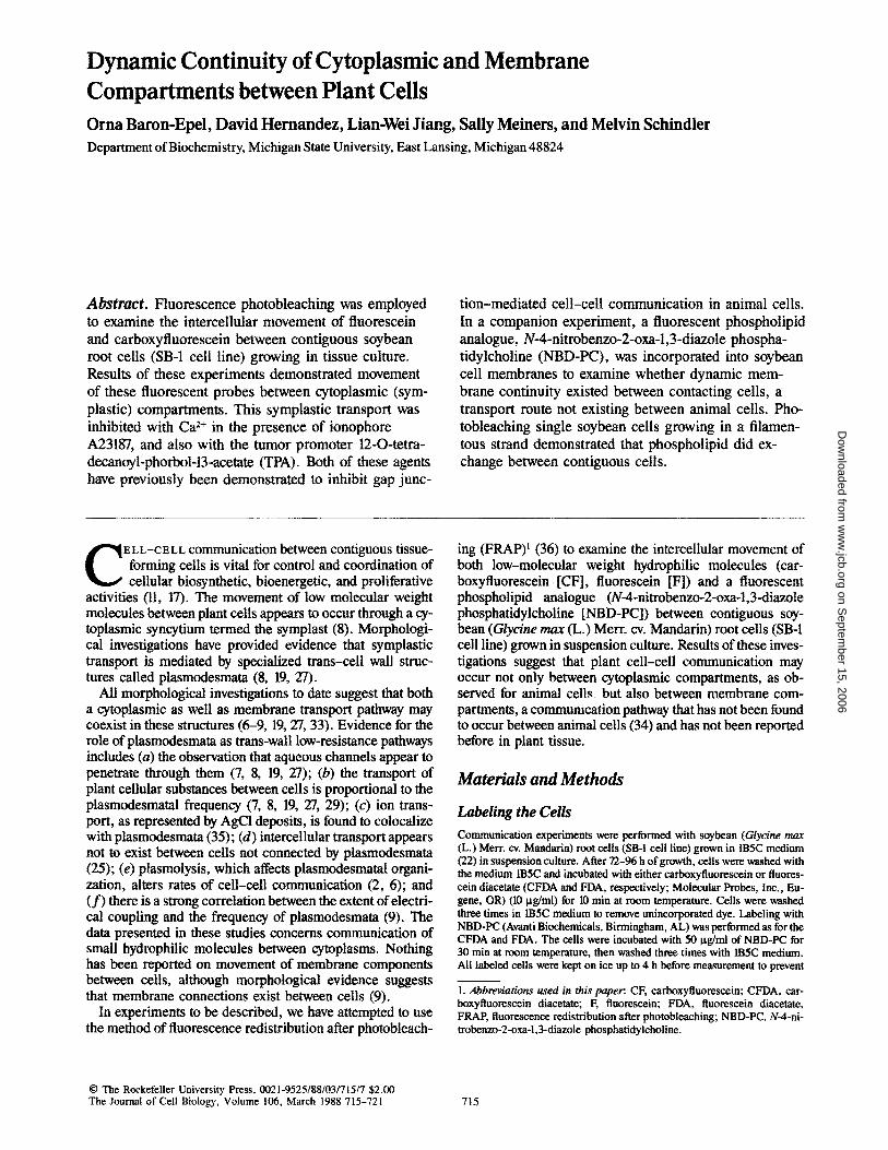
Dynamic Continuity of Cytoplasmic and Membrane ...
7
Dynamic Continuity of Cytoplasmic and Membrane Compartments between Plant Cells Orna Baron-Epel, David Hernandez, Lian-Wei Jiang, Sally Meiners, and Melvin Schindler Department of Biochemistry, Michigan State University, East Lansing, Michigan 48824 Abstract. Fluorescence photobleaching was employed to examine the intercellular movement of fluorescein and carboxyfluorescein between contiguous soybean root cells (SB-1 cell line) growing in tissue culture. Results of these experiments demonstrated movement of these fluorescent probes between cytoplasmic (sym- plastic) compartments. This symplastic transport was inhibited with Ca 2+ in the presence of ionophore A23187, and also with the tumor promoter 12-O-tetra- decanoyl-phorbol-13-acetate (TPA). Both of these agents have previously been demonstrated to inhibit gap junc- tion-mediated cell-cell communication in animal cells. In a companion experiment, a fluorescent phospholipid analogue, N-4-nitrobenzo-2-oxa-l,3-diazole phospha- tidylcholine (NBD-PC), was incorporated into soybean cell membranes to examine whether dynamic mem- brane continuity existed between contacting cells, a transport route not existing between animal cells. Pho- tobleaching single soybean cells growing in a filamen- tous strand demonstrated that phospholipid did ex- change between contiguous cells. C E L L-C E L L communication between contiguous tissue- forming cells is vital for control and coordination of cellular biosynthetic, bioenergetic, and proliferative activities (11, 17). The movement of low molecular weight molecules between plant cells appears to occur through a cy- toplasmic syncytium termed the symplast (8). Morphologi- cal investigations have provided evidence that symplastic transport is mediated by specialized trans-cell wall struc- tures called plasmodesmata (8, 19, 27). All morphological investigations to date suggest that both a cytoplasmic as well as membrane transport pathway may coexist in these structures (6--9, 19, 27, 33). Evidence for the role of plasmodesmata as trans-wall low-resistance pathways includes (a) the observation that aqueous channels appear to penetrate through them (7, 8, 19, 27); (b) the transport of plant cellular substances between cells is proportional to the plasmodesmatal frequency (7, 8, 19, 27, 29); (c) ion trans- port, as represented by AgCI deposits, is found to colocalize with plasmodesmata (35); (d) intercellular transport appears not to exist between cells not connected by plasmodesmata (25); (e) plasmolysis, which affects plasmodesmatal organi- zation, alters rates of cell-cell communication (2, 6); and (f) there is a strong correlation between the extent of electri- cal coupling and the frequency of plasmodesmata (9). The data presented in these studies concerns communication of small hydrophilic molecules between cytoplasms. Nothing has been reported on movement of membrane components between cells, although morphological evidence suggests that membrane connections exist between cells (9). In experiments to be described, we have attempted to use the method of fluorescence redistribution after photobleach- ing (FRAP) ~ (36) to examine the intercellular movement of both low-molecular weight hydrophilic molecules (car- boxyfluorescein [CF], fluorescein [F]) and a fluorescent phospholipid analogue (N-4-nitrobenzo-2-oxa-l,3-diazole phosphatidylcholine [NBD-PC]) between contiguous soy- bean (Glycine max (L.) Merr. cv. Mandarin) root cells (SB-1 cell line) grown in suspension culture. Results of these inves- tigations suggest that plant cell-cell communication may occur not only between cytoplasmic compartments, as ob- served for animal cell~, but also between membrane com- partments, a communication pathway that has not been found to occur between animal cells (34) and has not been reported before in plant tissue. Materials and Methods Labeling the Cells Communication experiments were performed with soybean (Glycine max (L.) Merr. ov. Mandarin) root cells (SB-I cell line) grown in 1B5C medium (22) in suspension cdture. After 72-96 h of growth, cells were washed with the medium tB5C and incubated with either carboxyfluorescein or fluores- cein diacetate (CFDA and FDA, respectively; Molecular Probes, Inc., Eu- gene, OR) (10 lag/ml) for 10 min at room temperature. Cells were washed three times in 1B5C medium to remove unincorporated dye. Labeling with NBD-PC (AvantiBioehemicals, Birmingham, AL) was performed as for the CFDA and FDA. The cells were incubated with 50 Ixg/ml of NBD-PC for 30 min at room temperature, then washed three times with 1B5C medium. All labeled cells were kept on ice up to 4 h before measurement to prevent 1. Abbreviations used in this paper: CF, carboxyfluorescein; CFDA, car- boxyfluorescein diacetate; F, fluorescein; FDA, fluorescein diaeetate, FRAP, fluorescence redistribution after photobleaching; NBD-PC, N-4-ni- trobenzo-2-oxa-1,3-diazole phosphatidylcholine. 9 The Rockefeller University Press, 0021-9525/88/03/715/7 $2.00 The Journal of Cell Biology, Volume 106, March 1988 715-721 715 on September 15, 2006 www.jcb.org Downloaded from
Transcript of Dynamic Continuity of Cytoplasmic and Membrane ...
Dynamic Continuity of Cytoplasmic and Membrane Compartments between Plant Cells Orna Baron-Epel , David Hernandez, Lian-Wei Jiang, Sally Meiners, and Melvin Schindler Department of Biochemistry, Michigan State University, East Lansing, Michigan 48824
Abstract. Fluorescence photobleaching was employed to examine the intercellular movement of fluorescein and carboxyfluorescein between contiguous soybean root cells (SB-1 cell line) growing in tissue culture. Results of these experiments demonstrated movement of these fluorescent probes between cytoplasmic (sym- plastic) compartments. This symplastic transport was inhibited with Ca 2+ in the presence of ionophore A23187, and also with the tumor promoter 12-O-tetra- decanoyl-phorbol-13-acetate (TPA). Both of these agents have previously been demonstrated to inhibit gap junc-
tion-mediated cell-cell communication in animal cells. In a companion experiment, a fluorescent phospholipid analogue, N-4-nitrobenzo-2-oxa-l,3-diazole phospha- tidylcholine (NBD-PC), was incorporated into soybean cell membranes to examine whether dynamic mem- brane continuity existed between contacting cells, a transport route not existing between animal cells. Pho- tobleaching single soybean cells growing in a filamen- tous strand demonstrated that phospholipid did ex- change between contiguous cells.
C E L L-C E L L communication between contiguous tissue- forming cells is vital for control and coordination of cellular biosynthetic, bioenergetic, and proliferative
activities (11, 17). The movement of low molecular weight molecules between plant cells appears to occur through a cy- toplasmic syncytium termed the symplast (8). Morphologi- cal investigations have provided evidence that symplastic transport is mediated by specialized trans-cell wall struc- tures called plasmodesmata (8, 19, 27).
All morphological investigations to date suggest that both a cytoplasmic as well as membrane transport pathway may coexist in these structures (6--9, 19, 27, 33). Evidence for the role of plasmodesmata as trans-wall low-resistance pathways includes (a) the observation that aqueous channels appear to penetrate through them (7, 8, 19, 27); (b) the transport of plant cellular substances between cells is proportional to the plasmodesmatal frequency (7, 8, 19, 27, 29); (c) ion trans- port, as represented by AgCI deposits, is found to colocalize with plasmodesmata (35); (d) intercellular transport appears not to exist between cells not connected by plasmodesmata (25); (e) plasmolysis, which affects plasmodesmatal organi- zation, alters rates of cell-cell communication (2, 6); and (f ) there is a strong correlation between the extent of electri- cal coupling and the frequency of plasmodesmata (9). The data presented in these studies concerns communication of small hydrophilic molecules between cytoplasms. Nothing has been reported on movement of membrane components between cells, although morphological evidence suggests that membrane connections exist between cells (9).
In experiments to be described, we have attempted to use the method of fluorescence redistribution after photobleach-
ing (FRAP) ~ (36) to examine the intercellular movement of both low-molecular weight hydrophilic molecules (car- boxyfluorescein [CF], fluorescein [F]) and a fluorescent phospholipid analogue (N-4-nitrobenzo-2-oxa-l,3-diazole phosphatidylcholine [NBD-PC]) between contiguous soy- bean (Glycine max (L.) Merr. cv. Mandarin) root cells (SB-1 cell line) grown in suspension culture. Results of these inves- tigations suggest that plant cell-cell communication may occur not only between cytoplasmic compartments, as ob- served for animal cell~, but also between membrane com- partments, a communication pathway that has not been found to occur between animal cells (34) and has not been reported before in plant tissue.
Materials and Methods
Labeling the Cells
Communication experiments were performed with soybean (Glycine max (L.) Merr. ov. Mandarin) root cells (SB-I cell line) grown in 1B5C medium (22) in suspension cdture. After 72-96 h of growth, cells were washed with the medium tB5C and incubated with either carboxyfluorescein or fluores- cein diacetate (CFDA and FDA, respectively; Molecular Probes, Inc., Eu- gene, OR) (10 lag/ml) for 10 min at room temperature. Cells were washed three times in 1B5C medium to remove unincorporated dye. Labeling with NBD-PC (Avanti Bioehemicals, Birmingham, AL) was performed as for the CFDA and FDA. The cells were incubated with 50 Ixg/ml of NBD-PC for 30 min at room temperature, then washed three times with 1B5C medium. All labeled cells were kept on ice up to 4 h before measurement to prevent
1. Abbreviations used in this paper: CF, carboxyfluorescein; CFDA, car- boxyfluorescein diacetate; F, fluorescein; FDA, fluorescein diaeetate, FRAP, fluorescence redistribution after photobleaching; NBD-PC, N-4-ni- trobenzo-2 -oxa-1,3-diazole phosphatidylcholine.
�9 The Rockefeller University Press, 0021-9525/88/03/715/7 $2.00 The Journal of Cell Biology, Volume 106, March 1988 715-721 715
on Septem
ber 15, 2006 w
ww
.jcb.orgD
ownloaded from

dye leakage from the cells into the medium. The dye concentration and labeling conditions did not affect cell viability.
Measurements on the ACAS 470 Workstation
The ACAS 470 Workstation was used to photobleach single cells and cells connected in a filament (31, 36). The labeled cells are placed on a slide which is mounted on the ACAS 470 stage. The automated stage moves the sample in 1.5-~tm steps in a two-dimensional grid past a microscope objec- tive that focuses the excitation beam (488 mM) from an argon ion laser to a 1-30-ktm diameter beam on the sample. A photomultiplier tube captures the emission intensities at each addressed excitation point. The emitted in- tensities are then color coded and displayed on a cathode ray tube as false color images of cellular fluorescence. Multiple photobleaching on the same cell did not alter the transport results.
Measurements on a F R A P Instrument
Cells were scanned and photobleached on a FRAP instrument as described (31, 36). A defocused laser beam (width approximately equal to the cell di- ameter) was employed, resulting in a linear fluorescence intensity profile across the cell; 20 scans were monitored at 15-s intervals for a total monitor- ing time of 5 min.
Results
Cell-cell communication between the cytoplasmic compart- ments of plant cells was determined by using fluorescence redistribution after photobleaching (FRAP) in conjunction with the fluorescent low-molecular weight compounds, CF and F (both added to the cells as the diacetylated derivative as described in Materials and Methods). This technique, first employed for the measurement of intercellular communica-
tion between mammalian cells (31, 36), is performed in the following manner. Briefly, a laser beam whose width is ap- proximately equal to the plant cell diameter photobleaches the fluorescent probes in a cell contacting other similarly la- beled cells. After photobleaching, a redistribution of dye molecules may occur between bleached and unbleached cells. Monitoring this process as a function of time yields a rate constant for dye movement. Photobleaching a single noncontacting cell provides a control that recovery is a func- tion of transport between contacting cells and not the physi- cal consequences of dye reactivation after photobleaching or uptake of dye from the media.
Fig. 1 demonstrates the cellular distribution of CF and E The acetates have been removed from the acetylated precur- sors by intracellular esterases; this traps the compounds in the cell over the time course of our measurements (1). A clear difference for CF and F localization is observed in soybean (SB-1) filaments. When cells are labeled and examined in medium at pH 5.5, fluorescein is found to localize predomi- nantly in the cytoplasm and the nucleus (Fig. 1, A and B), whereas CF at pH 5.5 appears to preferentially localize in the vacuole (Fig. 1, C and D). This corroborates a report by McCredy et al. (19a) that these two dyes differ in their intra- cellular localization. Both of these dyes were used to mea- sure intercellular communication (Figs. 2 and 3). Using the ACAS 470 Fluorescence Workstation (see Materials and Methods), a computer-generated false color image of fluo- rescence distribution is shown in Fig. 2 a for CF and in Fig. 3 a for F. Photobleaching results in a drop in the fluorescence
Figure 1. A phase contrast (A and C) and fluorescence image (B and D) of soybean tissue culture cells labeled with F (B) and CF (D). Cells were labeled with a dye concentrat ion of 10 Ixg/ml for 10 min at room temperature (Materials and Methods) , washed, and viewed with phase optics and epifluorescent illumination.
The Journal of Cell Biology, Volume 106, 1988 716
on Septem
ber 15, 2006 w
ww
.jcb.orgD
ownloaded from

Figure 2. Recovery of fluores- cence in photobleached soy- bean tissue culture cells la- beled with CE Labeling of the cells was performed as de- scribed in Materials and Meth- ods. The ACAS 470 Worksta- tion was used to photobleach the cells and record fluores- cence distribution as false color images visualized on a cathode ray tube. Before pho- tobleaching, there is a strong fluorescence intensity (a) that is reduced after photobleach- ing the cell located in the poly- gon (b). (c) Fluorescence in- tensity in the filament 10 min after photobleaching. (d) Phase view of the cells under analy- sis. Box denotes the photo- bleached cell. The color code presented relates color to an arbitrary numerical scale of fluorescence intensity.
intensity in the bleached cell and some bleaching in contact- ing cells (Fig. 2, a and b, and Fig. 3, a and b). Recovery is demonstrated to occur for both CF and F (Figs. 2 c and 3 c), although the extent of recovery for CF is less than E This correlates with the observation (see Fig. 1) that a high concentration of CF resides in the vacuole and may not be readily available for exchange between cells. Single isolated cells containing CF or F did not recover after photobleaching (data not shown). Fig. 2 d is a phase view of the SB-1 cells presented as pseudo-color images in Fig. 2, a - c .
Subsequent analytical transport experiments were per- formed on a FRAP instrument as described (14, 36). Cells labeled with the cytoplasmic localized dye F were photo- bleached with a defocused laser beam, the width of which was approximately equal to the cell diameter and the laser beam was run across the cell. The redistribution of fluores- cence data was analyzed according to the equation: F ( - ) - F ( t ) /F( - ) - F(o) = e -rv + e-~:2 ', where F ( - ) , F(o), and F(t) are fluorescence signals before photobleaching, after, and at time t (26). A sample recovery curve for dye transport between cells is represented in Fig. 4 A. Note that the recov- ery is represented by a fast component (K0 and a slower component (K2). Fig. 4 B demonstrates that a photo- bleached single cell, which should have no recovery, has
a fast component (K0 but no slower component (K2). These results are interpreted as demonstrating that component (K2) represents the intercellular flux, whereas component (K0 represents the rearrangement of bleached and unbleached molecules within the photobleached cell volume. The calcu-
Figure 3. Recovery of fluorescence in photobleached soybean tissue culture cells labeled with fluorescein. The experiment was per- formed as in Fig. 2. (a) Prebleach distribution. (b and c) The bleached cell immediately and 10 min after photobleaching, respec- tively.
Baron-Epel et al. Cytoplasmic and Membrane Continuity between Plant Cells 717
on Septem
ber 15, 2006 w
ww
.jcb.orgD
ownloaded from

lated flux rate derived from component (K2) for intercellu- lar dye transport is 1.46 + 0.43 • 10 -~ s -: (50 cells). o,ooo
-o.ioo
-o.zoo
Effect o f Ca 2+ and TPA on Plant - o ~
Cell-Cell Communication 5 *~~176
Previous measurements of plant intercellular communica- ~.--oi,oo tion have established that Ca z§ may prevent cell-cell trans- -~'** port (5) as initially observed in mammalian cell communica- -o,.o
-o , loo
tion studies (17). SB-1 cells incubated with 5 mM Ca 2+ and .~.~
ionophore A23187 (100 Ixg/ml) demonstrated inhibition of in- tercellular transport for F (Fig. 4 C). Inhibition was also ob- served at 1 mM Ca 2+ with ionophore. No inhibition by Ca 2+ .oooo
was observed without the presence of ionophore (data not ..... shown). Clearly, the Ca 2+ effect on transport is more com- - . . . . .
plicated, as can be discerned from the multiphasic response, . ~ ' ~ but nonetheless significant inhibition is observed (compare . ~ - ~ Fig. 4, A and C, for slope and extent of recovery). The com- ~ -~,oo plex response may represent an effect on a number of poten- -o.,o, tial Ca 2+ processes, such as movement of dye to the plus . . . . . modesmata by cytoplasmic streaming. Previous work on ..... animal cell-cell communication has demonstrated that TPA, a tumor promoter derived from croton oil (12), can inhibit cell-cell communication at ,o0.7 ng/ml (36 and references . . . . therein). When SB-1 cells were incubated with 0.4 ng/ml ...... TPA for 18 h, subsequent transport measurements demon- ~ o ~ strated complete inhibition of intercellular dye transport .~ ...... (Fig. 4 D). Partial inhibition was observed for 0.2 ng/ml, "7"~z ..... whereas no inhibitory effect was observed at 0.05 ng/ml (data ~ -~ctoo
not shown). Using 4-phorbol-12,13-didecanoate, a non . . . . . tumorigenic phorbol ester, we observed no inhibition (data ..... not shown). These results are identical to those reported for ...... the effect of tumorigenic and nontumorigenic phorbol esters on animal cell-cell communication (36 and references therein).
Intercellular Membrane Transport
Morphological investigations of plasmodesmata have pro- vided evidence of membrane continuity between plant cells across the cell wall (8, 9, 27). Both plasma membrane and endoplasmic reticulum have been suggested to traverse the cell wall at sites occupied by the plasmodesmata. In the case of endoplasmic reticulum, the electron micrographs suggest that endomembranes may thread through a central plasmo- desmatal channel. To explore this possibility, we labeled soy- bean cells with fluorescent phospholipid analogue NBD-PC, which has been demonstrated to partition into the plasma membrane and endomembranes of animal and plant cells (15, 21). Fig, 5 a shows an ACAS 470 false color image of the cel- lular distribution of NBD-PC fluorescence. Cells in the fila- ment were arbitrarily selected for photobleaching (Fig. 5 b), and fluorescence redistribution was observed (Fig. 5 c). The limited extents of recovery observed are most likely related to the incorporation of exogenously added fluorescent phos- pholipid into noncontinuous membrane compartments, i.e., vacuole, intracellular vesicle, or Golgi membranes. Fig. 6, a-c, demonstrates no recovery for a single cell stained with NBD-PC and then photobleacbed. This again excludes the possibility for this probe that the lipid fluorescence recovery is due to dye reactivation after photobleaching or uptake of unincorporated NBD-PC from the media. A differentiation
-o.ooo
-o.~oo
-ozoo
~ - o zoo
i ~ -o4oo
-a~oo
-r -oooo
-o.7oo
-oeoo
-o~oo
�9
A
\
8
\ Ill Illll Ill
2
~ Z ~: Z 2 2 2 2 p. 2 ~ <: 2 ~ 2
Sr r~mb~ I
C
9 2 0 : 2 :2
i ~ io is zo
numi~r
I 0
Saw,~m~
Figure 4. Fluorescence recovery in soybean tissue culture cells con- raining fluorescein as a function of Ca 2§ and TPA. (A) Analysis of the fluorescence recovery of a control cell within a filament. (B) No recovery for a single isolated cell. (C) A cell in a filament in- cubated with 5 mM Ca 2+ and the calcium ionophore, A23187 (100 txg/ml), for 10 rain at 25~ and then put on ice before the measure- ment. (D) The recovery profile of a cell in a filament incubated for 18 h at 25~ with TPA (0.4 ng/ml), labeled with fluorescein, and photobleached. The fluorescence recovery was obtained using a FRAP instrument (14). The laser scans occurred at 15-s intervals, with a total time (scan plus delay) of monitoring for each scan of 5 rain.
between fluorescence recovery due to diffusion of membrane lipids along continuous membrane strands or through lipid vesicle-mediated transport, or potentially, some aspect of all of these means is not, as of yet, possible.
The Journal of Cell Biology, Volume 106, 1988 718
on Septem
ber 15, 2006 w
ww
.jcb.orgD
ownloaded from

Figure 5. Recovery of fluorescence in photobleached soybean tissue culture cells labeled with the membrane probe, NBD-PC. Using the ACAS 470 Workstation in the same manner described in Figs. 2 and 3, a false color image of the fluorescence distribution of NBD-PC in the cells was determined (a). (b and c) Images immediately after and 20 min after photobleaching, respectively.
Discussion
Symplastic communication between plant cells has been sug- gested to be the dominant form of plant intercellular commu- nication (8). Morphological evidence has suggested that trans-wall tubular structures, termed plasmodesmata, pro- vide the channel(s) for the intercellular movement of hydro- philic low-molecular weight (~<1,000 mol wt) metabolic, bio- synthetic, and signaling molecules (8, 27, 32). In addition, it has been suggested that membrane continuity between cells may occur at the plasmodesmatal sites affording a trans- port route for hydrophobic compounds and membrane com- ponents. In previous attempts to measure cell-cell communi- cation, membrane impermeant dyes were injected into plant
cells and the movement of dye from the site of microinjection to the surrounding cells was optically monitored (2, 18, 25, 33). These experiments demonstrated a transfer size limit of <1,000 mol wt for model compounds and variations in the inhibitory effect of Ca 2+ on dye transport as a consequence of differing osmotic conditions (5, 6). A potential difficulty with these microinjection experiments may be inferred from the work of Davies and Schuster (4), who found that the properties of wounded tissues are markedly different from those of intact tissue. A needle breaching the cell wall barrier may, thus, lead to altered cellular responses. In an attempt to circumvent this potential problem, we employed fluores- cence photobleaching (31, 36) to measure the transport of fluorescein and carboxyfluorescein. These dyes are intro- duced passively into the cell as fluorescein and carboxyfluo- rescein diacetate, which subsequently become deacetylated by intracellular esterases. F and CF were found to transfer between cells with a calculated permeability of ~0.9 x 10 -6 cm/s. Ca 2+ (in the presence of ionophore A23187) or the tumorigenic phorbol ester TPA were individually found to inhibit transport. The inhibition of transport mediated by these compounds in plants was previously reported to occur in mammalian cells (36). In an examination of TPA effects on transmembrane signaling in animal cells, evidence was provided that TPA may serve as a diacylglycerol (DAG) ana- logue stimulating protein kinase C (3, 16). In this context, phosphorylation, albeit cAMP-dependent, has been suggest- ed to be a control mechanism for gap junction permeability (28, 37). Because recent observations in plant tissue have demonstrated Ca :+ and phorbol ester activated protein ki- nase activity (10, 23, 30), it may be reasonable to suggest similar posttranslational modifications of plasmodesmatal proteins through activation of the polyphosphoinositide path- way and the initiation of DAG, IP3, Ca ~§ mediated protein kinase activity. Recent preliminary results by Tucker and Rosenbaum (32a) demonstrating that IP3 inhibits cell-cell communication in plants support this view. The comparable results for TPA inhibition reported for gap junction mediated communication (36) may provide evidence that gap junction and plasmodesmatal mediated transport share similar cellu- lar control features. These similarities reported for transport properties and putative control mechanisms for gap junction and plasmodesmatal intercellular communication may now also be examined in the light of recent evidence presented by Meiners and Schindler (20) that a gap junction homolo- gous protein exists in the cell wall/membrane fraction of soy-
Figure 6. NBD-PC fluorescence recovery in unattached single soybean cells in tissue culture after photobleaching. The experi- ment was performed as described in Fig. 5. No recovery is observed.
Baron-Epcl et al. Cytoplasmic and Membrane Continui~ between Plant Cells 719
on Septem
ber 15, 2006 w
ww
.jcb.orgD
ownloaded from

bean plant cells. This has recently been topologically refined to suggest an enrichment of this material in the cell wall (Meiners, S., and M. Schindler, unpublished results). As of yet, plant gap junction homologous protein has yet to be definitively localized to plasmodesmatal structures.
Dynamic Assessment of Plasmodesmatal Frequency
By employing results from our dynamic measurements in conjunction with literature values for wall diameter and plas- modesmatal size, we may attempt to mathematically derive the number of structures required to provide observed trans- port rates. In our simplified model for transport, we assume cube-shaped soybean cells having an average dimension of 12 I~m organized in a filament (two contacting sides per monitored cell). Permeability coefficient (p) is related to a transport rate constant K by: p = (V/A)K, where V is the vol- ume of the model cell and A is the cell-cell contact area avail- able for communication through plasmodesmata (24). In the case of carboxyfluorescein transport, we obtained p ~ 0.9 x 10 -6 cm/s (K = 0.0015 s-l). Assuming the functional chan- nel diameter is diam -- 2.0 nm (obtained by analysis of mo- lecular exclusion [32]), the critical dimension of carboxyflu- orescein (0.7 x 1.12 • 1.27 nm) for radial transit through the channel is diamcF ~ 1.27 nm (3a) and the plasmodes- mata length is AX -~ 255 nm (average cell wall thickness from Robards [27]), then the area density of channels can be calculated by n = p AX/DcAe (26), where p is permeability coefficient, AX is plasmodesmatal length, and Dc is cyto- plasmic diffusion coefficient of CE Dc is in the range of 0.1-0.2 Dw (13), where Dw is the CF diffusion coefficient in pure water (Dw = kT/3rCrlwdiamcF). 2 We thereby calculated Dc --- 5.2 x 10 -7 cm2/s. For Ao, the mean effective area of a channel (ndiam2/4), correction factor K~ must be intro- duced describing the fractional resistance to diffusion in a cylindrical channel relative to that in free cytoplasmic pool. We estimated K~ = 12 for a value of diamcF = 1.27 nm and diam ", 2 nm (diamcF/diam = 0.64) (24), resulting in the expression Ae = rtdiam2/4K~. Given the above values, the channel density n ~ 169/lamL
The plasmodesmatal frequency in the literature varies from tissue to tissue, but Sauter and Kloth (29) report that in ray parenchyma cells of poplar wood, plasmodesmatal fre- quency is ~39/Ixm 2 in cell rows in the center and ~26/lxm 2 in cell rows at margins of the tissue. Considering this dispar- ity between our calculated result and the observed plas- modesmatal frequency, it may be important to note that Terry and Robards (32) have proposed a controversial model of the plasmodesma consisting of nine channels around the central desmotubule. Using this number, our calculated channel density drops to ~19/~tm:, in far better agreement with the observations of Sauter and Kloth (29). Interestingly, in the context of results presented by Meiners and Schindler (20), if we assume each channel is approximately the size of a con- nexon, then six to nine channels could be accommodated around a central desmotubule.
Intercellular Membrane Communication
The cell-cell communication experiments performed with NBD-PC suggest that, unlike contiguous mammalian cells in
2. k is Boltzmann's constant, T is absolute temperature; rlw is water vis- cosity; diamcF is critical dimension of CF taken as Stokes diameter.
tissue, plant cells may also communicate through continuous membrane paths. Our data suggest that this, indeed, can oc- cur for phospholipids. Whether membrane proteins or other lipophilic molecules can commute between plant cells through connecting membranes, or whether membrane con- tinuity also implies membrane potential continuity is pres- ently under investigation.
We wish to thank Dr, Margaret Wade of Meridian Instruments, Okemos, MI, 48864, for her helpful discussions and technical assistance, and Merid- ian Instruments for their use of an ACAS 470 Fluorescence Workstation.
This work was supported by National Institutes of Health grant GM 30158 and a Nitrogen Availability Program Fellowship from Michigan State University to O. Baron-Epel.
Received for publication 26 June 1987, and in revised form 3t August 1987.
R~feFellces
1. Admon, A., B. Jacoby, and E. E. Goldscbmidt. 1980. Assessment of cyto- plasmic contaminations in isolated vacuole preparations. Plant Physiol. (Bethesda). 65:85-87.
2. Barclay, G. F., C. A. Peterson, and M. T. Tyree. 1982. Transport of fluorescein in trichomes of Lycopersicon esculentium. Can. J. Bot. 60: 397--402.
3. Berridge, M. J. 1984. Inositol triphosphate and diacylglycerol as second messengers. Biochem. Z 220:345-360.
3a. Brink, P. R., S. W. Jaslove, and D. Joseph. 1986. Permeability character- istics of the intercellular channel of the gap junction. Biophys. J. 49:341a. (Abstr.)
4. Davies, E., and A. Schuster. 1981. Intercellular communication in plants: evidence for a rapidly generated, bidirectionally transmitted wound sig- nal. Proc. Natl. Acad. Sci. USA. 78:2422-2426.
5. Erwee, M. G., and P. B. Goodwin. 1983. Characterization of the Egeria densa planch. Leaf symplast, inhibition of the cellular movement of fluorescent probes by group II ions. Planta (Berl.). 158:320-328.
6. Erwee, M. G., and P. B. Goodwin. 1984. Characterization of Egeria densa leaf symplast: response to plasmolysis deplasmolysis and to aromatic amino acids. Protoplasma. 122:162-168.
7. Goodwin, P. B. 1976. Intercellular Communication in Plants: Studies on Plasmodesmata. B. E. S. Gunning and A. W. Robards, editors. Springer- Verlag, Berlin. 121-129.
8. Gunning, B. E. S. 1976. Intercellular Communication in Plants: Studies on Plasmodesmata. B. E. S. Gunning and A. W. Robards, editors. Springer- Verlag, Berlin. 1-13.
9. Gunning, B. E. S., and R. L. Overall. 1983. Plasmodesmata and cell to cell transport in plants. Bioscience. 33:260-265.
10. Helm, S., and K. G. Wagner. t986. Evidence of phosphorylated phos- phatidyl inositols in the growth cycle of suspension cultured plant cells. Biochem. Biophys. Res. Commun. 134:1175-t 181.
1 I. Hertzberg, E. L., T. S. Lawrence, and N. B. Gilula. 1981. Gap junctional communication. Annu. Rev. Physiol. 43:479--491.
12. Hirono, I. 1981. Natural carcinogenic products of plant origin. Crit. Rev. Toxicol. 199:235-265.
13. Horowitz, S. B. 1972. The permeability of the amphibian oocyte nucleus in situ. J. Cell Biol. 54:609-625.
14. Koppel, D. E. 1979. Fluorescence redistribution after photobleaching. A new multipoint analysis of membrane translational dynamics. Biophys. J. 28:281-292.
15. Koppel, D. E., M. P. Sheetz, and M. Schindler. 1981. Matrix control of protein diffusion in biological membranes. Proc. Natl. Acad. ScL USA. 78:3576-3580.
16. Leach, K. L., M. L. James, and P. M. Blumberg. 1983. Characterization of a specific phorbol ester aporeceptor in mouse brain cytosol. Proc. Natl. Acad. Sci. USA. 80:4208-4212.
17. Loewenstein, W. R. 1979. Junctional intercellular communication and the control of growth, Biochim. Biophys. Acta. 560:1-65.
18. Madore, M. A., J. W. Oross, and W. J. Lucas. 1986. Symplastic transport in Ipomea tricolor source leaves. Demonstration of functional symplastic connections from mesophyll to minor veins by a novel dye-tracer method. Plant PhysioL 82:432-442.
19. Marchant, H. J. 1976. Intercellular Communication in Plants: Studies on Plasmodesmata. B. E. S. Gunning and A. W. Robards, editors. Springer- Verlag, Berlin. 58-105.
19a. McCredy, D. W., A. C. Harmon, P. L. Pruett, M. Fechheimer, andJ. E. Wampler. 1986. Measurement of subeellular pH in single plant cells. Plant Physiol. (Bethesda). 80:484. (Abstr.)
20. Meiners, S., and M. Schindler. 1987. Immunological evidence for gap junction polypeptide in plant cells. J. Biol, Chem. 262:951-953.
21. Metcalf, T. N. III, J. L. Wang, and M. Schindler. 1986. Lateral diffusion of phospholipids in the plasma membrane of soybean protoplasts: evi-
The Journal of Cell Biology, Volume I06, 1988 720
on Septem
ber 15, 2006 w
ww
.jcb.orgD
ownloaded from

dence for membrane lipid domains. Proc. NatL Acad. Sci. USA. 83: 95-99.
22. Metcalf, T. N. III, J. L. Wang, K. R. Schubert, and M. Schindler. 1983. Lectin receptors on the plasma membrane of soybean cells. Binding and lateral diffusion of lectins. Biochemistry. 22:3969-3975.
23. Olah, Z., and Z. Kiss. 1986. Occurrence of lipid and phorbot ester activated protein kinase in wheat cells. FEBS (Fed. Eur. Biochem. Soc.) Left. 195:33-37.
24. Paine, P. L., L. L. Moore, and S. B. Horowitz. 1975. Nuclear envelope permeability. Nature (Lond. ). 256:109-113.
25. Palevitz, B. A., and P. K. Hepler. 1985. Changes in dye coupling of microinjection of lucifer yellow. Planta (Berl.). 164:473-479.
26. Peters, R. 1983. Nuclear envelope permeability measured by fluorescence microphotolysis of single liver cell nuclei. J. Biol. Chem. 258:11427- 11429.
27. Robards, A. W. 1976. Intercellular Communication in Plants: Studies on Plasmodesmata. B. E. S. Gunning and A. W. Robards, editors. Springer- Verlag, Berlin. 15-57.
28. Saez, J. C., D. C. Spray, A. C. Nairn, E. Hertzberg, P. Greengard, and M. V. L. Bennett. 1986. cAMP increases junctional conductance and stimulates phosphorylation of the 27-kDa principal gap junction polypep- tide. Proc. Natl. Acad. Sci. USA. 83:2473-2477.
29. Sauter, J. J., and S. Kloth. 1986. Ptasmodesmata frequency and radial translocation rates in ray cells of poplar (Populus • Camadensis Moench 'robreta'). Planta (Berl.). 168:377-380.
30. Sch~ifer, A., F. Bygrave, S. Matzenauer, and D. Marme. 1985. Identiflca-
tion of a calcium- and phospholipid-dependent protein kinase in plant tis- sue. FEBS (Fed. Eur. Biochem. Soc.) Lett. 187:25-28.
31~ Schindler, M., J. E. Trosko, and M. H. Wade. 1987. Cellular regulators. Calcium and lipids. Methods Enzymol. 144:439-447.
32. Terry, B. B., and A. W. Robards. 1987. Hydrodynamic radius alone governs the mobility of molecules through plasmodesmata, Planm (BerL). 171:145-157.
32a. Tucker, E. B., and R. Rosenbaum. 1987. Inositol triphosphate and cal- cium ionophore A23187 inhibit cell-to-cell passage of carboxy-ftuores- cein in staminal hairs of Setcreasea purpurea. Plant PhysioL (Bethesda). 83:64. (Abstr.)
33. Tucker, E. B., and R, M. Spanswick. 1985. Translocation in the staminal hairs of Setcreasea purpurea, II. Kinetics of intercellular transport. Pro- toplasma. 128:167-172.
34. Van Meer, G., B. Gumbiner, and K. Simons. 1986. The tight junction does not allow lipid molecules to diffuse from one epithelial cell to the next. Nature (Lond.). 322:639-641.
35. Van Steveninck, R. F. M. 1976. Intercellular Communication in Plants: Studies on Plasmodesmata. B. E. S. Gunning and A. W. Robards, edi- tors. Springer-Verlag, Berlin. 132-147.
36. Wade, M. H., J. E. Trosko, and M. Schindler. 1986. A fluorescence pho- tobleaching assay of gap junction-mediated communication between hu- man cells. Science (Wash. DC). 232:525-528.
37. Wiener, E. C., and W. R. Lowenstein. 1983. Correction of cell-cell com- munication defect by introduction of a protein kinase into mutant cells. Nature (Lond.). 305:433-435.
Baron-Epel et al. Cytoplasmic and Membrane Continuity between Plant Cells 721
on Septem
ber 15, 2006 w
ww
.jcb.orgD
ownloaded from