Draft - University of Toronto T-Space · Draft Introduction Liver dysfunction has been reported in...
22
Draft The effect dioxidovanadium complex (v) on hepatic function in streptozotocin-induced diabetic rats Journal: Canadian Journal of Physiology and Pharmacology Manuscript ID cjpp-2019-0369 Manuscript Type: Article Date Submitted by the Author: 12-Jul-2019 Complete List of Authors: Sibiya, Samukelisiwe; University of KwaZulu-Natal Msibi, Bonisiwe; University of KwaZulu-Natal Khathi, Andile; University of KwaZulu-Natal Sibiya, Ntethelelo; Rhodes University, Pharmacy Booysen, Irvin; University of KwaZulu-Natal Ngubane, Phikelelani; University of KwaZulu-Natal Is the invited manuscript for consideration in a Special Issue: Not applicable (regular submission) Keyword: hyperglycaemia, oxidative stress, liver dysfunction, liver function marker enzymes, C-reactive protein https://mc06.manuscriptcentral.com/cjpp-pubs Canadian Journal of Physiology and Pharmacology
Transcript of Draft - University of Toronto T-Space · Draft Introduction Liver dysfunction has been reported in...
Draft
The effect dioxidovanadium complex (v) on hepatic function in streptozotocin-induced diabetic rats
Journal: Canadian Journal of Physiology and Pharmacology
Manuscript ID cjpp-2019-0369
Manuscript Type: Article
Date Submitted by the Author: 12-Jul-2019
Complete List of Authors: Sibiya, Samukelisiwe; University of KwaZulu-NatalMsibi, Bonisiwe; University of KwaZulu-NatalKhathi, Andile; University of KwaZulu-NatalSibiya, Ntethelelo; Rhodes University, Pharmacy Booysen, Irvin; University of KwaZulu-NatalNgubane, Phikelelani; University of KwaZulu-Natal
Is the invited manuscript for consideration in a Special
Issue:Not applicable (regular submission)
Keyword: hyperglycaemia, oxidative stress, liver dysfunction, liver function marker enzymes, C-reactive protein
https://mc06.manuscriptcentral.com/cjpp-pubs
Canadian Journal of Physiology and Pharmacology

Draft
The effect dioxidovanadium complex (v) on hepatic function in streptozotocin-induced diabetic rats
Samukelisiwe Sibiya1, Bonisiwe Msibi1, Andile Khathi 1, Ntethelelo Sibiya3, Irvin Booysen2, Phikelelani Ngubane 1
1Schools of Laboratory Medicine and Medical Sciences, College of Health Sciences, University of KwaZulu-
Natal, Durban, South Africa
2School of Chemistry and Physics, College of Agriculture, Engineering and Science, University of KwaZulu-
Natal, Pietermaritzburg, South Africa
3Pharmacology Division, Faculty of Pharmacy, Rhodes University, Grahamstown, South Africa
Corresponding Author
Pharmacology Division, Pharmacy
Rhodes University
Artillery Road, Grahamstown, 6140, South Africa
Email: [email protected]
Page 1 of 21
https://mc06.manuscriptcentral.com/cjpp-pubs
Canadian Journal of Physiology and Pharmacology

Draft
ABSTRACT
Diabetics are susceptible hepatic dysfunction risks due to hyperglycaemia and insulin therapy. Diabetes
conventional treatments improve glycaemic control, however hepatic hazards associated with these agents
remains a challenge. Accordingly, this study sought to investigate the effect of a dioxidovanadium complex
(V) on the hepatic function in STZ-induced diabetic rats. Sprague-Dawley rats (240-250g) were divided into
4 groups (n=6) namely, non-diabetic control, diabetic control, insulin treated and vanadium complex group.
The dioxidovanadium (10, 20, 40 mg/kg) was administered twice every 2nd day for 5 weeks where blood
glucose concentration was monitored weekly. At the end of the experimental period, all the experimental
groups were sacrificed after which the lipid profile, liver superoxide dismutase (SOD), glutathione peroxidase
(GPx) and malondialdehyde (MDA), plasma alanine aminotransferase (ALT) and aspartate aminotransferase
(AST) and C reactive protein concentration were measured. The administration of dioxidovanadium
significantly alleviated hyperglyceamia with concomitant attenuation in oxidative stress as evidenced by
reduced MDA concentrations. Furthermore, vanadium complex abolished diabetes-induced dyslipidaemia.
Lastly, vanadium complex administration attenuated the increase in AST, ALT and plasma CRP. These
findings suggest that this metallo-compound (dioxidovanadium) may ameliorate liver dysfunction often
observed in diabetes.
Keywords: hyperglycaemia, oxidative stress, liver dysfunction, C-reactive protein, liver function marker
enzymes
Page 2 of 21
https://mc06.manuscriptcentral.com/cjpp-pubs
Canadian Journal of Physiology and Pharmacology

Draft
Introduction
Liver dysfunction has been reported in diabetes mellitus and associated with oxidative stress and low grade
inflammation (Amos et al. 1997). Hepatic oxidative stress is often marked with increases in lipid peroxidation
and cellular damage (Lewis et al. 2002). The cell membrane disruption due to accumulation of oxidative stress
products results in the release of hepatic function marker enzymes such as aspartate transaminase (AST) and
alanine transaminase (ALT) (Hamed 2014). In diabetic patients the activity of serum ALT and AST is
increased and strongly associated with signs of liver diseases (Harris 2005).
Some hypoglycaemic agents lower blood glucose in part via the liver. However, there is an ongoing
controversy regarding the long-term effect of these therapeutic modalities on the hepatic function and injury.
Troglitazone, an insulin sensitizer has been withdrawn mainly because of its hepatic hazardous effect which
outweighed the benefits of the drug (Bilal et al. 2016). The risks and benefits of metformin use in patients
with cirrhosis with diabetes are debated. The administration of subcutaneous insulin is the primary
intervention used in type 1 diabetes (Boden and Shulman 2002). Despite the tight glycaemic control rendered
by insulin, numerous and life threatening challenges exist (Saligram et al. 2012). Insulin is administered in
high doses which precipitate increase in hepatic lipogenesis, fat accumulation and glycogen deposition in the
liver resulting in hepatomegaly and risk of developing non-alcoholic fatty liver disease (NADFLD)
(Anderwald et al. 2002). NADFLD is becoming common in type 1 diabetic and is often associated with
elevated liver function enzymes such as AST and ALT. Additionally, this condition is also associated with
hepatic inflammation. Other types of diabetes treatments administered to patients have been shown to possess
undesirable hepatic effect, which result in tissue scarring and inflammation (Ishii and Takamura 2016). These
challenges justify the continuous search for novel compounds which may provide a therapeutic effect with
minimal hepatic hazards in diabetes. Additionally, it is imperative that any potential anti-diabetic agent is
screened and evaluated for potential hepatic toxicological effect.
Transition metals have been extensively investigated for their medicinal benefits and have displayed various
effects including anti-oxidant, anti-inflammatory as well as anti-diabetic effects (Frezza et al. 2010 Fedorova
et al. 2013). Vanadium is a transition metal that has also been reported to possess therapeutic effects including
antioxidant and anti-diabetic effects (Fedorova et al. 2013; Reul et al. 1999). The anti-diabetic effect of
vanadium is achieved through increase in glucose disposal in insulin target tissues. However, vanadium has
been associated with toxicity to organs such as the liver and kidneys if administered as an inorganic salt
(Sakurai 2002). Hence, in an effort to mitigate the vanadium associated toxicity, researchers are focusing on
developing and evaluating organic vanadium complexes. The use of heterocyclic ligands in the synthesis of
the vanadium complexes have been reported to provide stability and promote bio-availability (Goc 2006).
Additionally, organic metal complexes are reportedly less toxic, more stable and effective compared to the
vanadium salts. Research has shown that the organic ligand provides thermodynamic stability and efficient
transport of vanadium to target locations in the body making the vanadium complexes safer, more effective
and more stable than in their inorganic salt form (Sakurai et al. 2002).
Page 3 of 21
https://mc06.manuscriptcentral.com/cjpp-pubs
Canadian Journal of Physiology and Pharmacology

Draft
In this study, we are interested on effect of the novel organic vanadium complex, 2-phenolate-1H-
benzimadizole-dioxidovanadium which has been shown to attenuate hyperglycaemia in diabetic animals.
Although, such beneficial effect has been reported, for further development, it is essential that the potential
hepatic toxicological effect of this compound is elucidated. Therefore, this study sought to investigate the
effects of 2-phenolate-1H-benzimadizole-dioxidovanadium on liver function markers in STZ-induced diabetic
rats. We envisaged that measuring parameters such as oxidative stress markers, lipid profile, liver enzymes
and plasma C-reactive protein (CRP) may provide a holistic insight on the hepatic function and injury in
streptozotocin-induced diabetic rats.
Methods and materials
Chemicals and drugs
The following organic-, inorganic precursors and bio-reagents: insulin (NovoRapid pen refill, Novordisk Pty
Ltd, Sandton, South Africa); streptozotocin, thiobarbituric acid (TBA), ammonium metavanadate (NH4VO3),
2-hydroxyphenyl-1H-benzimidazole and butylate hydroxyl toluene (BHT) (Sigma Aldrich Chemical
Company, Missouri, St Louis, USA); Hydrochloric acid (HCl) (Merck Chemicals (PTY) LTD, Johannesburg,
South Africa); phosphoric acid (H3PO4) (NT Laboratory supplies (PTY) LTD, Johannesburg, South Africa);
USA Isofor inhalation anaesthetic (Safeline Pharmaceuticals (PTY) LTD, Weltevreden Park, Roodeport,
South Africa) were used. Reagent-, analytical-grade and deuterated NMR solvents were procured from Merck
SA. The metal complex, dioxidovanadium (V) cis-[VO2(obz)py] was synthesized according to an
experimental procedure previously reported. Purity confirmation of the compound was performed by 51V and 1H NMR spectroscopy as well as TOF-mass spectrometry.
Animals
Male Sprague-Dawley rats weighing between 250-300g, bred in the Biomedical Research Unit of the
University of KwaZulu-Natal were used in this study. The animals were kept and maintained under constant
laboratory conditions in terms of temperature and humidity, in a 12 hours days and 12 hours night cycle. The
animals had access to water ad libitum, and were given standard rat chow daily. All the animal experimentation
protocols followed the University of KwaZulu-Natal (South Africa) animal ethics guidelines as per approved
ethical clearance application (AREC/054/017D). The study adhered to the guidelines of the Guide for the Care
and Use of Laboratory Animals (8th edition, National Academies Press USA).
Page 4 of 21
https://mc06.manuscriptcentral.com/cjpp-pubs
Canadian Journal of Physiology and Pharmacology

Draft
Diabetes induction
Diabetes type 1 was induced by a single intravenous injection of freshly prepared STZ (60mg/kg) in 0.1M of
citrate buffer (pH 4.5). The control rats were only given an equal volume of citrate buffer. Animals with fasting
blood glucose concentrations higher than 20 mmol/L in STZ induced rats were considered as a stable diabetes.
Experimental protocol
STZ-induced experimental animals were divided into three groups of (n=6 per group). Group one were the
diabetic rats which remained untreated and acted as a negative control. Group 2 were diabetic rats which
received cis-[VO2(obz)py] (10, 20, 40 mg/kg). Group 3, were diabetic rats treated insulin (175 μg/kg, S.C)
treated animals serving as a positive control. Non-diabetic rats served as absolute control. All the animals
were housed individually in Makrolon polycarbonate metabolic cages (Techniplast, Labotec, South Africa).
The vanadium complex was administered twice every 3rd day at 09h00 and 15h00 by means of 18-gauge
gavage needle (Kyron Laboratories (Pty) LTD, Benrose, South Africa). Untreated control groups received
distilled water through the same route. Blood glucose concentration was monitored every 3rd day using the
OneTouch select glucometer (Lifescan, Mosta, Malta, United Kingdom). Body weight, water and food intake
was recorded every 3rd day.
Tissue sampling harvesting
At the end of the experimental period, all the animals were anaesthetized by exposing them to Isofor for 3 min
via a gas anesthetic chamber (100 mg kg-1). The blood samples were collected by cardiac puncture into
individual pre-cooled heparinized containers. The livers were removed, weighed, and snap frozen in liquid
nitrogen for storage in Bio Ultra freezer (Snijers Scientific, Tilburg, Netherlands) at −80°C until use. For
biochemical analysis, liver tissue homogenates were prepared in lysis buffer using an electric homogenizer.
Biochemical analysis
Plasma total cholesterol, triglycerides, LDL and HDL were measured by Global Clinical and Viral Laboratory
in Amanzimtoti, South Africa. ALT and AST were measured using AST/ALT assay kit based on colorimetric
method, following manufacturer’s instructions (ELabsciences). The liver SOD and GPx and plasma CRP were
analysed using separate specific ELISA kits (Elabscience and Biotechnology, WuHan) according to
manufacturer’s instructions.
MDA measurement
MDA concentration was measured using an established laboratory protocol [16]. Briefly, liver tissues (50 mg)
were homogenized in phosphoric acid (500 µL, 0.2%) after which the homogenate was supplemented with
phosphoric acid (400 L, 2%). The homogenate was then separated into two glass tubes, each receiving equal
volumes of the solution (200 µL). A further addition of phosphoric acid (200 µL, of 7 %) into both glass tubes
followed by the addition of butylated hydroxyl toulene (400 µL) into one glass tube (sample test) and
Page 5 of 21
https://mc06.manuscriptcentral.com/cjpp-pubs
Canadian Journal of Physiology and Pharmacology

Draft
hydrochloric acid (HCl) (400 µl, 3 mM) into the second glass tube (blank). Both solutions were heated at 100
C for 15 minutes and then allowed to cool down to room temperature. Butanol (1.5 ml) was added to the
cooled solutions. The sample solution was vortexed for 1 minute to ensure vigorous mixing and then allowed
to settle until 2 liquid phases could be distinguished in the tube. The butanol phase (top layer) was transferred
to Eppendorf tubes and centrifuged at 13200 xg in a Hermle Laborechnic GmBH centrifuge (Wehingen,
Germany) for 15 minutes. The samples were aliquoted into wells of a 96-well microtiter plate in triplicate and
the absorbance was read at 532 nm (reference λ 600nm) using a Nano spectrophotometer (BMG Labtech,
Ortenburg, Baden-Wurttemberg, Germany). The absorbance was used to calculate the concentration of MDA
using the Beer’s Law.
Concentration = Absorbance Final
Absorption coefficient (156mmol-1)
Statistical analysis
All data were expressed as mean ± SEM. The statistical analysis was performed using GraphPad prism Instant
Software (version 5.00, Graphpad software, San Diego, California, USA). Two way analysis of variance
followed by Bonferroni test was used to analyse the differences between the controls and experimental groups
for blood glucose concentration. One way analysis of variance followed by Tukey-Kramer post-hoc test was
used to analyse the discrepancies between the controls and experimental groups for following data set; liver
and body weights, lipid profile, oxidative stress markers, liver function marks and plasma CRP Statistical
significance was set at p<0.05 between the experimental groups. Two-way analysis was used for weekly blood
glucose concentration
Results
Blood glucose concentration
Figure 1 presents the weekly blood glucose concentration in non-diabetic (ND), diabetic control (DC), diabetic
rats treated with vanadium complex (VC) (10, 20 and 40 mg/kg) and subcutaneous insulin treated groups (SC)
for 5 weeks. The untreated STZ-induced diabetic animals presented with hyperglyceamia over the period of
5 weeks by comparison with non-diabetic control (p<0.05, DC vs ND). The administration of vanadium
complex attenuated the hyperglycaemia, showing a significant effect as from week 2 throughout the
experimental period compared to the diabetic control (p<0.05, VC vs DC, Figure 1). Similarly, the
administration of insulin attenuated hyperglycaemia in diabetic animals (p<0.05, SC vs DC).
Page 6 of 21
https://mc06.manuscriptcentral.com/cjpp-pubs
Canadian Journal of Physiology and Pharmacology

Draft
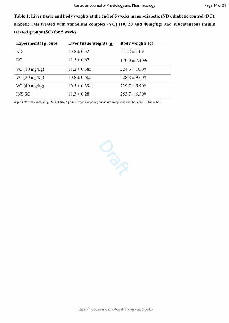
Liver and body weights
Table 1 presents the liver tissue and body weights in non-diabetic (ND), diabetic control (DC), diabetic rats
treated with vanadium complex (VC) (40 mg/kg) and subcutaneous insulin treated groups (SC) after the
experimental period of 5 weeks. The induction of diabetes resulted in a significant decrease in the body weight
in diabetic control compared to the non-diabetic control (p<0.05, DC vs ND), while no significant change was
observed in the liver weights (Table 1). However, the administration of vanadium complex showed an increase
in the body weights compared to the diabetic control (p<0.05, VC vs DC), yet no effect was observed in the
liver weights. The administration of insulin showed significant increases in both the liver and body weights
(p<0.05, SC vs DC).
Lipid profile
Table 2 compares the week 5 plasma total cholesterol (TC), triglycerides (TG), HDL and LDL concentrations
in non-diabetic control (ND), diabetic control (DC), diabetic animals treated with vanadium (10, 20, 40 mg)
and diabetic animals subcutaneously administered with insulin (S.C). Diabetes induction resulted in increased
TG and TC concentrations in comparison to non-diabetic control (p<0.05, DC vs ND). Diabetic control
animals showed increased TC and TG, respectively when compared to ND animals ★ (DC vs ND, p<0.05).
Diabetic rats treated with vanadium complex (10, 20, 40 mg/kg) decreased TC and TG concentrations in
comparison to STZ-induced diabetic rats. However, an increase in TGs concentration was observed in insulin
treated animals (p<0.05, SC vs DC). The diabetic rats presented with an increase in HDL concentration
(p<0.05, DC vs ND) and no effect was observed during VC administration (Table 2).
Oxidative stress markers
Table 3 shows superoxide dismutase (SOD), glutathione peroxidase (GPx) and malondialdehyde (MDA)
concentration measured in the liver tissues of non-diabetic (ND), diabetic control (DC), vanadium complex
(40 mg/kg) (VC) and diabetic animals subcutaneously administered with insulin (S.C) groups after 5 weeks.
SOD, GPx and MDA concentration in STZ-induced diabetic rats were significantly increased when compared
to non-diabetic control. The induction of diabetes resulted in an increment of SOD, GPx and MDA
concentration (DC vs ND, p<0.05, Table 3). Treatment with vanadium complex (40 mg/kg) resulted in a
significant decrease in MDA concentrations (p<0.05, VC vs DC). Similarly, insulin administration showed a
decrease in MDA concentrations (p<0.05, SC vs DC)).
Liver function enzymes
Figure 2 and 3 shows plasma alanine transaminase (ALT) and aspartate transaminase (AST) activities
respectively in non-diabetic (ND), diabetic control (DC), vanadium complex (VC) (40 mg/kg) and
subcutaneous insulin (SC) groups at the end of 5 weeks. The diabetic rats (DC) showed an increase in plasma
Page 7 of 21
https://mc06.manuscriptcentral.com/cjpp-pubs
Canadian Journal of Physiology and Pharmacology

Draft
ALT and AST activities when compared to non-diabetic rats (p<0.05, DC vs ND, Figure 2 and 3). However,
the administration of vanadium complex decreased plasma ALT activity (p<0.05, VC vs DC, Figure 2).
Plasma CRP
Figure 4 present the plasma C-reactive protein (CRP) concentration in non-diabetic (ND), diabetic control
(DC), diabetic rats treated with vanadium complex (VC) (40 mg/kg) and subcutaneous insulin treated groups
(SC) at the end of 5 weeks. The untreated STZ-induced diabetic animals showed an increase in plasma CRP
concentrations by comparison with non-diabetic control (p<0.05, DC vs ND). The administration of vanadium
complex decreased the plasma CRP at the end of the experimental period (p<0.05, VC vs DC, Figure 4).
Discussion
Previous studies have reported the development of metal based drugs with favourable pharmacological
application and may offer unique therapeutic opportunities. Indeed, glycaemic control has been achieved
through the administration of vanadium and zinc in form of inorganic salts (Fedorova et al. 2013). Depending
on the nature of ligand, vanadium complexes can be divided into inorganic and organic complexes (Sakurai
et al. 2002). Vanadium complexes with organic ligands have proved to be less toxic, with improved solubility
and lipophilicity (Goc 2006). The organic-ligand interactions are ubiquitous and play important roles in almost
every biological process (Sakurai et al. 2002). Research has shown that the organic ligands provides
thermodynamic stability and efficient transport of vanadium to target locations in the body making the
vanadium safer, more effective and stable than in the inorganic salt form (Sakurai et al. 2002). In this study,
we have investigated the effects of dioxidovanadium (V) complex with more focus on the liver function in
diabetes. This complex is fused with the organic ligands (phenol and benzimadizole) used in its synthesis. The
benzimadizole and phenolate derivatives have been shown to possess anti-diabetic and anti-oxidant properties.
Therefore, we envisaged that 2-phenolate-1H-benzimadizole-dioxidovanadium may have less potential
hepatic hazards, which may be beneficial in this context as most hypoglycaemic agents prove to detrimental.
The blood glucose concentrations were extremely high in the STZ-induced rats during the experimental
period. Streptozotocin (STZ) administration selectively destroys the beta insulin-producing cells of the
pancreas via DNA alkylation (Motala et al. 2008). However, in agreement with previous reports, the
administration of vanadium complex in STZ-induced diabetic rats attenuated blood glucose concentration in
Page 8 of 21
https://mc06.manuscriptcentral.com/cjpp-pubs
Canadian Journal of Physiology and Pharmacology

Draft
this study (Gurley et al. 2016). Vanadium administration has been shown to increase glucose transport and
oxidation and insulin-receptor tyrosine-kinase activity (Goc, 2006). Furthermore, previous study has reported
that vanadium inhibits microsomal glucose-6-phosphatase in STZ diabetic rats (Sakurai, 2002). The inhibition
of glucose-6 phosphatase by vanadium is thought to increase hepatic glucose-6 phosphate which serves as
substrate for glycogen synthesis (Goc 2006). The insulin-mimetic properties and anti-diabetic effects of
vanadium compounds have been widely documented both in vivo and in vitro. Vanadium induces the
recruitment of vesicles containing GLUT 4, by stimulating the tyrosine kinase activity of the β-subunit of the
insulin receptor (Reul et al. 1999). Another mechanism of reduction of blood glucose concentration by
vanadium compounds is the activation of PKB/Akt kinase leading to the increase of glucose uptake the cells
via GLUT4 transporter (Sakurai 2002).
STZ-induced diabetes is associated with decreased body weight (Sibiya et al. 2017a). The body weight loss is
attributable to the lack of insulin effect in diabetic animals (Musabayane et al. 2000). Insulin is a well-known
anabolic hormone, promoting lipid storage and protein synthesis, hence it absence leads body weight loss
(Musabayane et al. 2000). Indeed, the administration of insulin subcutaneously improved body weight. The
ability of vanadium to protect against weight loss might be due to its glucose lowering effect and perhaps also
other possible insulin mimetic effect (Juurinen et al. 2007). The liver has also been shown to be affected by
hyperglycaemia toxicity (Harris 2005). In this study, the increase (hypertrophy) in the liver weights in diabetic
control animals may be attributed to the increased influx of fatty acids into the liver and the low capacity of
excretion of lipoproteins secretion from the liver resulting from deficiency of Apo lipoprotein B synthesis
which is a protein involved in the metabolism of lipids (Khan and Newsome 2016).
Insulin is beneficial in restoring glycaemia and delaying the onset of diabetic complications, however, hepatic
hazards have been reported (Ishii and Takamura 2016). Studies have documented that subcutaneous insulin is
associated with hepatomegaly (Khan and Newsome 2016). Indeed, the animals on subcutaneous insulin
presented with an increase in liver weights which may perhaps allude to increase in fat and glycogen
deposition. Physiologically, when insulin is secreted by the beta-cells of the pancreas it is delivered to the
liver for clearance via the portal vein, therefore, less insulin concentration reaches the circulation (Havel et al.
1970). However, in diabetic patients, subcutaneous insulin injections are introduced directly into the blood
stream in high amounts, resulting in hyperinsulinaemia, since the first pass metabolism is by-passed (Havel et
Page 9 of 21
https://mc06.manuscriptcentral.com/cjpp-pubs
Canadian Journal of Physiology and Pharmacology

Draft
al. 1970). Thus, hyperinsulinaemia may act as a major contributor to the progression of liver damage and
dysfunction. The insulin-induced hepatomegaly is association with inflammation as a result of fatty
deposition, which may progress to scarring of the liver tissue to cirrhosis (Brunzell et al. 1973). In this study,
the administration of insulin subcutaneously, indeed resulted to hepatomegaly, further confirming the hepatic
hazards associated with insulin injections. Treatments with vanadium complex in diabetic rats significantly
restored liver and body weight to near normal (Sakurai 2002). Unlike insulin, vanadium has not been reported
dysregulate hepatic lipogenesis. These observations are of therapeutic important considering insulin-induced
hepatomegaly in diabetes.
Previous studies showed that vanadium treatment potentially ameliorate lipid metabolism derangement often
seen in diabetes (MacArthur et al. 2007). Reports suggest that dyslipidaemia is a frequent complication in all
types of diabetes which can range from hypercholesterolemia to hypolipoproteinaemia (Brunzell et al. 1973).
Hyperlipidaemia could be a factor for fatty liver development and has also been observed in diabetic patients
on insulin therapy (Lewis et al. 2002). In the present study, serum TG and TC concentrations were elevated
in the diabetic group compared to those in the control group, which is consistent with other studies and further
correlated with increases in liver weight. In the absence of insulin, lipolysis is increased thus leading to an
aggravated circulating triglycerides and cholesterol in the blood (Kowluru et al. 1999). However, the elevated
serum TG concentration in diabetic rats was significantly decreased after treatment with vanadium complex.
This result also suggest that liberation of fats as energy source was reduced, since tissues particularly skeletal
and adipose tissue could be able to utilize glucose as the energy source facilitated by dioxidovanadium
administered. This is in agreement with the evidence that decreasing high concentrations of serum and hepatic
TGs is often observed after treatment with vanadium compounds such as vanadium dipicolinate, vanadyl
sulphate and vanadate containing compounds (Sakurai 2002). The increase in the plasma triglycerides seen in
insulin injected animals may indicate increases in hepatic lipogenesis and may further explain the
hepatomegaly observed. Subcutaneous insulin administration have been shown to increase the expression of
sterol regulatory element-binding transcription factor 1 (SREBP1c). The SREBP1c is a major transcription factor
which regulates de novo lipogenesis through direct activation from AKT) in insulin signalling pathway. The
observations may further illuminate the potential cardiovascular risks associated insulin injection.
Page 10 of 21
https://mc06.manuscriptcentral.com/cjpp-pubs
Canadian Journal of Physiology and Pharmacology

Draft
Oxidative stress is the main cause for the development and advancement of liver dysfunction in diabetes
(Lewis et al. 2002). Hyperglycaemia evokes oxidative stress through activating of the following pathways;
polyol, hexosamine and protein kinase C pathway. In this study, the ability of vanadium complex to attenuate
hyperglycaemia might had a positive outcome in attenuating oxidative stress, as tight glycaemic control has
been shown to inactivate the above mentioned pathways (Sibiya et al. 2017b). Studies have indicated an
increase in liver antioxidant enzymes in response to elevated free radicals (Sakamaki et al. 1999). The results
of our study agree with other studies showing that induction of diabetes increase GPx and SOD activities
(Sakamaki et al. 1999). A decrease in SOD and GPx in the liver upon vanadium administration may be an
indication of vanadium complex’s ability to counteract free radical generation. Free radicals results in lipid
peroxidation and MDA has over the years been utilized as a prominent marker for lipid peroxidation. The
reduction of MDA concentration may owe to the improvement in glycaemic control and or the direct
antioxidant capacity of vanadium complex. The phenolate component in our vanadium complex is envisaged
have counter-acted the free radicals. The antioxidant therapy is amongst the treatment strategies for prevention
and slowing the progression of diabetic complications such including hepatic damage (Maritim et al. 2003).
Therefore, the ability of dioxidovanadium to alleviate oxidative stress may be beneficial in diabetes.
In diabetes mellitus, hepatocytes damage is inevitable, resulting in an increase in the circulating liver function
marker enzymes, such are aspartate transaminase (AST) and alanine transaminase (ALT) (Hamed 2014). The
increase in serum ALT and AST in diabetic patients is commonly associated with signs of hepatic diseases
such as NALFD (Frezza et al. 2010). The deleterious effect of hyperglyceamia in the liver of diabetic rats
observed in the present study was evidenced by elevation of liver damage biomarkers. In this study, animals
receiving insulin showed high levels of ALT and ALT thus indicating hepatic damage, these observations also
correlated with increase in liver mass which may perhaps indicate liver dysfunction and injury. The
hepatoprotective effects of vanadium is demonstrated by a significant reduction of serum ALT and AST.
Additionally, C Reactive protein CRP has been shown to be released by the hepatocytes in response to
inflammation or hepatic injury (Musabayane et al. 2000). CRP and diabetes associated inflammatory cytokines
such as tumour necrosis factor (TNF α) contribute to liver dysfunction and are associated with low grade
inflammation in diabetes. The elevation of liver enzymes is associated with higher CRP concentrations in
diabetic control as evidenced in this study, however administration of vanadium complex decreased plasma
Page 11 of 21
https://mc06.manuscriptcentral.com/cjpp-pubs
Canadian Journal of Physiology and Pharmacology

Draft
CRP at the end of 5 week experimental period. The attenuation of both liver enzymes and CRP may perhaps
be the outcome of glycaemic and oxidative stress attenuation by the vanadium complex administered.
Additionally, these observations may also suggest that our vanadium complex exert no or little toxicity to the
hepatocytes, unlike other oral hypoglycaemic agents such as troglitazone. Addionally, such observations
indicate that the incorporation of organic ligands mitigate the toxicity associated with inorganic vanadium
compounds. Perhaps such observations allude to the effective, non-toxic vanadium complex doses and dosing
schedule used in the study. Furthermore, the reduction in CRP concentration may also allude to the cardio
protective effect provided by vanadium, since CRP is also a prominent marker for screening cardiac injury
(Musabayane et al. 2000). Taken together, these results suggest that 2-phenolate-1H-benzimadizole-
dioxidovanadium possess no hepatic hazards and improves hepatic function in streptozotocin-induced-
diabetes. Moreover, the observations from this study further alludes and put more emphasis on the hepatic
hazards associated with subcutaneous insulin as evidence by high increase in liver mass, triglyceride, ALT,
AST and CRP.
Conclusion
The administration of dioxidovanadium complex attenuates hyerglycaemia, oxidative status, and lipid profile
disturbances in streptozotocin-induced diabetic animals. Additionally, dioxidovanadium administration exert
no apparent hepatic toxicological effect in diabetes induced animals. Taken together, the observations from
this study encourages further research and developments, regarding the use of vanadium therapy as an
alternative remedy for diabetes while highlighting the hepatic hazards associated insulin injections.
Conflict of interest
The authors declare that there is no conflict of interest.
Acknowledgement
The authors would like to acknowledge University of KwaZulu-Natal Biomedical Resource Unit personnel
for technical assistance. We also like to acknowledge National Research Funding South Africa for funding
the study.
Page 12 of 21
https://mc06.manuscriptcentral.com/cjpp-pubs
Canadian Journal of Physiology and Pharmacology

Draft
References
Amos, A. F. Mccarty, D. J. & Zimmet, P. 1997. The rising global burden of diabetes and its complications: estimates and projections to the year 2010. Diabetic medicine, 14, S7-S85.
Anderwald, C. Bernroider, E. Krssák, M. 2002. Effects of insulin treatment in type 2 diabetic patients on intracellular lipid content in liver and skeletal muscle. Diabetes, 51, 3025-3032.
Bilal, H. M. Riaz, F. Munir, K. Saqib, A. & SARWAR, M. R. 2016. Histological changes in the liver of diabetic rats: A review of pathogenesis of nonalcoholic fatty liver disease in type 1 diabetes mellitus. Cogent Medicine, 3, 1275415.
Boden, G. & Shulman, G. 2002. Free fatty acids in obesity and type 2 diabetes: defining their role in the development of insulin resistance and β-cell dysfunction. European journal of clinical investigation, 32, 14-23.
Brunzell, J. D. Porte, D. & Bierman, E. L. 1973. Evidence for a common, saturable, triglyceride removal mechanism for chylomicrons and very low density lipoproteins in man. The Journal of clinical investigation, 52, 1578-1585.
Fedorova, E. Buryakina, A. Vorobieva, N. & Baranova, N. 2013. The vanadium compounds: Chemistry, synthesis, insulinomimetic properties. Biochemistry (Moscow) Supplement Series B: Biomedical Chemistry, 7, 259-270.
Frezza, M. Hindo, S. Chen, D. Davenport, A. 2010. Novel metals anfd metal complexes as platforms for cancer therapy. Current pharmaceutical design, 16, 1813-1825.
Goc, A. 2006. Biological activity of vanadium compounds. Central European Journal of Biology, 1, 314-332.Gurley, J. M. Ilkayeva, O. Jackson, R. M.. 2016. Enhanced GLUT4-dependent glucose transport relieves nutrient stress in obese mice
through changes in lipid and amino acid metabolism. Diabetes, 65, 3585-3597.Hamed, A. E. 2014. The association between diabetes and liver disease: the need for a consensus.Harris, E. H. 2005. Elevated liver function tests in type 2 diabetes. Clinical diabetes, 23, 115-119.Havel, R. KANE, J. Balasse, E. SEGEL, N. & Basso, L. 1970. Splanchnic metabolism of free fatty acids and production of triglycerides of
very low density lipoproteins in normotriglyceridemic and hypertriglyceridemic humans. The Journal of clinical investigation, 49, 2017-2035.
Ishii, K. & Takamura, T. 2016. Non-alcoholic fatty liver disease (NAFLD)/non-alcoholic steatohepatitis (NASH) and nutrition. Clinical calcium, 26, 363-367.
Juurinen, L. Tiikkainen, M. Hakkinen, A.-M. Hakkarainen, A. & Yki-jarvinen, H. 2007. Effects of insulin therapy on liver fat content and hepatic insulin sensitivity in patients with type 2 diabetes. American Journal of Physiology-Endocrinology and Metabolism, 292, E829-E835.
Kowluru, R. A. Tang, J. & Kern, T. S. 2001. Abnormalities of retinal metabolism in diabetes and experimental galactosemia: VII. Effect of long-term administration of antioxidants on the development of retinopathy. Diabetes, 50, 1938-1942.
Lewis, G. F. Carpentier, A. Adeli, K. & Giacca, A. 2002. Disordered fat storage and mobilization in the pathogenesis of insulin resistance and type 2 diabetes. Endocrine reviews, 23, 201-229.
Macarthur, J. M. Bishop, J. R. 2007. Liver heparan sulfate proteoglycans mediate clearance of triglyceride-rich lipoproteins independently of LDL receptor family members. The Journal of clinical investigation, 117, 153-164.
Maritim, A. Sanders, R. & Watkins III, J. 2003. Effects of α-lipoic acid on biomarkers of oxidative stress in streptozotocin-induced diabetic rats. The Journal of nutritional biochemistry, 14, 288-294.
Motala, A. A. Esterhuizen, T. Gouws, E. Pirie, F. J. & Omar, M. A. 2008. Diabetes and other disorders of glycemia in a rural South African community: prevalence and associated risk factors. Diabetes care, 31, 1783-1788.
Musabayane, C. Munjeri, O. Bwititi, P. & OSim, E. 2000. Orally administered, insulin-loaded amidated pectin hydrogel beads sustain plasma concentrations of insulin in streptozotocin-diabetic rats. Journal of endocrinology, 164, 1-6.
Reul, B. A. Amin, S. S. Buchet, J. P.. 1999. Effects of vanadium complexes with organic ligands on glucose metabolism: a comparison study in diabetic rats. British journal of pharmacology, 126, 467-477.
Sakamaki, H. Akazawa, S. Urata, Y. & Kondo, T. 1999. Significance of glutathione-dependent antioxidant system in diabetes-induced embryonic malformations. Diabetes, 48, 1138-1144.
SakuraI, H. 2002. A new concept: the use of vanadium complexes in the treatment of diabetes mellitus. The Chemical Record, 2, 237-248.
Sakurai, H. Kojima, Y. Yoshikawa, Y. Kawabe, K. & YasuI, H. 2002. Antidiabetic vanadium (IV) and zinc (II) complexes. Coordination Chemistry Reviews, 226, 187-198.
Saligram, S. Williams, E. J. & Masding, M. G. 2012. Raised liver enzymes in newly diagnosed Type 2 diabetes are associated with weight and lipids, but not glycaemic control. Indian journal of endocrinology and metabolism, 16, 1012.
Sibiya, N. Ngubane, P. & Mabandla, M. 2017. Cardioprotective effects of pectin–insulin patch in streptozotocin-induced diabetic rats:. Journal of diabetes, 9, 1073-1081.
SibiyA, N. Ngubane, P. & MabandlA, M. 2017. The ameliorative effect of pectin-insulin patch on renal injury in streptozotocin-induced diabetic rats. Kidney and Blood Pressure Research, 42, 530-540.
Page 13 of 21
https://mc06.manuscriptcentral.com/cjpp-pubs
Canadian Journal of Physiology and Pharmacology
Draft
Table 1: Liver tissue and body weights at the end of 5 weeks in non-diabetic (ND), diabetic control (DC),
diabetic rats treated with vanadium complex (VC) (10, 20 and 40mg/kg) and subcutaneous insulin
treated groups (SC) for 5 weeks.
Experimental groups Liver tissue weights (g) Body weights (g)
ND 10.8 ± 0.32 345.2 ± 14.9
DC 11.5 ± 0.62 170.0 ± 7.40★
VC (10 mg/kg) 11.2 ± 0.38# 224.6 ± 10.0#
VC (20 mg/kg) 10.8 ± 0.50# 228.8 ± 9.60#
VC (40 mg/kg) 10.5 ± 0.39# 229.7 ± 5.90#
INS SC 11.3 ± 0.28 253.7 ± 6.50#★ p < 0.05 when comparing DC and ND, # p<0.05 when comparing vanadium complexes with DC and INS SC vs DC.
Page 14 of 21
https://mc06.manuscriptcentral.com/cjpp-pubs
Canadian Journal of Physiology and Pharmacology

Draft
Table 2: The lipid profile was assessed in non-diabetic control (ND), diabetic control (DC), diabetic
animals treated with vanadium complex (VC) (40mg/kg) and diabetic animals subcutaneously
administered with insulin (SC). Data is expressed as mean ± SEM (mean of samples per group, n=6).
Experimental
groups
Triglycerides
(mmol/L)
Cholesterol
(mmol/L)
HDL
(mmol/L)
LDL
(mmol/L)
ND 1.43 ± 0.07 4.08 ± 0.07 0.6 ± 0.2 2.8 ± 0.1
DC 2.50 ± 0.2★ 5.65 ± 0.20★ 1.3 ± 0.1★ 3.1 ± 0.1
40 (mg/kg) 1.00 ± 0.14# 3.30 ± 0.01# 1.5 ± 0.1 2.0 ±0.1#
SC INS 3.00 ± 0.26# 4.3 ± 0.8# 0.9 ± 0.1 2.1 ± 0.1#
★ p < 0.05 when comparing DC to ND, # p<0.05 when comparing vanadium complexes to DC and INS SC vs DC
Page 15 of 21
https://mc06.manuscriptcentral.com/cjpp-pubs
Canadian Journal of Physiology and Pharmacology

Draft
Table 3: The oxidative stress markers in liver tissues of non-diabetic control, diabetic control (DC),
diabetic rats treated with vanadium complex (VC) (40mg) and subcutaneous insulin treated groups
(SC) at the end of 5 weeks experimental period. Values are expressed as mean ± SEM.
EXPERIMENTAL
GROUPS
SOD (ng/ml) GPx (ng/ml) MDA (ng/ml)
ND 7.45 ± 0.06 1.61 ± 0.02 0.99 ± 0.02
DC 8.51 ± 0.08★ 1.94 ± 0.02★ 2.10 ± 0.01★
VC (40 mg/kg) 8.76 ± 0.08 1.83 ± 0.02# 1.09 ± 0.01#
SC INS 8.61 ± 0.12 1.97 ± 0.03 1.11 ± 0.01#
★ p < 0.05 when comparing DC and ND, # p<0.05 when comparing vanadium complexes with DC and INS SC vs DC.
Page 16 of 21
https://mc06.manuscriptcentral.com/cjpp-pubs
Canadian Journal of Physiology and Pharmacology

Draft
Figure 1: The weekly blood glucose concentration in non-diabetic (ND), diabetic control (DC), diabetic rats
treated with vanadium complex (VC) (10, 20 and 40mg/kg) and subcutaneous insulin treated groups (SC) for
5 weeks. Values are expressed as mean ± SEM, n=6 in each group. ★ p < 0.05 by comparison with diabetic
control.
Figure 2: Shows the liver function marker enzymes (ALT) activity in non-diabetic (ND), diabetic control
(DC), diabetic rats treated with vanadium complex (VC) (40mg/kg) and subcutaneous insulin treated groups
(SC) at the end of 5 weeks experimental period. Data is expressed as mean ± SEM (mean of samples per
group, n=6) ★ p < 0.05 when comparing DC and ND, # p<0.05 when comparing VC and SC with DC.
Figure 3: Shows the liver function marker enzymes (AST) activity in non-diabetic (ND), diabetic control
(DC), diabetic rats treated with vanadium complex (VC) (40mg/kg) and subcutaneous insulin treated groups
(SC) at the end of 5 weeks experimental period. Data is expressed as mean ± SEM (mean of samples per
group, n=6) ★ p < 0.05 when comparing DC and ND, # p<0.05 when comparing VC and SC with DC.
Figure 4: Shows the comparison of plasma CRP concentration in STZ-induced diabetic rats treated with
vanadium complex (40mg/kg) with positive control (SC), non-diabetic (ND) and untreated diabetic animals
(DC). Data is expressed as mean ± SEM (mean of samples per group, n=6) ★ p < 0.05 when comparing DC
and ND, # p<0.05 when comparing VC and SC with DC.
Page 17 of 21
https://mc06.manuscriptcentral.com/cjpp-pubs
Canadian Journal of Physiology and Pharmacology

Draft
Figure 1: The weekly blood glucose concentration in non-diabetic (ND), diabetic control (DC), diabetic rats treated with vanadium complex (VC) (10, 20 and 40mg/kg) and subcutaneous insulin treated groups (SC)
for 5 weeks. Values are expressed as mean ± SEM, n=6 in each group. ★ p < 0.05 by comparison with diabetic control.
193x109mm (300 x 300 DPI)
Page 18 of 21
https://mc06.manuscriptcentral.com/cjpp-pubs
Canadian Journal of Physiology and Pharmacology

Draft
Figure 2: Shows the liver function marker enzymes (ALT) activity in non-diabetic (ND), diabetic control (DC), diabetic rats treated with vanadium complex (VC) (40mg/kg) and subcutaneous insulin treated groups
(SC) at the end of 5 weeks experimental period. Data is expressed as mean ± SEM (mean of samples per group, n=6) ★ p < 0.05 when comparing DC and ND, # p<0.05 when comparing VC and SC with DC.
125x79mm (300 x 300 DPI)
Page 19 of 21
https://mc06.manuscriptcentral.com/cjpp-pubs
Canadian Journal of Physiology and Pharmacology

Draft
Figure 3: Shows the liver function marker enzymes (AST) activity in non-diabetic (ND), diabetic control (DC), diabetic rats treated with vanadium complex (VC) (40mg/kg) and subcutaneous insulin treated groups
(SC) at the end of 5 weeks experimental period. Data is expressed as mean ± SEM (mean of samples per group, n=6) ★ p < 0.05 when comparing DC and ND, # p<0.05 when comparing VC and SC with DC.
126x75mm (300 x 300 DPI)
Page 20 of 21
https://mc06.manuscriptcentral.com/cjpp-pubs
Canadian Journal of Physiology and Pharmacology

Draft
Figure 4: Shows the comparison of plasma CRP concentration in STZ-induced diabetic rats treated with vanadium complex (40mg/kg) with positive control (SC), non-diabetic (ND) and untreated diabetic animals (DC). Data is expressed as mean ± SEM (mean of samples per group, n=6) ★ p < 0.05 when comparing DC
and ND, # p<0.05 when comparing VC and SC with DC.
133x81mm (300 x 300 DPI)
Page 21 of 21
https://mc06.manuscriptcentral.com/cjpp-pubs
Canadian Journal of Physiology and Pharmacology