
Draft Benefit Definition: Unstable Angina/NSTEMI- … Definition Project... · Draft Benefit...
16
Draft Benefit Definition: Unstable Angina/NSTEMI- Acute Coronary Syndromes 28 August 2014 907E Acute and sub-acute ischaemic heart disease including myocardial infarction and unstable angina. Treatment: Medical management; surgery; percutaneous procedures
Transcript of Draft Benefit Definition: Unstable Angina/NSTEMI- … Definition Project... · Draft Benefit...

Draft Benefit Definition: Unstable Angina/NSTEMI- Acute Coronary Syndromes
28 August 2014 907E Acute and sub-acute ischaemic heart disease including myocardial infarction and
unstable angina.
Treatment: Medical management; surgery; percutaneous procedures

Contents
1. Introduction ................................................................................................................................... 3
2. Definition ....................................................................................................................................... 4
3. Pathophysiology ............................................................................................................................ 4
4. Clinical Presentation and Diagnosis: ................................................................................................ 4
4.1 Clinical Presentation ............................................................................................................... 4
4.2 Diagnostic Tools .................................................................................................................... 4
5. Risk- Stratification .......................................................................................................................... 8
6. Treatment ...................................................................................................................................... 8
6.1 Initial Pharmacological Treatment and Conservative Management .............................................. 9
6.2 Invasive Strategy ................................................................................................................. 10
7. Post Discharge Care .................................................................................................................... 13
8. Secondary prevention for N-STEMI Patients ................................................................................... 15
1. Introduction
The legislation governing the provision of the prescribed minimum benefits (PMBs) is contained in regulations
enacted under the Medical Schemes Act 131 of 1998. In respect of some of the diagnosis treatment pairs
(DTPs), medical scheme beneficiaries find it difficult to know in advance what their entitlements are. In addition,
medical schemes interpret these benefits differently resulting in a lack of uniformity of benefit entitlements.
The benefit definition project is coordinated by the Council for Medical Schemes and aims to define condition-
specific treatment guidelines, which will serve to guide the interpretation of the PMB provisions by relevant
stakeholders.
The benefit definition based on the available evidence of clinical and cost effectiveness taking into consideration
affordability constraints and financial viability of medical schemes in South Africa.
This benefit definition does not endorse explicitly one medicine/medical device within a particular therapeutic
class over another. Provision must be made for appropriate exceptions where this benefit definition has been
ineffective, causes, or will cause harm to a beneficiary, without penalty to that beneficiary. Health care providers
must provide written documentation for exceptions.
All patients who are treated successfully in an emergency setting must register with their scheme for chronic
management of Ischaemic heart disease. Scheme protocols and formularies developed taking into consideration
evidence-based medicine, cost-effectiveness, affordability should then apply.
It should be noted that benefit definitions are a minimum set of benefits and schemes may enrich the benefits but
not offer benefits less than stated here.
It should also be noted that management of ischaemic heart disease takes into consideration many clinical
aspects of the patients. This benefit definition does not address specific circumstances of high risk and
complicated patients who may need care more than specified here.
Alternatives must be made for patients where harm could be caused by treatment stated in the BD or scheme
formulary or protocol
Please note: procedure codes serve as a guideline for billing and many not include all relevant
procedure codes.

2. Definition
Acute coronary syndrome (ACS) represents a life-threatening manifestation of atherosclerosis. It is usually
precipitated by acute thrombosis induced by a ruptured or eroded atherosclerotic coronary plaque, with or
without concomitant vasoconstriction, causing a sudden and critical reduction in blood flow.
Among patients presenting with unstable angina, approximately 15% have one -vessel coronary artery disease
(CAD), 35% have 2-vessel CAD, and 50% have 3-vessel CAD. The incidence of left main disease is roughly 5-
10%. The rate of thrombus detected at coronary angiography varies widely, ranging from less than 10% among
those with chest pain in the previous month to more than 50% among those with rest angina in the preceding 24
hours.(1)
Unstable angina is considered to be an ACS in which there is no detectable release of the enzymes and
biomarkers of myocardial necrosis. N-STEMI is characterized by elevated cardiac enzymes. (2)
3. Pathophysiology
Atherosclerosis is a chronic, multifocal immune-inflammatory disease of medium-sized and large arteries mainly
driven by lipid accumulation.(3) NSTEMI usually occurs by developing a partial occlusion of a major coronary
artery or a complete occlusion of a minor coronary artery previously affected by atherosclerosis.(4)
ACS represent a life-threatening manifestation of atherosclerosis usually precipitated by acute thrombosis,
induced by a ruptured or eroded atherosclerotic plaque, with or without concomitant vasoconstriction, causing a
sudden and critical reduction in blood flow. (5, 6)
4. Clinical Presentation and Diagnosis:
4.1 Clinical Presentation
Patients present mainly with the following symptoms:(2)
Chest pain at rest that last longer than 20 minutes
New onset severe angina
Crescendo angina
Post myocardial infarct angina
4.2 Diagnostic Tools
4.2.1 Physical examination
Exclude non-cardiac and non-ischaemic causes of chest pain
To assess complications of acute coronary syndrome
To identify precipitating factors such as anaemia, fever, thyrotoxicosis etc.
4.2.2 Electrocardiogram
Twelve Lead ECG must be done as soon as possible
Continuous 12 – lead ECG monitoring must be done as 12 –ECG test may miss
some cases especially in patients with silent ischaemia
4.2.3 Blood tests
Cardiac Enzymes
Cardiac Troponins (cTnT or cTnI) are the preferred markers of myocardial injury, because they
are more specific and more sensitive than the traditional cardiac enzymes.(7) Troponin is also
a valuable prognostic test and useful in risk stratification.(8)

CKMB is important but limited by sensitivity
It should be noted that a single normal test may not be sufficient to exclude pathology in the
presence of suggestive symptoms and therefore a series of tests need to be done.
Inflammatory markers
C-reactive protein (CRP), although it has no diagnostic value in ACS, is a good predictor of
mortality.(2)
Novel biomarkers
Novel Biomakers such as myeloperixodase are not considered to be at PMB level of care as there is
insufficient data to confirm their ability to sensitively diagnose myocardial infarction.(2)
Other Blood test to assess baseline status and diagnose co-morbidities
Full blood count: -anaemia may precipitate myocardial ischemia and low HB,
Urea, creatinine and electrolytes
Serum glucose (may need to do a serial during admission)
Lipid profile
Thyroid function tests when thyrotoxicosis is suspected
Natriuretic peptides, such as brain- type B-type natriuretic peptide (BNP)] or its N-terminal
prohormone fragment (NT-proBNP) to detect left ventricular dysfunction. Although studies of
natriuretic peptides were done in hypertensive population, the sensitivity of this test in
diagnosing LVH is remains questionable. (9, 10) Therefore this tests are not considered to be
at PMB level of care
4.2.4 Non- Invasive Imaging
Chest-x ray; to exclude extra-cardiac causes of chest pain, detect heart failure and cardiomegaly
Echocardiography -is used to exclude other non-cardiac causes of chest pain such as aortic dissection
as well as diagnose ischaemia and detect complications of ischaemia such as left ventricular pathology
MRI and Scintigraphy may be used when there is diagnostic uncertainty.
4.2.5 Invasive Imaging
The gold standard remains angiography.
Cardiac computed tomography (CT) cannot be recommended as the coronary imaging modality in
NSTE-ACS, because of suboptimal diagnostic accuracy.(2)

Table 1: Possible codes for initial care of Acute Coronary Syndrome
Item Description Codes Additional comments
Professionals Paramedics For initial assessment , stabilisation and transportation to suitable facility
General practitioners and any other relevant health care provider in line scope of practice as per statutory body 0190-0192 For initial diagnosis and stabilisation
Physicians and cardiologist 0190-0192 For initial diagnosis, in-hospital management, Interventions and follow-up
Paramedics
ECG General Practitioner's fee for the taking of an ECG only: Without effort: ½ (item 1232) 1228 Serial ECG recording throughout assessment in Emergency room
General Practitioner's fee for the taking of an ECG only: Without and with effort: ½ (item 1233) 1229 Note: Items 1228 and 1229 deal only with the fees for taking of the ECG, the consultation fee must still be added
Physician's fee for interpreting an ECG: Without effort 1230 A specialist physician is entitled to the fees specified in item 1230 and 1231 for interpretation of an ECG tracing referred for interpretation. This applies also to a paediatrician when an ECG of a child is referred to him for interpretation
Physician's fee for interpreting an ECG: With and without effort 1231
Electrocardiogram: Without effort 1232
Electrocardiogram: With and without effort 1233 For inducible ischaemia
Pathology CKMB 4152,4153,4138 May be repeated every 6-8 hours
Troponin 4161
Full Blood Count- 3755 (Incl.
3739,3762,3783,3785,3791)
To rule out anaemia as secondary cause of
ACS
Platelet count 3797
Glucose-Hypo and hyperglycaemia affect treatment outcomes 4057
Lipogram-Lipid profile can change within 12-24 hours 4025
CRP 3947
ESR 3743
U & E and Creatinine 4171 Creatinine useful as baseline especially when

Creatinine-EGFR 4032 invasive strategy is considered. Potassium
abnormality must be corrected.
Magnesium 4094 or 4095 Low levels may predispose to arrhythmia
Non-invasive procedures
Single-photon emission computed tomography This test should not be used to
diagnose and will therefore not be
funded as PMB
Echocardiogram 3620,3621,3622,3623,3624,3625 To evaluate LV function
Stress Imaging In patients with non-diagnostic 12-lead ECGs
and negative cardiac biomarkers but suspected
ACS
Coronary CT angiography When troponin and ECG reading are non-
conclusive
Invasive imaging (coronary angiograph
Fractional Flow Reserve (FFR) FFR: First vessel. (add-on code) 1296
FFR: Each additional vessels add-
on code
1297
Coronary angiography 1249-54 To determine extend of coronary artery disease
or culprit lesion
Non-Invasive Radiology
Chest X-ray 1241 Evaluate patients for signs of congestive heart
failure (CHF) and for other causes of chest
symptoms, such as pneumothorax, pulmonary
infection or masses, pulmonary hypertension,
and mediastinal widening

5. Risk Stratification
Risk stratification should be performed as early as possible. Generally low risk patients will benefit from
conservative and selective invasive approach and high risk patients should be rapidly referred for angiography
and revascularization.
Tools for Risk Stratification
Full clinical history and examination including history of MI and previous interventions
The 12-lead ECG lies at the centre of the decision pathway for the evaluation and management of
patients with ischemic discomfort. A recording made during an episode of the presenting symptoms is
particularly valuable.
CK-MB has until recently been the principal serum cardiac marker used in the evaluation of ACS.
Despite its common use, CK-MB has several limitations (Loss of specificity in the presence of skeletal
muscle disease or injury. Low sensitivity during very early or later after the symptoms) (1). Despite its
limitations CK-MB remains a very useful marker for the detection of more than minor myocardial
damage.
The troponins offer greater diagnostic sensitivity due to their ability to identify patients with lesser
amounts of myocardial damage.
Various risk stratification tools are available however Global Registry of Acute Cardiac Events [GRACE]
score or TIMI are commonly used.
In a Cochrane review studying early invasive versus conservative strategies for unstable angina and non-ST
elevation myocardial infarction in the stent era, the invasive strategy did not reduce death on longer-term follow
up. Invasive strategy was however associated with reduced rates of refractory angina and re-hospitalization in
the shorter term and myocardial infarction in the longer term. The invasive strategy is associated with a doubled risk of
procedure-related to heart attack and increased risk of bleeding. It is suggested that an invasive strategy may be
particularly useful in those at high risk for recurrent events (1).
6. Treatment
Determination of the preferred strategy depends on the patient’s clinical characteristics and clinical risk. In a
Cochrane review studying early invasive versus conservative strategies for unstable angina and non-ST
elevation myocardial infarction in the stent era, the invasive strategy did not reduce death on longer-term follow
up. Invasive strategy was, however, associated with reduced rates of refractory angina and re-hospitalization in
the shorter term and myocardial infarction in the longer term. The invasive strategy is associated with a doubled
risk of procedure-related to heart attack and increased risk of bleeding. It is suggested that an invasive strategy
may be particularly useful in those at high risk for recurrent events.(1)
Generally, initial therapeutic approach is based on whether the patient is to be only medically treated, or in
addition referred to angiography and revascularization. Patients can be revascularised urgently or early (within
72 hours). Patient undergoing initial conservative management can be offered elective revascularization. (2)

6.1 Initial Pharmacological Treatment and Conservative Management
Any Medical Practitioner in line with scope of practice as regulated may initiate treatment. Patient must be
referred to a physician or cardiologist as soon as possible, taking into consideration limited number of
cardiologists.
6.1.1 Anti-ischaemic agents (2)
Beta-blockers are recommended in the absence of contraindications, particularly in patients with
hypertension or tachycardia
Intravenous or oral nitrates are effective for symptom relief in the acute management of anginal
episodes
Calcium channel blockers provide symptom relief inpatients already receiving nitrates and beta-
blockers; they are useful in patients with contraindications to nitrates and beta-blockers in the subgroup
of patients with vasospastic angina
Nifedipine, or other dihydropyridines, should not be used unless combined with beta-blockers
6.1.2 Anticoagulants
Anticoagulants are used in the treatment of NSTE-ACS to inhibit thrombin generation and/or activity, thereby
reducing thrombus-related events. Anticoagulation is recommended for all patients in addition to antiplatelet
therapy. Several anticoagulants are available; however the choice is dependent on the selected strategy.
6.1.3 Antiplatelets
Aspirin is recommended for all patients presenting with NSTE-ACS without contraindication at an initial
loading dose of 160–325 mg (non-enteric) (I-A), and at a maintenance dose of 75–100 mg long-term.
All patients should receive a loading dose of Clopidogrel (or similar thienopyridine-class antiplatelet agent)
and a maintenance dose of 300 mg for 12 months unless contraindicated.
6.1.4 Glycoprotein IIbIIIa inhibitors
These are recommended in intermediate to high risk patients. (1, 2)
Table 2: Pharmacological treatment of ACS
Type of drug Names Comments
Anti-ischaemic agents
Beta-blockers
Nitrates
Calcium channel blockers In patients with persistent symptoms despite receiving adequate beta blockers and Nitrates and in patient with contraindications to either Nitrates or beta blockers.
Angiotensin-converting enzyme inhibitors (ACEIs)
Recommended for high-risk patients, LV dysfunction, uncontrolled hypertension despite beta-blockers
Antiplatelet agents
Aspirin This is the first choice and administered indefinitely
Thienopyridine Indicated for patients who are sensitive to aspirin due to hypersensitivity or major gastrointestinal disturbance. Patients who are at high risk when non-invasive strategy is considered.
Ticagrelor Not included as PMB level of care as it is not registered with MCC. Will be subject to cost-effectiveness analysis upon registration.
Anticoagulants
Unfractionated heparin
Low molecular weight heparin Not recommended in patients with high risk of bleeding
Factor-Xa inhibitors (fondaparinux)
Recommended as it has the best efficacy/safety profile
Vitamin K antagonists
Direct thrombin inhibitors
Analgesia Morphine Sulphate Recommended in patients whose symptoms are not relieved after 3 serial sublingual NTG tablets or whose symptoms recur despite adequate anti-ischemic therapy

6.2 Invasive Strategy
Invasive strategy includes diagnostic catheterisation with instantaneous PCI or referral for coronary artery bypass
graft.
Indications for invasive strategy
i. Cardiogenic shock ii. Severe left ventricular dysfunction (<40%) iii. Angina refractory to medical therapy iv. Acute mitral regurgitation v. New ventricular septal defect vi. Unstable tachyarrhythmias vii. Hemodynamic instability viii. PCI within 6 months ix. High-risk findings on non-invasive stress testing x. New or presumably new ST-segment depression xi. High-risk score (e.g., TIMI, GRACE)
There are no RCTs comparing PCI with CABG in patients with NSTE-ACS. In all trials comparing an early with a
late strategy, or an invasive with a medical management strategy, the decision regarding whether to perform
CABG or PCI is left to the discretion of the investigator. (2) In patients stabilized after an episode of ACS, the
choice of revascularization modality can be made as in stable CAD.
6.2.1 Percutaneous Coronary Intervention
It is suitable for patients with single vessel disease or low risk double vessel disease. The risk of bleeding
complications should be balanced against the severity of ischaemia and the patient’s risk profile. The choice of
access site depends on operator expertise and local preference. Non-pharmacological strategies to reduce
access site bleeding complications include the use of closure devices and the radial approach.
Both DES and bare metal stents can be used. However bare metal stents should be considered in patients who
are likely to interrupt Clopidogrel or with Clopidogrel contraindications.

Table 3: Percutaneous Cardiac Intervention and Procedure Codes
Item Description Procedure code Discussion and conclusions
Catheter Laboratory
Clinicians Cardiologist Anaesthetist (Only when patient unstable) Physicians/2nd cardiologist (maybe required to assist in case of difficult anatomy) Nurse Radiographer Technologist
0190 0191 0192 0173-0175
Anaesthetist sometimes required for PCI of unstable patients when airway management is anticipated. Assistant cardiologist is sometimes required in patients with difficult anatomy
Clinical Technologist
Preparation and operation of pre-operative, intra-operative or post operative physiological monitoring per
patient, per admission
015
Cardiac catheterisation for the first hour. 063
Dilatation procedures and stents.
073
Radiographers Coronary angiogram per 30 minutes or part thereof provided that such part comprises 50% or more of the time
193
Stent procedure per 30 minutes or part thereof provided that such part comprises 50% or more of the time 197
Ancillary Drugs Glycoprotein IIb/IIIa inhibitor Low molecular weight heparin or unfractionated heparin Aspirin Clopidogrel or Prasugrel Beta Blocker or calcium channel blocker when beta-blockers are contraindicated. Prasugrel
Prasugrel has a fast onset of action and therefore more appropriate as a P2Y12 receptor inhibitors during PCI
Percutaneous procedure
PCI Invasive cardiology: Percutaneous translumical angioplasty
Percutaneous transluminal angioplasty: First cardiologist: Single lesion 1276
Percutaneous transluminal angioplasty: Second cardiologist: Single lesion 1277
Percutaneous transluminal angioplasty: First cardiologist: Second lesion 1278
Percutaneous transluminal angioplasty: Second cardiologist: Second lesion 1279
Percutaneous transluminal angioplasty: First cardiologist: Third or subsequent lesions (each) 1280
Percutaneous transluminal angioplasty: Second cardiologist: Third or subsequent lesions (each) 1281
Use of balloon procedures including: First cardiologist: Atrial septostomy; Pulmonary valve valvuloplasty; Aortic valve valvuloplasty; Coarctation dilation; Mitral valve valvuloplasty 1282
Use of balloon procedure as in item 1282: Second cardiologist 1283
Insertion of stents
Insertion of intravascular stent: First cardiologist 1286
The insertion of a stent(s) (item 1286 & 1267) may only be charged once per vessel regardless of the number of stents inserted in this vessel.
Insertion of intravascular stent: Second cardiologist 1287
Atherectomy Atherectomy: Single lesion: First cardiologist 1284
Atherectomy: Single lesion: Second cardiologist 1285
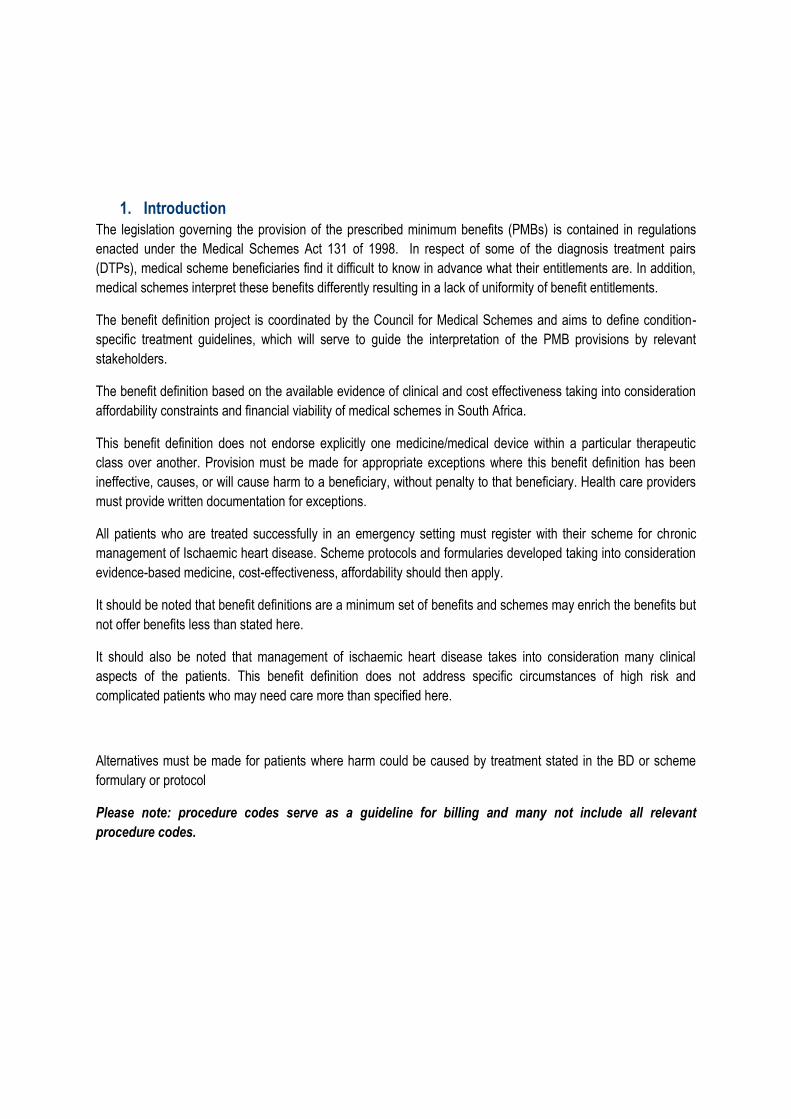
Stents Bare metal stent
Item Description Procedure code Discussion and conclusions
DES Drug eluting balloons and bioresorbable vascular scaffolds are currently not considered to be at PMB level of care due to lack of sufficient evidence on effectiveness and cost-effectiveness.
Drug Eluting Balloons
Bioresorbable vascular scaffolds
Imaging
IVUS
Diagnostic intravascular ultrasound (IVUS) imaging or wave wire mapping (without accompanying angioplasty). May be used only once per angiographic procedure. 5117
IVUS provides direct visualization and measurement of the inside of the blood vessels and may assist the doctor in selecting the appropriate size of balloons and/or stents, to ensure that a stent, if used, is properly opened, or to evaluate the use of other angioplasty instruments
Diagnostic intravascular ultrasound imaging or wave wire imaging (with accompanying angioplasty or accompanying intravascular ultrasound imaging or wave wire mapping in a different coronary artery [LAD (left anterior descending), Circumflex or Right coronary artery]). May be used a maximum of two per angiographic procedure 5118
Fractional Flow Reserve (FFR)
FFR: First vessel. (add-on code) 1296
FFR: Each additional vessels (add-on code) 1297

6.2.2 Coronary artery bypass surgery
Indications
Left main coronary artery stenosis >50%
Stenosis of proximal left anterior descending artery and proximal circumflex >70%
Three vessel disease
Three vessel disease with proximal LAD stenosis in patients with poor left ventricular (LV) function
Two-vessel disease and a large area of viable myocardium in high-risk area in patients with stable
angina
More than 70% proximal LAD stenosis with either ejection fraction < 50% or demonstrable ischemia on
non-invasive testing.
7. Post Discharge Care
All patients with NSTEMI-Unstable angina must have non-invasive stress testing. Echocardiogram must be done
post-discharge or immediately before discharge to evaluate left ventricular functioning.
The risk of mortality increases few months down the line. For this reason patient with unstable angina must be
followed more frequently than those with stable angina. Three monthly follow-ups are recommended.
Most of the patients with DES are also likely to default antiplatelet. These patients must be seen monthly for 3
months including post discharge follow-up between 4 and 6 weeks and there after 3 monthly. Stable patients can
then be seen 6 monthly.

Table 4: Possible procedure codes post- discharge
Item Description Code Comments
ECG
General Practitioner's fee for the taking of
an ECG only: Without effort: ½ (item
1232) 1228
Serial ECG recording throughout assessment in Emergency room
General Practitioner's fee for the taking of
an ECG only: Without and with effort: ½
(item 1233) 1229
Note: Items 1228 and 1229 deal only with the fees for taking of the ECG, the consultation fee must still be added
Physician's fee for interpreting an ECG:
Without effort 1230
A specialist physician is entitled to the fees specified in item 1230 and 1231 for interpretation of an ECG tracing referred for interpretation. This applies also to a paediatrician when an ECG of a child is referred to him for interpretation
Physician's fee for interpreting an ECG:
With and without effort 1231
Electrocardiogram: Without effort 1232
Electrocardiogram: With and without effort 1233 For inducible ischaemia
Exercise testing
Effort electrocardiogram with the aid of a special bicycle ergometer, monitoring apparatus and availability of associated apparatus
1252 Can be considered in patients without contradiction to exercise before discharge or early after discharge to assess inducible ischemia; to evaluate functional significance of coronary lesion; risk stratify according to likelihood of coronary events, establish ability and to exercise for life style modification
Multi-stage treadmill test 1234, 1235
Angiography Right and left cardiac catheterisation without coronary angiography (with or without biopsy) 1249
Indicated in patients with ECG changes of ischaemia post STEMI In patients with positive finding during non-invasive testing In patients who are persistently unstable For risk assessment in patients who had fibrinolytic therapy
Left heart catheterisation with coronary angiography (with or without biopsy) 1252
Right heart catheterisation (with or without biopsy) 1253
Catheterisation of coronary artery bypass grafts and/or internal mammary grafts 1254
Echocardiography Cardiac examination plus Doppler colour mapping 3620
It is indicated in patients with STEMI when there is a negative change in clinical status. It is reasonable to repeat the procedure in 1 to 3 months time. It is used to assess and re-evaluate LV function and to evaluate suspected complications. It can be used in patient with suspected RV infarction and inferior STEMI.
Cardiac examination (MMode) 3621
Cardiac examination: 2 Dimensional 3622
Cardiac examination + effort 3623
Cardiac examinations + contrast 3624
Cardiac examinations + doppler 3625
Cardiac examination + phonocardiography 3626
Pharmacological stress testing

8. Secondary prevention for N-STEMI Patients
i. Lifestyle modification(2, 11)
All persons with risk factors for ischaemic heart disease should be encouraged to make the
following lifestyle changes as appropriate:
Smoking cessation.
Weight reduction in the overweight patients, i.e. BMI > 25 kg/m2
Maintain ideal weight, i.e. BMI < 25 kg/m2
Reduce alcohol intake to no more than 2 standard drinks/day
Follow a prudent eating plan i.e. Low saturated fat, high fibre and unrefined carbohydrates, with
adequate fresh fruit and vegetables.
Moderate aerobic exercise, e.g. 30 minutes brisk walking at least 3 times a week
Members must be encouraged to participate in wellness and prevention activities as offered by the
scheme in line with scheme rules.
ii. Lipid Lowering Agents
The 2012 Essential drug list recommends lipid lowering agents in all Ischaemic heart disease irrespective of
cholesterol and triglyceride plasma concentration. The intention is to reduce LDL by at least 25%.(11)
iii. Control of diabetes
Maintain to HbA1 C < 7%
iv. Antiplatelets agents
Post STEMI patients must receive dual antiplatelet therapy. Aspirin must be continued indefinitely. Clopidogrel
must be used for at least a month if bare metal stents were used and for 6- 12 months if drug eluting stents were
used.
v. Blood pressure control
The main aim is to maintain BP at < 140/90 or < 130/80 in patients with chronic kidney disease and diabetes
mellitus.
Antihypertensive as per scheme’s formulary and CDL algorithm must be used. However, this should include beta
blocker and angiotensin converting enzyme inhibitors as a minimum benefit.(2)

Citation
1. Writing Committee M, Jneid H, Anderson JL, Wright RS, Adams CD, Bridges CR, et al. 2012 ACCF/AHA focused update of the guideline for the management of patients with unstable angina/Non-ST-elevation myocardial infarction (updating the 2007 guideline and replacing the 2011 focused update): a report of the American College of Cardiology Foundation/American Heart Association Task Force on practice guidelines. Circulation. 2012;126(7):875-910. 2. Task Force for D, Treatment of Non STSEACSoESoC, Bassand JP, Hamm CW, Ardissino D, Boersma E, et al. Guidelines for the diagnosis and treatment of non-ST-segment elevation acute coronary syndromes. European heart journal. 2007;28(13):1598-660. 3. Hamm C, Heeschen C, Falk E, KAA f. The ESC Textbook of Cardiovascular Medi-
cine.: Oxford; 2006. 4. Davies MJ. The pathophysiology of acute coronary syndromes. Heart. 2000;83(3):361-6. 5. Libby P. Current concepts of the pathogenesis of the acute coronary syndromes. Circulation. 2001;104(3):365-72. 6. Lupi Herrera E, Chuquiure Valenzuela E, Gaspar J, Ferez Santander SM. [From the single vulnerable plaque, to the multiple complex coronary plaques. From their basis, to the modern therapeutic approach. A clinical reality in the spectrum of the acute coronary syndromes]. Archivos de cardiologia de Mexico. 2006;76 Suppl 1:S6-34. 7. Wu AH, Feng YJ. Biochemical differences between cTnT and cTnI and their significance for diagnosis of acute coronary syndromes. European heart journal. 1998;19 Suppl N:N25-9. 8. Lindahl B, Diderholm E, Lagerqvist B, Venge P, Wallentin L. Mechanisms behind the prognostic value of troponin T in unstable coronary artery disease: a FRISC II substudy. Journal of the American College of Cardiology. 2001;38(4):979-86. 9. Morillas P, Castillo J, Quiles J, Nunez D, Guillen S, Maceira A, et al. Usefulness of NT-proBNP level for diagnosing left ventricular hypertrophy in hypertensive patients. A cardiac magnetic resonance study. Revista espanola de cardiologia. 2008;61(9):972-5. 10. Mouly-Bertin C, Bissery A, Milon H, Dzudie A, Rabilloud M, Bricca G, et al. N-terminal pro-brain natriuretic peptide--a promising biomarker for the diagnosis of left ventricular hypertrophy in hypertensive women. Archives of cardiovascular diseases. 2008;101(5):307-15. 11. Health Do. Essential Drug List -Hospital level 2012.


![Myocardial injury is distinguished from stable angina by a ... Injury Is... · NSTEMI/MI s group (n=15) comprised patients withcoronary atherosclerosis on angiogram coronary ... [HAc])](https://static.fdocuments.in/doc/165x107/606ccaf34234095c265d66c7/myocardial-injury-is-distinguished-from-stable-angina-by-a-injury-is-nstemimi.jpg)