Dr Stephen Goode MBChB, MRCS(Eng), FRCR, PhD Sheffield ... · identified angiographically and...
12
Title Page Manuscript Title: Targeted Contrast Enhanced Ultrasound Guidance for Liver TACE. Author Dr Stephen Goode MBChB, MRCS(Eng), FRCR, PhD Sheffield Vascular Institute, Northern General Hospital, Sheffield, S5 7AU, UK. [email protected]
Transcript of Dr Stephen Goode MBChB, MRCS(Eng), FRCR, PhD Sheffield ... · identified angiographically and...

Title Page
Manuscript Title:
Targeted Contrast Enhanced Ultrasound Guidance for Liver TACE.
Author
Dr Stephen Goode MBChB, MRCS(Eng), FRCR, PhD
Sheffield Vascular Institute, Northern General Hospital, Sheffield, S5 7AU, UK.
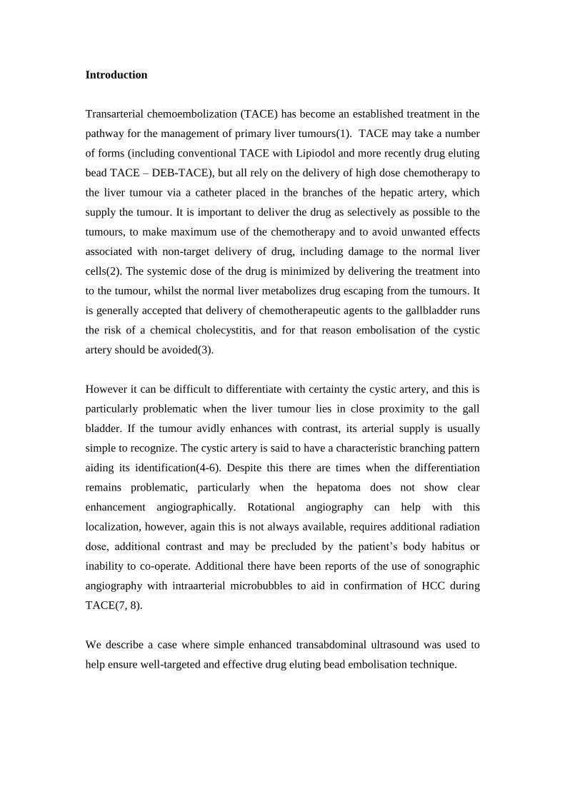
Introduction
Transarterial chemoembolization (TACE) has become an established treatment in the
pathway for the management of primary liver tumours(1). TACE may take a number
of forms (including conventional TACE with Lipiodol and more recently drug eluting
bead TACE – DEB-TACE), but all rely on the delivery of high dose chemotherapy to
the liver tumour via a catheter placed in the branches of the hepatic artery, which
supply the tumour. It is important to deliver the drug as selectively as possible to the
tumours, to make maximum use of the chemotherapy and to avoid unwanted effects
associated with non-target delivery of drug, including damage to the normal liver
cells(2). The systemic dose of the drug is minimized by delivering the treatment into
to the tumour, whilst the normal liver metabolizes drug escaping from the tumours. It
is generally accepted that delivery of chemotherapeutic agents to the gallbladder runs
the risk of a chemical cholecystitis, and for that reason embolisation of the cystic
artery should be avoided(3).
However it can be difficult to differentiate with certainty the cystic artery, and this is
particularly problematic when the liver tumour lies in close proximity to the gall
bladder. If the tumour avidly enhances with contrast, its arterial supply is usually
simple to recognize. The cystic artery is said to have a characteristic branching pattern
aiding its identification(4-6). Despite this there are times when the differentiation
remains problematic, particularly when the hepatoma does not show clear
enhancement angiographically. Rotational angiography can help with this
localization, however, again this is not always available, requires additional radiation
dose, additional contrast and may be precluded by the patient’s body habitus or
inability to co-operate. Additional there have been reports of the use of sonographic
angiography with intraarterial microbubbles to aid in confirmation of HCC during
TACE(7, 8).
We describe a case where simple enhanced transabdominal ultrasound was used to
help ensure well-targeted and effective drug eluting bead embolisation technique.

Case Description
A 65 year old man was referred having presented with upper abdominal pain. A CT
scan had revealed two large liver tumours, one each in segments 6 and 7, with an
associated subcapsular haematoma associated with the more inferior (segment 6)
lesion. It was considered that his pain was caused by a recent bleed from one of these
tumours. His body mass index was high (BMI= 45), the tumour locations and his co-
morbidities such that surgical resection or radio-frequency ablation were not
considered available treatment options. His clinical situation was discussed at the
Liver Multi-Disciplinary Team meeting, his imaging and biochemistry indicated that
these were primary liver tumours, and he was referred directly to the interventional
radiology outpatient clinic for consideration of, and consenting for, TACE.
His tumours were both located in the right lobe of his liver segments 6 and 7 (Figs. 1a
and 1b). The segment 6 lesion was lying adjacent to his gall bladder (Fig. 1b). As
previously noted his BMI was high, and the first TACE procedure was performed
from a femoral artery approach using 150mg of Doxorubicin loaded on to 100-300
micron beads (DEB-TACE). The larger lesion at the dome of the liver was easily
identified angiographically and embolised (Figs. 2a and 2b), whereas the lesion
located next to his gall bladder did not enhance as avidly, and could not be as reliably
delineated. Xper-CT (Philips) was tried and found to have very limited utility in this
gentleman, with significant usage of radiation and iodinated contrast. The full
treatment of 150mg of Doxorubicin was used, by which stage his skin radiation dose
was approaching the maximum advised level. No further imaging or embolization was
attempted on this occasion. A follow up CT scan showed a good response to the
segment 7 lesion but only a limited partial response to the segment 6 lesion (Figs. 3a
and 3b), and further TACE was planned.
It had proved problematic at the first TACE treatment to access the femoral artery due
to the distance from skin to femoral artery being significant with a large abdominal
apron. As a result the second treatment was performed from a left brachial approach.
Catheter angiography identified a branch arising proximally from the right hepatic
artery, with a branching pattern suggestive of it being the cystic artery (Figs. 4a and
4b). There was also a tissue blush, however it was not clear if this was actually

supplying the tumour close to the gall bladder in segment 6 or the gallbladder itself.
Xper-CT was not considered due to the previous difficulties.
The artery was selectively catheterized with a Progreat microcatheter (Terumo) (Fig.
4b). To further delineate the tumour, standard transabdominal ultrasound was
performed. Agitated saline was injected through the microcatheter whilst interrogating
the tumour with transabdominal ultrasound. Micro bubbles of air in the saline solution
were clearly seen within the tumour on ultrasound (Figs. 5a and 5b) thus indicating
that the artery was indeed supplying the tumour rather than the gall bladder. DEB-
TACE was performed via this artery, along with further DEB-TACE to the segment 7
tumour (easily identified angiographically again (Fig. 4a)).
The patient made an uneventful recovery and was discharged home the following day.
CT scanning 1 month later showed a good response to TACE and normal appearances
of the gallbladder (Figs. 6a and 6b)
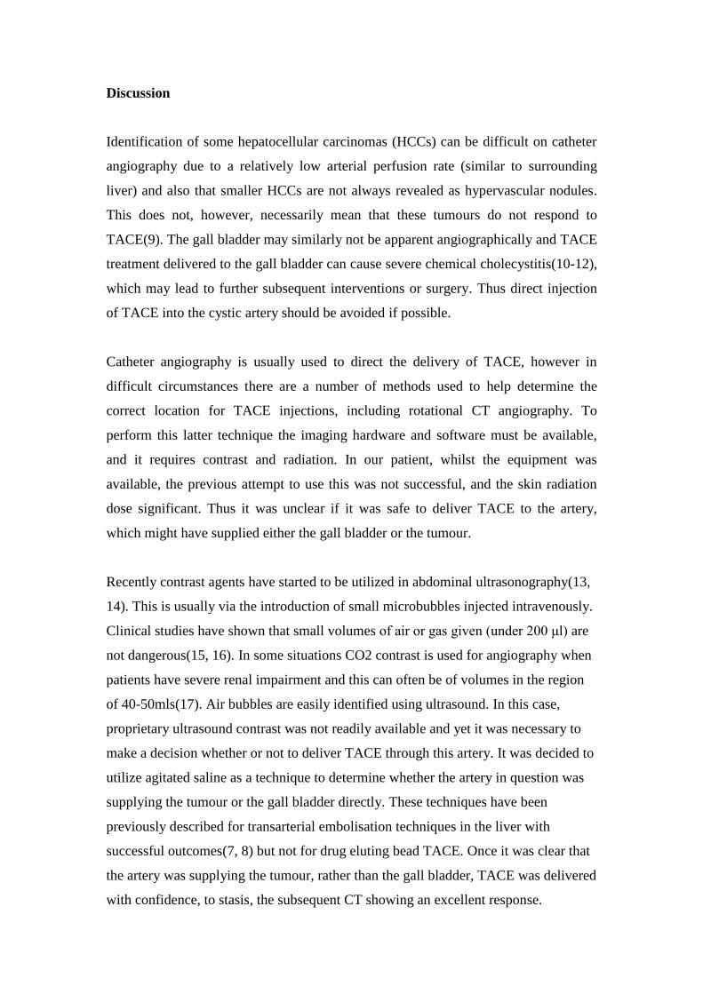
Discussion
Identification of some hepatocellular carcinomas (HCCs) can be difficult on catheter
angiography due to a relatively low arterial perfusion rate (similar to surrounding
liver) and also that smaller HCCs are not always revealed as hypervascular nodules.
This does not, however, necessarily mean that these tumours do not respond to
TACE(9). The gall bladder may similarly not be apparent angiographically and TACE
treatment delivered to the gall bladder can cause severe chemical cholecystitis(10-12),
which may lead to further subsequent interventions or surgery. Thus direct injection
of TACE into the cystic artery should be avoided if possible.
Catheter angiography is usually used to direct the delivery of TACE, however in
difficult circumstances there are a number of methods used to help determine the
correct location for TACE injections, including rotational CT angiography. To
perform this latter technique the imaging hardware and software must be available,
and it requires contrast and radiation. In our patient, whilst the equipment was
available, the previous attempt to use this was not successful, and the skin radiation
dose significant. Thus it was unclear if it was safe to deliver TACE to the artery,
which might have supplied either the gall bladder or the tumour.
Recently contrast agents have started to be utilized in abdominal ultrasonography(13,
14). This is usually via the introduction of small microbubbles injected intravenously.
Clinical studies have shown that small volumes of air or gas given (under 200 μl) are
not dangerous(15, 16). In some situations CO2 contrast is used for angiography when
patients have severe renal impairment and this can often be of volumes in the region
of 40-50mls(17). Air bubbles are easily identified using ultrasound. In this case,
proprietary ultrasound contrast was not readily available and yet it was necessary to
make a decision whether or not to deliver TACE through this artery. It was decided to
utilize agitated saline as a technique to determine whether the artery in question was
supplying the tumour or the gall bladder directly. These techniques have been
previously described for transarterial embolisation techniques in the liver with
successful outcomes(7, 8) but not for drug eluting bead TACE. Once it was clear that
the artery was supplying the tumour, rather than the gall bladder, TACE was delivered
with confidence, to stasis, the subsequent CT showing an excellent response.

In conclusion, the use of an agitated saline mixture injection and ultrasound detection
is a quick, cheap and easily available option for determining if TACE is delivered in
the correct area, in some circumstances. It is useful to keep this option in mind when
performing TACE.

Informed Consent
Informed consent was obtained from the patient included in the study.

References
1. Llovet JM, Burroughs A, Bruix J. Hepatocellular carcinoma. Lancet 2003;
362:1907-1917.
2. Raoul JL, Heresbach D, Bretagne JF, Ferrer DB, Duvauferrier R, Bourguet P,
et al. Chemoembolization of hepatocellular carcinomas. A study of the
biodistribution and pharmacokinetics of doxorubicin. Cancer 1992; 70:585-
590.
3. Basile A, Carrafiello G, Ierardi AM, Tsetis D, Brountzos E. Quality-
improvement guidelines for hepatic transarterial chemoembolization.
Cardiovasc Intervent Radiol; 35:765-774.
4. Daseler EH, Anson BJ, et al. The cystic artery and constituents of the hepatic
pedicle; a study of 500 specimens. Surg Gynecol Obstet 1947; 85:47-63.
5. Mlakar B, Gadzijev EM, Ravnik D, Hribernik M. Anatomical variations of the
cystic artery. Eur J Morphol 2003; 41:31-34.
6. Molmenti EP, Pinto PA, Klein J, Klein AS. Normal and variant arterial supply
of the liver and gallbladder. Pediatr Transplant 2003; 7:80-82.
7. Chen RC, Wang CK, Chiang LC, Lo HY, Duh SJ, Chen WT, et al. Intra-
arterial carbon dioxide-enhanced ultrasonogram of hepatocellular carcinoma
treated by transcatheter arterial embolization and percutaneous ethanol
injection therapy. J Gastroenterol Hepatol 1998; 13:41-46.
8. Hashimoto M, Watanabe O, Hirano Y, Kato K, Watarai J. Use of carbon
dioxide microbubble-enhanced sonographic angiography for transcatheter
arterial chemoembolization of hepatocellular carcinoma. AJR Am J
Roentgenol 1997; 169:1307-1310.
9. Hayano K, Desai GS, Kambadakone AR, Fuentes JM, Tanabe KK, Sahani
DV. Quantitative characterization of hepatocellular carcinoma and metastatic
liver tumor by CT perfusion. Cancer Imaging; 13:512-519.
10. Shah RP, Brown KT. Hepatic arterial embolization complicated by acute
cholecystitis. Semin Intervent Radiol; 28:252-257.
11. Karaman B, Battal B, Oren NC, Ustunsoz B, Yagci G. Acute ischemic
cholecystitis after transarterial chemoembolization with drug-eluting beads.
Clin Imaging; 36:861-864.
12. Wagnetz U, Jaskolka J, Yang P, Jhaveri KS. Acute ischemic cholecystitis after
transarterial chemoembolization of hepatocellular carcinoma: incidence and
clinical outcome. J Comput Assist Tomogr; 34:348-353.
13. Nicolau C. [Contrast-enhanced ultrasound in the diagnosis of hepatocellular
carcinoma. Yes, it is possible]. Radiologia; 54:363-365.
14. Nicolau C, Ripolles T. Contrast-enhanced ultrasound in abdominal imaging.
Abdom Imaging; 37:1-19.
15. Nanda nc Cc. Echo-enhancing agents: safety. 1997:115-131.
16. Blomley MJ, Cooke JC, Unger EC, Monaghan MJ, Cosgrove DO.
Microbubble contrast agents: a new era in ultrasound. BMJ 2001; 322:1222-
1225.
17. Shaw DR, Kessel DO. The current status of the use of carbon dioxide in
diagnostic and interventional angiographic procedures. Cardiovasc Intervent
Radiol 2006; 29:323-331.
Figures and Figure Legends
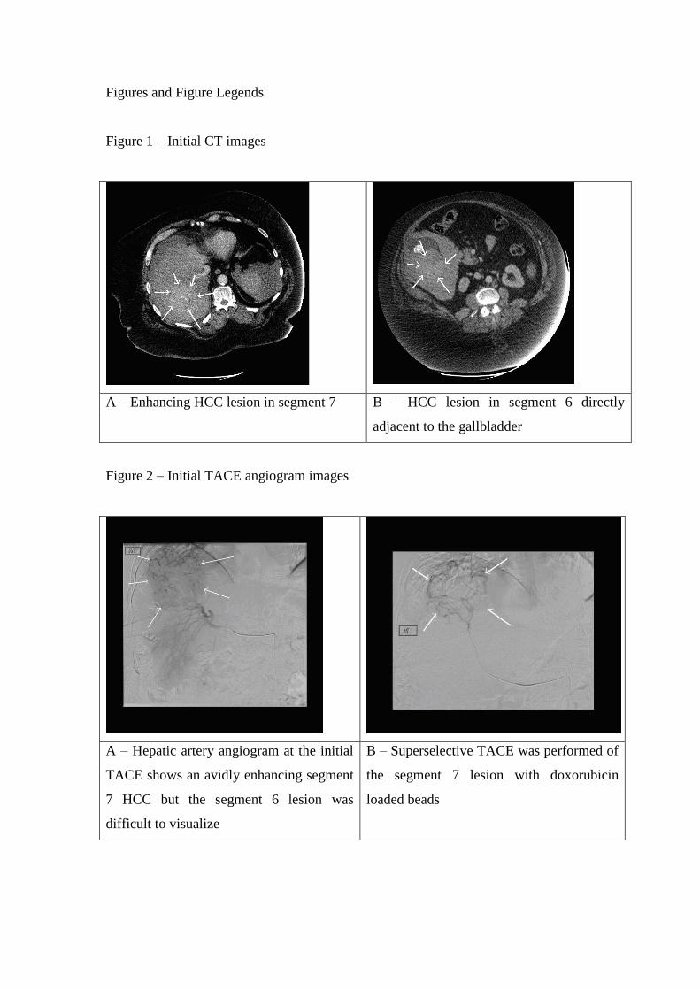
Figure 1 – Initial CT images
A – Enhancing HCC lesion in segment 7 B – HCC lesion in segment 6 directly
adjacent to the gallbladder
Figure 2 – Initial TACE angiogram images
A – Hepatic artery angiogram at the initial
TACE shows an avidly enhancing segment
7 HCC but the segment 6 lesion was
difficult to visualize
B – Superselective TACE was performed of
the segment 7 lesion with doxorubicin
loaded beads

Figure 3 – 1st follow up CT scan images
A – Good response to TACE for the
segment 7 lesion
B – minimal response to TACE for the
segment 6 lesion
Figure 4 – 2nd angiogram for repeat TACE treatment
A – Hepatic artery angiogram showing
segment 7 HCC but segment 6 lesion not well
visualized,
B – Selective angiogram of hepatic artery
branch thought to be supplying the
gallbladder. Progreat microcatheter seen in
good position in branch artery.

Figure 5 – Intraprocedural US imaging with use of air contrast
A – US image using curvilinear probe.
Segment 6 HCC (white arrows) visualized
with bubbles of air inside (white circle)
B – US image showing HCC (white arrows) in
segment 6 with GB (white rectangle) adjacent
and bubbles of gas within it (white circle).
Figure 6 – Follow up CT scan images
A - Axial portal venous CT image showing
good response to TACE in the segment 6
HCC lesion
B – Normal appearances of gallbladder on CT