Dr. Rath Research
13
MW Roomi. et al. Medical Research Archives vol 6 issue 12. December issue 12 Medical Research Archives Copyright 2018 KEI Journals. All Rights Reserved Chlorophyllin Suppresses Growth, MMP Secretion, Invasion and Cell Migration of Fibrosarcoma Cell Line HT-1080 Authors M Waheed Roomi, Bilwa Bhanap, Aleksandra Niedzwiecki, Matthias Rath Affiliations Dr. Rath Research Institute Correspondence Aleksandra Niedzwiecki Dr. Rath Research Institute [email protected] Abstract Fibrosarcoma is an aggressive and highly malignant cancer of connective tissue with the lungs being the most common site of metastasis. Surgery, chemotherapy, and radiation are the mainstay of treatments, yet the prognosis is very poor. A number of plant-based phytochemicals are increasingly being used as important treatment methods of cancers. Chlorophyll is a natural pigment that imparts the green color on plants. Chlorophyllin is a water soluble mixture of sodium-copper salts derived from chlorophyll. Chlorophyllin has been studied for its antioxidant potential. In the current study we tested the effects of chlorophyllin in fibrosarcoma HT-1080 cells on cell proliferation by MTT assay, modulation of matrix metalloproteinase (MMP) expression by zymography, cell invasive potential through Matrigel, cell migration by scratch test and morphology and apoptosis by H&E staining. Human fibrosarcoma cells HT-1080 were cultured in the media and were treated with chlorophyllin concentration at 10, 25 and 50 μM. The HT-1080 cell proliferation was significantly decreased at 50 μM dose of chlorophyllin. Expression of both, MMP-2, and MMP-9 decreased in a dose dependent manner. Both the MMPs were significantly inhibited at 25 μM and virtually undetectable at 50 μM. Cell invasion through Matrigel and cell migration was also reduced with the increasing concentrations of chlorophyllin with total inhibition of invasion at 50 μM. H&E staining at 10 μM of chlorophyllin showed a few cellular changes characteristic to apoptosis, while significant changes pertaining to apoptosis morphology were observed with increasing doses of chlorophyllin. Our results suggest that chlorophyllin may be a new chemotherapeutic strategy for fibrosarcoma patients and deserves further investigation as a potential agent in the treatment of this malignancy. RESEARCH ARTICLE
Transcript of Dr. Rath Research

MW Roomi. et al. Medical Research Archives vol 6 issue 12. December issue 12 Medical Research Archives
Copyright 2018 KEI Journals. All Rights Reserved
Chlorophyllin Suppresses Growth, MMP Secretion, Invasion and Cell
Migration of Fibrosarcoma Cell Line HT-1080
Authors
M Waheed Roomi, Bilwa Bhanap, Aleksandra Niedzwiecki, Matthias Rath
Affiliations
Dr. Rath Research Institute
Correspondence
Aleksandra Niedzwiecki
Dr. Rath Research Institute
Abstract
Fibrosarcoma is an aggressive and highly malignant cancer of connective tissue with the lungs
being the most common site of metastasis. Surgery, chemotherapy, and radiation are the
mainstay of treatments, yet the prognosis is very poor. A number of plant-based phytochemicals
are increasingly being used as important treatment methods of cancers. Chlorophyll is a natural
pigment that imparts the green color on plants. Chlorophyllin is a water soluble mixture of
sodium-copper salts derived from chlorophyll. Chlorophyllin has been studied for its antioxidant
potential. In the current study we tested the effects of chlorophyllin in fibrosarcoma HT-1080
cells on cell proliferation by MTT assay, modulation of matrix metalloproteinase (MMP)
expression by zymography, cell invasive potential through Matrigel, cell migration by scratch
test and morphology and apoptosis by H&E staining. Human fibrosarcoma cells HT-1080 were
cultured in the media and were treated with chlorophyllin concentration at 10, 25 and 50 µM.
The HT-1080 cell proliferation was significantly decreased at 50 µM dose of chlorophyllin.
Expression of both, MMP-2, and MMP-9 decreased in a dose dependent manner. Both the
MMPs were significantly inhibited at 25 µM and virtually undetectable at 50 µM. Cell invasion
through Matrigel and cell migration was also reduced with the increasing concentrations of
chlorophyllin with total inhibition of invasion at 50 µM. H&E staining at 10 µM of chlorophyllin
showed a few cellular changes characteristic to apoptosis, while significant changes pertaining to
apoptosis morphology were observed with increasing doses of chlorophyllin. Our results suggest
that chlorophyllin may be a new chemotherapeutic strategy for fibrosarcoma patients and
deserves further investigation as a potential agent in the treatment of this malignancy.
RESEARCH ARTICLE
MW Roomi. et al. Medical Research Archives vol 6 issue 12. December 2018 issue 11 Page 2 of 13
Copyright 2018 KEI Journals. All Rights Reserved http://journals.ke-i.org/index.php/mra
1. Introduction:
Fibrosarcoma is a tumor of the soft tissue of
mesenchymal origin and begins in fibrous
tissues which hold to the bones, muscles,
and other organs. Fibrosarcoma is a very
aggressive and highly metastatic cancer
primarily developing in metaphyses of the
long bones and the most common site of
metastasis is the lungs. It predominantly
affects children, adolescents, and young
adults.1 According to the American Cancer
Society, approximately 13,040 new cases of
soft tissue sarcomas, including
fibrosarcoma, are estimated to be diagnosed
in 2018, and close to 5,150 people are
expected to die due to this disease.2
Although the exact cause of fibrosarcoma is
unknown, genetic mutations may play a role
in causation. The most common mutation
includes allele loss, point mutations, and
chromosomal translocations.3, 4
Surgery, chemotherapy, and radiation are
the main modalities of treatment for
fibrosarcoma, however, overall this cancer
has poor prognosis.5 Prognosis in
fibrosarcoma patients depends on pathologic
grading and it worsens progressively with
increasing grade fibrosarcoma. However,
even when diagnosed and operated on early,
the probability of cancer recurrence of
fibrosarcoma at metastatic sites is greater
than 70% after surgery.1 The average 5-year
survival rates range from 50-80% for
moderate to low-grade fibrosarcoma, and
drops to 30% for high-grade fibrosarcoma.
Due to resistance to chemotherapy drugs and
limitation of other current treatment
modalities, there is an urgent need for a safe
and effective treatment approach.
A number of plant-based phytochemicals are
increasingly being used as important
treatment methods of cancers, due to their
antioxidant, chemo preventative, and
antitumor actions. Chlorophyll is a natural
pigment that imparts the green color on
plants. Plants and some algae use
chlorophyll to trap light for the process of
photosynthesis. Chlorophyllin is a water
soluble mixture of sodium-copper salts
derived from chlorophyll.6,7
Chlorophyllin
has been studied for its antioxidant
potential.8. 9
Anticanrcinogenic properties of
chlorophyllin have shown to block the
damage caused by free radicals derived from
smoking,10
certain heterocyclic amines from
processed and grilled food,11
and aflatoxin-
B1.12
In the current study we tested the
effects of chlorophyllin in fibrosarcoma HT-
1080 cells on cell proliferation, modulation
of matrix metalloproteinase (MMP)
expression, cell invasive potential, apoptosis
and cell morphology. Our results suggest
that chlorophyllin may be a new
chemotherapeutic strategy for fibrosarcoma
patients and deserves further investigation as
a potential agent in the treatment of this
malignancy.
MW Roomi. et al. Medical Research Archives vol 6 issue 12. December 2018 issue 11 Page 3 of 13
Copyright 2018 KEI Journals. All Rights Reserved http://journals.ke-i.org/index.php/mra
2. Materials and Methods:
2.1 Cell Culture and chlorophyllin:
The test compound chlorophyllin was
obtained from Sigma-Aldrich Corp. St.
Louis, MO, USA.
Human fibrosarcoma cells, HT-1080 were
obtained from ATCC (Rockville, MD) and
grown in MEM medium supplemented with
10% fetal bovine serum, penicillin G sodium
(100 U/ml), streptomycin (100 µg/ml) and
amphotericin (0.25 µg/ml) in 24-well tissue
culture plates (Costar, Cambridge, MA).
Cells were incubated with 1 ml of media at
37C in a tissue culture incubator
equilibrated with 95% air and 5% CO. At
near confluence the cells were treated with
chlorophyllin (obtained from Sigma Aldrich,
St. Luis, MO, USA) 10, 25, and 50 µM in
triplicate at each dose. The plates were then
returned to the incubator.
2.2 MTT Assay:
Cell proliferation was evaluated by MTT [3-
(4,5-dimethylthiazol-2-yl)-2,5-diphenyl
tetrazolium bromide] assay, a colorimetric
assay based on the ability of viable cells to
reduce a soluble yellow tetrazolium salt
(MTT) to a blue formazan crystal by
mitochondrial succinate dehydrogenase
activity of viable cells. This test is a good
index of mitochondrial activity and thus of
cell viability. After 24 hours of incubation,
the cells were washed with phosphate-
buffered saline (PBS) and 500 µl of MTT
(Sigma Catalog No. M-2128), and 0.5
mg/mL in media was added to each well.
The plates were covered and returned to the
37C incubator for 2 hours, the optimal time
for formazan product formation. Following
incubation, the supernatant was carefully
removed from the well, the formazan
product was dissolved in 1 mL DMSO, and
absorbance was measured at 570 nm in a
Bio Spec 1601 Shimadzu spectrometer. The
OD570 of the DMSO solution in each well
was considered to be proportional to the
number of cells. The OD570 of the control
(treatment without supplement) was
considered to be 100%.
2.3 Gelatinase Zymography:
MMP secretion in conditioned media was
determined by gelatinase zymography.
Gelatinase zymography was performed in
10% polyacrylamide precast Novex gel,
sodium dodecyl sulphate (Invitrogen Corp.),
in the presence of 0.1% gelatin under non-
reducing conditions. Culture medium (20 µl)
was loaded and SDS-Polyacrylamide Gel
Electrophoresis (SDS-PAGE) was
performed with Tris-Glycine SDS buffer as
described by the manufacturer (Novex).
Samples were not boiled before
electrophoresis. After electrophoresis, the
gels were washed with 5% Triton X-100 for
30 minutes at room temperature to remove
SDS. The gels were then incubated at 37C
overnight in the presence of 50 mM Tris-
HCl, 5 mm CaCl2, 5 µM ZnCl2 at pH 7.5,
stained with Coomasie Blue R 0.5% for 30
minutes, and destained. Protein standards
were run concurrently and approximate
molecular weights were determined by
plotting the relative mobilities of known
proteins.
2.4 Matrigel Invasion Studies:
Invasion studies were conducted using
Matrigel (Becton-Dickinson) inserts in 24-
MW Roomi. et al. Medical Research Archives vol 6 issue 12. December 2018 issue 11 Page 4 of 13
Copyright 2018 KEI Journals. All Rights Reserved http://journals.ke-i.org/index.php/mra
well plates. Suspended in medium
fibrosarcoma cells HT-1080 were
supplemented with chlorophyllin as
specified in the design of the experiment and
seeded on the insert in the well. Thus, both
the medium on the insert and in the well
contained the same supplements. The plates
with the inserts were then incubated in a
culture incubator equilibrated with 95% air
and 5% CO2 for 24 hours. After incubation,
the media from the wells were withdrawn.
The cells on the upper surface of the inserts
were gently scrubbed away with cotton
swabs. The cells that had penetrated the
Matrigel membrane and migrated onto the
lower surface of the Matrigel were stained
with hematoxylin and eosin and visually
counted under the microscope.
2.5 Morphology:
Morphology of cells cultured for 24 hours in
the test concentrations of chlorophyllin were
evaluated by H&E staining and observed
and photographed by microscopy.
2.6 Cell Migration:
To study cell migration, a 2 mm wide single
uninterrupted scratch test was made from the
top to the bottom of the culture plates of
HT-1080 cells grown to confluence. The
culture plates were washed with PBS,
incubated with chlorophyllin in the medium
and tested with 0, 10, 25, and 50 µM in
triplicate at each dose for 24 hours. The cells
were washed with PBS, fixed and strained
with H&E, and photomicrographs were
taken.
2.7 Statistical Analysis:
The results were expressed as means ± SD
for the groups. Data was analyzed by
independent sample “t” test.
3. Results
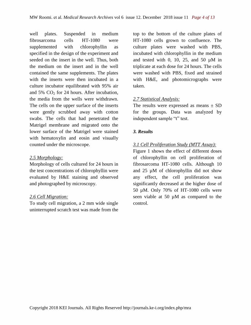
3.1 Cell Proliferation Study (MTT Assay):
Figure 1 shows the effect of different doses
of chlorophyllin on cell proliferation of
fibrosarcoma HT-1080 cells. Although 10
and 25 µM of chlorophyllin did not show
any effect, the cell proliferation was
significantly decreased at the higher dose of
50 µM. Only 70% of HT-1080 cells were
seen viable at 50 µM as compared to the
control.
MW Roomi. et al. Medical Research Archives vol 6 issue 12. December 2018 issue 11 Page 5 of 13
Copyright 2018 KEI Journals. All Rights Reserved http://journals.ke-i.org/index.php/mra
Figure 1: Effects of chlorophyllin on cell proliferation of fibrosarcoma HT-1080 (*-p<0.001)
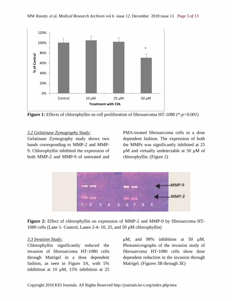
3.2 Gelatinase Zymography Study:
Gelatinase Zymography study shows two
bands corresponding to MMP-2 and MMP-
9. Chlorophyllin inhibited the expression of
both MMP-2 and MMP-9 of untreated and
PMA-treated fibrosarcoma cells in a dose
dependent fashion. The expression of both
the MMPs was significantly inhibited at 25
µM and virtually undetectable at 50 µM of
chlorophyllin. (Figure 2)
Figure 2: Effect of chlorophyllin on expression of MMP-2 and MMP-9 by fibrosarcoma HT-
1080 cells (Lane 1- Control, Lanes 2-4- 10, 25, and 50 µM chlorophyllin)
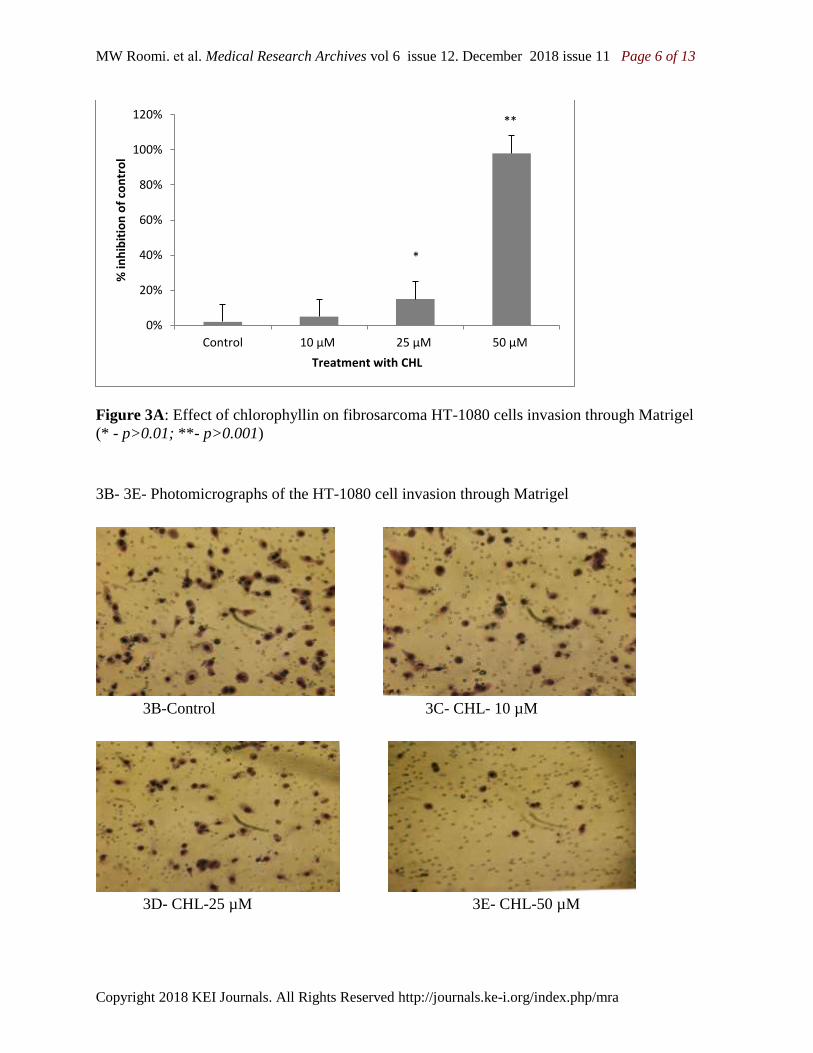
3.3 Invasion Study:
Chlorophyllin significantly reduced the
invasion of fibrosarcoma HT-1080 cells
through Matrigel in a dose dependent
fashion, as seen in Figure 3A, with 5%
inhibition at 10 µM, 15% inhibition at 25
µM, and 98% inhibition at 50 µM.
Photomicrographs of the invasion study of
fibrosarcoma HT-1080 cells show dose
dependent reduction in the invasion through
Matrigel. (Figures 3B through 3E)
0%
20%
40%
60%
80%
100%
120%
Control 10 µM 25 µM 50 µM
% o
f C
on
tro
l
Treatment with CHL
*
MW Roomi. et al. Medical Research Archives vol 6 issue 12. December 2018 issue 11 Page 6 of 13
Copyright 2018 KEI Journals. All Rights Reserved http://journals.ke-i.org/index.php/mra
Figure 3A: Effect of chlorophyllin on fibrosarcoma HT-1080 cells invasion through Matrigel
(* - p>0.01; **- p>0.001)
3B- 3E- Photomicrographs of the HT-1080 cell invasion through Matrigel
3B-Control 3C- CHL- 10 µM
3D- CHL-25 µM 3E- CHL-50 µM
0%
20%
40%
60%
80%
100%
120%
Control 10 µM 25 µM 50 µM
% in
hib
itio
n o
f co
ntr
ol
Treatment with CHL
*
**

MW Roomi. et al. Medical Research Archives vol 6 issue 12. December 2018 issue 11 Page 7 of 13
Copyright 2018 KEI Journals. All Rights Reserved http://journals.ke-i.org/index.php/mra
3.4. Morphology and Apoptosis Study (H&E
staining):
H&E staining revealed significant apoptotic
changes in dose dependent fashion in HT-
1080 fibrosarcoma cells treated with
moderate apoptotic changes at 10 µM, and
significant changes noticed at 25 and 50 µM
of chlorophyllin doses. These included
characteristic morphological changes such
as shrinkage of cytoplasm and darkly stained
and condensed nuclei with strongly
acidophilic cytoplasm (Figures 4A-4D).
Figure 4: Effect of chlorophyllin on human
Fibrosarcoma HT-1080 Cell morphology
showing apoptotic studied by H&E staining
4A- Control 4B- CHL-10 µM
4C-CHL-25 µM 4D-CHL-50 µM
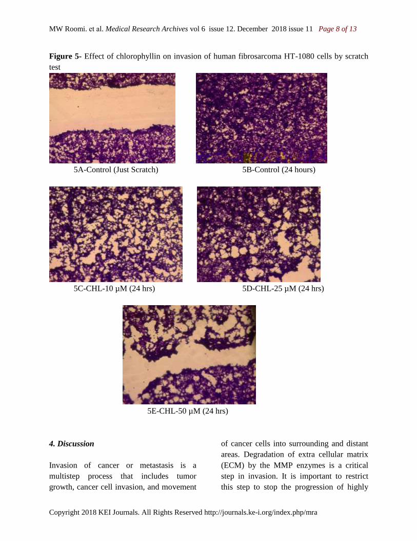
3.4. Cell Migration by Scratch Test:
Chlorophyllin reduced cell migration in a
dose dependent manner with a complete
block of fibrosarcoma HT-1080 cells at 50
µM. Photomicrographs of the results of the
scratch test are shown in Figures 5A-E.
MW Roomi. et al. Medical Research Archives vol 6 issue 12. December 2018 issue 11 Page 8 of 13
Copyright 2018 KEI Journals. All Rights Reserved http://journals.ke-i.org/index.php/mra
Figure 5- Effect of chlorophyllin on invasion of human fibrosarcoma HT-1080 cells by scratch
test
5A-Control (Just Scratch) 5B-Control (24 hours)
5C-CHL-10 µM (24 hrs) 5D-CHL-25 µM (24 hrs)
5E-CHL-50 µM (24 hrs)
4. Discussion
Invasion of cancer or metastasis is a
multistep process that includes tumor
growth, cancer cell invasion, and movement
of cancer cells into surrounding and distant
areas. Degradation of extra cellular matrix
(ECM) by the MMP enzymes is a critical
step in invasion. It is important to restrict
this step to stop the progression of highly
MW Roomi. et al. Medical Research Archives vol 6 issue 12. December 2018 issue 11 Page 9 of 13
Copyright 2018 KEI Journals. All Rights Reserved http://journals.ke-i.org/index.php/mra
aggressive cancer by tumor encapsulation.
Cancer cells that secrete higher amounts of
MMP are associated with poor prognosis
and decreased patient survival.13
Therefore,
control of ECM degradation provides an
opportunity to address common mechanism
of metastasis and tumor growth.
In the present study, we investigated effects
of chlorophyllin on human fibrosarcoma
HT-1080 cell line. The results indicate that
chlorophyllin effectively inhibits the HT-
1080 cell growth and cell invasion through
Matrigel in dose dependent manner. In
addition, chlorophyllin decreased the
expression of MMP-2 and MMP-9
fibrosarcoma HT-1080 cells in dose
dependent fashion. Exposure of HT-1080
cells to chlorophyllin in increasing
concentrations also altered their
morphological characteristics of apoptosis
and inhibited cell migration. MMPs are
produced by the cancer cells as well as
surrounding stromal cells and are tumor
specific.14
Human fibrosarcoma cells express
several MMPs, and of that, MMPs -2 and -9
are of particular importance in tumor
invasion, metastasis, and angiogenesis.15, 16
In an earlier in vivo study and in vitro
studies, we have demonstrated that a
specific micronutrient mixture significantly
inhibited cell growth, invasion and MMP
expression in vitro. Using xenograft athymic
mice, we also demonstrated that the nutrient
mixture inhibited the tumor growth and
tumor burden of human fibrosarcoma HT-
1080 cells.17
Chlorophyll is a plant pigment that gives
them the green color and is used to trap the
sunlight for photosynthesis, and
chlorophyllin is a semi synthetic mixture of
sodium and copper salts derived from
chlorophyll. Chlorophyllin is said to be one
of the most potent antioxidants and protects
against free radicals, heterocyclic amines
generated from grilled or processed meats,
smoking, and Aflatoxin-B1 (AFB1)
generated by a fungus growing on grains
and nuts. It is well known that the free
radical injury plays a key role in cancer
initiation and progression. Chlorophyllin has
been shown to be effective in blocking the
effects from heterocyclic amines and
aflatoxin. 8, 9
Chlorophyllin protects against
oxidative damage by inducing the
expression of heme-oxigenase-1.18
Chlorophyllin is an effective inhibitor of
numerous mutagens, including AFB1,
polycyclic
aromatic hydrocarbons (PAHs), heterocyclic
amines, direct acting compounds and
complex mixtures.19
AFB1is a known
carcinogen and is a major risk factor for
hepatocarcinoma. Breinholt, et al., 20
studied
chlorophyllin as an antimutagenic agent and
showed that chlorophyllin inhibits actions of
AFB1 and may prevent progression to
cancer. Chlorophyllin acts as an inceptor
molecule and blocks the action of
carcinogens by interfering with their
absorption by forming a reversible complex.
Simonich, et al., 21, 22
proved that
chlorophyllin is an effective anticancer
agent in rats as well as in rainbow trout
claiming that it has a species-independent
mechanism of action. An in vivo study by
Priyadarshini, et al., 23
showed that
MW Roomi. et al. Medical Research Archives vol 6 issue 12. December 2018 issue 11 Page 10 of 13
Copyright 2018 KEI Journals. All Rights Reserved http://journals.ke-i.org/index.php/mra
chlorophyllin effectively reversed the
expression of 104 genes associated with cell
adhesion, cell-cell communication, and
invasion and TGF-b signaling in a hamster
carcinogenesis model.
Radiation therapy and surgery are the
mainstays of treatment of fibrosarcoma.
Radiotherapy has wide spread damaging
effects from skin changes, damage to the
lungs and other organs, and DNA damage
that may lead to future cancers.
Occasionally, chemotherapy is used
especially in fibrosarcoma with bone
lesions. Studies show that chlorophyllin is
also effective in protecting the chromosomal
damage caused by gamma-radiation,
chemotherapeutic drug cyclophosphomide,
and carcinogenic agents such as N-nitroso-
N-ethylurea and urathane.24
In vivo and in
vitro studies by Kumar SS, et al., show that
chlorophyllin offers protection against
whole body irradiation in BALB/c mice and
chlorophyllin increased the intracellular
antioxidant enzymes such as superoxide
dismutase and glutathione peroxidase.25
Furthermore, Boloor, et al.,26
showed that
chlorophyllin not only is effective in
protection against cell damage caused by
radiation, but it offers a higher degree of
protection as compared to other
antioxidants, ascorbic acid and glutathione.
Chlorophyllin has been shown to work in
association with indol-3-carbinol in
carcinogenesis of colon cancer in rats by
altering the uptake or metabolism of
chemical carcinogens.27
It has also been
studied as a promising anti-carcinogen in
human breast cancer cells.28
Lawrence, et
al.,29
showed that chlorophyllin can induce
cell cycle arrest and apoptosis in breast
cancer cells by deactivating a family of
mitogen –activated protein kinases.
Furthermore, Diaz, et al.,30
showed that
chlorophyllin effectively blocks the
initiation of carcinogenesis, and induces
apoptosis in colon cancer cells.
Due to the aggressiveness of fibrosarcoma,
surgery, radiotherapy and chemotherapy
have not been able to show adequate
promise. A higher rate of recurrence
contributes to the poor prognosis. Therefore,
it is important to look into other methods of
cancer prevention and cure. Our studies
suggest that chlorophyllin is an excellent
candidate for treatment in fibrosarcoma by
inhibiting MMP expression, and Matrigel
invasion while avoiding potential side
effects commonly associated with
chemotherapy and radiation.
Acknowledgements
The research study was funded by Dr. Rath
Foundation (Santa Clara, CA, USA) a non-
profit organization. Ms. Cathy Flowers
provided the proofreading assistance.
MW Roomi. et al. Medical Research Archives vol 6 issue 12. December 2018 issue 11 Page 11 of 13
Copyright 2018 KEI Journals. All Rights Reserved http://journals.ke-i.org/index.php/mra
References:
1. Papagelopoulos PJ, Galanis EC,
Trantafyllidis P, Boscainos PJ, Sim FH,
Unni KK. Clinicopathologic features,
diagnosis, and treatment of fibrosarcoma
of bone. Am J Orthop 2002; 31(5): 253–
257
2. https://www.cancer.org/cancer/soft-
tissue-sarcoma/about/key-statistics.html
3. Ferrari A, Casanova M, Bisogno G,
Cecchetto G, Meazza C, Gandola L, et al
Malignant vascular tumors in children
and adolescents: a report from the Italian
and German Soft Tissue Sarcoma
Cooperative Group. Med Pediatr Oncol
2002; 39(2): 109–114.
DOI:10.1002/mpo.10078
4. Collin CF, Friedrich C, Godbold J, Hajdu
S, Brennan MF. Prognostic factors for
local recurrence and survival in patients
with localized extremity soft-tissue
sarcoma. Semin Surg Oncol 1988; 4(1):
30–37
5. Weitz J, Antonescu CR, Brennan MF.
Localized extremity soft tissue sarcoma:
improved knowledge with unchanged
survival over time. J Clin Oncol 2003;
15(14): 2719–2725. DOI:
10.1200/JCO.2003.02.026
6. Sudakin DL. Dietary aflatoxin exposure
and chemoprevention of cancer: a clinical
review. J Toxicol Clin Toxicol.
2003;41(2):195-204.
7. Dashwood RH. The importance of using
pure chemicals in (anti) mutagenicity
studies: chlorophyllin as a case in point.
Mutat Res. 1997;381(2):283-286.
8. Kumar SS, Devasagayam TP, Bhushan
B, Verma NC. Scavenging of reactive
oxygen species by chlorophyllin: an ESR
study. Free Radic Res. 2001;35(5):563-
574.],
9. Kamat JP, Boloor KK, Devasagayam TP.
Chlorophyllin as an effective antioxidant
against membrane damage in vitro and ex
vivo. Biochim Biophys Acta.
2000;1487(2-3):113-127.
10. Tachino N, Guo D, Dashwood WM,
Yamane S, Larsen R, Dashwood R.
Mechanisms of the in vitro antimutagenic
action of chlorophyllin against
benzo[a]pyrene: studies of enzyme
inhibition, molecular complex formation
and degradation of the ultimate
carcinogen. Mutat Res. 1994;308(2):191-
203.
11. Dashwood R, Yamane S, Larsen R.
Study of the forces of stabilizing
complexes between chlorophylls and
heterocyclic amine mutagens. Environ
Mol Mutagen. 1996;27(3):211-218.
DOI:10.1002/(SICI)1098-
2280(1996)27:3<211::AID-
EM6>3.0.CO;2-H
12. Breinholt V, Schimerlik M, Dashwood R,
Bailey G. Mechanisms of chlorophyllin
anticarcinogenesis against aflatoxin B1:
complex formation with the carcinogen.
Chem Res Toxicol. 1995;8(4):506-514.
13. Fang W, Li H, Kong L, Niu G, Gao Q,
Zhou K, et al.,. Role of matrix
metalloproteinases (MMPs) in tumor
invasion and metastasis: serial studies on
MMPs and TIMPs. Beijing Da Xue Xue
Bao Yi Xue Ban. 2003;35(4):441-3.
[Article in Chinese]
MW Roomi. et al. Medical Research Archives vol 6 issue 12. December 2018 issue 11 Page 12 of 13
Copyright 2018 KEI Journals. All Rights Reserved http://journals.ke-i.org/index.php/mra
14. van der Horst G, Bos L, van der Pluijm
G. Epithelial plasticity, cancer stem cells,
and the tumor-supportive stroma in
bladder carcinoma. Mol Cancer Res. Aug
2012; 10(8):995-1009. doi:
10.1158/1541-7786.MCR-12-0274
15. Giambernardi TA, Grant GM, Taylor GP,
Hay RJ, Maher VM, McCormick JJ, et
al,. Overview of matrix metalloproteinase
expression in cultured human cells.
Matrix biology. 1998;16:483–496.
16. Sternlicht MD, Werb Z. How matrix
metalloproteinases regulate cell behavior.
Annual review of cell and developmental
biology. 2001:17463–516.
DOI:10.1146/annurev.cellbio.17.1.463
17. Roomi MW, Ivanov V, Kalinovsky T,
Niedzwiecki A, Rath M. In Vivo and In
Vitro Antitumor Effect of Ascorbic Acid,
Lysine, Proline, Arginine and Green Tea
Extract on Human Fibrosarcoma Cells
HT-1080, Medical Oncology 2006, 23(1):
105-112
18. Zhang YL, Guan L, Zhou PH, Mao LJ,
Zhao ZM, Li SQ, Xu et al., The
protective effect of chlorophyllin against
oxidative damage and its mechanism.
Zhonghua Nei Ke Za Zhi.
2012;51(6):466-70.[Article in Chinese]
19. Waters MD1, Stack HF, Jackson MA,
Brockman HE, De Flora S. Activity
profiles of antimutagens: in vitro and in
vivo data. Mutat
Res.1996;19;350(1):109-29.
20. Breinholt V, Schimerlik M, Dashwood R,
Bailey G. Mechanisms of chlorophyllin
anticarcinogenesis against aflatoxin B1:
complex formation with the carcinogen.
Chem Res Toxicol. 1995;8(4):506-14.
21. Simonich MT, McQuistan T, Jubert C, et
al. Low-dose dietary chlorophyll inhibits
multi-organ carcinogenesis in the
rainbow trout. Food and chemical
toxicology : an international journal
published for the British Industrial
Biological Research Association.
2008;46(3):1014-1024.
doi:10.1016/j.fct.2007.10.034.
DOI:10.1016/j.fct.2007.10.034
22. Simonich MT, Egner PA, Roebuck BD,
Orner GA, Jubert C, Pereira C, et al.
Natural chlorophyll inhibits aflatoxin B1-
induced multi-organ carcinogenesis in the
rat. Carcinogenesis. 2007
Jun;28(6):1294-302.
DOI:10.1093/carcin/bgm027
23. Vidya Priyadarsini R, Kumar N, Khan I,
Thiyagarajan P, Kondaiah P, Nagini S.
Gene expression signature of DMBA-
induced hamster buccal pouch
carcinomas: modulation by chlorophyllin
and ellagic acid. PLoS One.
2012;7(4):e34628. doi:
10.1371/journal.pone.0034628.
24. Abraham SK, Sarma L, Kesavan PC.
Role of chlorophyllin as an in vivo
anticlastogen: protection against gamma-
radiation and chemical clastogens. Mutat
Res. 1994,:322(3):209-12.
25. Kumar SS, Shankar B, Sainis KB. Effect
of chlorophyllin against oxidative stress
in splenic lymphocytes in vitro and in
vivo. Biochim Biophys Acta.
2004;3;1674(2):100-11. DOI:
10.1016/j.bbagen.2004.03.002
26. Boloor KK, Kamat JP, Devasagayam TP.
Chlorophyllin as a protector of
mitochondrial membranes against
gamma-radiation and
MW Roomi. et al. Medical Research Archives vol 6 issue 12. December 2018 issue 11 Page 13 of 13
Copyright 2018 KEI Journals. All Rights Reserved http://journals.ke-i.org/index.php/mra
photosensitization.Toxicology. Nov
2000;155(1-3):63-71
27. Guo D, Schut HA, Davis CD,
Snyderwine EG, Bailey GS, Dashwood
RH. Protection by chlorophyllin and
indole-3-carbinol against 2-amino-1-
methyl-6-phenylimidazo[4,5-b]pyridine
(PhIP)-induced DNA adducts and colonic
aberrant crypts in the F344 rat.
Carcinogenesis. 1995;16(12):2931-7.
28. Smith WA, Freeman JW, Gupta RC.
Effect of chemopreventive agents on
DNA adduction induced by the potent
mammary carcinogen dibenzo[a,l]pyrene
in the human breast cells MCF-7.Mutat
Res. 2001;1;480-481:97-108
29. Chiu LC, Kong CK, Ooi VE. The
chlorophyllin-induced cell cycle arrest
and apoptosis in human breast cancer
MCF-7 cells is associated with ERK
deactivation and Cyclin D1 depletion. Int
J Mol Med. 2005 Oct;16(4):735-40.
30. Díaz GD, Li Q, Dashwood RH. Caspase-
8 and apoptosis-inducing factor mediate a
cytochrome c-independent pathway of
apoptosis in human colon cancer cells
induced by the dietary phytochemical
chlorophyllin.Cancer
Res. 2003;15;63(6):1254-61