DOI: 10.5585/ConsSaude.v11n4.3988 Recebido em 29 out. 2012 ... · rior (TI) estão associados a...
8
ConScientiae Saúde, 2012;11(4):660-667. 660 Lower trapezius and serratus anterior activation: which exercise to use for scapular neuromuscular reeducation? Ativação do trapézio inferior e serrátil anterior: qual o exercício para reeducação neuromuscular da escápula? Rodrigo Py Gonçalves Barreto 1 ; Caroline Cabral Robinson 2 ; Clarice Sperotto dos Santos Rocha 3 ; Fernando Carlos Mothes 4 ; Fábio Matsumoto 4 ; Luís Henrique Telles da Rosa 5 ; Tiago Kiefer 6 ; Marcelo Faria Silva 7 1 Physiotherapist, Grupo de Cirurgia do Ombro da Santa Casa de Porto Alegre, Master’s Program of Rehabilitation Science – UFCSPA. Porto Alegre, RS – Brasil. 2 Physiotherapist, Master degree in Neuroscience, Doctorate’s Program of Health Sciences – UFCSPA. Porto Alegre, RS – Brasil. 3 Physiotherapist, Doctorate degree in Neuroscience, Professor of Physical Therapy from Centro Universitário Metodista do IPA. Porto Alegre, RS – Brasil. 4 Medical Doctor, Shoulder Surgeon from Grupo de Cirurgia do Ombro da Santa Casa de Porto Alegre. Porto Alegre, RS – Brasil. 5 Physiotherapist, Professor of Physical Therapy – UFCSPA. Porto Alegre, RS – Brasil. 6 Physiotherapist, Master’s Program of Rehabilitation Sciences – UFCSPA. Porto Alegre, RS – Brasil. 7 Physiotherapist, Doctorate degree in Human Movement Sciences, Professor of Physical Therapy – UFCSPA. Porto Alegre, RS – Brasil. Postal address Rodrigo Py Gonçalves Barreto R. Lobo da Costa, 247/204, Cidade Baixa 90050-110 – Porto Alegre – RS [Brasil] [email protected] Recebido em 29 out. 2012. Aprovado em 7 dez. 2012 DOI: 10.5585/ConsSaude.v11n4.3988 Abstract Introduction: Low levels of activation of the serratus anterior (SA) and lower tra- pezius (LT) muscles are associated with kinematics dysfunctions of the scapular belt, for which the focus of functional recovery is neuromuscular reeducation. Hence, the proposed exercises should keep muscular activation at levels between 20% and 40% of the maximal voluntary contraction. Objectives: To compare the activation of SA and LT muscles in different exercises by using surface electromy- ography. Methods: Five exercises (modified crucifix, scaption, modified military press, pull over and low row) were executed by ten healthy subjects. Results: The highest SA activation was found during scaption, and the adequate activation occurred in the modified military press. The highest LT activation was found during scaption and low row exercises. Conclusions: The exercises that kept the recommended range of activation for neuromuscular reeducation were the mili- tary press, for the SA muscle, and the low row and scaption, for the LT muscle. Key words: Electromyography; Exercise; Physical Therapy Modalities; Scapula; Shoulder. Resumo Introdução: Baixos níveis de ativação do serrátil anterior (SA) e do trapézio infe- rior (TI) estão associados a disfunções cinemáticas da cintura escapular cujo foco da reabilitação funcional é a reeducação neuromuscular. Para tanto, os exercí- cios propostos devem manter a ativação muscular entre 20% e 40% da contração voluntária máxima. Objetivos: Comparar a ativação do SA e TI em diferentes exercícios por eletromiografia de superfície. Métodos: Cinco exercícios (modified crucifix, scaption, modified military press, pull over e low row) foram executados por dez sujeitos saudáveis. Resultados: A maior ativação para o SA foi encontrada durante o scaption e sua ativação adequada ocorreu durante o modified military press. Para o TI, a maior ativação ocorreu durante os exercícios scaption e low row. Conclusões: A ativação muscular dentro da faixa recomendada para a reeduca- ção neuromuscular ocorreu no modified military press para o AS; e no low row e scaption, para o TI. Descritores: Eletromiografia, Escápula; Exercício; Modalidades de Fisioterapia; Ombro.
Transcript of DOI: 10.5585/ConsSaude.v11n4.3988 Recebido em 29 out. 2012 ... · rior (TI) estão associados a...

ConScientiae Saúde, 2012;11(4):660-667.660
Lower trapezius and serratus anterior activation: which exercise to use for scapular neuromuscular reeducation?Ativação do trapézio inferior e serrátil anterior: qual o exercício para reeducação neuromuscular da escápula?
Rodrigo Py Gonçalves Barreto1; Caroline Cabral Robinson2; Clarice Sperotto dos Santos Rocha3; Fernando Carlos Mothes4; Fábio Matsumoto4; Luís Henrique Telles da Rosa5; Tiago Kiefer6; Marcelo Faria Silva7
1 Physiotherapist, Grupo de Cirurgia do Ombro da Santa Casa de Porto Alegre, Master’s Program of Rehabilitation Science – UFCSPA. Porto Alegre, RS – Brasil.
2 Physiotherapist, Master degree in Neuroscience, Doctorate’s Program of Health Sciences – UFCSPA. Porto Alegre, RS – Brasil.3 Physiotherapist, Doctorate degree in Neuroscience, Professor of Physical Therapy from Centro Universitário Metodista do IPA. Porto
Alegre, RS – Brasil.4 Medical Doctor, Shoulder Surgeon from Grupo de Cirurgia do Ombro da Santa Casa de Porto Alegre. Porto Alegre, RS – Brasil.5 Physiotherapist, Professor of Physical Therapy – UFCSPA. Porto Alegre, RS – Brasil.6 Physiotherapist, Master’s Program of Rehabilitation Sciences – UFCSPA. Porto Alegre, RS – Brasil.7 Physiotherapist, Doctorate degree in Human
Movement Sciences, Professor of Physical Therapy – UFCSPA. Porto Alegre, RS – Brasil.
Postal addressRodrigo Py Gonçalves BarretoR. Lobo da Costa, 247/204, Cidade Baixa 90050-110 – Porto Alegre – RS [Brasil] [email protected]
Recebido em 29 out. 2012. Aprovado em 7 dez. 2012DOI: 10.5585/ConsSaude.v11n4.3988
Abstract
Introduction: Low levels of activation of the serratus anterior (SA) and lower tra-pezius (LT) muscles are associated with kinematics dysfunctions of the scapular belt, for which the focus of functional recovery is neuromuscular reeducation. Hence, the proposed exercises should keep muscular activation at levels between 20% and 40% of the maximal voluntary contraction. Objectives: To compare the activation of SA and LT muscles in different exercises by using surface electromy-ography. Methods: Five exercises (modified crucifix, scaption, modified military press, pull over and low row) were executed by ten healthy subjects. Results: The highest SA activation was found during scaption, and the adequate activation occurred in the modified military press. The highest LT activation was found during scaption and low row exercises. Conclusions: The exercises that kept the recommended range of activation for neuromuscular reeducation were the mili-tary press, for the SA muscle, and the low row and scaption, for the LT muscle.
Key words: Electromyography; Exercise; Physical Therapy Modalities; Scapula; Shoulder.
Resumo
Introdução: Baixos níveis de ativação do serrátil anterior (SA) e do trapézio infe-rior (TI) estão associados a disfunções cinemáticas da cintura escapular cujo foco da reabilitação funcional é a reeducação neuromuscular. Para tanto, os exercí-cios propostos devem manter a ativação muscular entre 20% e 40% da contração voluntária máxima. Objetivos: Comparar a ativação do SA e TI em diferentes exercícios por eletromiografia de superfície. Métodos: Cinco exercícios (modified crucifix, scaption, modified military press, pull over e low row) foram executados por dez sujeitos saudáveis. Resultados: A maior ativação para o SA foi encontrada durante o scaption e sua ativação adequada ocorreu durante o modified military press. Para o TI, a maior ativação ocorreu durante os exercícios scaption e low row. Conclusões: A ativação muscular dentro da faixa recomendada para a reeduca-ção neuromuscular ocorreu no modified military press para o AS; e no low row e scaption, para o TI.
Descritores: Eletromiografia, Escápula; Exercício; Modalidades de Fisioterapia; Ombro.
ConScientiae Saúde, 2012;11(4):660-667. 661
Barreto RPG, Robinson CC, Santos Rocha CSS, Mothes FC, Matsumoto F, Rosa LHT, Kiefer T, Silva MF
Ed
itorial
Ciên
cias
bá
sicas
Ciên
cias
ap
licad
as
Revisões
de litera
tura
Ava
liad
oresIn
struções
pa
ra os a
utores
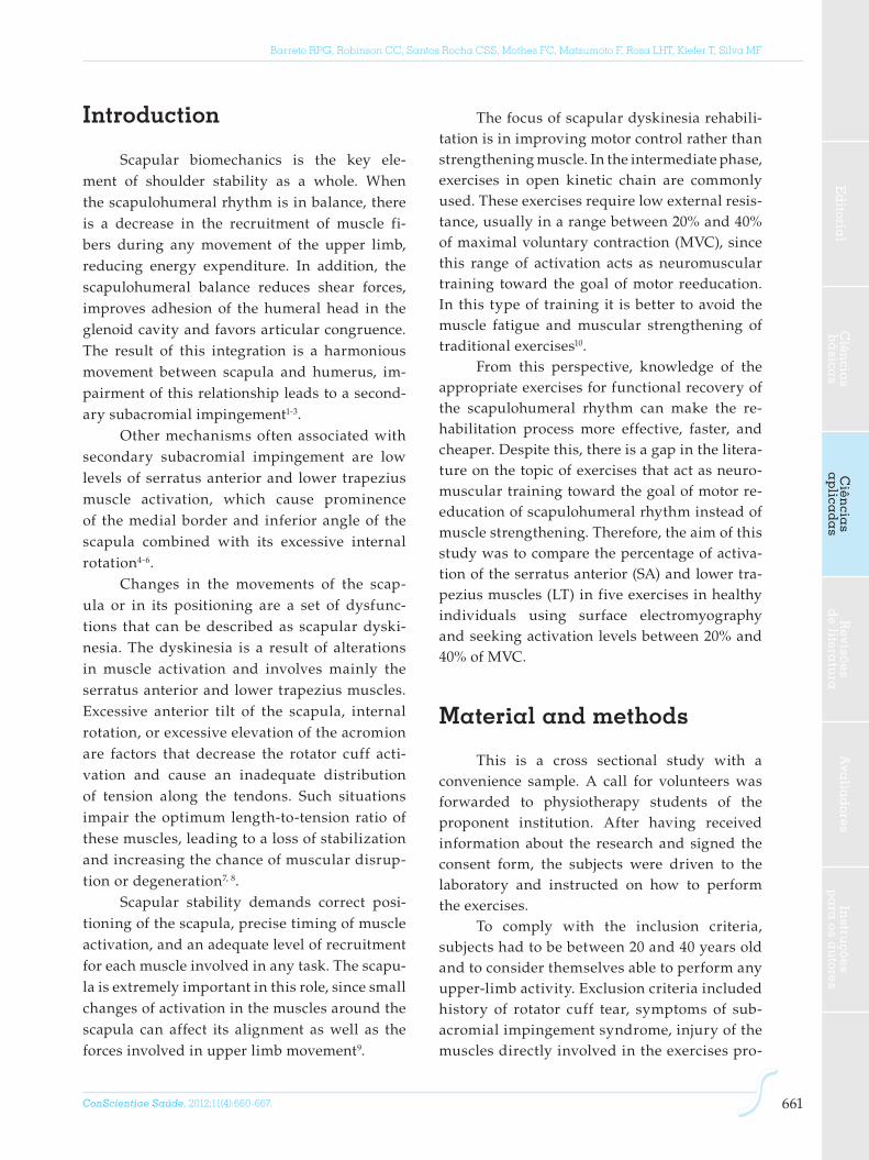
Introduction
Scapular biomechanics is the key ele-ment of shoulder stability as a whole. When the scapulohumeral rhythm is in balance, there is a decrease in the recruitment of muscle fi-bers during any movement of the upper limb, reducing energy expenditure. In addition, the scapulohumeral balance reduces shear forces, improves adhesion of the humeral head in the glenoid cavity and favors articular congruence. The result of this integration is a harmonious movement between scapula and humerus, im-pairment of this relationship leads to a second-ary subacromial impingement1-3.
Other mechanisms often associated with secondary subacromial impingement are low levels of serratus anterior and lower trapezius muscle activation, which cause prominence of the medial border and inferior angle of the scapula combined with its excessive internal rotation4-6.
Changes in the movements of the scap-ula or in its positioning are a set of dysfunc-tions that can be described as scapular dyski-nesia. The dyskinesia is a result of alterations in muscle activation and involves mainly the serratus anterior and lower trapezius muscles. Excessive anterior tilt of the scapula, internal rotation, or excessive elevation of the acromion are factors that decrease the rotator cuff acti-vation and cause an inadequate distribution of tension along the tendons. Such situations impair the optimum length-to-tension ratio of these muscles, leading to a loss of stabilization and increasing the chance of muscular disrup-tion or degeneration7, 8.
Scapular stability demands correct posi-tioning of the scapula, precise timing of muscle activation, and an adequate level of recruitment for each muscle involved in any task. The scapu-la is extremely important in this role, since small changes of activation in the muscles around the scapula can affect its alignment as well as the forces involved in upper limb movement9.
The focus of scapular dyskinesia rehabili-tation is in improving motor control rather than strengthening muscle. In the intermediate phase, exercises in open kinetic chain are commonly used. These exercises require low external resis-tance, usually in a range between 20% and 40% of maximal voluntary contraction (MVC), since this range of activation acts as neuromuscular training toward the goal of motor reeducation. In this type of training it is better to avoid the muscle fatigue and muscular strengthening of traditional exercises10.
From this perspective, knowledge of the appropriate exercises for functional recovery of the scapulohumeral rhythm can make the re-habilitation process more effective, faster, and cheaper. Despite this, there is a gap in the litera-ture on the topic of exercises that act as neuro-muscular training toward the goal of motor re-education of scapulohumeral rhythm instead of muscle strengthening. Therefore, the aim of this study was to compare the percentage of activa-tion of the serratus anterior (SA) and lower tra-pezius muscles (LT) in five exercises in healthy individuals using surface electromyography and seeking activation levels between 20% and 40% of MVC.
Material and methods
This is a cross sectional study with a convenience sample. A call for volunteers was forwarded to physiotherapy students of the proponent institution. After having received information about the research and signed the consent form, the subjects were driven to the laboratory and instructed on how to perform the exercises.
To comply with the inclusion criteria, subjects had to be between 20 and 40 years old and to consider themselves able to perform any upper-limb activity. Exclusion criteria included history of rotator cuff tear, symptoms of sub-acromial impingement syndrome, injury of the muscles directly involved in the exercises pro-

ConScientiae Saúde, 2012;11(4):660-667.662
Lower trapezius and serratus anterior activation: which exercise to use for scapular neuromuscular reeducation?
posed in this research, and having performed any kind of strength training or regular physi-cal activities that use the muscles being as-sessed.
The principles of the Declaration of Helsinki and Resolution 196/96 of the National Health Council11, 12 were applied, and all pa-tients provided written informed consent to participate. The study was approved by the Ethics Committee of the Centro Universitário Metodista do IPA, Rio Grande do Sul, Brazil, in decision 256/2010.
Data acquisitionThe acquisition of Electromyographic
(EMG) signals was performed using an elec-tromyography device (Miotec®, model Miottol 400) with four channels, 14-bit resolution, elec-trical isolation of 5000 volts, 2000 Hz per chan-nel, common mode rejection ratio of 110db, and connection to a computer by USB port. Data were collected and analyzed using Miograph 2.0 software. A notebook was used to collect and process data. To capture EMG signals, Ag/AgCl (silver/silver chloride) surface electrodes (Meditrace®, Canada) with diameter of 2.2 cm were used in bipolar configuration on the skin overlying the belly of the serratus anterior and the lower trapezius, according to guidelines of the International Society of Electrophysiology and Kinesiology (ISEK)13.
Procedures for data collectionThe skin impedance was reduced by asep-
sis with alcohol (70%) and gentle skin abrasion. The placement of electrodes followed the recom-mendations of ISEK13. For the lower trapezius muscle, the subject was placed in humeral flex-ion of 90°, and the electrodes were positioned at an oblique and vertical angle 5 cm apart from the base of the spine of the scapula, parallel to the muscle fibers. For the serratus anterior mus-cle, the shoulder was abducted 90°, and the elec-trodes positioned horizontally in line with the
lower angle of the scapula, just anterior to the latissimus dorsi muscle. The reference electrode was positioned on the anterior tibial tuberosity of the right leg.
After having the electrodes placed on them, the subjects performed some upper-limb movements: elevation, shoulder flexion, external rotation, and internal rotation of the scapula only to confirm the position of the elec-trodes and the reception of myoeletric signals. Immediately after these procedures, the subjects were instructed on accomplishing maximum voluntary contraction (MVC) of the serratus an-terior and lower trapezius muscles, following Kendall s14 protocol. The MVC was followed by a self-determined rest period, and the subjects began the exercises when they considered them-selves able to do so.
Data analysisMiograph 2.0 software was used for analy-
sis, and the following procedures were adopt-ed: EMG signals were filtered by a third-order band-pass Butterworth filter with cut-off fre-quencies between 20 and 500Hz; EMG signals were clipping (considering the beginning and end of each contraction) and the average RMS (root mean square) was calculated for the two muscles of interest. For data analysis, we used all RMS values normalized for MVC.
Physical exercisesThe exercise protocol was started with a
verbal communication and was composed of one series of five repetitions for each of the five different exercises, with an interval of at least one minute of rest between every new exercise or until the subjects had considered themselves able to continue. Initially, each exercise was demonstrated by the same researcher for each participant, and, immediately after, the subject was instructed to perform the exercise until the completion of the protocol.

ConScientiae Saúde, 2012;11(4):660-667. 663
Barreto RPG, Robinson CC, Santos Rocha CSS, Mothes FC, Matsumoto F, Rosa LHT, Kiefer T, Silva MF
Ed
itorial
Ciên
cias
bá
sicas
Ciên
cias
ap
licad
as
Revisões
de litera
tura
Ava
liad
oresIn
struções
pa
ra os a
utores
The rhythm of each exercise was con-trolled by a digital metronome; thus, all partici-pants had the same execution rhythm. A load of 2 kg was used for the purpose of neuromuscu-lar training, since this load can induce appropri-ate levels of activation. Moreover, this load can serve as a direct reference to other professionals in clinical practice10.
Modified crucifix (CM): in supine posi-tion, headboard elevated 45°, horizontal flex-ion of the shoulder with the forearm in prona-tion and the elbow in full extension, bilateral execution with a starting position in a 90° ab-duction, and a final position in a 115° abduc-tion (Figure 1a).
Scaption (SP): in a standing position, a uni-lateral execution of 130° elevation in the scapu-lar plane with the forearm in neutral position (Figure 1b).
Modified military press (MMP): in a sitting position with headboard elevated 45°, initial po-sition of both upper limbs with abduction (90°) in the plane of the scapula, elbow flexion (90°), and forearm supination. Final position with el-bow extension and shoulder forearm pronation (120°) (Figure 1c).
Pull-over (PO): in a supine position, shoul-der flexion, holding a dumbbell with both hands, an initial position of 90° shoulder flexion and elbow in full extension, and a final posi-tion on the edge of the active range of shoulder flexion with 15° of maximum tolerance of elbow flexion (Figure 1d).
Low-row (LR): in a standing position with the arm alongside the body and the forearm in pronation, internal rotation of the humerus; the subject performs combined motion of shoulder extension and scapular retraction (Figure 1e).
Statiscal analysisNormality of data distribution was veri-
fied with the Shapiro-Wilk test, and data explo-ration was conducted using descriptive statis-tics (mean and standard deviation or median, minimum, and maximum). The normalized
signals of the muscles studied were compared among the exercises with the Wilcoxon test. We adopted the significance level of 5%. Data were analyzed by SPSS for Windows (version 17.0) sta-tistical software.
Results
A total of 16 subjects agreed to partici-pate in the study, but six were excluded because they were undergoing strength training. Thus, ten subjects, all female, with a mean age of 24.5 years (3.1), remained in the sample.
The EMG signals normalized for MCV with median, minimum and maximum values, arranged by type of exercise, are reported in Table 1.
The five exercises elicited significant dif-ferences in muscular activation of the SA and LT muscles.
The highest SA muscle percentage of MVC was found during the scaption, and adequate percentage of MVC occurred during the modi-fied military press. Regarding LT muscle activa-tion, the highest percentage of MVC was found in the scaption and low row exercises.
Figure 1: Exercises performed by the subjectsA: Modified crucifix; B: Scaption; C: Pull Over; D: Modified Military Press; E: Low row.

ConScientiae Saúde, 2012;11(4):660-667.664
Lower trapezius and serratus anterior activation: which exercise to use for scapular neuromuscular reeducation?
Discussion
Our study investigated some exercises appropriate for the intermediate phase of func-tional recovery in patients who need greater activation of the serratus anterior and lower trapezius muscles, following the activation range proposed for neuromuscular reeduca-tion. These exercises were chosen because al-most all of them are conducted in open kinetic chain, requiring minimum scapular control and avoiding compensation with the scapula or trunk.
We found high levels of activation of the SA in SP – 53% (24-76) – and adequate levels of activation in MMP – 29% (10-55). These two exercises were those that exhibited the highest percentages of MVC (Table 1).
Regarding the activation of the lower tra-pezius muscle, we found higher percentages of MVC in SP – 27% (8-47) and LR – 26.5% (17-56) (Table 1).
The MC and PO exercises exhibited medi-ans that can be considered close to adequate for neuromuscular reeducation, but only in the ser-ratus anterior muscle.
There is considerable evidence in the scien-tific literature that in scapular dyskinesia there is a lack of muscle coordination that is related to poor muscular activation of the lower trapezius
and serratus anterior muscles. It is suggested that this incoordination contributes to second-ary impingement syndrome. Therefore, the ba-sis of this disorder is mainly neuromuscular. This could not be linked to muscular weakness, given that it is related to motor control. Thus, as previously mentioned in our work, the exercises for functional recovery of patients with this im-balance must be performed with reduced activa-tion, around 20% to 40%15.
Exercises that exceed this activation threshold are producing the effect of muscle strengthening. Moreover, exercises that come close to 100% activation cause the recruitment of other muscle fibers, not only those of the target muscle, thereby causing adaptive movements which do not contribute to neuromuscular re-education.
In this context, there are other studies with similar purposes investigating exercises that could be proposed for functional recovery, be-cause they are executed in open kinetic chain re-quiring a minimum of motor control. However, most of these studies do not contribute to the complete elucidation of the exercises that are suitable for a functional recovery having the goal of improving motor control.
In the Ekstrom et al.16 study, it is pos-sible to note that only three of the ten inves-tigated exercises are in the activation range of interest. The first was an isometric exercise in which the upper limb was positioned with humeral elevation above the head and aligned with the fibers of the lower trapezius muscle in a prone decubitus showing 43%±17% MVC for serratus anterior and 97%±16% MVC for the lower trapezius muscle. It should be noted that this exercise elicited almost 100% MVC of the lower trapezius. Since it was conducted in the prone decubitus and in almost maximal acti-vation with the limb fully extended above the head, especially in this position it could lead to the coactivation of the upper trapezius mus-cles because of the need of neck extension. So this exercise can only be performed if the sub-ject has moderate or good motor control. The
Table 1: Muscular activation normalized for MVC
ExerciseLower
trapezius (%MVC)
Serratus anterior (%MVC)
p
Modified crucifix 7 (2-12) 15 (9-31) 0.005
Scaption 27 (8-47) 53.5 (24-76) 0.032
Modified military press
6 (3-12) 29 (10-55) 0.005
Pull over 4.5 (3-11) 13 (6-18) 0.011
Low row 26.5 (17-56) 10.5 (5-17) 0.005
Values of median with minimum and maximum. MVC: maximal voluntary contraction, significan-ce when p<0,05.

ConScientiae Saúde, 2012;11(4):660-667. 665
Barreto RPG, Robinson CC, Santos Rocha CSS, Mothes FC, Matsumoto F, Rosa LHT, Kiefer T, Silva MF
Ed
itorial
Ciên
cias
bá
sicas
Ciên
cias
ap
licad
as
Revisões
de litera
tura
Ava
liad
oresIn
struções
pa
ra os a
utores
second exercise was the unilateral row, which showed 45%±17% MVC for the lower trapezius muscle and 14%±6% MVC for the serratus an-terior muscle; and the third was a combined exercise in a position of 90° shoulder flexion, adduction, and external rotation, which caused 100%±24% MVC for the serratus anterior mus-cle and 39%±15% MVC for the lower trapezius.
In another study, Cools et al.17, had an ex-cellent proposal comparing 12 exercises and measuring the activation ratio of the serratus anterior, middle, and lower trapezius muscles, all in relation to the upper trapezius muscle. This study presented some important method-ological issues worth highlighting. The partici-pants performed three repetitions of MVC with five-second rest intervals, which would provide fatigue influence on EMG signals, decreasing their reliability. Finally, the authors did not ex-plain clearly how the analysis of electromyo-graphic signals was performed.
Arlotta et al.18 measured the lower trapezius activity during five exercises. Only three of them showed activation in the recommended range for neuromuscular reeducation. In the modified prone cobra, 44.67%±14.60% activation was ob-served; in the prone row, 36.44%±14.35%; and in the latissimus pull down, 35.31%±20% MVC. So all these exercises would be suitable in regard to the activation threshold. Although the author has used only isometric exercises, these exercises are interesting because they match the findings of our study. The factor that could be an inter-fering variable in this study is that the external resistance was imposed by the researcher’s hand and always by the same person; but even so, it is impossible to be sure whether resistance and duration was the same in all subjects. Thus, the isometric load would only apply if other instru-ments were inserted into the methodology, such as a load cell.
A very interesting study conducted by Decker et al.19 measured the activity of the ser-ratus anterior muscle in eight exercises. There is little information about the imposed load for each individual; moreover, it is noted that MVC
was repeated five times with a small rest in-terval of three to five seconds, which could in-fluence myoelectric signals by muscle fatigue. Three exercises proposed by Decker were con-ducted in open kinetic chain (the serratus ante-rior punch, the scaption and the dynamic hug). It is noted that all muscle activations reached were very much above 40% MVC. Therefore, those exercises should be contemplated when the aim is muscle strengthening, as concluded by the authors.
Another interesting study that aimed at helping in the choice of exercises for motor con-trol recovery of the serratus anterior and lower trapezius muscles and that involved healthy individuals only was the study of Witt et al.20. Twenty-one subjects performed exercises with an elastic resistance band and dumbbells, all in diagonal patterns; however, for statistical analysis, the authors did not perform any test to verify the normality of data distribution, and in the results analysis the absence of symmetrical distribution was evident. The standard devia-tion exceeds the mean, and even doubles it, for several variables; but the authors used a robust test for parametric outcomes, which greatly in-creases the chance of a type I error, leading to a non-reliable conclusion.
In this study, like other studies using sur-face electromyography to measure muscular electrical activity in dynamic contractions, there is the possibility of classic EMG bias, which is based on the fact that the electrodes move over the skin of the individual during the muscle contractions and that this can change the read-ing of the muscle fibers, capturing signals from different ones. Moreover, we stress the fact that the fibers show differences in length and con-traction velocity during the dynamic exercises. However, the same linear relationship present-ed in isometric exercises may be reproduced in the dynamic exercise if the speed of exercise is constant, which happened in our study, as de-scribed in the methodology21-23.
Another factor for consideration is that the sample used in this study was composed of

ConScientiae Saúde, 2012;11(4):660-667.666
Lower trapezius and serratus anterior activation: which exercise to use for scapular neuromuscular reeducation?
women only. Some authors suggest that there would be differences in activation between genders, but there is still controversy about this, especially in relation to the muscles of the shoulder complex. The most specific study that used EMG and tried to prove the differences in activation has some methodological questions related to signal analysis and to the utilized tasks. Specifically, all tasks were isometric, and the analysis was performed using relative mus-cle activation, differing from our study24.
This research was conducted with a re-duced sample and healthy individuals; hence, without scapular dyskinesia. It would be inter-esting to include a group with the dysfunction of interest. For these reasons we suggest cau-tion in extrapolating the results of our study to different populations. Nevertheless, our results contribute to the choice of suitable exercises in clinical practice during functional recov-ery from neuromuscular balance and provide insights for future studies that evaluate more muscles, such as the upper trapezius and its relationship with the serratus anterior, or that include a group with scapular dyskinesia to measure the effects of an intervention, measur-ing the degree of activation induced by other exercises.
Conclusions
The exercises that stayed within the rec-ommended range of activation for neuromuscu-lar reeducation among the five proposed exer-cises were the modified military press, for the SA muscle, and the low row and scaption, for the LT muscle.
In view of the methodological questions of some studies that used exercises in open ki-netic chain or exercises that require a minimum scapular control, we conducted a study that provides more information regarding muscle activation of the lower trapezius and serratus anterior muscles, filling the gap in this area of scientific study.
References1. Kibler WB. Management of the scapula in
glenohumeral instability. Techniques Shoulder and
Elbow Surgery. 2003;4(3):89-98.
2. Karandikar N, Vargas OOO. Kinetic chains: a review
of the concept and its clinical applications. PM R.
2011;3(8):739-45.
3. Maenhout A, Praet KV, Pizzi L, Herzeele MV, Cools
A. Electromyographic analysis of knee push up
plus variations: what is the influence of the kinetic
chain on scapular muscle activity? Br J Sports Med.
2010;44(14):1010-5.
4. Nijs J, Roussel N, Struyf F,Mottram S Meeusen
R. Clinical assessment of scapular positioning
in patients with shoulder pain: state of the art. J
Manipulative Physiol Ther. 2007;30(1):69-75.
5. Lewis J, Green A, Dekel S. The aetiology of
subacromial impingement syndrome. Physiotherapy.
2001;87(9):458-69.
6. Budoff J, Nirschl RP, Guidi EJ. Débridement of
partial-thickness tears of the rotator cuff without
acromioplasty: long-term follow-up and review of
the literature. J Bone Joint Surg AM. 1998;80(5):733-
48.
7. Hébert L, Moffet H, McFadyen BJ, Dionne CE.
Scapular behavior in shoulder impingement
syndrome. Arch Phys Med Rehabil. 2002;83(1):60-9.
8. Dickens V, Williams J, Bhamra M. Role of
physiotherapy in the treatment of subacromial
impingement syndrome: a prospective study.
Physiotherapy. 2005;91(3):159-64.
9. Cools A, Witvrouw EE, Declercq GA, Danneels
LA, Cambier DC. Scapular muscle recruitment
patterns: trapezius muscle latency with and
without impingement symptoms. Am J Sports Med.
2003;31(4):542-9.
10. Tucci HT, Ciol MA, Araújo RC Andrade R de,
Martins J, McQuade KJ, Oliveira AS. Activation of
selected shoulder muscles during unilateral wall
and bench press tasks under submaximal isometric
effort. J Orthop Sports Phys Ther. 2011;41(7):520-5.
11. World Medical Association. Declaration of Helsinki:
Ethical Principles for medical Research Involving
Human Subjects, as amended by the 52nd WMA
Assembly, Edinburgh, Scotland, October 2000; Note
of Clarification in Paragraph 29 added by the WMA
General Assembly, Washington, DC; 2002.

ConScientiae Saúde, 2012;11(4):660-667. 667
Barreto RPG, Robinson CC, Santos Rocha CSS, Mothes FC, Matsumoto F, Rosa LHT, Kiefer T, Silva MF
Ed
itorial
Ciên
cias
bá
sicas
Ciên
cias
ap
licad
as
Revisões
de litera
tura
Ava
liad
oresIn
struções
pa
ra os a
utores
12. Conselho Nacional de Saúde (Brasil). Resolução
196/96. Diretrizes e Normas Regulamentadoras de
Pesquisas Envolvendo Seres Humanos. Brasília;
Conselho Nacional de Saúde; 1996.
13. ISEK-online.org [internet]. International Society of
Electrophysiology and Kinesiology [Accessed at:
2012 Dec]. Available from: <http://www.isek-online.
org/standards_emg.html>.
14. Kendall, P. Músculos, provas e funções. 4ª ed. São
Paulo: Manole; 1995.
15. Chester R, Smith TO, Hooper L, Dixon J. The
impact of subacromial impingement, syndrome on
muscle activity patterns of the shoulder complex:
a systematic review of electromyographic studies.
BMC Musculoskelet Disord. 2010;11(45):1-12.
16. Ekstrom RA, Donatelli RA, Soderberg GL. Surface
electromyographic analysis of exercises for the
trapezius and serratus anterior muscles. J Orthop
Sports Phys Ther. 2003;33(5):247-58.
17. Cools A, Dewitte V, Lanszweert F, Notebaert D,
Roets A, Soetens B, et al. Rehabilitation of scapular
muscle balance: which exercises to prescribe? Am J
Sports Med. 2007;35(10):1744-51.
18. Arlotta M, Lovasco G, McLean L. Selective
recruitment of the lower fibers of the trapezius
muscle. J Electromyogr Kinesiol. 2011;21(3):403-10.
19. Decker MJ, Hintermeister R, Faber KJ, Hawkins RJ.
SA muscle activity during selected rehabilitation
exercises. Am J Sports Med. 1999;27(6):784-91.
20. Witt D, Talbott N, Kotowski S. Electromyographic
activity of scapular muscles during diagonal
patterns using elastic resistance and free weights.
Int J Sports Phys Ther. 2011;6(4):322-32.
21. Basmajian JV. Electromyography Comes of Age.
Science. 1972;176(40325):603-9.
22. De Luca CJ. The Use of Surface Electromyography in
Biomechanics. J Appl Biomech. 1997;13:135-63.
23. Hug F. Can muscle coordination be precisely studied
by surface electromyography? J Electromyogr
Kinesiol. 2011;21:1-12.
24. Anders C, Bretschneider S, Bernsdorf A, Erler K,
Schneider W. Activation of shoulder muscles in
healthy men and women under isometric conditions.
J Electromyogr Kinesiol. 2004;14(6):699-707.