Dnmt1 Overexpression Causes Genomic Hypermethylation, Loss
12
MOLECULAR AND CELLULAR BIOLOGY, Apr. 2002, p. 2124–2135 Vol. 22, No. 7 0270-7306/02/$04.000 DOI: 10.1128/MCB.22.7.2124–2135.2002 Copyright © 2002, American Society for Microbiology. All Rights Reserved. Dnmt1 Overexpression Causes Genomic Hypermethylation, Loss of Imprinting, and Embryonic Lethality Detlev Biniszkiewicz, 1 Joost Gribnau, 1 Bernard Ramsahoye, 2 François Gaudet, 1,3 Kevin Eggan, 1 David Humpherys, 1 Mary-Ann Mastrangelo, 1 Zhan Jun, 4 Jörn Walter, 4 and Rudolf Jaenisch 1 * Whitehead Institute for Biomedical Research and Massachusetts Institute of Technology, Cambridge, Massachusetts 02142 1 ; Department of Oncology, John Hughes Bennett Laboratory, Western General Hospital, Edinburgh EH4 2XU, United Kingdom 2 ; and Max-Delbrück-Center for Molecular Medicine, 13125 Berlin, 3 and Max-Planck Institute for Molecular Genetik, 14195 Berlin, 4 Germany Received 8 October 2001/Returned for modification 30 November 2001/Accepted 4 January 2002 Biallelic expression of Igf2 is frequently seen in cancers because Igf2 functions as a survival factor. In many tumors the activation of Igf2 expression has been correlated with de novo methylation of the imprinted region. We have compared the intrinsic susceptibilities of the imprinted region of Igf2 and H19, other imprinted genes, bulk genomic DNA, and repetitive retroviral sequences to Dnmt1 overexpression. At low Dnmt1 methyltrans- ferase levels repetitive retroviral elements were methylated and silenced. The nonmethylated imprinted region of Igf2 and H19 was resistant to methylation at low Dnmt1 levels but became fully methylated when Dnmt1 was overexpressed from a bacterial artificial chromosome transgene. Methylation caused the activation of the silent Igf2 allele in wild-type and Dnmt1 knockout cells, leading to biallelic Igf2 expression. In contrast, the imprinted genes Igf2r, Peg3, Snrpn, and Grf1 were completely resistant to de novo methylation, even when Dnmt1 was overexpressed. Therefore, the intrinsic difference between the imprinted region of Igf2 and H19 and of other imprinted genes to postzygotic de novo methylation may be the molecular basis for the frequently observed de novo methylation and upregulation of Igf2 in neoplastic cells and tumors. Injection of Dnmt1-overexpressing embryonic stem cells in diploid or tetraploid blastocysts resulted in lethality of the embryo, which resembled embryonic lethality caused by Dnmt1 deficiency. The two most common mechanisms to induce tumor growth are the activation of oncogenes and the inactivation of tumor suppressor genes. Both genetic alterations or epigenetic events can result in activation of oncogenes (8, 9) or in the silencing of tumor suppressor genes (17, 40). Imprinted genes that affect cellular growth are particularly vulnerable targets in tumori- genesis because only one allele of an imprinted gene is ex- pressed. Indeed, activation of the silent Igf2 allele leading to biallelic Igf2 expression is a frequent event in neoplasia, which provides the cell with a strong growth signal. Loss of imprinting (LOI) and biallelic Igf2 expression have been described to occur in over 20 different tumor types, including cancers of the liver, breast, pancreas, and colon (16, 27, 30, 35, 40). In addi- tion, it has been shown that experimental overexpression of Igf2 in mice leads to increased cell proliferation, overgrowth, and increased probability of malignant transformation (2, 34, 38). It has been suggested that deregulation of Igf2 expression is caused by de novo methylation of the differentially methylated domain (DMD), which controls expression of Igf2 and H19 (43, 52). In normal cells the DMD region is methylated on the paternal chromosome and unmethylated on the maternal chro- mosome, leading to paternal expression of Igf2 and maternal expression of H19. During tumorigenesis, the unmethylated DMD region is frequently methylated, and this has been cor- related with biallelic expression of Igf2 and silencing of H19 (27, 40). In contrast, the active allele of imprinted tumor sup- pressor genes such as Igf2r is inactivated during tumorigenesis through genetic mechanisms and not by epigenetic changes (16). Although the imprinted status of the human Igf2r gene remains unclear, during liver tumorigenesis in rats Igf2r func- tion is always deleted by mutations and loss of heterozygosity, which is in contrast to the LOI of Igf2 and H19 (16). These observations raise the question of whether Igf2 and H19 have an intrinsic susceptibility to de novo methylation that is differ- ent from other imprinted genes. Inheritable DNA methylation patterns are established through a two-step process that converts two unmethylated cytosine residues within palindromic CpG dinucleotides into two methylated CpG dinucleotides with hemi-methylated DNA as an intermediate. During early development the estab- lishment of genomic methylation patterns is accomplished by the concerted action of Dnmt3 and Dnmt1 DNA methyltrans- ferases (MTases). Dnmt3a and Dnmt3b, both expressed in early embryos, have been shown to “de novo methylate” un- modified DNA after implantation of blastocysts and in trans- genic flies, resulting in hemi-methylated DNA (26, 31). How- ever, the activity of the hemi-MTase Dnmt1 is crucial to achieving a normal inheritable methylation level of the post- gastrulation embryo, because this enzyme acts as a mainte- nance methylase that recognizes the hemi-methylated CpG sites established by Dnmt3a and -b, methylates the comple- mentary CpG and, thus, converts the respective CpG site to a fully methylated state. Consistent with this notion is the obser- vation that the deficiency of Dnmt1 leads to genomic hypo- methylation and embryonic lethality after gastrulation (23). * Corresponding author. Mailing address: Whitehead Institute for Biomedical Research, Nine Cambridge Center, Cambridge MA 02142. Phone: (617) 258-5186. Fax: (617) 258-6505. E-mail: [email protected] .edu. 2124 Downloaded from https://journals.asm.org/journal/mcb on 03 February 2022 by 59.25.78.181.
Transcript of Dnmt1 Overexpression Causes Genomic Hypermethylation, Loss

MOLECULAR AND CELLULAR BIOLOGY, Apr. 2002, p. 2124–2135 Vol. 22, No. 70270-7306/02/$04.00�0 DOI: 10.1128/MCB.22.7.2124–2135.2002Copyright © 2002, American Society for Microbiology. All Rights Reserved.
Dnmt1 Overexpression Causes Genomic Hypermethylation, Loss ofImprinting, and Embryonic Lethality
Detlev Biniszkiewicz,1 Joost Gribnau,1 Bernard Ramsahoye,2 François Gaudet,1,3 Kevin Eggan,1David Humpherys,1 Mary-Ann Mastrangelo,1 Zhan Jun,4 Jörn Walter,4 and Rudolf Jaenisch1*
Whitehead Institute for Biomedical Research and Massachusetts Institute of Technology, Cambridge, Massachusetts 021421;Department of Oncology, John Hughes Bennett Laboratory, Western General Hospital, Edinburgh EH4 2XU, United Kingdom2;
and Max-Delbrück-Center for Molecular Medicine, 13125 Berlin,3 and Max-Planck Institute for Molecular Genetik,14195 Berlin,4 Germany
Received 8 October 2001/Returned for modification 30 November 2001/Accepted 4 January 2002
Biallelic expression of Igf2 is frequently seen in cancers because Igf2 functions as a survival factor. In manytumors the activation of Igf2 expression has been correlated with de novo methylation of the imprinted region.We have compared the intrinsic susceptibilities of the imprinted region of Igf2 and H19, other imprinted genes,bulk genomic DNA, and repetitive retroviral sequences to Dnmt1 overexpression. At low Dnmt1 methyltrans-ferase levels repetitive retroviral elements were methylated and silenced. The nonmethylated imprinted regionof Igf2 and H19 was resistant to methylation at low Dnmt1 levels but became fully methylated when Dnmt1 wasoverexpressed from a bacterial artificial chromosome transgene. Methylation caused the activation of the silentIgf2 allele in wild-type and Dnmt1 knockout cells, leading to biallelic Igf2 expression. In contrast, the imprintedgenes Igf2r, Peg3, Snrpn, and Grf1 were completely resistant to de novo methylation, even when Dnmt1 wasoverexpressed. Therefore, the intrinsic difference between the imprinted region of Igf2 and H19 and of otherimprinted genes to postzygotic de novo methylation may be the molecular basis for the frequently observed denovo methylation and upregulation of Igf2 in neoplastic cells and tumors. Injection of Dnmt1-overexpressingembryonic stem cells in diploid or tetraploid blastocysts resulted in lethality of the embryo, which resembledembryonic lethality caused by Dnmt1 deficiency.
The two most common mechanisms to induce tumor growthare the activation of oncogenes and the inactivation of tumorsuppressor genes. Both genetic alterations or epigenetic eventscan result in activation of oncogenes (8, 9) or in the silencingof tumor suppressor genes (17, 40). Imprinted genes that affectcellular growth are particularly vulnerable targets in tumori-genesis because only one allele of an imprinted gene is ex-pressed. Indeed, activation of the silent Igf2 allele leading tobiallelic Igf2 expression is a frequent event in neoplasia, whichprovides the cell with a strong growth signal. Loss of imprinting(LOI) and biallelic Igf2 expression have been described tooccur in over 20 different tumor types, including cancers of theliver, breast, pancreas, and colon (16, 27, 30, 35, 40). In addi-tion, it has been shown that experimental overexpression ofIgf2 in mice leads to increased cell proliferation, overgrowth,and increased probability of malignant transformation (2, 34,38).
It has been suggested that deregulation of Igf2 expression iscaused by de novo methylation of the differentially methylateddomain (DMD), which controls expression of Igf2 and H19 (43,52). In normal cells the DMD region is methylated on thepaternal chromosome and unmethylated on the maternal chro-mosome, leading to paternal expression of Igf2 and maternalexpression of H19. During tumorigenesis, the unmethylatedDMD region is frequently methylated, and this has been cor-related with biallelic expression of Igf2 and silencing of H19
(27, 40). In contrast, the active allele of imprinted tumor sup-pressor genes such as Igf2r is inactivated during tumorigenesisthrough genetic mechanisms and not by epigenetic changes(16). Although the imprinted status of the human Igf2r generemains unclear, during liver tumorigenesis in rats Igf2r func-tion is always deleted by mutations and loss of heterozygosity,which is in contrast to the LOI of Igf2 and H19 (16). Theseobservations raise the question of whether Igf2 and H19 havean intrinsic susceptibility to de novo methylation that is differ-ent from other imprinted genes.
Inheritable DNA methylation patterns are establishedthrough a two-step process that converts two unmethylatedcytosine residues within palindromic CpG dinucleotides intotwo methylated CpG dinucleotides with hemi-methylatedDNA as an intermediate. During early development the estab-lishment of genomic methylation patterns is accomplished bythe concerted action of Dnmt3 and Dnmt1 DNA methyltrans-ferases (MTases). Dnmt3a and Dnmt3b, both expressed inearly embryos, have been shown to “de novo methylate” un-modified DNA after implantation of blastocysts and in trans-genic flies, resulting in hemi-methylated DNA (26, 31). How-ever, the activity of the hemi-MTase Dnmt1 is crucial toachieving a normal inheritable methylation level of the post-gastrulation embryo, because this enzyme acts as a mainte-nance methylase that recognizes the hemi-methylated CpGsites established by Dnmt3a and -b, methylates the comple-mentary CpG and, thus, converts the respective CpG site to afully methylated state. Consistent with this notion is the obser-vation that the deficiency of Dnmt1 leads to genomic hypo-methylation and embryonic lethality after gastrulation (23).
* Corresponding author. Mailing address: Whitehead Institute forBiomedical Research, Nine Cambridge Center, Cambridge MA 02142.Phone: (617) 258-5186. Fax: (617) 258-6505. E-mail: [email protected].
2124
Dow
nloa
ded
from
http
s://j
ourn
als.
asm
.org
/jour
nal/m
cb o
n 03
Feb
ruar
y 20
22 b
y 59
.25.
78.1
81.
It has been shown that the deletion of Dnmt1 in mice leads,in addition to genome-wide hypomethylation and embryoniclethality, to loss of monoallelic expression of imprinted genes(23). Dnmt1 mutant mice show biallelic expression of H19 andsilencing of the active Igf2 and Igf2r alleles. In addition, dereg-ulation of several other imprinted genes, including p57Kip2and Kvlqt1, has been observed in Dnmt1 mutant mouse em-bryos, further demonstrating the involvement of DNA meth-ylation in the maintenance of genomic imprinting (4). In con-trast, Mash2 was unaffected by the lack of Dnmt1 MTase,suggesting either that not every imprinted gene is controlled byDNA methylation or that the Dnmt1 mutant embryos diedprior to complete demethylation of this locus. Reexpression ofthe Dnmt1 cDNA in Dnmt1 homozygous mutant (Dnmt1�/�)embryonic stem (ES) cells at low levels did not result in rem-ethylation of parental imprints despite restoration of the over-all genomic methylation level (45), implying that methylationimprints can only be imposed on imprinted genes during ga-metogenesis and that postzygotic cells lack the ability to denovo methylate imprinting boxes.
Increased MTase activity and changes of DNA methylationpatterns are commonly seen in neoplastic cells and tumors ofhumans and mice (3, 6, 19, 49), and this increase in MTaseactivity has been linked to the deregulation of tumor suppres-sor genes, oncogenes, and imprinted genes (47). Because denovo methylation and biallelic expression of Igf2 is a frequentalteration in neoplastic cells and tumors, we tested whether theimprinted region of Igf2 and H19 is particularly susceptible toelevated levels of Dnmt1 expression, distinguishing Igf2 andH19 from other imprinted genes. In this study, we generatedES cells with a wide range of Dnmt1 expression levels in wild-type and in Dnmt1�/� ES cells to determine whether the im-printed region of Igf2 and H19 has a different intrinsic sensi-tivity to postzygotic de novo methylation in comparison toother imprinted genes.
MATERIALS AND METHODS
Dnmt1 BACs. PCR screening with two different sets of primers of a 129Sv/Jmouse bacterial artificial chromosome (BAC) library from the Whitehead Insti-tute Genome Center identified a BAC clone containing the Dnmt1 gene. TheBAC clone Dnmt1-BAC has a length of 150 kb as determined by pulsed-field gelelectrophoresis. The BAC clone was shown to contain the complete Dnmt1 geneby a combination of PCR and Southern hybridization techniques.
ES cell culture and transfection. Wild-type ES cells (J1 ES cells) and Dnmt1c/c
homozygous mutant ES cells (22) were cultivated on �-irradiated murine em-bryonic fibroblasts (mEF) as described previously (24) or without mEF by usinga high concentration of leukemia inhibitory factor (LIF; 1,000 U/ml). All trans-fections were done using the cationic liposome reagent DOTAP from Boehr-inger Mannheim. To prepare BAC DNA for transfection, bacteria containing theartificial chromosomes were grown on Luria-Bertani medium with 12.5 �g ofchloramphenicol/ml. BAC DNA was prepared using standard Qiagen columns.The quality of the BAC DNA was controlled by pulsed-field gel electrophoresis.Uncut BAC DNA was used for lipofection, due to the lack of an unique restric-tion site for linearization.
DNA preparation and methylation analysis. Cells were digested in lysis buffer(100 mM Tris-HCl [pH 8.5], 5 mM EDTA, 0.2% sodium dodecyl sulfate [SDS],200 mM NaCl, 100 to 300 �g of proteinase K/ml) for several hours at 55°C,phenol-chloroform extracted, and precipitated with an equal volume of isopro-panol. Ten micrograms of DNA was digested with the stated restriction endo-nuclease for 12 to 16 h. The products were resolved on an agarose gel, trans-ferred to nylon membranes, and hybridized in Church buffer (0.5 M NaPO4 [pH7.5], 7% SDS, 2 mM EDTA) with a radioactively labeled probe for 8 to 16 h.Radioactively labeled probes were synthesized with random labeling. Final washwas with 0.1� SSC (1� SSC is 0.15 M NaCl plus 0.015 M sodium citrate), 0.1%
SDS at 65°C. All methylation-sensitive assays were performed two to four timeswith independently derived genomic DNAs. Methylation analysis of repetitiverepeats was performed by comparison of the intensity of individual low-molec-ular-weight bands to the intensity of all bands. Details for the methylation-sensitive digests were as follows [gene name, enzyme(s), probe (reference)]:Igf2r, PvuII and MluI, region 2 probe, (41); Peg3, KpnI and SacII (25); H19, SacIand HhaI, DMD probe (nucleotides [nt] 1440 to 3332; GenBank accession no.U19619); Grf1, EcoRV and HhaI (EcoRV, NotI) (33); Snrpn, EcoRI and MluI(10); intracisternal type A particle sequence (IAP), HpaII, probe homologous togag coding region (47a).
Northern blotting. Poly(A)-RNA was purified using the OligoTex system (Qia-gen), separated on formaldehyde-agarose gels, and transferred to nylon mem-branes (Hybond). Hybridization was carried out at 63°C for 8 to 16 h in Churchbuffer. All details for the probes are described in reference 42. The IAP probe ishomologous to the gag coding region (nt 1570 to 1899; GenBank accession no.M17551). RNA loading was normalized to hybridization with Gapdh.
In vitro differentiation. In vitro differentiation of ES cells was induced withretinoic acid in the absence of a fibroblast feeder layer and with LIF, as describedin references 12 and 45. After 9 days of differentiation, a monolayer of cells washarvested and mRNA transcripts were purified. To ensure complete differenti-ation of the cell lines, we measured the onset of a differentiation marker (Fgf5)and the transcriptional downregulation of an ES cell marker (Oct3/4). Indeed, wefound that Oct3/4 mRNA was undetectable in all differentiated cells. In addition,Fgf5 mRNA levels were increased to similar levels in all differentiated cell lines.
Western blotting. Protein extracts were harvested from ES cells, which weregrown for at least two passages without mEF and with 1,000 U of LIF/ml. ES cellswere harvested in 10 volumes of sample buffer (2% SDS, 100 mM dithiothreitol,60 mM Tris [pH 6.8], 0.01% bromophenol blue), boiled, sonicated, and sepa-rated on an SDS–7% polyacrylamide gel. All protein extracts were controlled byCoomassie-stained gel to ensure equal loading. Antibodies against the C termi-nus and N terminus of Dnmt1 are described in references 11 and 46. mEF werederived from tetraploid embryos as described in references 24 and 12. Cells weretransformed with large SV40 antigen, harvested in 10 volumes of sample buffer,and analyzed using antibodies against the N terminus of Dnmt1 (46). Proteinextracts were controlled by Coomassie-stained gel.
In vitro methylation assay. ES cells were grown without mEF to avoid anyDnmt1 contamination from the mEF. ES cell nuclear lysates were prepared andthe protein concentrations were normalized. Methyltransferase activity of thelysates was measured by their ability to incorporate the methyl group from[3H]S-adenosyl-L-methionine into a synthetic poly(dIdC) substrate or on a dou-ble-stranded oligonucleotide (30-mer) containing one unmethylated CpG site(20). The in vitro assays were performed with three independently derived sets ofprotein extracts and their protein concentration was normalized. All resultswithin a set were normalized to wild-type activity, which was defined as 100%.
Bisulfite modification. One to 2 �g of DNA was digested for 6 h with 10 U ofrestriction endonuclease, precipitated in ethanol, and resuspended. The DNAfragments were modified by bisulfite treatment using a final concentration of 2.0M sodium metabisulfite (Sigma) in 474 �l for 16 h at 50°C (32, 48). Thedeaminated restriction fragments were desalted using a DNA clean-up column(Promega). Bisulfite-converted DNA was used for PCR amplification usingprimers directed at the deaminated sequence. After 40 cycles of amplification(94°C denaturation for 1 min, 55°C annealing for 1 min, 72°C extension for 1min), PCR products were size fractionated on a 1% agarose gel. The PCRproducts were recovered, cloned into the pGEM T-Easy vector (Promega), andsequenced (ABI Automated Sequencer).
Reverse-phase HPLC. Thirty micrograms of DNA was incubated with RNaseA (250 �g/ml) and RNase T1 (5,000 U/ml) and recovered by ethanol precipita-tion. The DNA was then digested to completion with DNase and nuclease P1 andfiltered with 0.2-�m spin columns. The resulting nucleotides were separated byisocratic reverse-phase high-pressure liquid chromatography (HPLC). The mo-bile phase was 50 mM ammonium orthophosphate (pH 4.1; flow rate, 1 ml/min)and the solid phase was a 25- by 0.4-cm, 5-�m APEX ODS column (JonesChromatography Limited, Wales, United Kingdom). The nucleotides were de-tected by UV absorption (280 nm) and the area under each peak was convertedto a molar equivalent by dividing by the respective extinction coefficient of eachnucleotide. These have been previously determined as 11.5 � 10�3 and 10.1 �10�3 at pH 4.3 and 280 nm for dCMP and mdCMP, respectively (39). Thepercent cytosine methylation was then found by the equation [mdCMP/(mdCMP� dCMP)] � 100.
RNA fluorescence in situ hybridization (RNA FISH). ES cells were differen-tiated for 5 days with retinoic acid, trypsinized, and fixed onto poly-L-lysine-coated slides with 4% formaldehyde–5% acetic acid in phosphate-buffered saline(PBS) for 18 min at room temperature (RT). Subsequently, the slides were
VOL. 22, 2002 Dnmt1 OVEREXPRESSION CAUSES LOSS OF IMPRINTING 2125
Dow
nloa
ded
from
http
s://j
ourn
als.
asm
.org
/jour
nal/m
cb o
n 03
Feb
ruar
y 20
22 b
y 59
.25.
78.1
81.

washed with PBS (three times for 5 min) and stored in 70% ethanol at �20°C.Pretreatment of slides included washing for 5 min with 70% ethanol and 0.1 MTris, 0.15 M NaCl. Next, slides were subjected to pepsin digestion (4 min at 37°C;0.01% pepsin in 0.01 M HCl), rinsed in water, and postfixed in 4% formalde-hyde–PBS (5 min). Slides were washed with PBS (10 min), dehydrated in 70, 90,and 100% ethanol, and air dried. The hybridization mixture (1 ng of probe/�l,50% formamide, 2� SSC, 200 ng of salmon sperm DNA/�l, 5� Denhardtsolution, 50 mM phosphate buffer, 1 mM EDTA) was applied (12 ml per 24- by24-mm coverslip), and slides were incubated at 37°C in a moisturized chamberfor 12 h. Igf2 and H19 probes were nick translated (Boehringer) with digoxigeninand biotin-conjugated nucleotides, respectively. The Igf2 probe contains an8.6-kb Igf2 fragment (accession no. U71085; nt 8017 to 16706), and the H19probe is a 582-bp fragment (accession no. AF049091; nt 7550 to 8132). Afterhybridization, slides were washed in 2� SSC (four times for 10 min; 37°C) andin 0.1 M Tris, 0.15 M NaCl, 0.05% Tween 20 (twice for 5 min; RT) and thenincubated in 2 mg of bovine serum albumin/ml in 0.1 M Tris, 0.15 M NaCl in ahumidified chamber (30 min; RT). Detection was with subsequent incubationsteps with antidigoxigenin (Boehringer), -sheep (fluorescein isothiocyanate[FITC]-conjugated; Jackson Laboratory), -rabbit (FITC-conjugated; JacksonLaboratory), -biotin (Boehringer), -mouse (rhodamine red-conjugated; JacksonLaboratory), and -horse (rhodamine red-conjugated; Jackson Laboratory) anti-bodies in 0.1 M Tris, 0.15 M NaCl (30 min; RT). Slides were washed twice inbetween each detection step with 0.1 M Tris, 0.15 M NaCl, 0.05% Tween 20,mounted with Vectashield (Vector Laboratories), and stored at 4°C. Fluores-cence was detected by epifluorescence–charge-coupled device. Between 100 and300 cells were counted for each cell line. Between 29 and 32% of ES cells in eachline were informative and Igf2 expression was detected. Igf2 expression wasdetected in 15% of the fetal liver cells.
Tetraploid blastocyst injection. Two-cell embryos for tetraploid electrofusionwere produced by superovulation of B6D2F1 females with intraperitoneal injec-tion of 7.5 IU of pregnant mare serum gonadotropin (Calbiochem) and 7.5 IU ofhuman chorionic gonadotropin (HCG; Calbiochem) (46 to 50 h later). Afterinjection of HCG, females were mated with B6D2F1 males and fertilized zygoteswere isolated from the oviduct 24 h later. Zygotes were washed in HEPES-buffered Chatot-Ziomek-Bavister (CZB) with 0.1% bovine testicular hyaluroni-dase (2 to 10 min; RT) to remove remaining cumulus cells. Zygotes were cul-tured overnight at 37° to obtain two-cell embryos. Forty hours after HCGinjection, the blastomeres of two-cell embryos were electrofused to produceone-cell tetraploid embryos. Electrofusion was carried out on an inverted mi-croscope. Platinum wires were used as electrodes. Two-cell embryos were placedon the stage in a 200-�l drop of M2 medium (Sigma). Embryos were aligned withthe interface between their two blastomeres perpendicular to the electrical field,and each embryo was fused with a single electrical pulse of 100 V for 100 �s.Embryos that had not undergone membrane fusion within 1 h in CZB mediumat 37°C were discarded. ES cells were injected into tetraploid blastocysts with aflat-tip microinjection pipette (internal diameter, 12 to 15 �m) using a PiezoMicromanipulator (Primetech Pmm, Ibaraki, Japan). The injection pipette con-taining the ES cells was pressed against the zona opposite the inner cell mass. Abrief pulse of the Piezo Micromanipulator was applied, and the injection needlewas pushed through the zona and trophectoderm layer into the blastocoel cavity.About 10 ES cells were then expelled from the injection pipette and placedagainst the inner cell mass. After injection, blastocysts were placed in CZBmedium at 37°C until transfer to recipient females. Ten injected blastocysts weretransferred to each uterine horn of 2.5-days postcoitum (dpc) pseudopregnantSwiss females that had mated with vasectomized males. Recipient mothers weresacrificed at the indicated time points.
Regular blastocyst injection to obtain chimeric mice was performed as de-scribed in references 24 and 23. Recipient blastocysts were BALB/c derived,while injected ES cells originated from 129/SvJae mice. Teratomas were gener-ated by the injection of 107 ES cells into the flank of isogenic 129/SvJae mice andexcision 3 weeks after injection (11).
RESULTS
Genomic methylation is dependent on the Dnmt1 expres-sion level. A 150-kb BAC containing the complete Dnmt1gene (Fig. 1A) was introduced into wild-type and Dnmt1�/�
ES cells (22). Wild-type (Dnmt1�/�;BAC) and knockout(Dnmt1�/�;BAC) ES cells with insertion of the Dnmt1 BACwere analyzed for Dnmt1 expression (Fig. 1B). Additionally,we analyzed expression of Dnmt1�/� ES cells with random
integration of a Dnmt1 minigene (Dnmt1�/�;cDNA ES cells)(46) and of Dnmt1�/� ES cells with homologous insertion of apartial Dnmt1 cDNA into the cognate Dnmt1 locus(Dnmt1chip/� ES cells) (45).
To determine the Dnmt1 level in the different ES cells,protein extracts were analyzed by Western blotting using twodifferent Dnmt1-specific antibodies (11, 46). A Dnmt1-specificband of 190 kDa was detected in Dnmt1�/� ES cells, while noDnmt1 band was seen in Dnmt1�/� ES cells (Fig. 1B).Dnmt1chip/� ES cells displayed approximately 10% of wild-typeDnmt1 protein levels. Insertion of the Dnmt1 minigene inDnmt1�/� ES cells resulted in low Dnmt1 expression in 24independent clones (data not shown). In contrast, the level ofDnmt1 protein was elevated to approximately 200% of wild-type levels in Dnmt1�/�;BAC ES cells and to approximately400% in Dnmt1�/�;BAC ES cells.
To confirm that an increase in Dnmt1 expression led toelevation of MTase activity, we performed a standard in vitromethylation assay with synthetic poly(dIdC) as the substrate(Fig. 1C) (20). The results were normalized to wild-type activ-ity, which was defined as 100%. Dnmt1�/� ES cells had resid-ual activity of 20%, which probably reflects Dnmt3a/b activity(31, 37, 51). Dnmt1chip/� ES cells showed an activity level of47%, while Dnmt1�/�;BAC ES cells had a MTase activity ele-vated to about 130%. Dnmt1�/�;BAC ES cells further increasedMTase activity to 251%. We also measured de novo methyl-ation activity on an unmethylated oligonucleotide (30-mer)and detected no increased MTase activity in Dnmt1�/�;BAC
wild-type ES cells when compared to Dnmt1�/� and Dnmt1�/�
ES cells (data not shown).Next, we determined whether the observed Dnmt1 overex-
pression caused changes in the genomic DNA methylationlevel. The 5-methyl-cytosine content of genomic DNA wasmeasured by reverse-phase HPLC. A total of 4.0% of all cy-tosine residues were methylated in the genome of Dnmt1�/�
ES cells, while the 5-methyl-cytosine content in Dnmt1�/� EScells was only 1.1% (Fig. 1D). Dnmt1�/�;BAC ES cells displayedan increase in 5-methyl-cytosine content to about 4.5%, whileDnmt1�/�;BAC ES cells contained about 4.6%. The biologicalsignificance of the 0.6% increase in 5-methyl-cytosine contentin Dnmt1�/�;BAC ES cells was demonstrated by nearest neigh-bor analysis, which showed that the level of methylation atCpG dinucleotides increased from 62% in Dnmt1�/� ES cellsto 72% in Dnmt1�/�;BAC ES cells (36). Therefore, insertion ofthe Dnmt1 BAC into ES cells resulted in the generation of twocell lines with elevated Dnmt1 expression, increased MTaseactivity, and hypermethylated genomic DNA. We note thatwild-type ES cells are slightly less methylated than somatictissues, as observed previously (36).
Methylation assays with methyl-sensitive restriction enzymeswere used to compare methylation of specific genomic se-quences such as retroviral elements, centromeric repeats, CpGislands from nonimprinted genes, and differentially methylatedregions of imprinted genes. The minor satellite centromericrepeat and the IAP (47a), both repetitive sequences, showed ahigh level of DNA methylation in wild-type ES cells but wereseverely hypomethylated in Dnmt1�/� ES cells, as indicated bythe increased density of the lower-molecular-weight bands(Fig. 2A). DNA methylation of bulk repetitive DNA was al-most restored to normal in Dnmt1chip/� ES cells but not in
2126 BINISZKIEWICZ ET AL. MOL. CELL. BIOL.
Dow
nloa
ded
from
http
s://j
ourn
als.
asm
.org
/jour
nal/m
cb o
n 03
Feb
ruar
y 20
22 b
y 59
.25.
78.1
81.

Dnmt1�/�;cDNA ES cells. Dnmt1�/�;BAC and Dnmt1�/�;BAC EScells de novo methylated both repetitive elements to a levelhigher than that observed in wild-type ES cells. It has beenshown that expression of IAP elements is controlled by meth-ylation (47a). Northern analysis confirmed that the level ofIAP expression was inversely correlated to the level of DNAmethylation detected at the IAP retroviral elements (Fig. 2B).All three major classes of IAP elements were highly expressedin Dnmt1�/� cells (50-fold induction). IAP expression wasreduced in Dnmt1�/�;cDNA cells compared to Dnmt1�/� cellsand was only expressed at barely detectable levels inDnmt1chip/�, Dnmt1�/�;BAC, Dnmt1�/�;BAC, and Dnmt1�/� EScells. We conclude that Dnmt1 overexpression in wild-type orDnmt1�/� ES cells caused hypermethylation of repetitivegenomic DNA, including centromeric repeats and retroviralIAP elements, and that IAP expression was highly sensitive togenomic demethylation.
DNA methylation of imprinted genes. The methylation sta-tus of several imprinted genes, including Igf2r (41), Peg3 (25),Grf1 (33), and Snrpn (21; data not shown), was determined attheir differentially methylated regions (Fig. 3). Dnmt1�/� cellsdisplayed one unmethylated allele and one methylated allele ineach of the four imprinted genes, while both alleles were un-methylated in the Dnmt1�/� ES cells (Fig. 3 and data notshown). Reexpression of Dnmt1 by the CHIP construct, cDNAconstruct, or BAC in Dnmt1�/� ES cells did not result inremethylation of the unmethylated alleles, even when Dnmt1was expressed at high levels, as observed in Dnmt1�/�;BAC EScells. Similarly, when high levels of Dnmt1 were expressed inwild-type ES cells (Dnmt1�/�;BAC), no de novo methylation ofthe unmethylated allele was detected. In addition, we mea-sured the methylation level of CpG islands of several nonim-printed genes, such as p21 (nt 3900 to 4500; accession no.MMU24171), p16 (promoter and exon1), and telomerase (pro-
FIG. 1. (A) Schematic diagram and restriction map of the BAC containing the Dnmt1 gene. Restriction sites are abbreviated as follows: N, NotI;P, PstI. PCR screening of a mouse 129/Sv BAC library identified a BAC clone containing the Dnmt1 gene. The 150-kb BAC clone was shown tocontain the complete Dnmt1 gene by a combination of PCR and Southern hybridization techniques (data not shown). (B) Western blot analysisof ES cell extracts using an antibody which was generated against the catalytic domain of Dnmt1. Similar results were obtained with a polyclonalantibody against the N-terminal region of Dnmt1 and are therefore not shown. All protein extracts were controlled by Coomassie-stained gel toensure equal loading. (C) In vitro MTase activity assay measuring incorporation of the methyl group from [3H]S-adenosyl-L-methionine into asynthetic poly(dIdC) substrate. The in vitro assay was performed with three independently derived sets of protein extracts and their proteinconcentration was normalized. All results within a set were normalized to wild-type activity, which was defined as 100%. Each value represents themean � SEM. (D) 5-Methyl-cytosine content of genomic DNA from different ES cells and from differentiated tissues (liver, kidney), as measuredby HPLC. Each value represents the mean � SEM.
VOL. 22, 2002 Dnmt1 OVEREXPRESSION CAUSES LOSS OF IMPRINTING 2127
Dow
nloa
ded
from
http
s://j
ourn
als.
asm
.org
/jour
nal/m
cb o
n 03
Feb
ruar
y 20
22 b
y 59
.25.
78.1
81.

moter and exon1), by bisulfite sequencing and detected nosignificant increase in DNA methylation in the Dnmt1�/�;BAC
ES cells compared to levels in Dnmt1�/� ES cells. Therefore,Dnmt1 overexpression in ES cells did not lead to changes inDNA methylation of the imprinted region of Igf2r, Peg3, Snrpn,and Grf1 or of CpG islands of nonimprinted genes.
A strikingly different result was obtained when the methyl-ation status of several imprinted HhaI sites within the DMD ofthe imprinted genes Igf2 and H19 was assayed (Fig. 4A).Dnmt1�/� ES cells contained one fully methylated paternalband and several weaker undermethylated lower bands fromthe maternal allele (Fig. 4B) (1, 44, 48). Dnmt1�/� ES cells
were completely unmethylated and displayed several lower-molecular-weight bands. Minimal remethylation of the im-printed region was observed in Dnmt1chip/� ES cells andDnmt1�/�;cDNA ES cells, which express Dnmt1 at a low level.In contrast, Dnmt1�/�;BAC ES cells showed significant remeth-ylation of the imprinted H19/Igf2 region. Remarkably, theDnmt1�/�;BAC ES cells also showed an increase in methylationof the undermethylated maternal allele, as indicated by thedisappearance of low-molecular-weight bands. To confirmthese results, we measured the DNA methylation status of 10CpG dinucleotides within the DMD (nt 1089 to 1481; acces-sion no. U19619) by using genomic bisulfite sequencing (data
FIG. 2. (A) Methylation-sensitive restriction enzyme assays with HpaII using two repetitive probes, minor satellite centromeric repeat and anIAP. In these assays, low-molecular-weight bands indicate demethylated restriction sites, while high-molecular-weight bands indicate methylatedrestriction sites. (B) Northern blot analysis of IAP expression in differentiated cells. RNA loading was controlled for by hybridization with Gapdh.
FIG. 3. The imprinted region of the genes Igf2r (A), Peg3 (B), and Grf1 (C) are resistant to de novo methylation by overexpression of Dnmt1in ES cells. For the Igf2r gene, we assayed an MluI site in region 2, which resulted in a maternally methylated band and a paternally unmethylatedband in wild-type ES cells (42). For the Peg3 gene, a SacII site was assayed which is normally methylated on the maternal allele (25). A differentiallymethylated HhaI site was assayed for the Grf1 gene, which is paternally methylated in wild-type cells (34).
2128 BINISZKIEWICZ ET AL. MOL. CELL. BIOL.
Dow
nloa
ded
from
http
s://j
ourn
als.
asm
.org
/jour
nal/m
cb o
n 03
Feb
ruar
y 20
22 b
y 59
.25.
78.1
81.

not shown). Previously, this region was shown to be differen-tially methylated in Dnmt1�/� ES cells, with one allele com-pletely methylated and the other undermethylated (48). Theoverall methylation level in Dnmt1�/� ES cells in this regionwas between 56 and 78% (48). Dnmt1�/� ES cells displayed aDNA methylation level of 3.3%, which was slightly lower thanthat in Dnmt1chip/� ES cells (8.8%) (48). In contrast,Dnmt1�/�;BAC ES cells remethylated this region to approxi-mately 70%. Dnmt1�/�;BAC ES cells increased DNA methyl-ation in this region to approximately 88%. Therefore, we con-clude that the unmethylated allele(s) of imprinted region ofH19 and Igf2 is susceptible to de novo methylation in ES cells,which overexpress Dnmt1. This is in contrast to the imprintedregions of Igf2r, Snrpn, Peg3, Grf1, and Snrpn, which werecompletely resistant to de novo methylation.
Expression of imprinted genes. To determine if de novomethylation of the DMD region resulted in altered expressionof H19 and Igf2, we measured Igf2 and H19 expression in thedifferent ES cell lines. Because undifferentiated ES cells do notexpress H19 and Igf2 (45), expression of both genes was in-duced upon differentiation in vitro by culturing the cells in thepresence of retinoic acid and without LIF and embryonic fi-
broblasts (12, 45). Differentiation was monitored by the ap-pearance of the differentiation marker Fgf5 and the repressionof the ES cell marker Oct3/4 9 days after retinoic acid exposure(data not shown). As expected, several Igf2 RNA transcripts ofdifferent sizes were detected in differentiated Dnmt1�/� EScells (Fig. 4C) (7). Little or no Igf2 expression was observed inDnmt1�/� cells, Dnmt1chip/� cells, or Dnmt1�/�;cDNA cells,which is consistent with the DMD region being unmethylated.In contrast, Igf2 was highly expressed in Dnmt1�/�;BAC cells,where the DMD region was de novo methylated on both al-leles. Furthermore, Igf2 expression level in Dnmt1�/�;BAC cellswas increased to approximately twice the level in wild-typecells (mean � standard error of the mean [SEM], 1.86 � 0.084;n � 3), suggesting biallelic expression. Conversely, the H19gene was expressed at a higher level than in wild-type cellswhen the DMD region was unmethylated, such as inDnmt1�/�, Dnmt1chip/�, and Dnmt1�/�;cDNA cells (Fig. 4C).Expression of H19 was significantly downregulated inDnmt1�/�;BAC cells to a level still higher than in wild-type cells.H19 expression was reduced below the wild-type expressionlevel in Dnmt1�/�;BAC cells, which showed increased methyl-ation at the DMD imprinted box. These results indicate that
FIG. 4. (A) Map of the upstream region of H19, which includes the DMD that regulates expression of Igf2 and H19. Location of the mainimprinted region and the Southern probe (nt 1440 to 3332; GenBank accession no. U19619) are shown. Restriction sites are abbreviated as follows:E, EcoRI; S, SacI. HhaI sites are indicated beneath the line. The region (nt 1089 to 1481; accession no. U19619) which was assayed by bisulfitemodification is also indicated. (B) Methylation analysis of the DMD Igf2/H19 imprinted region, performed using the methylation-sensitiverestriction enzyme HhaI and the methylation-insensitive enzyme SacI. ES cells show one paternal methylated band and there are severalundermethylated lower bands from the maternal allele. (C) Northern blot analysis of Igf2, H19, p57Kip2, and Igf2r expression in differentiated cells.RNA loading was controlled for by hybridization with Gapdh. The last two lanes on the right show a longer exposure of H19 expression only forthe wild-type and Dnmt1�/�;BAC cells.
VOL. 22, 2002 Dnmt1 OVEREXPRESSION CAUSES LOSS OF IMPRINTING 2129
Dow
nloa
ded
from
http
s://j
ourn
als.
asm
.org
/jour
nal/m
cb o
n 03
Feb
ruar
y 20
22 b
y 59
.25.
78.1
81.

2130 BINISZKIEWICZ ET AL. MOL. CELL. BIOL.
Dow
nloa
ded
from
http
s://j
ourn
als.
asm
.org
/jour
nal/m
cb o
n 03
Feb
ruar
y 20
22 b
y 59
.25.
78.1
81.

DMD methylation controls expression of Igf2 and H19. Igf2was only active when the DMD was methylated, and Igf2 ex-pression was completely silenced when the DMD was unmeth-ylated. H19 expression was inhibited by methylation of DMDbut with lesser stringency.
We also investigated the expression level of Igf2r in differ-entiated ES cells (Fig. 4C). Igf2r was expressed at comparablelevels in wild-type and Dnmt1�/�;BAC cells, but it was notdetected in Dnmt1�/�, Dnmt1chip/�, Dnmt1�/�;cDNA, andDnmt1�/�;BAC cells. This is consistent with the lack of rem-ethylation of the Igf2r imprinted region in these cells, which isonly expressed when the gene is methylated. Importantly, theunmethylated and inactive copies of Igf2r, carried in Dnmt1�/�
and Dnmt1�/� ES cells, cannot be reactivated through anepigenetic mechanism because the Igf2r imprinted region 2 isresistant to de novo methylation even when the Dnmt1 MTaseis overexpressed.
Similar to Igf2 expression, expression of p57Kip2 was alsoderegulated in differentiated ES cells overexpressing Dnmt1(Fig. 4C). While no or only residual p57Kip2 expression wasdetected in Dnmt1�/�, Dnmt1chip/�, or Dnmt1�/�;cDNA cells,reactivation of p57Kip2 was observed in Dnmt1�/�;BAC cells byNorthern blotting. p57Kip2 expression was also increased inDnmt1�/�;BAC cells compared to that in wild-type cells, sug-gesting that p57Kip2 is activated by DNA methylation. Becauseof the absence of restriction site polymorphisms in our ES celllines, we cannot determine if the increased p57Kip2 expressionis the result of biallelic expression or upregulation of expres-sion of the active maternal allele. Our results are in contrastwith a former report that suggests that the maternal allele ofp57Kip2 is activated in Dnmt1�/� embryos as measured bynonquantitative reverse transcription-PCR (RT-PCR) (4). OurNorthern analysis suggests that the detected expression of thematernal p57Kip2 allele in dying Dnmt1�/� embryos by non-quantitative RT-PCR is likely the result of increased nonspe-cific expression at the p57Kip2 locus by activated endogenousretroviral elements. Due to the lack of a defined imprintedregion, we were unable to analyze the imprinted p57Kip2 genein the Dnmt1-overexpressing cells for methylation differences.
Biallelic expression of Igf2. The increased Igf2 expression inDnmt1�/�;BAC or Dnmt1�/�;BAC cells could be due either toactivation of the inactive maternal Igf2 allele, resulting in bi-allelic Igf2 expression, or to overexpression of the active pa-ternal Igf2 allele. To distinguish between these two possibili-ties, we performed Igf2 RNA FISH. As expected, wild-type EScells as well as control wild-type fetal liver cells showed mono-allelic Igf2 expression (Fig. 5A). A small percentage of fetalliver cells (8%) and ES cells (7%) showed biallelic Igf2 expres-sion, which is consistent with previously reported results (18).In contrast, Igf2 expression was biallelic in 68% of theDnmt1�/�;BAC cells, demonstrating that Dnmt1 overexpressioncauses activation of the silent maternal allele (Fig. 5A).
To independently confirm that Dnmt1 overexpression causesactivation of silenced Igf2 alleles, we also analyzed Igf2 expres-sion in Dnmt1�/� cells, Dnmt1chip/� cells, and Dnmt1�/�;BAC
cells by RNA FISH. No expression of Igf2 was detected inDnmt1�/� or Dnmt1chip/� cells, as measured by FISH analysis(Fig. 5B). In contrast, a high level of Dnmt1 expression fromthe Dnmt1-BAC transgene in Dnmt1�/�;BAC cells caused acti-vation of both silenced Igf2 alleles, as indicated by 81% biallelicexpression in these cells (Fig. 5B). When globin expression wasanalyzed as a control for biallelic expression, 91% of fetal livercells expressed both alleles (data not shown). We conclude thatoverexpression of Dnmt1 activates inactive Igf2 alleles.
We also analyzed H19 expression in wild-type andDnmt1�/�;BAC cells by RNA FISH analysis. While wild-typeES cells showed monoallelic expression of the paternal Igf2allele and of the maternal H19 allele (Fig. 5C), Dnmt1�/�;BAC
cells demonstrated biallelic expression of both Igf2 and H19(Fig. 5C). This is consistent with the high level of H19 expres-sion in Dnmt1�/�;BAC cells as measured by Northern analysis,indicating that partial methylation might not be sufficient forsilencing of the H19 gene.
Overexpression of Dnmt1 causes embryonic lethality. Todetermine whether overexpression of the Dnmt1 MTase wouldimpair the developmental potential of totipotent ES cells,we injected Dnmt1�/�;BAC and Dnmt1�/�;BAC ES cells intowild-type blastocysts. Injection of each of the 129Sv/Jae-de-rived Dnmt1�/�;BAC and Dnmt1�/�;BAC ES cells into over 300BALB/c blastocysts resulted in few newborns but no chimericpups (Fig. 6A). In contrast, injection of Dnmt1chip/� ES cellsinto wild-type blastocysts generated chimeric mice, with thegerm line contribution of the injected cells indicating that therescued Dnmt1�/� ES cells can regain developmental totipo-tency (45).
To further investigate the developmental potency of theseES cells, we injected Dnmt1�/�;BAC cells and Dnmt1�/�;BAC
cells into tetraploid host blastocysts (Fig. 6A). Because tet-raploid blastocysts, generated by electrofusion of the blas-tomeres in a two-cell embryo, cannot contribute to embryoniclineages, the composite embryos give rise to mice that areentirely derived from the descendants of ES cells injected intothe blastocyst (28, 29). Wild-type control cells (Dnmt1�/�) andtargeted control cells (Dnmt1chip/�) implanted efficiently intothe uterus of pseudopregnant females and showed normal de-velopment at dpc 14.5, similar to previously reported results(5). Although Dnmt1�/�;BAC cells and Dnmt1�/�;BAC ES cellsinjected into tetraploid blastocysts showed a similar percentageof implantation, 33% (29 of 88) and 35% (34 of 98), respec-tively, embryonic development of these ES cells was severelyimpaired after implantation (Fig. 6A). None of the embryosderived from Dnmt1�/�;BAC cells survived past dpc 14.5 (0 of88), with three malformed and developmentally delayed em-bryos recovered between dpc 10.5 and 12.5. Similarly, embryos
FIG. 5. Igf2 RNA FISH in wild-type (A), Dnmt1�/�;BAC (A), Dnmt1�/� (B), Dnmt1chip/� (B), and Dnmt1�/�;BAC (B) cells. Content of imagesfor each cell line is as follows, from left to right: Igf2 expression (green), nuclear 4�,6�-diamidino-2-phenylindole (DAPI) staining (blue), andmerged image of Igf2 and DAPI. The graph to the right indicates the monoallelic versus biallelic Igf2 expression observed in each cell line. (C) Igf2and H19 RNA FISH of wild-type and Dnmt1�/�;BAC cells. Images, from right to left, are Igf2 expression (green), H19 expression (red), a mergedimaged of Igf2 and H19 expression, and a merged image of Igf2 and H19 expression with nuclear DAPI staining.
VOL. 22, 2002 Dnmt1 OVEREXPRESSION CAUSES LOSS OF IMPRINTING 2131
Dow
nloa
ded
from
http
s://j
ourn
als.
asm
.org
/jour
nal/m
cb o
n 03
Feb
ruar
y 20
22 b
y 59
.25.
78.1
81.

derived from Dnmt1�/�;BAC ES cells were unable to developbeyond dpc 12.5 (0 of 98), with one malformed and develop-mentally delayed embryo recovered at dpc 10.5. Even thoughDnmt1-overexpressing ES cells exhibit developmental deficien-cies, Dnmt1�/�;BAC ES cells (7 of 8) and Dnmt1�/�;BAC EScells (8 of 8) were able to form teratomas at a similar rate aswild-type ES cells (8 of 8) when injected into the flank of129Sv/Jae mice. In contrast, Dnmt1�/� ES cells were unable toform teratomas (0 of 8).
Protein extracts of Dnmt1�/�;BAC, Dnmt1chip/�, and wild-type mEF, which were cultured from the recovered tetraploidembryos, were analyzed by Western blotting to determine theDnmt1 expression level (Fig. 6B). All analyzed fibroblast celllines were transformed with SV40 large T antigen, as we wereunable to long-term culture the nontransformed Dnmt1�/�;BAC mEF. Dnmt1�/�;BAC fibroblasts from two independentembryos displayed significant overexpression when comparedto the Dnmt1 protein level of wild-type fibroblasts. Dnmt1chip/�
fibroblasts displayed approximately 10% of wild-type Dnmt1protein levels. Thus, Dnmt1 expression levels in differentiatedfibroblasts recapitulate the expression profile of the corre-
sponding ES cells. We conclude that Dnmt1 overexpression inDnmt1�/�;BAC and Dnmt1�/�;BAC ES cells results in embry-onic lethality resembling the embryonic fatality observed inDnmt1-deficient ES cells.
DISCUSSION
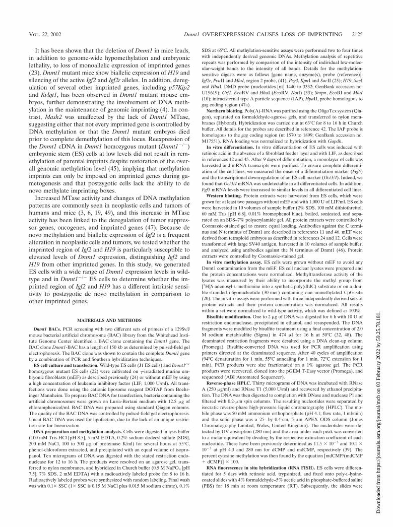
The establishment of genomic methylation patterns requiresthe concerted activity of two enzymes, the de novo MTaseDnmt3 and the hemi-MTase Dnmt1 (26, 31). Our study definesthree classes of sequences which are subject to gain of meth-ylation at different levels of Dnmt1 expression, as summarizedin Fig. 7. (i) Retroviral elements like IAP and other repetitivesequences such as centromeric repeats are highly susceptible togain of methylation even under conditions of low Dnmt1 ex-pression (Dnmt1chip/� cells). (ii) Unmethylated alleles of mostimprinted genes, including Igf2r, Peg3, Snrpn, and Grf1 and theCpG islands of several nonimprinted genes, are completelyresistant to de novo methylation. (iii) The DMD imprintedregion, which has been shown to control expression of Igf2 andH19, is resistant to de novo methylation at low Dnmt1 expres-
FIG. 6. (A) Summary of the development (implantation) and survival of ES cell tetraploid blastocyst-derived mice or ES cell blastocyst-derivedmice. Implantation is indicated in percent, while survial at dpc 14.5 is indicated in total numbers. ND, not determined. (B) Western blot analysisof mEF cell extracts using an antibody against the N-terminal domain of Dnmt1. All protein extracts were controlled by Coomassie-stained gel toensure equal loading.
2132 BINISZKIEWICZ ET AL. MOL. CELL. BIOL.
Dow
nloa
ded
from
http
s://j
ourn
als.
asm
.org
/jour
nal/m
cb o
n 03
Feb
ruar
y 20
22 b
y 59
.25.
78.1
81.
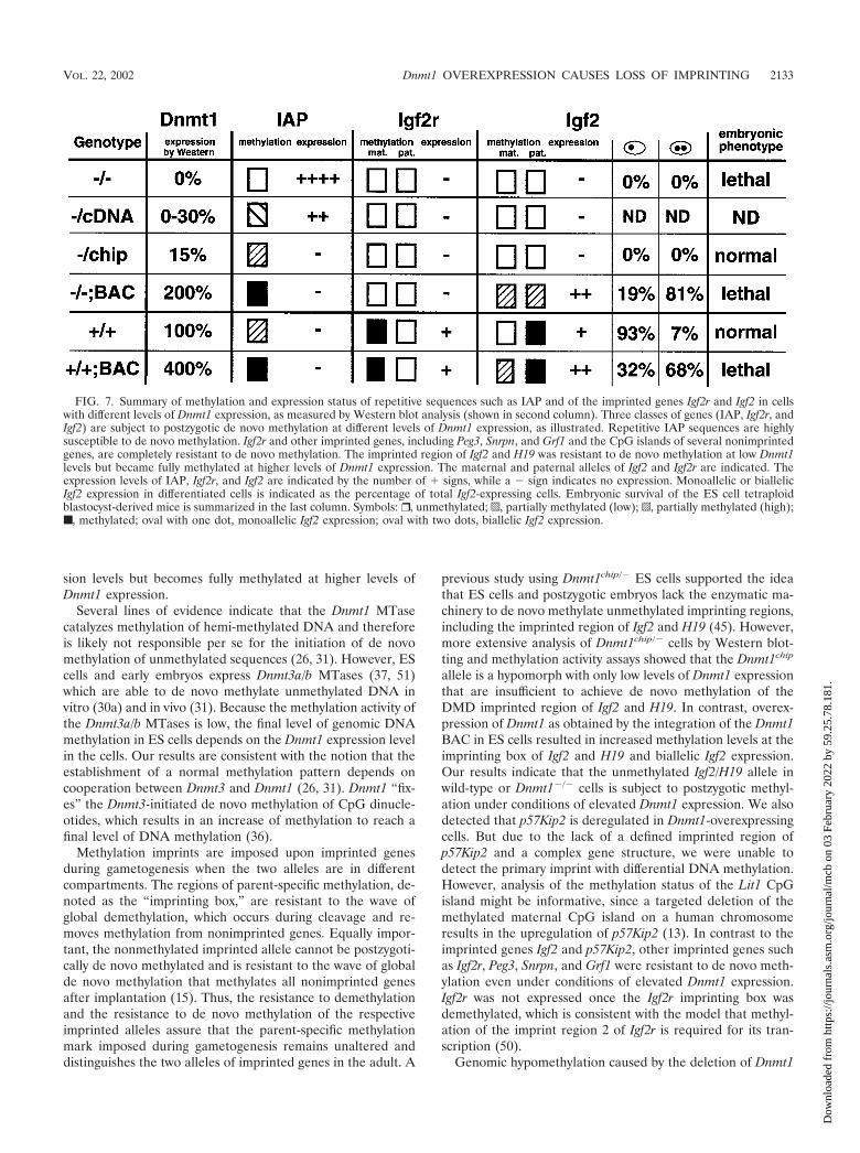
sion levels but becomes fully methylated at higher levels ofDnmt1 expression.
Several lines of evidence indicate that the Dnmt1 MTasecatalyzes methylation of hemi-methylated DNA and thereforeis likely not responsible per se for the initiation of de novomethylation of unmethylated sequences (26, 31). However, EScells and early embryos express Dnmt3a/b MTases (37, 51)which are able to de novo methylate unmethylated DNA invitro (30a) and in vivo (31). Because the methylation activity ofthe Dnmt3a/b MTases is low, the final level of genomic DNAmethylation in ES cells depends on the Dnmt1 expression levelin the cells. Our results are consistent with the notion that theestablishment of a normal methylation pattern depends oncooperation between Dnmt3 and Dnmt1 (26, 31). Dnmt1 “fix-es” the Dnmt3-initiated de novo methylation of CpG dinucle-otides, which results in an increase of methylation to reach afinal level of DNA methylation (36).
Methylation imprints are imposed upon imprinted genesduring gametogenesis when the two alleles are in differentcompartments. The regions of parent-specific methylation, de-noted as the “imprinting box,” are resistant to the wave ofglobal demethylation, which occurs during cleavage and re-moves methylation from nonimprinted genes. Equally impor-tant, the nonmethylated imprinted allele cannot be postzygoti-cally de novo methylated and is resistant to the wave of globalde novo methylation that methylates all nonimprinted genesafter implantation (15). Thus, the resistance to demethylationand the resistance to de novo methylation of the respectiveimprinted alleles assure that the parent-specific methylationmark imposed during gametogenesis remains unaltered anddistinguishes the two alleles of imprinted genes in the adult. A
previous study using Dnmt1chip/� ES cells supported the ideathat ES cells and postzygotic embryos lack the enzymatic ma-chinery to de novo methylate unmethylated imprinting regions,including the imprinted region of Igf2 and H19 (45). However,more extensive analysis of Dnmt1chip/� cells by Western blot-ting and methylation activity assays showed that the Dnmt1chip
allele is a hypomorph with only low levels of Dnmt1 expressionthat are insufficient to achieve de novo methylation of theDMD imprinted region of Igf2 and H19. In contrast, overex-pression of Dnmt1 as obtained by the integration of the Dnmt1BAC in ES cells resulted in increased methylation levels at theimprinting box of Igf2 and H19 and biallelic Igf2 expression.Our results indicate that the unmethylated Igf2/H19 allele inwild-type or Dnmt1�/� cells is subject to postzygotic methyl-ation under conditions of elevated Dnmt1 expression. We alsodetected that p57Kip2 is deregulated in Dnmt1-overexpressingcells. But due to the lack of a defined imprinted region ofp57Kip2 and a complex gene structure, we were unable todetect the primary imprint with differential DNA methylation.However, analysis of the methylation status of the Lit1 CpGisland might be informative, since a targeted deletion of themethylated maternal CpG island on a human chromosomeresults in the upregulation of p57Kip2 (13). In contrast to theimprinted genes Igf2 and p57Kip2, other imprinted genes suchas Igf2r, Peg3, Snrpn, and Grf1 were resistant to de novo meth-ylation even under conditions of elevated Dnmt1 expression.Igf2r was not expressed once the Igf2r imprinting box wasdemethylated, which is consistent with the model that methyl-ation of the imprint region 2 of Igf2r is required for its tran-scription (50).
Genomic hypomethylation caused by the deletion of Dnmt1
FIG. 7. Summary of methylation and expression status of repetitive sequences such as IAP and of the imprinted genes Igf2r and Igf2 in cellswith different levels of Dnmt1 expression, as measured by Western blot analysis (shown in second column). Three classes of genes (IAP, Igf2r, andIgf2) are subject to postzygotic de novo methylation at different levels of Dnmt1 expression, as illustrated. Repetitive IAP sequences are highlysusceptible to de novo methylation. Igf2r and other imprinted genes, including Peg3, Snrpn, and Grf1 and the CpG islands of several nonimprintedgenes, are completely resistant to de novo methylation. The imprinted region of Igf2 and H19 was resistant to de novo methylation at low Dnmt1levels but became fully methylated at higher levels of Dnmt1 expression. The maternal and paternal alleles of Igf2 and Igf2r are indicated. Theexpression levels of IAP, Igf2r, and Igf2 are indicated by the number of � signs, while a � sign indicates no expression. Monoallelic or biallelicIgf2 expression in differentiated cells is indicated as the percentage of total Igf2-expressing cells. Embryonic survival of the ES cell tetraploidblastocyst-derived mice is summarized in the last column. Symbols: ❒ , unmethylated; p, partially methylated (low); o, partially methylated (high);■ , methylated; oval with one dot, monoallelic Igf2 expression; oval with two dots, biallelic Igf2 expression.
VOL. 22, 2002 Dnmt1 OVEREXPRESSION CAUSES LOSS OF IMPRINTING 2133
Dow
nloa
ded
from
http
s://j
ourn
als.
asm
.org
/jour
nal/m
cb o
n 03
Feb
ruar
y 20
22 b
y 59
.25.
78.1
81.

MTase, though with no obvious effect on in vitro growth ofundifferentiated ES cells, results in abnormal development andembryonic lethality (23). Similarly, overexpression of Dnmt1and genomic hypermethylation had no obvious effect on EScell proliferation. However, injection of the cells into blasto-cysts resulted in embryonic lethality of the chimeric embryos.Our results are consistent with the notion that DNA methyl-ation has no obvious role in the survival of embryonic cells butis crucial for normal physiology of somatic cells (15). It hasbeen shown that conditional deletion of Dnmt1 in fibroblastsleads to genomic demethylation and widespread ectopic geneactivation, which may explain the cell death of Dnmt1 mutantcells (14). The mechanism of lethality induced by overexpres-sion of Dnmt1, however, is unclear. It has been reported thatDnmt1 can be overexpressed in rare clones of tumor cells (47),suggesting that genomic hypermethylation is compatible withsurvival of transformed cells in contrast to that of primary cells.Our inability to long-term culture untransformed Dnmt1-over-expressing fibroblasts is consistent with that observation.
The susceptibility of Igf2 and H19 to postzygotic de novomethylation is of potential relevance for tumorigenesis becausederegulation of Igf2 imprinting has been shown to occur in over20 different tumor types, including Wilms’ tumor (16, 27, 30,35, 40). Igf2 is a potent cell survival factor that stimulates cellproliferation, and its overexpression leads to conditions thatare favorable to increased cell proliferation and overgrowth.The oncogenic function of Igf2 has been confirmed by theexperimental overexpression of Igf2 in mice leading to an in-creased probability of tumor development (2, 34, 38). Thesusceptibility of the imprinted region of Igf2 and H19 to postzy-gotic de novo methylation may be the basis for the high fre-quency of biallelic Igf2 expression observed in many cancers(27, 40). This appears to distinguish the Igf2 gene from otherimprinted genes, such as Igf2r, Grf1, Snrpn, and Peg3, that werenot susceptible to postzygotic de novo methylation. Consistentwith this notion is the observation that tumor-specific alter-ations of these imprinted genes involve genetic mutationsrather than epigenetic changes (16).
The results described in this paper suggest that Dnmt1 ac-tivity may be crucial for the final level of methylation of bulkgenomic DNA and repetitive genes and for the imprintedregion of Igf2 and H19. Our results imply that even a moderateincrease in Dnmt1 expression in cells with low intrinsic de novoactivity from Dnmt3 may be sufficient to shift the balancetowards de novo methylation and activation of the silenced Igf2allele. Thus, overexpression of Dnmt1 and activation of Dnmt3as observed in many transformed cells may be of selectiveadvantage for the incipient tumor cell (37, 51).
ACKNOWLEDGMENTS
We thank H. J. Gross, Kerry Tucker, and W. M. Rideout III forstimulating discussions, Ruth Curry, Jessie Dausman, and Jan Loringfor expert technical assistance, and C. Plass for providing the Grf1probe.
This work was supported by NIH grant NIH/NCI 5-R35-CA44339and in part by the ERC Program of the National Science Foundationunder award EEC-9843342. D.B. was supported by the Deutsche Aka-demische Austauschdienst and the Fritz-Thyssen Stiftung. B.R. is sup-ported by a Medical Research Council (United Kingdom) ClinicianScientist Fellowship.
REFERENCES
1. Bartolomei, M. S., A. L. Webber, M. E. Brunkow, and S. M. Tilghman. 1993.Epigenetic mechanisms underlying the imprinting of the mouse H19 gene.Genes Dev. 7:1663–1673.
2. Bates, P., R. Fisher, A. Ward, L. Richardson, D. J. Hill, and C. F. Graham.1995. Mammary cancer in transgenic mice expressing insulin-like growthfactor II (IGF-II). Br. J. Cancer 72:1189–1193.
3. Belinsky, S. A., K. J. Nikula, S. B. Baylin, and J. P. Issa. 1996. Increasedcytosine DNA-methyltransferase activity is target-cell-specific and an earlyevent in lung cancer. Proc. Natl. Acad. Sci. USA 93:4045–4050.
4. Caspary, T., M. A. Cleary, C. C. Baker, X. J. Guan, and S. M. Tilghman.1998. Multiple mechanisms regulate imprinting of the mouse distal chromo-some 7 gene cluster. Mol. Cell. Biol. 18:3466–3474.
5. Eggan, K., H. Akutsu, J. Loring, L. Jackson-Grusby, M. Klemm, W. M.Rideout, R. Yanagimachi, and R. Jaenisch. 2001. Hybrid vigor, fetal over-growth, and viability of mice derived by nuclear cloning and tetraploidembryo complementation. Proc. Natl. Acad. Sci. USA 98:6209–6214.
6. el-Deiry, W. S., B. D. Nelkin, P. Celano, R. W. Yen, J. P. Falco, S. R.Hamilton, and S. B. Baylin. 1991. High expression of the DNA methyltrans-ferase gene characterizes human neoplastic cells and progression stages ofcolon cancer. Proc. Natl. Acad. Sci. USA 88:3470–3474.
7. Feil, R., T. Moore, J. Oswald, J. Walter, F. Sun, and W. Reik. 1997. Theimprinted insulin-like growth factor 2 gene, p. 70–97. In W. Reik and A.Surani (ed.), Genomic imprinting. IRL Press, Oxford, England.
8. Feinberg, A. P., and B. Vogelstein. 1983. Hypomethylation distinguishesgenes of some human cancers from their normal counterparts. Nature 301:89–92.
9. Feinberg, A. P., and B. Vogelstein. 1983. Hypomethylation of ras oncogenesin primary human cancers. Biochem. Biophys. Res. Commun. 111:47–54.
10. Gabriel, J. M., T. A. Gray, L. Stubbs, S. Saitoh, T. Ohta, and R. D. Nicholls.1998. Structure and function correlations at the imprinted mouse Snrpnlocus. Mamm. Genome 9:788–793.
11. Gaudet, F., D. Talbot, H. Leonhardt, and R. Jaenisch. 1998. A short DNAmethyltransferase isoform restores methylation in vivo. J. Biol. Chem. 273:32725–32729.
12. Hogan, B., R. Beddington, F. Costantini, and F. Lacy. 1994. Manipulatingthe mouse embryo: a laboratory manual, 2nd ed. Cold Spring Harbor Lab-oratory Press, Cold Spring Harbor, N.Y.
13. Horike, S., K. Mitsuya, M. Meguro, N. Kotobuki, A. Kashiwagi, T. Notsu,T. C. Schulz, Y. Shirayoshi, and M. Oshimura. 2000. Targeted disruption ofthe human LIT1 locus defines a putative imprinting control element playingan essential role in Beckwith-Wiedemann syndrome. Hum. Mol. Genet.9:2075–2083.
14. Jackson-Grusby, L., C. Beard, R. Possemato, M. Tudor, D. Fambrough, G.Csankovszki, J. Dausman, P. Lee, C. Wilson, E. Lander, and R. Jaenisch.2001. Loss of genomic methylation causes p53-dependent apoptosis andepigenetic deregulation. Nat. Genet. 27:31–39.
15. Jaenisch, R. 1997. DNA methylation and imprinting: why bother? TrendsGenet. 13:323–329.
16. Jirtle, R. L. 1999. Genomic imprinting and cancer. Exp. Cell Res. 248:18–24.17. Jones, P. A., and P. W. Laird. 1999. Cancer epigenetics comes of age. Nat.
Genet. 21:163–167.18. Jouvenot, Y., F. Poirier, J. Jami, and A. Paldi. 1999. Biallelic transcription of
Igf2 and H19 in individual cells suggests a post-transcriptional contributionto genomic imprinting. Curr. Biol. 9:1199–1202.
19. Kautiainen, T. L., and P. A. Jones. 1986. DNA methyltransferase levels intumorigenic and nontumorigenic cells in culture. J. Biol. Chem. 261:1594–1598.
20. Kumar, S., X. Cheng, J. W. Pflugrath, and R. J. Roberts. 1992. Purification,crystallization, and preliminary X-ray diffraction analysis of an M.HhaI-AdoMet complex. Biochemistry 31:8648–8653.
21. Leff, S. E., C. I. Brannan, M. L. Reed, T. Ozcelik, U. Francke, N. G.Copeland, and N. A. Jenkins. 1992. Maternal imprinting of the mouse Snrpngene and conserved linkage homology with the human Prader-Willi syn-drome region. Nat. Genet. 2:259–264.
22. Lei, H., S. P. Oh, M. Okano, R. Juttermann, K. A. Goss, R. Jaenisch, and E.Li. 1996. De novo DNA cytosine methyltransferase activities in mouse em-bryonic stem cells. Development 122:3195–3205.
23. Li, E., C. Beard, and R. Jaenisch. 1993. Role for DNA methylation ingenomic imprinting. Nature 366:362–365.
24. Li, E., T. H. Bestor, and R. Jaenisch. 1992. Targeted mutation of the DNAmethyltransferase gene results in embryonic lethality. Cell 69:915–926.
25. Li, L. L., I. Y. Szeto, B. M. Cattanach, F. Ishino, and M. A. Surani. 2000.Organization and parent-of-origin-specific methylation of imprinted peg3gene on mouse proximal chromosome 7. Genomics 63:333–340.
26. Lyko, F., B. H. Ramsahoye, H. Kashevsky, M. Tudor, M. A. Mastrangelo,T. L. Orr-Weaver, and R. Jaenisch. 1999. Mammalian (cytosine-5) methyl-transferases cause genomic DNA methylation and lethality in Drosophila.Nat. Genet. 23:363–366.
27. Moulton, T., W. Y. Chung, L. Yuan, T. Hensle, P. Waber, P. Nisen, and B.
2134 BINISZKIEWICZ ET AL. MOL. CELL. BIOL.
Dow
nloa
ded
from
http
s://j
ourn
als.
asm
.org
/jour
nal/m
cb o
n 03
Feb
ruar
y 20
22 b
y 59
.25.
78.1
81.

Tycko. 1996. Genomic imprinting and Wilms’ tumor. Med. Pediatr. Oncol.27:476–483.
28. Nagy, A., E. Gocza, E. M. Diaz, V. R. Prideaux, E. Ivanyi, M. Markkula, andJ. Rossant. 1990. Embryonic stem cells alone are able to support fetaldevelopment in the mouse. Development 110:815–821.
29. Nagy, A., J. Rossant, R. Nagy, W. Abramow-Newerly, and J. C. Roder. 1993.Derivation of completely cell culture-derived mice from early-passage em-bryonic stem cells. Proc. Natl. Acad. Sci. USA 90:8424–8428.
30. Ogawa, O., M. R. Eccles, J. Szeto, L. A. McNoe, K. Yun, M. A. Maw, P. J.Smith, and A. E. Reeve. 1993. Relaxation of insulin-like growth factor II geneimprinting implicated in Wilms’ tumour. Nature 362:749–751.
30a.Okano, M., S. Xie, and E. Li. 1998. Cloning and characterization of a familyof novel mammalian DNA (cytosine-5) methyltransferases. Nat. Genet. 19:219–220.
31. Okano, M., D. W. Bell, D. A. Haber, and E. Li. 1999. DNA methyltrans-ferases Dnmt3a and Dnmt3b are essential for de novo methylation andmammalian development. Cell 99:247–257.
32. Olek, A., J. Oswald, and J. Walter. 1996. A modified and improved methodfor bisulphite based cytosine methylation analysis. Nucleic Acids Res. 24:5064–5066.
33. Plass, C., H. Shibata, I. Kalcheva, L. Mullins, N. Kotelevtseva, J. Mullins, R.Kato, H. Sasaki, S. Hirotsune, Y. Okazaki, W. A. Held, Y. Hayashizaki, andV. M. Chapman. 1996. Identification of Grf1 on mouse chromosome 9 as animprinted gene by RLGS-M. Nat. Genet. 14:106–109.
34. Pravtcheva, D. D., and T. L. Wise. 1998. Metastasizing mammary carcinomasin H19 enhancers-Igf2 transgenic mice. J. Exp. Zool. 281:43–57.
35. Rainier, S., L. A. Johnson, C. J. Dobry, A. J. Ping, P. E. Grundy, and A. P.Feinberg. 1993. Relaxation of imprinted genes in human cancer. Nature362:747–749.
36. Ramsahoye, B. H., D. Biniszkiewicz, F. Lyko, V. Clark, A. P. Bird, and R.Jaenisch. 2000. Non-CpG methylation is prevalent in embryonic stem cellsand may be mediated by DNA methyltransferase 3a. Proc. Natl. Acad. Sci.USA 97:5237–5242.
37. Robertson, K. D., E. Uzvolgyi, G. Liang, C. Talmadge, J. Sumegi, F. A.Gonzales, and P. A. Jones. 1999. The human DNA methyltransferases(DNMTs) 1, 3a and 3b: coordinate mRNA expression in normal tissues andoverexpression in tumors. Nucleic Acids Res. 27:2291–2298.
38. Rogler, C. E., D. Yang, L. Rossetti, J. Donohoe, E. Alt, C. J. Chang, R.Rosenfeld, K. Neely, and R. Hintz. 1994. Altered body composition andincreased frequency of diverse malignancies in insulin-like growth factor-IItransgenic mice. J. Biol. Chem. 269:13779–13784.
39. Sinsheimer, R. 1954. The action of pancreatic deoxyribonuclease. I. Isolationof mono- and dinucleotides. J. Biol. Chem. 208:445–459.
40. Steenman, M. J., S. Rainier, C. J. Dobry, P. Grundy, I. L. Horon, and A. P.Feinberg. 1994. Loss of imprinting of IGF2 is linked to reduced expressionand abnormal methylation of H19 in Wilms’ tumour. Nat. Genet. 7:433–439.
41. Stoger, R., P. Kubicka, C. G. Liu, T. Kafri, A. Razin, H. Cedar, and D. P.Barlow. 1993. Maternal-specific methylation of the imprinted mouse Igf2rlocus identifies the expressed locus as carrying the imprinting signal. Cell73:61–71.
42. Szabo, P., and J. R. Mann. 1994. Expression and methylation of imprintedgenes during in vitro differentiation of mouse parthenogenetic and andro-genetic embryonic stem cell lines. Development 120:1651–1660.
43. Thorvaldsen, J. L., K. L. Duran, and M. S. Bartolomei. 1998. Deletion of theH19 differentially methylated domain results in loss of imprinted expressionof H19 and Igf2. Genes Dev. 12:3693–3702.
44. Tremblay, K. D., K. L. Duran, and M. S. Bartolomei. 1997. A 5� 2-kilobase-pair region of the imprinted mouse H19 gene exhibits exclusive paternalmethylation throughout development. Mol. Cell. Biol. 17:4322–4329.
45. Tucker, K. L., C. Beard, J. Dausmann, L. Jackson-Grusby, P. W. Laird, H.Lei, E. Li, and R. Jaenisch. 1996. Germ-line passage is required for estab-lishment of methylation and expression patterns of imprinted but not ofnonimprinted genes. Genes Dev. 10:1008–1020.
46. Tucker, K. L., D. Talbot, M. A. Lee, H. Leonhardt, and R. Jaenisch. 1996.Complementation of methylation deficiency in embryonic stem cells by aDNA methyltransferase minigene. Proc. Natl. Acad. Sci. USA 93:12920–12925.
47. Vertino, P. M., R. W. Yen, J. Gao, and S. B. Baylin. 1996. De novo methyl-ation of CpG island sequences in human fibroblasts overexpressing DNA(cytosine-5-)-methyltransferase. Mol. Cell. Biol. 16:4555–4565.
47a.Walsh, C. P., J. R. Chaillet, and T. H. Bestor. 1998. Transcription of IAPendogenous retroviruses is constrained by cytosine methylation. Nat. Genet.20:116–117.
48. Warnecke, P. M., D. Biniszkiewicz, R. Jaenisch, M. Frommer, and S. J.Clark. 1998. Sequence-specific methylation of the mouse H19 gene in em-bryonic cells deficient in the Dnmt-1 gene. Dev. Genet. 22:111–121.
49. Wu, J., J. P. Issa, J. Herman, D. E. Bassett, Jr., B. D. Nelkin, and S. B.Baylin. 1993. Expression of an exogenous eukaryotic DNA methyltrans-ferase gene induces transformation of NIH 3T3 cells. Proc. Natl. Acad. Sci.USA 90:8891–8895.
50. Wutz, A., O. W. Smrzka, N. Schweifer, K. Schellander, E. F. Wagner, andD. P. Barlow. 1997. Imprinted expression of the Igf2r gene depends on anintronic CpG island. Nature 389:745–749.
51. Xie, S., Z. Wang, M. Okano, M. Nogami, Y. Li, W. W. He, K. Okumura, andE. Li. 1999. Cloning, expression and chromosome locations of the humanDNMT3 gene family. Gene 236:87–95.
52. Zhang, Y., T. Shields, T. Crenshaw, Y. Hao, T. Moulton, and B. Tycko. 1993.Imprinting of human H19: allele-specific CpG methylation, loss of the activeallele in Wilms tumor, and potential for somatic allele switching. Am. J.Hum. Genet. 53:113–124.
VOL. 22, 2002 Dnmt1 OVEREXPRESSION CAUSES LOSS OF IMPRINTING 2135
Dow
nloa
ded
from
http
s://j
ourn
als.
asm
.org
/jour
nal/m
cb o
n 03
Feb
ruar
y 20
22 b
y 59
.25.
78.1
81.


![Promoter hypermethylation profiling of distant breast ... · phenotype of distant breast cancer metastases [14–16]. Extensive knowledge of the hypermethylation status of tumor suppressor](https://static.fdocuments.in/doc/165x107/5d21f00788c993722e8c67ea/promoter-hypermethylation-profiling-of-distant-breast-phenotype-of-distant.jpg)
