![Untitled-2 [medicare.lk] · gem hospital institute of gastroenterology & laparoscopy vamsam fertility research centre narayana hrudayalaya limited parampara fertility and gynaec centre](https://static.fdocuments.in/doc/165x107/5ec5b02076c4b41a435bf125/untitled-2-gem-hospital-institute-of-gastroenterology-laparoscopy-vamsam.jpg)
DNB Cardiology from Narayana Hrudayalaya • Associate ...
17
• DNB Cardiology from Narayana Hrudayalaya • Associate Professor of Cardiology at SJMCH • Has more than 45 International publications and authored 3 Books ( Springer and Ta ylor and Francis publications) • Practices adult interventional cardiology with interest in pediatric cardiology. www.wincarsassociation.com
Transcript of DNB Cardiology from Narayana Hrudayalaya • Associate ...

• DNB Cardiology from Narayana Hrudayalaya
• Associate Professor of Cardiology at SJMCH
• Has more than 45 International publications and authored 3 Books ( Springer and Ta ylor and Francis publications)
• Practices adult interventional cardiology with interest in pediatric cardiology.
www.wincarsassociation.com

IMAGING INSIGHTS IN EISENMENGER’S SYNDROME: FROM BASICS TO SOPHISTICATION
Dr M A Srilakshmi DNB CardiologyAssociate ProfessorSt. John’s Medical College HospitalBangalore
www.wincarsassociation.com

Introduction
• There is an increasing dependence on investigations which has compromised clinical skills of all clinicians.
• We describe an unusual case of a large Aorto-pulmonary window which was not very symptomatic and presented as Eisenmenger syndrome at the age of 20 years in a male patient.
• We take this opportunity to highlight some of the basic investigations like Chest X Ray and Contrast Echocardiography which can delineate the level of the shunt.
• Attention should be given to the subtle bedside clinical signs which can give clues to the diagnosis.
www.wincarsassociation.com

Case Report-History
• We describe a 20 year old male who presented to us with 2 to 3 episodes of hemoptysis per day of 10 ml each since 1 week.
• He had a childhood history of breathlessness and failure to thrive which was noticed when he was 5 months of age. He was hospitalized for the same and administered medications. The details of which were not available.
• Further, he had history of breathlessness on exertion at the age of 5 years due to which he could not play and keep up with his peers in out- door activities. Besides, he developed repeated lower respiratory tract infections requiring hospitalization and systemic antibiotics.
• After the age of 14 years, the episodes of productive cough and breathlessness decreased
www.wincarsassociation.com

Clinical Findings
• Clinical examination revealed a right ventricular apex, left parasternal heave, single second heart sound which was a loud P2 and a long early diastolic murmur along his left parasternal region.
• He had uniform pan-digital cyanosis and clubbing.
www.wincarsassociation.com

www.wincarsassociation.com

Chest X Ray
• His Chest X Ray ( PA view) demonstrated that the cardiac apex was formed by the right ventricle, with an aneurysmal main pulmonary artery segment.
• There was peripheral pruning of pulmonary vasculature.
• An interesting observation was that the right pulmonary artery was conspicuous by its absence, while a branching pulmonary artery was seen through the cardiac silhouette.
• The aortic arch was right sided as it coursed to the right of the vertebral column
www.wincarsassociation.com

2D Echocardiography
www.wincarsassociation.com
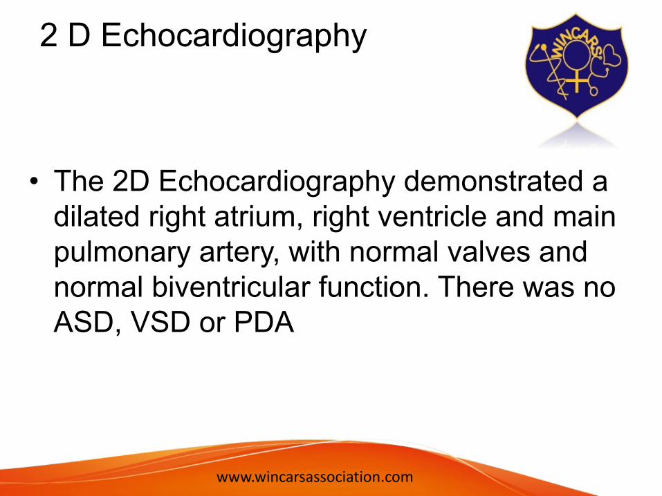
2 D Echocardiography
• The 2D Echocardiography demonstrated a dilated right atrium, right ventricle and main pulmonary artery, with normal valves and normal biventricular function. There was no ASD, VSD or PDA
www.wincarsassociation.com

Contrast EchocardiographyA contrast echocardiography with agitated saline injected into the ante-cubital vein, showed contrast bubbles in the ascending aorta after 5 cardiac cycles. The contrast bubbles were seen in the descending aorta much later
www.wincarsassociation.com

Clinical Signs of Eisenmenger syndrome in individual lesions
• Certain subtle clinical features have been defined as hallmarks of individual lesions, mainly involving the second heart sound.
• A single second sound is a feature of reversal of flow through a ventricular septal defect, while a normally split second sound with loud P2 features reversal of flow through a patent ductus arteriosus (PDA). In an atrial septal defect(ASD), reversal of flow may shorten the gap between the two components of the second heart sound with a fixed split, the P2 being loud. The split in the second heart sound remains fixed although it may shorten.
• Differential cyanosis is a feature of reversal of flow in a PDA. ASD and VSD usually exhibit uniform pandigital cyanosis and clubbing. The cardiac size usually regresses with reversal of flow in a ventricular septal defect (VSD), while in an ASD, cardiomegaly persists
www.wincarsassociation.com
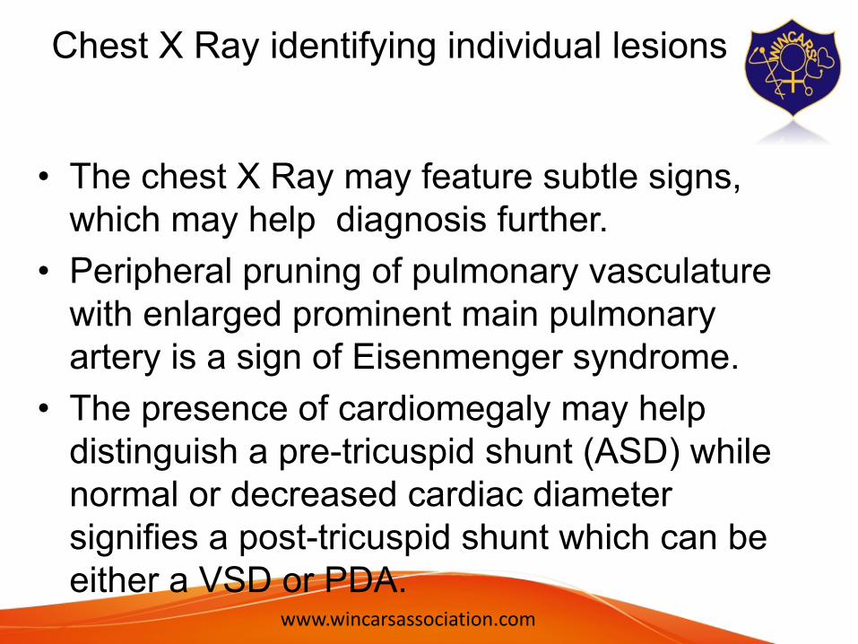
Chest X Ray identifying individual lesions
• The chest X Ray may feature subtle signs, which may help diagnosis further.
• Peripheral pruning of pulmonary vasculature with enlarged prominent main pulmonary artery is a sign of Eisenmenger syndrome.
• The presence of cardiomegaly may help distinguish a pre-tricuspid shunt (ASD) while normal or decreased cardiac diameter signifies a post-tricuspid shunt which can be either a VSD or PDA.
www.wincarsassociation.com

Contrast Echocardiography
• A contrast Echocardiogram can define the anatomical position of the shunt. Injection of agitated saline into the ante-cubital vein can define the right to left shunt when the bubbles appear in the left heart after 5 to 7 cardiac cycles.
• In the aorta, if the bubbles appear in the descending aorta, the reversal of flow is through a PDA, while if the bubbles appear in the ascending aorta, the reversed flow is through an aorto-pulmonary window
www.wincarsassociation.com

CT
• In our patient, a cardiac CT scan was done which confirmed a Type1 aorto-pulmonary window with enlarged pulmonary arteries
www.wincarsassociation.com

CT scan
www.wincarsassociation.com

Take Home Message
• Here, the clinical history and examination pointed towards Eisenmenger syndrome.
• The chest X Ray showed that the shunt was at the great arterial level with a possiblity of hemitruncus or Truncus Arteriosus.
• The contrast Echocardiography pointed towards a diagnosis of aorto-pulmonary window. This was confirmed by a Cardiac CT scan.
• We wish to emphasize the importance of bedside clinical examination and basic imaging techniques which are still relevant in their contribution to the diagnosis of Eisenmenger syndrome defining the level of the intra-cardiac shunt.
www.wincarsassociation.com

THANK YOU
www.wincarsassociation.com


![Untitled-2 [medicare.lk]medicare.lk/images/Medicare 2019_brochure-V5.pdf · gem hospital institute of gastroenterology & laparoscopy vamsam fertility research centre narayana hrudayalaya](https://static.fdocuments.in/doc/165x107/5ec5b02176c4b41a435bf129/untitled-2-2019brochure-v5pdf-gem-hospital-institute-of-gastroenterology.jpg)