Crosslinking- Gr … . evolution,protocols,controversies &newer insights . Dr. Rohit shetty . Dnb,...
50
Gr Crosslinking- evolution,protocols,controversies &newer insights Dr. Rohit shetty Dnb, frcs Vice-chairman Narayana Nethralaya , Bangalore
Transcript of Crosslinking- Gr … . evolution,protocols,controversies &newer insights . Dr. Rohit shetty . Dnb,...

Gr Crosslinking-
evolution,protocols,controversies
&newer insights
Dr. Rohit shetty
Dnb, frcs
Vice-chairman
Narayana Nethralaya , Bangalore

This presentation is dedicated to Mr Rudolph Peschke, a great friend and guide…


Diagnostics revisited Pathophysiology redefined? Crosslinking - protocols and dilemma

Keratoconus Pathophysiology?


Can Biomarkers predict the treatment outcomes of
Crosslinking in Keratoconus?

Keratoconus Disease known to us for more than 150 years now!
“Keratoconus is a progressive, noninflammatory, usually bilateral corneal disease, characterized by
paraxial stromal thinning and weakening that leads to corneal surface distortion. “

Is inflammation driving Keratoconus ?
A holistic study of molecular pathways

Hidden biomarkers in tears & corneal epithelium

This cartoon depicts five Greeks representing the cardinal signs of inflammation
“heat, redness, swelling, pain and loss of function”
— described by Celsus more than 2000 years ago
Inflammation

Normal Cornea
Keratoconus Cornea
Keratoconus cornea

EPITHELIUM ,TEARS & KERATOCONUS

14

15
Hidden biomarkers in tears....
• Accepted in IOVS,2014,DECEMBER

Tear MMP9 levels Tear MMP9 levels by grade
Tear IL6 levels Tear IL6 levels by grade
MMP9 and IL6 levels in tears increase with increasing severity of KC. Thus, these two markers may be useful to use as biomarkers for KC. Also suggests their utility for
therapeutic intervention.
Shetty et al, IOVS (in revision)

Kera
toco
nus
Don
or c
ontro
l
LOX Collagen I Collagen IV
Immunohistochemical validation of gene expression data
Reduced LOX expression in the patient epithelium may be an important contributing factor to the loss of collagen structure and ectasia.
Shetty et al, Mol. Vis. (in revision)

Can immunomodulatory drugs like Cyclosporine A (0.05%, topical) block the inflammatory pathway in Keratoconus?
• 20 patients with progressive keratoconus were treated with topical CsA 0.05%
• Topography was analyzed pre and post treatment (3-6 months)
In cultured human corneal epithelial cells, CyA treatment could inhibit TNFα induced inflammation.
Shetty et al, IOVS (in revision)

Corneal topography of KC patients after treatment with CyclosporineA
Short term treatment with CysclosporineA led to local flattening of the cornea and concomitant reduction in tear
MMP9 levels.
Shetty et al, IOVS (in revision)

MMP 9 Levels Pre and Post

MMP 9 Levels Pre and Post

22

Clinical correlation to the biomarkers

Diagnosing Inflammatory Dry eye

MMP-9 and Dry Eye Severity
Patient’s Dysfunctional Tear Syndrome Level
Average MMP-9 level Statistical significance vs
Normal
Normal 8.39 ng/ml No
Severity level 1 35.57 ng/ml No
Severity level 2 66.17 ng/ml Yes
Severity level 3 101.42 ng/ml Yes
Severity level 4 381.24 ng/ml Yes
Positive result= Chronic Dry eye >40 ng/ml

MMP 9 Levels Pre and Post

HOW IMPORTANT IS BOWMANS IN KERATOCONUS ??

`
Radial Scan on hand held OCT (Bioptigen) correlating with steepest
part on topography

Factors associated with change in BCVA (improvement) after surgery. R2 of the model=0.86.
• Improvement in BCVA was greater if the concentration of LOX and COLA1 were higher
Postoperative Km flattening was greater if the KC grade of higher and ColA4 was higher.
R2 of the model=0.80.
. Factors associated with change in Km (flattening) after surgery. (R2 of the model=0.80.)

Why is epithelium removal important…?
Type 4 Collagen in Bowman’s membrane might have a role in outcomes of crosslinking , Smooth epithelium removal thus is vital

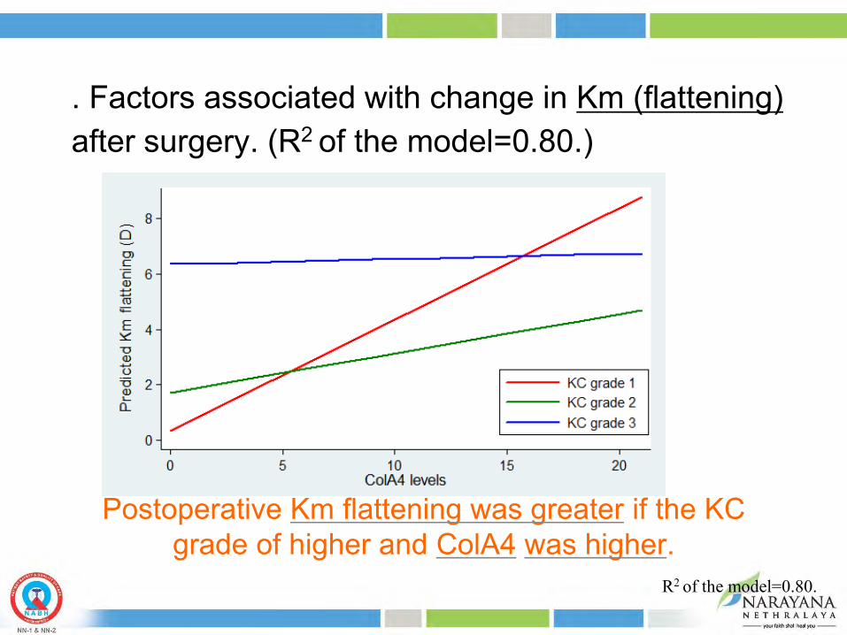
USE OF A NOVEL DEVICE FOR REMOVAL OF EPITHELIUM IN PHOTOREFRACTIVE
KERECTECTOMY

33
OD PRK - EPI CLEAR™ OS PRK – MECHANICAL SCRAPER
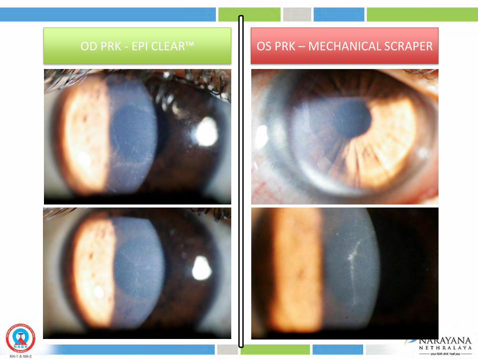
Post operative visual recovery was faster and less painful in RE as compared to LE. AS OCT done @ 2 weeks post op
OD more regularized and smooth corneal epithelium healing
OS showing irregular healing of epithelium Superior quadrant shows poor recovery and epithelial thinning
EPICLEAR BLADE
MECHANICAL SCRAPER

36


1. High-intensity CXL 2. Optimized beam profile 3. Transepithelial CXL 4. Biochemical mechanism of CXL 5. Detection of CXL 6. Combinations and new indications
New developments


Young Patient (< 14yrs)
Thickness < 425 microns
Female (20 -25yrs)
Documented Progression
Family History
Severe Allergy
Immediate Treatment
Wait
Age > 25
Thickness > 450microns
Male / Female
No Documented Progression
No Allergy
Caution: Thin cornea / Scar / Poor Endothelium
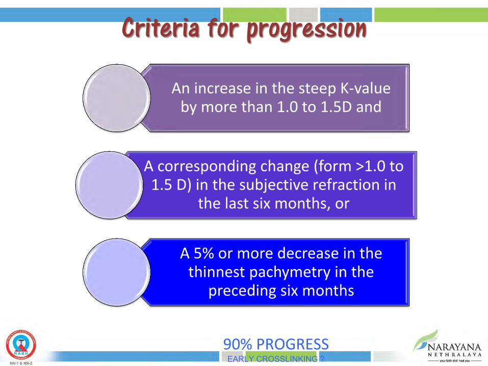
Criteria for progression
An increase in the steep K-value by more than 1.0 to 1.5D and
A corresponding change (form >1.0 to 1.5 D) in the subjective refraction in
the last six months, or
A 5% or more decrease in the thinnest pachymetry in the
preceding six months
90% PROGRESS EARLY CROSSLINKING ?

Playing devil’s advocate
• Ideal UVA intensity • Is faster better • Cross-linking with refractive surgery • Transepithelial cross-linking

Current status of corneal collagen cross-linking for keratoconus: a review Clin Exp Optom 2013; 96: 155–164 DOI:10.1111/cxo.12020 Elsie Chan FRANZCO

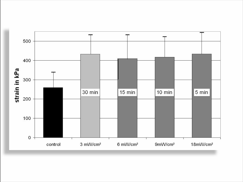
CROSS LINKING PROTOCOLS
• 3 mW/cm2 @ 30 minutes • 10 mW/cm2 @ 9 minutes • 18 mW/cm2 @ 5 minutes • 30 mW/cm2 @ 3 minutes • 45 mW/cm2 @ 2 minutes 40 seconds
Which is better?
Pulsed oxygen therapy – increase cross-linking effect

Photochemical processes depend on the absorbed energy dose
Unfortunately, it is known from photography that this law is only valid for a certain range. Question: How large is this range for CXL?

Second-harmonic microscopy of porcine corneas (in reflection) Second-harmonic microscopy of porcine corneas (in reflection)
Untreated 3mW/cm² 10mW/cm² 100mW/cm²
DEPTH (100µm)
Rebecca McQuaid1,2, JiaJun Li1, Arthur Cummings2, Michael Mrochen3, Brian Vohnsen1
1AOI Group, School of Physics UCD, Dublin 4, IRELAND 2Wellington Eye Clinic, Dublin, IRELAND 3IROC, Zurich, SWITZERLAND

• From 3 to 30 mW
(continuously adjustable within that range.)
• Modes 1. Continuous 2. Pulsed 3. Lasik mode. • Self-calibrating
• Beam diameter from 2
to 16 mm.
Collagen Crosslinking device CXL Phoenix system