Diversity and Morphological Characteristics of Aspergillus Species and Fusarium...
14
ISSN: 1511-3701 Pertanika J. Trop. Agric. Sci. 35 (1): 103 - 116 (2012) © Universiti Putra Malaysia Press Received: 31 December 2009 Accepted: 20 July 2010 * Corresponding Author INTRODUCTION Corn is categorized as a main staple food and industrial material which serves as an additional food and nutritional supplement for both human and animals. It also serves as an important food component for poultry and swine consumption and is directly used in the farming industry, hence contributing to the high demand of corn worldwide. Corn plantations are vulnerable to fungal infection caused by Fusarium species, especially in cool and humid climates (Lacey, 1989; Sewram et al., 1999; Vanara et al., 2009). Fusarium species play a significant role as plant pathogens, causing a broad range of diseases on various host plants, such as vascular wilt, pre and post-appearance blight as well as root and stem rots (Pascale et al., 2002; Schollenberger et al., 2006). Fusarium is also considered as a vital genre associated with cereal mycology, where this pre-harvest fungal infection prolongs until post-harvest and storage stage (Hussein et al., 1991; Plattner et al., 1991; Mubatanhema et al., 1999; Ezekiel et al., 2008). According to Ezekiel et al. (2008), poor storage condition, storage period, temperature, humidity levels and suitable climate could lead to infection caused by various storage fungi, such as Aspergillus Diversity and Morphological Characteristics of Aspergillus Species and Fusarium Species Isolated from Cornmeal in Malaysia Nithiyaa, P. 1 , Nur Ain Izzati, M.Z. 1* , Umi Kalsom, Y. 1 , Salleh, B. 2 1 Department of Biology, Faculty of Science, Universiti Putra Malaysia, 43400 Serdang, Selangor, Malaysia 2 School of Biological Sciences, Universiti Sains Malaysia, 11800 Minden, Penang, Malaysia *E-mail: [email protected] ABSTRACT Corn is a vital food component that serves as a nutritional diet element for human and feedstuff for livestock. Despite its vast importance, corn frequently faces contamination problem caused by a range of microbes especially fungi. For the purpose of this study, cornmeal samples were collected from nine states in Malaysia, and were cultured onto Peptone Pentachloronitrobenzene Agar (PPA) to isolate the fungi. Single spore isolation was done on Potato Dextrose Agar (PDA) to obtain the pure culture. A total of 314 isolates of microscopic fungi were obtained, 284 isolates belonging to the Aspergillus species, namely A. flavus (241), A. niger (24), A. nidulans (14) and A. fumigatus (5). Another 30 isolates were Fusarium species, identified as F. verticillioides (14), F. semitectum (10) and F. proliferatum (6). The diversity of the fungi was determined by using Shannon- Weiner Index. The diversity index indicated that A. flavus was the most abundant, recorded as 0.203. Keywords: Aspergillus flavus, A. niger, A. nidulans, A. fumigatus, Fusarium verticillioides, F. semitectum, F. proliferatum, cornmeal
Transcript of Diversity and Morphological Characteristics of Aspergillus Species and Fusarium...

ISSN: 1511-3701Pertanika J. Trop. Agric. Sci. 35 (1): 103 - 116 (2012) © Universiti Putra Malaysia Press
Received: 31 December 2009Accepted: 20 July 2010*Corresponding Author
INTRODUCTIONCorn is categorized as a main staple food and industrial material which serves as an additional food and nutritional supplement for both human and animals. It also serves as an important food component for poultry and swine consumption and is directly used in the farming industry, hence contributing to the high demand of corn worldwide.
Corn plantations are vulnerable to fungal infection caused by Fusarium species, especially in cool and humid climates (Lacey, 1989; Sewram et al., 1999; Vanara et al., 2009). Fusarium species play a significant role as plant
pathogens, causing a broad range of diseases on various host plants, such as vascular wilt, pre and post-appearance blight as well as root and stem rots (Pascale et al., 2002; Schollenberger et al., 2006). Fusarium is also considered as a vital genre associated with cereal mycology, where this pre-harvest fungal infection prolongs until post-harvest and storage stage (Hussein et al., 1991; Plattner et al., 1991; Mubatanhema et al., 1999; Ezekiel et al., 2008). According to Ezekiel et al. (2008), poor storage condition, storage period, temperature, humidity levels and suitable climate could lead to infection caused by various storage fungi, such as Aspergillus
Diversity and Morphological Characteristics of Aspergillus Species and Fusarium Species Isolated from Cornmeal in Malaysia
Nithiyaa, P.1, Nur Ain Izzati, M.Z.1*, Umi Kalsom, Y.1, Salleh, B.2
1Department of Biology,Faculty of Science, Universiti Putra Malaysia,
43400 Serdang, Selangor, Malaysia2School of Biological Sciences,
Universiti Sains Malaysia,11800 Minden, Penang, Malaysia
*E-mail: [email protected]
ABSTRACTCorn is a vital food component that serves as a nutritional diet element for human and feedstuff for livestock. Despite its vast importance, corn frequently faces contamination problem caused by a range of microbes especially fungi. For the purpose of this study, cornmeal samples were collected from nine states in Malaysia, and were cultured onto Peptone Pentachloronitrobenzene Agar (PPA) to isolate the fungi. Single spore isolation was done on Potato Dextrose Agar (PDA) to obtain the pure culture. A total of 314 isolates of microscopic fungi were obtained, 284 isolates belonging to the Aspergillus species, namely A. flavus (241), A. niger (24), A. nidulans (14) and A. fumigatus (5). Another 30 isolates were Fusarium species, identified as F. verticillioides (14), F. semitectum (10) and F. proliferatum (6). The diversity of the fungi was determined by using Shannon-Weiner Index. The diversity index indicated that A. flavus was the most abundant, recorded as 0.203.
Keywords: Aspergillus flavus, A. niger, A. nidulans, A. fumigatus, Fusarium verticillioides, F. semitectum, F. proliferatum, cornmeal

Nithiyaa, P., Nur Ain Izzati, M.Z., Umi Kalsom, Y., Salleh, B.
104 Pertanika J. Trop. Agric. Sci. Vol. 35 (1) 2012
species, Penicillium species, Curvularia species Alternaria alternata, Cladosporium cladosporioides and Phoma species (Horn et al., 2000; Reddy et al., 2009).
The fungi that contaminate the feed, produce a wide range of secondary metabolites that give negative effects to animal and human. They are known as mycotoxin. Mycotoxin causes several losses in the quality of the corn. It has been recorded to affect the health of it consumers, human and animal (Mubatanhema et al., 1999; Sewram et al., 1999; Zinedine & Manes, 2009). Livestock fed with contaminated feeds were diagnosed with symptoms of weight decrease, liver or kidney cancer, leukoencephalomalacia and death (Munkvold et al., 1998; Pascale et al., 2002). Since contamination on grain cereal has been proven to be one of the major causes of toxicity affecting animals and human, special attention must therefore be paid during the processes of storage, handling and packaging.
The objectives of this study were to investigate the diversity and distribution of microscopic fungi associated with cornmeal, as well as to identify the fungi based on morphological diagnostics.
MATERIALS AND METHODS
Samples Collection and Isolation of Aspergillus and Fusarium Isolates The cornmeal samples were collected from 9 states throughout Malaysia, namely, Pulau Pinang, Selangor, Melaka, Negeri Sembilan, Johor, Pahang, Terengganu, Sabah and Sarawak (Table 1). The samples were surface sterilized with 0.525% Sodium hypochlorite (NaOCl), soaked for 10 seconds and rinsed with sterile distilled water twice. The sterilized samples were cultured onto Peptone Pentachloronitrobenzene Agar (PPA) to isolate the fungi (Papavizas, 1967). Single spore isolation was then carried out onto Potato Dextrose Agar (PDA) to obtain pure cultures. These cultures were incubated for 7 days at room temperature (25 ± 2ºC) and were further proceeded for identification. Fusarium was identified according to the Fusarium
Laboratory Manual by Leslie and Summerell, (2006), whereas Aspergillus identified according to Raper and Fennell (1965).
The Macromorphology of Aspergillus and FusariumThe Fusarium and Aspergillus isolates were cultured on PDA and incubated for 7 days under standard temperature, lighting and period of incubation conditions (Salleh & Sulaiman, 1984). The macromorphological features of Aspergillus and Fusarium isolates, such as the colony features, pigmentation and presence or absence of sporodochia on PDA (only Fusarium), were observed. Meanwhile, the growth rates of the isolates were examined according to Diba et al., (2007), with minor modification by measuring the diameter of the fungal colony after 72 hours of incubation.
The Micromorphology of Aspergillus and FusariumFusarium were grown on the Carnation Leaf Agar (CLA) and incubated for 7-10 days. The carnation leaves were obtained from a local florist and washed under running tap water for 12 hours. The clean leaves were cut into 1cm length and dried in the oven (Memmert) at 60ºC until the leaf pieces became dry and brittle. The leaves were then autoclaved at 121ºC for 15 minutes. After that, the water Agar (WA) was prepared by dissolving 20g of straw agar into 1L distilled water and autoclaved. The WA was poured into sterile petri dish and 5 pieces of sterile carnation leaves were placed onto the medium and allowed to dry before culturing the Fusarium isolates. After 7-10 days of incubation, an in situ observation was done by cutting a block of Fusarium colonized agar and placed on a clean slide with no cover slip. The agar cube was observed of light microscope (Carl Zeiss) to examine the arrangement of the conidia. Water mount of CLA cultures was done by scrapping the fungal mycelia and placing on a drop of sterile distilled water on a clean slide. The shape and size of conidia, conidiophores

Diversity and Morphological Characteristics of Aspergillus Species and Fusarium Species
105Pertanika J. Trop. Agric. Sci. Vol. 35 (1) 2012
and presence and the absence of chlamydospores were observed (Burgess et al., 1994; Summerell et al., 2003; Leslie & Summerell, 2006).
Moist chamber cultures were prepared for Aspergillus by cutting a block (1cm2) uninoculated PDA and placed on a clean slide. The Aspergillus isolate was cultured on all four sides of the agar block and covered with a cover slip and incubated for 7 days in a sterile petri dish layered with a damp filter paper. The inner surface of the cover slip colonized by the mycelia was placed on a new slide containing a drop of sterile distilled water. The slide culture was observed using light microscope (Carl Zeiss) to examine the size and arrangement of conidia on the conidial heads, conidiophores and vesicle structures (Samson, 1979).
Diversity of Aspergillus and FusariumThe diversity of all the microfungi isolates was calculated by using the Shannon-Weiner Index, based on the following formula (Cuenca & Meneses, 1996).
s
H’= - ∑ pi (ln pi) i=1
Where: H’= value of Shannon-Weiner Index ∑= total pi= proportion of ith species ln= natural log
RESULTS AND DISCUSSIONA total of 314 isolates of microscopic fungi consisting of Fusarium species and Aspergillus species were obtained from the cornmeal samples collected throughout Malaysia (Table 1). Two hundred and eighty four isolates (90.4%) were Aspergillus species, identified as A. flavus, A. nidulans, A. niger and A. fumigatus. The remaining 30 isolates (9.6%) belonging to Fusarium species were identified as F. semitectum, F. proliferatum and F. verticillioides.
The colony features of A. flavus on PDA were initially white and turned yellowish green to light green (Fig. 1A-B). Some isolates have black sclerotia upon maturation. The colony’s growth rates ranged between 0.77-0.83 cm/day at room temperature (25 ± 2ºC). The conidial heads appeared green with radiating and columnar measuring 30.0-100.0 µm in diameter (Fig. 1C-D). The conidiophores were smooth and moderate in length, measuring 800.0 µm. The vesicles were globose upon maturity with
TABLE 1 Distribution of Aspergillus and Fusarium isolated from cornmeal according to the state
of origin.
StatesIsolates
TotalA. flavus A. nidulans A. niger A. fumigatus F. semitectum F. proliferatum F. verticillioides
Pulau Pinang 26 0 4 0 2 1 1 34Selangor 25 3 2 0 0 5 11 46Melaka 11 8 0 0 0 0 0 19Negeri Sembilan 3 0 0 0 0 0 1 4Johor 17 1 2 0 0 0 1 21Pahang 81 1 8 4 3 0 0 97Terengganu 18 0 5 0 0 0 0 23Sabah 35 0 0 0 5 0 0 40Sarawak 25 1 3 1 0 0 0 30
Total 241 14 24 5 10 6 14 314Percentage 76.8 4.5 7.6 1.6 3.2 1.9 4.5 100.0

Nithiyaa, P., Nur Ain Izzati, M.Z., Umi Kalsom, Y., Salleh, B.
106 Pertanika J. Trop. Agric. Sci. Vol. 35 (1) 2012
heads, and 20.0-45.0 µm in diameter, and with both uniseriate and biseriate sterigmata (Fig. 1E). The conidia are 3.0-6.0 µm in size with smooth cell wall (Fig. 1F). The morphological identification needs sufficient growth in order to evaluate the colony characteristics
and microscopic features. Hence, PDA was used to accelerate the growth rate and the production of conidia, as reported by Diba et al., (2007).
A. niger isolates grew rapidly on PDA and were visibly white initially but they appeared
Fig. 1: Macroscopic and microscopic characteristics of A. flavus. A-B: colony features on PDA, upper and lower surface, respectively; C-D: conidial heads; E: vesicle
(arrow); F: conidia
A
C
E F
D
B

Diversity and Morphological Characteristics of Aspergillus Species and Fusarium Species
107Pertanika J. Trop. Agric. Sci. Vol. 35 (1) 2012
black and powdery on the second day of incubatison (Fig. 2A-B). After three days, the growth rates were determined to be 0.73-0.83 cm/day. Some isolates grew rapidly and were uniformly distributed and scattered. Meanwhile, the conidial head was brownish black and it split into several irregular and regular columns of
conidial chain (Fig. 2C-D). The diameter of the conidial heads ranged from 70.0-120.0 µm. The conidiophores were hyaline, brown in colour, measuring more than 500.0 µm. Vesicles were hyaline, globose, and also brown in colour, 30.0-75.0 µm in diameter with uniseriate sterigmata (Fig. 2E). The conidia were brown to black,
Fig. 2: Macroscopic and microscopic characteristics of A. niger. A-B: colony features on PDA upper and lower surface, respectively; C-D: conidial heads; E: vesicle; F:
coarsely roughened conidia
A
C
E F
D
B

Nithiyaa, P., Nur Ain Izzati, M.Z., Umi Kalsom, Y., Salleh, B.
108 Pertanika J. Trop. Agric. Sci. Vol. 35 (1) 2012
globose, very rough, and with a diameter ranging between 4.0-5.0 µm (Fig. 2F) (Raper & Fennell, 1965; Samson, 1979).
A. nidulans isolates initially appeared yellow and gradually turned orange and completely
brown on matured cultures (Fig. 3A-B). The growth rates of the colony were 0.50-0.67 cm/day after three days of incubation. The conidial heads have short columnar, with a diameter of 40.0-80.0 µm (Fig. 3C). Conidiophores were
Fig.3: Macroscopic and microscopic characteristics of A. nidulans. A-B: colony features on PDA upper and lower surface, respectively; C: conidia head; D:
conidiophore with vesicle containing chains of conidia; E: conidia; F: smooth walled ascospores as indicated by arrows
A B
C D
E F

Diversity and Morphological Characteristics of Aspergillus Species and Fusarium Species
109Pertanika J. Trop. Agric. Sci. Vol. 35 (1) 2012
moderate in length (less than 150 µm) and brown in colour. The vesicles were hemispherical in shape, small, globose and flattened at the apical part, and diameters ranging from 8.0-12.0 µm, with uniseriate and biseriate sterigmata (Fig. 3D). Meanwhile, conidia were light brown in colour, globose and rough walled and
measuring at 3.0-4.0 µm in diameter (Fig. 3E). The ascospore have rough wall, reddish brown in colour, and with a diameter of 4.0-5.0 µm (Fig. 3F). The conidial heads of this species were small and compactly columnar (Raper & Fennell, 1965; Samson, 1979).
Fig. 4: Macroscopic and microscopic characteristics of A. fumigatus. A-B: colony on PDA upper and lower surface, respectively; C-D: conidial heads; E: short
conidiophores with vesicle; F: conidia
A B
C D
E F

Nithiyaa, P., Nur Ain Izzati, M.Z., Umi Kalsom, Y., Salleh, B.
110 Pertanika J. Trop. Agric. Sci. Vol. 35 (1) 2012
The colony of A. fumigatus on PDA grew white initially and then turned dark green, while its old cultures turned smoky gray (Fig. 4A-B). The growth was rapid, i.e. at the growth rates of 0.47-0.67 cm/day after three days of
incubation with dense colony. The conidial heads, measuring 70.0-80.0 µm with short conidiophores rising vertically from the hyphae (Fig. 4C-D). The conidiophores measured around 300.0 µm in length. The conidial head
Fig. 5: Macroscopic and microscopic characteristics of F. verticillioides. A-B: colony features on PDA upper and lower surface respectively; C: macroconidia (arrow); D: microconidia; E: microconidia forms long chains (a) and false head (b); F: branched
monophialides
A B
C D
E F
a
b

Diversity and Morphological Characteristics of Aspergillus Species and Fusarium Species
111Pertanika J. Trop. Agric. Sci. Vol. 35 (1) 2012
comprises a vesicle-like structure with size, 20.0-30.0 µm that holds the uniseriate sterigmata (Fig. 4E). Meanwhile, the conidia were green in colour and sub-globose with a diameter between 2.4-3.7 µm (Fig. 4F). A. fumigatus can be characterized by usually having subclavate vesicles (Samson et al., 2006).
F. verticillioides isolates on the PDA formed aerial mycelia and produced violet pigments, ranging from a pinkish orange to dark violet (Fig. 5A-B). The growth rate of the colony ranged from 0.90-0.93 cm/day after three days. The macroconidia produced were long, slender, straight and thin walled, measuring around
37.0-55.0 µm x 4.0-4.2 µm, with three septates (Fig.5C). The apical cell of the macroconidia was curved and tapered, and the basal cell was notched. Microconidia were oval or abovoid with a flat base with 0-2 septate and 4.0-33.0 µm x 2.4-3.3 µm in size (Fig. 5D). The microconidia were abundant in aerial mycelia, formed long chains or false head (Fig. 5E) attached at branched monophialides (Fig. 5F). The cultures did not produce chlamydospores. Microconidia always formed from monophialides that occur in V-shaped pairs and were found in chains that are moderately long (Leslie & Summerell, 2006).
Fig. 6: Macroscopic and microscopic characteristics of F. semitectum. A-B: colony features on PDA upper and lower surface, respectively; C: macroconidia; D:
microconidia; E-F: mesoconidia froms singly on aerial hyphae; G: monophialides (arrow); H: polyphialides; I: intercalary chlamydospore (arrow)
A B C
D
G H I
E F

Nithiyaa, P., Nur Ain Izzati, M.Z., Umi Kalsom, Y., Salleh, B.
112 Pertanika J. Trop. Agric. Sci. Vol. 35 (1) 2012
The colony features of F. semitectum on the PDA exhibited a thick layer of aerial mycelia. The colony was initially white, while some produced light orange pigments, and turned beige and brown in matured cultures (Fig. 6A-B).
The growth rates after three days were 0.80-0.93 cm/day. Macroconidia has a curved surface on vertical and a straight surface at the dorsal (Fig. 6C). The apical cell was curved and tapered and the basal cell was foot-shaped, measuring
Fig. 7: Macroscopic and microscopic characteristics of F. proliferatum. A-B: colony features on PDA upper and lower surface, respectively; C: macroconidia (a) and microconidia (b); D: microconidia in false head (a) and in chain (b); E: branched
monophialides; F: polyphialides.
A B
C D
E F
b b
a
b
Diversity and Morphological Characteristics of Aspergillus Species and Fusarium Species
113Pertanika J. Trop. Agric. Sci. Vol. 35 (1) 2012
around 39.0-79.0 µm x 4.9-6.6 µm in size with 4-6 septate. Microconidia is oval-shaped with 1 septate (Fig. 6D). The size of microconidia is 20.0-37.4 µm x 4.0-4.2 µm. Single mesoconidia with 3 septates are abundance in the aerial mycelia, (Fig. 6E-F), measuring 25.0-37.4 µm x 4.0-5.0 µm. Conidiophores were monophialides (Fig. 6G) with/without polyphialides (Fig. 6H). Intercalary chlamydospore formed with diameter 22.0-26.0 µm (Fig. 6I) were also present. Mesoconidia were abundant in the aerial mycelia and were easily observed microscopically in situ, whereas microconidia are sparse and often difficult to find (Nelson et al., 1983; Burgess et al., 1994; Leslie & Summerell, 2006).
F. proliferatum producing aerial mycelia and pigmentation was violet, ranging from light to dark violet (Fig. 7A-B), and with growth rates of 0.97-1.07 cm/day. The macroconidia are thin-walled, slender and straight, with 3-5 septates (Fig. 7C). The apical cell was curved and measuring around 29.0-75.0 µm x 3.3-4.2 µm in size. The microconidia were abovoid with flattened base (Fig. 7C), 0-1 septate and formed in chains of moderate length and also in false heads (Fig. 7D), measuring 4.0-23.0 µm x 1.6-4.2 µm in size, while microconidia formed in chains of moderate length and in false heads borne from monophialides (Fig. 7E) and polyphialides (Fig. 7F). The chains of conidia were usually shorter than those formed by F. verticillioides. However, no chlamydospores were produced (Gerlach & Nirenberg, 1982).
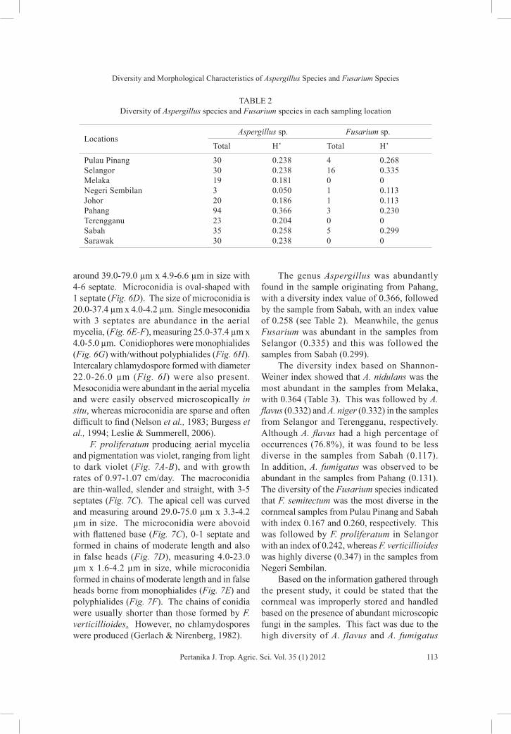
The genus Aspergillus was abundantly found in the sample originating from Pahang, with a diversity index value of 0.366, followed by the sample from Sabah, with an index value of 0.258 (see Table 2). Meanwhile, the genus Fusarium was abundant in the samples from Selangor (0.335) and this was followed the samples from Sabah (0.299).
The diversity index based on Shannon-Weiner index showed that A. nidulans was the most abundant in the samples from Melaka, with 0.364 (Table 3). This was followed by A. flavus (0.332) and A. niger (0.332) in the samples from Selangor and Terengganu, respectively. Although A. flavus had a high percentage of occurrences (76.8%), it was found to be less diverse in the samples from Sabah (0.117). In addition, A. fumigatus was observed to be abundant in the samples from Pahang (0.131). The diversity of the Fusarium species indicated that F. semitectum was the most diverse in the cornmeal samples from Pulau Pinang and Sabah with index 0.167 and 0.260, respectively. This was followed by F. proliferatum in Selangor with an index of 0.242, whereas F. verticillioides was highly diverse (0.347) in the samples from Negeri Sembilan.
Based on the information gathered through the present study, it could be stated that the cornmeal was improperly stored and handled based on the presence of abundant microscopic fungi in the samples. This fact was due to the high diversity of A. flavus and A. fumigatus
TABLE 2 Diversity of Aspergillus species and Fusarium species in each sampling location
Locations Aspergillus sp. Fusarium sp.
Total H’ Total H’
Pulau Pinang 30 0.238 4 0.268Selangor 30 0.238 16 0.335Melaka 19 0.181 0 0Negeri Sembilan 3 0.050 1 0.113Johor 20 0.186 1 0.113Pahang 94 0.366 3 0.230Terengganu 23 0.204 0 0Sabah 35 0.258 5 0.299Sarawak 30 0.238 0 0

Nithiyaa, P., Nur Ain Izzati, M.Z., Umi Kalsom, Y., Salleh, B.
114 Pertanika J. Trop. Agric. Sci. Vol. 35 (1) 2012
which are well known storage fungi that colonize post-harvested grains, specifically when provided with suitable conditions (Reddy et al., 2009; Lacey, 1989). Conversely, A. niger is a saprophyte that grows readily on organic matters, as well as a pathogen that infects crops and stays dormant till storage (Raper & Fennell, 1965). According to Soriano and Dragacci (2004), amongst the corn-based products, cornmeal is one of the items that receives the mildest processing and treatment compared to other food stuff. Thus, it is very susceptible to fungal contamination, specifically air-borne fungi. The fungi that infected the cornmeal are capable of producing secondary metabolites that are responsible in causing hazardous diseases in both human and animal. Although the Fusarium species, such as F. verticillioides (Sacc.) Nirenberg (synonym, F. moniliforme Sheldon) and F. proliferatum (Matsushima) Nirenberg (Torres et al., 2001), are commonly known as plant pathogen, their invasion in the field could proceed till storage. F. semitectum is normally isolated from soil and possibly exists as soil inhabitants. Although this species is
not regarded as significant plant pathogens, the species have been reported to cause diseases on several crops and widely distributed as saprophytes in soil, as well as on diseased tissues (Latiffah et al., 2007). Moreover, there are occasions where other researchers managed to obtain Fusarium species in corn-based products, besides solely from corn plantation (Jimenez & Mateo, 1997; Pascale et al., 2002). The prevalence of the Fusarium species is however lower compared to the Aspergillus species. Besides, the Fusarium species favours tropical and temperate areas to inhabit, therefore it has less adaptation to storage condition (Presello et al., 2008).
CONCLUSIONMost of the Aspergillus and Fusarium isolates were obtained from the cornmeal samples from Pahang (H’=0.596), and this was followed by Selangor (H’=0.573). Meanwhile, the least number of isolates was obtained from Negeri Sembilan (H’=0.163). With the aid of the morphological identification, the diversity and
TABLE 3 Diversity of Microscopic Fungi based on Shannon-Weiner Index
Locations
Aspergillus Fusarium
A. fl
avus
A. n
idul
ans
A. n
iger
A. fu
mig
atus
F. se
mite
ctum
F. p
rolif
erat
um
F. v
ertic
illio
ides
*H’ *H’ *H’ *H’ *H’ *H’ *H’
Pulau Pinang 0.205 0 0.252 0 0.167 0.103 0.103Selangor 0.332 0.177 0.135 0 0 0.242 0.342Melaka 0.316 0.364 0 0 0 0 0Negeri Sembilan 0.216 0 0 0 0 0 0.347Johor 0.171 0.146 0.224 0 0 0 0.146Pahang 0.151 0.05 0.205 0.131 0.108 0 0Terengganu 0.192 0 0.332 0 0 0 0Sabah 0.117 0 0 0 0.260 0 0Sarawak 0.152 0.113 0.230 0.113 0 0 0
*Calculation: pi= ∑ Aspergillus or Fusarium species in each state ∑ Aspergillus or Fusarium in all states H’= pi(ln pi)
Diversity and Morphological Characteristics of Aspergillus Species and Fusarium Species
115Pertanika J. Trop. Agric. Sci. Vol. 35 (1) 2012
abundance of individual isolates of Aspergillus and Fusarium were attained instantly. The cornmeal samples were probably stored at a very poor condition since the diversity of storage fungi, specifically A. flavus, is tremendously high.
ACKNOWLEDGEMENTThe authors are thankful to the financial support received from the UPM Research University Grants Scheme No. 05-03-08-0484RU/91489.
REFERENCESBurgess, L. W., Summerell, B. A., Bullock, S., Gott,
K. P., & Backhouse, D. (1994). Laboratory Manual for Fusarium Research. Sydney: University of Sydney.
Cuenca, G., & Meneses, E. (1996). Diversity patterns of arbuscular mycorrhizal fungi associated with cacao in Venezuela. Plant and Soil, 183, 315-322.
Diba, K., Kordbacheh, P., Mirhendi, S. H., Rezaie, S., & Mahmoudi, M. (2007). Identification of Aspergillus sp. using morphological characteristics. Pakistan Journal of Medical Science, 23, 867-872.
Ezekiel, C. N., Odebode, A. C., & Fapohunda, S. O. (2008). Zearalenone Production by naturally occurring Fusarium sp. on maize, wheat and soybeans from Nigeria. Journal of Biology and Environment Science, 6, 77-82.
Gerlach, W., & Nirenberg, H. I. (1982). The genus Fusarium: A pictorial atlas. Berlin: Mitt. Biol. Bundes. Land- Forst. (Berlin - Dahlem).
Horn, B. W., Greene, R. L., & Dorner, J. W. (2000). Inhibition of Aflatoxin B1 Production by Aspergillus parasiticus Using Nonaflatoxigenic Strains: Role of Vegetative Compatibility. Biological Control, 17, 147-154.
Hussein, H. M., Baxter, M., Andrew, I. G., & Franich, R. A. (1991). Mycotoxin production by Fusarium species isolated from New Zealand maize fields. Mycopathologia, 113, 35-40.
Jimenez, M., & Mateo, R. (1997). Determination of mycotoxins produced by Fusarium isolates from
banana fruits by capillary gas chromatography and highperformance liquid chromatography. Journal of Chromatography, 778, 363-372.
Lacey, J. (1989). Pre- and post-harvest ecology of fungi causing spoilage of foods and other stored products. Journal of Applied Bacteriology Symposium Supplement, 11-25.
Latiffah, Z., Mohd Zariman, M, & Baharuddin, S. (2007). Diversity of Fusarium species in cultivated soils in Penang. Malaysian Journal of Microbiology, 3, 27-30.
Leslie, J. F., & Summerell, B. A. (2006). The Fusarium laboratory manual. South Africa: Wiley-Blackwell Publishing.
Mubatanhema, W., Moss, M. O., Frank, M. J., & Wilson, D. M. (1999). Prevalence of Fusarium species of the Liseola section on Zimbabwean corn and their ability to produce the mycotoxins zearalenone, moniliformin and fumonisin B1. Mycopathologia, 148, 157-163.
Munkvold, G., Stahr, H. M., Logrieco, A., Moretti, A., & Ritieni, A. (1998). Occurrence of fusaproliferin and beauvericin in Fusarium-contaminated livestock feed in Iowa. Applied and Environmental Microbiology, 64, 3923-3926.
Nelson, P. E., Toussoun, T. A., & Marasas, W. F. O. (1983). Fusarium Species: An Illustrated Manual for Identification. University Park, Pennsylvania : Pennsylvania State University Press.
Papavizas, G. C. (1967). Evaluation of various media and antimicrobial agents for isolation of Fusarium from soil. Phytopathology, 57, 848-852.
Pascale, M., Visconti, A., & Chelkowski, J. (2002). Ear rot susceptibility and mycotoxin contamination of maize hybrids inoculated with Fusarium species under field conditions. European Journal of Plant Pathology, 108, 645-651.
Plattner, R. D., Ross, P. F., Reagor, J., Stedelin, J. and Rice, L. G. (1991). Analysis of corn and cultured corn for fumonisin B1 by HPLC and GC/MS by four laboratories. Journal of Veterinary Diagnosis and Investigation, 3, 357-358.
Presello, D. A., Botta, G., Iglesias, J., & Eyherabide, G. H. (2008). Effect of disease severity on yield and grain fumonisin concentration of maize

Nithiyaa, P., Nur Ain Izzati, M.Z., Umi Kalsom, Y., Salleh, B.
116 Pertanika J. Trop. Agric. Sci. Vol. 35 (1) 2012
hybrids inoculated with Fusarium verticillioides. Crop Protection, 27, 572-576.
Raper, K. B., & Fennell, D. I. (1965). Description and morphology. The genus Aspergillus. The Williams & Wilkins Company. (p. 17-29). United States of America: The Waverly Press.
Reddy, K. R. N., Reddy, C. S., & Muralidharan, K. (2009). Potential of botanicals and biocontrol agents on growth and aflatoxin production by Aspergillus flavus infecting rice grains. Food Control, 20, 173-178.
Salleh, B., & Sulaiman, B. (1984). Fusaria associated with naturally diseasedplants in Penang. Journal of Plant Protection in the Tropics, 1, 47-53.
Samson, R. A. (1979). A compilation of the Aspergilli described since 1965. Studies in Mycology, 18, 1-38.
Samson, R. A., Hongs, S. B., & Frisvad, J. C. (2006). Old and new concepts of species differentiation in Aspergillus. Medical Mycology, 44, 133-148.
Schollenberger, M., Muller, H. M., Rufle, M., Suchy, S., Plank, S., & Drochner, W. (2006). Natural occurrence of 16 Fusarium toxins in grains and feedstuffs of plant origin from Germany. Mycopathologia, 161, 43-52.
Soriano, J. M., & Dragacci, S. (2004). Occurrence of fumonisins in foods. Food Research International, 37, 985-1000.
Sewram, V., Nieuwoudt, T. W., Marasas, W. F. O., Shephard, G. S., & Ritieni, A. (1999). Determination of the mycotoxin moniliformin in cultures of Fusarium subglutinans and in naturally contaminated maize by high-performance liquid chromatography– atmospheric pressure chemical ionization mass spectrometry. Journal of Chromatography, 848, 185-191.
Soriano, J. M., & Dragacci, S. (2004). Occurrence of fumonisins in foods. Food Research International, 37, 985-1000.
Summerell, B. A., Salleh, B. and Leslie, J.F. (2003). A Utilitarian Approach to Fusarium Identification. American Phytopathological Society, 87, 117-128.
Torres, A. M., Reynoso, M. M., Rojo, F. G., Ramirez, M.L., & Chulze, S.N. (2001). Fusarium species (section Liseola) and its mycotoxins in maize harvested in northern Argentina. Food Additives and Contaminants, 18, 836-843.
Vanara, F., Reyneri, A., & Blandino, M. (2009). Fate of fumonisin B1 in the processing of whole maize kernels during dry-milling. Food Control, 20, 235-238.
Zinedine, A., & Manes, J. (2009). Occurrence and legislation of mycotoxins in food and feed from Morocco. Food Control, 20, 334-344.