disorders 5 head - carrsconsulting.comDefinition Atrophic rhinitis covers any disorder that results...
15
Disorders of the head Disorders present in Australia Europe/Asia North America Clinical gross anatomy of the head Progressive atrophic rhinitis Yes Yes Yes Congenital tremor Yes Yes Yes Conjunctivitis Yes Yes Yes Meningitis Yes Yes Yes Post-weaning sneezing Yes Yes Yes Other conditions: Middle Ear Yes Yes Yes Aural Haematoma Yes Yes Yes
Transcript of disorders 5 head - carrsconsulting.comDefinition Atrophic rhinitis covers any disorder that results...
Disorders of the head
Disorders present in Australia Europe/Asia North America Clinical gross anatomy of the head Progressive atrophic rhinitis Yes Yes Yes Congenital tremor Yes Yes Yes Conjunctivitis Yes Yes Yes Meningitis Yes Yes Yes Post-weaning sneezing Yes Yes Yes Other conditions: Middle Ear Yes Yes Yes Aural Haematoma Yes Yes Yes

Clinical gross anatomy of the head
The head from the front Detail of the nose
The skeletal head lateral Detail of the teeth in the skull
The skull, ventral view The lower jaw, dorsal view
The hard and soft palate Detail of the teeth and gums

The tongue, dorsal view The tongue, ventral view
The tongue and larynx The palatine tonsils
Longitudinal section of the nasal cavity Cross section of the nasal cavity
The brain, dorsal view Longitudinal section of the cranial vault
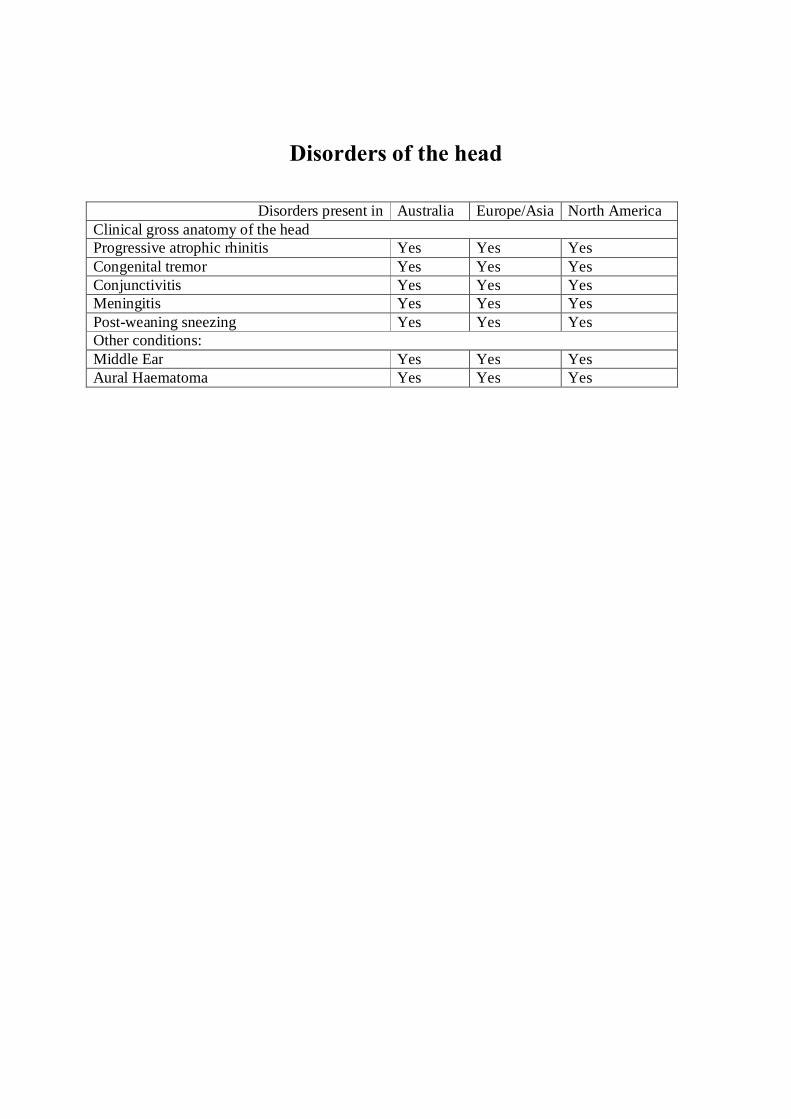
Aging piglets, weaners and growers by their teeth
(the non erupted teeth have been blacked out to create clarity)
Birth – the incisors 3 and canines are present 1 week – the upper premolars 3 and 4 erupt
2 to 4 weeks: lower premolars 3 and 4 and incisors 1 erupt
5 to 7 weeks: Premolars 2 and lower incisors 2 erupt
Permanent teeth eruptions 4 to 6 months M1 8 to 12 months M2 and I3 9 to 10 months C 12 months I1 12 to 15 months P1, P2, P3, P4 16 to 20 months I2 18 to 20 months M3
6 to 12 weeks upper incisors 2 erupt
Adult lower and upper jaw with teeth labeled
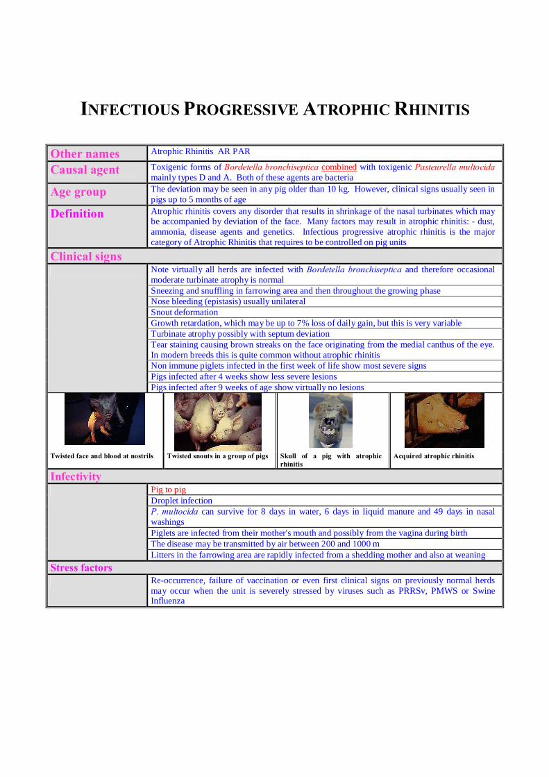
INFECTIOUS PROGRESSIVE ATROPHIC RHINITIS
Other names Atrophic Rhinitis AR PAR
Causal agent Toxigenic forms of Bordetella bronchiseptica combined with toxigenic Pasteurella multocida mainly types D and A. Both of these agents are bacteria
Age group The deviation may be seen in any pig older than 10 kg. However, clinical signs usually seen in pigs up to 5 months of age
Definition Atrophic rhinitis covers any disorder that results in shrinkage of the nasal turbinates which may be accompanied by deviation of the face. Many factors may result in atrophic rhinitis: - dust, ammonia, disease agents and genetics. Infectious progressive atrophic rhinitis is the major category of Atrophic Rhinitis that requires to be controlled on pig units
Clinical signs Note virtually all herds are infected with Bordetella bronchiseptica and therefore occasional
moderate turbinate atrophy is normal Sneezing and snuffling in farrowing area and then throughout the growing phase Nose bleeding (epistasis) usually unilateral Snout deformation Growth retardation, which may be up to 7% loss of daily gain, but this is very variable Turbinate atrophy possibly with septum deviation Tear staining causing brown streaks on the face originating from the medial canthus of the eye. In modern breeds this is quite common without atrophic rhinitis Non immune piglets infected in the first week of life show most severe signs Pigs infected after 4 weeks show less severe lesions Pigs infected after 9 weeks of age show virtually no lesions
Twisted face and blood at nostrils Twisted snouts in a group of pigs Skull of a pig with atrophic rhinitis
Acquired atrophic rhinitis
Infectivity Pig to pig
Droplet infection P. multocida can survive for 8 days in water, 6 days in liquid manure and 49 days in nasal washings Piglets are infected from their mother's mouth and possibly from the vagina during birth The disease may be transmitted by air between 200 and 1000 m Litters in the farrowing area are rapidly infected from a shedding mother and also at weaning
Stress factors Re-occurrence, failure of vaccination or even first clinical signs on previously normal herds
may occur when the unit is severely stressed by viruses such as PRRSv, PMWS or Swine Influenza
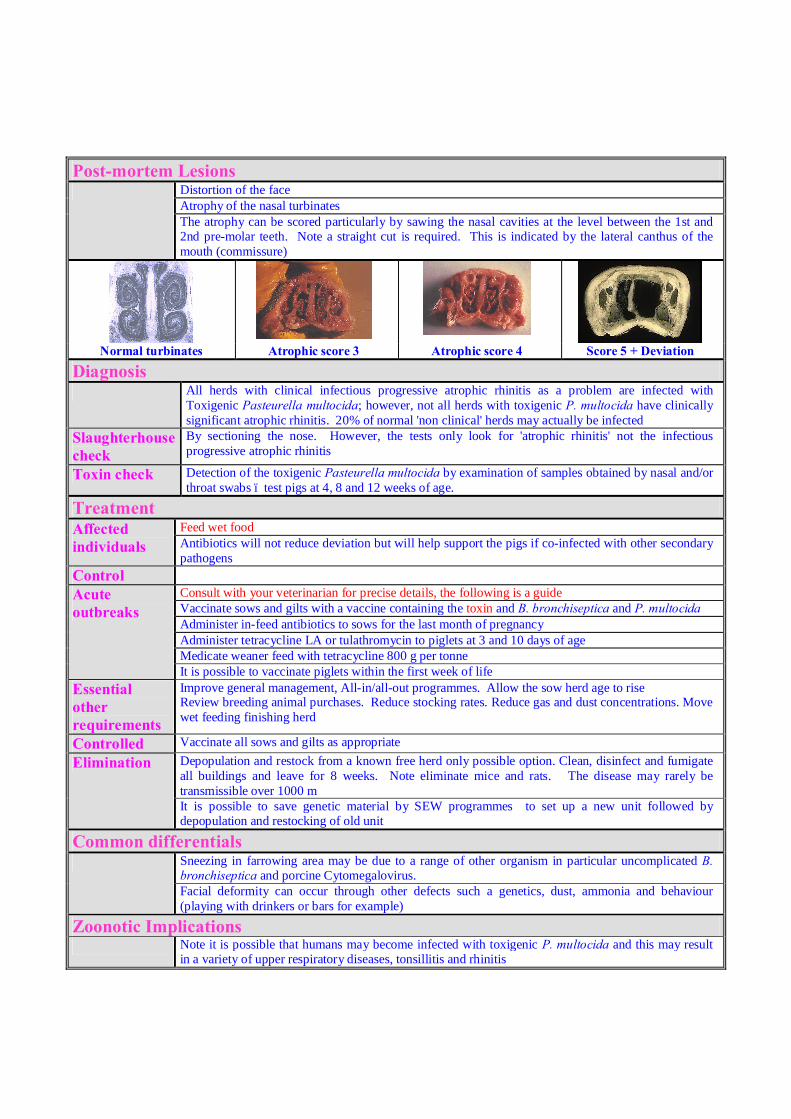
Post-mortem Lesions Distortion of the face
Atrophy of the nasal turbinates The atrophy can be scored particularly by sawing the nasal cavities at the level between the 1st and 2nd pre-molar teeth. Note a straight cut is required. This is indicated by the lateral canthus of the mouth (commissure)
Normal turbinates Atrophic score 3 Atrophic score 4 Score 5 + Deviation
Diagnosis All herds with clinical infectious progressive atrophic rhinitis as a problem are infected with
Toxigenic Pasteurella multocida; however, not all herds with toxigenic P. multocida have clinically significant atrophic rhinitis. 20% of normal 'non clinical' herds may actually be infected
Slaughterhouse check
By sectioning the nose. However, the tests only look for 'atrophic rhinitis' not the infectious progressive atrophic rhinitis
Toxin check Detection of the toxigenic Pasteurella multocida by examination of samples obtained by nasal and/or throat swabs – test pigs at 4, 8 and 12 weeks of age.
Treatment Affected individuals
Feed wet food Antibiotics will not reduce deviation but will help support the pigs if co-infected with other secondary pathogens
Control Acute outbreaks
Consult with your veterinarian for precise details, the following is a guide Vaccinate sows and gilts with a vaccine containing the toxin and B. bronchiseptica and P. multocida Administer in-feed antibiotics to sows for the last month of pregnancy Administer tetracycline LA or tulathromycin to piglets at 3 and 10 days of age Medicate weaner feed with tetracycline 800 g per tonne It is possible to vaccinate piglets within the first week of life
Essential other requirements
Improve general management, All-in/all-out programmes. Allow the sow herd age to rise Review breeding animal purchases. Reduce stocking rates. Reduce gas and dust concentrations. Move wet feeding finishing herd
Controlled Vaccinate all sows and gilts as appropriate Elimination Depopulation and restock from a known free herd only possible option. Clean, disinfect and fumigate
all buildings and leave for 8 weeks. Note eliminate mice and rats. The disease may rarely be transmissible over 1000 m It is possible to save genetic material by SEW programmes to set up a new unit followed by depopulation and restocking of old unit
Common differentials Sneezing in farrowing area may be due to a range of other organism in particular uncomplicated B.
bronchiseptica and porcine Cytomegalovirus. Facial deformity can occur through other defects such a genetics, dust, ammonia and behaviour (playing with drinkers or bars for example)
Zoonotic Implications Note it is possible that humans may become infected with toxigenic P. multocida and this may result
in a variety of upper respiratory diseases, tonsillitis and rhinitis

CONGENITAL TREMOR
Agent A variety of events, diseases, toxins etc can cause a piglet to present with congenital tremor
1 The most common cause is an unidentified viral agent 2 There are a number of genetic causes – Landrace trembles,
Saddleback tremors 3 There are other known congenital infections for example
Classical Swine Fever Virus which can result in the production of trembling piglets
This technical note will concentrate on the most common cause Clinical signs
The problem classically occurs in naïve gilt litters from newly introduced stock.
New born piglets
Piglets are born trembling in all the muscles. They may be unable to walk and often are unable to suckle efficiently. Once attached to the teat by the stockman the piglet will suckle vigorously, but once let go by the stockman the piglet will often shake itself away from the teat. Excluding sporadic cases it is normal that there will be an increase in pre-weaning mortality and diseases, in particularly overlaids, increased lameness from trauma and outbreaks of scour. A trembling piglet which has problems suckling will clearly have problems ingesting sufficient colostrum. Mortality rates of 75 to 100% are common. Sometimes the trembling will be noticeably reduced when the piglet is asleep. In the piglets which survive, the clinical signs subside with age. However, the tremble is often noticeable if the animal is watched over time and then relaxes, then mild muscle trembles (fasciculation) will be evident over the ears and muscles on the back.
Start up herds
The problem can be very severe and potentially disastrous with almost 100% of litters presenting with congenital tremor piglets
Closed herds And new stock
In small closed herds it is possible for the resident animals to be or become naive. Under these circumstances the introduction of a new boar or other gilts will invoke the problem in piglets born to any pregnant sow. In these circumstances the farm’s original sows and gilts have problems.
Sporadic cases
Congenital tremor is seen occasionally on most farms as an incidental occurrence and probably results from gilt or possibly sow which has by chance remained naive until she became pregnant.
All other age groups
There are no clinical signs to born piglets, weaners, growers or adults who become infected for the first time. The disease only affects the foetus.
Infectivity The agent would appear to be very infective. However, on modern farms disease transmission can be slow and it is possible for the disease to die out on a farm. For this reason nucleus farms may produce naive gilts. All materials are infective – faeces, placenta, nasal droplets, macerated piglets/foetuses and possibly semen
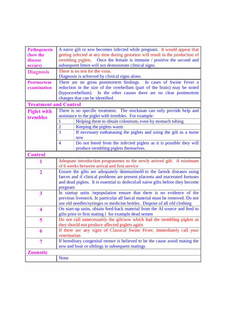
Pathogenesis (how the disease occurs)
A naive gilt or sow becomes infected while pregnant. It would appear that getting infected at any time during gestation will result in the production of trembling piglets. Once the female is immune / positive the second and subsequent litters will not demonstrate clinical signs.
Diagnosis There is no test for the virus. Diagnosis is achieved by clinical signs alone.
Postmortem examination
There are no gross postmortem findings. In cases of Swine Fever a reduction in the size of the cerebellum (part of the brain) may be noted (hypocerebellum). In the other causes there are no clear postmortem changes that can be identified.
Treatment and Control Piglet with trembles
There is no specific treatment. The stockman can only provide help and assistance to the piglet with trembles. For example: 1 Helping them to obtain colostrum, even by stomach tubing 2 Keeping the piglets warm 3 If necessary euthanasing the piglets and using the gilt as a nurse
sow 4 Do not breed from the infected piglets as it is possible they will
produce trembling piglets themselves. Control
1 Adequate introduction programmes to the newly arrived gilt. A minimum of 6 weeks between arrival and first service
2 Ensure the gilts are adequately ‘immunised’ to the farm’s diseases using faeces and if clinical problems are present placenta and macerated foetuses and dead piglets. It is essential to ‘infect’ all naive gilts before they become pregnant
3 In startup units /repopulation ensure that there is no evidence of the previous livestock. In particular all faecal material must be removed. Do not use old needles/syringes or medicine bottles. Dispose of all old clothing
4 On start-up units, obtain feed-back material from the AI source and feed to gilts prior to first mating – for example dead semen
5 Do not cull unnecessarily the gilt/sow which had the trembling piglets as they should not produce affected piglets again
6 If there are any signs of Classical Swine Fever, immediately call your veterinarian
7 If hereditary congenital tremor is believed to be the cause avoid mating the sow and boar or siblings in subsequent matings
Zoonotic None

CONJUNCTIVITIS
Causal agent Unknown. Probably a combination of Bordetella bronchiseptica and Chlamydia psittici.
Age group Weaners and progresses into grow/finish and young adults
Clinical signs Sneezing and runny eyes in the late farrowing house and nursery.
Conjunctiva of both eyes become injected and inflamed Third eyelid prolapses. Once prolapsed condition seems to become static. Many only affect individuals, but on some farms all pigs in the pen are affected. The pigs otherwise do not seem affected by the condition
Growing pigs with conjunctivitis Both eyes are equally affected Note prolapse of the third eyelid
Infectivity Unknown
Transmission Probable nose to nose contact. Organisms isolated are very common on all farms with or
without the problem
Post-mortem lesions
Severe conjunctivitis with prolapse of the third eyelid. Sclera and cornea unaffected Tear staining on face. Young pigs moderate to severe purulent rhinitis
Diagnosis
Clinical signs Chlamydia suspected as a secondary invader following swollen exposed conjunctiva
Treatment Individual/group Review Progressive atrophic rhinitis vaccine programme.
Test and eliminate toxigenic Pasteurella multocida as a problem Antimicrobials appear to have little effect. Overuse of antimicrobials may encourage Chlamydia as a problem
Control Review management of the farm buildings Avoid chilling and draughts Reduce ammonia concentrations in the air Reduce dust levels in the grower and finishing house – cover feeders, consider wet feeding Minimise the clinical effects of PRRSv
Common differentials
Progressive Atrophic Rhinitis. Swine Influenza Zoonotic
None

MENINGITIS
Causal agent
The classical cause of meningitis in piglets, weaners and growers is Streptococcus suis II. However, several other bacteria can cause meningitis namely Haemophilus parasuis and septicaemic Escherichia coli or Bowel Oedema (F18)
Age group
Any. Meningitis classically occurs between 3-60 kg. Haem. parasuis in naive finishing and adult pigs can cause a devastating acute fatal meningitis
Clinical signs Acute The pig becomes incoordinated often with uncontrolled eye movements
Rectal temperature is increased to 40-41°C As the condition progresses the pig will fall over and thrash with all four legs on the floor. The pig may traumatise itself during these thrashing movements Death can occur quickly especially if the pig is stressed. In the farrowing house the condition can then be misdiagnosed as an overlaid pig If treatment is late a neurologically damaged pig may result
Normal pig No clinical signs, the organism lives on the tonsils and upper respiratory tract of the normal healthy pig
Two growing pigs with meningitis
Infectivity Most pigs are infected
Transmission
From the mother at birth by nose to nose contact Between pigs by nose to nose contact
Post-mortem Lesions None particularly obvious. Meningeal
congestion and tags are suspicious but most veterinarians examine so few brains it would be difficult for them to differentiate between the normal range of meningeal changes. The picture shows the brain in a sectioned head. Take a piece of brain and meninges for histology in all <30kg pigs

Diagnosis Culture of Streptococcus suis II from cerebrospinal fluid (CSF)
Using an 18 gauge 1.5 inch needle. Place the pig in a dorsal position. Flex the head over the edge of the post-mortem table. Feel for the atlas occipital junction. Swab the skin with surgical spirit. Insert the needle and ‘walk’ the needle down the brain case off the occipital joint. Attach a sterile 2 ml syringe. 1 ml of clear CSF should be readily easy to extract The reason for using CSF is that Strep. suis is widespread on the skin and upper respiratory tract and post-mortem knives are easily contaminated leading to a misdiagnosis Histological examination of the meninges including a Gram stain
Treatment Individual The pig is dying
Treatment must be rapid and vigorous Ceftiofur 3mg/kg by intramuscular injection is the medicine of choice Treat with procaine penicillin every 12 hours until clinical signs subside A few pigs can develop a procaine allergy and go into anaphylactic shock If weaned isolate the pig. If not weaned place in creep area Keep in a darken room If fits are extreme give a sedative as necessary Provide water by mouth from a syringe is necessary. Note a pig drinks 1 litre per 10 kg per day so a few syringe fulls is not sufficient
Control Most pigs carry Strep. suis on their tonsils and upper respiratory tracts. The causal factor that results in the clinical signs is that the pig is subjected to too much stress or other disease Draughts particularly in farrowing houses and nurseries Check colostrum management when meningitis occurs in piglets. Review feedback programme Wet and cold environments, common if weaners placed into cleaned houses. In outdoor situations use of damp mouldy straw often precipitates a meningitis ‘outbreak’. Excessive change in temperature in the pigs sleeping area Pigs being moved and mixed resulting in disturbed social hierarchy Gas levels being too high. Carbon monoxide poisoning from gas heaters
Common differentials Haemophilus parasuis and septicaemic E.coli. Intoxication with brewer
products. Water deprivation. Porcine Stress Syndrome. Trauma to the head. Aujeszky’s Disease.
Zoonotic Implications Strep. suis can rarely cause a fatal meningitis in man following infection into a
cut from the pig’s skin typically following cutting a finger following injection of a pig. It is an occupational hazard
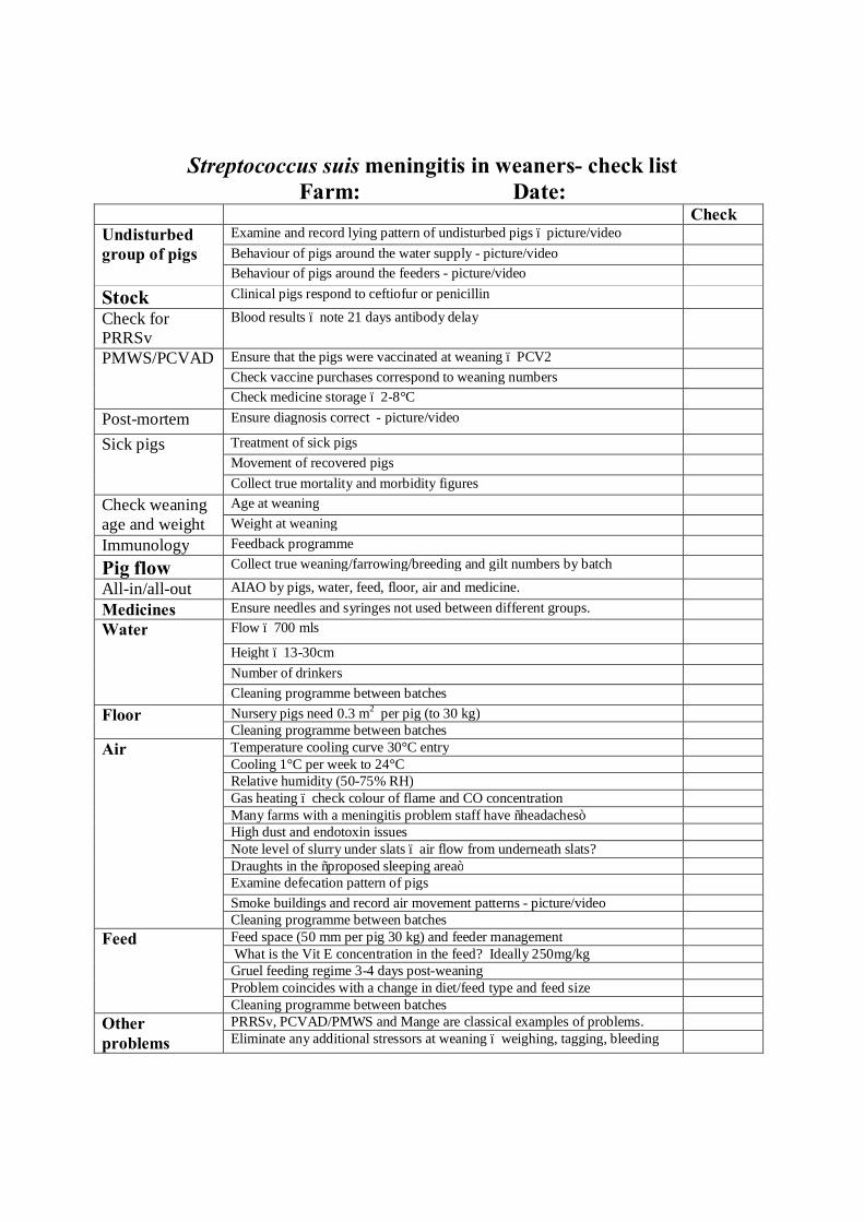
Streptococcus suis meningitis in weaners- check list Farm: Date:
Check Undisturbed group of pigs
Examine and record lying pattern of undisturbed pigs – picture/video Behaviour of pigs around the water supply - picture/video Behaviour of pigs around the feeders - picture/video
Stock Clinical pigs respond to ceftiofur or penicillin Check for PRRSv
Blood results – note 21 days antibody delay
PMWS/PCVAD Ensure that the pigs were vaccinated at weaning – PCV2 Check vaccine purchases correspond to weaning numbers Check medicine storage – 2-8°C
Post-mortem Ensure diagnosis correct - picture/video Sick pigs Treatment of sick pigs
Movement of recovered pigs Collect true mortality and morbidity figures
Check weaning age and weight
Age at weaning Weight at weaning
Immunology Feedback programme Pig flow Collect true weaning/farrowing/breeding and gilt numbers by batch All-in/all-out AIAO by pigs, water, feed, floor, air and medicine. Medicines Ensure needles and syringes not used between different groups. Water Flow – 700 mls
Height – 13-30cm Number of drinkers Cleaning programme between batches
Floor Nursery pigs need 0.3 m2 per pig (to 30 kg) Cleaning programme between batches
Air Temperature cooling curve 30°C entry Cooling 1°C per week to 24°C Relative humidity (50-75% RH) Gas heating – check colour of flame and CO concentration Many farms with a meningitis problem staff have “headaches” High dust and endotoxin issues Note level of slurry under slats – air flow from underneath slats? Draughts in the “proposed sleeping area” Examine defecation pattern of pigs Smoke buildings and record air movement patterns - picture/video Cleaning programme between batches
Feed Feed space (50 mm per pig 30 kg) and feeder management What is the Vit E concentration in the feed? Ideally 250mg/kg Gruel feeding regime 3-4 days post-weaning Problem coincides with a change in diet/feed type and feed size Cleaning programme between batches
Other problems
PRRSv, PCVAD/PMWS and Mange are classical examples of problems. Eliminate any additional stressors at weaning – weighing, tagging, bleeding
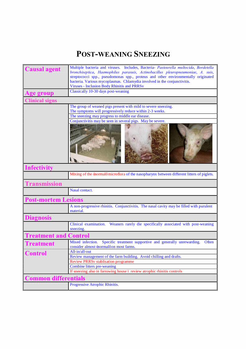
POST-WEANING SNEEZING
Causal agent Multiple bacteria and viruses. Includes, Bacteria- Pasteurella multocida, Bordetella bronchiseptica, Haemophilus parasuis, Actinobacillus pleuropneumoniae, A. suis, streptococci spp., pseudomonas spp., proteus and other environmentally originated bacteria. Various mycoplasmas. Chlamydia involved in the conjunctivitis. Viruses - Inclusion Body Rhinitis and PRRSv
Age group Classically 10-30 days post-weaning
Clinical signs The group of weaned pigs present with mild to severe sneezing.
The symptoms will progressively reduce within 2-3 weeks. The sneezing may progress to middle ear disease. Conjunctivitis may be seen in several pigs. May be severe.
Infectivity Mixing of the ‘normal’ microflora of the nasopharynx between different litters of piglets.
Transmission Nasal contact.
Post-mortem Lesions A non-progressive rhinitis. Conjunctivitis. The nasal cavity may be filled with purulent
material.
Diagnosis Clinical examination. Weaners rarely die specifically associated with post-weaning
sneezing.
Treatment and Control Treatment Mixed infection. Specific treatment supportive and generally unrewarding. Often
consider almost ‘normal’ on most farms.
Control All-in/all-out Review management of the farm building. Avoid chilling and drafts. Review PRRSv stablisation programme Combine litters pre-weaning If sneezing also in farrowing house – review atrophic rhinitis controls
Common differentials Progressive Atrophic Rhinitis.

MIDDLE EAR DISEASE
This condition is characterized by the pig walking with a pronounced head tilt. Radiological examination reveals an abscess and occlusion of the middle ear on the side of the tilt. There is some evidence that M. hyorhinis may play a role in the pathogenesis. Culture at post-mortem tends to reveal a mixed bacterial population. The condition is more common in weaners who develop colds and sneezing 10-20 days post-weaning. Treatment of individuals may be unrewarding. Injections of ampicillin have been effective. Control is to avoid chilling and drafting pigs. However, the problem tends to be sporadic.
A CT scan on a middle ear case, demonstrating occlusion of the right middle ear – left indicated by the black circular hole
In sows the condition may occur due to misplacement of drip cooling systems – which drip into the sow’s ear.
AURAL HAEMATOMA
Characterized by a swelling of the external pinna (ear). The swelling is soft and fluctuated. The swelling is associated with blood leaking under the skin of the ear. The condition is associated with head shaking – fighting and mange are important predisposing factors.
Treatment is varied depending on the farm Do nothing – the lesion with resolve with time, but the ears can be very heavy and they may become infected
This ear has healed after the resolution of the blood clot
Open the ear, but there is a risk of the pig bleeding to death if the internal bleeding has not stopped Placing a band around the base of the ear and the ear will drop off.