
Digestion (Core) Stephen Taylor i-Biology.net.
33
Digestion (Core) Stephen Taylor i-Biology.net
-
Upload
rhoda-flowers -
Category
Documents
-
view
241 -
download
0
Transcript of Digestion (Core) Stephen Taylor i-Biology.net.


Gall
Liver: Secretion of surfactants in bile to break up lipid droplets
Gall Bladder- storage and regulated release of bile
Pancrease- secretion of lipase, amylase and protease
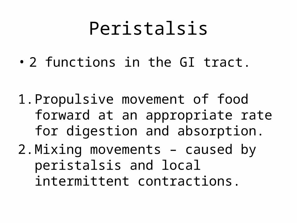
Peristalsis
• 2 functions in the GI tract.
1. Propulsive movement of food forward at an appropriate rate for digestion and absorption.
2. Mixing movements – caused by peristalsis and local intermittent contractions.

Peristalsis
• The propulsive movement in the GI is achieved by peristalsis.

Peristalsis
• Constriction of circular muscles behind the food constricts muscles behind the food preventing it from being pushed back towards the mouth

Muscle Contraction for Peristalsis
The smooth muscle of the gut is formed from short cells, not elongated fibers.Often continuous interspersed with short periods of vigorous contraction.

Peristalsis
• Occurs in one direction• Vomiting uses abdominal muscles

Enzymes review:
• What type of biological molecule is an enzyme?
• How do enzymes work?
• How are enzymes produced through protein synthesis?
• What are the effects of temperature, pH and substrate concentration on the rate of enzyme activity?
• How are immobilized enzymes used in the production of lactose-free milk?
http://www.northland.cc.mn.us/biology/biology1111/animations/enzyme.html

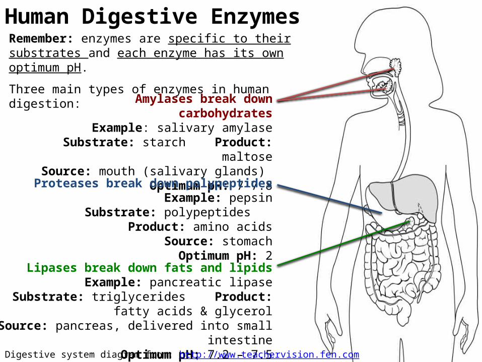
Human Digestive EnzymesRemember: enzymes are specific to their substrates and each enzyme has its own optimum pH.
Three main types of enzymes in human digestion:
Amylases break down carbohydratesExample: salivary amylase
Substrate: starch Product: maltoseSource: mouth (salivary glands)
Optimum pH: 7-7.8
Proteases break down polypeptidesExample: pepsin
Substrate: polypeptides Product: amino acidsSource: stomachOptimum pH: 2
Lipases break down fats and lipidsExample: pancreatic lipase
Substrate: triglycerides Product: fatty acids & glycerolSource: pancreas, delivered into small intestine
Optimum pH: 7.2 – 7.5
Digestive system diagram from: http://www.teachervision.fen.com/digestive-system/printable/57730.html

http://highered.mcgraw-hill.com/sites/0072495855/student_view0/chapter26/animation__organs_of_digestion.html

Pancreatic
• 2 types gland tissue– Islets of Langerhans secrete hormones insulin and
glucagon into blood– Reminder synthesizes and secretes digestive
enzymes into gut in response to eating– Glands secrete enzymes into ducts then into
intestions

Pancreatic Juice
Contains enzymes digesting 3 macromolecules– Amylase: starch– Lipases: trigycerides, phospholipids– Proteases: proteins, peptides

Intestine enzymes
• Starch digested to maltose by amylase• Triglycerides digested to fatty acids and
glycerol or fatty acids and monoglycerides by lipase
• Phospholipds digested into fatty acids, glycerol, and phosphate by phospholipase
• Proteins and polypeptides digested to shorter peptides by protease
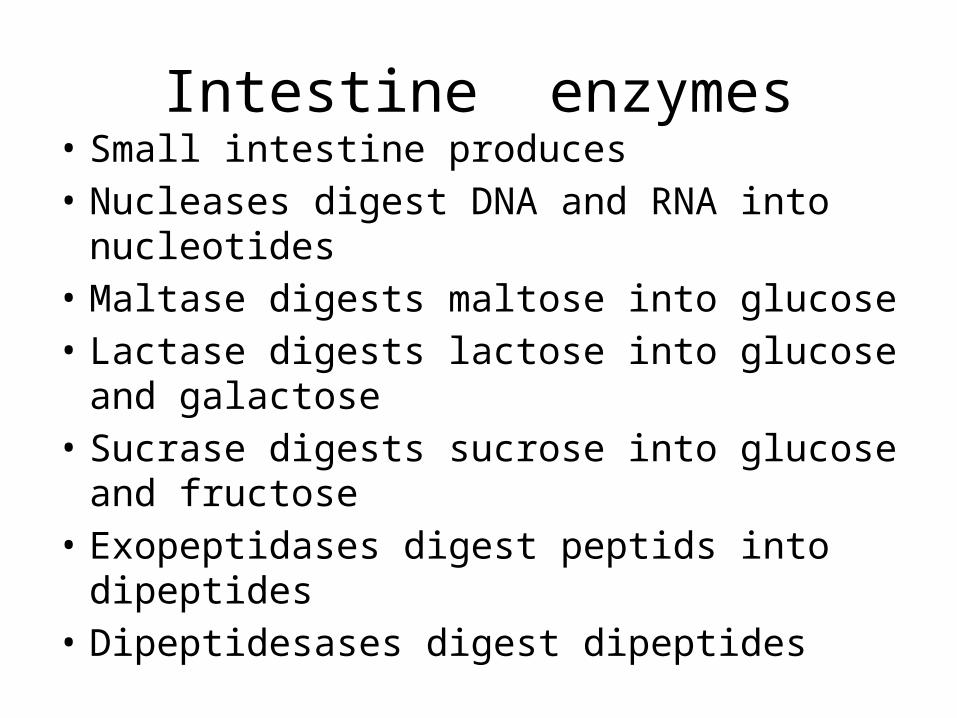
Intestine enzymes• Small intestine produces• Nucleases digest DNA and RNA into
nucleotides• Maltase digests maltose into glucose• Lactase digests lactose into glucose and
galactose• Sucrase digests sucrose into glucose and
fructose• Exopeptidases digest peptids into dipeptides• Dipeptidesases digest dipeptides
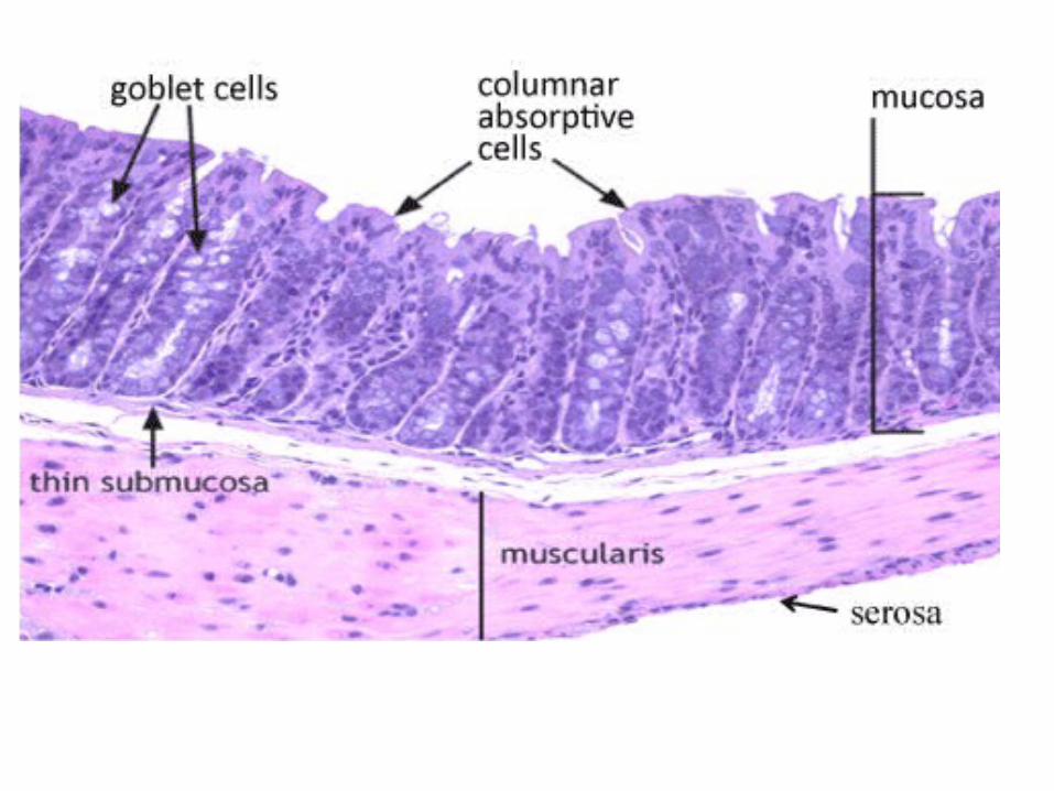

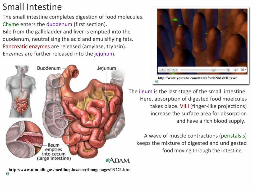
Intestinal Wall

Adaptations to Absorption Getting digested food molecules into the blood from the lumen of the ileum.
Single-cell layer of epithelial cellsShort path for diffusion.
Capillaries close to epitheliumShort path for diffusion, rich supply of blood.
Lacteals (lymph vessels)Allow for rapid absorption and transport of lipids.
Rich blood supplyMaintains concentration gradients between lumen and blood.
Many villi protrude into the lumen, greatly increasing the surface area for absorption.
Images from: http://en.wikipedia.org/wiki/Intestinal_villi
Microvilli on the surface of each cell increase surface
area even further.

Microvasculature of Intestines
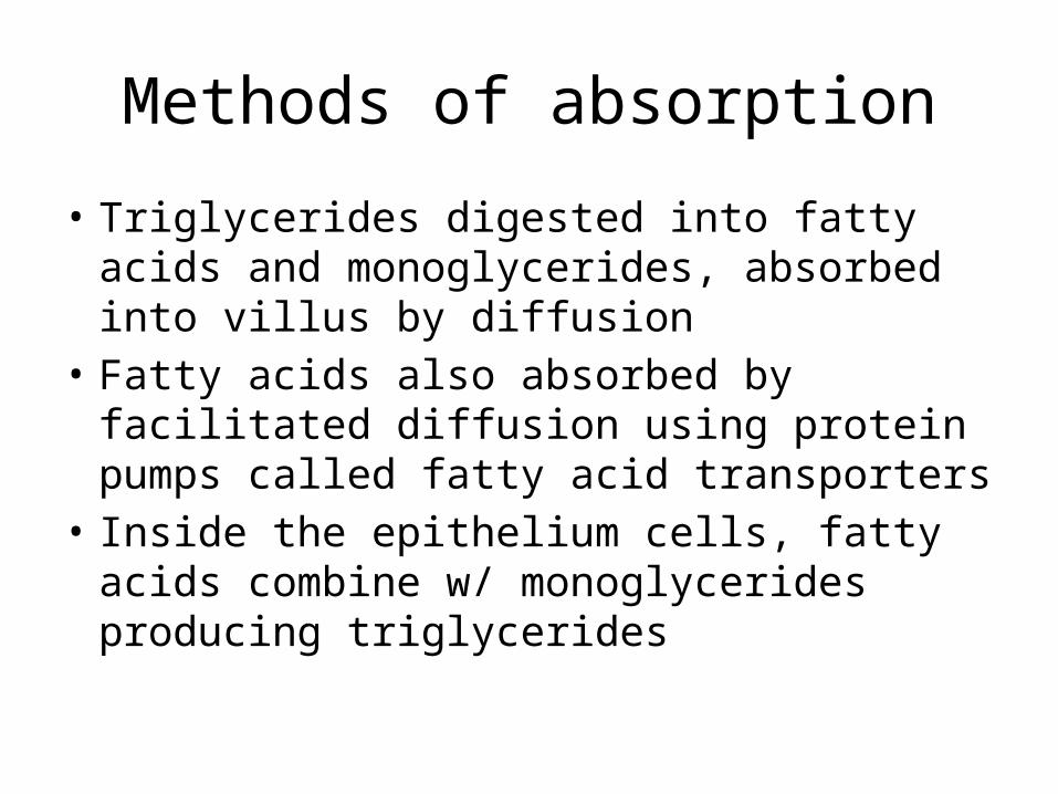
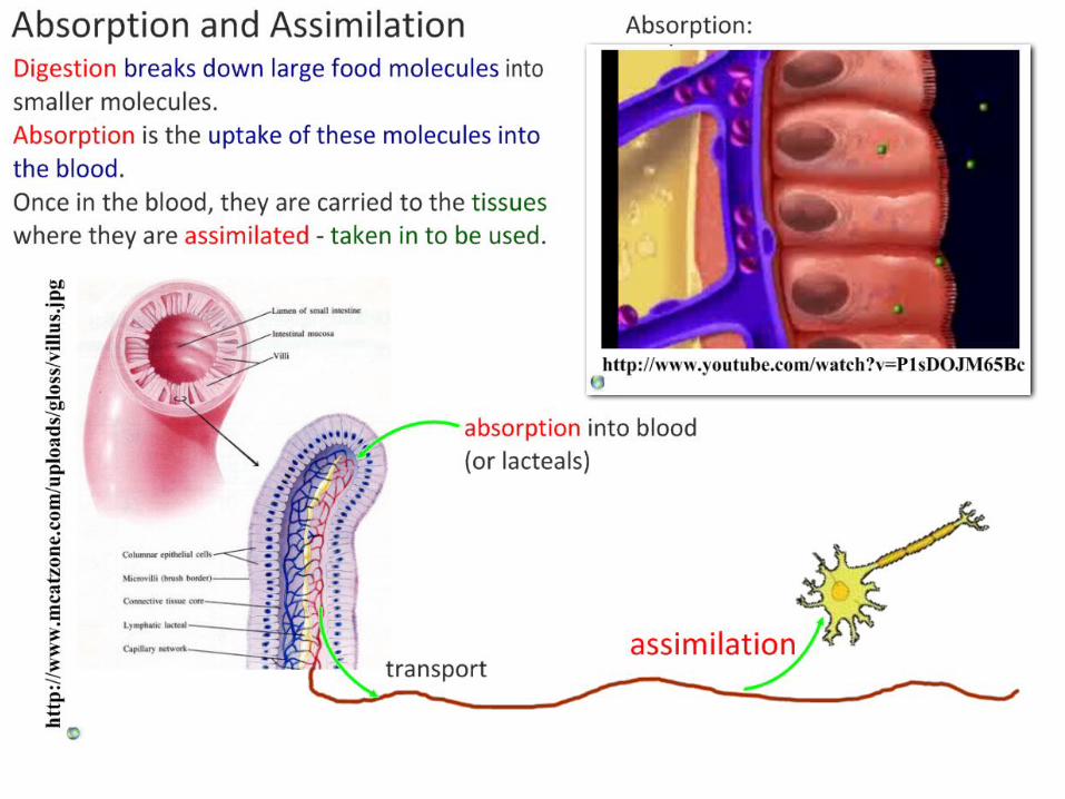
Methods of absorption
• Triglycerides digested into fatty acids and monoglycerides, absorbed into villus by diffusion
• Fatty acids also absorbed by facilitated diffusion using protein pumps called fatty acid transporters
• Inside the epithelium cells, fatty acids combine w/ monoglycerides producing triglycerides

Methods of absorption
• Triglycerides coalesce w/ cholesterol to form lipoprotein (droplets coated in phospholipids and protein)
• Lipoproteins released by exocytosis through plasma membrane where they enter the lacteal or enter blood stream

Methods of absorption
• Glucose cannot pass through plasma membrane by simple diffusion
• Sodium-potassium pump• Sodium-glucose co-transporter proteins in
microvilli transfer sodium and glucose together from lumen to cytoplasm of epithelium. Passive facilitated fusion
• Glucose channels allow facilitated diffusion from villus into blood capillaries.
This is a Creative Commons presentation. It may be linked and embedded but not sold or re-hosted.
Please consider a donation to charity via Biology4Good.Click here for more information about Biology4Good charity donations.
@IBiologyStephen