Digestion 9.3, 9.4. Digestion 4 steps ingestion digestion absorption egestion.
Upload
omkar-khandagaleCategory
view
123download
0
Digestion and AbsorptionMade by Omkar B. Khandagale

Digestion : enzymatic conversion of complex food substances to simple absorbable forms in the alimentary canal.
DIGESTIVE SYSTEM :
• Alimentary canal
• Digestive glands or associated glands.

Alimentary canal :
• The alimentary canal begins with mouth and ends with anus.• Mouth leads to buccal cavity or oral cavity.• Oral cavity has teeth and muscular tongue.• Each tooth embedded in a socket of jaw bone: such attachment called thecodont.• Diphyodont : human has two sets of teeth in their life time:

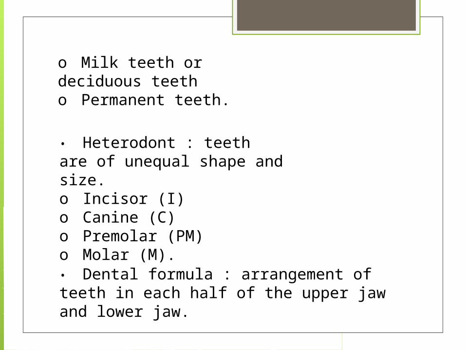
o Milk teeth or deciduous teetho Permanent teeth.
• Heterodont : teeth are of unequal shape and size.o Incisor (I)o Canine (C)o Premolar (PM)o Molar (M).
• Dental formula : arrangement of teeth in each half of the upper jaw and lower jaw.


• Dental formula of human adult is • The hard chewing surface of the teeth made up of enamel.• The tongue is a freely movable muscular organ attached to the floor of the oral cavity by the frenulum.• The upper surface of tongue has small projections called papillae, some of which bears taste buds.• The oral cavity leads into a short pharynx which serves as a common passage for food and air.

• Oesophagus and the trachea open into the pharynx.• Opening of wind pipe or trachea called glottis, and that of oesophagus is called gullet.• The cartilaginous epiglottis prevents the entry of food into the glottis during swallowing.• Oesophagus connects pharynx with stomach.• Opening of oesophagus is regulated by gastro-oesophageal sphincter.• The stomach has three parts:o Cardiac: into which oesophagus opens.o Fundus: air filled portion of stomach.

o Pyloric: portion opens into the small intestine.
• Small intestine distinguished into three parts:o Duodenum: ‘U’ shaped first part.o Jejunum: longer, coiled middle portion.o Ileum: highly coiled posterior part.
• The opening of stomach into the duodenum is guarded by pyloric sphincter.• Large intestine consists of three parts:o Caecumo Colono Rectum.

• Caecum is a small blind sac which hosts some symbiotic micro-organisms.• Caecum has a finger-like blind tubular projection called vermiform appendix.• The Caecum opens into colon, which has three distinct part-o Ascending colono Transverse colono Descending colon
• The descending colon opens into rectum which opens to out through anus.

Histolology of alimentary canal :• Alimentary canal from oesophagus to rectum has four layers.o Serosa.o Muscularis.o Sub mucosa.o Mucosa.
• Serosa is the outermost layer and is made up of a thin mesothelium with some connective tissues.• Muscularis is formed by smooth muscles arranged outer longitudinal and inner circular layers.

• Sub-mucosa is formed by loose connective tissues containing nerves, blood and lymph vessels.• Mucosa is the innermost layer made of endothelium.• Mucosa forms irregular folds (rugae) in the stomach and small finger like folding called villi in the small intestine.
• The cells lining the villi produce numerous microscopic projections called microvilli giving a brush border appearance.

• These modifications increase the surface area for absorption.• Villi are supplied with a network of capillaries and a central lymphatic vessel called lacteal.• Epithelial cells of mucosa contain secretory cells which secretes digestive enzymes.• Mucosa also forms glands in the stomach (gastric gland)• Mucosa forms crypts in between the bases of villi in the intestine called Crypts of Lieberkuhn.


Digestive glands :• The digestive glands associated with the alimentary canal includes-o Salivary glando Livero Pancreas.
• There are three pairs of salivary gland present in the buccal cavity.o Parotid gland (below internal ear)o Sub-maxillary / submandibular (below lower jaw)o Sub-lingual (below tongue)

salivary glands

• All salivary glands produce saliva into the buccal cavity.
Liver :• Largest gland of the body weighing about 1.2 to 1.5 kg in adult.• Located below diaphragm and has two lobes.• Structural and functional unit of liver is the hepatic lobules.• Hepatic lobules consist of hepatic cells arranged in the form of cords.

Liver

original human liver

• Each lobule is covered by a thin connective tissue sheath called Glisson’s capsule.• The bile secreted by the hepatic cells passes through the hepatic ducts and stored in the gall bladder in concentrated form.• Bile from the gall bladder is transported by cystic duct.• Cystic duct along with hepatic duct forms the common bile duct.• Bile duct joined with pancreatic duct to form hepato-pancreatic duct which open into the duodenum• Hepato-pancreatic has a swelling portion called ampulla of Vater; the opening is guarded by sphincter of Oddi.

Pancreas
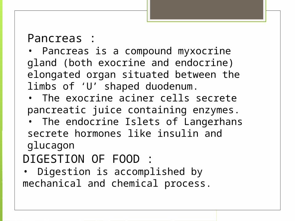
Pancreas :• Pancreas is a compound myxocrine gland (both exocrine and endocrine) elongated organ situated between the limbs of ‘U’ shaped duodenum.• The exocrine aciner cells secrete pancreatic juice containing enzymes.• The endocrine Islets of Langerhans secrete hormones like insulin and glucagon
DIGESTION OF FOOD :• Digestion is accomplished by mechanical and chemical process.

buccal cavity :• Buccal cavity performs two major functions;o Mastication of food.o Facilitation of swallowing.
• The teeth and tongue with the help of saliva masticate and mix up the food.• The saliva composed of ;o Electrolytes (Na+, K+, Cl- HCO-3)o Enzyme- salivary amylase or ptyalin.o Lysozyme.

buccal cavity

• About 30% of starch is hydrolyzed into disaccharide (maltose) by salivary amylase in optimum pH 6.8).• Lysozyme acts as antibacterial agent preventing infections.• Mucus in the saliva helps in lubricating and adhering the masticated food particle into a bolus.• The bolus is then passed into oesophagus through pharynx by swallowing or deglutition.• By peristalsis the bolus from the oesophagus passed into the stomach.

In the stomach :• The mucosa of stomach has gastric glands.• Gastric glands have three major types of cells namely –o Mucus neck cells – secretes mucus.o Peptic or chief or zymogen cells – secretes proenzymes pepsinogen.o Parietal or oxyntic cells – secretes HCl and castles intrinsic factor (factor essential for absorption of vitamin B12)
• The stomach stores the food for 4-5 hours.• The food mixed with the acidic gastric juice and form chyme.

• Pepsinogen converted into active pepsin in presence of HCl.• Active pepsin converts proteins into proteose and peptones (peptides).• Mucus and bicarbonate ions play important role in lubrication and protection of mucosal epithelium from excoriation by HCl and active enzymes.• HCl provides the acidic pH of stomach (pH1.8)• Rennin is an enzyme present in gastric juice helps in digestion of milk proteins.• Small amount of lipases are present in gastric juice helps in digestion of fats
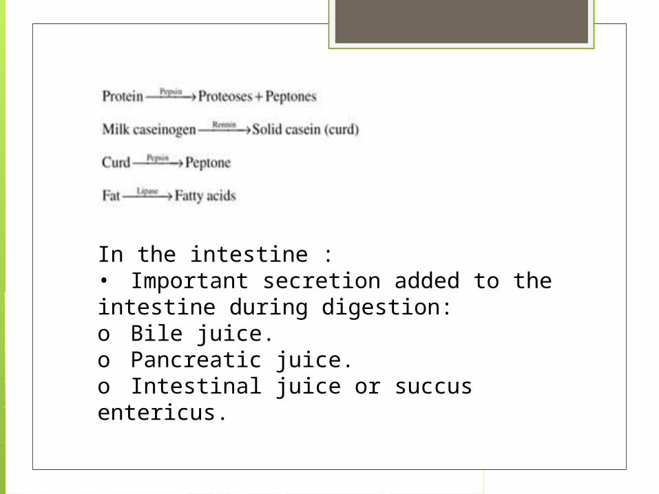
In the intestine :• Important secretion added to the intestine during digestion:o Bile juice.o Pancreatic juice.o Intestinal juice or succus entericus.

• The pancreatic juice contain following enzymes:o Trypsinogeno Chymotrypsinogeno Procarboxypeptidase.o Amylaseso Lipaseso Nucleases.
• Trypsinogen is activated by an enzyme, enterokinase secreted by intestinal mucosa into active trypsin.• Active trypsin activates other enzymes in the pancreatic juice in the intestine.

• The bile released into the duodenum contains –o Bile pigments (bilirubin and bili-verdin)o Bile salts. (Bicarbonate, tourocholate, glycolate)o Cholesterol ando Phospholipids.
• Bile salt helps in emulsification of fat, i.e. breakdown fats into small micelles.• Bile also activates lipases.• The intestinal mucosa contains goblet cells which secrete mucus.

• The secretion of brush border cells of intestinal mucosa and the goblet cells constitute the intestinal juice or succusentericus.• The intestinal juice contains variety of enzymes –o Disaccharidases (maltase, lactase and invertase)o Dipeptidases.o Lipases.o Nucleosidases.

• Sub-mucosal glands (Brunner’s glands) also secrete alkaline fluid to counter act acidic chyme before secretion of bile and pancreatic juice.

ABSORPTION OF DIGESTED PRODUCTS :• Absorption is the process by which the end product of digestion passes through the intestinal mucosa into the blood or lymph.• Absorption is carried out by passive, active or facilitated transport mechanism.• Glucose, amino acids and electrolytes are absorbed by simple diffusion into the blood in the concentration gradient.• Fructose and some amino acids absorbed with the help of carrier ions like Na+. This is called facilitated diffusion.

• Active transport of digested food and electrolytes takes place against the concentration gradients hence require energy.Absorption of fatty acid and glycerol.• Fatty acids and glycerol being insoluble cannot be absorbed into blood.• They are transported into mucosal epithelium and triglycerides are formed
• Triglycerides are covered by a protein coat to form small fat globules called chylomicron, which are incorporated into the lacteal in the villi.• These lymphatic vessels ultimately release the absorbed substances into the blood stream later on.

Assimilation and egestion :• The absorbed substances finally reach the tissues which utilize them for their activities. This process is calledassimilation.• The digestive wastes, solidified into coherent faeces in the rectum and removed to outside periodically by the process called defaecation.
DISORDERS OF DIGESTIVE SYSTEM :Jaundice :• Affected organ is the liver.• Skin and eyes turn yellow
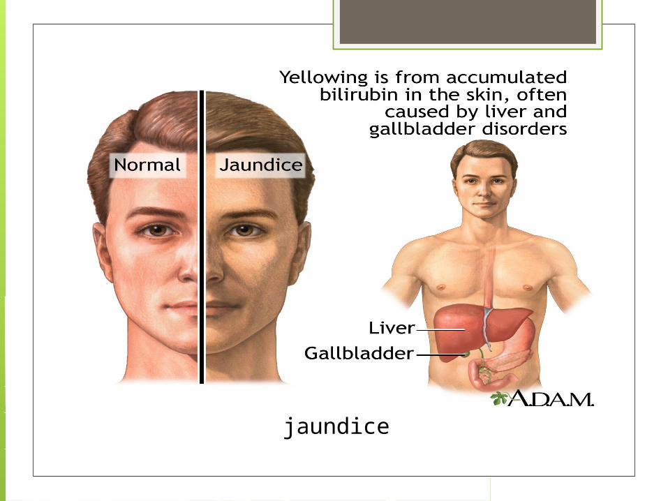
jaundice

eye become yellowish , person suffering from jaundice

Vomiting :• Ejection of stomach contents through the mouth.• It is controlled by the vomit centre in the medulla oblongata.• A feeling of nausea precedes vomiting.
:• Abnormal frequency of bowel movement and increased liquidity of the faecal discharge.• It reduces the absorption of food.
diarrhea

vomiting

Constipation :• The faeces are retained in the rectum as the bowel movements occurs irregularly.Indigestion :• The food is not properly digested leading to a feeling of fullness.Causes are inadequate enzymes secretion, anxiety, food poisoning, over eating and spicy food.

Indigestion

Historical depiction of the digestive system, 17th century Persia

THANK YOU FOR WATCHING THE END.