DIFFICULT TO TREAT SLE (“NOT YOUR ROUTINE CASE OF SLE”) · This study demonstrated...
32
Lupus Challenging Cases Richard Furie, MD Professor of Medicine Hofstra North Shore LIJ School of Medicine Albert Einstein College of Medicine Chief, Division of Rheumatology and Allergy-Clinical Immunology North Shore-Long Island Jewish Health System Lake Success, NY CASE 1 Case: 58 y/o female – 30 year history of SLE, which was quiet on no medicines until 2009 – Onset of AHA controlled with mycophenolate and rituximab – During initial visit in 2009: • Beta-2 glycoprotein I IgG Ab: >150 (<15) • Anticardiolipin IgG Ab: 120 (<15) – No history of miscarriage or thrombosis
Transcript of DIFFICULT TO TREAT SLE (“NOT YOUR ROUTINE CASE OF SLE”) · This study demonstrated...

Lupus Challenging Cases
Richard Furie, MD
Professor of Medicine
Hofstra North Shore LIJ School of Medicine
Albert Einstein College of Medicine
Chief, Division of Rheumatology and Allergy-Clinical Immunology
North Shore-Long Island Jewish Health System
Lake Success, NY
CASE 1
Case: 58 y/o female
– 30 year history of SLE, which was quiet on no
medicines until 2009
– Onset of AHA controlled with mycophenolate
and rituximab
– During initial visit in 2009:
• Beta-2 glycoprotein I IgG Ab: >150 (<15)
• Anticardiolipin IgG Ab: 120 (<15)
– No history of miscarriage or thrombosis

CASE 1
Would you advise therapy for thrombosis prevention?
If so, which medicine?
1. ASA
2. Clopidogrel
3. Warfarin
4. Prednisone
5. Direct thrombin inhibitor
CASE 1
Case continued
– Placed on ASA 81 mg per day in 2009
– Stroke Oct 2010
– Treated with heparin and then warfarin

Predictors of Thrombotic Risk?
• Titer of aPL Ab
• Isotype of aPL Ab
– IgG > IgA > IgM
• Degree of PTT elevation
• Other factors
– Nephrosis
– Smoking
– BCP
Predictors of Thrombotic Risk
• Triple positivity (LAC, β2GPI Ab, aCL Ab)
– 104 asymptomatic patients
– Mean F/U: 4.5 years
• 25 first thrombotic events
– 5.3%/y vs. 1.4% in single positivity patients
– 37% cumulative incidence over 10 y
Pengo et al. Blood 2011

Predictors of Thrombotic Risk
• Additional risk factors:
– Male gender
– Venous thrombosis risk factors
• Thrombosis rates on:
– ASA: 7/37 (not protective)
– Hydroxychloroquine: 2/18: 11%
Pengo et al. Blood 2011
Preventing the First Clot
• Risk of thrombosis < 4% per year
• Confounded by other factors
• APLASA (Erkan 2007): ASA 81 mg vs placebo
– N=98; f/u 2.3 years
– ASA (3 events); placebo (0)
– ASA did not protect against thrombosis
• ?? ASA for primary prevention ??

Preventing the Second Clot
• Retrospective Studies
– Warfarin better than ASA
– Warfarin HD better than LD
• Prospective Studies
– Warfarin Mod Intensity = High Intensity
Targeting a specific INR is
easier said than done
Khamashta M et al. N Engl J Med 1995;332:993-997
Comparison of Antithrombotic Treatments Used

Crowther M et al. N Engl J Med 2003;349:1133-1138
Outcomes and Duration of Follow-up in the High-Intensity and Moderate-Intensity Warfarin Groups
6
CASE 2
Medical History
A 24 year old female with recently diagnosed steroid-
responsive thrombocytopenia was referred for evaluation
of possible SLE. She had no other inflammatory
manifestations. During the interview, she had
spontaneous flailing movements of the right wrist as well
as choreic movements of the fingers of the right hand.

CASE 2 Physical Exam
BP: 110/70 P: 70
HEENT normal
Chest normal
Cardiac normal
Abdomen normal
Skin livedo reticularis
Joints normal
Neurologic flailing movements of the right wrist;
choreic movements of the right fingers;
occasional contractions of the right side
of her face
CASE 2

CASE 2
All of the following tests would be appropriate to
order except:
1. Antiphospholipid antibodies
2. Lupus serologies
3. ASO titer
4. Cerebral angiogram
5. PET scan
CASE 2
Laboratory Results
WBC normal
Hb normal
Plt 125,000
ANA 1/160 (speckled)
DNA absent (as was Sm, RNP, SSA, SSB)
PTT 39 seconds (<35 seconds)
LAC positive (by dRVVT and PNP)
RPR positive
ACL GPL: 51 MPL: <5 APL: <5
ASO normal
Brain MRI normal
EEG normal

CASE 2 PET Scan
CASE 2
Course The patient was diagnosed with PAPS and referred to the Center
for Movement Disorders at NSUH. A diagnosis of right hemichorea
was made, and the patient underwent PET. This study demonstrated
contralateral striatal 18F-fluorodeoxyglucose hypermetabolism.
Her symptoms remitted spontaneously. A follow-up PET was normal.
Nearly one year later, the patient developed chorea on the opposite
side of the body. PET once again demonstrated contralateral striatal 18F-fluorodeoxyglucose hypermetabolism. The patient was treated
with haloperidol 4 mg/d with improvement over the ensuing 4 weeks.

CASE 2
Chorea
• Associated with SLE and with PAPS
• Striatal hypermetabolism
• Pathogenesis unknown
• microvascular occlusion
• aPL binding to striatal tissue
CASE 3
Medical History A 27 year old female with a 5 year history of SLE received 60 mg/d of
prednisone for lupus nephritis. Three weeks after the dosage
increase, she heard a “pop” while stepping on a bus. The leg gave
out, and a few seconds later, she heard a second “pop.” Both legs
gave out, and she was not able to stand. She otherwise felt fine.
Physical Exam Joints: mild Jaccoud’s arthropathy
high-riding patellae bilaterally; no knee effusions

CASE 3
What is the most likely diagnosis?
1. Torn menisci
2. Ruptured ACLs
3. Tibial plateau fractures from osteonecrosis
4. Ruptured quadriceps tendons
5. Ruptured patellar tendons
CASE 3

CASE 3
Tendon Rupture
• Acute in onset
• May be bilateral
• Knee (patellar tendon), Achilles’ tendon
• Steroids, trauma as risk factors
• Mild inflammation
• Repair approached surgically or medically
CASE 4
Medical History A 44 year old female with a 9 year history of SLE was
hospitalized in septic shock complicated by DIC and
oliguric renal failure. Prior manifestations of lupus
included arthritis and nephritis. However, she never had
required steroids or other immunosuppressive therapies.
Physical Exam BP: 100/60 P: 100 T: 39
Skin scattered raised violaceous papules with necrotic
centers; ischemic toes

CASE 4
CASE 4
What is the most likely diagnosis?
1. Vasculitis from active SLE
2. Libman-Sacks with peripheral embolization
3. Infection with Neisseria meningitidis
4. Cholesterol embolization syndrome
5. Infection with Streptococcus pneumoniae

CASE 4
Laboratory Results WBC 58,000 (91% PMN, 6 Bands K/uL)
Hb 12.8 g/dL
Plt 127 K/uL
Blood cultures Streptococcus pneumoniae
Smear Howell-Jolly Bodies
CASE 4
Why?
• Pneumococcal sepsis
• No immunospressive therapy
• Howell-Jolly bodies
without splenectomy

CASE 4
CASE 4
Functional Asplenia
• Anatomic presence but functional absence of spleen
• Seen in SLE, celiac sprue
• Infections with Salmonella or Pneumococcus
• 3% of SLE patients observed to have H-J bodies
• Pathogenesis unknown
• Pneumococcal vaccination recommended

CASE 5 Medical History
A 52 year old female with a history of stroke (maintained
on warfarin) was hospitalized for evaluation of 2 months of
fever, diarrhea, and weight loss.
Physical Exam was unremarkable except for T= 39.3 and
mild synovitis of the PIP joints.
Laboratory:
WBC: 1.2 (50% PMN’s; 50% L) AST: 173
Hb: 7.6 ALT: 104
Plt: 66,000 Ferritin: 2597
LDH: 728
Haptoglobin: 49 (normal) Creat: 0.8
Coombs (direct): negative No schistocytes
ANA: 1/320
CASE 5
Hospital Course Extensive evaluation included imaging studies and serologies.
Body CT: normal
CT brain: old left cerebellar infarct
ANA: 1:320 (H)
DNA: 562
APL Ab: neg

CASE 5
Impression: Systemic Lupus complicated by fever, arthritis and
pancytopenia
CASE 5
What are possible causes of the pancytopenia?
1. Lymphoma
2. Autoimmune hemolytic anemia and thrombocytopenia
3. Macrophage activation syndrome with
hemophagocytosis
4. Aplastic anemia
5. Thrombotic microangiopathy
CASE 5
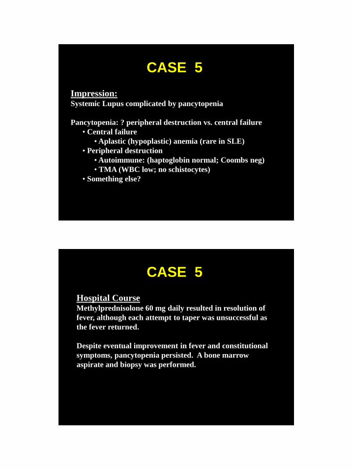
Impression: Systemic Lupus complicated by pancytopenia
Pancytopenia: ? peripheral destruction vs. central failure
• Central failure
• Aplastic (hypoplastic) anemia (rare in SLE)
• Peripheral destruction
• Autoimmune: (haptoglobin normal; Coombs neg)
• TMA (WBC low; no schistocytes)
• Something else?
CASE 5
Hospital Course Methylprednisolone 60 mg daily resulted in resolution of
fever, although each attempt to taper was unsuccessful as
the fever returned.
Despite eventual improvement in fever and constitutional
symptoms, pancytopenia persisted. A bone marrow
aspirate and biopsy was performed.

CASE 5
Hospital Course Bone marrow aspirate and biopsy demonstrated
hemophagocytosis, increased histiocytes and macrophages;
increased iron stores.
Cyclosporin 300 mg per day was added with improvement
in panyctopenia.
Outpatient Course
Arthritis, fever, pancytopenia all disappeared. Steroids
and CsA were tapered and eventually discontinued.
David Fisman, MD
Erythrocyte Phagocytosis Neutrophil Phagocytosis

CASE 5
Thrombotic Microangiopathy
LDH
Haptoglobin
Reticulocyte count
Direct Coombs test
Blood smear
1200
not detectable
15.5 %
negative
CASE 5 Hemophagocytic Syndrome
• Seen in JIA (triggered by viral infections)
• = Macrophage Activation Syndrome
• Manifestations ─ Pancytopenia
─ Fever
─ Liver, spleen, and lymph node enlargement
─ DIC (predicts a poor prognosis)
─ Abnormal LFT’s, increased ferritin
• T cells, macrophages (TNF, IL-6, IL-1)
• Rx: high dose steroids, CsA, pheresis, IVIG

CASE 5 Key Teaching Points
• Cytopenias (other than anemia of chronic disease)
typically result from peripheral destruction
− Antibody-mediated
− ADCC >> CDC
− Hypercellular marrow
• Marrow hypoplasia/aplasia is rare
• Consider central problem especially when:
− Pancytopenia exists
− Initial steroid therapy is unsuccessful
CASE 6 Medical History A 22 year old female with a 3 year history of SLE underwent a kidney
biopsy for worsening proteinuria (13 grams/24 hours). She had a
creatinine of 0.5, very elevated anti-DNA antibodies, and
hypocomplementemia. The histology was consistent with WHO class
IV; little chronicity was present. She received pulse steroids but
refused cyclophosphamide. However, she agreed to mycophenolate
mofetil 2000 mg/day and enalapril. Seven months later she had 4
grams/24 hour of proteinuria, creatinine 0.8, hypocomplementemia,
and anti-DNA antibodies. A repeat biopsy once again showed a WHO
class IV lesion and surprisingly little in the way of chronicity. She
agreed to parenteral cyclophosphamide for 6 months and was
maintained on mycophenolate and cyclosporin.
A subsequent major relapse treated with rituximab resulted in
miraculous results with normalization of proteinuria, DNA Ab, and C’.

CASE 6
Comment: • Is mycophenolate mofetil equal in efficacy
to cyclophosphamide?
• Is mycophenolate mofetil safer than
cyclophosphamide?
• Is rituximab the answer when all else fails?
• Where have we been and where are we
going in drug development in SLE?
CASE 7
Medical History A 47 year old female with a 5 year history of severe discoid
lupus had unsuccessfully tried numerous topical steroids,
azathioprine, methotrexate, hydroxychloroquine, and
systemic steroids. She was given 100 mg/d of thalidomide
with an amazing response. Active skin lesions regressed,
and hair began to grow on her scalp.

CASE 7
Discoid Lupus
CASE 7
Biologic Properties of Thalidomide
1. Inhibition of angiogenesis
2. Inhibition of TNF, IL8
3. Inhibition of COX-2
4. Down regulation of IL6
5. Increased IL10 production

CASE 7
Side Effects of Thalidomide
1. Somnolence
2. Rash
3. Leukopenia
4. Orthostatic hypotension
5. Peripheral neuropathy (primarily sensory)
6. Constipation
7. BIRTH DEFECTS
CASE 1
Medical History
A 27 year old female with a 5 year history of SLE was
admitted to the hospital because of confusion and fever.
Past manifestations of SLE have included polyarthritis,
rash, recurrent episodes of pericarditis, and anemia. A
flare 2 months prior to admission, consisting of
pericarditis, fever, hypocomplementemia, and a 2-fold
rise in anti-DNA antibodies, was successfully treated with
prednisone 40 mg/day; prednisone was subsequently
tapered. At the time of admission, medicines included
hydroxychloroquine 400 mg/day, prednisone 15 mg/day,
calcium, and a vitamin.
CASE 1
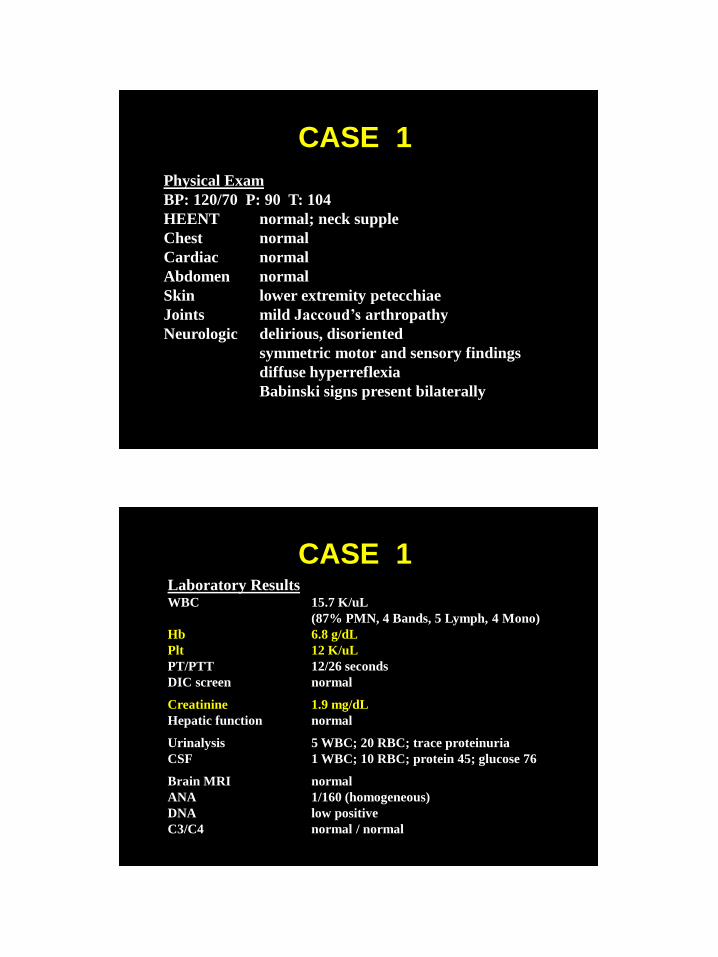
Physical Exam
BP: 120/70 P: 90 T: 104
HEENT normal; neck supple
Chest normal
Cardiac normal
Abdomen normal
Skin lower extremity petecchiae
Joints mild Jaccoud’s arthropathy
Neurologic delirious, disoriented
symmetric motor and sensory findings
diffuse hyperreflexia
Babinski signs present bilaterally
CASE 1 Laboratory Results WBC 15.7 K/uL
(87% PMN, 4 Bands, 5 Lymph, 4 Mono)
Hb 6.8 g/dL
Plt 12 K/uL
PT/PTT 12/26 seconds
DIC screen normal
Creatinine 1.9 mg/dL
Hepatic function normal
Urinalysis 5 WBC; 20 RBC; trace proteinuria
CSF 1 WBC; 10 RBC; protein 45; glucose 76
Brain MRI normal
ANA 1/160 (homogeneous)
DNA low positive
C3/C4 normal / normal

CASE 1
1. Sepsis
2. Bacterial meningitis
3. Fungal meningitis
4. Viral encephalitis
5. Vasculitis
6. Central Nervous System Lupus
7. Intracranial Hemorrhage
8. Drug intoxication
What is the Differential Diagnosis?
CASE 1
Hospital Course The patient was given broad spectrum antibiotics for the treatment
of sepsis and/or bacterial meningitis. Methylprednisolone 60 mg/day
was also administered. However, the patient’s mental status
worsened, and she became comatose. All cultures were sterile.
The impression was that of a flare of SLE complicated by
autoimmune thrombocytopenia and CNS disease. “Pulse” steroids
were administered for 3 days without subsequent improvement.
Intravenous gamma-globulin (IVIG) failed to improve the
thrombocytopenia, and cyclophosphamide was being considered.
What should be done next? Rheumatology to the rescue!

CASE 1
The Rheumatologist’s View
A consulting rheumatologist asked the following:
1. If this is a lupus flare, why…
a. no hypocomplementemia?
b. only a modest elevation of anti-DNA antibodies?
c. no response to high doses of steroids?
d. no response to IVIG?
2. What is the origin of the anemia and thrombocytopenia?
3. What is the pathogenesis of the cerebral dysfunction?
CASE 1
The Rheumatologist’s View
1. Is this “difficult to treat SLE”?
a. Vasculitis
b. Vasculopathy
2. Is the anemia autoimmune, microangiopathic, or from
marrow failure?
3. Is the thrombocytopenia autoimmune,
microangiopathic, or marrow failure?
4. What is the pathogenesis of the cerebral dysfunction?

CASE 1
Needed Lab Tests
LDH
Haptoglobin
Reticulocyte count
Direct Coombs test
Blood smear
1200
not detectable
15.5 %
negative
CASE 1
Diagnosis
Thrombotic Microangiopathy
1. thrombocytopenia
2. microangiopathic hemolytic
anemia (schistocytes)
3. organ dysfunction (kidney; brain)
Treatment
Plasmapheresis with complete response
within one week

CASE 1
Thrombotic Microangiopathy
• Hemolytic-uremic syndrome
• TTP
• Familial TTP
• SLE
• aPL
• Drug-induced (mitomycin, cyclosporine)
• Pregnancy/post-partum
• Scleroderma renal crisis
CASE 1

CASE 1
CASE 1

CASE 1
CASE 1

CASE 1 Summary
1. Manifestations of TM can mimic more typical
“inflammatory” complications of SLE.
2. In the presence of thrombocytopenia and anemia,
think of TM to explain new neurologic and/or
renal disease.
3. Look for schistocytes!!; determine haptoglobin, LDH,
reticulocyte count, and direct Coomb’s.
4. Although patients are typically treated with high dose
steroids before the diagnosis is recognized, consider
initiating plasmapheresis.
5. Affected organs include brain, kidney, and heart.
6. TM can occur in PAPS.


![Pharmacokinetic modeling of [18F]fluorodeoxyglucose (FDG ...](https://static.fdocuments.in/doc/165x107/61886b54df681277ae16a602/pharmacokinetic-modeling-of-18ffluorodeoxyglucose-fdg-.jpg)