Different promoter usage for CYP19 gene expression in buffalo ovary and placenta
10
Different promoter usage for CYP19 gene expression in buffalo ovary and placenta Deepti Sharma, Sandeep Ghai, Dheer Singh * Molecular Endocrinology Lab, Animal Biochemistry Division, National Dairy Research Institute, Karnal, Haryana 132001, India article info Article history: Received 2 February 2009 Revised 4 April 2009 Accepted 8 April 2009 Available online 16 April 2009 Keywords: CYP19 gene Aromatase RT-PCR 5 0 RACE Promoter Buffalo Tissue-specific abstract Aromatase is the key enzyme for estrogen biosynthesis and is coded by CYP19 gene. The expression of CYP19 gene is regulated in tissue-specific manner by alternate use of different promoters. In this study, we have analyzed tissue-specific expression and their regulation of CYP19 gene in preovulatory and pos- tovulatory stages of buffalo (Bubalus bubalis) ovary and placenta. RT-PCR analysis showed that the CYP19 gene expression was significantly (p < 0.05) higher in granulosa cells of large follicles as compared to the other tissues. The transcript analysis and transcriptional start site identified by 5 0 -RLM RACE for CYP19 expression indicated different transcriptional start sites within the different sized follicles during follicu- logenesis. Sequence analysis showed that the transcription start site of transcript isolated from buffalo granulosa cells of small follicles was 37 bases upstream of the transcript isolated from granulosa cells of large follicles. However, both the transcripts were found to be derived from proximal promoter II. Dif- ference in the transcriptional start site indicates the different promoter sequence usage in granulosa cells of different sized follicles. Further, in silico analysis of the difference in promoter sequence based on the 5 0 -UTRs isolated from granulosa cells of small follicles (151 bases) and large follicles (114 bases) showed that consensus sequence for certain important trans-elements (viz., TATA binding protein, E2F and CAAT binding protein) would lie in the promoter sequence isolated from the granulosa cells of large follicles. These transcription factors may be involved in regulation of CYP19 gene expression in ovary, either directly or indirectly. The difference in the size of 5 0 -UTR among the granulosa cells of ovary reflects the possible mechanism for the differential regulation of CYP19 gene during development. The transcripts isolated from buffalo corpus luteum and placental cotyledons were having same 5 0 -UTR comprises of 168 bases and found to be derived from PI.1. Estimates of CYP19 gene transcript concentration in the different tissues revealed that the CYP19 gene expression in granulosa cells is predominantly regulated by PII and to a minor extent by PI.1. However, PI.1 is almost exclusively responsible for CYP19 gene expression in placenta and residual expression in corpus luteum. In order to understand the complex CYP19 gene reg- ulation in these tissues, further studies are needed to elucidate the activity of different promoters and define regulatory elements for binding of transcription factors. Ó 2009 Elsevier Inc. All rights reserved. 1. Introduction Estrogen plays vital role in animal reproduction, particularly, in proliferation of uterine endometrium, hypertrophy and growth of endometrial glands to maintain zygote for implantation (Murray et al., 2000). Placental estrogen protects mother and fetus from exposure to excess of androgens produced by adrenals and pla- centa (Grumbach and Auchus, 1999). Estrogen biosynthesis is cat- alyzed by cytochrome P450 aromatase, encoded by CYP19 gene (Nelson et al., 1996). Though estrogen production is generally thought as an endocrine product of ovary, several other tissues also have the capacity to synthesize estrogen from androgen. These in- clude granulosa cells of ovary, placenta, brain, adipose tissue, skin, bone osteoblasts and Leydig cells of testis (Simpson et al., 1994). Increasing evidence suggests that switching of aromatase expression from one mRNA variant to another may be a key regu- latory mechanism in several physiological and pathological pro- cesses. Distinct aromatase transcripts are expressed in early vs. midpregnancy in porcine placenta (Choi et al., 1996; Choi et al., 1997a,b; Conley et al., 1997) as well as in fetal vs. adult human li- ver (Harada et al., 1993). Several studies have shown that a similar switch occurs in healthy vs. cancerous human breast adipose tissue (Harada et al., 1993; Agarwal et al., 1996; Utsumi et al., 1996; Zhou et al., 1996; Harada, 1997; Chen, 1998). It has been speculated that a switching mechanism may be involved in the ovulation/luteini- zation process (Harada et al., 1993). However, results from the study of Jenkins et al. (1993) did not support this concept, as only promoter II-derived aromatase transcripts were detected in human follicles and corpora lutea. In most mammals, expression of CYP19 gene is regulated in tissue-specific manner by alternative use of different promoters. 0016-6480/$ - see front matter Ó 2009 Elsevier Inc. All rights reserved. doi:10.1016/j.ygcen.2009.04.009 * Corresponding author. Fax: +91 182 2250042. E-mail address: [email protected] (D. Singh). General and Comparative Endocrinology 162 (2009) 319–328 Contents lists available at ScienceDirect General and Comparative Endocrinology journal homepage: www.elsevier.com/locate/ygcen
-
Upload
deepti-sharma -
Category
Documents
-
view
213 -
download
1
Transcript of Different promoter usage for CYP19 gene expression in buffalo ovary and placenta

General and Comparative Endocrinology 162 (2009) 319–328
Contents lists available at ScienceDirect
General and Comparative Endocrinology
journal homepage: www.elsevier .com/locate /ygcen
Different promoter usage for CYP19 gene expression in buffalo ovary and placenta
Deepti Sharma, Sandeep Ghai, Dheer Singh *
Molecular Endocrinology Lab, Animal Biochemistry Division, National Dairy Research Institute, Karnal, Haryana 132001, India
a r t i c l e i n f o
Article history:Received 2 February 2009Revised 4 April 2009Accepted 8 April 2009Available online 16 April 2009
Keywords:CYP19 geneAromataseRT-PCR50RACEPromoterBuffaloTissue-specific
0016-6480/$ - see front matter � 2009 Elsevier Inc. Adoi:10.1016/j.ygcen.2009.04.009
* Corresponding author. Fax: +91 182 2250042.E-mail address: [email protected] (D. Sing
a b s t r a c t
Aromatase is the key enzyme for estrogen biosynthesis and is coded by CYP19 gene. The expression ofCYP19 gene is regulated in tissue-specific manner by alternate use of different promoters. In this study,we have analyzed tissue-specific expression and their regulation of CYP19 gene in preovulatory and pos-tovulatory stages of buffalo (Bubalus bubalis) ovary and placenta. RT-PCR analysis showed that the CYP19gene expression was significantly (p < 0.05) higher in granulosa cells of large follicles as compared to theother tissues. The transcript analysis and transcriptional start site identified by 50-RLM RACE for CYP19expression indicated different transcriptional start sites within the different sized follicles during follicu-logenesis. Sequence analysis showed that the transcription start site of transcript isolated from buffalogranulosa cells of small follicles was 37 bases upstream of the transcript isolated from granulosa cellsof large follicles. However, both the transcripts were found to be derived from proximal promoter II. Dif-ference in the transcriptional start site indicates the different promoter sequence usage in granulosa cellsof different sized follicles. Further, in silico analysis of the difference in promoter sequence based on the50-UTRs isolated from granulosa cells of small follicles (151 bases) and large follicles (114 bases) showedthat consensus sequence for certain important trans-elements (viz., TATA binding protein, E2F and CAATbinding protein) would lie in the promoter sequence isolated from the granulosa cells of large follicles.These transcription factors may be involved in regulation of CYP19 gene expression in ovary, eitherdirectly or indirectly. The difference in the size of 50-UTR among the granulosa cells of ovary reflectsthe possible mechanism for the differential regulation of CYP19 gene during development. The transcriptsisolated from buffalo corpus luteum and placental cotyledons were having same 50-UTR comprises of 168bases and found to be derived from PI.1. Estimates of CYP19 gene transcript concentration in the differenttissues revealed that the CYP19 gene expression in granulosa cells is predominantly regulated by PII andto a minor extent by PI.1. However, PI.1 is almost exclusively responsible for CYP19 gene expression inplacenta and residual expression in corpus luteum. In order to understand the complex CYP19 gene reg-ulation in these tissues, further studies are needed to elucidate the activity of different promoters anddefine regulatory elements for binding of transcription factors.
� 2009 Elsevier Inc. All rights reserved.
1. Introduction
Estrogen plays vital role in animal reproduction, particularly, inproliferation of uterine endometrium, hypertrophy and growth ofendometrial glands to maintain zygote for implantation (Murrayet al., 2000). Placental estrogen protects mother and fetus fromexposure to excess of androgens produced by adrenals and pla-centa (Grumbach and Auchus, 1999). Estrogen biosynthesis is cat-alyzed by cytochrome P450 aromatase, encoded by CYP19 gene(Nelson et al., 1996). Though estrogen production is generallythought as an endocrine product of ovary, several other tissues alsohave the capacity to synthesize estrogen from androgen. These in-clude granulosa cells of ovary, placenta, brain, adipose tissue, skin,bone osteoblasts and Leydig cells of testis (Simpson et al., 1994).
ll rights reserved.
h).
Increasing evidence suggests that switching of aromataseexpression from one mRNA variant to another may be a key regu-latory mechanism in several physiological and pathological pro-cesses. Distinct aromatase transcripts are expressed in early vs.midpregnancy in porcine placenta (Choi et al., 1996; Choi et al.,1997a,b; Conley et al., 1997) as well as in fetal vs. adult human li-ver (Harada et al., 1993). Several studies have shown that a similarswitch occurs in healthy vs. cancerous human breast adipose tissue(Harada et al., 1993; Agarwal et al., 1996; Utsumi et al., 1996; Zhouet al., 1996; Harada, 1997; Chen, 1998). It has been speculated thata switching mechanism may be involved in the ovulation/luteini-zation process (Harada et al., 1993). However, results from thestudy of Jenkins et al. (1993) did not support this concept, as onlypromoter II-derived aromatase transcripts were detected in humanfollicles and corpora lutea.
In most mammals, expression of CYP19 gene is regulated intissue-specific manner by alternative use of different promoters.

320 D. Sharma et al. / General and Comparative Endocrinology 162 (2009) 319–328
Aromatase promoter-switching phenomenon was reported (Boer-boom et al., 1999) in granulosa cells of cattle during follicularluteinization (Drummond and Findlay, 1999). During growth ofsmall to large ovarian follicles in cattle, CYP19 transcripts were ob-served to be derived predominantly from promoter II (Lenz et al.,2004). However, these were almost exclusively derived from pro-moter I.1 in corpus luteum (Doody et al., 1990). In bovine placenta,major transcript variant was from exon I.1 (Vanselow and Furbass,1995). The ovary-specific transcripts are conserved among mam-malian species whereas in cow and sheep placenta, different tran-scripts are expressed (Vanselow et al., 1999), suggesting thatunrelated placenta-specific promoters are used in different species.Recently, increased P450 aromatase expression has been reportedin goat placenta during pregnancy (Mondragon et al., 2007). TheCYP19 gene and its species and tissue-specific expression are wellstudied in human, cattle and sheep. However, there is no such re-port in buffalo; the present study thus has been conducted to ana-lyze the tissue-specific expression pattern and transcript variantsin buffalo.
2. Materials and methods
2.1. Collection of ovaries and other tissues
Buffalo ovaries (200 approx.) were collected from commercialabattoir, Delhi within 10–20 min after slaughtering and placentalcotyledons (from five different animals) were collected from NDRIcattle yard in chilled normal saline (0.9% NaCl) containing 100 U ofpenicillin and 50 lg of streptomycin per liter, and transported tothe laboratory as quickly as possible (2 h approx.). All the tissueswere washed three times in saline, immersed in 70% ethanol for30 s, and washed again with saline and processed immediately.
2.2. Isolation of granulosa cells, luteal and placental tissues
Healthy, developing follicles were assessed by the presence ofvascularized theca externa and clear amber follicular fluid withno debris. The follicular fluid was aspirated from small (65 mm),and large antral follicles (P10 mm), using 20-gauge needle andsterile, non-toxic, non-pyrogenic monoinjected brand syringes(Dispovan, 5-ml). The follicular fluid collected in 2-ml Eppendorftube (for RNA isolation) and the granulosa cells were isolated bycentrifugation at a low speed (2000 rpm) to pellet the cells. Pel-leted cells were used for total RNA isolation. The postovulatory tis-sue i.e. corpus luteum and the buffalo’s placental cotyledon wasexcised with the help of sterilized scissors and kept in ice.
2.3. RNA and DNA isolation
The total RNA was isolated from granulosa cells of small andlarge follicles, corpus luteum and placental cotyledons using TRIreagent [Life Technologies (India) Pvt. Ltd.] with modified method(Chomczynski and Sacchi, 1987). The DNA was isolated using DNA-zol reagent [Life Technologies (India) Pvt. Ltd.]. The RNA and DNAwere quantified spectrophotometrically and RNA integrity wasevaluated by denaturing agarose gel electrophoresis.
2.4. Reverse transcription-polymerase chain reaction (RT-PCR)
The cDNA was synthesized using Fermantas First strand cDNAsynthesis kit (Fermantas, Germany). The reaction mixture con-tained 2 lg of total RNA, 1 ll of random hexamer (0.2 lg/ll) andDEPC treated water up to 11 ll. The contents were incubated at65 �C for 10 min followed by 2 min incubation at room tempera-ture. The reagents added further were: 4 ll of 5� reaction buffer(250 mM Tris–HCl, pH 8.3; 250 mM KCl, 20 mM MgCl2, 50 mM
DTT), 1 ll of RNase inhibitor (20 IU), 2 ll of dNTP mix (10 mM),2 ll of M-MuLV reverse transcriptase (200 IU) to a final volumeof 20 ll. The contents were incubated at 25 �C for 10 min, 42 �Cfor 30 min and 95 �C for 3 min.
The cDNA was amplified in a reaction mixture containing 2 ll ofRT product, 0.2 lM primers (gene-specific forward and reverseprimers), 1� PCR buffer [10 mM Tris–HCl, pH 9; 50 mM KCl,1.5 mM MgCl2, 0.01% gelatin], 0.2 mM dNTP mix, 1U Taq polymer-ase (1U/ll) made to 50 ll with nuclease free water. The amplifica-tion was done in a thermocycler under two different cycleconditions. For CYP19 aromatase gene expression, two-step PCRwas performed as nested PCR. For the first amplification, 2 ll ofcDNA was used as template, for the second amplification, 5 ll ofPCR product from the first reaction was used. PCRs were performedby incubating the contents at 94 �C for 2 min, followed by 32 cyclesof 94 �C for 1 min, 60 �C for 1 min, 72 �C for 1 min and the finalextension of 4 min at 72 �C. The b-actin was used as housekeepinggene.
To selectively detect the expression of CYP19 gene transcriptswith different 50-UTRs that were identified in prior 50-RACE exper-iments, specific sense primers for each of the 50-UTRs were de-signed (Table 1). PCR was performed with cycle conditions; 94 �Cfor 2 min, followed by 32 cycles of 94 �C for 1 min, 58–60 �C for1 min, 72 �C for 1 min and final extension at 72 �C for 4 min.
2.5. 50-RACE
50-RACE was carried out with First Choice RLM RACE kit (Ambi-on, India). Briefly, total RNA (10 lg) was treated with Calf IntestineAlkaline Phosphatase (CIP) to remove the phosphate group fromthe 50-end of uncapped RNA and incubated at 37 �C for 1 h fol-lowed by acid:phenol:chloroform extraction. The CIP-treated RNAwas then incubated with Tobacco Acid Pyrophosphatase (TAP) toremove phosphates from the 50-end of capped (intact) mRNA, at37 �C for 1 h. The CIP/TAP-treated RNA was ligated with the 50-RACE adapter by incubation with 2.5 U of T4 RNA ligase at 37 �Cfor 1 h. After cDNA library synthesis, the 50-end of RNA was ob-tained by two rounds of PCR amplification using RNA linker prim-ers provided by the kit and the gene-specific primers. The firstround of PCR was performed using a universal primer and a 20-bp antisense primer (50-GAGAAGGAGAGCTTGCCATG-30). A nestedPCR amplification of the PCR mix was performed using a nesteduniversal primer and 23-bp antisense primer (50-AATGAGGGGGCCCAATTCCCAGA-30). The resulting PCR products were fractionatedon a 1% agarose gel.
2.6. Gel extraction of PCR products and sequencing
The gel extraction of PCR products was carried out using Wiz-ard� SV Gel and PCR Clean-Up System, (Promega, USA) as per man-ufacturer’s instructions. After the gel extraction, the PCR productswere custom sequenced (Sigma Genosys Lab., Bangalore).
2.7. Cloning of CYP19 gene promoter II and promoter I.1
To isolate the CYP19 gene promoter II and promoter I.1, therespective primers DP2F and DP2R and P1.1F and P1.1R weredesigned using PRIMER 3 software (Whitehead Institute for Biomed-ical Research, MIT) to a genomic database sequence for the bovineCYP19 gene (GenBank Accession Nos. Z69242 and Z69241) andsynthesized by Ocimum Biosolutions Ltd., India. The PCR was per-formed in a 50 ll of reaction mixture consisting of 120–150 ng ofDNA, 1� PCR buffer [10 mM Tris–HCl, pH 9; 50 mM KCl, 1.5 mMMgCl2, 0.01% gelatin], 200 lM of dNTPs, 0.4 lM of each primerand 1U Taq DNA polymerase. The amplification was done in thethermocycler (Biometra) using cycle conditions as 94 �C for

Table 1Primers used to isolate CYP19 gene, b-actin gene, promoter I.1 and promoter II.
Primer name Primer sequence Accession No. Amplification product
b-ActinF 50-CGTGGGCCGCCCTAGGCACCA-30 NM 007393.3 243 bpb-ActinR 50-TTGGCCTTAGGGTTCAGGGGGG-30 NM 007393.3P6 50-CATGGCAAGCTCTCCTTCTC-30 NM_174305 866 bpP7 50-GCAGGGACTGACCAAACTTC-30 NM_174305P5 50-TCAACAGCAGAGAAGCTGGA-30 NM_174305 302 bpP8 50-TGGTACCGCATGCTCTCATT-30 NM_174305T1.1F 50-CTGTGGTGATGACGAAGGAC-30 Z69241 378 bpATR 50-AATGAGGGGGCCCAATTCCCAGA-30 NM_174305AT2a(c)f 50-CTGAAGCAACAGGAGTCCTAAATGTACA-30 Z69242 261 bpT1.1R 50-CTGGGACCTGGTATTGAGGA-30 NM_174305P1.1F 50-CCTCACATTCCCTGACATCC-30 Z69241 734 bpP1.1R 50-TGCGTTGGCTCACCTACCT-30 Z69241DP2F 50-GTCCTGTTGAATTCAATAGACAA-30 Z69242 625 bpDP2R 50-GGGTTCAGCACTTCCAAAA-30 NM_174305
D. Sharma et al. / General and Comparative Endocrinology 162 (2009) 319–328 321
5 min, 35 cycles of 94 �C for 1 min, 54–56 �C for 1 min, 72 �C for1 min and final extension at 72 �C for 4 min.
PCR products were purified using a Wizard� SV Gel and PCRClean-Up System (Promega, USA) and ligated into the pCRII vector(Invitrogen, USA) and transformed into DH5a competent Esche-richia coli cells according to manufacturer’s instructions (Invitro-gen, USA). Small-scale preparations of plasmid DNA were madeby using QIAprep� spin Miniprep Kit (Qiagen, Australia). Therecombinants were confirmed by PCR amplification of the insertusing specific primers. Sequencing of plasmids was performed byGenomBiotechnologies Pvt. Ltd., Bangalore, India.
2.8. In silico analysis of promoter I.1 and II
Promoter sequences were analyzed using TRANSFAC 4.0 data-base of transcription factor using the MatInspector V2.2 program(Quandt et al., 1995) for the identification of the putative func-tional cis-acting elements.
3. Results
3.1. Expression of cytochrome P450 aromatase (CYP19) mRNA inbuffalo ovary and placenta using RT-PCR
The relative expression of aromatase mRNA in different buffalotissues is shown in Fig. 1. The expression was exhibited by granu-losa cells of ovarian large follicles (Fig. 1A, lane 5) and placenta(Fig. 1A, lane 3). The nested PCR experiment was performed to con-firm the aromatase mRNA expression in granulosa cells of smalland large follicles, corpus luteum and placenta. The results showedthe CYP19 gene expression in both outer (Fig. 1B, lane 4) and inner(Fig. 1B, lane 5) PCRs for granulosa cells from large follicles. How-ever, the expression was detected in granulosa cells from small fol-licles (Fig. 1B, lane 3) and corpus luteum (Fig. 1B, lane 7) andplacenta (Fig. 1D, lane 3) only after second amplification. The com-parative expression of aromatase mRNA (quantified by GelQuantsoftware) is shown in Fig. 1F, where granulosa cells of large folli-cles shown to exhibit the highest expression followed by that ingranulosa cells of small follicles and placenta, while corpus luteumshowing lowest expression.
3.2. Isolation of tissue-specific transcripts
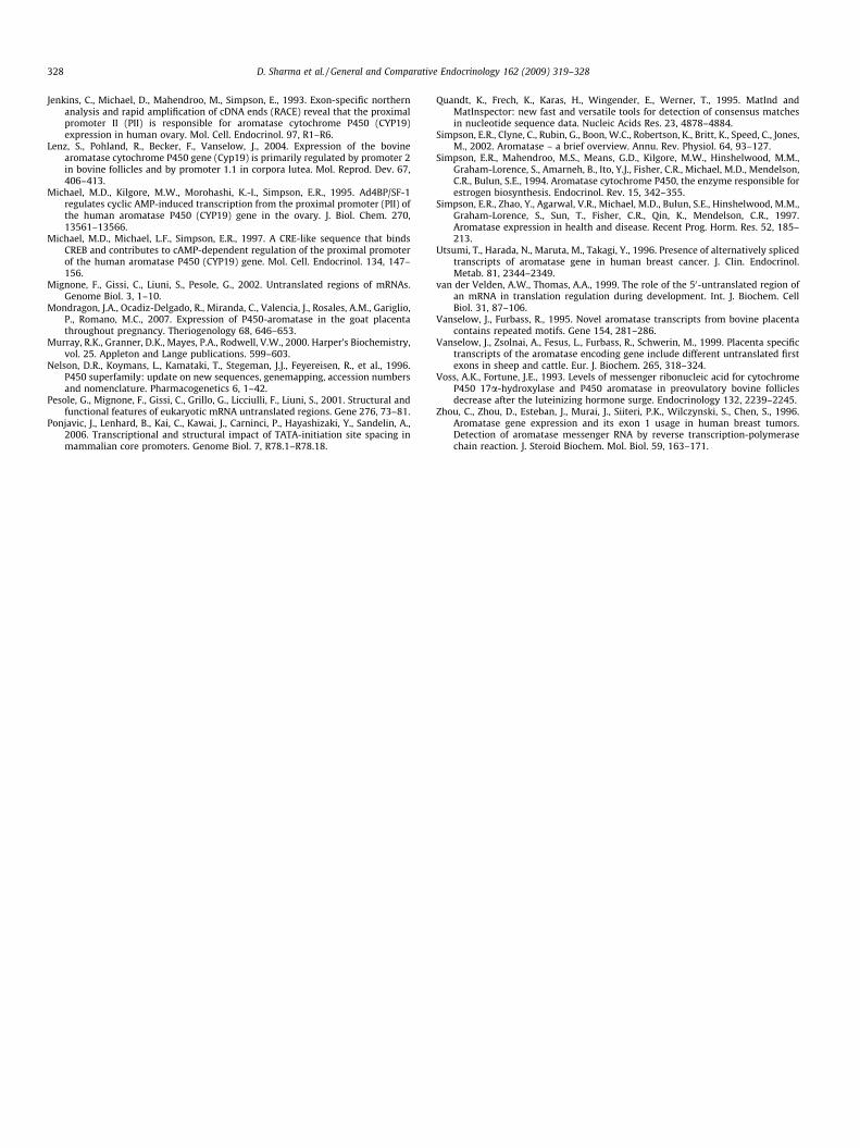
RNA samples from granulosa cells of small follicles, corpus lut-eum and placental cotyledons were screened for CYP19 transcriptsusing 50-RLM RACE and nested PCR. RACE experiment generatesspecific 50-RACE products from all the tissues with detectableCYP19 expression. The 50-RACE products of aromatase from buffalo
granulosa cells of small follicles included a 50-UTR that was homol-ogous to the region upstream from the splice acceptor site of exonII in bovine. However, the 50-UTRs of the transcript variants fromcorpus luteum and placenta were consisting of an additional firstexon and a short part of exon II. The transcripts identified in corpusluteum and placental cotyledons were having the same 50-UTR de-rived from exon I.1. During the processing of these CYP19 tran-scripts, the dinucleotide AG within exon II, located 40nucleotides upstream from the translational start codon (ATG), isused as a splice acceptor for the various first exons. The electropho-retic profile of the RACE products isolated from granulosa cells ofsmall follicles, corpus luteum and placental cotyledons are shownin Fig. 2. The sequences of these transcripts have been submitted toGenBank.
3.3. Tissue-specific CYP19 transcript variants
In order to estimate the relative amount of the transcripts iden-tified with the 50-RACE in ovary and placental cotyledons, RT-PCRusing transcript specific primers was performed. The presence oftranscripts comprising the 50-UTR of aromatase exon I.1 and exonII was determined in granulosa cells of small follicles and large fol-licles, corpus luteum and placental cotyledons. A high expressionof transcript comprising 50-UTR of exon II was found to be exhib-ited in granulosa cells of small and large follicles (Fig. 3A, lanes 2and 3) as compared to very low expression in corpus luteum(Fig. 3A, lane 4) and placenta (Fig. 3C, lane 2). There was strongexpression of CYP19 gene transcript comprising 50-UTR of exonI.1 in placenta (Fig. 3G, lane 2) followed by relatively lower expres-sion in corpus luteum (Fig. 3E, lane 2). There was weak expressionof this transcript in granulosa cells of small (Fig. 3E, lane 3) andlarge (Fig. 3E, lane 4) follicles. The comparative expression of aro-matase transcript I.1 and II in different tissues (quantified by Gel-Quant software) is shown in Fig. 3I.
3.4. Isolation and cloning of the CYP19 gene promoter I.1 and promoterII
In order to determine the complete structure of the 50-UTR ofeach aromatase transcript and identify the potential regulatoryelements involved in their expression, the 50-flanking regions ofexon I.1 and exon II were amplified from genomic DNA and cloned.Isolation of buffalo ovary and placenta-specific CYP19 gene pro-moter was achieved using PCR technique. The putative promoterI.1 of 734 bp and promoter II of 625 bp was obtained after PCRamplification. These fragments were purified and cloned into pCRIIvector. PCR amplification of these plasmids was done using sameprimer set for the confirmation of the insert. BlastN analysis of

301 bp
(CXCR-4) 373 bp
(CYP19)
387 bp
(CYP19)
AA B C
L 1 L 2 L 3 L 4
387 bp
(CYP19)
DFig. 2. 50-RLM RACE experiment for isolation of CYP19 transcripts from granulosa cells and corpus luteum of buffalo ovary. 50-RLM RACE was performed using total RNAisolated from granulosa cells of small follicles and corpus luteum of buffalo and control RNA using Ambion’s first choice RLM RACE kit (Cat # 1700). (A) Lane 1, control 50-RLMRACE products. The two bands in lane 1 could be due to the higher yield of PCR products as described in manufacturer’s instructions. (B) Lane 2, 50-RLM RACE product ofCYP19 gene in small follicles. (C) Lane 3, 50-RLM RACE product of CYP19 gene in corpus luteum. (D) Lane 4, 50-RLM RACE product of CYP19 gene in placenta. L, 100 bp ladder.Isolation of transcripts from the tissues was done two times.
CYP19 (866 bp)
CYP19 (302 bp)
CYP19 (302 bp)
β-actin (243 bp)
β-actin (243 bp)
Lane 1 2 3 4 5 6 7 Lane 1 2 3LPLCLCGSCG
B
C
D
E
F
GCS GCL CL PL1.01.21.41.61.82.02.22.42.62.83.0
Aro
mat
ase
mR
NA
/ β-a
ctin
Lane 1 2 3 4 5 6
CYP19(302 bp)
A
Fig. 1. Expression of CYP19 (aromatase) gene mRNA in buffalo ovary and placenta by semi-quantitative RT-PCR and nested PCR. PCR was carried out in 1.5 mM MgCl2 at 60 �C(annealing) for 32 cycles. Lane 1, 100 bp DNA ladder. (A) Lane 2, corpus luteum; lane 3, placenta; lane 4, granulosa cells of small follicles; lane 5, granulosa cells of largefollicles; lane 6, negative control. (B) Lanes 2, 4, 6, first amplification; lanes 3, 5, 7, second amplification. (D) Lane 2, first amplification; lane 3, second amplification. (F)Quantitative analysis of CYP19 gene mRNA in different tissues. (C and E) b-Actin mRNA expression in respective tissues. GCS, granulosa cells of small follicles; GCL, granulosacells of large follicles; CL, corpus luteum; PL, placenta.
322 D. Sharma et al. / General and Comparative Endocrinology 162 (2009) 319–328
promoter I.1 (734 bp) and promoter II (625 bp) sequences indicatedthat these are the fragment of CYP19 gene and extended upstream
of the exon I.1 and exon II in the bovine genomic database se-quence Z69241 and Z69242, respectively. The sequences PII and

Lane 1 2 3 4
Transcript exon II (261 bp)
β-actin(243 bp)
A
B β-actin(243 bp)
C
D
Transcript exon II (261 bp)
Lane 1 2
Transcript exon I.1 (378 bp)
Lane 1 2 3 4
Transcript exon I.1 (378 bp)
β-actin (243 bp)
E
F β-actin (243 bp)
G
H
Lane 1 2
SF LF CL PL0 .0
0 .2
0 .4
0 .6
0 .8
1 .0
1 .2
1 .4
T IIT I.1
CY
P19
gen
e tr
ansc
ript
var
iant
s/β
-act
in m
RN
A
I
Fig. 3. Expression of CYP19 (aromatase) gene transcript with 50-UTR of exon II and exon I.1 by RT-PCR in different tissues of buffalo. PCR was carried out in 1.5 mM MgCl2 at58–60 �C (annealing) for 32 cycles. Lane 1, 100 bp DNA ladder. (A) Lane 2, granulosa cells of small follicles; lane 3, granulosa cells of large follicles; lane 4, corpus luteum. (Cand G) Lane 2, placenta. (E) Lane 2, corpus luteum; lane 3, granulosa cells of small follicles; lane 4, granulosa cells of large follicles. (B, D, F and H) b-Actin mRNA expression inrespective tissues. (I) Quantitative analysis of expression of various CYP19 gene transcripts in different tissues of buffalo. The graph shows that transcript II is significantly(p < 0.01) higher in granulosa cells of small follicles (GCS) and large follicle (GCL) while transcript I.1 is significantly (p < 0.01) higher in corpus luteum (CL) and placenta (PL).
D. Sharma et al. / General and Comparative Endocrinology 162 (2009) 319–328 323
PI.1 of the two respective promoters have been submitted to theNCBI data base (GenBank Accession Nos. EU308111 andEU621845).
3.5. Identification of the putative functional trans-acting elements
BLAST analysis of 50-UTR obtained from granulosa cells of smalland large follicle showed 100% homology and that of corpus lute-um and placenta showed 98% homology with the bovine promoterII (Fig. 4) and I.1 (Fig. 5), respectively. Further, sequence alignmentof 50-UTR from corpus luteum and placenta showed that the tran-scripts in the two tissues were transcribed from the same TSS(transcription start site). However, this TSS does not coincide withthat of the bovine. Analysis of the promoter I.1 and II sequences,using TRANSFAC 4.0 database of transcription factor sequenceusing the MatInspector V2.2 program (Quandt et al., 1995), identi-fied a number of conserved potential transcription factor bindingsites based on comparison with the respective human and bovineCYP19 gene promoter regions (Fig. 6). The buffalo aromatase pro-
moter II contains a highly conserved adrenal 4 binding protein/ste-roidogenic factor-1 (SF-1) element involved in cAMP-dependentand -independent regulation in granulosa cells of other species(Fig. 7A). However, a cAMP response element (CRE)-like regionknown to bind cAMP-binding protein (CREB) in rat and human(Fitzpatrick and Richards, 1994), but not in bovine (Hinshelwoodet al., 1997) and horse (Boerboom et al., 1999), ovarian tissues isnot conserved in the buffalo (Fig. 7B).
4. Discussion
Estrogen, an important steroid hormone, is indispensable forthe development of anatomical, functional and behavioral char-acteristics necessary for female reproduction. The female repro-ductive cycle in animals is controlled through an integratedsystem involving hypothalamus, pituitary gland, ovary andreproductive tract. Since estrogen is synthesized by cytochromeP450 aromatase (CYP19) gene, the present work is done to elu-cidate the tissue-specific expression, regulation and the transcript

Fig. 4. Comparison of 50-UTRs of CYP19 transcripts from buffalo ovarian small and large follicles with corresponding exon II of cattle sequence. 50-UTRs were obtainedthrough 50-RLM RACE experiments. Using T-COFFEE, Version 5.31 multiple sequence software tool, sequences of buffalo aromatase 50-UTR from small follicles (151 bases) andlarge follicles (114 bases) were aligned with the cattle CYP19 exon II (Accession No. Z69242). The 50-UTRs of 151 bases and 114 bases showed 100% homology with cattlesequence. The bold arrows indicate the transcription start site 1 (small follicles) and 2 (large follicles) in buffalo. The stars indicate complete conservation of that particularnucleotide in the aligned sequences. BBSF-RACE product from small follicles of buffalo ovary; BBLF-RACE product from large follicles of buffalo ovary; BOSE2-Bovine CYP19exon 2 sequence.
Fig. 5. Comparison of 50-UTR sequence of CYP19 transcript from corpus luteum and placenta with exon I.1 of cattle sequence. Using T-COFFEE, Version_5.31 multiplesequence software tool, sequence of buffalo aromatase 50-UTR from corpus luteum (168 bases) was aligned with the cattle CYP19 exon I.1 (Accession No. Z69241). The 50-UTRs of 168 bases showed 93% homology with that of cattle sequence. This indicates that this transcript may be directed by promoter I.1. The bold arrows indicate thetranscription start site 1 (buffalo) and 2 (cattle) sequences. The stars indicate complete conservation of that particular nucleotide in the aligned sequences. BBCL-RACEproduct from corpus luteum of buffalo ovary; BBPL-RACE product from placenta of buffalo ovary; BOSE2-Bovine CYP19 exon I.1 sequence.
324 D. Sharma et al. / General and Comparative Endocrinology 162 (2009) 319–328
variants of mRNA encoding aromatase in buffalo ovary andplacenta. Significant variation was found in the expression of
buffalo CYP19 gene in preovulatory and postovulatory stages ofreproductive cycle. Granulosa cells of large follicles and placenta

Fig. 6. Isolation and characterization of buffalo CYP19 gene promoter I.1 and promoter II region. A DNA fragment located immediately upstream of exon I.1 and exon II wasisolated from buffalo. Selected potential cis-acting promoter elements are underlined, whereas sequences representing exon I.1 and exon II are indicated by uppercase letters.Transcription start sites are represented as arrows. Translation of CYP19 transcripts starts at ATG with the amino acid Met. The sequences PII and PI.1 of the two respectivepromoters have been submitted to the NCBI data base (GenBank Accession Nos. EU308111 and EU621845).
Fig. 7. Homology analysis of putative aromatase cis-acting promoter elements.Selected buffalo putative aromatase promoter elements are aligned with corre-sponding sequences from all species homologs characterized to date. (A) CRE-likesequence in aromatase promoter II. (B) Adrenal 4 binding protein Ad4BP/SF1element in aromatase promoter II.
D. Sharma et al. / General and Comparative Endocrinology 162 (2009) 319–328 325
showed comparatively high expression than small follicle andcorpus luteum. Nested PCR was subsequently performed toascertain and compare aromatase expression in different tissuesof buffalo.
The increased expression of aromatase mRNA from small tolarge follicles indicated the high 17b-estradiol synthesis in antralfollicles, an essential requirement for the follicular developmentand maturation. This also prevents the follicular atresia in buffaloovary, as it down regulates the genes responsible for apoptosis(Evans et al., 2004). These results are in concurrence with theexpression studies of aromatase in granulosa cells of different spe-cies (Conley and Hinshelwood, 2001). The presence of higher(p < 0.01) levels of aromatase mRNA in granulosa cells of buffalolarge follicles than that in small follicles coincides with the studieson the rat ovary (Hickey et al., 1988). The detection of aromatasemRNA in corpus luteum by nested PCR, indicated that aromatasemRNA can be localized in buffalo luteal tissues as observed in hu-man, rodents and bovine (Doody et al., 1990). The level of expres-sion in corpus luteum was less (p < 0.01) as compared to that ingranulosa cells of small and large ovarian follicles which suggeststhat with the onset of luteinization, there is a decline in the expres-sion of aromatase, thereby low level of estradiol synthesis (Vossand Fortune, 1993).

326 D. Sharma et al. / General and Comparative Endocrinology 162 (2009) 319–328
The presence of high expression of aromatase mRNA in placen-tal cotyledons of buffalo, as detected by nested PCR, is in goodagreement with the results obtained in bovine (Furbass et al.,1997). This suggested that like human and cattle (Simpson et al.,1994), buffalo also has high placental aromatase activity, thus pla-centa is acting as a primary site of estrogen synthesis in pregnantanimals.
Gene expression is finely regulated at the post-transcriptionallevel. UTRs are known to play crucial role in the post-transcrip-tional regulation of gene expression including, modulation of thetransport of mRNAs out of the nucleus and of translation efficiency(van der Velden and Thomas, 1999), sub-cellular localization (Jan-sen, 2001) and stability (Bashirullah et al., 2001). Nucleotide pat-terns or motifs located in the 50-UTRs and 30-UTRs can interactwith specific RNA-binding proteins. It is striking that the lengthof both 50- and 30-UTRs varies a lot within a species, ranging froma dozen nucleotides to a few thousand (Pesole et al., 2001). In fact,it has been shown using a mammalian in vitro system that even asingle nucleotide is a sufficient 50-UTR for the initiation of transla-tion (Hughes and Andrews, 1997). Secondary structures in 50-UTRsare also important in the regulation of translation. Accurately pre-dicting UTRs is important because transcriptional regulatory sig-nals are often located adjacent to the transcription start site(TSS), and post-transcriptional regulatory sites can often be foundin the 50-UTR (Brown et al., 2005).
In the present study, we have done the 50-RLM RACE to deter-mine the 50-UTRs of aromatase transcripts in ovarian granulosacells of small follicles, corpus luteum and placental cotyledons ofbuffalo. The electrophoretic profile of 50-RACE products of granu-losa cells from these tissues indicated the presence of two differenttranscript variants of aromatase in buffalo ovarian and placentaltissues. Sequencing of 50-RLM RACE product (373 bp) of granulosacells of small follicles showed that the 50-UTR region was 151nucleotides, just upstream of translation start site ATG. The se-quence comparison of 50-UTR (151 bases) of aromatase from gran-ulosa cells of small follicles with corresponding 50-UTR sequences(114 bases) from granulosa cells of large follicles (Gali, 2006)showed 100% conservation. The 40 bases downstream of the com-mon splice site, AG, have shown 100%, 80% and 65% conservationwith corresponding aromatase 50-UTR of cattle (GenBank AccessionNo. S80143), sheep (GenBank Accession No. AJ012153) and goat(GenBank Accession No. AY148883).
BLASTN analysis showed that the transcripts, isolated fromgranulosa cells of small follicles, were having transcription startsite upstream to that of large follicles. The sequence alignment ofthe transcripts from buffalo granulosa cells of small follicles andlarge follicles (Gali, 2006) with the corresponding sequence of cat-tle indicated that both the transcripts in buffalo were derived fromthe proximal promoter II. To the best of our knowledge, this is thefirst report showing the difference in the position of transcriptionstart site of buffalo ovarian CYP19 gene in the granulosa cells ofsmall and large follicle. Moreover, the difference in the size of 50-UTR of transcripts from granulosa cells of small follicles (151bases) and large follicles (114) might be responsible for the differ-ential expression of the CYP19 gene during different stages of fol-licular development. As already discussed the secondary structuresformed with in the 50-UTRs are responsible for the translationalregulation in eukaryotes. The transcriptional start site in the buf-falo granulosa cells of small follicles (151 bases) was found up-stream to that of large follicles (114 bases). Therefore, the regionupstream of the transcriptional start site in granulosa cells of largefollicle might form the secondary structures that may lead to theinhibition of aromatase expression and thus less estrogen produc-tion in granulosa cells of small follicle (Mignone et al., 2002) or itcould be the part of promoter sequence in large follicles and mayinvolved in the regulation.
In silico analysis of the 50-UTRs isolated from granulosa cells ofsmall follicles (151 bases) and large follicles (114 bases) showedcertain important trans-elements (viz., TATA binding protein, E2Fand CAAT binding protein) which could bind within the 50-UTR oftranscript isolated from small follicles. These transcription factorshave been found to be involved in regulation of CYP19 geneexpression, either directly or indirectly (Furbass et al., 1997). Thefunctional significance of the two start sites is validated by thelocalization of the TATA binding protein (TBP) motifs, present atpositions �31 and �29 in small and large follicle, respectively.Our results are in agreement with the recent finding which showsthat the TATA box position relative to the TSS is constraint to a nar-row window (�32 to �29), where positions �31 and �30 are theoptimal positions for achieving high tissue specificity (Ponjavicet al., 2006). Thus upstream region which is acting as a promoterfor CYP19 gene in large follicles may thus be responsible for the en-hanced expression of aromatase in the same which is not the casewith small follicles.
Similarly, the sequencing of 50-RLM RACE products isolatedfrom corpus luteum and placental cotyledons showed that the sizeof this product was 387 bp in which, the 50-UTR region was 168nucleotides. BlastN analysis of 50-RLM RACE product of buffalo cor-pus luteum and placental cotyledons showed 93% homology withmajor placental transcript of bovine (Vanselow and Furbass,1995) which comprised of exon I.1. This clearly indicated thatthe buffalo corpus luteum and placental cotyledons transcriptswere having a transcription start site other than those reportedin bovine placenta. It suggests that PI.1 is the major promoterresponsible for CYP19 gene expression in buffalo corpus luteumand placental cotyledons.
Computational analysis of transcription factor binding sites onPI.1 showed the lack of binding site for TATA binding protein(TBP). As discussed above, that the TATA box is the core promoterelement associated with the expression of gene, it is speculatedthat lack of TBP binding motif in PI.1 might be responsible for com-paratively low expression of CYP19 gene in corpus luteum andplacenta.
There are numerous untranslated first exons that occur in aro-matase transcripts in a tissue-specific fashion due to differentialsplicing as a consequence of the use of tissue-specific promoters(Simpson et al., 1997). In the present study, three different tran-scripts comprising 50-UTR of exon II and exon I.1 were isolatedby RT-PCR in ovary and placenta of buffalo. The results showedthat in granulosa cells from follicles of buffalo ovary, transcriptsdriven by the region upstream from exon II, promoter II showedthe highest concentration, coinciding with the study in bovine(Furbass et al., 1997). Promoter II, the most proximal promoter ofCYP19 gene, has been described in several other species. It isthought to be the primordial CYP19 promoter and is regulated pri-marily by cAMP (Simpson et al., 1994). The level of CYP19 pro-moter II derived transcripts in granulosa cells is a marker forfollicular differentiation towards selection and dominance.
Detection of intermediate levels of CYP19 gene transcript, com-prising 50-UTR of exon I.1 in granulosa cells from follicles in buffaloovary, indicated the possible functional role of promoter adjacentto exon I.1, promoter I.1. It is the most distally located promoterof CYP19 gene (Simpson et al., 1994) and transcripts driven by thispromoter were also detected previously in granulosa cells in cattle(Lenz et al., 2004; Vanselow and Furbass, 1995). This suggestedthat not only promoter II but promoter I.1 is also involved in theregulation of CYP19 gene expression in granulosa cells of buffaloovarian follicles. Detection of transcript variants in buffalo corpusluteum showed that level of CYP19 transcripts driven by promoterII reduced drastically after luteinization whereas the major tran-scripts present are driven by promoter I.1. This suggested that pro-moter II virtually gets switched off whereas the residual transcripts

D. Sharma et al. / General and Comparative Endocrinology 162 (2009) 319–328 327
were almost exclusively derived from promoter I.1, afterluteinization.
In placental tissue of buffalo, the major transcript of CYP19 genecomprises 50-UTR of exon I.1 and thus is suggested as the majorplacental transcript. Studies in humans showed that this constitu-tively active distal promoter I.1 in placenta is the basis of strikinglyelevated levels of circulating estrogen in pregnant women (Simp-son et al., 2002). Thus the recruitment of this most distal promotermay have an evolutionary aspect. The existence of CYP19 tran-scripts driven by promoter I.1 in both placenta and corpus luteumof buffalo, suggested the role of promoter I.1 for CYP19 expressionduring pregnancy in both corpus luteum and placenta though atvery different levels (Lenz et al., 2004).
This study reports the cloning and characterization of buffaloaromatase promoter I.1 and II. Some of the molecular mechanismsinvolved in regulation of these promoters have been studied in dif-ferent species. This is the first report showing the putative regula-tory mechanism of aromatase expression in buffalo. By in silicoanalysis we identified several perfectly conserved cis-elementswith in the 50-flanking regions of PI.1 and II. Among the various ele-ments present in the PII, at least two cis-elements appear to be cru-cial for cAMP-dependent and -independent expression in humanand rat granulosa cells (Michael et al., 1995, 1997; Carlone andRichards, 1997). A first element, a consensus SF1 binding site con-served in all species, appears to mediate both constitutive andinducible aromatase transcription (Michael et al., 1995; Carloneand Richards, 1997). A second element, a CRE-like element towhich CREB can bind, appears to be required to achieve optimaltranscriptional activity in rats and humans (Hasegawa et al.,1995; Carlone and Richards, 1997). However, this later elementis poorly conserved in other species and contains a 1-bp deletionin buffalo (this study), bovine (Hinshelwood et al., 1997), porcine(Choi et al., 1997a,b) and equine (Boerboom et al., 1999). In cattle,this deletion was initially thought to be responsible for the lack ofaromatase expression in bovine luteal cells. However, a site direc-ted mutagenesis study designed to render the bovine CRE-like siteidentical to its human counterpart resulted only in partial restora-tion of cAMP-inducible promoter activity in luteal cells, suggestingthat other elements are also involved (Hinshelwood et al., 1997).
It is concluded that aromatase expression in ovary and placentais regulated by the use of tissue-specific promoters that may em-ploy entirely different signaling pathways and thus different co-horts of transcription factors. Along with this different CYP19gene transcripts were reported, which might be responsible forvariable expression of aromatase during follicular developmentand differentiation. In spite of having difference in expression ofCYP19 gene, transcripts isolated from granulosa cells of smalland large follicles are derived from proximal promoter II. Thusthe difference in expression may be due to the variations in the sizeof 50-UTR of aromatase mRNA, leading to differential regulation ofCYP19 gene during folliculogenesis. This study is the first to reportan aromatase promoter-switching phenomenon in buffalo granu-losa cells during follicular luteinization, characterized by a down-regulation of promoter II- and an up-regulation of promoter I.1-de-rived transcript after differentiation of granulosa cells. However,experimental evidence is still missing, to understand the complexpost-transcriptional regulation of the CYP19 gene expression dur-ing estrous cycle in buffalo.
Acknowledgments
The authors are grateful to the Director, National Dairy Re-search Institute, Karnal for providing necessary facilities for thiswork. The financial assistance as Institute SRF to Ms. Deepti andMr. Sandeep Ghai is thankfully acknowledged. This work is sup-ported by DBT grant (to DS) also.
References
Agarwal, V.R., Bulun, S.E., Leitch, M., Rohrich, R., Simpson, E.R., 1996. Use ofalternative promoters to express the aromatase cytochrome P450 (CYP19) genein breast adipose tissues of cancer-free and breast cancer patients. J. Clin.Endocrinol. Metab. 81, 3843–3849.
Bashirullah, A., Cooperstock, R.L., Lipshitz, H.D., 2001. Spatial and temporal controlof RNA stability. Proc. Natl. Acad. Sci. USA 98, 7025–7028.
Boerboom, D., Kerban, A., Sirois, R., 1999. Dual regulation of promoter II- andpromoter 1f-derived cytochrome P450 aromatase transcripts in equinegranulose cells during human chorionic gonadotropin-induced ovulation: anovel model for the study of aromatase promoter switching. Endocrinology 140(9), 4133–4141.
Brown, R.H., Gross, S.S., Brent, M.R., 2005. Begin at the beginning: predicting geneswith 50 UTRs. Genome Res. 15, 742–747.
Carlone, D.L., Richards, J.S., 1997. Functional interactions, phosphorylation, andlevels of 30 , 50-cyclic adenosine monophosphate-regulatory element bindingprotein and steroidogenic factor-1 mediate hormone-regulated and constitutiveexpression of aromatase in gonadal cells. Mol. Endocrinol. 11, 292–304.
Chen, S., 1998. Aromatase and breast cancer. Front. Biosci. 3, d922–d933.Choi, I., Simmen, R.C., Simmen, F.A., 1996. Molecular cloning of cytochrome P450
aromatase complementary deoxyribonucleic acid from periimplantationporcine and equine blastocysts identifies multiple novel 59-untranslatedexons expressed in embryos, endometrium, and placenta. Endocrinology 137,1457–1467.
Choi, I., Collante, W.R., Simmen, R.C.M., Simmen, F.A., 1997a. A developmentalswitch in expression from blastocyst to endometrial/placental-type cytochromeP450 aromatase genes in the pig and horse. Biol. Reprod. 56, 688–696.
Choi, I., Troyer, D.L., Cornwell, D.L., Kirby-Dobbels, K.R., Collante, W.R., Simmen, F.A.,1997b. Closely related genes encode developmental and tissue isoforms ofporcine cytochrome P450 aromatase. DNA Cell Biol. 16, 769–777.
Chomczynski, P., Sacchi, N., 1987. Single method of RNA isolation by acidguanidinium–thiocynate–phenol–chloroform extraction. Anal. Biochem. 162,156–160.
Conley, A., Hinshelwood, M., 2001. Mammalian aromatase. Reproduction 121, 685–695.
Conley, A., Corbin, J., Smith, T., Hinshelwood, M., Liu, Z., Simpson, E., 1997. Porcinearomatases: studies on tissue-specific, functionally distinct isozymes from asingle gene? J. Steroid Biochem. Mol. Biol. 61, 407–413.
Doody, K.J., Lorence, M.D., Mason, J.I., Simpson, E.R., 1990. Expression of ribonucleicacid species encoding steroidogenic enzymes in human follicles and corporalutea throughout the menstrual cycle. J. Clin. Endocrinol. Metab. 70, 1041–1045.
Drummond, A.E., Findlay, J.K., 1999. The role of estrogens in folliculogenesis. Mol.Endocrinol. 151, 57–64.
Evans, A.C.O., Ireland, J.L.H., Winn, M.E., Longergan, P., Smith, G.W., Coussens, P.M.,Ireland, J.J., 2004. Identification of genes involved in apoptosis and dominantfollicle development during follicular waves in cattle. Biol. Reprod. 70, 1475–1484.
Fitzpatrick, S.L., Richards, J.S., 1994. Identification of a cyclic adenosine 30 , 50-monophosphate-response element in the rat aromatase promoter that isrequired for transcriptional activation in rat granulosa cell and R2C Leydigcells. Mol. Endocrinol. 8, 1309–1319.
Furbass, R., Kalbe, C., Vanselow, J., 1997. Tissue-specific expression of the bovinearomatase-encoding gene uses multiple transcriptional start sites andalternative first exons. Endocrinology 138, 2813–2819.
Gali, J.M., 2006. Characterization of 50-end of aromatase cytochrome p450 (cyp19)transcripts in buffalo ovary, M.Sc., Thesis, NDRI Deemed University, Karnal,India.
Grumbach, M.M., Auchus, R.J., 1999. Estrogen: consequences and implications ofhuman mutations in synthesis and action. J. Clin. Endocrinol. Metab. 84, 4677–4694.
Harada, N., Utsumi, T., Takagi, Y., 1993. Tissue-specific expression of the humanaromatase cytochrome P450 gene by alternative use of multiple exons 1 andpromoters and switching of tissue-specific exons 1 in carcinogenesis. Proc. Natl.Acad. Sci. USA 90, 11312–11316.
Harada, N., 1997. Aberrant expression of aromatase in breast cancer tissues. J.Steroid Biochem. Mol. Biol. 61, 175–184.
Hasegawa, T., Mukoyama, H., Yoshida, S., Takahashi, M., 1995. Molecular cloningand nucleotide sequence of equine testicular cytochrome P-450 steroid17ahydroxylase/C17,20 lyase messenger ribonucleic acid. Biol. Reprod.[Monogr 1] 52, 615–622.
Hickey, G.J., Chen, S.A., Besmen, M.J., Shively, J.E., Hall, P.F., Gaddy-Kurten, D.,Richards, J.S., 1988. Hormonal regulation, tissue distribution and content ofaromatase cytochrome P450 messenger ribonucleic acid and enzyme in ratovarian follicles and corpora lutea: relationship to estradiol biosynthesis.Endocrinology 122, 1426–1436.
Hinshelwood, M.M., Michael, M.D., Simpson, E.R., 1997. The 50-flanking region of theovarian promoter of the bovine CYP19 gene contains a deletion in a cyclicadenosine 30 , 50-monophosphate-like responsive sequence. Endocrinology 138,3704–3710.
Hughes, M.J., Andrews, D.W., 1997. A single nucleotide is a sufficient 50-untranslated region for translation in an eukaryotic in vitro system. FEBS Lett.414, 19–22.
Jansen, R.P., 2001. MRNA localization: message on the move. Nat. Rev. Mol. Cell Biol.2, 247–256.
328 D. Sharma et al. / General and Comparative Endocrinology 162 (2009) 319–328
Jenkins, C., Michael, D., Mahendroo, M., Simpson, E., 1993. Exon-specific northernanalysis and rapid amplification of cDNA ends (RACE) reveal that the proximalpromoter II (PII) is responsible for aromatase cytochrome P450 (CYP19)expression in human ovary. Mol. Cell. Endocrinol. 97, R1–R6.
Lenz, S., Pohland, R., Becker, F., Vanselow, J., 2004. Expression of the bovinearomatase cytochrome P450 gene (Cyp19) is primarily regulated by promoter 2in bovine follicles and by promoter 1.1 in corpora lutea. Mol. Reprod. Dev. 67,406–413.
Michael, M.D., Kilgore, M.W., Morohashi, K.-I., Simpson, E.R., 1995. Ad4BP/SF-1regulates cyclic AMP-induced transcription from the proximal promoter (PII) ofthe human aromatase P450 (CYP19) gene in the ovary. J. Biol. Chem. 270,13561–13566.
Michael, M.D., Michael, L.F., Simpson, E.R., 1997. A CRE-like sequence that bindsCREB and contributes to cAMP-dependent regulation of the proximal promoterof the human aromatase P450 (CYP19) gene. Mol. Cell. Endocrinol. 134, 147–156.
Mignone, F., Gissi, C., Liuni, S., Pesole, G., 2002. Untranslated regions of mRNAs.Genome Biol. 3, 1–10.
Mondragon, J.A., Ocadiz-Delgado, R., Miranda, C., Valencia, J., Rosales, A.M., Gariglio,P., Romano, M.C., 2007. Expression of P450-aromatase in the goat placentathroughout pregnancy. Theriogenology 68, 646–653.
Murray, R.K., Granner, D.K., Mayes, P.A., Rodwell, V.W., 2000. Harper’s Biochemistry,vol. 25. Appleton and Lange publications. 599–603.
Nelson, D.R., Koymans, L., Kamataki, T., Stegeman, J.J., Feyereisen, R., et al., 1996.P450 superfamily: update on new sequences, genemapping, accession numbersand nomenclature. Pharmacogenetics 6, 1–42.
Pesole, G., Mignone, F., Gissi, C., Grillo, G., Licciulli, F., Liuni, S., 2001. Structural andfunctional features of eukaryotic mRNA untranslated regions. Gene 276, 73–81.
Ponjavic, J., Lenhard, B., Kai, C., Kawai, J., Carninci, P., Hayashizaki, Y., Sandelin, A.,2006. Transcriptional and structural impact of TATA-initiation site spacing inmammalian core promoters. Genome Biol. 7, R78.1–R78.18.
Quandt, K., Frech, K., Karas, H., Wingender, E., Werner, T., 1995. MatInd andMatInspector: new fast and versatile tools for detection of consensus matchesin nucleotide sequence data. Nucleic Acids Res. 23, 4878–4884.
Simpson, E.R., Clyne, C., Rubin, G., Boon, W.C., Robertson, K., Britt, K., Speed, C., Jones,M., 2002. Aromatase – a brief overview. Annu. Rev. Physiol. 64, 93–127.
Simpson, E.R., Mahendroo, M.S., Means, G.D., Kilgore, M.W., Hinshelwood, M.M.,Graham-Lorence, S., Amarneh, B., Ito, Y.J., Fisher, C.R., Michael, M.D., Mendelson,C.R., Bulun, S.E., 1994. Aromatase cytochrome P450, the enzyme responsible forestrogen biosynthesis. Endocrinol. Rev. 15, 342–355.
Simpson, E.R., Zhao, Y., Agarwal, V.R., Michael, M.D., Bulun, S.E., Hinshelwood, M.M.,Graham-Lorence, S., Sun, T., Fisher, C.R., Qin, K., Mendelson, C.R., 1997.Aromatase expression in health and disease. Recent Prog. Horm. Res. 52, 185–213.
Utsumi, T., Harada, N., Maruta, M., Takagi, Y., 1996. Presence of alternatively splicedtranscripts of aromatase gene in human breast cancer. J. Clin. Endocrinol.Metab. 81, 2344–2349.
van der Velden, A.W., Thomas, A.A., 1999. The role of the 50-untranslated region ofan mRNA in translation regulation during development. Int. J. Biochem. CellBiol. 31, 87–106.
Vanselow, J., Furbass, R., 1995. Novel aromatase transcripts from bovine placentacontains repeated motifs. Gene 154, 281–286.
Vanselow, J., Zsolnai, A., Fesus, L., Furbass, R., Schwerin, M., 1999. Placenta specifictranscripts of the aromatase encoding gene include different untranslated firstexons in sheep and cattle. Eur. J. Biochem. 265, 318–324.
Voss, A.K., Fortune, J.E., 1993. Levels of messenger ribonucleic acid for cytochromeP450 17a-hydroxylase and P450 aromatase in preovulatory bovine folliclesdecrease after the luteinizing hormone surge. Endocrinology 132, 2239–2245.
Zhou, C., Zhou, D., Esteban, J., Murai, J., Siiteri, P.K., Wilczynski, S., Chen, S., 1996.Aromatase gene expression and its exon 1 usage in human breast tumors.Detection of aromatase messenger RNA by reverse transcription-polymerasechain reaction. J. Steroid Biochem. Mol. Biol. 59, 163–171.