DIAGNOSTIC*IMMUNODERMATOLOGY*SERVICES:* … · Lichen%planus(LP)% Lesional%...
4
Page 1 of 4 File name Immunodermatology User Guide Version 1.9 Author John Mee Issue/reviewed date Feb 2016 Authorised by Fiona Denham DIAGNOSTIC IMMUNODERMATOLOGY SERVICES: USER GUIDE The Immunodermatology Laboratory was founded by Professor Martin Black in 1975 and is now under the leadership of Dr Richard Groves. Initially, it functioned as an experimental unit, developing immunofluorescence (IMF) techniques and looking at specific immunoglobulin deposition patterns in autoimmune diseases. The unit is the largest skin immunofluorescence referral laboratory in the UK. The workload comprises approximately 7,000 samples per year for direct and indirect IMF, of which, over 80% are from external centres. Dermatologists from all over the UK send us their complex and interesting immunobullous cases for further evaluation. Specimens are also referred to the laboratory internationally. The laboratory also performs research, including immunoblotting and immunoelectron microscopy, into autoimmune blistering diseases, in collaboration with other international centres. The findings are frequently presented at national and international meetings. The Immunodermatology Laboratory specialises in contributing to the diagnoses of the following diseases using immunofluorescence and enzymelinked immunosorbent assay (ELISA) techniques: Condition Biopsy site(s) for IMF studies Pemphigus (all forms) Perilesional & uninvolved Pemphigoid (all forms) Perilesional & uninvolved Pemphigoid (herpes) gestationis Perilesional Epidermolysis bullosa acquisita (EBA) Perilesional Linear IgA bullous dermatosis (LAD & CBDC) Perilesional & uninvolved Discoid lupus erythematosus (DLE) Lesional Systemic lupus erythematosus (SLE) Lesional & uninvolved (nonsun exposed) Lichen planus (LP) Lesional Dermatitis herpetiformis (DH) Perilesional Porphyria Lesional Vasculitis Lesional Amyloidosis Lesional
Transcript of DIAGNOSTIC*IMMUNODERMATOLOGY*SERVICES:* … · Lichen%planus(LP)% Lesional%...
Page 1 of 4
File name Immunodermatology User Guide Version 1.9
Author John Mee Issue/reviewed date Feb 2016
Authorised by Fiona Denham
DIAGNOSTIC IMMUNODERMATOLOGY SERVICES:
USER GUIDE
The Immunodermatology Laboratory was founded by Professor Martin Black in 1975 and is now under the leadership of Dr Richard Groves.
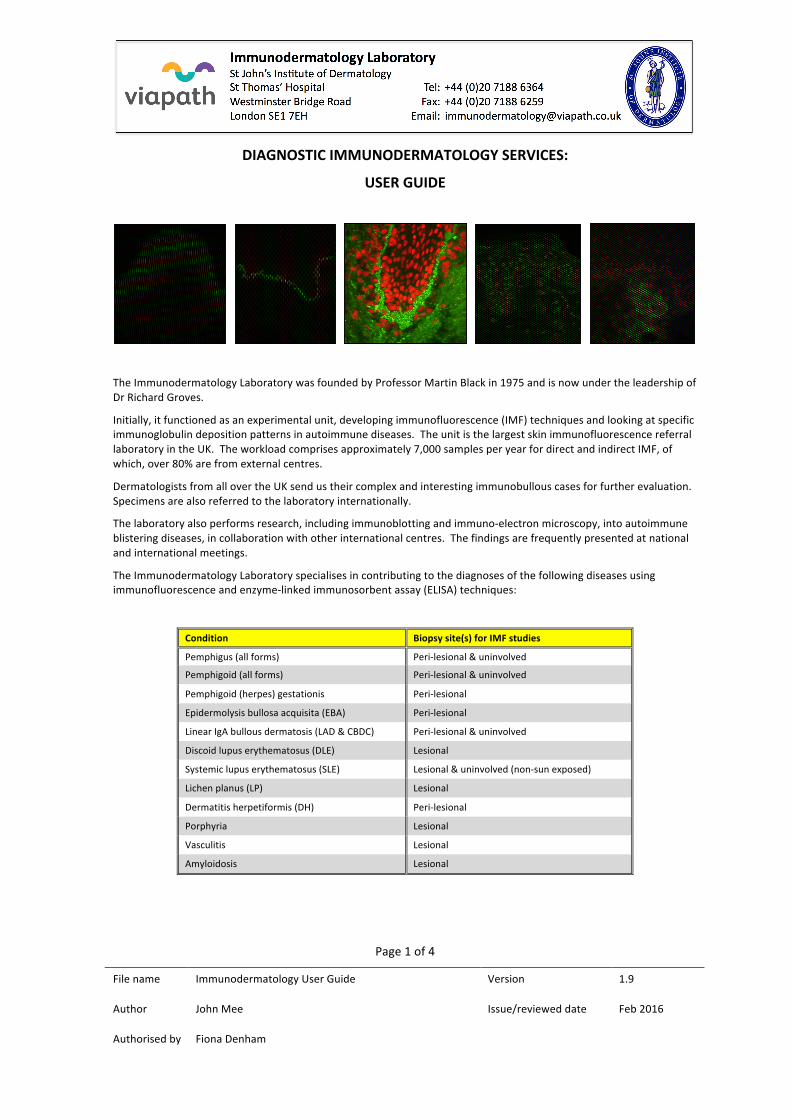
Initially, it functioned as an experimental unit, developing immunofluorescence (IMF) techniques and looking at specific immunoglobulin deposition patterns in autoimmune diseases. The unit is the largest skin immunofluorescence referral laboratory in the UK. The workload comprises approximately 7,000 samples per year for direct and indirect IMF, of which, over 80% are from external centres.
Dermatologists from all over the UK send us their complex and interesting immunobullous cases for further evaluation. Specimens are also referred to the laboratory internationally.
The laboratory also performs research, including immunoblotting and immuno-‐electron microscopy, into autoimmune blistering diseases, in collaboration with other international centres. The findings are frequently presented at national and international meetings.
The Immunodermatology Laboratory specialises in contributing to the diagnoses of the following diseases using immunofluorescence and enzyme-‐linked immunosorbent assay (ELISA) techniques:
Condition Biopsy site(s) for IMF studies
Pemphigus (all forms) Peri-‐lesional & uninvolved
Pemphigoid (all forms) Peri-‐lesional & uninvolved
Pemphigoid (herpes) gestationis Peri-‐lesional
Epidermolysis bullosa acquisita (EBA) Peri-‐lesional
Linear IgA bullous dermatosis (LAD & CBDC) Peri-‐lesional & uninvolved
Discoid lupus erythematosus (DLE) Lesional
Systemic lupus erythematosus (SLE) Lesional & uninvolved (non-‐sun exposed)
Lichen planus (LP) Lesional
Dermatitis herpetiformis (DH) Peri-‐lesional
Porphyria Lesional
Vasculitis Lesional
Amyloidosis Lesional
Page 2 of 4
File name Immunodermatology User Guide Version 1.9
Author John Mee Issue/reviewed date Feb 2016
Authorised by Fiona Denham
Specimen Requirements
Immunofluorescence can be performed on either epithelial biopsies (direct IMF), serum or blister fluid (indirect IMF). Enzyme-‐linked immunosorbent assays (ELISAs) can be performed on either serum or blister fluid. (Please note that the analysis of blister fluid falls outside the scope of the current UKAS accreditation of the laboratory to ISO15189.)
Direct IMF
This is a one-‐step procedure for detecting in vivo deposition of immunoglobulins, complement components and fibrinogen in epithelial tissues, including skin, buccal mucosa and conjunctiva.
Sample required Tissue biopsy/biopsies from perilesional sites
Biopsy preparation Biopsies (see page 3 for excision details) should be rinsed in saline, then placed in a specimen pot containing Michel’s media (provided by us). Biopsies are stable at room temperature in this medium for up to 6 months. Containers must be labelled with patient name and date of birth.
Request form IMF request forms are provided with the Michel’s media specimen pots. These should be fully completed to include:
• Patient name (forename and surname)
• Date of birth
• Requesting clinician name
• Sender details
• Report destination
• Biopsy site
• Clinical history (including question(s) to be answered and differential diagnoses)
Transport • The specimen container must be placed in a sealed plastic bag, with the request form outside the plastic bag.
• The bagged specimen should be placed into a primary container, with adequate absorbent material, in case of leakage.
• Securely seal the primary container, then place it in a secondary shipping container, which should also contain enough absorbent material to prevent any leakage from escaping outside the container.
• Specimens should be sent by either courier service or registered post to the above address and shipped at ambient temperature.
Rejection criteria for sample • Tissue not transported in Michel’s media
• Tissue contaminated with formalin
• Request form inadequately filled out or absent
• Specimen pot details fail to match those on the request form
Turnaround time 5 working days from receipt in laboratory
Page 3 of 4
File name Immunodermatology User Guide Version 1.9
Author John Mee Issue/reviewed date Feb 2016
Authorised by Fiona Denham
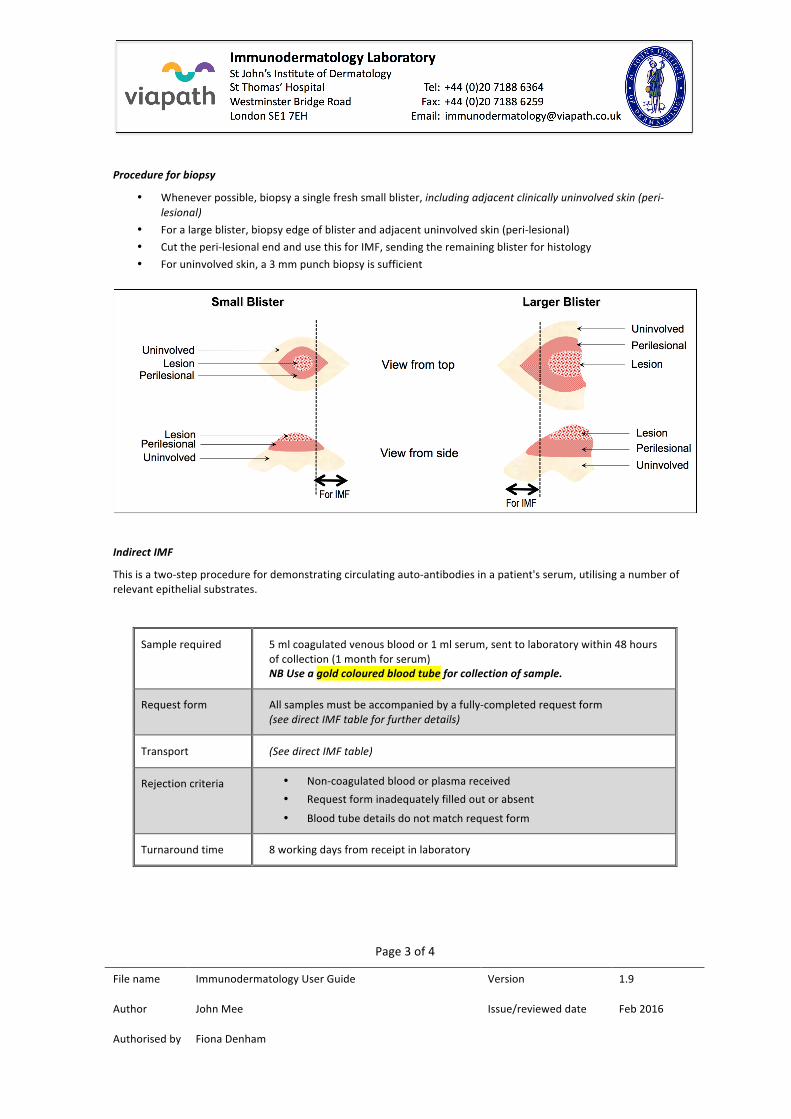
Procedure for biopsy
• Whenever possible, biopsy a single fresh small blister, including adjacent clinically uninvolved skin (peri-‐lesional)
• For a large blister, biopsy edge of blister and adjacent uninvolved skin (peri-‐lesional)
• Cut the peri-‐lesional end and use this for IMF, sending the remaining blister for histology
• For uninvolved skin, a 3 mm punch biopsy is sufficient
Indirect IMF
This is a two-‐step procedure for demonstrating circulating auto-‐antibodies in a patient's serum, utilising a number of relevant epithelial substrates.
Sample required 5 ml coagulated venous blood or 1 ml serum, sent to laboratory within 48 hours of collection (1 month for serum) NB Use a gold coloured blood tube for collection of sample.
Request form All samples must be accompanied by a fully-‐completed request form (see direct IMF table for further details)
Transport (See direct IMF table)
Rejection criteria • Non-‐coagulated blood or plasma received
• Request form inadequately filled out or absent
• Blood tube details do not match request form
Turnaround time 8 working days from receipt in laboratory
Page 4 of 4
File name Immunodermatology User Guide Version 1.9
Author John Mee Issue/reviewed date Feb 2016
Authorised by Fiona Denham
ELISA
The laboratory performs the following 5 assays for quantification and monitoring of specific circulating antibodies:
Antibody Disease Positivity threshold
Desmoglein 1 Pemphigus >30 U/ml
Desmoglein 3 Pemphigus >30 U/ml
BP180/collagen XVII Pemphigoid >20 U/ml
BP230/dystonin Pemphigoid >10 U/ml
Collagen VII Epidermolysis bullosa acquisita >6 U/ml
Each ELISA can be performed on 5 µl of serum (or blister fluid) and results are reported in (arbitrary) U/ml. ELISAs are normally performed in combination with indirect IMF studies, hence sample requirements are identical to those for indirect IMF. Relevant ELISA testing will be determined by lab staff, dependent on immunofluorescence results and information provided on the request form.
Laboratory opening hours:
09:00 – 17:00, Monday to Friday
Contact details:
Tel: 020 7188 6364 Fax: 020 7188 6259
Email: [email protected]
Results are available to clinicians by telephone, fax or email (nhs.net addresses only), using the above numbers. No information will be given to patients or their relatives, in accordance with the laboratory policy on protection of personal information.
Staff:
• Pushpaharan Balachandran, Associate Practitioner
• Peter Carrington, Medical Laboratory Assistant
• Dr Richard Groves, Consultant Dermatologist (Clinical Lead)
• Dr John Mee, Trainee Clinical Scientist (Lab Manager)
• Chinele Ukachukwu, Biomedical Scientist
• Joanne Warrick, Biomedical Scientist
• Lynn Westmarland, Biomedical Scientist
For clinical advice, including patient management issues, please contact Dr Groves on 020 7188 6279. For IMF interpretation or any other lab-‐related queries, contact Dr Mee, on the main telephone number above.