Diagnostic Characteristics of Papaya Ringspot Virus ... · Diagnostic Characteristics of Papaya...
9
Research Article Volume 1 Issue 4 - January 2017 DOI: 10.19080/JOJIV.2017.01.555567 JOJ Immuno Virology Copyright © All rights are reserved by Awasthi LP Diagnostic Characteristics of Papaya Ring Spot Virus Isolates Infecting Papaya (Carica papaya L.) in India Shyam Singh 1 , LP Awasthi 2 *, Pankaj Kumar 3 and Anjeet Jagre 1 1 S.K College of Agriculture and Research Station (IGKV), Kawardha, India 2 Amity Center for Research and Innovation(Amity University Uttar Pradesh),Varanasi-2200,India 3 Department of Plant Pathology, Indira Gandhi Krishi Vishwavidyalaya, India Submission: November 22, 2016; Published: January 30, 2017 *Corresponding author: Awasthi LP, Amity Center for Research and Innovation(Amity University Uttar Pradesh),Varanasi-2200, India, Email: Introduction Papaya (Carica papaya L.) is a popular and economically important fruit tree of tropical and subtropical countries in the World. Papaya provides economically important edible fruits and is considered to be one of the most important sources of vitamins A and C. In addition, papaya contains enzyme papain and chymopapain, both of which are widely used in the food industry and for medical purposes. It is consumed world-wide as fresh ripen fruit as well as vegetable and also used for the preparation of value added products. This crop is badly affected by biotic factors such as fungi, bacteria, viruses and nematodes. Among the biotic factors, viruses are the limiting factor for cultivation of papaya in India especially northern India. Large numbers of viruses have been reported time to time on papaya which belong to cucumo-, Gemini-, ilar-, poty-, rhabdo-, tobra- and tospo- virus group [1]. Among the viruses Papaya ringspot virus (PRSV) is a major and ubiquitous limiting factor for papaya production throughout the papaya plantation worldwide. Papaya ringspot virus is a member of the genus Potyvirus in the family Potyviridae, which causes severe yield losses (20%) in papaya. PRSV also infect cucurbits and other plants. Particles of PRSV are flexuous rod measuring 760-800 nm x 12 nm [2]. It consists of positive sense single stranded RNA with 9000 to 10,326 nucleotides in length excluding the poly ‘A’ tail [3] encapsulated by 30 36 kD coat protein. According to the host range specificity, PRSV is classified into two biotypes: (i) PRSV-W, formerly water mosaic virus 1, which naturally infects Cucurbitaceae crops but is unable to infect papaya; and (ii) PRSV-P, which naturally infects papaya (Carica papaya) and can be transmitted experimentally to cucurbits. Bio-physical properties of PRSV isolates in Uttar Pradesh have not been well characterized. In order to determine the bio-physical properties, such as symptomatology, host rage, transmission, dilution end point, thermal inactivation point and longevity in vitro, of PRSV isolates from different geographical locations of Uttar Pradesh studies were carried out to establish the variability among all PRSV isolates. 001 Abstract A survey conducted, on orchards of papaya in different districts revealed various type of symptoms on papaya plants viz., mild to severe mosaic, mottling, filiformy, shoestring, puckering, distortion of leaves, leaf curling, leaf rolling, vein clearing, ringspot and yellowing on different plant parts (leaves, stems and fruits) and stunted growth of plants due to PRSV infection in all the locations. In case of host studies, virus produced systemic symptoms in the form of mosaic mottling and leaf distortion in Carica papaya, Citrullus lanatus var. fistulosus, C. vulgaris, C. melo (local and harichal), C. sativus, C. anguria var. anguria, C. metuliferous, C. melo var. utilissimus, Cucurbita moschata, C. pepo, Luffa acutangula, L. cylindrical, Lagenaria siceraria, Momordica charantia and Ricinus communis. Ricinus communis was found to be a new host of the virus and necrotic lesions were produced on Chenopodium amaranticolor, C. quinon and Gopherana globosa leaves. transmissible by mechanical sap inoculation in papaya seedlings and Chenopodium amaranticolor leaves and usually gave 100.00 per cent infection followed by insect vector (Myzus persicae 93.33 %, Aphis gossypii 90.00 %, Aphis craccivora 83.33 %) and seeds (23.40 %) on papaya. Dilution end point of was recorded between 1 x 10 -3 to 1 x 10 -4 , thermal inactivation point between 50 55°C and longevity in vitro between 8 to 10 hrs. Keywords: Bio-physical properties; Carica papaya L; Host range; Papaya ringspot Virus; Transmission JOJ Immuno Virology 1(4): JOJIV.MS.ID.555567 (2017)
Transcript of Diagnostic Characteristics of Papaya Ringspot Virus ... · Diagnostic Characteristics of Papaya...

Research ArticleVolume 1 Issue 4 - January 2017DOI: 10.19080/JOJIV.2017.01.555567
JOJ Immuno VirologyCopyright © All rights are reserved by Awasthi LP
Diagnostic Characteristics of Papaya Ring Spot Virus Isolates Infecting Papaya (Carica papaya L.) in India
Shyam Singh1, LP Awasthi2*, Pankaj Kumar3 and Anjeet Jagre1
1S.K College of Agriculture and Research Station (IGKV), Kawardha, India2Amity Center for Research and Innovation(Amity University Uttar Pradesh),Varanasi-2200,India3Department of Plant Pathology, Indira Gandhi Krishi Vishwavidyalaya, India
Submission: November 22, 2016; Published: January 30, 2017
*Corresponding author: Awasthi LP, Amity Center for Research and Innovation(Amity University Uttar Pradesh),Varanasi-2200, India, Email:
IntroductionPapaya (Carica papaya L.) is a popular and economically
important fruit tree of tropical and subtropical countries in the World. Papaya provides economically important edible fruits and is considered to be one of the most important sources of vitamins A and C. In addition, papaya contains enzyme papain and chymopapain, both of which are widely used in the food industry and for medical purposes. It is consumed world-wide as fresh ripen fruit as well as vegetable and also used for the preparation of value added products. This crop is badly affected by biotic factors such as fungi, bacteria, viruses and nematodes. Among the biotic factors, viruses are the limiting factor for cultivation of papaya in India especially northern India. Large numbers of viruses have been reported time to time on papaya which belong to cucumo-, Gemini-, ilar-, poty-, rhabdo-, tobra- and tospo- virus group [1]. Among the viruses Papaya ringspot virus (PRSV) is a major and ubiquitous limiting factor for papaya production throughout the papaya plantation worldwide. Papaya
ringspot virus is a member of the genus Potyvirus in the family Potyviridae, which causes severe yield losses (20%) in papaya. PRSV also infect cucurbits and other plants. Particles of PRSV are flexuous rod measuring 760-800 nm x 12 nm [2]. It consists of positive sense single stranded RNA with 9000 to 10,326 nucleotides in length excluding the poly ‘A’ tail [3] encapsulated by 30 36 kD coat protein. According to the host range specificity, PRSV is classified into two biotypes: (i) PRSV-W, formerly water mosaic virus 1, which naturally infects Cucurbitaceae crops but is unable to infect papaya; and (ii) PRSV-P, which naturally infects papaya (Carica papaya) and can be transmitted experimentally to cucurbits. Bio-physical properties of PRSV isolates in Uttar Pradesh have not been well characterized. In order to determine the bio-physical properties, such as symptomatology, host rage, transmission, dilution end point, thermal inactivation point and longevity in vitro, of PRSV isolates from different geographical locations of Uttar Pradesh studies were carried out to establish the variability among all PRSV isolates.
001
Abstract
A survey conducted, on orchards of papaya in different districts revealed various type of symptoms on papaya plants viz., mild to severe mosaic, mottling, filiformy, shoestring, puckering, distortion of leaves, leaf curling, leaf rolling, vein clearing, ringspot and yellowing on different plant parts (leaves, stems and fruits) and stunted growth of plants due to PRSV infection in all the locations. In case of host studies, virus produced systemic symptoms in the form of mosaic mottling and leaf distortion in Carica papaya, Citrullus lanatus var. fistulosus, C. vulgaris, C. melo (local and harichal), C. sativus, C. anguria var. anguria, C. metuliferous, C. melo var. utilissimus, Cucurbita moschata, C. pepo, Luffa acutangula, L. cylindrical, Lagenaria siceraria, Momordica charantia and Ricinus communis. Ricinus communis was found to be a new host of the virus and necrotic lesions were produced on Chenopodium amaranticolor, C. quinon and Gopherana globosa leaves. transmissible by mechanical sap inoculation in papaya seedlings and Chenopodium amaranticolor leaves and usually gave 100.00 per cent infection followed by insect vector (Myzus persicae 93.33 %, Aphis gossypii 90.00 %, Aphis craccivora 83.33 %) and seeds (23.40 %) on papaya. Dilution end point of was recorded between 1 x 10⁻³ to 1 x 10⁻⁴, thermal inactivation point between 50 55°C and longevity in vitro between 8 to 10 hrs.
Keywords: Bio-physical properties; Carica papaya L; Host range; Papaya ringspot Virus; Transmission
JOJ Immuno Virology 1(4): JOJIV.MS.ID.555567 (2017)

How to cite this article: Shyam S, LP Awasthi, Pankaj K and Anjeet J. Diagnostic Characteristics of Papaya Ring Spot Virus Isolates Infecting Papaya (Carica papaya L.) in India. JOJ Immuno Virology. 2017;1(4):555567.DOI: 10.19080/JOJIV.2017.01.555567.002
Juniper Online Journal of Immuno Virology
Material and Methods
SymptomatologyField survey was carried out at four locations of Etawah,
Faizabad, Lucknow and Varanasi districts of Uttar Pradesh. Each locality was surveyed during the month of August or September, December or January and April or May. Data were recorded on appearance of symptoms on different plant parts from each location.
TransmissionTransmission through seeds: Some viruses are carried on
the seed coat (testa) as surface contaminants. However, some other viruses carried in the embryo. Seeds of ten varieties (CO-7, Pusa Nanha, Coorghneydew, CO-2, Washington, CO-5, Pusa Gaint, Pusa Delicious, Pusa Dwarf, CO-1, CO-3) were collected from virus infected papaya fruits and dried under shade at room temperature. These seeds were sown in polythene bags having sandy loam soil and FYM (9:1) mixture. After the germination, observations were made daily for appearance of symptoms. Percentage of seed transmission was calculated as follows:
Diseased plants
Percent transmission = x 100 Total plants (diseased + healthy)
Transmission through mechanical sap inoculation: The inocula were prepared as described earlier. Four vigorously growing young leaves of hypersensitive host (Chenopodium amaranticolor) and systemic host (Carica papaya) were inoculated with cotton pad soaked in filtered sap and rubbed on the leaves of test plants. Local lesions on Chenopodium amaranticolor leaves, appeared after 6-8 days of inoculation were counted. The characteristic symptoms were observed on young leaves of inoculated plants of Carica papaya and per cent transmission was calculated.
Transmission through aphids: Myzus parsicae, Aphis gossypii and A. craccivora were collected from the fields and maintained on Solanum melongina and Lagenaria siceraria plants. These aphids were collected from pure culture and transferred in the glass vials for pre-acquisition fasting. After two hrs of pre-acquisition fasting, aphids were placed on virus infected leaf for 5 to 10 minutes for acquisition feeding. Stylet feedings were observed under stereoscopic binocular. These aphids (10) were released one by one on test seedlings with the help of a fine hair brush. The seedlings were covered with cages after acquisition access and placed in dark chamber overnight. Next day the seedlings were sprayed with the metasystox @ 0.01% to kill the aphids. Daily observations on appearance of symptoms were recorded and per cent transmission was calculated.
Host range: Young infected leaves of papaya with distinct symptoms under natural conditions were collected and
ground in a mortar in 0.1M phosphate buffer (pH, 7.0) of 1:1 ratio (w/v). The slurry was squeezed through muslin cloth. Sap was centrifuged at 3000 rpm for five minutes and the supernatant thus obtained was used as standard inoculum. Plants of different families (Table 1) grown in earthen pots in a net house were inoculated at 4 leaves stage with sap of virus infected leaves. Before inoculation upper surface of the leaves was dusted uniformly with carborandum powder (600 meshes) and the inoculation was done by gently rubbing these leaves with forefinger dipped in inoculum. Inoculation was done very carefully without damaging the leaf surface by maintaining homogenous pressure. These leaves were washed with distilled water just after inoculation. Reactions on tested plants were assessed by visual observations.Table 1: Host range studies of the causal virus.
S. No. Family Plants
1 Caricaceae Carica papaya L.
2 ChenopodiaceaeChenopodium amaranticolor
Chenopodium quinon
3 Cucurbitaceae
Citrullus lanatus var. fstulosus
Citrullus vulgaris
Cucumis melo (Local)
Cucumis melo (Harichal)
Cucumis sativus
Cucumis anguria var. anguria
Cucumis metuliferous
Cucumis melo var. utilissimus
Cucurbita moschata
Cucurbita pepo
Luffa acutengula
Luffa cylindrica
Lagenaria siceraria
Momordica charantia
4 Solanaceae
Nicotiana xanthi
Nicotiana glutinosa
Nicotiana tabaccum
Nicotiana tabaccum var. burley Ky-58
Nicotiana rustica
Lycopersicum esculentum
Datura stramonium

How to cite this article: Shyam S, LP Awasthi, Pankaj K and Anjeet J. Diagnostic Characteristics of Papaya Ring Spot Virus Isolates Infecting Papaya (Carica papaya L.) in India. JOJ Immuno Virology. 2017;1(4):555567.DOI: 10.19080/JOJIV.2017.01.555567.003
Juniper Online Journal of Immuno Virology
5 Legumenasae
Vigna radiata
Vigna mungo
Vigna sinensis
Pisum sativum
6 Euphorbiace Ricinus communis
7 Amaranthaceae Gomphrena globosa
Dilution end point: The dilution end point exist between two dilution i.e. between the higher dilution that was still infectious and the next higher the non infection one. The test was performed by inoculating hypersensitive hosts with sap diluted repeatedly x 10. In case the local lesion host was not known, at least 5 plants, which reacted systemically, were inoculated with each sample. Symptomatic young leaves were collected from diseased plants. In the laboratory such leaves were washed properly and gently blotted dry with blotting paper. Fifty gram leaves were ground in a mortar and extracts were collected by passing through cheese cloth. Dilutions were made in a series like undiluted, 10-1, 10-2, 10-3, 10-4, 10-5, 10-6 and 10-7. Eight test tubes were placed in a row in a test tube stand. Second of these test tubes were filled with 9 ml water with help of a pipette. One ml sap was transferred in the second test tube to make dilution 10-1. Sap was mixed thoroughly with water in test tube and 1 ml of this dilution (10-1) was transferred to the third test tube to be make the dilution (10-2). This procedure was repeated till 10-7. The leaves of Chenopodium amaranticolor were inoculated with sap at different dilutions to test infectivity. There were five replicates for each dilution level. Symptoms were observed after 10-15 days and data were recorded for each treatment separately.
Thermal inactivation point: The thermal inactivation point of a virus in crude juice is “the temperature required for the complete inactivation of a virus in untreated crude juice during a 10 minutes exposure” to heat. The term is used to state one temperature as the inactivation temperature or to mention two temperatures in between at which the virus is inactivated completely. Symptomatic young leaves were collected from diseased plants. In the laboratory such leaves were washed properly and gently blotted dry with blotting paper. Fifty gram infected leaves were ground in a mortar and pressed through cheese cloth. Two ml of sap was transferred in 16 test tubes separately. The water bath was filled with water until the level was at least 3 cm above the level of the sap in the test tube. Water was heated to 30, 35, 40, 45, 50, 55, 60, 65, 70, 75, 80, 85, 90, 95, and 1000C temperature. One test tube was placed in the rack of water bath when water reaches at 30oC temperature (lowest). A thermometer was placed in water bath close to test tube at same level. The temperature in each case was maintained for 10 minutes. Test tube was removed from bath after 10 minutes and cooled in running water. After heating the bath to the next
temperature treated a second tube in the same manner. When all test tubes were treated at specified temperatures, the leaves of Chenopodium amaranticolor were inoculated with each sample separately, including one untreated control, kept at ambient temperature (20±°C). Regular observations were recorded for the appearance of symptoms in different treatments.
Longevity in vitro: Longevity in vitro may be defined as “the time expressed in days, weeks, hours for which the virus in crude juice kept at room temperature remains invectives. It is usual to store the crude juice in closed tubes and to lost a sample on test plants at a series of intervals. The inoculum was prepared as earlier and two ml sap was pipetted to each test tube and the tubes were closed with a stopper or aluminium foil. Tubes were stored at room temperature for 2, 4, 6, 8, 10, 12, 14, 16, 18, 20, 22 and 24 hrs. After the specified duration of storage the samples were inoculated on the leaves of Chenopodium amaranticolor. Regular observations were made for the appearance of symptoms and data were recorded from each plant separately.
Results and Discussion
SymptomatologyMost of the plants surveyed at different locations showed
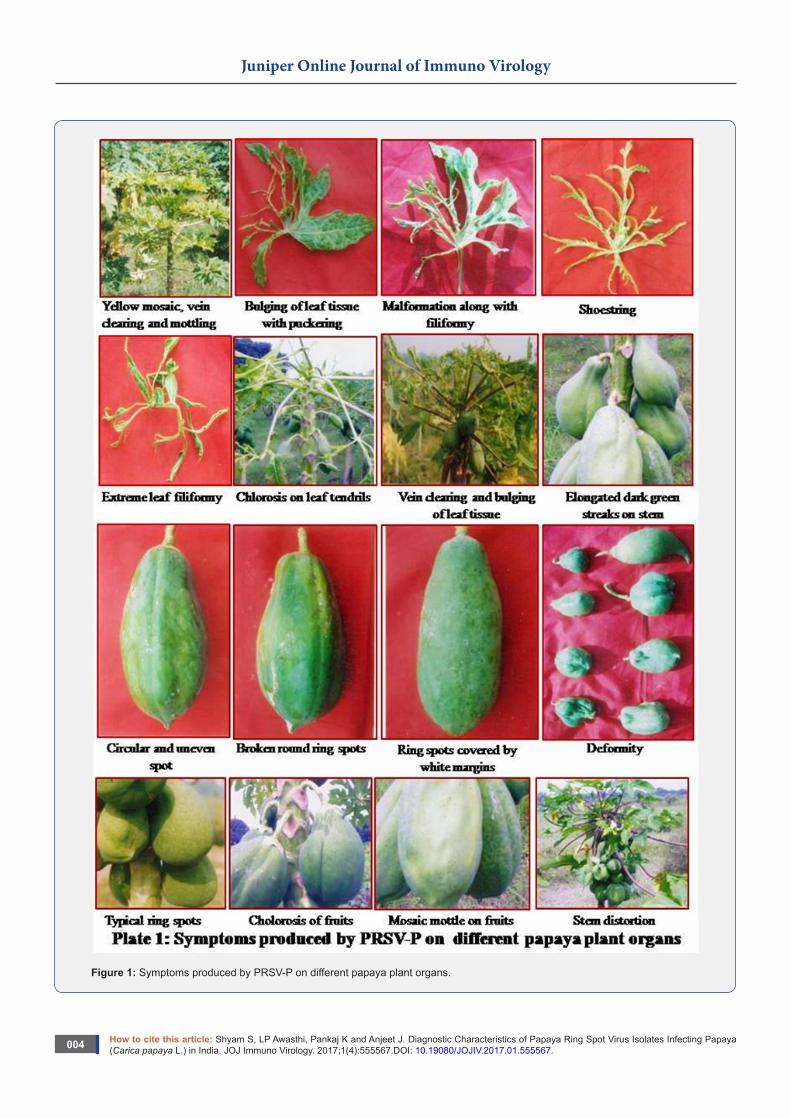
symptoms of papaya ringspot disease. Symptoms varied from chlorotic mottling of the leaves to severe rugosity. Infected plants showed chlorosis on the youngest leaves, vein clearing rugosity and mottling of leaf lamina interveinal puckering or bulging of the leaf tissues on the upper surface of the young leaves. In the severe cause’s filiformy, shoestring and distinct chlorotic streaks were found on the leaf tendrils and younger portion of the shoot. ringspot pattern was observed on the skin of affected fruits. Size of these necrotic spots ranged from 3-20 mm in diameter. The number of ringspots in a single fruit varied from a few to more than 200 depending upon the size of fruits and severity of the disease. Maximum number of spots on fruit skin was observed on the sunny side i.e. opposite to stem. These spots coalesce to each other and form distorted ringspots on most of the fruits. Virus infected ripened fruits when peeled out had a characteristic broken or elongated rings on the surface of fruits. The ringspots were bigger in size and surrounded by white margin. The size of these spots ranged between 8-20 mm. Elongated dark green streaks were observed on petioles and upper half of the stem symptoms of blistering and deformity of large number of fruits were observed. Some fruits had clorosis mosaic mottling symptoms and upper portion of the stem was distorted (Figure 1&2). Most of field surveyed revealed characteristic symptoms of papaya ringspot virus. Various types of symptoms like mild to severe mosaic, mottling, ringspot on fruits, leaves and stems, distortion of fruits, leaves and stems, filiform leaf, shoesting leaf, vein clearing, vein curling, vein distortion, puckering, leaf curling, leaf rolling, fruit yellowing, vein zigzag and stunting growth of plants were observed in all the locations. Several workers have described same type of symptoms for PRSV [4-10].
How to cite this article: Shyam S, LP Awasthi, Pankaj K and Anjeet J. Diagnostic Characteristics of Papaya Ring Spot Virus Isolates Infecting Papaya (Carica papaya L.) in India. JOJ Immuno Virology. 2017;1(4):555567.DOI: 10.19080/JOJIV.2017.01.555567.004
Juniper Online Journal of Immuno Virology
Figure 1: Symptoms produced by PRSV-P on different papaya plant organs.

How to cite this article: Shyam S, LP Awasthi, Pankaj K and Anjeet J. Diagnostic Characteristics of Papaya Ring Spot Virus Isolates Infecting Papaya (Carica papaya L.) in India. JOJ Immuno Virology. 2017;1(4):555567.DOI: 10.19080/JOJIV.2017.01.555567.005
Juniper Online Journal of Immuno Virology
Figure 2: Symptoms produced by PRSV-P on different host plants.

How to cite this article: Shyam S, LP Awasthi, Pankaj K and Anjeet J. Diagnostic Characteristics of Papaya Ring Spot Virus Isolates Infecting Papaya (Carica papaya L.) in India. JOJ Immuno Virology. 2017;1(4):555567.DOI: 10.19080/JOJIV.2017.01.555567.006
Juniper Online Journal of Immuno Virology
Host rangeTable 2: Reaction of PRSV on different hosts of different family.
S. No. Families Plants Reaction
1 Caricaceae Carica papaya L. Mosaic mottling, leaf distortion
2Chenopodiaceae
Chenopodium amaranticolor
Necrotic local lesions
Chenopodium quinoa
Necrotic local lesions
3 Cucurbitaceae
Citrullus lanatus var. lanatus Mosaic mottling
Citrullus vulgaris Mosaic mottling
Cucumis melo (Local) Mosaic mottling
Cucumis melo (Harichal) Mosaic mottling
Cucumis sativus Mosaic mottling
Cucumis anguria var. anguria Mosaic mottling
Cucumis metuliferous
Cucumis melo var. utilissimus Mosaic mottling
Cucurbita moschata Mosaic mottling
Cucurbita pepo Mosaic mottling
Luffa acutangula Mosaic mottling
Luffa cylindrica Mosaic mottling
Lagenaria siceraria Mosaic mottling
Momordica charantia Mosaic mottling
4 Solanaceae
Nicotiana xanthi No symptoms
Nicotiana glutinosa No symptoms
Nicotiana tabaccum No symptoms
Nicotiana burley No symptoms
Nicotiana rustica No symptoms
Lycopersicum esculentum No symptoms
Datura stramonium No symptoms
5 Legumenasae
Vigna radiata No symptoms
Vigna mungo No symptoms
Vigna sinensis No symptoms
Pisum sativum No symptoms
6 Euphorbiace Ricinus communis
Mosaic mottling, leaf distortion
7 Amaranthaceae Gomphrena globosa
Necrotic local lesions
The causal virus was easily transmitted by sap inoculation to Carica papaya plants. Results presented in Table 2 indicated that the virus could be transmitted mechanically in Citrullus lanatus var. lanatus, Citrullus vulgaris, Cucumis melo (Local), Cucumis melo (Harichal), Cucumis sativus, Cucumis anguria var. anguria, Cucumis metuliferous, Cucumis melo var. utilissimus, Cucurbita moschata, Cucurbita pepo, Luffa acutangula, Luffa cylindrica, Lagenaria siceraria, Momordica charantia, Ricinus communis and produced systemic mosaic mottling and leaf distortion symptoms. Whereas, necrotic local lesions were observed on Chenopodium amarenticolor, Chenopodium quinon and Gomphrena globosa plants. The virus under study did not produce any symptom on Nicotiana xanthi, Nicotiana glutinosa, Nicotiana tabaccum, Nicotiana tabaccum var. burley Ky-58, Nicotiana rustica, Lycopersicom esculentum, Datura stramonium, Vigna radiata, Vigna mungo, Vigna sinensis and Pisum sativum which indicated their non host status (Figure 2). Papaya ringspot virus was easily mechanically transmitted in papaya, cucurbits and some other plants. Experimental findings showed that the virus was successfully transmitted by the sap inoculation method in plants belonging to families Caricaceae (Carica papaya), cucurbitaceae (Citrullus lanatus var. fistulosus, C. vulgaris, C. melo ( local and harichal), C. sativus, C. anguria var. anguria, C. metuliferous, C. melo var. utilissimus, Cucurbita moschata, C. pepo, Luffa acutangula, L. cylindrical, Lagenaria siceraria, Momordica charantia) and Euphorbiaceae (Ricinus communis) with systemic mosaic mottling symptoms. However, plants of families Chenopodiaceae (Chenopodium amaranticolor, C. quinon) and Amaranthaceae (Gopherana globosa) produced necrotic lesions served as hypersensitive host. Ricinus communis of family Euphorbiaceae was found to be a new hosts of papaya ring spot virus. It has been reported earlier also that papaya ring spot virus infects plants of families Caricaceae, Chenopodiaceae and Cucurbitaceae [3,11-12]. It has been reported that squash and C. metuliferous were suitable host of PRSV-P. Dahal, et al. [13] reported PRSV disease incidence on Bennicasa hispida, Momordica charantia, Citrullus vulgaris,Cucurbita maxima, C. melo, C. sativus, C. pepo, Luffa acutangula, Lagenaria siceraria, Triohosanthes cucurmeria and Sechium edule confirmed by ELISA. Many cucurbitatious plants were reported as natural hosts of papaya ring spot virus [14,15].
Transmission of causal virusResults have indicated that virus under study was easily
transmissible by mechanical sap inoculation in papaya seedlings and Chenopodium amaranticolor leaves and usually gave 100.00 per cent infection followed by insect vector (Myzus persicae 93.33 %, Aphis gossypii 90.00 %, Aphis craccivora 83.33 %) and seeds (23.40 %). Bemisia tabaci however could not transmit. Causal virus of papaya ringspot disease was easily transmitted by sap inoculation and exhibited 100 per cent transmission. As for as insect transmission is concerned maximum transmission was observed with Myzus persicae followed by Aphis gossypii

How to cite this article: Shyam S, LP Awasthi, Pankaj K and Anjeet J. Diagnostic Characteristics of Papaya Ring Spot Virus Isolates Infecting Papaya (Carica papaya L.) in India. JOJ Immuno Virology. 2017;1(4):555567.DOI: 10.19080/JOJIV.2017.01.555567.007
Juniper Online Journal of Immuno Virology
A. craccivora while no transmission was observed by Bemisia tabaci. The seeds from infected plants were capable for transmitting PRSV as 23.40 per cent (Table 3-5). Transmission of PRSV by infected sap from papaya to papaya and cucurbits was reported [16-18]. Papaya ringspot virus type P transmission was
reported by 21 species in 11 genera including Myzus persicae, Aphis gossypii, A. nerii, A. spiraccola, A. craccivora, Corolina cypere, Dactynotus ambraisae [19,9]. Talens, et al. [20] have reported transmission of PRSV through seeds. In our studies we have also observed seed transmission upto 23.40 per cent.
Table 3: Transmission of PRSV through mechanically sap inoculation in systemic and hypersensitive hosts.
S. No. Hosts Type of Infection No. of Plants Inoculated
No. of Local
Lesion Per Leaf
Percent Infection
Diseased Healthy Total
1 Carica papaya Systemic 30 0 30 - 100
2 Chenopodium amaranticolor Hypersensitive 5 0 5 23.45 100
Table 4: Transmission of PRSV through infected seeds of different varieties of papaya.
S. No. Varieties Number of Seedlings Transmission (%)Diseased Healthy Total
1 CO-7 85 215 300 28.33
2 Pusa Nanha 60 240 300 20
3 Coorghoneydew 81 219 300 27
4 CO-2 55 260 300 18.33
5 Washington 75 225 300 25
6 CO-5 70 230 300 23.33
7 Pusa Gaint 64 236 300 21.33
8 Pusa Delicious 68 232 300 22.66
9 Pusa dwarf 78 222 300 26
10 CO-3 66 234 300 22
Mean 70.2 229.8 300 23.4
Table 5: Transmission of PRSV through insect vectors on papaya plants.
S. No.Methods of
Transmission/ Vectors
Total Number of Inoculated
Plants
Diseased Plants Healthy Plants Percent
Transmission
1 Myzus persicae 30 28 2 93.33
2 Aphis gossypii 30 27 3 90
3 Aphis craccivora 30 25 5 83.33
4 Bemisia tabaci 30 0 30 0

How to cite this article: Shyam S, LP Awasthi, Pankaj K and Anjeet J. Diagnostic Characteristics of Papaya Ring Spot Virus Isolates Infecting Papaya (Carica papaya L.) in India. JOJ Immuno Virology. 2017;1(4):555567.DOI: 10.19080/JOJIV.2017.01.555567.008
Juniper Online Journal of Immuno Virology
Dilution end point of causal virusThe virus remained infective in sap extracted from diseased
leaves of papaya at 1: 1000 dilutions but not at 1: 10000 dilutions,
which indicated the dilution end point between 1: 1000 and 1: 10000 (Table 6).
Table 6: Effect of dilution on PRSV inoculated in non-systemic host plant.
S. No. Dilution (Concentration) Average no. of Local Lesion on Chenopodium Amaranticolor Leaves
1 1:01 26.65
2 1:10 16.7
3 0.111111111 9.05
4 0.736111111 3.1
5 1:10000 No lesions
6 1:100000 No lesions
7 1:1000000 No lesions
8 1:10000000 No lesions
9 1:100000000 No lesions
10 1:1000000000 No lesions
Thermal inactivation point of causal virusThe virus was found active at a temperature upto 50°C but it
was inactivated at 55°C or more which indicated that the virus
was inactivated between 50 and 55°C as the sap treated at 55°C for ten minutes could not produce any lesion on Chenopodium amaranticolor plants (Table 7).
Table 7: Effect of temperature on PRSV inoculated in non-systemic host plants.
S. No. Temperature (°C) Average no. of Local Lesion on Chenopodium Amaranticolor Leaves
1 30 26.95
2 35 24.6
3 40 21.65
4 45 13.45
5 50 4.45
6 55 No lesions
7 60 No lesions
8 65 No lesions
9 70 No lesions
10 75 No lesions
11 80 No lesions
12 85 No lesions
13 90 No lesions
14 95 No lesions
15 100 No lesions

How to cite this article: Shyam S, LP Awasthi, Pankaj K and Anjeet J. Diagnostic Characteristics of Papaya Ring Spot Virus Isolates Infecting Papaya (Carica papaya L.) in India. JOJ Immuno Virology. 2017;1(4):555567.DOI: 10.19080/JOJIV.2017.01.555567.009
Juniper Online Journal of Immuno Virology
Longevity in vitro of causal virusTable 8: Effect duration on survivability of PRSV.
S. No. Duration (hrs.) Reaction1 0 30.5
2 2 23.1
3 4 19.15
4 6 10.15
5 8 4.6
6 10 No lesions
7 12 No lesions
8 14 No lesions
9 16 No lesions
10 18 No lesions
11 20 No lesions
12 22 No lesions
13 24 No lesions
Data presented in (Table 8) indicated that virus was infectious upto 8 hrs of storage at room temperature and it was inactivated after 10 hrs of storage. The longevity of virus was recorded between 8 and 10 hrs at room temperature. In our case we have observed dilution end point of between 1 x 10⁻³ to 1 x 10⁻⁴, thermal inactivation point between 50-55°C and longevity in vitro up to 8 hrs. Similar results were reported by [3,21,22]. While, [11] reported a higher thermal inactivation point between 60-65°C and some other worker reported longevity upto 24 hrs [22,23,5].
References1. Purcifull DE, Hiebert E (1978) Serological distinction of Watermelon
mosaic virus isolates. Phytopathology 69: 112-116.
2. Yeh SD, Gonsalves D (1984) Purification and immunological analysis of cylindrical inclusion protein induced by Papaya ringspot virus and Watermelon mosaic virus-1. Phytopath 74(11): 1273-1278.
3. Wang HL, Wang CC, Chiu RJ, Sun MH (1978) A preliminary study of papaya ring spot virus in Taiwan. Pl Prot Bull 20(2): 133-140.
4. Khurana SMP, Bhargava KS (1970) Induced apocarpy and “double papaya” fruit formation in papaya with Distortion ringspot virus infection. Plant Disease Reptr 54(2): 181-183.
5. Surekha SK, Mathur K, Shukla DD (1978) Virus diseases of papaya (Carica papaya) in Udaipur Indian. J Mycol Pl Pathol 7: 115-121.
6. Verma HN, Prasad V (1986) Virus diseases in papaw (papaya) Rev Trop Pl Path 9: 311-327.
7. Verma AK (1996) Viral and Mycoplasmal Diseases of papaya (Carica
papaya L). Disease scenario in crop plants. Vol. 1-Fruits and Vegetables (eds) Agnihotri VP, Om Prakash, Ram Kishun, Mishra A. International Books and Periodical Supply Service, India, pp. 156- 175.
8. Marys EE, Carballo O, Izaguirre Maycoral ML (2000) Occurrence and relative incidence of virus infected papaya in Venezuela. Ann Appl Biol 136(2): 121-124.
9. Singh SJ (2003) Virus and phytoplasma disease of papaya, passion fruit and pineapple. Kalyani Publishers Ludhiana pp. 147.
10. Jain RK, Sharma J and Verma A (2004) Present status of Papaya ring spot virus population profile in India. Annu Rev Plant Pathol 3: 1-15.
11. Yemewar SI, Mali VR (1980) On the identity of a sap transmissible virus of papaya in Marathwada. Indian J Myco Pl Path 10(2): 155-160.
12. Yeh SD, Gonsalves D, Provvidenti R (1984) Comparative studies on host range and serology of Papaya ringspot virus and Water melon mosaic virus-1. Phytopath 74(9): 1081-1085.
13. Dahal C, Lecoq H, Albrechtten SE (1997) Occurrences of Papaya ringspot potyvirus and cucurbit viruses in Nepal. Annals of Applied Biology 130(3): 491-502.
14. Roggero P, Gotta P, Stravato VM, Dellavalle G, Ciuffo M (1999) Further spread of Moroccan water melon mosaic poty virus in Italy. J Plant Pathology 82(3): 351.
15. Moura MC, Lima JA, Oliveira VB, Gonsalves MFS (2001) Serological identification virus species infecting cucurbit in producing areas of the state species of Maranhao, Brazil. Fitopatiologia Bras 26(1): 90-92.
16. Khurana SMP (1974) Studies on three virus disease of papaya in Gorakhpur, India. Proc 19th International Hort, Cong, Warsa, Poland, 1A: 260.
17. Ray G, Jain RK, Bhat AI, Varma A (1999) Comparative host range and serological studies of Papaya ringspot potyvirus isolates. Indian Phytopathology 52: 14-17.
18. Singh A, Upadhyaya PP, Rao GP (2005) Distribution and serological Diagnosis of Papaya ring spot virus in Eastern Uttar Pradesh. India J Mycol Pl Pathol 35(2): 237-241.
19. Reddy LCN (2000) Studies on Papaya ringspot virus disease on papaya (Carica papaya L). M.Sc. (Agri.) Thesis, University of Agricultural Sciences, Banglore, pp. 105.
20. Talens AD, Alcantara BS, Yeh SD (1995) Studies of papaya viruses in the Philippines: seed transmission of Papaya ring spot virus in the Philippines. Philippines J Crop Sci 20(1): 23.
21. Wu FC, Peng XX, Xu SH (1983) Preliminary studies on identification, purification and properties of Papaw ringspot virus in South China. Acta Phytopathologica Sinica 13(3): 21-28.
22. Rosa DLM, Lastra R (1977) Identification and characterization of Papaya ring spot virus in Venezuela. Proc Amer Phytopathol Soc 4: 175-176.
23. Zettler FW, Pureifull DE, Endwardson JR (1968) Some properties and classification of Papaya mosaic virus and Papaya ringspot virus. Phytopath 58: 554.
Your next submission with Juniper Publishers will reach you the below assets
• Quality Editorial service• Swift Peer Review• Reprints availability• E-prints Service• Manuscript Podcast for convenient understanding• Global attainment for your research• Manuscript accessibility in different formats
( Pdf, E-pub, Full Text, Audio) • Unceasing customer service
Track the below URL for one-step submission https://juniperpublishers.com/online-submission.php
This work is licensed under CreativeCommons Attribution 4.0 LicenseDOI:10.19080/JOJIV.2017.01.555567