Diagnosis Standards for Determining Cause of Death · Kwashiorkor ... Marasmus or unspecified...
62
Transcript of Diagnosis Standards for Determining Cause of Death · Kwashiorkor ... Marasmus or unspecified...

2
Contents Abbreviations .......................................................................................................................................................... 6
Definitions ............................................................................................................................................................... 6
I. Overview ............................................................................................................................................................. 7
II. Non-infectious Congenital and Neonatal Conditions ....................................................................................... 10
Birth trauma (ND, SB) ....................................................................................................................................................... 10
Intrauterine Hypoxia (ND, SB) ......................................................................................................................................... 10
Birth Asphyxia (ND) .......................................................................................................................................................... 11
Hypoxic Ischemic Encephalopathy of Newborn (ND) ..................................................................................................... 12
Congenital Malformations, Deformations and Chromosomal Abnormalities (ND, SB) ................................................ 12
Intracranial Hemorrhage of the Fetus and Newborn (nontraumatic) (ND, SB) ............................................................. 13
Hemolytic Disease of the Fetus and Newborn (Erythroblastosis fetalis) (ND, SB) ........................................................ 13
Kernicterus (ND) ............................................................................................................................................................... 14
Meconium Aspiration (ND) .............................................................................................................................................. 14
Extremely Low Birth Weight (ND, SB) .............................................................................................................................. 15
Other Low Birth Weight (ND, SB) ..................................................................................................................................... 15
Extreme immaturity (ND, SB)........................................................................................................................................... 15
Other preterm infants (ND, SB) ....................................................................................................................................... 16
Respiratory Distress Syndrome (RDS) (Hyaline Membrane Disease) (ND) .................................................................... 16
III. Congenital and Neonatal Infections ................................................................................................................ 17
Congenital pneumonia (ND) ............................................................................................................................................ 17
Congenital Rubella Syndrome (ND, SB) ........................................................................................................................... 18
Congenital Cytomegalovirus Infection (CMV) (ND, SB) .................................................................................................. 18
Congenital Herspesviral (herpes simplex) Infection (ND, SB) ......................................................................................... 19
Congenital Viral Hepatitis (ND, SB) .................................................................................................................................. 19
Other or Unspecified Congenital Viral Diseases (ND, SB) ............................................................................................... 20
Congenital Varicella Infection (ND, SB) ........................................................................................................................... 20
Congenital Parvovirus Infection (ND, SB) ........................................................................................................................ 20
Bacterial sepsis of the newborn (ND) ............................................................................................................................. 21
Congenital Tuberculosis (ND, SB) .................................................................................................................................... 22
Congenital Toxoplasmosis (ND, SB) ................................................................................................................................ 22
Neonatal (disseminated) Listeriosis (ND, SB) .................................................................................................................. 23
Congenital Malaria (ND, SB) ............................................................................................................................................ 23
Neonatal Candidiasis (ND) ............................................................................................................................................... 24
Neonatal Tetanus (ND) .................................................................................................................................................... 24
Congenital Syphilis (ND) ................................................................................................................................................... 24
CHAMPS Diagnosis Standards for DeCoDe 3
IV. Infectious Diseases .......................................................................................................................................... 25
Gastroenteritis/Enteritis (unspecified origin) ................................................................................................................. 25
Cholera ......................................................................................................................................................................... 25
Typhoid or Paratyphoid Fever ..................................................................................................................................... 26
Salmonellosis due to other Salmonella (non-typhoid) .............................................................................................. 26
Gastroenteritis/Enteritis due to Enteroinvasive Escherichia coli (EIEC) or Shigellosis ............................................. 27
Gastroenteritis/Enteritis due to Enteropathogenic Escherichia coli (EPEC) .............................................................. 27
Gastroenteritis/Enteritis due to Enterotoxigenic Escherichia coli (ETEC) ................................................................. 27
Gastroenteritis/Enteritis due to Campylobacter ........................................................................................................ 27
Gastroenteritis/Enteritis due to Yersinia enterocolitica ............................................................................................ 28
Gastroenteritis/Enteritis due to Clostridium difficile ................................................................................................. 28
Gastroenteritis/Enteritis due to other specified bacterial intestinal infections ....................................................... 28
Amoebic Dysentery due to Entamoeba histolytica .................................................................................................... 28
Giardiasis ...................................................................................................................................................................... 29
Cryptosporidiosis ......................................................................................................................................................... 29
Gastroenteritis/Enteritis due to Rotavirus ................................................................................................................. 29
Gastroenteritis/Enteritis due to Norovirus ................................................................................................................ 30
Gastroenteritis/Enteritis due to Adenovirus .............................................................................................................. 30
Gastroenteritis/Enteritis due to Astrovirus ................................................................................................................ 30
Gastroenteritis/Enteritis due to Sapovirus ................................................................................................................. 30
Gastroenteritis/Enteritis due to Enterovirus .............................................................................................................. 31
Ascariasis ...................................................................................................................................................................... 31
Trichuriasis ................................................................................................................................................................... 31
Arthropod-borne viral fevers and Viral Hemorrhagic Fevers (VHF) ............................................................................... 31
Brucellosis ........................................................................................................................................................................ 32
Meliodosis ........................................................................................................................................................................ 33
Leptospirosis .................................................................................................................................................................... 33
Rickettsioses ..................................................................................................................................................................... 34
HIV Disease ....................................................................................................................................................................... 35
Asymptomatic HIV Infection ............................................................................................................................................ 36
Malaria ............................................................................................................................................................................. 36
Measles............................................................................................................................................................................. 37
Bacterial Meningitis ......................................................................................................................................................... 37
Bacterial Meningitis due to a specific pathogen ............................................................................................................. 38
Viral Meningitis or Encephalitis ....................................................................................................................................... 39
Pertussis ........................................................................................................................................................................... 39
Pneumonia ....................................................................................................................................................................... 40
Pneumonia due to Streptococcus pneumoniae .......................................................................................................... 41
Pneumonia due to Haemophilus influenzae ............................................................................................................... 41
4
Pneumonia due to Klebsiella pneumoniae ................................................................................................................. 42
Pneumonia due to Pseudomonas aeruginosa ............................................................................................................ 42
Pneumonia due to Staphylococcus aureus ................................................................................................................. 43
Pneumonia due to Streptococcus, Group B ................................................................................................................ 43
Pneumonia Due to other streptococci (Not Group B) ................................................................................................ 44
Pneumonia due to Escherichia coli ............................................................................................................................. 44
Pneumonia due to other aerobic gram negative bacteria ......................................................................................... 45
Pneumonia due to Mycoplasma pneumoniae ............................................................................................................ 45
Pneumonia due to other specified bacteria ............................................................................................................... 45
Influenza with pneumonia .......................................................................................................................................... 46
Influenza with other respiratory manifestations (not pneumonia) .......................................................................... 46
Pneumonia due to Adenovirus.................................................................................................................................... 46
Pneumonia due to Respiratory Syncytial Virus (RSV) ................................................................................................ 47
Pneumonia due to Human Metapneumovirus (HMPV) ............................................................................................. 47
Pneumonia due to Chlamydia ..................................................................................................................................... 48
Pneumonia due to Pneumocystis (PCP) ...................................................................................................................... 48
Pneumonia due to other infectious organism, not elsewhere classified .................................................................. 49
Sepsis ................................................................................................................................................................................ 49
Sepsis due to a specific pathogen .................................................................................................................................... 50
Tuberculosis ..................................................................................................................................................................... 50
V. Malnutrition ...................................................................................................................................................... 51
Kwashiorkor ..................................................................................................................................................................... 51
Marasmus or unspecified severe protein-energy malnutrition ..................................................................................... 51
Moderate protein-energy malnutrition .......................................................................................................................... 52
Marasmic kwashiorkor .................................................................................................................................................... 52
Stunting ............................................................................................................................................................................ 52
VI. Other Conditions .............................................................................................................................................. 53
Anemia ............................................................................................................................................................................. 53
Congestive Heart Failure .................................................................................................................................................. 53
Malignant Neoplasms of a Specific Site .......................................................................................................................... 54
Malignant Neoplasms of lymphoid, hematopoietic and related tissues (Leukemias and Lymphomas) ...................... 54
Sickle Cell Disease ............................................................................................................................................................ 54
VII. External Causes ............................................................................................................................................... 55
Trauma ............................................................................................................................................................................. 55
Burns ................................................................................................................................................................................. 55
Poisoning .......................................................................................................................................................................... 56
Environmental Exposures ................................................................................................................................................ 56
Non-Accidental Trauma (NAT) ......................................................................................................................................... 56
Drug Resistance ................................................................................................................................................................ 57

CHAMPS Diagnosis Standards for DeCoDe 5
Hospital acquired ............................................................................................................................................................. 57
VIII. Undetermined................................................................................................................................................ 57
Fetal death of unspecified cause ..................................................................................................................................... 57
Other ill-defined and unspecified causes of mortality ................................................................................................... 57
IX. Maternal Conditions in Perinatal Death .......................................................................................................... 57
M1 Complications of placenta, cord and membranes .................................................................................................... 57
M2 Maternal complications of pregnancy ...................................................................................................................... 58
M3 Other complications of labour and delivery ............................................................................................................. 58
M4 Maternal medical and surgical conditions ................................................................................................................ 58
M5 No Maternal Condition .............................................................................................................................................. 59
References ............................................................................................................................................................ 60
6
Abbreviations AIDS Acquired Immunodeficiency Syndrome CHAMPS Child Health and Mortality Prevention Surveillance Network CSF Cerebrospinal Fluid CRP C-reactive Protein DeCoDe Determination of the Cause of Death DNA Deoxyribonucleic acid DS Diagnosis Standards ELISA Enzyme-linked Immunosorbent Assay HIV Human Immunodeficiency Virus ICD-10 International Statistical Classification of Diseases and Related Health Problems 10th Revision MITS Minimally Invasive Tissue Sampling NAT Non-accidental Trauma ND Neonatal Death NP/OP Nasopharyngeal/Oropharyngeal PCP Pneumocystis pneumonia PCR Polymerase Chain Reaction RDT Rapid Diagnostic Test RNA Ribonucleic Acid RSV Respiratory Syncytial Virus SB Stillbirth SMEs Subject Matter Experts Spp Species TAC TaqMan Array Card VA Verbal Autopsy VHF Viral Hemorrhagic Fever WBC White Blood Cell WHO World Health Organization
Definitions Child CHAMPS focuses on children under 60 months (5 years) DeCoDe Panel Local panels comprised of pathologists, clinicians, epidemiologists, and microbiologists who will
review all available information for each case in order to determine cause of death I:T Ratio Ratio of immature to total neutrophils, with the absolute number of all immature forms
included in the numerator and absolute number of neutrophils included in the denominator. Infant A child under the age of 12 months Neonatal An infant under the age of 28 days (0-27 days) Stillbirth No spontaneous breathing or movement at time of delivery AND at least one of the following: 1)
weighing 1000 grams or more or 2) estimated gestational age > 28 weeks TaqMan Array Cards Multiplex PCR Assays
CHAMPS Diagnosis Standards for DeCoDe 7
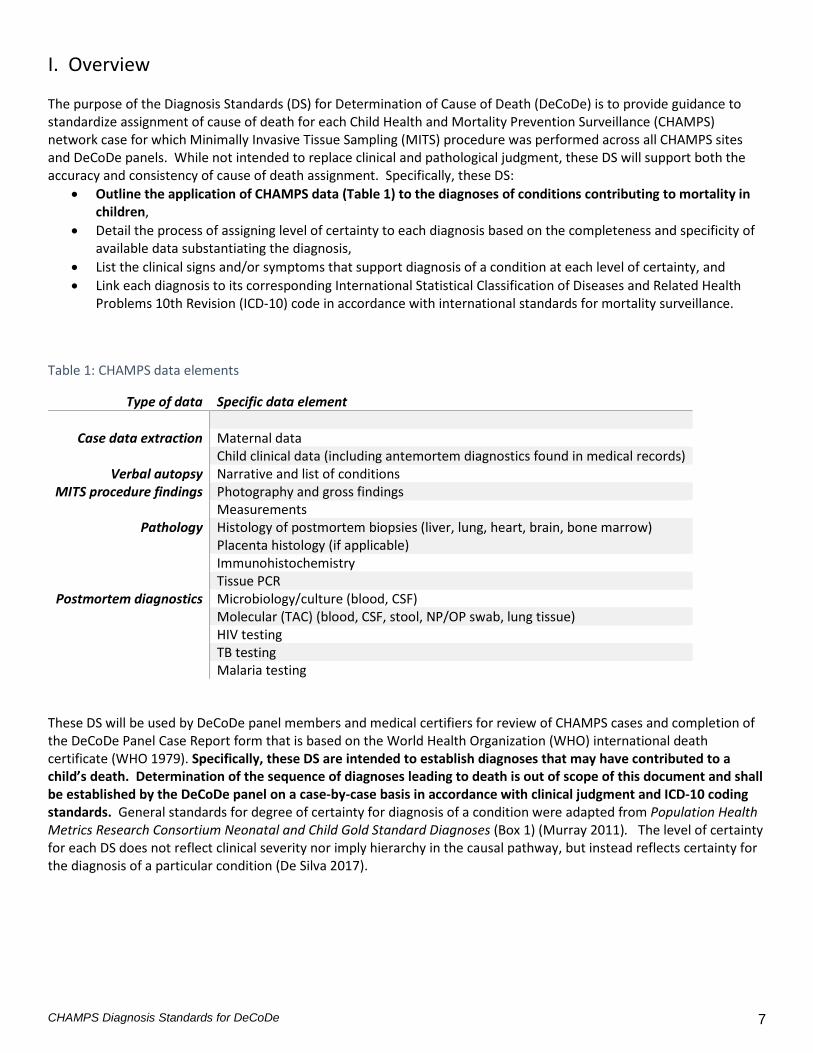
I. Overview The purpose of the Diagnosis Standards (DS) for Determination of Cause of Death (DeCoDe) is to provide guidance to standardize assignment of cause of death for each Child Health and Mortality Prevention Surveillance (CHAMPS) network case for which Minimally Invasive Tissue Sampling (MITS) procedure was performed across all CHAMPS sites and DeCoDe panels. While not intended to replace clinical and pathological judgment, these DS will support both the accuracy and consistency of cause of death assignment. Specifically, these DS:
• Outline the application of CHAMPS data (Table 1) to the diagnoses of conditions contributing to mortality in children,
• Detail the process of assigning level of certainty to each diagnosis based on the completeness and specificity of available data substantiating the diagnosis,
• List the clinical signs and/or symptoms that support diagnosis of a condition at each level of certainty, and • Link each diagnosis to its corresponding International Statistical Classification of Diseases and Related Health
Problems 10th Revision (ICD-10) code in accordance with international standards for mortality surveillance.
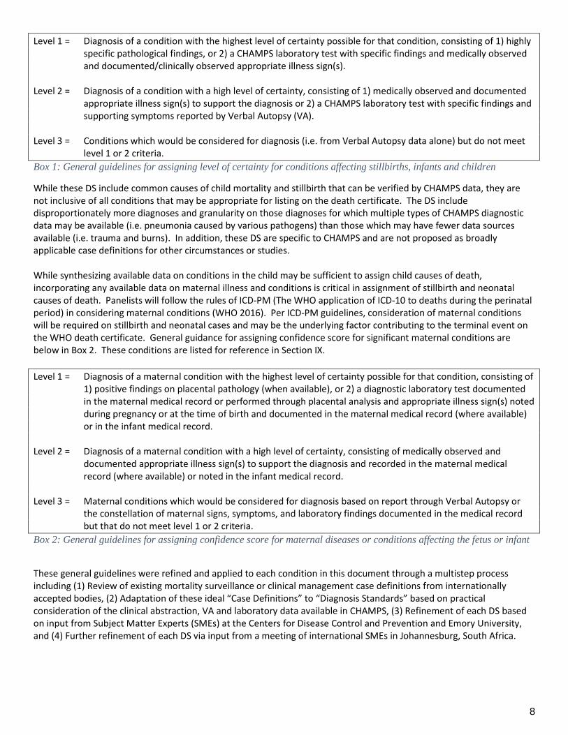
Table 1: CHAMPS data elements
These DS will be used by DeCoDe panel members and medical certifiers for review of CHAMPS cases and completion of the DeCoDe Panel Case Report form that is based on the World Health Organization (WHO) international death certificate (WHO 1979). Specifically, these DS are intended to establish diagnoses that may have contributed to a child’s death. Determination of the sequence of diagnoses leading to death is out of scope of this document and shall be established by the DeCoDe panel on a case-by-case basis in accordance with clinical judgment and ICD-10 coding standards. General standards for degree of certainty for diagnosis of a condition were adapted from Population Health Metrics Research Consortium Neonatal and Child Gold Standard Diagnoses (Box 1) (Murray 2011). The level of certainty for each DS does not reflect clinical severity nor imply hierarchy in the causal pathway, but instead reflects certainty for the diagnosis of a particular condition (De Silva 2017).
Type of data Specific data element
Case data extraction Maternal data Child clinical data (including antemortem diagnostics found in medical records)
Verbal autopsy Narrative and list of conditions MITS procedure findings Photography and gross findings
Measurements Pathology Histology of postmortem biopsies (liver, lung, heart, brain, bone marrow)
Placenta histology (if applicable) Immunohistochemistry Tissue PCR
Postmortem diagnostics Microbiology/culture (blood, CSF) Molecular (TAC) (blood, CSF, stool, NP/OP swab, lung tissue) HIV testing TB testing Malaria testing
8
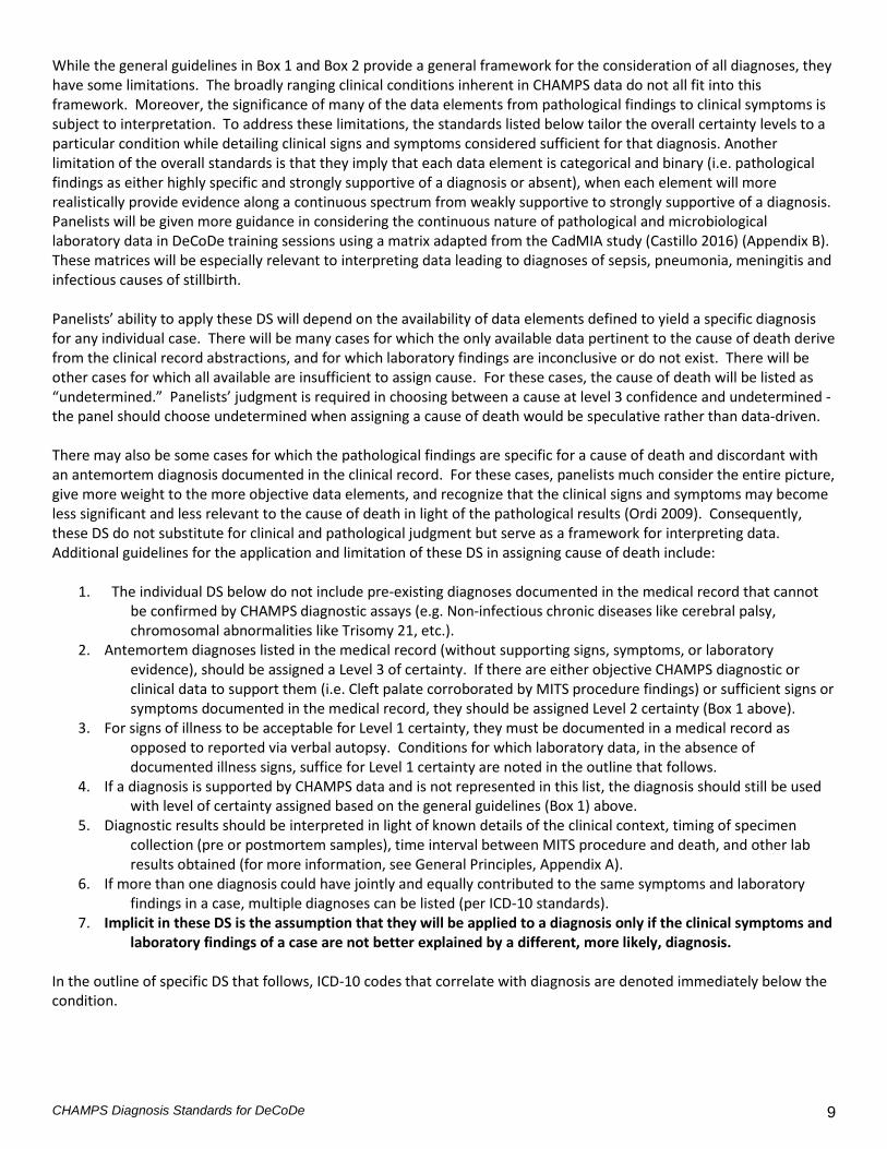
Level 1 = Diagnosis of a condition with the highest level of certainty possible for that condition, consisting of 1) highly specific pathological findings, or 2) a CHAMPS laboratory test with specific findings and medically observed and documented/clinically observed appropriate illness sign(s).
Level 2 = Diagnosis of a condition with a high level of certainty, consisting of 1) medically observed and documented
appropriate illness sign(s) to support the diagnosis or 2) a CHAMPS laboratory test with specific findings and supporting symptoms reported by Verbal Autopsy (VA).
Level 3 = Conditions which would be considered for diagnosis (i.e. from Verbal Autopsy data alone) but do not meet
level 1 or 2 criteria. Box 1: General guidelines for assigning level of certainty for conditions affecting stillbirths, infants and children
While these DS include common causes of child mortality and stillbirth that can be verified by CHAMPS data, they are not inclusive of all conditions that may be appropriate for listing on the death certificate. The DS include disproportionately more diagnoses and granularity on those diagnoses for which multiple types of CHAMPS diagnostic data may be available (i.e. pneumonia caused by various pathogens) than those which may have fewer data sources available (i.e. trauma and burns). In addition, these DS are specific to CHAMPS and are not proposed as broadly applicable case definitions for other circumstances or studies. While synthesizing available data on conditions in the child may be sufficient to assign child causes of death, incorporating any available data on maternal illness and conditions is critical in assignment of stillbirth and neonatal causes of death. Panelists will follow the rules of ICD-PM (The WHO application of ICD-10 to deaths during the perinatal period) in considering maternal conditions (WHO 2016). Per ICD-PM guidelines, consideration of maternal conditions will be required on stillbirth and neonatal cases and may be the underlying factor contributing to the terminal event on the WHO death certificate. General guidance for assigning confidence score for significant maternal conditions are below in Box 2. These conditions are listed for reference in Section IX. Level 1 = Diagnosis of a maternal condition with the highest level of certainty possible for that condition, consisting of
1) positive findings on placental pathology (when available), or 2) a diagnostic laboratory test documented in the maternal medical record or performed through placental analysis and appropriate illness sign(s) noted during pregnancy or at the time of birth and documented in the maternal medical record (where available) or in the infant medical record.
Level 2 = Diagnosis of a maternal condition with a high level of certainty, consisting of medically observed and
documented appropriate illness sign(s) to support the diagnosis and recorded in the maternal medical record (where available) or noted in the infant medical record.
Level 3 = Maternal conditions which would be considered for diagnosis based on report through Verbal Autopsy or
the constellation of maternal signs, symptoms, and laboratory findings documented in the medical record but that do not meet level 1 or 2 criteria.
Box 2: General guidelines for assigning confidence score for maternal diseases or conditions affecting the fetus or infant
These general guidelines were refined and applied to each condition in this document through a multistep process including (1) Review of existing mortality surveillance or clinical management case definitions from internationally accepted bodies, (2) Adaptation of these ideal “Case Definitions” to “Diagnosis Standards” based on practical consideration of the clinical abstraction, VA and laboratory data available in CHAMPS, (3) Refinement of each DS based on input from Subject Matter Experts (SMEs) at the Centers for Disease Control and Prevention and Emory University, and (4) Further refinement of each DS via input from a meeting of international SMEs in Johannesburg, South Africa.
CHAMPS Diagnosis Standards for DeCoDe 9
While the general guidelines in Box 1 and Box 2 provide a general framework for the consideration of all diagnoses, they have some limitations. The broadly ranging clinical conditions inherent in CHAMPS data do not all fit into this framework. Moreover, the significance of many of the data elements from pathological findings to clinical symptoms is subject to interpretation. To address these limitations, the standards listed below tailor the overall certainty levels to a particular condition while detailing clinical signs and symptoms considered sufficient for that diagnosis. Another limitation of the overall standards is that they imply that each data element is categorical and binary (i.e. pathological findings as either highly specific and strongly supportive of a diagnosis or absent), when each element will more realistically provide evidence along a continuous spectrum from weakly supportive to strongly supportive of a diagnosis. Panelists will be given more guidance in considering the continuous nature of pathological and microbiological laboratory data in DeCoDe training sessions using a matrix adapted from the CadMIA study (Castillo 2016) (Appendix B). These matrices will be especially relevant to interpreting data leading to diagnoses of sepsis, pneumonia, meningitis and infectious causes of stillbirth. Panelists’ ability to apply these DS will depend on the availability of data elements defined to yield a specific diagnosis for any individual case. There will be many cases for which the only available data pertinent to the cause of death derive from the clinical record abstractions, and for which laboratory findings are inconclusive or do not exist. There will be other cases for which all available are insufficient to assign cause. For these cases, the cause of death will be listed as “undetermined.” Panelists’ judgment is required in choosing between a cause at level 3 confidence and undetermined - the panel should choose undetermined when assigning a cause of death would be speculative rather than data-driven. There may also be some cases for which the pathological findings are specific for a cause of death and discordant with an antemortem diagnosis documented in the clinical record. For these cases, panelists much consider the entire picture, give more weight to the more objective data elements, and recognize that the clinical signs and symptoms may become less significant and less relevant to the cause of death in light of the pathological results (Ordi 2009). Consequently, these DS do not substitute for clinical and pathological judgment but serve as a framework for interpreting data. Additional guidelines for the application and limitation of these DS in assigning cause of death include:
1. The individual DS below do not include pre-existing diagnoses documented in the medical record that cannot be confirmed by CHAMPS diagnostic assays (e.g. Non-infectious chronic diseases like cerebral palsy, chromosomal abnormalities like Trisomy 21, etc.).
2. Antemortem diagnoses listed in the medical record (without supporting signs, symptoms, or laboratory evidence), should be assigned a Level 3 of certainty. If there are either objective CHAMPS diagnostic or clinical data to support them (i.e. Cleft palate corroborated by MITS procedure findings) or sufficient signs or symptoms documented in the medical record, they should be assigned Level 2 certainty (Box 1 above).
3. For signs of illness to be acceptable for Level 1 certainty, they must be documented in a medical record as opposed to reported via verbal autopsy. Conditions for which laboratory data, in the absence of documented illness signs, suffice for Level 1 certainty are noted in the outline that follows.
4. If a diagnosis is supported by CHAMPS data and is not represented in this list, the diagnosis should still be used with level of certainty assigned based on the general guidelines (Box 1) above.
5. Diagnostic results should be interpreted in light of known details of the clinical context, timing of specimen collection (pre or postmortem samples), time interval between MITS procedure and death, and other lab results obtained (for more information, see General Principles, Appendix A).
6. If more than one diagnosis could have jointly and equally contributed to the same symptoms and laboratory findings in a case, multiple diagnoses can be listed (per ICD-10 standards).
7. Implicit in these DS is the assumption that they will be applied to a diagnosis only if the clinical symptoms and laboratory findings of a case are not better explained by a different, more likely, diagnosis.
In the outline of specific DS that follows, ICD-10 codes that correlate with diagnosis are denoted immediately below the condition.
10
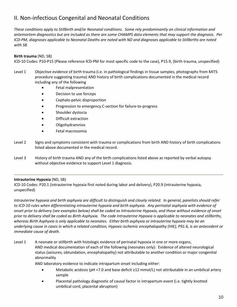
II. Non-infectious Congenital and Neonatal Conditions These conditions apply to Stillbirth and/or Neonatal conditions. Some rely predominantly on clinical information and antemortem diagnostics but are included as there are some CHAMPS data elements that may support the diagnosis. Per ICD-PM, diagnoses applicable to Neonatal Deaths are noted with ND and diagnoses applicable to Stillbirths are noted with SB. Birth trauma (ND, SB) ICD-10 Codes: P10-P15 (Please reference ICD-PM for most specific code to the case), P15.9, (birth trauma, unspecified) Level 1 Objective evidence of birth trauma (i.e. in pathological findings in tissue samples, photographs from MITS
procedure suggesting trauma) AND history of birth complications documented in the medical record including any of the following
• Fetal malpresentation • Decision to use forceps • Cephalo-pelvic disproportion • Progression to emergency C-section for failure-to-progress • Shoulder dystocia • Difficult extraction • Oligohydramnios • Fetal macrosomia
Level 2 Signs and symptoms consistent with trauma or complications from birth AND history of birth complications
listed above documented in the medical record. Level 3 History of birth trauma AND any of the birth complications listed above as reported by verbal autopsy
without objective evidence to support Level 1 diagnosis.
Intrauterine Hypoxia (ND, SB) ICD-10 Codes: P20.1 (intrauterine hypoxia first noted during labor and delivery), P20.9 (intrauterine hypoxia, unspecified) Intrauterine hypoxia and birth asphyxia are difficult to distinguish and closely related. In general, panelists should refer to ICD-10 rules when differentiating intrauterine hypoxia and birth asphyxia. Any perinatal asphyxia with evidence of onset prior to delivery (see examples below) shall be coded as Intrauterine Hypoxia, and those without evidence of onset prior to delivery shall be coded as Birth Asphyxia. The code Intrauterine Hypoxia is applicable to neonates and stillbirths, whereas Birth Asphyxia is only applicable to neonates. Either birth asphyxia or intrauterine hypoxia may be an underlying cause in cases in which a related condition, Hypoxic-ischemic encephalopathy (HIE), P91.6, is an antecedent or immediate cause of death. Level 1 A neonate or stillbirth with histologic evidence of perinatal hypoxia in one or more organs,
AND medical documentation of each of the following (neonates only): Evidence of altered neurological status (seizures, obtundation, encephalopathy) not attributable to another condition or major congenital abnormality AND laboratory evidence to indicate intrapartum onset including either:
• Metabolic acidosis (pH <7.0 and base deficit ≥12 mmol/L) not attributable in an umbilical artery sample
• Placental pathology diagnostic of causal factor in intrapartum event (i.e. tightly knotted umbilical cord, placental abruption)
CHAMPS Diagnosis Standards for DeCoDe 11
Level 2 A neonate or stillbirth with EITHER, histologic evidence of perinatal hypoxia in one or more organs, OR,
TWO or more of the following clinical criteria in the 24 hours after birth: • Evidence of altered neurological status (seizures, obtundation, encephalopathy) not attributable
to another condition or major congenital abnormality • Poor feeding or hypotonia • Apgar score ≤3 at 5 minutes of life • Pulse less than 100 per minute at birth and falling or not improving or 100 or above but without
establishment of normal respirations at 1 minute of life • Failure to cry at birth or to cry, initiate, and/or maintain adequate respirations after birth • Failed resuscitation at birth
AND clinical evidence to indicate intrapartum onset, including any of the following:
• A known sentinel hypoxic event occurring immediately before or during labor • Sudden and sustained fetal bradycardia or absent fetal heart rate • Meconium staining in the amniotic fluid • Documentation of prolapsed umbilical cord • Maternal cardiovascular collapse • Decision to proceed to emergency C-section for fetal factors
Level 3 Cases meeting Level 3 criteria for Birth Asphyxia below, in a neonate or stillbirth, but with findings
suggestive of intrapartum onset not meeting Level 1 or Level 2 definitions above. References: (Murray 2011) (Gerosa 2014) (Antonucci 2014) (American College of Obstetrics and Gynecology and American Academy of Pediatrics 2003) (ACOG Task Force on Neonatal Encephalopathy 2014)
Birth Asphyxia (ND) ICD-10 Codes: P21.0 (severe birth asphyxia), P21.1 (moderate birth asphyxia), P21.9 (birth asphyxia, unspecified) See guidance related to use of codes for Birth Asphyxia or Intrauterine Hypoxia above, in the definition for Intrauterine Hypoxia. Level 1 One of the following laboratory findings in a neonate:
• Metabolic acidosis (pH <7.0 and base deficit ≥12 mmol/L) not attributable to another cause and within 5 minutes of birth
• Histologic evidence of perinatal hypoxia in one or more organs AND medical documentation of each of the following:
• Evidence of altered neurological status (seizures, obtundation, encephalopathy) not attributable to another condition or major congenital abnormality
• Apgar score 0-3 at 1 minute of life (severe – P20.0) or Apgar score 4-7 at 1 minute of life (moderate – P20.1)
• Pulse less than 100 per minute at birth and falling or not improving (severe) or 100 or above but without establishment of normal respirations (moderate) at 1 minute of life
• Not a stillbirth Level 2 EITHER, histologic evidence of perinatal hypoxia in one or more organs, OR,
TWO or more of the following clinical criteria in the 24 hours after birth: • Evidence of altered neurological status (seizures, obtundation, encephalopathy) not attributable
to another condition or major congenital abnormality
12
• Poor feeding or hypotonia • Apgar score 0-3 at 1 minute of life (severe – P20.0) or Apgar score 4-7 at 1 minute of life
(moderate – P20.1) • Pulse less than 100 per minute at birth and falling or not improving (severe) or 100 or above but
without establishment of normal respirations (moderate) at 1 minute of life • Failure to cry at birth or to cry, initiate, and/or maintain adequate respirations after birth • Failed resuscitation at birth
Level 3 Medical documentation or verbal autopsy report of the above criteria insufficient for Level 1 or Level 2
diagnosis above. References: (Murray 2011) (Gerosa 2014) (Antonucci 2014) (American College of Obstetrics and Gynecology and American Academy of Pediatrics 2003) (ACOG Task Force on Neonatal Encephalopathy 2014)
Hypoxic Ischemic Encephalopathy of Newborn (ND) ICD-10 Code: P91.6 Level 1 Evidence of birth asphyxia or intrapartum hypoxia AND either of the following:
• Axonal necrosis or other strong pathological evidence of hypoxic-ischemic insult • Moderate pathological evidence of hypoxic-ischemic insult, AND Medical documentation of
altered neurological status (seizures, obtundation, encephalopathy) not attributable to another condition or major congenital abnormality
Level 2 Evidence of birth asphyxia or intrapartum hypoxia AND either of the following:
• Clinical documentation of altered neurological status (seizures, obtundation, encephalopathy) not attributable to another condition or major congenital abnormality
• Moderate pathological evidence of hypoxic-ischemic insult Level 3 Evidence of birth asphyxia or intrapartum hypoxia AND either VA report of altered neurological status not
attributed to another condition or major congenital anomaly, OR pathological findings suggestive of hypoxic ischemic encephalopathy in the absence of available signs and symptoms to meet criteria for Level 1 or Level 2 definitions above.
(E. Graham 2008) (Allen 2011)
Congenital Malformations, Deformations and Chromosomal Abnormalities (ND, SB) ICD-10 Codes: Q00-Q99 (Please reference ICD-PM for most specific code to the case) Level 1 Both of the following documented medically or during MITS:
• Congenital malformation that is externally visible OR established by an imaging study • Congenital malformation assessed to have been or reasonably could have been or contributed
to the cause of death Examples: Esophageal atresia; Gastroschisis; Anencephaly; Imperforate anus; Intestinal obstruction; Omphalocele
Level 2 External or internal congenital abnormality suspected by medical provider as documented in the medical
record and likely to have caused or contributed to death but not meeting the above criteria for level 1. Level 3 Congenital malformation reported by VA (i.e. physical abnormality at the time of delivery) to have caused or
contributed to death and not meeting the above criteria for level 1.

CHAMPS Diagnosis Standards for DeCoDe 13
References: (Murray 2011)
Intracranial Hemorrhage of the Fetus and Newborn (nontraumatic) (ND, SB) ICD-10 Codes: P52.0 (intraventricular hemorrhage [IVH], grade 1), P52.1 (IVH, grade 2), P52.2 (IVH, grade 3-4), P52.3 (unspecified IVH), P52.4 (intracerebral hemorrhage), P52.5 (subarachnoid hemorrhage), P52.6 (cerebellar and posterior fossa hemorrhage), P52.8 (other intracranial hemorrhage), P52.9 (unspecified intracranial hemorrhage) Level 1 Documentation of intracranial hemorrhage by imaging modality (i.e. Ultrasound) OR strong evidence
pathological evidence of intracranial hemorrhage. Level 2 Either of the following:
• Documentation of EITHER rapid change in hemoglobin not attributable to another cause OR bloody CSF in the absence of a traumatic tap AND change in neurologic status of the infant
• Moderate pathological evidence of intracranial hemorrhage
Hemolytic Disease of the Fetus and Newborn (Erythroblastosis fetalis) (ND, SB) ICD-10 Code: P55.0 (Rh isoimmunization), P55.1 (ABO isoimmunization), P55.8 (other antibody isoimmunization), P55.9 (unspecified), P56.0 (hydrops fetalis due to isoimmunization), P56.9 (hydrops fetalis due to other and unspecified hemolytic disease) Level 1 Laboratory confirmation of Rhesus (Rh) antigen or ABO blood type discordance in a mother-newborn pair
with the following additional laboratory findings: • Positive Direct Antiglobulin test AND • Anemia, hyperbilirubinemia, increased nucleated RBCs, hypoalbuminemia OR reticulocytosis (if
available) Level 2 Laboratory evidence to suggest hemolytic disease of the newborn, including:
• Positive Direct Antiglobulin test, OR • Pathological evidence of kernicterus on evaluation of brain tissues during MITS, OR • TWO of the following: unexplained anemia, rapidly developing indirect hyperbilirubinemia,
increased nucleated RBCs, hypoalbuminemia, reticulocytosis (if available) AND clinical evidence of hemolytic disease of the newborn, including:
• Hydrops fetalis (ascites, pleural effusions, pericardial effusions, skin edema) without evidence of parvovirus or infections cause, OR
• TWO of the following documented in the medical record or during MITS: o Hepatosplenomegaly o Early Jaundice o Pallor o Respiratory distress
Level 3 Clinical suspicion or laboratory evidence to suggest hemolytic disease of the newborn but not meeting
criteria for Level 1 or Level 2 diagnosis. (Murray 2007) (Osaro 2010)
14
Kernicterus (ND) ICD-10 Code: P57.0 (due to isoimmunization), P57.8 (other, specified), P57.9 (unspecified) Kernicterus is grouped with neonatal conditions, but the ICD-10 codes above can be used for any age child and criteria are included below for older children with bilirubin-induced neurologic dysfunction as well. Level 1 Pathological evidence of kernicterus on evaluation of brain tissues during MITS (if obtained). Level 2 Jaundice OR documentation of very elevated serum bilirubin in a neonate (>25 mg/dL) AND clinical evidence
of acute bilirubin encephalopathy in a neonate including TWO or more of the following documented in the medical record:
• Within 1-7 days of life o Somnolence o Hypotonia o Loss of Moro reflex o Decreased feeding o High-pitched cry
• After 7 days of life o Retrocolis (back arching of the neck) o Opisthonus (back arching of the trunk) o Choreoathetosis o Setting sun sign (impaired upward gaze)
OR evidence of bilirubin-induced neurologic dysfunction in an older child, including TWO or more of the following documented in the medical record:
• Movement disorder • Auditory dysfunction, including hearing loss or deafness • Ocular dysfunction, especially impaired upward gaze • Dental enamel hypoplasia
Level 3 Marked jaundice documented in the medical record or reported by verbal autopsy and suspected bilirubin
encephalopathy in a neonate but not meeting the criteria for Level 1 or Level 2 diagnosis above OR evidence of bilirubin-induced neurologic dysfunction in an older child, including TWO or more of the
signs above reported by verbal autopsy. (Olusanya 2015) (Shapiro 2005)
Meconium Aspiration (ND) ICD-10 Code: P24.0 Level 1 Lung tissue with meconium in the airspaces AND onset of respiratory distress within 12 hours of life as
documented by medical personnel including ONE of the following: • Hypoxia • Grunting respirations • Retractions • Coarse, patchy infiltrates on chest radiograph
Level 2 ONE of the following:
• Medically documented meconium stained amniotic fluid or meconium staining of a neonate with ONE of the signs of respiratory distress above observed within 12 hours of life
CHAMPS Diagnosis Standards for DeCoDe 15
• Lung tissue with meconium in the airspaces AND ONE sign of respiratory distress as reported by verbal autopsy: difficulty breathing, breathlessness, lower chest wall/ribs being pulled in, or grunting breath sounds.
Level 3 Death assessed to have been caused by meconium aspiration syndrome but not meeting the above criteria. (van Ierland 2009) (Kakimoto 2015)
Extremely Low Birth Weight (ND, SB) ICD-10 Code: P07.0 The following 4 codes (P07.0-P07.4) all relate to prematurity and are coded together. Panelists should follow ICD-10 coding rules for applying these codes and use low birth weight preferentially when both are available. The other may be noted in Part II of the WHO death certificate. Level 1 Birth weight of 999g or less documented in the medical record at the time of birth or noted in early neonatal
deaths ≤7 days at the time of MITS. Level 3 Birth weight of 999g or less documented in the medical record upon readmission or admission ≥7 days from
birth or as reported by VA and unable to be confirmed at the time of MITS.
Other Low Birth Weight (ND, SB) ICD-10 Code: P07.1 Level 1 Birth weight of 1000-2499 documented in the medical record at the time of birth or noted in early neonatal
deaths ≤7 days in the medical record or at the time of MITS. Level 3 Birth weight of 1000-2499g documented in the medical record upon readmission or admission ≥7 days from
birth OR as reported by VA and unable to be confirmed at the time of MITS.
Extreme immaturity (ND, SB) ICD-10 Code: P07.2 Level 1 Not a stillbirth as documented in the medical record AND birth at <28 weeks gestation based on history of
prenatal c of gestation.
Level 2 Not a stillbirth as documented in the medical record AND birth at <28 weeks based on one of the following: • History of prenatal US between 14 weeks and ≤27 6/7 weeks of gestation • Documentation in the medical record based on birth date minus mother’s certain last menstrual
period AND consistent with first trimester exam Level 3 Not a stillbirth AND birth at <28 weeks gestation based one of the following:
• Verbal Autopsy report • A physician or clinical officer’s Ballard gestational age assessment • Uncertain or certain last menstrual period as documented in the clinical record AND consistent
with birth weight Reference: (Dargaville 2005) (Quinn 2016)
16
Other preterm infants (ND, SB) ICD-10 Code: P07.3 Level 1 Not a stillbirth as documented in the medical record AND birth at ≥28 weeks and <37 weeks based on
history of prenatal US ≤13 6/7 weeks of gestation.
Level 2 Not a stillbirth as documented in the medical record AND birth at ≥28 weeks and <37 weeks based on one of the following:
• History of prenatal US between 14 weeks and ≤27 6/7 weeks of gestation • Documentation in the medical record based on birth date minus mother’s certain last menstrual
period AND consistent with first trimester physical exam Level 3 Not a stillbirth AND birth at ≥28 weeks and <37 weeks based on any of the following:
• Verbal Autopsy report of birth <37 weeks or a positive response to “more than one month early”
• A physician or clinical officer’s Ballard gestational age assessment • Uncertain or certain last menstrual period as documented in the clinical record AND consistent
with birth weight Reference: (Dargaville 2005) (Quinn 2016)
Respiratory Distress Syndrome (RDS) (Hyaline Membrane Disease) (ND) ICD-10 Code: P22.0 This standard and corresponding ICD-10 code is specific to Respiratory Distress Syndrome/Hyaline Membrane Disease and should not be used for respiratory distress in general. Level 1 EITHER, Strong histological evidence of RDS (i.e. hyaline membranes) in lung tissue
OR Moderate histological evidence of RDS in lung tissue AND Medical documentation of TWO or more of the following clinical/radiographic criteria:
• Chest x-ray positive for characteristic “ground glass” appearance • Respiratory rate >70/minute • Central cyanosis (dusky, bluish lips or mucus membranes) • Severe retractions/lower chest wall indrawing • Grunting • Nasal flaring
Level 2 One of the following:
• Medical documentation of birth at <37 weeks AND TWO or more of the above clinical/radiographic criteria PLUS inadequate post mortem lung biopsy.
• Moderate histologic evidence of RDS without availability of supporting clinical signs and symptoms necessary for level 1 diagnosis.
Level 3 VA report of birth more than one month early and TWO or more signs of respiratory distress: difficulty
breathing, breathing fast, breathlessness, lower chest wall/ribs being pulled in, or grunting breath sounds PLUS inadequate post mortem lung biopsy.
References: (Murray 2011)
CHAMPS Diagnosis Standards for DeCoDe 17
III. Congenital and Neonatal Infections This section includes infections specific to Stillbirths and/or the neonatal period. For other infectious entities, please reference appropriate entry in Section IV, Infectious Diseases. ICD-PM, diagnoses applicable to Neonatal Deaths are noted with ND and diagnoses applicable to Stillbirths are noted with SB. Congenital pneumonia (ND) ICD-10 Codes: P23.0 (due to a viral agent), P23.1 (due to Chlamydia), P23.2 (due to staphylococcus), P23.2 (due to streptococcus, Group B, P23.4 (due to Escherichia coli), P23.5 (due to Pseudomonas), P23.6 (due to other bacterial agents), P23.7 (due to other organisms), and P23.9 (unspecified) This code should be used for infective pneumonia acquired in utero or during birth. Level 1 EITHER Strong histological evidence of pneumonia in lung tissue with death within 48 hours of birth, OR One of the following from samples taken within 48 hours of birth:
• Moderate histological evidence of pneumonia in lung tissue with death within 48 hours of birth • Detection of a pathogen by PCR (TAC) (on the respiratory card) in lung tissue • Isolation of a pathogen from lung culture that is a plausible cause of pneumonia in the host • Isolation of a pathogen from blood culture that is a plausible cause of pneumonia in the host
with inadequate postmortem lung tissue for analysis • New infiltrate or pleural effusion on chest radiograph
AND TWO or more of the following clinical signs documented in the medical record: • Tachypnea (Per WHO Clinical Case Definitions defined as respiratory rate >60/minute in 0-2
months, >50/minute for infants 2-12 months, >40 in children 12 months -5 years) • Respiratory distress as chest indrawing, grunting or nasal flaring • Abnormal breath sounds (i.e. decreased breath sounds, crackles, crepitations) • Hypoxia, cyanosis or desaturations (oxygen saturation <95%) • Fever >38.0 or hypothermia <36.0
Level 2 One of the following:
• Moderate histological evidence of pneumonia in lung tissue with death within 48 hours of birth AND EITHER detection of a pathogen by PCR (TAC) in lung tissue, OR isolation of a pathogen from lung culture that is a plausible cause of pneumonia in the host
• No laboratory or imaging data available and ALL of the following documented in the medical record: fever or hypothermia, hypoxia or abnormal breath sounds, and tachypnea or respiratory distress.
• One of the laboratory or imaging findings above with TWO or more of clinical signs of pneumonia above reported by verbal autopsy (difficulty breathing, fast breathing or breathlessness, lower chest wall/ribs being pulled in or grunting, or fever).
Level 3 One of the following • Histological evidence of pneumonia in lung tissue with death within 48 hours of birth AND
detection of a pathogen on NP/OP swab • Acute febrile illness or hypothermia with tachypnea, respiratory distress, abnormal breath
sounds, hypoxia or cyanosis documented in the medical record or reported by verbal autopsy, but not meeting the criteria for Level 1 or Level 2 diagnosis above.
(Hooven 2017)
18
Congenital Rubella Syndrome (ND, SB) ICD-10 Code: P35.0 Level 1: An infant with EITHER of the following lab findings:
• Detection of Rubella in fluid or tissue (blood, CSF, nasopharyngeal/oropharyngeal swab, lung) by PCR (TAC) within 2 weeks of birth, OR
• A positive blood test for Rubella-specific IgM documented in the medical record AND EITHER two of the complications listed below in (a) OR one in (a) and one in (b) documented in the medical
record or noted during MITS: • (a) Cataracts, congenital glaucoma, congenital heart disease, loss of hearing, or pigmentary
retinopathy • (b) Purpura, extramedullary hematopoiesis (blueberry muffin spots), splenomegaly,
microcephaly, mental retardation, meningoencephalitis, radiolucent bone disease, or jaundice that begins within 24 hours after birth
Level 2: A case without laboratory confirmation of infection AND EITHER two of the complications listed above in (a)
OR one in (a) and one in (b) documented in the medical record or noted at the time of MITS. References: (World Health Organization (WHO) 2003) (CDC, Rubella, Congenital Syndrome, Case Definition. 2010)
Congenital Cytomegalovirus Infection (CMV) (ND, SB) ICD-10 Code: P35.1 This code should not be used for older, immunocompromised children with positive CMV laboratory findings and clinical illness. Because of the high prevalence and uncertain significance of CMV by PCR in neonates and stillbirths, subject matter experts recommended noting any cases that meet criteria for “DISEASE” (as noted below) in the causal pathway while noting any cases that only meet criteria for “INFECTION” (as noted below) in Section II of the WHO death certificate. Level 1: INFECTION: Detection of CMV in nasopharyngeal specimen, lung tissue, CSF, or blood by PCR (TAC) within 2
weeks of birth or in a stillbirth Level 1: DISEASE: Histopathologic evidence of CMV inclusion disease from an appropriate clinical specimen within
the first 2 weeks of life or in a stillbirth, OR evidence for disseminated CMV with detection of CMV by PCR from 2 or more tissues within 2 weeks of birth or in a stillbirth
Level 2: DISEASE: Laboratory detection of CMV by any of the methods above in a child older than 2 weeks of life AND TWO or more signs of clinical illness documented in the medical record as present in the first month of
life including: • Intrauterine growth retardation or small for gestational age • Premature birth • Hepatosplenomegaly • Petechial rash • Microcephaly • Motor disability • Chorioretinitis • Cerebral calcifications • Seizures

CHAMPS Diagnosis Standards for DeCoDe 19
AND no laboratory evidence to suggest an alternative etiology for the clinical syndrome (i.e. no evidence for congenital toxoplasmosis or congenital syphilis).
(CDC National Center for Immunization and Respiratory Diseases, Division of Viral Diseases 2016) (Government of Alberta 2011)
Congenital Herspesviral (herpes simplex) Infection (ND, SB) ICD-10 Code: P35.2 Level 1 Laboratory evidence of Herpes Simplex Virus (HSV) in blood, CSF, or tissues of a neonate (<28 days) by PCR
or immunohistochemical (IHC) evidence of HSV in tissues of a neonate <28 days with or without clinical illness.
Level 2 A child born to a mother with active herpetic lesions at the time of delivery AND TWO or more signs of clinical illness documented in the medical record as present in the first month of
life including: • Cutaneous scaring or vesicular skin, eye, or mouth lesions • Jaundice • Hepatosplenomegaly • Pneumonitis (respiratory distress or chest radiograph findings) • Seizures or lethargy • Chorioretinitis • Microophthalmia • Prematurity
AND no laboratory evidence to suggest an alternative etiology for the clinical syndrome (i.e. no evidence for congenital CMV or congenital syphilis).
Level 3 A child with unknown maternal HSV status and THREE or more signs of clinical illness documented in the
medical record or reported by verbal autopsy as presented in the first month of life, AND no laboratory evidence to suggest an alternative etiology for the clinical syndrome.
(Jones 2014) (Corey 2009)
Congenital Viral Hepatitis (ND, SB) ICD-10 Code: Use code P35.3 AND appropriate code for specific virus, if known Level 1 Laboratory evidence of hepatitis virus per the guidelines below in a child <24 months old born to a mother
with known viral hepatitis infection documented in the medical record. • Pathological evidence of hepatitis • Hepatitis E virus - Detectable HEV DNA by PCR (TAC) in blood or CSF • Hepatitis B virus
o Positive hepatitis B surface antigen test (if >4 weeks since hepatitis b vaccine) o Positive hepatitis B e antigen test in an infant 9-24 months of age o Detectable HBV DNA
• Hepatitis C virus o Detectable HCV DNA o Positive hepatitis C antibody test in a child 18-24 months of age

20
Level 2 Laboratory evidence of specific hepatitis virus as above in a child <24 months born to a mother whose hepatitis status is unknown.
Level 3 Clinical or laboratory evidence to suggest congenital viral hepatitis but not meeting the criteria for Level 1 or
Level 2 diagnosis as above. (CDC 2017) (Krain 2014) (Mirazo 2014) (Davison 2006)
Other or Unspecified Congenital Viral Diseases (ND, SB) ICD-10 Code: P35.8 (other – use with the specific code for the infectious agent, if known, i.e. for congenital varicella and parvovirus DS below), P35.9 (unspecified) Level 1 A positive laboratory test (either suggested by histopathology or detected by PCR/TAC) for a congenital
infection with signs and symptoms of illness documented in the medical record that are consistent with that congenital viral infection.
Level 3 A case without laboratory confirmation of infection but with suspected congenital viral infection based on
signs and symptoms documented in the medical record or noted during MITS procedure (use code 35.9 for unspecified congenital viral infection unless pathognomonic findings are present), OR a case with only a positive laboratory test for a congenital viral infection in the absence of sufficient clinical data for Level 1 diagnosis.
Congenital Varicella Infection (ND, SB) ICD-10 Code: P35.8 AND B01.9 Level 1 Clinical evidence of Congenital Varicella Syndrome defined by typical cicatricial skin scarring AND one or
more additional clinical findings suggestive of congenital varicella syndrome documented in the medical record or observed at the time of MITS:
• Limb hypoplasia • Rudimentary digits • Microcephaly • Cataracts • Nystagmus • Chorioretinitis
Level 3 Clinical evidence of Congenital Varicella Syndrome not meeting the criteria for Level 1 diagnosis above and
without laboratory findings to suggest an alternative congenital infection.
Congenital Parvovirus Infection (ND, SB) ICD-10 Code: P35.8 AND B34.3 Level 1 One or more of the following laboratory findings documented in the medical record or during MITS in an
infant <2 weeks old regardless of clinical signs and symptoms • Detection of Parvovirus B19 in blood or CSF by PCR (TAC) • Immunohistochemical (IHC) evidence of Parvovirus B19 in tissues or placenta
Level 2 A child born to a mother with evidence of Parvovirus B19 infection during pregnancy (if known) by either:
• Detection of Parvovirus B19 by PCR (TAC) • Positive Parvovirus IgM

CHAMPS Diagnosis Standards for DeCoDe 21
AND with clinical evidence of congenital parvovirus infection, including any of the following documented in the medical record or during MITS:
• Severe anemia at birth • Hydrops fetalis • Pleural effusion • Subcutaneous edema • Placental edema
(Giorgio 2010) (Bonvicini 2011)
Bacterial sepsis of the newborn (ND) ICD-10 Codes: P36.0 (due to streptococcus, Group B), P36.1 (due to other and unspecified streptococci), P36.2 (due to Staphylococcus aureus), P36.3 (due to other and unspecified staphylococci), P36.4 (due to Escherichia coli), P36.5 (due to anaerobes), P36.8 (other bacterial sepsis), P36.9 (unspecified) This DS should be used for sepsis in neonates <28 days old. The Sepsis DS in Section IV should be used for older infants and children. Level 1 Strong pathological evidence of pyogenic infection in 2 or more tissues with isolation of an organism by
culture or immunohistochemical (IHC) evidence of an organism from one or more tissues OR Infection suggested by one of the following laboratory findings:
• Isolation of a pathogen by culture from a normally sterile body site and judged by panelists not to reflect postmortem contamination
• Detection of a pathogen by PCR (TAC) in 2 or more tissues • Immunohistochemical (IHC) evidence of a pathogen in 2 more tissues • Histological evidence of pyogenic infection in 2 or more tissues • Metabolic acidosis (Base excess <10 mmoL/L)
PLUS THREE or more of the following clinical signs or clinical laboratory findings (if available) documented in the medical record:
• Temperature >38oC or <360C • Tachycardia or new episodes of bradycardia • Altered mental status, abnormally sleepy, difficult to wake, lethargic or reduced or no
spontaneous movement, irritable, or agitated • Absent or weak cry, weak suck, or difficulty in feeding • New or increased episodes of apnea, tachypnea, or increased requirement for ventilator support
(if available) • Mottled, pale, cyanotic, delayed capillary refill, diminished pulses, cool extremities or
hypotension • Elevated C-reactive Protein (CRP) • Increased White Blood Cell (WBC) count for age (based on Table 14.1 in the Harriet Lane
Handbook) • I/T ratio >0.2
Level 2 One of the following:
• Moderate pathological evidence of sepsis in 2 or more tissues with isolation or detection of an organism consistent with the infection from one or more tissues
22
• Isolation of an organism by culture in 2 or more tissues • Isolation of an organism by culture from one tissue and detection of the organism by PCR in one
or more different tissues • No Level 1 laboratory tests available AND THREE or more clinical signs of sepsis as above
documented in the medical record • One or more of the laboratory findings outlined for Level 1 diagnosis of sepsis above AND THREE
or more clinical signs of sepsis above reported by verbal autopsy Level 3 Cases that meet the Level 1 or Level 2 clinical criteria of sepsis above, with suspected infection and
symptoms not more likely attributable to another condition, but without sufficient laboratory findings for Level 1 or Level 2 diagnosis, OR cases with laboratory evidence of sepsis that is not attributable to perimortem overgrowth or contamination but that does not meeting criteria for Level 1 or Level 2 diagnosis above.
(Simonson 2014) (Vergagno 2016) (Shane 2014) (Wynn 2010)
Congenital Tuberculosis (ND, SB) ICD-10 Code: P37.0 Level 1 1) A stillbirth with evidence of Mycobacterium tuberculosis by either histology OR isolation of M.
tuberculosis by culture of any specimen 2) An infant with evidence of Mycobacterium tuberculosis by either histology OR isolation of M. tuberculosis
by culture of any specimen; AND one of the following additional findings:
• Tuberculosis lesions in the first week of life • Primary hepatic complex or caseating granulomas • Exclusion of postnatal transmission • Tuberculosis infection of the maternal genital tract or placenta
Level 2 An infant or stillbirth with evidence of Mycobacterium tuberculosis by ONE of the following laboratory
findings: • Detection of M. tuberculosis by PCR (Xpert, MTB/RIF) in the lung, gastric aspirate, NP/OP
aspirate, or stool • Detection of M. tuberculosis by PCR (TAC) in lung tissue, NP/OP swab, or CSF • M. tuberculosis observed on special stain (e.g. Ziehl-Nielson method) or fluorescence
microscopy of a specimen (e.g. sputum, induced sputum, gastric aspirate, CSF, nasopharyngeal aspirate, pleural fluid, ascitic fluid)
AND one of the following additional findings (in infants only): • Tuberculosis lesions in the first week of life • Primary hepatic complex or caseating granulomas • Exclusion of postnatal transmission • Tuberculosis infection of the maternal genital tract or placenta
(Cantwell 1994)
Congenital Toxoplasmosis (ND, SB) ICD-10 Code: P37.1

CHAMPS Diagnosis Standards for DeCoDe 23
Level 1 Detection of Toxoplasma gondii in blood or CSF of a neonate (<28 days) by PCR (TAC) OR immunohistochemical (IHC) evidence of T. gondii in tissues of a neonate with or without clinical illness.
Level 2 A child born to a seropositive mother (if known) OR with laboratory evidence of infection by:
• Detection of T. gondii in blood or CSF by PCR (TAC) in a child >28 days • Detection IgA or IgM antibodies to T. gondii in a neonate • Demonstration of rising IgG titers to T. gondii in a neonate
AND TWO or more signs of clinical illness documented in the medical record as present in the first month of life including:
• Intrauterine growth retardation or small for gestational age • Premature birth • Jaundice • Hepatosplenomegaly • Petechial rash • Microcephaly • Motor disability • Chorioretinitis • Cerebral calcifications • Seizures
AND no laboratory evidence to suggest an alternative etiology for the clinical syndrome (i.e. no evidence for congenital CMV or congenital syphilis).
(Hughes 2000) (Government of Alberta 2011)
Neonatal (disseminated) Listeriosis (ND, SB) ICD-10 Code: P37.2 Level 1 Laboratory evidence of Listeria monocytogenes in a neonate documented in the medical record or during
MITS including any of the following: • Immunohistochemical (IHC) evidence of L. monocytogenes in tissues • Isolation of L. monocytogenes by from blood or CSF culture • Isolation of L. monocytogenes from placental tissue • Evidence of disseminated inflammatory granuloma on pathological review of tissues
Level 2 Detection of L. monocytogenes in blood or CSF by PCR (TAC) in and infant with clinical illness consistent with
listeriosis including ANY of the following documented in the medical record: • TWO or more clinical symptoms of pneumonia as described below • TWO or more clinical symptoms of meningitis as described below • THREE or more clinical symptoms of sepsis as described below
Level 3 Detection of L. monocytogenes in blood or CSF by PCR (TAC) in an infant with clinical illness consistent with
listeriosis as above with symptoms reported by verbal autopsy. (McKinney 2016) (Lamont 2011)
Congenital Malaria (ND, SB) ICD-10 Code: P37.3 (Congenital falciparum malaria), P37.4 (Other congenital malaria)
24
Level 1 The presence of asexual stages of malaria in cord blood smear at delivery or peripheral blood smear of an infant in the first 7 days of life.
Level 2 Detection of malaria by PCR (TAC) or RDT in the peripheral blood of an infant in the first 7 days of life. (Uneke 2007) (Stassinjs 2016)
Neonatal Candidiasis (ND) ICD-10 Code: P37.5 Infection meeting criteria for Level 1-3 neonatal sepsis but with isolation of Candida species by culture or detection of candida species by TAC (PCR). (Shane 2014) (Benjamin 2010)
Neonatal Tetanus (ND) ICD-10 Code: A33 Level 1 Any neonate with normal ability to suck and cry during the first 2 days of life with BOTH of the following
documented in the medical record: • loses ability to open mouth or suck normally between 3 and <28 days of age • becomes stiff, has opisthotonus, or has spasms (i.e. jerking of the muscles)
Level 2 Any neonate with normal ability to suck and cry during the first 2 days of life with BOTH of the following
reported by verbal autopsy: • loses ability to open mouth or suck normally between 3 and 28 days of age • becomes stiff, has spasms, backward arching of the head, neck and spine, or jerking of the
muscles References: (Murray 2011) (World Health Organization (WHO) 2003)
Congenital Syphilis (ND) ICD-10 Code: A50 Level 1 One or more of the following laboratory findings documented in the medical record or during MITS
• Demonstration of Treponema pallidum by darkfield microscopy from any bodily fluid • Detection of T. pallidum in blood or CSF by PCR (TAC) • Immunohistochemical (IHC) evidence of T. pallidum in tissues
Level 2 One or more of the following documented in the medical record:
• An infant born to a mother with inadequately treated syphilis at delivery • An infant or child with reactive non-treponemal test for syphilis (Venereal Disease Research
Laboratory [VDRL] or rapid plasma reagin [RPR] AND ANY of the following documented in the medical record or during MITS
• ANY evidence of congenital syphilis on physical exam, including o In an infant: Hepatosplenomegaly, rash, condyloma lata, snuffles, jaundice,
pseudoparalysis, anemia, edema

CHAMPS Diagnosis Standards for DeCoDe 25
o In older children: Interstitial keratitis, deafness, anterior bowing of the shins, frontal bossing, mulberry molars, Hutchinson teeth, saddle nose, rhagades, Clutton joints
• ANY evidence of congenital syphilis on radiographs of long bones • In a non-traumatic lumbar puncture, an elevated CSF white blood cell count or protein not
attributable to another cause, with suggested parameters per CDC case definition below. The panelists should interpret the CSF findings in the context of the specific patient.
o <30 days old: CSF WBC >15WBC/mm3, CSF protein >120 mg/dL o >30 days old: CSF WBC >5WBC/mm3, CSF protein >40mg/dL
Level 3 Clinical findings consistent with congenital syphilis as documented in the medical record or reported by
verbal autopsy without sufficient laboratory data for Level 1 or Level 2 diagnosis. (CDC 2015)
IV. Infectious Diseases Gastroenteritis/Enteritis (unspecified origin) ICD-10 Code: A09.0 (Other and unspecified gastroenteritis and colitis of infectious origin) Use this ICD-10 code of the etiologic agent is not known. Level 1 Either diarrhea OR vomiting as defined below, medically observed or by history and documented in the
medical record: • Diarrhea: liquid or watery or loose stools with increase of 3 episodes of liquid or watery or loose
stools above baseline a day for at least 1 day • Vomiting: forceful expulsion of abdominal contents with more than 1 episode per day for at
least 1 day
OR ANY report of diarrhea or vomiting documented in the medical record PLUS ONE of the following, medically observed and documented in the medical record:
• Dehydration: decreased skin turgor (tenting or prolonged tenting), sunken eyes, dry mucous membranes, or capillary refill >2 seconds OR a decision by a clinician to administer oral rehydration solution or intravenous fluids
• Non-anion gap metabolic acidosis (arterial pH < 7.35 and base deficit ≥4 mmol/L) prior to administration of IV fluids (if administered)
Level 2 Either diarrhea OR vomiting as defined above, medially observed and documented in the medial record,
without documented dehydration or metabolic acidosis. Level 3 Reported acute illness with liquid, watery or loose stools or vomiting but not meeting the criteria above. (Gidudu 2011) (Majowicz 2008) (WHO 2013) (WHO and UNICEF 2013) (Kotloff 2013) (Liu 2016)
Cholera ICD-10 Code: A00 Level 1 Illness meeting criteria for Level 1 diagnosis of Gastroenteritis/Enteritis as above AND acute onset of

26
symptoms (<7 days duration) AND detection of Vibrio cholerae by PCR (TAC) in stool. Level 2 Detection of Vibrio cholerae by PCR (TAC) in stool with signs and symptoms of acute diarrheal illness (<7
days duration) reported by verbal autopsy OR documented in the medical record but not meeting criteria for Level 1 diagnosis.
Level 3 Detection of Vibrio cholerae by PCR (TAC) in stool in the context of diarrheal illness but without sufficient
clinical symptoms for Level 1 or Level 2 diagnosis.
Typhoid or Paratyphoid Fever ICD-10 Code: A01 .0 (Typhoid fever), A01.1 (Paratyphoid fever A), A01.2 (Paratyphoid fever B), A01.3 (Paratyphoid fever C), A01.4 (Paratyphoid fever, unspecified) Level 1 ONE or more of the following laboratory findings:
• Detection of Salmonella Typhi or Salmonella Paratyphi by PCR (TAC) in blood • Detection of Salmonella Typhi or Salmonella Paratyphi by PCR (TAC) in stool
AND illness with acute onset including • Fever >38.0 • AND ONE or more of the following clinical signs and symptoms documented in the medical
record: o Headache o Malaise o Anorexia o Abdominal pain o Diarrhea or constipation
Level 2 Detection of Salmonella Typhi or Salmonella Paratyphi by PCR (TAC) in blood or stool AND acute febrile
illness AND ONE or more symptoms above as reported by verbal autopsy (headache, belly pain, or loose stools).
Level 3 Detection of Salmonella Typhi or Salmonella Paratyphi by PCR (TAC) in blood or stool in the context of
febrile or diarrheal illness but in the absence of sufficient clinical data or verbal autopsy for Level 1 or Level 2 diagnosis.
Salmonellosis due to other Salmonella (non-typhoid) ICD-10 Code: A02 Level 1 One or more of the following laboratory findings:
• Detection of Salmonella species (spp) by PCR (TAC) in blood • Detection of Salmonella spp by PCR (TAC) in stool
AND illness with acute onset including • Fever >38.0 • AND ONE or more of the following clinical signs and symptoms documented in the medical
record: o Headache o Malaise o Abdominal pain o Diarrhea or constipation o Nausea

CHAMPS Diagnosis Standards for DeCoDe 27
o Vomiting Level 2 Detection of Salmonella spp by PCR (TAC) in blood or stool with AND acute febrile illness AND ONE or more
symptoms as above as reported by verbal autopsy (headache, belly pain, or loose stools). Level 3 Detection of Salmonella spp by PCR (TAC) in blood or stool in the context of a diarrheal illness but in the
absence of sufficient symptoms for Level 1 or Level 2 diagnosis.
Gastroenteritis/Enteritis due to Enteroinvasive Escherichia coli (EIEC) or Shigellosis ICD-10 Code: A04.2 and A03 Level 1 Illness meeting the criteria for Level 1 diagnosis of Gastroenteritis/Enteritis with detection of EIEC/ Shigella
spp by PCR (TAC) in stool. Level 2 Detection of EIEC/ Shigella spp by PCR (TAC) in stool with illness meeting the criteria for Level 2 diagnosis of
Gastroenteritis/Enteritis. Level 3 Detection of EIEC/ Shigella spp by PCR (TAC) in stool in the context of a diarrheal illness but in the absence
of sufficient symptoms for Level 1 or Level 2 diagnosis.
Gastroenteritis/Enteritis due to Enteropathogenic Escherichia coli (EPEC) ICD-10 Code: A04.0 Level 1 Illness meeting the criteria for Level 1 diagnosis of Gastroenteritis/Enteritis with detection of EPEC by PCR
(TAC) in stool. Level 2 Detection of EPEC by PCR (TAC) in stool with illness meeting the criteria for Level 2 diagnosis of
Gastroenteritis/Enteritis. Level 3 Detection of ETEC by PCR (TAC) in stool in the context of a diarrheal illness but in the absence of sufficient
symptoms for Level 1 or Level 2 diagnosis.
Gastroenteritis/Enteritis due to Enterotoxigenic Escherichia coli (ETEC) ICD-10 Code: A04.1 Level 1 Illness meeting the criteria for Level 1 diagnosis of Gastroenteritis/Enteritis with detection of ETEC by PCR
(TAC) in stool. Level 2 Detection of ETEC by PCR (TAC) in stool with illness meeting the criteria for Level 2 diagnosis of
Gastroenteritis/Enteritis. Level 3 Detection of ETEC by PCR (TAC) in stool in the context of a diarrheal illness but in the absence of sufficient
symptoms for Level 1 or Level 2 diagnosis.
Gastroenteritis/Enteritis due to Campylobacter ICD-10 Code: A04.5 Level 1 Illness meeting the criteria for Level 1 diagnosis of Gastroenteritis/Enteritis with detection of Campylobacter
28
spp by PCR (TAC) in stool. Level 2 Detection of Campylobacter spp by PCR (TAC) in stool with illness meeting criteria for Level 2 diagnosis of
Gastroenteritis/Enteritis. Level 3 Detection of Campylobacter spp by PCR (TAC) in stool in the context of a diarrheal illness but in the absence
of sufficient symptoms for Level 1 or Level 2 diagnosis.
Gastroenteritis/Enteritis due to Yersinia enterocolitica ICD-10 Code: A04.6 Level 1 Detection of Yersinia spp by PCR (TAC) in stool AND either illness meeting the criteria for Level 1 diagnosis of
Gastroenteritis/Enteritis OR illness defined by fever >38.0 and abdominal pain/tenderness as documented in the medical record.
Level 2 Detection of Yersinia spp by PCR (TAC) in stool with illness meeting criteria for Level 2 diagnosis of
Gastroenteritis/Enteritis. Level 3 Detection of Yersinia spp by PCR (TAC) in stool in the context of a diarrheal illness but in the absence of
sufficient symptoms for Level 1 or Level 2 diagnosis.
Gastroenteritis/Enteritis due to Clostridium difficile ICD-10 Code: A04.7 Level 1 Illness meeting the criteria for Level 1 diagnosis of Gastroenteritis/Enteritis with detection of Clostridium
difficile toxin by PCR (TAC) in stool from a child >2 years of age. Level 2 Detection of Clostridium difficile toxin by PCR (TAC) in stool from a child >2 years of age with illness meeting
criteria for Level 2 diagnosis of Gastroenteritis/Enteritis. Level 3 Detection of Clostridium difficile toxin by PCR (TAC) in stool from a child >2 years of age in the context of a
diarrheal illness but in the absence of sufficient symptoms for Level 1 or Level 2 diagnosis.
Gastroenteritis/Enteritis due to other specified bacterial intestinal infections ICD-10 Code: A04.8 Level 1 Illness meeting the criteria for Level 1 diagnosis of Gastroenteritis/Enteritis with detection of a specific
bacterial diarrheagenic pathogen (not included above) in stool by PCR. Level 2 Illness meeting the criteria for Level 2 diagnosis of Gastroenteritis/Enteritis with detection of a specific
bacterial diarrheagenic pathogen (not included above) in stool by PCR. Level 3 Detection of a specific bacterial diarrheagenic pathogen (not include above) in stool by PCR in the context of
a diarrheal illness but in the absence of sufficient symptoms for Level 1 or Level 1 diagnosis above.
Amoebic Dysentery due to Entamoeba histolytica ICD-10 Code: A06.0 (acute amoebic dysentery)

CHAMPS Diagnosis Standards for DeCoDe 29
This code refers to acute amoebic dysentery. For other types of Amoebiasis, please reference section A06 of ICD-10. Level 1 Illness meeting the criteria for Level 1 diagnosis of Gastroenteritis/Enteritis with detection of Entamoeba
histolytica by PCR (TAC) in stool. Level 2 Detection of Entamoeba histolytica by PCR (TAC) in stool with illness meeting criteria for Level 2 diagnosis of
Gastroenteritis/Enteritis. Level 3 Detection of Entamoeba histolytica by PCR (TAC) in stool in the context of a diarrheal illness but in the
absence of sufficient symptoms for Level 1 or Level 2 diagnosis.
Giardiasis ICD-10 Code: A07.1 Level 1 Illness meeting the criteria for Level 1 diagnosis of Gastroenteritis/Enteritis with detection of Giardia by PCR
(TAC) in stool. Level 2 Detection of Giardia by PCR (TAC) in stool with illness meeting criteria for Level 2 diagnosis of
Gastroenteritis/Enteritis. Level 3 Detection of Giardia by PCR (TAC) in stool in the context of a diarrheal illness but in the absence of sufficient
symptoms for Level 1 or Level 2 diagnosis.
Cryptosporidiosis ICD-10 Code: A07.2 Level 1 Illness meeting the criteria for Level 1 diagnosis of Gastroenteritis/Enteritis with detection of
Cryptosporidium parvum by PCR (TAC) in stool. Level 2 Detection of Cryptosporidium parvum by PCR (TAC) in stool with illness meeting criteria for Level 2 diagnosis
of Gastroenteritis/Enteritis. Level 3 Detection of Cryptosporidium parvum by PCR (TAC) in the context of a diarrheal illness but in the absence of
sufficient symptoms for Level 1 or Level 2 diagnosis.
Gastroenteritis/Enteritis due to Rotavirus ICD-10 Code: A08.0 Level 1 Illness meeting the criteria for Level 1 diagnosis of Gastroenteritis/Enteritis with detection of rotavirus by
PCR (TAC) in stool. Level 2 Detection of rotavirus by PCR (TAC) in stool with illness meeting criteria for Level 2 diagnosis of
Gastroenteritis/Enteritis. Level 3 Detection of rotavirus by PCR (TAC) in stool in the context of a diarrheal illness but in the absence of
sufficient symptoms for Level 1 or Level 2 diagnosis.
30
Gastroenteritis/Enteritis due to Norovirus ICD-10 Code: A08.1 Level 1 Illness meeting the criteria for Level 1 diagnosis of Gastroenteritis/Enteritis with detection of norovirus by
PCR (TAC) in stool. Level 2 Detection of norovirus by PCR (TAC) in stool with illness meeting criteria for Level 2 diagnosis of
Gastroenteritis/Enteritis. Level 3 Detection of norovirus by PCR (TAC) in stool in the context of a diarrheal illness but in the absence of
sufficient symptoms for Level 1 or Level 2 diagnosis.
Gastroenteritis/Enteritis due to Adenovirus ICD-10 Code: A08.2 Level 1 Illness meeting the criteria for Level 1 diagnosis of Gastroenteritis/Enteritis with detection of adenovirus by
PCR (TAC) in stool. Level 2 Detection of adenovirus by PCR (TAC) in stool with illness meeting criteria for Level 2 diagnosis of
Gastroenteritis/Enteritis. Level 3 Detection of adenovirus by PCR (TAC) in stool in the context of a diarrheal illness but in the absence of
sufficient symptoms for Level 1 or Level 2 diagnosis.
Gastroenteritis/Enteritis due to Astrovirus ICD-10 Code: A08.3 This code refers to other viral enteritis, please list Astrovirus on the etiology line of the Panel Case Report Form. Level 1 Illness meeting the criteria for Level 1 diagnosis of /Enteritis with detection of astrovirus by PCR (TAC) in
stool. Level 2 Detection of astrovirus by PCR (TAC) in stool with illness meeting criteria for Level 2
Gastroenteritis/Enteritis. Level 3 Detection of astrovirus by PCR (TAC) in stool in the context of a diarrheal illness but in the absence of
sufficient symptoms for Level 1 or Level 2 diagnosis.
Gastroenteritis/Enteritis due to Sapovirus ICD-10 Code: A08.3 This code refers to other viral enteritis, please list Sapovirus on the etiology line of the Panel Case Report Form. Level 1 Illness meeting the criteria for Level 1 diagnosis of Gastroenteritis/Enteritis with detection of sapovirus by
PCR (TAC) in stool.

CHAMPS Diagnosis Standards for DeCoDe 31
Level 2 Detection of sapovirus by PCR (TAC) in stool with illness meeting the criteria for Level 2 diagnosis of Gastroenteritis/Enteritis.
Level 3 Detection of sapovirus by PCR (TAC) in stool in the context of a diarrheal illness but in the absence of
sufficient symptoms for Level 1 or Level 2 diagnosis.
Gastroenteritis/Enteritis due to Enterovirus ICD-10 Code: A08.3 AND B97.1 Level 1 Illness meeting the criteria for Level 1 diagnosis for Gastroenteritis/Enteritis with detection of enterovirus by
PCR (TAC) in stool. Level 2 Detection of enterovirus by PCR (TAC) in stool with illness meeting criteria for Level 2 diagnosis of
Gastroenteritis/Enteritis. Level 3 Detection of enterovirus by PCR (TAC) in stool in the context of a diarrheal illness but in the absence of
sufficient symptoms for Level 1 or Level 2 diagnosis.
Ascariasis ICD-10 Code: B77 Level 1 Chronic history of loose stools, poor appetite, or abdominal discomfort as documented in the medical
record and detection of Ascaris by PCR (TAC) in stool. Level 2 Chronic history of loose stools and belly (abdominal) pain as reported by verbal autopsy and detection of
Ascaris by PCR (TAC) in stool. Level 3 Detection of Ascaris by PCR (TAC) in stool in the context of a diarrheal illness but in the absence of sufficient
symptoms for Level 1 or Level 2 diagnosis.
Trichuriasis ICD-10 Code: B79 Level 1 Chronic history of loose stools, poor appetite, or abdominal discomfort as documented in the medical
record with detection of Trichuris spp by PCR (TAC) in stool. Level 2 Chronic history of loose stools and belly (abdominal) pain as reported by verbal autopsy and detection of
Trichuris spp by PCR (TAC) in stool. Level 3 Detection of Trichuris spp by PCR (TAC) in stool in the context of a diarrheal illness but in the absence of
sufficient symptoms for Level 1 or Level 2 diagnosis.
Arthropod-borne viral fevers and Viral Hemorrhagic Fevers (VHF) ICD-10 Codes: A92.0 (Chikungunya), A92.3 (West Nile Virus), A92.4 (Rift Valley Fever), A92.8 (other specified, including Japanese Encephalitis and Zika Virus); A95 (Yellow Fever); A97 (Dengue Fever); A98.0 (Crimean-Congo Hemorrhagic Fever)
32
Level 1 One or more of the following laboratory findings:
• Detection of virus by PCR (TAC) • Detection of viral antigens in tissues by immunohistochemistry
AND illness with acute onset and • Fever >38.5°C • AND ONE or more of the following clinical signs and symptoms documented in the medical
record: o Severe headache o Muscle pain o Erythematous maculopapular rash on the trunk with or without fine desquamation 3–4
days after rash onset o Vomiting o Diarrhea o Pharyngitis (arenavirus only) o Abdominal pain o Bleeding not related to injury o Retrosternal chest pain (arenavirus only) o Proteinuria, if available (arenavirus only) o Thrombocytopenia o Anasarca or abdominal distension
Level 2 One of the following:
• Laboratory evidence of virus as noted above AND Fever AND ONE or more findings consistent with the clinical signs and symptoms above as reported by verbal autopsy: (headache, rash on the trunk, loose stools, vomiting, pain upon swallowing, belly pain, chest pain, generalized puffiness all over the body or protruding abdomen)
• Illness with acute onset AND Fever >38.5°C AND ONE or more of the clinical signs and symptoms noted above as documented in the medical record AND an epidemiological link, without laboratory data to meet Level 1 criteria
Level 3 Suspected arthropod-borne viral fever or VHF based on laboratory or clinical findings but not meeting the
above criteria for Level 1 or Level 2 diagnosis. References: (CDC, Viral Hemorrhagic Fever Case Definition 2011)
Brucellosis ICD-10 Codes: A23 Level 1 One or more of the following laboratory findings:
• Detection of Brucella spp by PCR (TAC) • Detection of Brucella spp in tissues by immunohistochemistry
AND illness with acute onset with • Fever • AND ONE or more of the following clinical signs and symptoms documented in the medical
record: o Headache o Pain in muscles, joints and/or back o Anorexia o Sweats

CHAMPS Diagnosis Standards for DeCoDe 33
o Malaise Level 2 One of the following:
• Laboratory evidence of Brucella spp as noted above AND Fever AND ONE or more of the clinical signs and symptoms as reported by verbal autopsy.
• Illness with acute onset AND Fever AND ONE or more of the clinical signs and symptoms noted above as documented in the medical record AND an epidemiological link or exposure risk, without laboratory data to meet Level 1 criteria
Level 3 Suspected brucellosis based on laboratory or clinical findings but not meeting the above criteria for Level 1
or Level 2 diagnosis. References: CDC
Meliodosis ICD-10 Codes: A24.4 Level 1 One or more of the following laboratory findings:
• Detection of Burkholderia pseudomallei by PCR (TAC) • Detection of B. pseudomallei in tissues by immunohistochemistry
AND illness with acute onset with • Fever • AND ONE or more of the following clinical signs and symptoms documented in the medical
record: o Headache o Respiratory distress (retractions, nasal flaring, increased work of breathing, tachypnea,
or irregular respirations) o Cellulitis or soft tissue abscesses
Level 2 One of the following:
• Laboratory evidence of B. pseudomallei as noted above AND Fever AND ONE or more of the clinical signs and symptoms above as reported by verbal autopsy
• Illness with acute onset AND Fever AND ONE or more of the clinical signs and symptoms noted above as documented in the medical record AND an epidemiological link or exposure risk, without laboratory data to meet Level 1 criteria
Level 3 Suspected meliodosis based on laboratory or clinical findings but not meeting the above criteria for Level 1
or Level 2 diagnosis. References: CDC
Leptospirosis ICD-10 Codes: A27 Level 1 One or more of the following laboratory findings:
• Detection of Leptospira spp by PCR (TAC) • Detection of Leptospira spp in tissues by immunohistochemistry
AND illness with acute onset with • Fever • AND ONE or more of the following clinical signs and symptoms documented in the medical
record:

34
o Headache o Muscle pain o Vomiting o Jaundice o Diarrhea o Abdominal pain o Rash
Level 2 One of the following:
• Laboratory evidence of Leptospira spp as noted above AND Fever AND ONE or more of the clinical signs and symptoms as reported by verbal autopsy
• Illness with acute onset AND Fever AND ONE or more of the clinical signs and symptoms noted above as documented in the medical record AND an epidemiological link or exposure risk, without laboratory data to meet Level 1 criteria
Level 3 Suspected leptospirosis based on laboratory or clinical findings but not meeting the above criteria for Level
1 or Level 2 diagnosis. References: CDC
Rickettsioses ICD-10 Codes: A75 (Typhus fever) A77 (Spotted fever) A78 (Q fever) Level 1 One or more of the following laboratory findings:
• Detection of Rickettsia spp (Typhus fever and Spotted fever) or Coxiella burnetti (Q fever) by PCR (TAC)
• Detection of Rickettsia spp or C. burnetti in tissues by immunohistochemistry AND illness with acute onset with
• Fever • AND ONE or more of the following clinical signs and symptoms documented in the medical
record: o Headache o Muscle pain o Vomiting o Jaundice o Diarrhea o Abdominal pain o Rash
Level 2 One of the following:
• Laboratory evidence of Rickettsia spp or C. burnetti as noted above AND Fever AND ONE or more of the clinical signs and symptoms as reported by verbal autopsy
• Illness with acute onset AND Fever AND ONE or more of the clinical signs and symptoms noted above as documented in the medical record AND an epidemiological link or exposure risk, without laboratory data to meet Level 1 criteria
Level 3 Suspected rickettsioses based on laboratory or clinical findings but not meeting the above criteria for Level
1 or Level 2 diagnosis. References: CDC
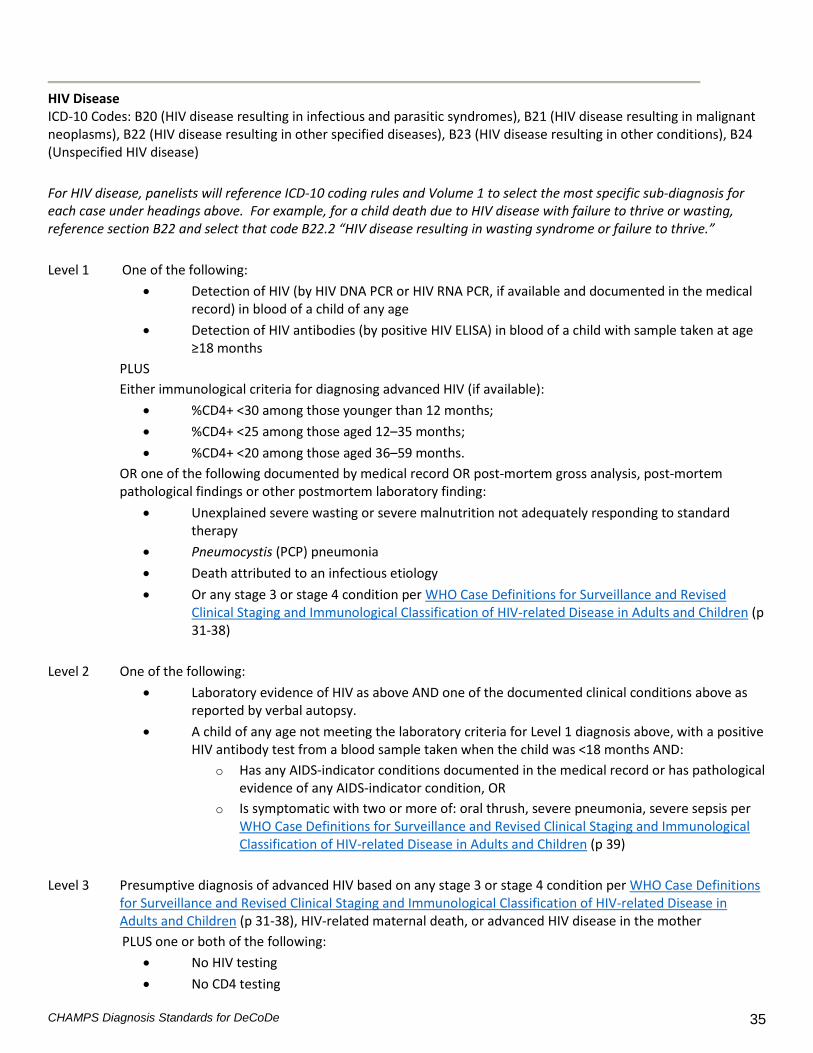
CHAMPS Diagnosis Standards for DeCoDe 35
HIV Disease ICD-10 Codes: B20 (HIV disease resulting in infectious and parasitic syndromes), B21 (HIV disease resulting in malignant neoplasms), B22 (HIV disease resulting in other specified diseases), B23 (HIV disease resulting in other conditions), B24 (Unspecified HIV disease) For HIV disease, panelists will reference ICD-10 coding rules and Volume 1 to select the most specific sub-diagnosis for each case under headings above. For example, for a child death due to HIV disease with failure to thrive or wasting, reference section B22 and select that code B22.2 “HIV disease resulting in wasting syndrome or failure to thrive.” Level 1 One of the following:
• Detection of HIV (by HIV DNA PCR or HIV RNA PCR, if available and documented in the medical record) in blood of a child of any age
• Detection of HIV antibodies (by positive HIV ELISA) in blood of a child with sample taken at age ≥18 months
PLUS Either immunological criteria for diagnosing advanced HIV (if available):
• %CD4+ <30 among those younger than 12 months; • %CD4+ <25 among those aged 12–35 months; • %CD4+ <20 among those aged 36–59 months.
OR one of the following documented by medical record OR post-mortem gross analysis, post-mortem pathological findings or other postmortem laboratory finding:
• Unexplained severe wasting or severe malnutrition not adequately responding to standard therapy
• Pneumocystis (PCP) pneumonia • Death attributed to an infectious etiology • Or any stage 3 or stage 4 condition per WHO Case Definitions for Surveillance and Revised
Clinical Staging and Immunological Classification of HIV-related Disease in Adults and Children (p 31-38)
Level 2 One of the following:
• Laboratory evidence of HIV as above AND one of the documented clinical conditions above as reported by verbal autopsy.
• A child of any age not meeting the laboratory criteria for Level 1 diagnosis above, with a positive HIV antibody test from a blood sample taken when the child was <18 months AND:
o Has any AIDS-indicator conditions documented in the medical record or has pathological evidence of any AIDS-indicator condition, OR
o Is symptomatic with two or more of: oral thrush, severe pneumonia, severe sepsis per WHO Case Definitions for Surveillance and Revised Clinical Staging and Immunological Classification of HIV-related Disease in Adults and Children (p 39)
Level 3 Presumptive diagnosis of advanced HIV based on any stage 3 or stage 4 condition per WHO Case Definitions
for Surveillance and Revised Clinical Staging and Immunological Classification of HIV-related Disease in Adults and Children (p 31-38), HIV-related maternal death, or advanced HIV disease in the mother
PLUS one or both of the following: • No HIV testing • No CD4 testing
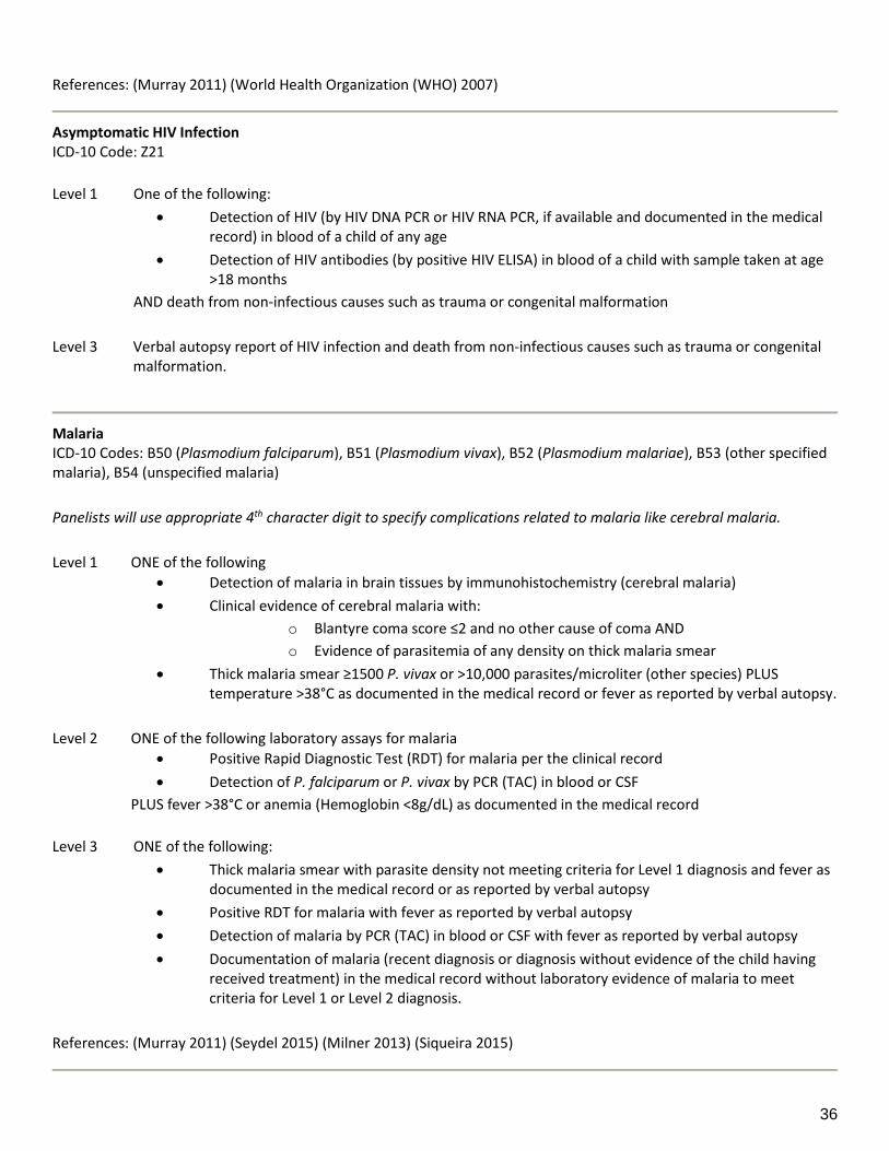
36
References: (Murray 2011) (World Health Organization (WHO) 2007)
Asymptomatic HIV Infection ICD-10 Code: Z21 Level 1 One of the following:
• Detection of HIV (by HIV DNA PCR or HIV RNA PCR, if available and documented in the medical record) in blood of a child of any age
• Detection of HIV antibodies (by positive HIV ELISA) in blood of a child with sample taken at age >18 months
AND death from non-infectious causes such as trauma or congenital malformation Level 3 Verbal autopsy report of HIV infection and death from non-infectious causes such as trauma or congenital
malformation.
Malaria ICD-10 Codes: B50 (Plasmodium falciparum), B51 (Plasmodium vivax), B52 (Plasmodium malariae), B53 (other specified malaria), B54 (unspecified malaria) Panelists will use appropriate 4th character digit to specify complications related to malaria like cerebral malaria. Level 1 ONE of the following
• Detection of malaria in brain tissues by immunohistochemistry (cerebral malaria) • Clinical evidence of cerebral malaria with:
o Blantyre coma score ≤2 and no other cause of coma AND o Evidence of parasitemia of any density on thick malaria smear
• Thick malaria smear ≥1500 P. vivax or >10,000 parasites/microliter (other species) PLUS temperature >38°C as documented in the medical record or fever as reported by verbal autopsy.
Level 2 ONE of the following laboratory assays for malaria
• Positive Rapid Diagnostic Test (RDT) for malaria per the clinical record • Detection of P. falciparum or P. vivax by PCR (TAC) in blood or CSF
PLUS fever >38°C or anemia (Hemoglobin <8g/dL) as documented in the medical record Level 3 ONE of the following:
• Thick malaria smear with parasite density not meeting criteria for Level 1 diagnosis and fever as documented in the medical record or as reported by verbal autopsy
• Positive RDT for malaria with fever as reported by verbal autopsy • Detection of malaria by PCR (TAC) in blood or CSF with fever as reported by verbal autopsy • Documentation of malaria (recent diagnosis or diagnosis without evidence of the child having
received treatment) in the medical record without laboratory evidence of malaria to meet criteria for Level 1 or Level 2 diagnosis.
References: (Murray 2011) (Seydel 2015) (Milner 2013) (Siqueira 2015)
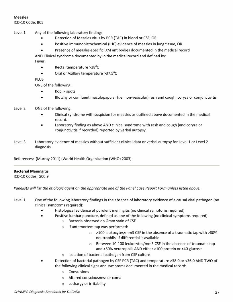
CHAMPS Diagnosis Standards for DeCoDe 37
Measles ICD-10 Code: B05 Level 1 Any of the following laboratory findings
• Detection of Measles virus by PCR (TAC) in blood or CSF, OR • Positive Immunohistochemical (IHC) evidence of measles in lung tissue, OR • Presence of measles-specific IgM antibodies documented in the medical record
AND Clinical syndrome documented by in the medical record and defined by: Fever:
• Rectal temperature >380C • Oral or Axillary temperature >37.50C
PLUS ONE of the following:
• Koplik spots • Blotchy or confluent maculopapular (i.e. non-vesicular) rash and cough, coryza or conjunctivitis
Level 2 ONE of the following:
• Clinical syndrome with suspicion for measles as outlined above documented in the medical record.
• Laboratory finding as above AND clinical syndrome with rash and cough (and coryza or conjunctivitis if recorded) reported by verbal autopsy.
Level 3 Laboratory evidence of measles without sufficient clinical data or verbal autopsy for Level 1 or Level 2
diagnosis. References: (Murray 2011) (World Health Organization (WHO) 2003)
Bacterial Meningitis ICD-10 Codes: G00.9 Panelists will list the etiologic agent on the appropriate line of the Panel Case Report Form unless listed above. Level 1 One of the following laboratory findings in the absence of laboratory evidence of a causal viral pathogen (no
clinical symptoms required): • Histological evidence of purulent meningitis (no clinical symptoms required) • Positive lumbar puncture, defined as one of the following (no clinical symptoms required)
o Bacteria observed on Gram stain of CSF o If antemortem tap was performed:
o >100 leukocytes/mm3 CSF in the absence of a traumatic tap with >80% neutrophils, if differential is available
o Between 10-100 leukocytes/mm3 CSF in the absence of traumatic tap and >80% neutrophils AND either >100 protein or <40 glucose
o Isolation of bacterial pathogen from CSF culture • Detection of bacterial pathogen by CSF PCR (TAC) and temperature >38.0 or <36.0 AND TWO of
the following clinical signs and symptoms documented in the medical record: o Convulsions o Altered consciousness or coma o Lethargy or irritability
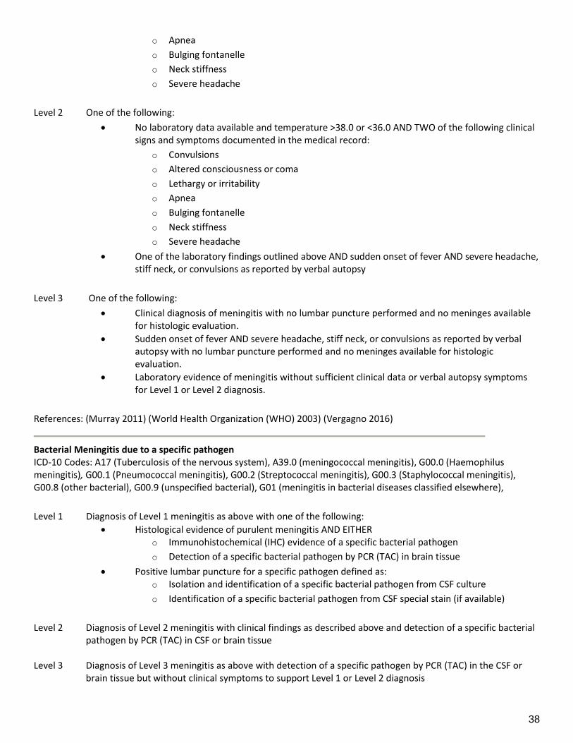
38
o Apnea o Bulging fontanelle o Neck stiffness o Severe headache
Level 2 One of the following:
• No laboratory data available and temperature >38.0 or <36.0 AND TWO of the following clinical signs and symptoms documented in the medical record:
o Convulsions o Altered consciousness or coma o Lethargy or irritability o Apnea o Bulging fontanelle o Neck stiffness o Severe headache
• One of the laboratory findings outlined above AND sudden onset of fever AND severe headache, stiff neck, or convulsions as reported by verbal autopsy
Level 3 One of the following:
• Clinical diagnosis of meningitis with no lumbar puncture performed and no meninges available for histologic evaluation.
• Sudden onset of fever AND severe headache, stiff neck, or convulsions as reported by verbal autopsy with no lumbar puncture performed and no meninges available for histologic evaluation.
• Laboratory evidence of meningitis without sufficient clinical data or verbal autopsy symptoms for Level 1 or Level 2 diagnosis.
References: (Murray 2011) (World Health Organization (WHO) 2003) (Vergagno 2016)
Bacterial Meningitis due to a specific pathogen ICD-10 Codes: A17 (Tuberculosis of the nervous system), A39.0 (meningococcal meningitis), G00.0 (Haemophilus meningitis), G00.1 (Pneumococcal meningitis), G00.2 (Streptococcal meningitis), G00.3 (Staphylococcal meningitis), G00.8 (other bacterial), G00.9 (unspecified bacterial), G01 (meningitis in bacterial diseases classified elsewhere), Level 1 Diagnosis of Level 1 meningitis as above with one of the following:
• Histological evidence of purulent meningitis AND EITHER o Immunohistochemical (IHC) evidence of a specific bacterial pathogen o Detection of a specific bacterial pathogen by PCR (TAC) in brain tissue
• Positive lumbar puncture for a specific pathogen defined as: o Isolation and identification of a specific bacterial pathogen from CSF culture o Identification of a specific bacterial pathogen from CSF special stain (if available)
Level 2 Diagnosis of Level 2 meningitis with clinical findings as described above and detection of a specific bacterial
pathogen by PCR (TAC) in CSF or brain tissue Level 3 Diagnosis of Level 3 meningitis as above with detection of a specific pathogen by PCR (TAC) in the CSF or
brain tissue but without clinical symptoms to support Level 1 or Level 2 diagnosis
CHAMPS Diagnosis Standards for DeCoDe 39
Viral Meningitis or Encephalitis ICD-10 Codes: A83 (Mosquito-borne viral encephalitis), A84 (Tick-borne viral encephalitis), A85 (other viral encephalitis), A86 (unspecified viral encephalitis), A87.0 (Enteroviral meningitis), A87.1 (Adenoviral meningitis), A87.2 (Lymphocytic choriomeningitis), A87.8 (other viral meningitis), A87.9 (unspecified viral meningitis), G02.0 (meningitis in viral diseases classified elsewhere) Panelists will reference ICD-10 and use the most specific etiologic agent for each case. If a specific code for that etiology does not exist, list the etiologic agent on the appropriate line on the Panel Case Report Form. Level 1 One of the following laboratory findings:
• Lumbar puncture consistent with viral etiology, defined as all of the following: o >10 leukocytes/ mm3 and >50% lymphocytes in the absence of a traumatic tap o No bacteria observed on Gram stain of CSF o No bacteria recovered from CSF culture (if performed) o Negative latex agglutination test of CSF (if performed)
• Detection of a virus likely to cause encephalitis in CSF by PCR (TAC) • CSF PCR positive for organism likely to cause encephalitis
AND one of the following medically documented clinical signs: • Convulsions • Headache • Neck stiffness • Confusion or altered mental status • Vomiting • Bulging fontanelle • Cranial nerve palsies
Level 2 One of the laboratory findings outlined above AND TWO or more of the clinical signs above (convulsions,
headache, neck stiffness, confusion, or vomiting) as reported by verbal autopsy. Level 3 Laboratory data, medical records or verbal autopsy supporting a diagnosis of viral meningitis or encephalitis
but not meeting the criteria for Level 1 or Level 2 above. References: (Murray 2011) (Vergagno 2016)
Pertussis ICD-10 Code: A37 Level 1 Immunohistochemical (IHC) evidence of Bordetella pertussis in the lung tissue OR both of the following:
• Isolation of B. pertussis from any respiratory culture documented in the medical record • A cough illness of any duration documented in the medical record
OR both of the following: • Detection of B. pertussis by PCR (TAC) from lung and/or nasopharyngeal/oropharyngeal (NP/OP)
swab • AND ONE or more of the following symptoms documented in the medical record
40
o Apnea (for infants <1 year, defined as cessation of breathing for >20 seconds associated with bradycardia or cyanosis)
o Paroxysms of coughing o Inspiratory whoop o Posttussive vomiting
OR both of the following: • Epidemiological link to a case confirmed by culture or PCR • AND one or more of the symptoms above documented in the medical record
Level 2 An illness meeting any of the laboratory or epidemiological criteria for diagnosis of pertussis above but with
symptoms (if required) reported by verbal autopsy. Level 3 An illness with > 2 weeks of cough suggestive of pertussis but not meeting the above criteria OR no clinical
symptom information available with isolation of B. pertussis from any respiratory culture or detection of B. pertussis by PCR (TAC) from lung and/or nasopharyngeal/oropharyngeal swab.
References: (Murray 2011) (World Health Organization (WHO) 2003) (CDC 2014)
Pneumonia ICD-10 Codes: J12.9 (viral not elsewhere classified), J15.9 (unspecified bacteria), J18 (unspecified organism) For pneumonia due to a specific pathogen, please reference the DS that follow. Level 1 EITHER Strong histological evidence of pyogenic pneumonia in lung tissue OR One of the following laboratory or imaging findings
• Moderate histological evidence of pneumonia in lung tissue • New infiltrate or pleural effusion on chest radiograph
AND TWO or more of the following clinical signs documented in the medical record: • Tachypnea (Per WHO Clinical Case Definitions defined as respiratory rate >60/minute in 0-2
months, >50/minute for infants 2-12 months, >40 in children 12 months -5 years) • Respiratory distress as chest indrawing, grunting or nasal flaring • Abnormal breath sounds (i.e. decreased breath sounds, crackles, crepitations) • Hypoxia, cyanosis or desaturations (oxygen saturation <95%) • Fever >38.0 or hypothermia <36.0
Level 2 One of the following:
• No laboratory or imaging data available and ALL of the following documented in the medical record: fever or hypothermia, hypoxia or abnormal breath sounds, and tachypnea or respiratory distress.
• One of the laboratory or imaging findings above with TWO or more of clinical signs of pneumonia above reported by verbal autopsy (difficulty breathing, fast breathing or breathlessness, lower chest wall/ribs being pulled in or grunting, or fever).
Level 3 Acute febrile illness or hypothermia with tachypnea, respiratory distress, abnormal breath sounds, hypoxia or cyanosis documented in the medical record or reported by verbal autopsy, but not meeting the criteria for Level 1 or Level 2 diagnosis above OR laboratory evidence of pneumonia but not meeting the criteria for Level 1 or Level 2 diagnosis above.
CHAMPS Diagnosis Standards for DeCoDe 41
References: (Scott 2012) (Rambaud-Althaus 2015) (McIntosh 2002) (Beard 2015) (Murray 2011) (WHO Pneumonia Vaccine Trial Investigators' Group 2001) (WHO and UNICEF 2013) (WHO 1995) (WHO 2000)
Pneumonia due to Streptococcus pneumoniae ICD-10 Code: J13 Level 1 One of the following:
• Strong histological evidence of pneumonia and detection of S. pneumoniae in lung tissue • Moderate histological evidence of pneumonia and detection of S. pneumoniae in lung tissue
with TWO of the clinical criteria for diagnosis of pneumonia as above, documented in medical record or reported by verbal autopsy.
• Pneumonia meeting TWO of the clinical criteria for diagnosis of pneumonia as above, as documented in the medical record, with one of the following:
o Isolation of S. pneumoniae from blood culture with inadequate postmortem lung tissue o Detection of S. pneumoniae in blood by PCR (TAC) with inadequate postmortem lung
tissue o Detection of S. pneumoniae by PCR (TAC) in lung tissue
Level 2 EITHER Moderate histological evidence of pneumonia and detection of S. pneumoniae in lung tissue, OR Pneumonia meeting the clinical criteria for diagnosis of Level 2 pneumonia above, as reported by verbal
autopsy, with one of the following: • Isolation of S. pneumoniae from blood culture with inadequate postmortem lung tissue • Detection of S. pneumoniae in blood by PCR (TAC) with inadequate postmortem lung tissue • Detection of S. pneumoniae in lung tissue by PCR (TAC)
Level 3 Detection of S. pneumoniae in lung tissue or detection of S. pneumoniae in the blood in a patient with a
primary respiratory illness but in the absence of sufficient clinical information to meet criteria for Level 1 or Level 2 diagnosis.
Pneumonia due to Haemophilus influenzae ICD-10 Code: J14 Level 1 One of the following:
• Strong histological evidence of pneumonia and detection of H. influenzae in lung tissue • Histological evidence of pneumonia and detection of H. influenzae in lung tissue with TWO of
the clinical criteria for diagnosis of pneumonia as above, documented in medical record or reported by verbal autopsy.
• Pneumonia meeting TWO of the clinical criteria for diagnosis of pneumonia as above, as documented in the medical record, with one of the following:
o Isolation of H. influenzae from blood culture with inadequate postmortem lung tissue o Detection of H. influenzae in blood by PCR (TAC) with inadequate postmortem lung
tissue o Detection of H. influenzae in lung tissue by PCR (TAC)
Level 2 EITHER Moderate histological evidence of pneumonia and detection of H. influenzae in lung tissue, OR Pneumonia meeting the clinical criteria for diagnosis of Level 2 pneumonia as above, as reported by
verbal autopsy, with one of the following: • Isolation of H. influenzae from blood culture with inadequate postmortem lung tissue • Detection of H. influenzae in blood by PCR (TAC) with inadequate postmortem lung tissue

42
• Detection of H. influenzae in lung tissue by PCR (TAC) Level 3 Detection of H. influenzae in lung tissue or detection of H. influenzae in the blood in a patient with a primary
respiratory illness but in the absence of sufficient clinical information to meet criteria for Level 1 or Level 2 diagnosis.
Pneumonia due to Klebsiella pneumoniae ICD-10 Code: J15.0 Level 1 One of the following:
• Strong histological evidence of pneumonia and detection of K. pneumoniae in lung tissue • Histological evidence of pneumonia and detection of K. pneumoniae in lung tissue with TWO of
the clinical criteria for diagnosis of pneumonia as above, documented in medical record or reported by verbal autopsy.
• Pneumonia meeting TWO of the clinical criteria for diagnosis of pneumonia as above, as documented in the medical record, with one of the following:
o Isolation of K. pneumoniae from blood culture with inadequate postmortem lung tissue o Detection of K. pneumoniae in blood by PCR (TAC) with inadequate postmortem lung
tissue o Detection of K. pneumoniae in lung tissue by PCR (TAC)
Level 2 EITHER Moderate histological evidence of pneumonia and detection of K. pneumoniae in lung tissue, OR Pneumonia meeting TWO of the clinical criteria for diagnosis of Level 2 pneumonia as above, as reported
by verbal autopsy, with one of the following: • Isolation of K. pneumoniae from blood culture with inadequate postmortem lung tissue • Detection of K. pneumoniae in blood by PCR (TAC) with inadequate postmortem lung tissue • Detection of K. pneumoniae in lung tissue by PCR (TAC
Level 3 Detection of K. pneumoniae in lung tissue or detection of K. pneumoniae in the blood in a patient with a
primary respiratory illness but in the absence of sufficient clinical information to meet criteria for Level 1 or Level 2 diagnosis.
Pneumonia due to Pseudomonas aeruginosa ICD-10 Code: J15.1 Level 1 One of the following:
• Strong histological evidence of pneumonia and detection of P. aeruginosa in lung tissue • Histological evidence of pneumonia and detection of P. aeruginosa in lung tissue with TWO of
the clinical criteria for diagnosis of pneumonia as above, documented in medical record or reported by verbal autopsy.
• Pneumonia meeting TWO of the clinical criteria for diagnosis of pneumonia as above, as documented in the medical record, with one of the following:
o Isolation of P. aeruginosa from blood culture with inadequate postmortem lung tissue o Detection of P. aeruginosa in blood by PCR (TAC) with inadequate postmortem lung
tissue o Detection of P. aeruginosa in lung tissue by PCR (TAC)
Level 2 EITHER Moderate histological evidence of pneumonia and detection of P. aeruginosa in lung tissue OR Pneumonia meeting TWO of the clinical criteria for diagnosis of Level 2 pneumonia above, as reported by
verbal autopsy, with one of the following:
CHAMPS Diagnosis Standards for DeCoDe 43
• Isolation of P. aeruginosa from blood culture with inadequate postmortem lung tissue • Detection of P. aeruginosa in blood by PCR (TAC) with inadequate postmortem lung tissue • Detection of P. aeruginosa in lung tissue by PCR (TAC)
Level 3 Detection P. aeruginosa in lung tissue or detection of P. aeruginosa in the blood in a patient with a primary
respiratory illness but in the absence of sufficient clinical information to meet criteria for Level 1 or Level 2 diagnosis.
Pneumonia due to Staphylococcus aureus ICD-10 Code: J15.2 Level 1 One of the following:
• Strong histological evidence of pneumonia and detection of P. aeruginosa in lung tissue • Histological evidence of pneumonia and detection of S. aureus in lung tissue with TWO of the
clinical criteria for diagnosis of pneumonia as above, documented in medical record or reported by verbal autopsy.
• Pneumonia meeting TWO of the clinical criteria for diagnosis of pneumonia as above, as documented in the medical record, with one of the following:
o Isolation of S. aureus from blood culture with inadequate postmortem lung tissue o Detection of S. aureus in blood by PCR (TAC) with inadequate postmortem lung tissue o Detection of S. aureus in lung tissue by PCR (TAC)
Level 2 EITHER Moderate histological evidence of pneumonia and detection of S. aureus in lung tissue OR Pneumonia meeting TWO of the clinical criteria for diagnosis of Level 2 pneumonia above, as reported by
verbal autopsy, with one of the following: • Isolation of S. aureus from blood culture with inadequate postmortem lung tissue • Detection of S. aureus in blood by PCR (TAC) with inadequate postmortem lung tissue • Detection of S. aureus in lung tissue by PCR (TAC)
Pneumonia due to Streptococcus, Group B ICD-10 Code: J15.3 Level 1 One of the following:
• Strong histological evidence of pneumonia and Group B Streptococcus in lung tissue • Histological evidence of pneumonia and Group B Streptococcus in lung tissue with TWO of the
clinical criteria for diagnosis of pneumonia as above, documented in medical record or reported by verbal autopsy.
• Pneumonia meeting TWO of the clinical criteria for diagnosis of pneumonia as above, as documented in the medical record, with one of the following:
o Isolation of Group B Streptococcus from blood culture with inadequate postmortem lung tissue
o Detection of Group B Streptococcus in blood by PCR (TAC) with inadequate postmortem lung tissue
o Detection of Group B Streptococcus in lung tissue by PCR (TAC) Level 2 EITHER Moderate histological evidence of pneumonia and detection of Group B Streptococcus in lung tissue OR Pneumonia meeting TWO of the clinical criteria for diagnosis of Level 2 pneumonia above, as reported by
verbal autopsy, with one of the following:
44
• Isolation of Group B Streptococcus from blood culture with inadequate postmortem lung tissue • Detection of Group B Streptococcus in blood by PCR (TAC) with inadequate postmortem lung
tissue • Detection of Group B Streptococcus in lung tissue by PCR (TAC)
Pneumonia Due to other streptococci (Not Group B) ICD-10 Code: J15.4 Level 1 One of the following:
• Strong histological evidence of pneumonia and detection of Group A or D Streptococcus in lung tissue
• Histological evidence of pneumonia and detection of Group A or D Streptococcus in lung tissue with TWO of the clinical criteria for diagnosis of pneumonia as above, documented in medical record or reported by verbal autopsy.
• Pneumonia meeting TWO of the clinical criteria for diagnosis of pneumonia as above, as documented in the medical record, with one of the following:
o Isolation of Group A or D Streptococcus from blood culture with inadequate postmortem lung tissue
o Detection of Group A Streptococcus in blood by PCR (TAC) with inadequate postmortem lung tissue
o Detection of Group A Streptococcus in lung tissue by PCR (TAC) Level 2 EITHER Moderate histological evidence of pneumonia and detection of Group A or D Streptococcus in lung
tissue OR Pneumonia meeting TWO of the clinical criteria for diagnosis of Level 2 pneumonia as above, as reported
by verbal autopsy, with one of the following: • Isolation of Group A or D Streptococcus from blood culture with inadequate postmortem lung
tissue • Detection of Group A Streptococcus in blood by PCR (TAC) with inadequate postmortem lung
tissue • Detection of Group A Streptococcus in lung tissue by PCR (TAC)
Pneumonia due to Escherichia coli ICD-10 Code: J15.5 Level 1 One of the following:
• Strong histological evidence of pneumonia and detection of E. coli in lung tissue • Histological evidence of pneumonia and detection of E. coli in lung tissue with TWO of the
clinical criteria for diagnosis of pneumonia as above, documented in medical record or reported by verbal autopsy.
• Pneumonia meeting TWO of the clinical criteria for diagnosis of pneumonia as above, as documented in the medical record, with one of the following:
o Isolation of E. coli from blood culture with no other pathogen detected and with inadequate postmortem lung tissue
o Detection of E. coli in blood by PCR (TAC) with no other pathogen detected and with inadequate postmortem lung tissue
o Detection of E. coli in lung tissue by PCR (TAC)

CHAMPS Diagnosis Standards for DeCoDe 45
Level 2 EITHER Moderate histological evidence of pneumonia and detection of E. coli in lung tissue OR Pneumonia meeting TWO of the clinical criteria for diagnosis of pneumonia as above, as reported by
verbal autopsy, with one of the following: • Isolation of E. coli from blood culture with no other pathogen detected and with inadequate
postmortem lung tissue • Detection of E. coli in blood by PCR (TAC) with no other pathogen detected and with inadequate
postmortem lung tissue • Detection of E. coli in lung tissue by PCR (TAC)
Pneumonia due to other aerobic gram negative bacteria ICD-10 Code: J15.6 Diagnosis of Level 1 or Level 2 Pneumonia (per the general pneumonia definition above) with one of the following:
• Immunohistochemical (IHC) evidence of a specific aerobic gram negative organism not listed above in lung tissue
• Detection of a specific aerobic gram negative organism not listed above in lung tissue by PCR (TAC) with histological evidence of pneumonia in postmortem biopsy
Pneumonia due to Mycoplasma pneumoniae ICD-10 Code: J15.7 Level 1 One of the following:
• Strong histological evidence of pneumonia and detection of M. pneumoniae in lung tissue • Histological evidence of pneumonia and detection of M. pneumoniae in lung tissue with TWO of
the clinical criteria for diagnosis of pneumonia as above, documented in medical record or reported by verbal autopsy.
• Pneumonia meeting TWO of the clinical criteria for diagnosis of pneumonia as above, as documented in the medical record, with one of the following:
o Detection of M. pneumoniae in NP/OP swab by PCR (TAC) o Detection of M. pneumoniae in lung tissue by PCR (TAC)
Level 2 EITHER Moderate histological evidence of pneumonia and detection of M. pneumoniae in lung tissue OR Pneumonia meeting TWO of the clinical criteria for diagnosis of pneumonia as above, as reported by
verbal autopsy, with one of the following: • Detection of M. pneumoniae in NP/OP swab by PCR (TAC) • Detection of M. pneumoniae in lung tissue by PCR (TAC)
Level 3 Detection of M. pneumoniae in the lung tissue or NP/OP swab in the absence of sufficient clinical criteria to
meet criteria for Level 1 or Level 2 diagnosis.
Pneumonia due to other specified bacteria ICD-10 Code: J15.8 Diagnosis of Level 1 or Level 2 Pneumonia as noted above with one of the following:
• Immunohistochemical (IHC) evidence of a specific bacteria not listed above in lung tissue • Detection of a specific bacteria not listed above by TAC (PCR) with histological evidence of
pneumonia in postmortem biopsy

46
Influenza with pneumonia ICD-10 Code: J10.0 Level 1 One of the following:
• Strong histological evidence of pneumonia and detection of influenza virus in lung tissue • Histological evidence of pneumonia and detection of influenza virus in lung tissue with TWO of
the clinical criteria for diagnosis of pneumonia above, documented in medical record or reported by verbal autopsy.
• Pneumonia meeting TWO of the clinical criteria for diagnosis of pneumonia above, as documented in the medical record, with detection of influenza virus in lung tissue by PCR (TAC)
Level 2 EITHER Moderate histological evidence of pneumonia and detection of influenza virus in lung tissue OR Pneumonia meeting TWO of the clinical criteria for diagnosis of Level 2 pneumonia above, as reported
by verbal autopsy, with detection of influenza virus in lung tissue by PCR (TAC). Level 3 Acute febrile illness or hypothermia with tachypnea, respiratory distress, abnormal breath sounds, hypoxia
or cyanosis documented in the medical record or reported by verbal autopsy, and detection of influenza virus in NP/OP swab by PCR (TAC).
Influenza with other respiratory manifestations (not pneumonia) ICD-10 Code: J10.1 Level 1 Detection of influenza virus in NP/OP swab by PCR (TAC) AND fever plus cough or sore throat documented in
medical record, without signs and symptoms of pneumonia. Level 2 Detection of influenza virus in NP/OP swab by PCR (TAC) AND fever plus cough or sore throat as reported by
verbal autopsy, without signs and symptoms of pneumonia.
Pneumonia due to Adenovirus ICD-10 Code: J12.0 Level 1 One of the following:
• Strong histological evidence of viral pneumonia and detection of adenovirus virus in lung tissue by PCR (TAC)
• Histological evidence of pneumonia and detection of adenovirus virus in lung tissue by PCR (TAC) with TWO of the clinical criteria for diagnosis of pneumonia as above, documented in medical record or reported by verbal autopsy.
• Pneumonia meeting TWO of the clinical criteria for diagnosis of pneumonia as above, as documented in the medical record, with detection of adenovirus in lung tissue by PCR (TAC)
Level 2 Pneumonia meeting TWO of the clinical criteria for diagnosis of Level 2 pneumonia above, as reported by
verbal autopsy, with detection of adenovirus in lung tissue by PCR (TAC). Level 3 One of the following:
• Acute febrile illness or hypothermia with tachypnea, respiratory distress, abnormal breath sounds, hypoxia or cyanosis documented in the medical record or reported by verbal autopsy, and detection of adenovirus in NP/OP swab by PCR (TAC).
• Detection of adenovirus in the lung tissue in the absence of sufficient clinical information for Level 1 or Level 2 diagnosis.

CHAMPS Diagnosis Standards for DeCoDe 47
Pneumonia due to Respiratory Syncytial Virus (RSV) ICD-10 Code: J15.1 Level 1 One of the following:
• Strong histological evidence of viral pneumonia and detection of RSV in lung tissue by PCR (TAC) or immunohistochemistry (IHC).
• Histological evidence of pneumonia and detection of RSV in lung tissue by PCR (TAC) or immunohistochemistry (IHC) with TWO of the clinical criteria for diagnosis of pneumonia as above, documented in medical record or reported by verbal autopsy.
• Pneumonia meeting TWO of the clinical criteria for diagnosis of pneumonia as above, as documented in the medical record, with detection of RSV in lung tissue by PCR (TAC)
Level 2 Pneumonia meeting TWO of the clinical criteria for diagnosis of Level 2 pneumonia above, as reported by
verbal autopsy, with detection of RSV in lung tissue by PCR (TAC). Level 3 One of the following:
• Acute febrile illness or hypothermia with tachypnea, respiratory distress, abnormal breath sounds, hypoxia or cyanosis documented in the medical record or reported by verbal autopsy, and detection of RSV in NP/OP swab by PCR (TAC).
• Detection of RSV in the lung tissue by PCR (TAC) in the absence of sufficient information for Level 1 or Level 2 diagnosis.
Pneumonia due to Parainfluenza ICD-10 Code: J15.2 Level 1 One of the following:
• Strong histological evidence of viral pneumonia and detection of parainfluenza virus in lung tissue by PCR (TAC)
• Histological evidence of pneumonia and detection of parainfluenza virus in lung tissue by PCR (TAC) with TWO of the clinical criteria for diagnosis of pneumonia as above, documented in medical record or reported by verbal autopsy.
• Pneumonia meeting TWO of the clinical criteria for diagnosis of pneumonia as above, as documented in the medical record, with detection of parainfluenza virus in lung tissue by PCR (TAC).
Level 2 Pneumonia meeting TWO of the clinical criteria for diagnosis of Level 2 pneumonia above, as reported by
verbal autopsy, with detection of parainfluenza virus in lung tissue by PCR (TAC). Level 3 One of the following:
• Acute febrile illness or hypothermia with tachypnea, respiratory distress, abnormal breath sounds, hypoxia or cyanosis documented in the medical record or reported by verbal autopsy, and detection of parainfluenza virus in NP/OP swab by PCR (TAC).
• Detection of parainfluenza virus in the lung tissue by PCR (TAC) in the absence of sufficient clinical information for Level 1 or Level 2 diagnosis.
Pneumonia due to Human Metapneumovirus (HMPV) ICD-10: J15.3 Level 1 One of the following:
• Strong histological evidence of viral pneumonia and detection of HMPV in lung tissue by PCR
48
(TAC) or immunohistochemistry (IHC). • Histological evidence of pneumonia and detection of HMPV in lung tissue by PCR (TAC) with
TWO of the clinical criteria for diagnosis of pneumonia as above, documented in medical record or reported by verbal autopsy.
• Pneumonia meeting TWO of the clinical criteria for diagnosis of pneumonia as above, as documented in the medical record, with detection of HMPV in lung tissue by PCR (TAC).
Level 2 Pneumonia meeting TWO of the clinical criteria for diagnosis of Level 2 pneumonia above, as reported by
verbal autopsy, with detection of HMPV in lung tissue by PCR (TAC). Level 3 One of the following:
• Acute febrile illness or hypothermia with tachypnea, respiratory distress, abnormal breath sounds, hypoxia or cyanosis documented in the medical record or reported by verbal autopsy, and detection of HMPV in NP/OP swab by PCR (TAC).
• Detection of HMPV in the lung tissue by PCR (TAC) in the absence of sufficient clinical information for Level 1 or Level 2 diagnosis.
Pneumonia due to Chlamydia ICD-10 Code: J16.0 Level 1 One of the following:
• Strong Histological evidence of pneumonia and detection of C. pneumoniae (at all ages) or C. trachomatis (infants <6 months) in lung tissue by PCR (TAC)
• Histological evidence of pneumonia and detection of C. pneumoniae (at all ages) or C. trachomatis (infants <6 months) in lung tissue by PCR (TAC) with TWO of the clinical criteria for diagnosis of pneumonia as above, documented in medical record or reported by verbal autopsy.
• Pneumonia meeting TWO of the clinical criteria for diagnosis of pneumonia as above, as documented in the medical record, with detection of C. pneumoniae (at all ages) or C. trachomatis (infants <6 months) in lung tissue by PCR (TAC).
Level 2 Pneumonia meeting TWO of the clinical criteria for diagnosis of Level 2 pneumonia above, as reported by
verbal autopsy, with detection of C. pneumoniae (at all ages) or C. trachomatis (infants <6 months) in lung tissue by PCR (TAC).
Level 3 One of the following:
• Acute febrile illness or hypothermia with tachypnea, respiratory distress, abnormal breath sounds, hypoxia or cyanosis documented in the medical record or reported by verbal autopsy, with detection of C. pneumoniae (at all ages) or C. trachomatis (infants <6 months) in NP/OP swab by PCR (TAC).
• Detection of C. pneumoniae (at all ages) or C. trachomatis (infants <6 months) in lung tissue by PCR (TAC) in the absence of sufficient clinical information for Level 1 or Level 2 diagnosis.
Pneumonia due to Pneumocystis (PCP) ICD-10 Code: B59 AND J17.3 Level 1: Either of the following findings in an immunocompromised, severely malnourished, or HIV+ patient:
• Immunohistochemical (IHC) evidence of PCP in lung tissue • Detection of PCP in lung tissue by PCR (TAC) with histological evidence of pneumonia in
postmortem biopsy Level 3: Either of the following findings in an immunocompromised, severely malnourished, or HIV+ patient:
CHAMPS Diagnosis Standards for DeCoDe 49
• Chest radiograph with “ground glass” infiltrates without known alternative etiology and without sufficient lung tissue for analysis
• Detection of PCP by PCR in lung tissue
Pneumonia due to other infectious organism, not elsewhere classified ICD-10 Code: J15 Diagnosis of Level 1 or Level 2 Pneumonia as noted above with one of the following:
• Immunohistochemical (IHC) evidence of non-bacterial pathogen not listed above in lung tissue • Detection of a non-bacterial pathogen in lung tissue by PCR (TAC) with histological evidence of
pneumonia in postmortem biopsy
Sepsis ICD-10 Codes: A41.9 (unspecified) This DS should be used for sepsis in in children 28 days or older. The DS for Bacterial Sepsis in a Newborn should be used for infants <28 days. For specific etiologies, please see the following DS. Level 1 Infection suggested by histological evidence of pyogenic infection in 2 or more tissues PLUS THREE or more
of the following clinical signs or clinical laboratory findings (if available) documented in the medical record: • Temperature >38oC or <360C • Tachycardia or new episodes of bradycardia • Altered mental status, abnormally sleepy, difficult to wake, lethargic or reduced or no
spontaneous movement, irritable, or agitated • Unconscious • Absent or weak cry, weak suck, or difficulty in feeding • New or increased episodes of apnea, tachypnea, or increased requirement for ventilator support
(if available) • Mottled, pale, cyanotic, delayed capillary refill, diminished pulses, cool extremities or
hypotension • Elevated C-reactive Protein (CRP) • Metabolic acidosis (pH <7.35 and base deficit >4.0mmoL/L) • Increased White Blood Cell (WBC) count for age (based on Table 14.1 in the Harriet Lane
Handbook) Level 2 One of the following:
• No Level 1 laboratory tests available AND THREE or more clinical signs of sepsis as above documented in the medical record
• One or more of the laboratory findings outlined for Level 1 diagnosis of sepsis above AND THREE or more clinical signs of sepsis above reported by verbal autopsy
Level 3 Cases that meet the Level 1 or Level 2 clinical criteria of sepsis above, with suspected infection and
symptoms not more likely attributable to another condition, but without sufficient laboratory findings for Level 1 or Level 2 diagnosis, OR cases with laboratory evidence of sepsis that is not attributable to perimortem overgrowth or contamination but that does not meeting criteria for Level 1 or Level 2 diagnosis above.
References: (Murray 2011) (Vergagno 2016) (Randolph 2014) (Goldstein 2005) (Flerlage 2015) (Weinberg 2016)
50
Sepsis due to a specific pathogen ICD-10 Codes: A02.1 (Salmonella sepsis), A22.7 (Anthrax sepsis), A32.7 (Listerial sepsis), A40.0 (Streptococcus, Group A), A40.1 (Streptococcus, Group B), A40.2 (Streptococcus, Group D), A40.3 (Streptococcus pneumoniae), A40.8 (other streptococcal species), A40.9 (Streptococcal species, unspecified), A41.0 (Staphylococcus aureus), A41.1 (other specified Staphylococcus), A41.2 (unspecified Staphylococcus aureus), A41.3 (Haemophilus influenzae), A41.4 (anaerobes), A41.5 (other Gram-negative organisms), A41.8 (other specified sepsis), B37.7 (Candidal sepsis) This DS should be used for sepsis in in children 28 days or older. The DS for Bacterial Sepsis in a Newborn should be used for infants <28 days. Panelists will reference ICD-10 for the most specific code relevant for each case. If the etiologic agent is not listed above, panelists will list it on the appropriate line of the Panel Case Report Form. Level 1 Strong pathological evidence of pyogenic infection in 2 or more tissues with isolation of an organism by
culture or immunohistochemical (IHC) evidence of an organism from one or more tissues OR
Detection of a single or at most two pathogens by PCR (TAC) in blood or CSF or isolation of a single or at most two pathogens in two or more normally sterile body sites by culture, and judged not to be post-mortem contamination based on time to culture positivity, corroborating molecular or immunohistochemical data; PLUS THREE or more of the clinical signs of sepsis or clinical laboratory findings of sepsis noted above documented in the medical record.
Level 2 One of the following:
• Moderate pathological evidence of sepsis in 2 or more tissues with isolation or detection of an organism consistent with the infection from one or more tissues
• Isolation of an organism by culture in 2 or more tissues • Isolation of an organism by culture from one tissue and detection of the organism by PCR in one
or more different tissues Level 3 Cases with laboratory evidence of sepsis due to a specific pathogen that is not attributable to perimortem
overgrowth or contamination but that does not meeting criteria for Level 1 or Level 2 diagnosis above.
Tuberculosis ICD-10 Codes: A15-A19 Tuberculosis codes are broken down in ICD-10 by system affected and method of confirmation. Please reference Vol 1 of ICD-10 in choosing the appropriate code. Level 1 ONE of the following laboratory findings:
• Histologic evidence of Mycobacterium tuberculosis • Isolation of M. tuberculosis by culture of any specimen
Level 2 ONE of the following laboratory findings:
• Detection of M. tuberculosis by PCR (Xpert, MTB/RIF) in the lung, gastric aspirate, induced sputum, or NP/OP aspirate, or stool
• Detection of M. tuberculosis by PCR (TAC) in lung tissue, NP/OP swab, or CSF • M. tuberculosis observed on special stain (e.g. Ziehl-Nielson method) or fluorescence
microscopy of a specimen (e.g. sputum, induced sputum, gastric aspirate, CSF, nasopharyngeal aspirate, pleural fluid, ascitic fluid)
Level 3 Persistent unexplained cough, fever, or failure to thrive (documented crossing of percentile lines on growth
chart in the preceding 3 months or moderate or severe wasting) not responding to therapy for alternative

CHAMPS Diagnosis Standards for DeCoDe 51
causes AND a known close contact with TB disease in the last year reported by verbal autopsy, but without the above laboratory evidence necessary for Level 1 or Level 2 diagnosis.
References: (Murray 2011) (Graham 2015)
V. Malnutrition The interpretation of the measurements taken during the MITS procedure will be automated and the z scores will be presented to the panelists. For special circumstances for which specific growth charts exist – i.e. prematurity, Trisomy 21, Turner Syndrome, Cerebral Palsy – panelists should reference the appropriate chart. Kwashiorkor ICD-10 Codes: E40 Level 1 Clinical diagnosis of kwashiorkor (malnutrition with edema) prior to death with histologic evidence of fatty
infiltration of the liver (macrocytic steatosis, especially in a periportal distribution) or documented low albumin in the course of clinical care prior to CHAMPs intervention
Level 2 Clinical diagnosis of kwashiorkor documented in the medical record prior to death OR Documentation in the medical record of edema not attributed to an infectious cause (note: a child with
kwashiorkor will usually be underweight, but the edema may mask the true weight) PLUS two or more of the following documented in the medical record
• Loss of appetite • Dyspigmentation of skin • Dyspigmentation of hair • Enlarged fatty liver • Protruding abdomen • Loss of muscle mass • Pitting edema
Level 3 Symptoms of kwashiorkor as above but with edema PLUS two additional findings reported by verbal
autopsy.
Marasmus or unspecified severe protein-energy malnutrition ICD-10 Codes: E41 and E43
These ICD-10 codes are most consistent with clinical diagnoses of marasmus and Severe Acute Malnutrition as defined by WHO and will be grouped together for CHAMPS purposes. Level 1 One or more of the following:
• Severe wasting, defined as weight-for-height z-score (WHZ) < -3 • No height measurement available and severe underweight, defined as weight-for-age z-score
(WAZ) < -3 • For ages 6-59 months only using mid-upper arm circumference (MUAC):
o Severe wasting: MUAC < 11.5 cm Level 2 Clinical diagnosis of marasmus or severe acute malnutrition documented in the medical record prior to
death.

52
Level 3 No measurement indicators available or measurement indicators do not meet Level 1 criteria PLUS one or more of the following:
• Clinical evidence of severe acute malnutrition, wasting, or weight loss • Verbal autopsy positive response to severe thinness • Photographic evidence of wasting (e.g. protruding ribs)
References: (World Health Organization 2006) (Grover 2009) (Black 2013) (Stevens 2012) (Asfaw 2015) (WHO 2017)
Moderate protein-energy malnutrition ICD-10 Code: E44.0 This code is most consistent with Moderate Acute Malnutrition or Underweight as defined by WHO. Level 1 One or more of the following:
• Moderate wasting, defined as weight-for-height z-score (WHZ) < -2 • No height measurement available and moderate underweight, defined as weight-for-age z-score
(WAZ): -3 ≤ z-score < -2 • For ages 6-59 months only using mid-upper arm circumference (MUAC):
o Moderate wasting: 11.5 cm ≤ MUAC < 12.5 cm Level 3 No measurement indicators available or measurement indicators do not meet Level 1 criteria PLUS one or more of the following:
• Clinical evidence of moderate acute malnutrition, wasting, or weight loss • Verbal autopsy positive response to weight loss
References: (World Health Organization 2006) (Grover 2009) (Black 2013) (Stevens 2012) (Asfaw 2015)
Marasmic kwashiorkor ICD-10 Code: E42 Level 1 A case which meets criteria for Level 1 diagnosis of both kwashiorkor and marasmus Level 3 Evidence of kwashiorkor and marasmus, but not meeting criteria for Level 1 diagnosis above
Stunting ICD-10 Code: E45 This code is most consistent with chronic malnutrition or stunting as defined by WHO. Level 1 Using Height-for-age z-score (HAZ):
• Moderate stunting: -3 ≤ z-score < -2 • Severe stunting: z-score < -3
References: (World Health Organization 2006) (Grover 2009)

CHAMPS Diagnosis Standards for DeCoDe 53
VI. Other Conditions Anemia ICD-10 Codes D50-D64 Panelists will use the appropriate and most specific ICD-10 code to specify the type of anemia, if known, or use D64, other anemias, if not known. Level 1 Anemia defined by hemoglobin 2 standard deviations below normal for age:
• Birth, <13.5 g/dL • 1-3 days, <14.5 g/dL • 4 days to <2 weeks, <13.5 g/dL • 2 weeks to <1 month, <12.5 g/dL • 1 month to <2 months, <10.0 g/dL • 2 months to <3 months, <9.0 g/dL • 3 months to <6 months, <9.5 g/dL • 6 months to <60 months, <11.0 g/dL • Pregnant women, <12.0 g/dL
Level 3 Documentation of anemia in the medical record but without laboratory evidence to support the diagnosis. (WHO, UNICEF, and UN University 2001) (Marks 2009)
Congestive Heart Failure ICD-10 Code: I50 Level 1 Objective findings of congestive heart failure based on imaging study (echocardiogram with decreased
function or chest radiograph with enlarged cardiac silhouette) PLUS clinical findings of congestive heart failure including TWO or more:
• Laterally displaced point of maximal impulse • S3 heart sound gallop • Cyanosis • Diffuse crackles in the lungs • Enlarged liver • Edema • Jugular venous distension
OR Pathological findings of congestive heart failure on examination of tissues, such as pulmonary edema and liver congestion, not attributable to another cause AND TWO or more clinical findings of congestive heart failure as above.
Level 2 One of the following:
• TWO or more clinical findings of congestive heart failure not attributable to another condition without sufficient tissue for pathological analysis.
• Pathological findings of congestive heart failure on examination of tissues with any symptoms of congestive heart failure reported by verbal autopsy.
Level 3 Suspicion of congestive heart failure not meeting criteria for Level 1 or Level 2 diagnosis above.
54
Malignant Neoplasms of a Specific Site ICD-10 Codes: C00-C80 Panelists will use the appropriate and most specific ICD-10 code to specify the location of the confirmed or reported neoplasm or solid tumor. Level 1 Objective evidence of a solid tumor including ANY of the following:
• Histological confirmation of malignant neoplasm on examination of tissues • Imaging findings consistent with malignant neoplasm • Solid tumor visible on gross examination during MITS • Laboratory confirmation of malignant neoplasm based on assays for specific tumor markers (if
available) Level 2 Clinical exam findings consistent with a malignant neoplasm but without studies above to further
characterize the neoplasm, including a mass noted on abdominal exam, tracheal deviation and reduced lung sounds on one side to suggest an intrathoracic mass, etc.)
Level 3 Report of neoplasm or solid tumor that cannot be confirmed by objective evidence above.
Malignant Neoplasms of lymphoid, hematopoietic and related tissues (Leukemias and Lymphomas) ICD-10 Codes: C81-C96 Panelists will use the appropriate and most specific ICD-10 code to the confirmed or reported leukemia or lymphoma. Level 1 One of the following
• Histopathological evidence of lymphoid or hematopoietic neoplasm on examination of tissues • Diagnosis of acute myeloid leukemia or acute lymphoblastic leukemia based presence of at least
20% blasts of the specified lineage from blood or bone marrow biopsy prior to any treatment • Evidence of chromosomal changes associated with malignancy by cytogenetic analysis
Level 2 Clinical symptoms consistent with hematologic malignancy that are not more likely attributable to an
infectious pathogen, including THREE or more of the following • Pallor, easy bruising or bleeding (from the gums or nosebleeds), or petechia • Bone pain or joint pain • Lymphadenopathy • Decreased energy • Weight loss • Fever • Night sweats
Level 3 Documentation of leukemia or lymphoma in the medical record without laboratory evidence or clinical
criteria sufficient for Level 1 or Level 2 diagnosis. (Vardimann 2009) (Chiarreti 2014)
Sickle Cell Disease ICD-10: D57.0 (with crisis), D57.1 (without crisis), D57.2 (double heterozygous sickling disorders, i.e. Hb-SC, Hb-SD, Hb-SE and sickle cell thalassemia), D57.8 (other sickle cell disorders)

CHAMPS Diagnosis Standards for DeCoDe 55
Level 1 Diagnosis of sickle cell disease in the medical record OR evidence of sickle cells in tissue by histology AND
clinical signs and symptoms judged by panelists to be consistent with ONE or more sickle cell crisis: • Sepsis (clinical signs and symptoms defined above) • Acute chest syndrome (tachypnea, respiratory distress, hypoxia, chest or back pain, new
infiltrate on chest radiograph, if available) • Splenic sequestration syndrome (new or worsening splenomegaly, hypotension, poor perfusion,
hemoglobin and platelet count lower than baseline, if available) • Vaso-occlusive crisis (hypotension, poor perfusion, rapid death, and very low hemoglobin, if
available) • Aplastic crisis (detection of parvovirus by in blood or CSF by PCR (TAC), hypotension, poor
perfusion, hemoglobin lower than baseline and low reticulocyte count, if available. Level 2 Suspicion of sickle cell disease based on clinical findings consistent with sickle cell crisis or laboratory
evidence of sickle cell disease but not meeting the criteria for Level 1 diagnosis above. Level 3 Report of sickle cell disease on verbal autopsy that cannot be confirmed by laboratory data. (Chakravorty 2015) (Yusuf 2014) (Williams 2015) (Manci 2015) (Karacoaglu 2016) (Mulumba 2015)
VII. External Causes Trauma ICD-10 Codes: S00-T19 Panelists will use the appropriate and most specific ICD-10 code to the location of injury as well as accompanying ICD-10 Code for mechanism of injury, if applicable. Level 1 Objective evidence of trauma (i.e. in pathological findings in tissue samples, severe anemia suggesting blood
loss, photographs from MITS procedure suggesting trauma, or signs and symptoms of trauma or complications from trauma documented in the medical record) AND history of trauma.
Level 3 History of severe trauma leading to death as reported by verbal autopsy without objective evidence to
support Level 1 diagnosis.
Burns ICD-10 Codes: T20-T32 Panelists will use the appropriate and most specific ICD-10 code to the location of burn or percentage body surface area (if known) as well as accompanying ICD-10 Code for mechanism of burn, if applicable. Level 1 Objective evidence of burns (i.e. in pathological findings in tissue samples, photographs from MITS
procedure suggesting burns, or signs and symptoms of burns or complications from burns documented in the medical record) AND history of burns.
Level 3 History of burns as reported by verbal autopsy without objective evidence to support Level 1 diagnosis.

56
Poisoning ICD-10 Codes: T36-T65 Panelists will use the appropriate and most specific ICD-10 code to the toxin or exposure. Level 1 Laboratory evidence supporting poisoning AND history of poisoning. Level 2 History of poisoning with signs and symptoms of poisoning reported by medical personnel OR strong
suspicion by medical personnel based on specific clinical toxidrome in the absence of reported history of ingestion/poisoning but without laboratory confirmation of ingestion/poisoning.
Level 3 History of poisoning as reported by verbal autopsy OR suspicion by medical personnel of toxic
ingestion/poisoning but without sufficient evidence for a specific toxidrome to assign Level 2 certainty.
Environmental Exposures ICD-10 Codes: X01 (Exposure to uncontrolled fire), X08 (Exposure to other fire or smoke), X30 (Exposure to excessive natural heat), X31 (Exposure to excessive natural cold), X34 (Exposure to earthquake), X36 (Exposure to landslide or avalanche), X37 (Exposure cataclysmic storms), and X38 (Exposure to flood from a storm) Panelists will use the above codes to specify environmental exposures or events as contributors to mortality. Level 1 Objective evidence of environmental exposure noted on gross examination during MITS or by other
laboratory means AND history of environmental exposure. Level 2 Clinical findings suggestive of environmental exposure documented in the medical record AND history of
environmental exposure. Level 3 History of environmental exposure but without evidence to meet Level 1 or Level 2 criteria.
Non-Accidental Trauma (NAT) ICD-10 Code: T74 (confirmed- use for Level 1 diagnosis), T76 (suspected – use for Level 2 or 3 diagnosis) Level 1 Objective evidence to suggest NAT including any ONE of the following:
• Pathological findings consistent with NAT on examination of tissues • Photographic evidence consistent with NAT on MITS • Documentation of a pattern of injuries or multiple bruises consistent with NAT in the medical
record • Radiographic findings consistent with NAT documented in the medical record: (Two or more
fractures in multiples stages of healing, rib fractures, spiral fractures of long bones, femur fractures in children who cannot yet walk, displaced skull fracture, or intracranial hemorrhage)
• Documentation of retinal hemorrhages in an infant AND history consistent with non-accidental trauma (observed or reported) Level 2 Any objective evidence as above of possible NAT in the absence of any more likely cause for the findings but
without history to suggest NAT.
CHAMPS Diagnosis Standards for DeCoDe 57
Level 3 Suspected NAT based on historical findings or clinical records but not meeting criteria for Level 1 or Level 2 diagnosis
(Paul 2014)
Drug Resistance The ICD-10 codes U82-U84 are supplemental codes that should be added to a diagnosis when the etiologic agent for that condition has documented drug resistance on susceptibility testing.
Hospital acquired The ICD-10 code Y95 is a supplemental code should be added to any condition that is deemed “Nosocomial” or hospital acquired in addition to the primary code.
VIII. Undetermined Panelists will use these ICD-10 codes in cases where no definitive diagnosis can be applied after review of all available data Fetal death of unspecified cause ICD-10 Code: P95 Level 1 Cause of stillbirth not otherwise specified, unknown cause of mortality. Insufficient clinical and post-mortem
laboratory data to determine cause of death Other ill-defined and unspecified causes of mortality ICD-10 Code: R99 Level 1 Cause of death not otherwise specified, unknown cause of mortality. Insufficient clinical and post-mortem
laboratory data to determine cause of death
IX. Maternal Conditions in Perinatal Death Below is referenced Annex D from ICD-PM which lists maternal conditions affecting the fetus or infant that may be used in the maternal portion of the death certificate. In assigning levels of certainty, panelists should reference the “General guidelines for assigning level of certainty for maternal conditions affecting the fetus or infant” in Box 2 of this document.
M1 Complications of placenta, cord and membranes
P02 Fetus and newborn affected by complications of placenta, cord and membranes
• P02.0 Fetus and newborn affected by placenta praevia • P02.1 Fetus and newborn affected by other forms of placental separation and hemorrhage: Abruptio
placentae, Accidental hemorrhage, Antepartum hemorrhage, Damage to placenta from amniocentesis, caesarean section or surgical induction, Maternal blood loss, Premature separation of placenta
58
• P02.2 Fetus and newborn affected by other and unspecified morphological and functional abnormalities of placenta Placental: • dysfunction • infarction •
• P02.3 Fetus and newborn affected by placental transfusion syndromes: Placental and cord abnormalities resulting in twin-to-twin or other transplacental transfusion (Use additional code, if desired, to indicate resultant condition in the fetus or newborn).
• P02.4 Fetus and newborn affected by prolapsed cord • P02.5 Fetus and newborn affected by other compression of umbilical cord: Cord (tightly) around neck,
Entanglement of cord, Knot in cord • P02.6 Fetus and newborn affected by other and unspecified conditions of umbilical cord: Short cord,
Vasa praevia • P02.7 Fetus and newborn affected by chorioamnionitis: Amnionitis, Membranitis, Placentitis • P02.8 Fetus and newborn affected by other abnormalities of membranes • P02.9 Fetus and newborn affected by abnormality of membranes, unspecified
M2 Maternal complications of pregnancy
P01 Fetus and newborn affected by maternal complications of pregnancy
• P01.0 Fetus and newborn affected by incompetent cervix • P01.1 Fetus and newborn affected by premature rupture of membranes • P01.2 Fetus and newborn affected by oligohydramnios • P01.3 Fetus and newborn affected by polyhydramnios, Hydramnios • P01.4 Fetus and newborn affected by ectopic pregnancy, Abdominal pregnancy • P01.5 Fetus and newborn affected by multiple pregnancy: Triplet (pregnancy), Twin (pregnancy) • P01.6 Fetus and newborn affected by maternal death • P01.7 Fetus and newborn affected by malpresentation before labour • P01.8 Fetus and newborn affected by other maternal complications of pregnancy, Spontaneous
abortion, fetus • P01.9 Fetus and newborn affected by maternal complication of pregnancy, unspecified
M3 Other complications of labour and delivery
P03 Fetus and newborn affected by other complications of labour and delivery
• P03.0 Fetus and newborn affected by breech delivery and extraction • P03.1 Fetus and newborn affected by other malpresentation, malposition and dispro portion during
labour and delivery: Contracted pelvis, Fetus or newborn affected by conditions classifiable to O64–O66, Persistent occipitoposterior Transverse lie
• P03.2 Fetus and newborn affected by forceps delivery • P03.3 Fetus and newborn affected by delivery by vacuum extractor [ventouse] • P03.4 Fetus and newborn affected by caesarean delivery • P03.5 Fetus and newborn affected by precipitate delivery, Rapid second stage • P03.6 Fetus and newborn affected by abnormal uterine contractions, Fetus or newborn affected by
conditions classifiable to O62.-, except O62.3 Hypertonic labour, Uterine inertia • P03.8 Fetus and newborn affected by other specified complications of labour and delivery, Abnormality
of maternal soft tissues, Destructive operation to facilitate delivery, Fetus or newborn affected by conditions classifiable to O60–O75 and by procedures used in labour and delivery not included in P02.– and P03.0–P03.6 Induction of labour
• P03.9 Fetus and newborn affected by complication of labour and delivery, unspecified
M4 Maternal medical and surgical conditions
P00 Fetus and newborn affected by maternal conditions that may be unrelated to present pregnancy
• P00.0 Fetus and newborn affected by maternal hypertensive disorders, Fetus or newborn affected by maternal conditions classifiable to O10–O11, O13–O16
o Pre-existing hypertension complicating pregnancy and childbirth (O10)

CHAMPS Diagnosis Standards for DeCoDe 59
o Pre-existing hypertension and new-onset proteinuria (chronic hypertension and pre-eclampsia (O11))
o Gestational edema and proteinuria without hypertension (O12) o Gestation hypertension – Hypertension that did not exist prior to pregnancy (O13) o Pre-eclampsia – Gestational Hypertension associated with proteinuria (O14) o Eclampsia – Pre-eclampsia with convulsions (O15) o Unspecified maternal hypertension (O16) o HELLP Syndrome – Severe pre-eclampsia characterized by hemolysis, thrombocytopenia, and
elevated liver enzymes (O14.2) • P00.1 Fetus and newborn affected by maternal renal and urinary tract diseases, Fetus or newborn
affected by maternal conditions classifiable to N00–N39 • P00.2 Fetus and newborn affected by maternal infectious and parasitic diseases Fetus or newborn
affected by maternal infectious disease classifiable to A00–B99 and J09–J11, but not itself manifesting that disease
• P00.3 Fetus and newborn affected by other maternal circulatory and respiratory diseases Fetus or newborn affected by maternal conditions classifiable to I00–I99, J00–J99, Q20–Q34 and not included in P00.0, P00.2
• P00.4 Fetus and newborn affected by maternal nutritional disorders Fetus or newborn affected by maternal disorders classifiable to E40–E64 Maternal malnutrition NOS
• P00.5 Fetus and newborn affected by maternal injury Fetus or newborn affected by maternal conditions classifiable to S00–T79
• P00.6 Fetus and newborn affected by surgical procedure on mother • P00.7 Fetus and newborn affected by other medical procedures on mother, not else where classified
Fetus or newborn affected by radiology on mother • P00.8 Fetus and newborn affected by other maternal conditions Fetus or newborn affected by: •
conditions classifiable to T80–T88 • maternal genital tract and other localized infections • maternal systemic lupus erythematosus
• P00.9 Fetus and newborn affected by unspecified maternal condition
P04 Fetus and newborn affected by noxious influences transmitted via placenta or breast-milk Incl.: nonteratogenic effects of substances transmitted via placenta P04.0 Fetus and newborn affected by maternal anaesthesia and analgesia in pregnancy, labour and delivery Reactions and intoxications from maternal opiates and tranquillizers administered during labour and delivery
• P04.1 Fetus and newborn affected by other maternal medication Cancer chemotherapy Cytotoxic drugs • P04.2 Fetus and newborn affected by maternal use of tobacco • P04.3 Fetus and newborn affected by maternal use of alcohol • P04.4 Fetus and newborn affected by maternal use of drugs of addiction • P04.5 Fetus and newborn affected by maternal use of nutritional chemical substances • P04.6 Fetus and newborn affected by maternal exposure to environmental chemical substances • P04.8 Fetus and newborn affected by other maternal noxious influences • P04.9 Fetus and newborn affected by maternal noxious influence, unspecified
M5 No Maternal Condition

60
References ACOG Task Force on Neonatal Encephalopathy. 2014. Neonatal Encephalopathy and Neurologic Outcome, Second
Edition. Washington, DC: ACOG. Allen, K et al. 2011. "Hypoxic Ischemic Encephalopathy: Pathophysiology and Experimental Treatments." Newborn Infant
Nurs Rev 11 (3): 125-133. American College of Obstetrics and Gynecology and American Academy of Pediatrics. 2003. "Neonatal Encephalopathy
and Cerebral Palsy: Defining the Pathogenesis and Pathophysiology." Washington, DC: American College of Obstetricians and Gynecologists.
Antonucci, R et al. 2014. "Perinatal asphyxia in the term newborn." J Pediatr Neonat Individual Med 3 (2). Asfaw, Mandefro, et al. 2015. "Prevalence of undernutrition and associated factors among children aged between six to
fifty nine months in Bule Hora district, South Ethiopia." BMC Public health 15 (1): 41. 2015. "Pediatric Parameters, Equipment, and Resuscitation Medications." In Harriet Lane Handbook, by Lauren Beard.
Saunders. Benjamin, D et al. 2010. "Neonatal candidiasis: Epidemiology, Risk Factors, and Clinical Judgment." Pediatrics 126 (4). Black, Robert E., et al. 2013. "Maternal and child undernutrition and overweight in low-income and middle-income
countries." The Lancet 382 (9890): 427-451. Bonvicini, F et al. 2011. "Gestational and Fetal Outcomes in B19 Maternal Infection: a Problem of Diagnosis." Journal of
Clinical Microbiology 49 (10). CDC. 2015. "Congenital Syphilis Case Definition." CDC NNDSS CSTE Position Statement.
https://wwwn.cdc.gov/nndss/conditions/congenital-syphilis/case-definition/2015/. —. 2017. "Hepatitis B, Perinatal Virus Infection, 2017 Case Definition." CDC NNDSS CSTE Position Statement.
https://wwwn.cdc.gov/nndss/conditions/hepatitis-b-perinatal-virus-infection/case-definition/2017/. CDC National Center for Immunization and Respiratory Diseases, Division of Viral Diseases. 2016. "Congenital CMV."
June 17. https://www.cdc.gov/cmv/clinical/congenital-cmv.html. CDC. 2014. "Pertussis/Whooping Cough, Case Definition." CDC NNDSS CSTE Position Statement.
https://wwwn.cdc.gov/nndss/conditions/pertussis/case-definition/2014/. CDC. 2010. "Rubella, Congenital Syndrome, Case Definition." https://wwwn.cdc.gov/nndss/conditions/rubella-
congenital-syndrome/case-definition/2010/. —. 2011. "Viral Hemorrhagic Fever Case Definition." CDC NNDSS CSTE Position Statement.
https://wwwn.cdc.gov/nndss/conditions/viral-hemorrhagic-fever/case-definition/2011/. Chakravorty, S et al. 2015. "Sickle cell disease: a neglected chronic disease of increasing global health importance." Arch
Dis Child 100: 48-53. Chiarreti, S et al. 2014. "Diagnosis and Subclassifications of Acute Lymphoblastic Leukemia." Mediterranean Journal of
Hematology 6 (1). Corey, L and A Wald. 2009. "Maternal and Neonatal Herpes Simplex Virus Infections." NEJM 361: 1376-1385. Dargaville, Peter et al. 2005. "The Epidemiology of Meconium Aspiration Syndrome: Incidence, Risk Factors, Therapies,
and Outcomes." Pediatrics 117 (5): 1712-1721. Davison, SM, et al. 2006. "Perinatal hepatitis C virus infection: diagnosis and management." Arch Dis Childhood 91 (9):
781-785. Flerlage, J and B. Engorn, ed. 2015. The Harriet Lane Handbook. Philadelphia: Saunders. Gerosa, C et al. 2014. "Histological markers of neonatal asphyxia: the relevant role of vascular changes." Journal of
Pediatric and Neonatal Individualized Medicine. Vol. 3. no. 2. Gidudu, J et al. 2011. "Diarrhea: case definition and guidelines for collection, analysis, and presentation of immunization
safety data." Vaccine 29 (5): 1053-1071. Giorgio, E et al. 2010. "Parvovirus B19 during pregnancy: a review." J Prenat Med 4: 63-66. Goldstein, B and A Giroir Randolph. 2005. "International pediatric sepsis consensus conference: definitions for sepsis
and organ dysfunction in pediatrics." Pediatric Critical Care Medicine 6 (1): 2-8. Government of Alberta. 2011. "Congenital Cytomegalovirus." Public Health Notifiable Disease Management Guidelines.
Alberta, August. http://www.health.alberta.ca/documents/Guidelines-Congenital-Cytomegalovirus-CMV-2011.pdf.
—. 2011. "Congenital Toxoplasmosis." Alberta Health and Wellness Public Health Notifiable Disease Management Guidelines. http://www.health.alberta.ca/documents/Guidelines-Congenital-Toxoplasmosis-2011.pdf.

CHAMPS Diagnosis Standards for DeCoDe 61
Graham, Ernest. 2008. "A systematic review of the role of intrapartum hypoxia-ischemia in the causation of neonatal encephalopathy." AJOG 199 (6): 587-595.
Graham, S et al. 2015. "Clinical Case Definitions for Classification of Intrathoracic Tuberculosis in Children: An Update." Clinical Infectious Diseases 61 (S3): S179-187.
Grover, Zubin, and Looi C. Ee. 2009. "Protein energy malnutrition." Pediatric Clinics of North America 56 (5): 1055-1068. Hooven, T and R Polin. 2017. "Pneumonia." Seminars in fetal and neonatal medicine. Hughes, J et al. 2000. "Preventing Congenital Toxoplasmosis." 49 (RR-2): 57-68.
https://www.cdc.gov/mmwr/preview/mmwrhtml/rr4902a5.htm. Jones, C et al. 2014. "Population-based Surveillance of Neonatal Herpes Simplex Virus Infection in Australia, 1997-2011."
Clinical Infectious Diseases 59 (4): 525-531. Kakimoto, S, et al. 2015. "Implication of Meconium-aspirated Lungs in Stillborns and Neonatal Deaths." Austin Journal of
Forensic Science and Criminology 2 (4). Karacoaglu, P. et al. 2016. "East mediterranean region sickle cell disease mortality trial: retrospective multicenter cohort
analysis of 735 patients." Annals of Hematology (95): 993-1000. Kotloff, KL et al. 2013. "Burden and aetiology of diarrheal disease in infants and young children in developing countries
(the Global Enteric Multicenter Study, GEMS): a prospective, case-control study." Lancet 382 (9888): 209-22. Krain, et al. 2014. "Review Article: Fetal and Neonatal Health Consequences of Vertically Transmitted Hepatitis E Virus
Infection." Am J Trop Med Hyg 90 (2): 365-370. Lamont, R. 2011. "Listeriosis in human pregnancy: A systematic review." J Perinat Med 39 (3): 227-236. Liu, J et al. 2016. "Use of quantitative molecular diagnostic methods to identify causes of diarrhoea in children: a
reanalysis of the GEMS case-control study." Lancet 388: 1291-301. Majowicz, SE et al. 2008. "A common, symptom-based case definition for gastroenteritis." Epidemiol Infect 136 (7): 886-
894. Manci, E et al. 2015. "Causes of death in sickle cell disease: an autopsy study." British Journal of Haematology 123: 359-
365. McIntosh, K. 2002. "Community-Acquired Pneumonia in Children." N England J Med 429-437. McKinney, J. 2016. "Listera infection in noenates." NeoReviews 17 (9). Milner, D. et al. 2013. "A histological method for quantifying Plasmodium falciparum in the brain in fatal paediatric
cerebral malaria." Malaria Journal 12 (191). Mirazo, S et al. 2014. "Transmission, diagnosis, and management of Hepatitis E: an update." Hepat Med 6: 45-59. Mulumba, L et al. 2015. "Sickle cell disease among children in Africa: An integrative literature review and global
recommendations." International Journal of Africa Nursing Science 56-54. Murray. 2007. "Haemolytic Disease of the Newborn." Arch Dis Child Fetal Neonatal Ed 92 (2): F83-F88. Murray, CJL et al. 2011. "Population Health Metrics Research Consortium gold standard verbal autopsy validation study:
design, implementation, and development of analysis datasets; Additional file 7: Gold standard (GS) definitions in the PHMRC study for children/neonates." Population Health Metrics 9 (27): 1-15.
Olusanya, B et al. 2015. "Risk Factors for Severe Neonatal Hyperbilirubinemia in Low and Middle-Income Countries: A Systematic Review and Meta-Analysis." PLOS ONE 10 (2).
Ordi, J, et al. 2009. "Clinico-Pathological Discrepancies in the Diagnosis of Causes of Maternal Death in Sub-Saharan Africa: Retrospective Analysis." PLOS Medicine 6 (2): 0174-0180.
Osaro, E and AT Charles. 2010. "Rh isoimmunization in Sub-Saharan Africa indicates need for universal access to Anti-RhD immunoglobulin and effective management of D-negative pregnancies." International Journal of Women's Health 429-437.
Paul, R. 2014. "Non-accidental trauma in pediatric patients: a review of epidemiology, pathophysiology, diagnosis, and treatment." Translational Pediatrics 3 (3): 195-207.
Quinn, J et al. 2016. "Preterm birth: Case definitions and guidelines for data collection, analysis, and presentation of immunisation safety data." Vaccine 34: 6047-6056.
Rambaud-Althaus, C et al. 2015. "Clinical features for diagnosis of pneumonia in children younger than 5 years: a systematic review and meta-analysis." The Lancet 15 (4): 439-450.
Randolph, Adrienne, and Russel McCulloh. 2014. "Pediatric Sepsis: Important considerations for diagnosing and managing severe infections in infants, children, and adolescents." Virulence 5 (1): 179-189.
Scott, JA et al. 2012. "The definition of pneumonia, the assessment of severity, and clinical standardization in the Pneumonia Etiology Research for Child Health study." Clinical Infectious Diseases S109-116.
62
Seydel, K. et al. 2015. "Brain Swelling and Death in Children with Cerebral Malaria." New England Journal of Medicine 372 (12): 1126-1137.
Shane, A and B Stoll. 2014. "Neonatal Sepsis: Progress Towards Improved Outcomes." Journal of Infection S24-S32. Shapiro, S. 2005. "Definition of the Clinical Spectrum of Kernicterus and Bilirubin-Induced Neurologic Disfunction
(BIND)." Journal of Perinatology 25: 54-59. Simonson, K et al. 2014. "Early-Onset Neonatal Sepsis." Clinical Microbiology Reviews 27 (1): 21-47. Siqueira, A et al. 2015. "Characterization of Plasmodium vivax-associated admissions to reference hospitals in Braxil and
India." BMC Medicine 13 (57). Stevens, Gretchen A., et al. . 2012. "Trends in mild, moderate, and severe stunting and underweight, and progress
towards MDG 1 in 141 developing countries: a systematic analysis of population representative data." The Lancet 380 (9844): 834-834.
van Ierland, Y., and A.J. de Beaufort. 2009. "Why does meconium cause meconium aspiration syndrome? Current concepts of MAS pathophysiology." Early Human Development 85: 617-620.
Vardimann, JW et al. 2009. "The 2008 Revision of the World Health Organization (WHO) classification of myeloid neoplasms and acute leukemia: rationale and important changes." Blood 114: 937-951.
Vergagno, S. et al. 2016. "Neonatal infections: Case definition and guidelines for data collection, analysis, and presentation of immunisation safety data." Vaccine 34 (49): 6038-6046.
Weinberg, G. and C. D'Angio. 2016. "Laboratory Aids for Diagnois of Neonatal Sepsis." In Remington and Klein's Infectious Diseases of the Fetus and Newborn Infant, 1132-1146. Philadelphia: Saunders.
WHO and UNICEF. 2013. "Ending Preventable Child Deaths from Pneumonia and Diarrhoea by 2025: The integrated global action plan for pneumonia and diarrhea ." Geneva: WHO Press.
WHO. 2013. "Diarrhoeal disease: Fact sheet No 330." WHO. http://www.who.int/mediacentre/factsheets/fs330/en/. —. 2000. Handbook IMCI: Integrated management of childhood illnesses. Geneva: World Health Organization. —. 2017. Management of Severe Acute Malnutrition in Infants and Children. Accessed 04 18, 2017.
http://www.who.int/elena/titles/full_recommendations/sam_management/en/index7.html. WHO. 1979. Medical certification of cause of death : instructions for physicians on use of international form of medical
certificate of cause of death. Geneva: World Health Organization. http://apps.who.int/iris/handle/10665/40557. WHO Pneumonia Vaccine Trial Investigators' Group. 2001. "Standardization of interpretation of chest radiographs for
the diagnosis of pnuemonia in children." Geneva: WHO. http://apps.who.int/iris/bitstream/10665/66956/1/WHO_V_and_B_01.35.pdf.
WHO. 1995. The Management of Acute Respiratory Infections in Children: Practical Guidelines for Outpatient Care. Geneva: World Health Organization.
Williams, T. 2015. "An accurate and affordable test for the rapid diagnosis of sickle cell disease could revolutionize the outlook for affected children born in resource-limited settings." BMC Medicine 13 (238).
World Health Organization (WHO). 2007. WHO case definitions of HIV for surveillance and revised clinical staging and immunological classification of HIV-related disease in adults and children. Geneva: WHO Press. http://www.who.int/hiv/pub/guidelines/HIVstaging150307.pdf?ua=1.
World Health Organization (WHO). 2003. WHO-recommended standards for surveillance of selected vaccine-preventable diseases. Geneva: WHO Press. http://apps.who.int/iris/bitstream/10665/68334/1/WHO_V-B_03.01_eng.pdf?ua=1.
World Health Organization. 2006. "Global burden of protein-energy malnutrition in the year 2000." 1-12. http://www.who.int/healthinfo/statistics/bod_malnutrition.pdf.
Wynn, J et al. 2010. "Pathophysiology and Treatment of Septic Shock in Neonates." Clinical Perinatology 37 (2): 439-479. Yusuf, BJ et al. 2014. "Acute Chest Syndrome." Sub-Saharan African Journal of Medicine 1 (3): 111-118.