DIAGNOSIS OF STREPTOCOCCOSIS IN TILAPIALaboratory diagnosis of Streptococcosis in tilapia includes...
32
THAI AGRICULTURAL STANDARD TAS 10453-2010 DIAGNOSIS OF STREPTOCOCCOSIS IN TILAPIA National Bureau of Agricultural Commodity and Food Standards Ministry of Agriculture and Cooperatives ICS 11.220 ISBN978-974-403-756-5
Transcript of DIAGNOSIS OF STREPTOCOCCOSIS IN TILAPIALaboratory diagnosis of Streptococcosis in tilapia includes...

THAI AGRICULTURAL STANDARD
TAS 10453-2010
DIAGNOSIS OF STREPTOCOCCOSIS IN TILAPIA
National Bureau of Agricultural Commodity and Food Standards
Ministry of Agriculture and Cooperatives
ICS 11.220 ISBN978-974-403-756-5

THAI AGRICULTURAL STANDARD
TAS 10453-2010
DIAGNOSIS OF STREPTOCOCCOSIS IN TILAPIA
National Bureau of Agricultural Commodity and Food Standards
Ministry of Agriculture and Cooperatives
50 Phaholyothin Road, Chatuchak, Bangkok 10900
Telephone (662) 561 2277 Facsimile (662) 561 3357
www.acfs.go.th
Published in the Royal Gazette, Announcement and General Publication Volume 127, Special Section 150ง (Ngo),
Dated 28 December B.E. 2553 (2010)
UNOFFICIAL TRANSLATION
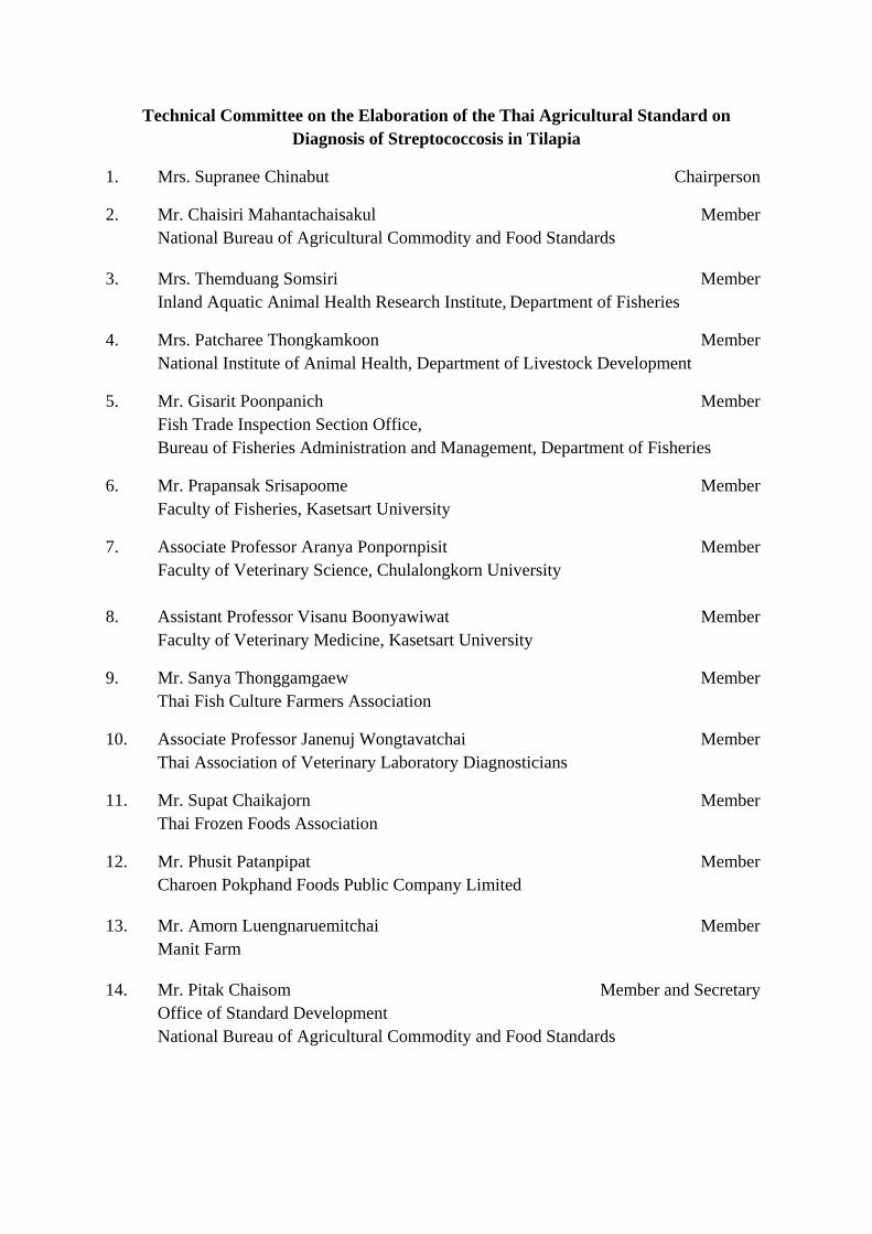
Technical Committee on the Elaboration of the Thai Agricultural Standard on Diagnosis of Streptococcosis in Tilapia
1. Mrs. Supranee Chinabut Chairperson
2. Mr. Chaisiri Mahantachaisakul Member National Bureau of Agricultural Commodity and Food Standards
3. Mrs. Themduang Somsiri Member Inland Aquatic Animal Health Research Institute, Department of Fisheries
4. Mrs. Patcharee Thongkamkoon Member National Institute of Animal Health, Department of Livestock Development
5. Mr. Gisarit Poonpanich Member Fish Trade Inspection Section Office, Bureau of Fisheries Administration and Management, Department of Fisheries
6. Mr. Prapansak Srisapoome Member Faculty of Fisheries, Kasetsart University
7. Associate Professor Aranya Ponpornpisit Member Faculty of Veterinary Science, Chulalongkorn University 8. Assistant Professor Visanu Boonyawiwat Member Faculty of Veterinary Medicine, Kasetsart University
9. Mr. Sanya Thonggamgaew Member Thai Fish Culture Farmers Association
10. Associate Professor Janenuj Wongtavatchai Member Thai Association of Veterinary Laboratory Diagnosticians
11. Mr. Supat Chaikajorn Member Thai Frozen Foods Association
12. Mr. Phusit Patanpipat Member Charoen Pokphand Foods Public Company Limited
13. Mr. Amorn Luengnaruemitchai Member Manit Farm
14. Mr. Pitak Chaisom Member and Secretary Office of Standard Development National Bureau of Agricultural Commodity and Food Standards
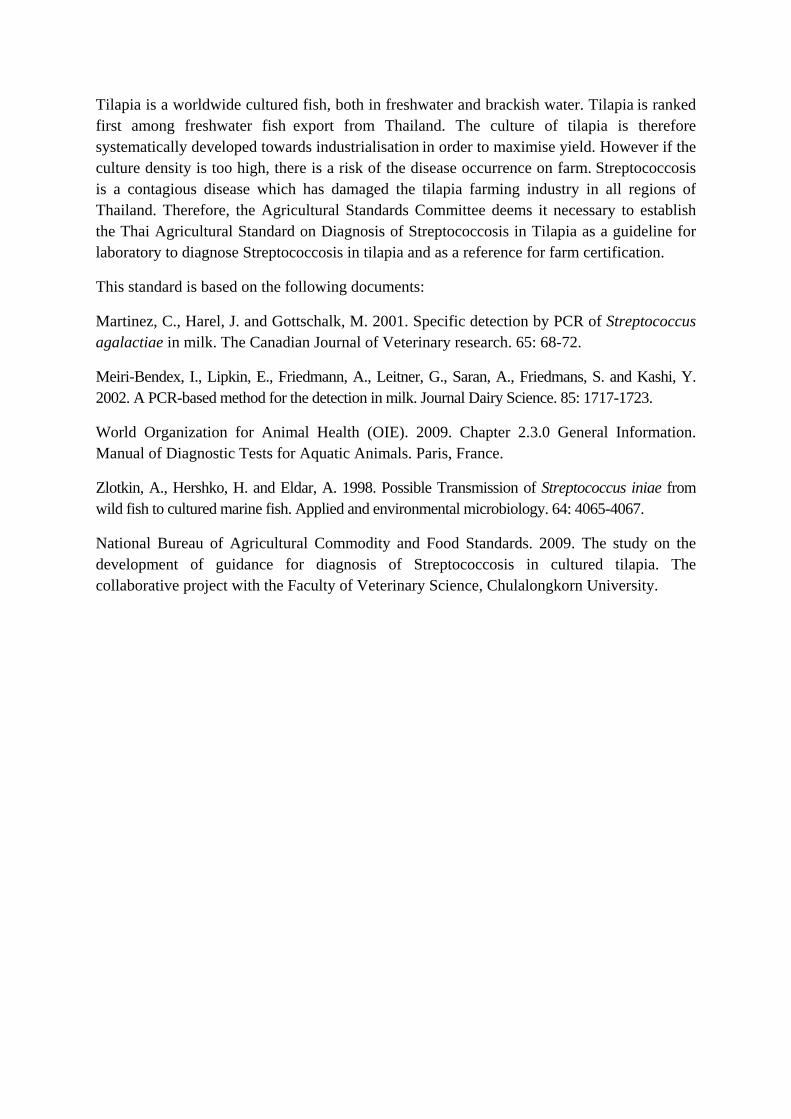
Tilapia is a worldwide cultured fish, both in freshwater and brackish water. Tilapia is ranked first among freshwater fish export from Thailand. The culture of tilapia is therefore systematically developed towards industrialisation in order to maximise yield. However if the culture density is too high, there is a risk of the disease occurrence on farm. Streptococcosis is a contagious disease which has damaged the tilapia farming industry in all regions of Thailand. Therefore, the Agricultural Standards Committee deems it necessary to establish the Thai Agricultural Standard on Diagnosis of Streptococcosis in Tilapia as a guideline for laboratory to diagnose Streptococcosis in tilapia and as a reference for farm certification.
This standard is based on the following documents:
Martinez, C., Harel, J. and Gottschalk, M. 2001. Specific detection by PCR of Streptococcus agalactiae in milk. The Canadian Journal of Veterinary research. 65: 68-72.
Meiri-Bendex, I., Lipkin, E., Friedmann, A., Leitner, G., Saran, A., Friedmans, S. and Kashi, Y. 2002. A PCR-based method for the detection in milk. Journal Dairy Science. 85: 1717-1723.
World Organization for Animal Health (OIE). 2009. Chapter 2.3.0 General Information. Manual of Diagnostic Tests for Aquatic Animals. Paris, France.
Zlotkin, A., Hershko, H. and Eldar, A. 1998. Possible Transmission of Streptococcus iniae from wild fish to cultured marine fish. Applied and environmental microbiology. 64: 4065-4067.
National Bureau of Agricultural Commodity and Food Standards. 2009. The study on the development of guidance for diagnosis of Streptococcosis in cultured tilapia. The collaborative project with the Faculty of Veterinary Science, Chulalongkorn University.
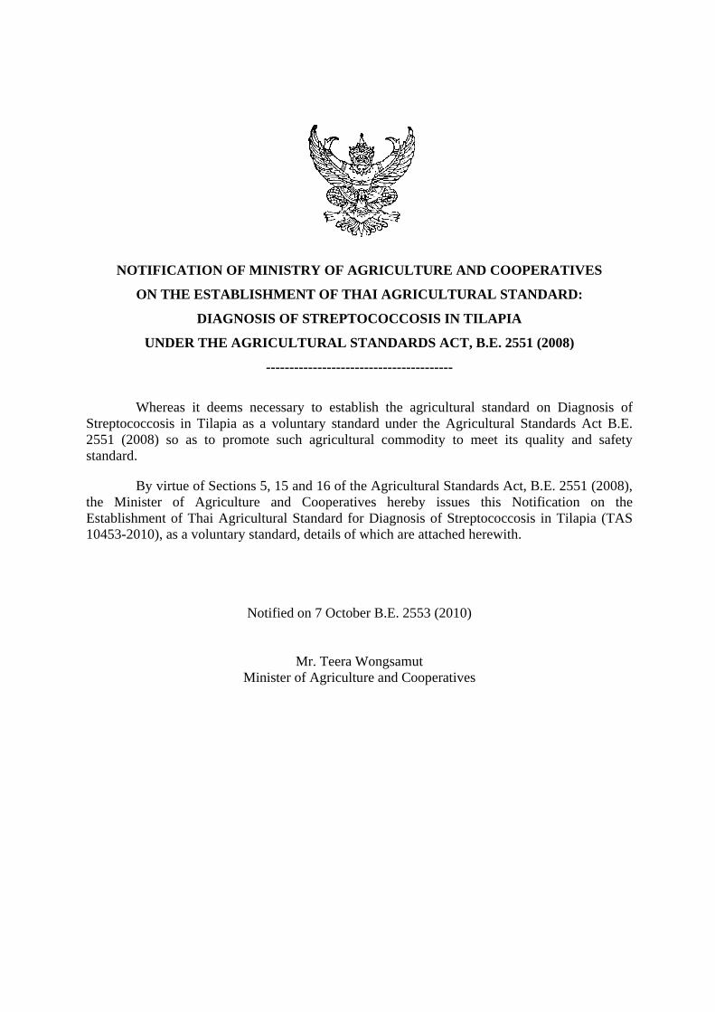
NOTIFICATION OF MINISTRY OF AGRICULTURE AND COOPERATIVES
ON THE ESTABLISHMENT OF THAI AGRICULTURAL STANDARD:
DIAGNOSIS OF STREPTOCOCCOSIS IN TILAPIA
UNDER THE AGRICULTURAL STANDARDS ACT, B.E. 2551 (2008)
----------------------------------------
Whereas it deems necessary to establish the agricultural standard on Diagnosis of
Streptococcosis in Tilapia as a voluntary standard under the Agricultural Standards Act B.E. 2551 (2008) so as to promote such agricultural commodity to meet its quality and safety standard.
By virtue of Sections 5, 15 and 16 of the Agricultural Standards Act, B.E. 2551 (2008), the Minister of Agriculture and Cooperatives hereby issues this Notification on the Establishment of Thai Agricultural Standard for Diagnosis of Streptococcosis in Tilapia (TAS 10453-2010), as a voluntary standard, details of which are attached herewith.
Notified on 7 October B.E. 2553 (2010)
Mr. Teera Wongsamut Minister of Agriculture and Cooperatives

TAS 10453-2010
THAI AGRICULTURAL STANDARD
DIAGNOSIS OF STREPTOCOCCOSIS IN TILAPIA
1. SCOPE
This Thai Agricultural Standard provides essential details for the laboratory diagnosis of Streptococcosis in tilapia, covering post-mortem examination in combination with rapid staining test, and the methods of histopathology, microbiology and polymerase chain reaction (PCR).
2. DEFINITIONS
For the purpose of this standard: 2.1 Tilapia means fish belong to the family Oreochromis. 2.2 Streptococcosis means epidemic disease in fish caused by the infection of gram-positive Streptococcal bacteria. 2.3 Diagnosis means the investigation processes to identify diseases. 2.4 Blood means red fluid circulating through blood vessels and heart. Blood composes of erythrocyte, leucocyte and thrombocyte. 2.5 Carrier means infected fish without showing any sign of illness. 2.6 Presumptive test means convenient and rapid laboratory test such as rapid staining test and histopathological method. 2.7 Confirmatory test means laboratory test used to confirm disease diagnosis. The test have been proved and recognised for its high specificity and sensitivity. 2.8 Specificity means ability of a test method that yields a negative result in a group of un-infected samples. 2.9 Sensitivity means ability of a test method that yields a positive result in a group of infected samples. 2.10 Positive control set means a test panel containing chemicals and standard microbe which will be subjected to the same diagnostic procedure as unknown sample in order to compare the test results. 2.11 Negative control set means a test panel containing chemicals but without standard microbe which will be subjected to the same diagnostic procedure as unknown sample in order to compare the test results. 2.12 Fresh carcass means a sample that is collected from moribund or euthanised fish.

TAS 10453-2010
2
3. DIAGNOSIS
Laboratory diagnosis of Streptococcosis in tilapia includes post-mortem examination in combination with rapid staining test andthe methods of histopathology, microbiology and polymerase chain reaction (PCR).
The responsible authority under the Animal Epidemics Act, B.E.2499 (1956) and its amendments should examine laboratory results together with the epidemiology, pathogenesis and clinical signs of Streptococcosis (Annex A) in order to ensure the effectiveness of disease prevention and control.
The objectives of the diagnosis of Streptococcosis are to screen and confirm disease in sick fish or carrier and to conduct a surveillance program for rapid disease control, which will reduce both production loss and spreading of disease to the environment.
The efficacy of each diagnostic method is different (Annex C). Selection of diagnostic method depends on purposes and sample types. The recommended presumptive methods of this standard are post-mortem examination in combination with rapid staining test or histopathological method. Negative result should be further confirmed by microbiological method or PCR method.
For example: microbiological method can be applied to infected tilapia with clinical lesions.
Microbiological method or PCR method can be used with suspected tilapia or larvae in hatchery.
PCR method is used for certification and testing of imported tilapia.
3.1 Sampling
Number of samples collected shall follow Table B.1, Annex B.
3.2 Post-mortem examination in combination with rapid staining test
3.2.1 Principle
This is a presumptive test by examining gross lesions (Annex A) in combination with fresh tissue staining. The fresh tissues can be collected from skin or mucous membrane with pustules or inflammation, kidney or brain. They are then stained with Gram’s stain and observed under light microscope.
3.2.2 Sample collection and preservation
Samples shall be fresh or chilled to prevent autolysis.
3.2.3 Procedure
(1) Spray 70% alcohol onto skin, mucous membraneor internal organ.
In case of pustule on skin or epithelial surface, use a tip of sterile sharp knife or sterile gauge needle (No. 18-24) to prick open the pustule. Use wire loop, flamed and cooled, to pick up pus from the laceration and spread onto glass slide. If pus is hardor sticky, apply 1 drop of sterile normal saline to the glass slide before spreading pus onto the slide. Leave it to dry at room temperature.
In case of kidney or brain tissues, dissect into the size of 0.5 cm3, using sterile technique.
Press tissue onto glass slide to allow film visible on the slide. Leave it to dry at room temperature.
(2) Heat-fix the prepared slide 2-3 times and then process to Gram’s stain (Annex D).

TAS 10453-2010
3
3.2.4 Interpretation of results
Presence of bacterial cells is commonly found in infected fish with Streptococcosis. Streptococcus bacteria are Gram-positive, basophilic and cocci shape at the size approximately 0.5 µm. Streptococci may arrange in single, pair or short-chain forms.
3.3 Histopathology
3.3.1 Principle
This method investigates disease at tissue level. The preserved tissues are stained with hematoxylin and eosin (H&E) and observed under light microscope.
3.3.2 Sample collection and preservation
(1) Use fresh carcass. Dissect carcass and collect target organ(s) or tissue. Immerse organ(s) or tissue in 10% formalin buffer at the 10-20 times volume to the sample.
- Small fish less than 1 cm long: immerse whole fish in formalin buffer.
- Fish of 1-5 cm long: cut the fish transversely in front of vent and immerse frontal part in formalin buffer.
-Fish larger than 5 cm long: collect internal organs with lesions. Lesions often appear on brain, liver, spleen, kidney, ovary and testis. Dissect the organs into small pieces of not more than 0.3 cm thick in order to let formalin buffer penetrate tissue thoroughly.
(2) Immerse tissue in formalin buffer for at least 24 h.
3.3.3 Procedure
Tissue samples from section 3.2.2 shall be examined by histopathological method (Annex E)
3.3.4 Interpretation of results
Infected fish with Streptococcus presents signs of acute inflammatory response on the tissues of multiple organs, which caused by exotoxin of the bacteria. Leucocytes such as neutrophils, macrophages and the clump of basophilic, cocci-shaped bacteria, which approximately 0.5 µ m/cell are commonly found at the lesions. Fibrinous exudates may also be detected at the lesions.
3.4 Microbiological method
3.4.1 Principle
This method investigates disease at tissue level. Streptococcus bacteria are isolated and cultured from organs or tissues of infected fish. Bacteria can be identified by testing the biochemical characteristics and bacterial morphology.
3.4.2 Sample collection and preservation
Sample should be collected from fish with or without clinical signs from the same population in order to compare the test results. Eggs and fish of all sizes can be collected.
(1) Collect and separate live samplesfrom each pond. Clearly provide details of samples (Annex H) and deliver to laboratory.
(2) If samples cannot be delivered alive, euthanise the fish and put them into plastic bags. Tightly close the bags and put on ice in a closed container. Deliver samples to laboratory immediately or within 6 h. If samples cannot be delivered within the day of

TAS 10453-2010
4
collection, keep the samples of whole fish on dry ice or at -20oC or below and deliver the next day.
(3) Egg samples can be collected as of (2)
3.4.3 Procedure
(1) Streptococcus isolation
If fish are still alive, they should be anesthezied or euthanized. Bacteria can be isolated from target organs, including liver, kidney, spleen and brain.
(1.1) small fish of less than 1 cm long or fish eggs
- Wash samples with 70% alcohol and rinse twice with distilled water
- Grind fish or egg samples in sterile normal saline (0.9% NaCl), at the ratio of 1:1 weight by volume.
- C entrifuge ground sam ple at 100 g for 2 m in, and collect 0 .1 m l of supernatant for bacterial culture. Spread on 5% sheep blood agar and incubate at 28-30oC for 24-48 h.
(1.2) Fish of 1-5 cm long
- Wash sample with 70% alcohol and rinse twice with distilled water
- Use only frontal part. If possible, use sterile knife, surgical blade or scissors to dissect abdominal cavity and remove gastrointestinal tract. Add sample with sterile normal saline at the ratio of 1:1 weight by volume into sterile hand homogenizer.
- Centrifuge ground sam ple at 100 g for 2 m in, and collect 0.1 m l of supernatant for bacterial culture then spread on 5% sheep blood agar and incubate at 28-30oC for 24-48 h.
(1.3) Fish larger than 5 cm long
- Spray 70% alcohol at the dissected point, use sterile scissors or surgical blade to dissect through skin until target organs are reached by wire loop.
- Wire loop should be flamed and cooled before touching tissue. Swab from liver, kidney, spleen and brain and streak on 5% sheep blood agar. Incubate culture agar at 28-30oC for 24-48 h.
(1.4) Streptococcus may be presumed if pin-point colonies of approximately 0.5-1 mm diameter, cream color, concave and smooth edge are observed. Hemolysis zone surrounding the colonies may also be found.
(2) Pure culture isolation
- Select the colonies from (1.4) and streak on Tryptic Soy Agar (TSA) in order to obtain pure culture. Incubate culture agar at 28-30oC for 24-48 h.
- Test purity of the bacteria by using Gram’s stain (AnnexD).
(3) Testing of Streptococcal bacteria
- Streptococcus colony morphology
Observe Streptococcus colony morphology on blood agar that has been incubated in 28-30oC for 24-48 h.
(4) Catalase test
- Swab suspected bacterial colony using platinum wire loop or glass rod and smear on a clean glass slide.

TAS 10453-2010
5
- Add 1 drop of 3% hydrogen peroxide.
- Observe bubble formation, catalase positive is indicated by the presence of bubble formation while negative result is noted when bubble is not formed.
(5) Biochemical tests can be performed according to the standard methods (Bergey’s manual) or by a commercial kit, API 20 STREP (BioMerieux, France), as described in Annex F.
3.4.4 Interpretation of results
(1) Streptococcus colony morphology on 5% blood agar that has been incubated at 28-30oC, under aerobic condition for 24-48 h, is approximately 0.5-1 mm diameter, smooth and concave, and cream-opaque colour with clear-hemolysis zone visible surrounding the colony.
(2) Streptococcus are Gram-positive bacteria, basophilic, cocci shape with the size of approximately 0.5 µm. Bacteria may arrange in single or pair or short-chain forms.
(3) Catalase test is used to differentiate between cocci shape, Gram-positive bacteria, such as Staphylococcus and Streptococcus. Streptococcus does not react with 3% hydrogen peroxide, whereas Staphylococcus and Micrococcus do.
3.5 PCR method
3.5.1 Principle
PCR assay amplifies DNA of Streptococcus using specific primers, which increase the amounts of DNA to a detectable level. Amplified DNA is then visualized by electrophoresis.
3.5.2 Sample collection and preservation
(1) Collect and separate live fish individually and clearly provide details of samples (Annex J). Deliver live samples to laboratory.
(2) If samples cannot be delivered alive, put the samples into plastic bags. Tightly close the bags and put on ice in a closed container. Deliver samples to laboratory immediately or within 6 h. If the samples cannot be delivered within 6 h, keep the sample on dry ice or at -20oC or below and deliver the samples the next day.
(3) If the delivery of live or frozen samples is not applicable, preserve the samples in 90-95% ethanol, at 4oC. Small fish can be immersed as whole, while large fish should be collected only their target organs. Use ethanol 10 times the volume by weight of samples (ml/g) and deliver to laboratory within 7 days.
(4) Egg samples are treated in the same manner as small fish
(5) Blood samples are collected from caudal vessel by sterile syringe and needle. Load blood samples into the equal volume of anticoagulant (Annex G), store at 4 o C a n d deliver sample to laboratory within 6 h. If the sample cannot be delivered within 6 h, keep the samples on dry ice or at -20oC or below. Place the samples on ice in cold storage container and deliver the sample within 24 h.
TAS 10453-2010
6
3.5.3 Procedure
3.5.3.1 Target DNA extraction
(1) Sample preparation depends on sample types, as follows:
(1.1) Fish tissues - For large fish, whose target organs can be collected. Collect 100-200 mg of
organ tissues and thoroughly grind the tissues in sterile distilled water. In addition, for blood sample, collect 0.1 ml.
- For eggs or relatively small fish that the organs cannot be collected, collect 100-200 mg of whole samples and thoroughly grind them in sterile distilled water.
(1.2) Pure bacterial colony
Adjust concentration of pure bacterial isolate to McFarland No. 0 . 5 (1x 10⁸cells/ml to 2 x 10⁸cells/ml), using sterile distilled water.
(2) Add 1 ml of sample from (1) into 1.5 ml microcentrifuge tube, centrifuge at 15,000 g for 15 min and discard the supernatant.
(3) Add 150-200 µl of 5 0 X Tris and ethylediaminetetraacetic acid (TE) buffer and 30 µl of lysozyme (20 mg/ml) and place in incubator shaker at 37 oC for 40 min.
(4) Add 20 µl of 10% sodium dodecyl sulfate (SDS) and place in incubator at 65 oC for 10 min. Add 30 µl of proteinase K (10 mg/ml) and place in incubator shaker at 37 oC for 24 h.
(5) Add 150-200 µl of phenol-chloroform-isoamyl alcohol (25: 24: 1), m ix w ell and centrifuge at 15,000 g for 15 min. Transfer 200 µl of supernatant into a new microcentrifuge tube.
(6) Add 20 µl of 3 M-ammonium acetate and 1 ml of absolute ethanol to the supernatant from (5) and store at -20oC for 24 h.
(7) Centrifuge at 15,000 g for 15 min then discard the supernatant and leave the precipitate to dry at room temperature for complete evaporation of ethanol.
(8) Dissolve the precipitate with 100 μl 1X TE buffer or sterile double distilled water (DDW) to obtain DNA solution for further steps. Keep the solution at -20oC.
3.5.3.2 Amplification of target DNA
(1) prepare PCR cocktail solution, which consists of
- 2 μl of 10X PCR buffer (100 mmol Tris-HCL (pH 8.3), 500 mmol KCl, 20 mmol MgCl2, enhancer solution)
- 2 μl of 10 mmol dNTPs (dATP, dTTP, dGTP, dCTP)
- 0.5 μl of 2.5 Unit Taq polymerase
- 1 μl of 10 μmol Forward primer
- 1 μl of 10 μmol Reverse primer
The three primers pairs, C1/C2 or F1/IMOD or Sin-1/Sin-2,can be used for diagnosis of the target bacteria.
Add DDW to obtain the final volume of 15 μl per test sample.
(2) add 5 μl of DNA sample into tube with PCR cocktail solution.
For each diagnosis the test will consist of positive (DNA of the standard Streptococcus), negative and non-template controls.
(3) put the above samples into thermocycler by settingstepwise conditions as follows
Step 1 incubate the samples at 94oC for 2 min
TAS 10453-2010
7
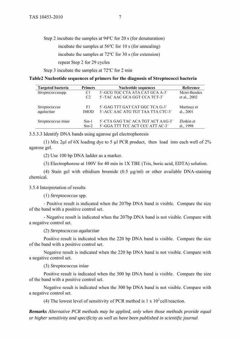
Step 2 incubate the samples at 94oC for 20 s (for denaturation)
incubate the samples at 56oC for 10 s (for annealing)
incubate the samples at 72oC for 30 s (for extension)
repeat Step 2 for 29 cycles
Step 3 incubate the samples at 72oC for 2 min
Table2 Nucleotide sequences of primers for the diagnosis of Streptococci bacteria
Targeted bacteria Primers Nucleotide sequences Reference Streptococcusspp. C1 5´-GCG TGC CTA ATA CAT GCA A-3´ Meiri-Bendex C2 5´-TAC AAC GCA GGT CCA TCT-3´ et al., 2002 Streptococcus F1 5´-GAG TTT GAT CAT GGC TCA G-3´ Martinez et agalactiae IMOD 5´-ACC AAC ATG TGT TAA TTA CTC-3´ al., 2001 Streptococcus iniae Sin-1 5´-CTA GAG TAC ACA TGT ACT AAG-3´ Zlotkin et Sin-2 5´-GGA TTT TCC ACT CCC ATT AC-3´ al., 1998
3.5.3.3 Identify DNA bands using agarose gel electrophoresis
(1) Mix 2µl of 6X loading dye to 5 µl PCR product, then load into each well of 2% agarose gel.
(2) Use 100 bp DNA ladder as a marker.
(3) Electrophorese at 100V for 40 min in 1X TBE (Tris, boric acid, EDTA) solution.
(4) Stain gel with ethidium bromide (0.5 µg/ml) or other available DNA-staining chemical.
3.5.4 Interpretation of results
(1) Streptococcus spp.
- Positive result is indicated when the 207bp DNA band is visible. Compare the size of the band with a positive control set.
- Negative result is indicated when the 207bp DNA band is not visible. Compare with a negative control set.
(2) Streptococcus agalactiae
Positive result is indicated when the 220 bp DNA band is visible. Compare the size of the band with a positive control set.
Negative result is indicated when the 220 bp DNA band is not visible. Compare with a negative control set.
(3) Streptococcus iniae
Positive result is indicated when the 300 bp DNA band is visible. Compare the size of the band with a positive control set.
Negative result is indicated when the 300 bp DNA band is not visible. Compare with a negative control set.
(4) The lowest level of sensitivity of PCR method is 1 x 10² cell/reaction.
Remarks Alternative PCR methods may be applied, only when those methods provide equal or higher sensitivity and specificity as well as have been published in scientific journal.
TAS 10453-2010
8
ANNEX A
EPIDEMIOLOGY, PATHOGENESIS AND CLINICAL SIGNS OF STREPTOCOCCOSIS
(Section 3)
A.1 Epidemiology
Streptococcosis is a bacterial disease in fish that causes economic losses in the production of freshwater and marine fish, which is economically important in many countries, including tilapia industry in Thailand. Streptococcosis outbreaks may either reoccur in the same area or emerge from a new area which has never been reported of such disease due to inappropriate farm management and biosecurity system. Streptococcus species that cause disease in fish include Streptococcus agalactiae, S. dysgalactiae, and S. equi, S. equisimilis, S. faecium, S. pyogenes, S. zooepidemicus and S. iniae. Previous case reports showed that S. agalactiae and S. iniae are the main causative agents of Streptococcosis in tilapia. Moreover, the World Organisation for Animal Health (OIE) has declared that S. iniae is zoonosis.
Streptococcosis outbreaks in tilapia were reported in many countries, such as the United States of America, South Africa, Japan, Israel, Italy and Thailand. The disease was found in black and red tilapia, of all sizes. The disease occurrences were reported in all regions of tilapia producing countries.
Disease pattern and severity are different depending on bacterial strains and farm management system. Abrupt change in farm management, intensive farming, improper harvest or handling of fish, resulting in skin laceration, as well as poor water quality, such as low dissolved oxygen, high ammonia or nitrite, induce stress which has negative impact on fish immunity.
A.2 Pathogenesis
The main predisposing causes of Streptococcus infection in fish are inappropriate management practices and/or abrupt change of the environment. For example, relatively high water temperature above 30 o C during summer may induce stress in fish. Moreover, such temperature is favourable for the growth of Streptococcus. As a result, fish are more susceptible to the infection. Infected fish with clinical signs, carrier and non-infected fish may be presented in the same water source. Bacteria may enter the fish by cannibalism (fish consume moribund or dead fish which were infected with this bacteria), or through skin laceration and mucous membrane of several organs. Fish respond to the infection by initiating inflammatory process, which neutralise pathogens by white blood cells such as macrophage and polymorphonuclear leukocyte. Cell-mediated and humoral immunity also play an important role in preventing the bacteria to reach blood circulation and internal organs. However, some of the bacteria are able to evade the immune system using its particular properties to cause systemic infection, for instance:
(1) Surface antigens of Streptococcal bacteria can attach to the fish’s cell surface, which prevent them to be eliminated by fish’s lysozyme. Consequently, the bacteria replicate in lymph and blood (septicemia) and spread to target organs, such as liver, kidney, spleen and brain.
(2) Toxin production, the important hemolytic toxin of Streptococcus is streptolysin, which is further classified as streptolysin S and streptolysin O. The toxins cause complete hemolysis on blood agar. Streptolysin S produces hemolysis on the surface of blood agar (surface hemolysis), whereas streptolysin O produces hemolysis under anaerobic condition,

TAS 10453-2010
9
in a deeper layer of blood agar (deep hemolysis). The toxin rapidly damages cells and tissues, including white blood cells, liver and heart.
(3) Enzyme production, most of the enzymes produced by Streptococcus are capable of digesting large molecules, such as fibrin clump and connective tissue. This enables the bacteria to easily penetrate the tissue, especially at skin lacerations and mucous membrane of several organs.
A.3 Clinical signs
Clinical signs of infected fish with Streptococcus are commonly detected in fish larger than 300 g. There are 2 forms of clinical signsas follows:
A.3.1 Acute
Signs of acute Streptococcosis in fish include abnormal swimming, such as non-directional, imbalance or spiral swimming. Gross signs include enlarge abdomen, eye lesions, exophthalmia, corneal opacity and hemorrhage, which can be found on oneor both eyes. Moreover, hemorrhagic lesions at the base of the fins, operculum, skin and tissue around ventcan also be found. Significant internal lesions are presence of clear, viscous to hemorrhagic fluid in abdominal cavity, pale and enlarge liver, darken and enlarge spleen, hemorrhage and congestion at mucosal side of the intestine. These gross lesions are clearly visible in relatively large fish, however, they may be absent from infected juvenile or larvae. Mortality rate of the moribund fish may reach up to 50% within 3-7 days post infection or since when the first clinical sign was observed. Mortality rate may reach up to 80-100%, in case of the severe outbreaks.
A.3.2 Chronic
Chronically infected fish may be found floating. Black tilapia presents darkening body colour, whereas, red tilapia showspale body colour. External lesions may include presence of pustules at caudal peduncle or under the month. Internal lesions may include fibrinous peritonitis, adhesion of internal organs, pericarditis and/or absence ofinternal lesion. Mortality rate is low or none. Feed intake and growth rate are reduced, which prolong cultured period. The carcass is abnormal and defected, resulting in low quality meat.
TAS 10453-2010
10
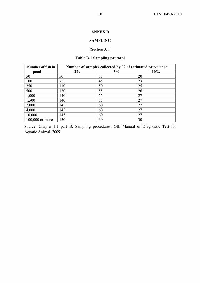
ANNEX B
SAMPLING
(Section 3.1)
Table B.1 Sampling protocol
Number of fish in pond
Number of samples collected by % of estimated prevalence 2% 5% 10%
50 50 35 20 100 75 45 23 250 110 50 25 500 130 55 26 1,000 140 55 27 1,500 140 55 27 2,000 145 60 27 4,000 145 60 27 10,000 145 60 27 100,000 or more 150 60 30
Source: Chapter 1.1 part B: Sampling procedures, OIE Manual of Diagnostic Test for Aquatic Animal, 2009

TAS 10453-2010
11
ANNEX C
COMPARISON OF THE EFFICACY OF DIFFERENT DIAGNOSTIC METHODS
(Section 3)
Table C.1 Comparison of the efficacy of different diagnostic methodsfor Streptococcosis
Diagnostic methods Presumptive Confirmatory
Post-mortem examination in combination with rapid staining test
+ -
Histopathology ++ + Microbiology - Light microscope morphology
++ +
- Biochemical property +++ +++ PCR method +++ +++
Source: Results of a research project for establishing agriculture standard, “Diagnosis of Streptococcosis in cultured Tilapia” Remark
- = not availableor not suitable + = can be used in some situations due to limitation of its application ++ = standard method with good diagnostic sensitivity and specificity +++ = recommended method due to the high specificity and sensitivity

TAS 10453-2010
12
ANNEX D
GRAM’S STAIN
(Section 3.2.3 and Section 3.4.3) D.1 Gram’s stain procedures are as follows:
(1) Flood the slide on the area where sample has been placed with crystal violet solution for 1 min and rinsewith clean water.
(2) Flood the slide on the area where sample has been placed with Gram’s iodine (Lugol’s solution) for 1 min and rinse with clean water.
(3) Remove excess stains with decolorizer (95% ethanol or ethanol/acetone) for 2-5 s and rinse with clean water.
(4) Flood the slide on the area where sample has been placed with safranin solution for 1 min and rinse with clean water.
(5) Let the slide dry and observe the slide under light microscope at 1000x magnification. Observe Gram reaction (colour), morphology and arrangement of suspected bacterial cells.
TAS 10453-2010
13
ANNEX E
SAMPLE PREPARATION FOR HISTOPATHOLOGY
(Section 3.3.3) E.1 Sample preparation for histopathology: paraffin embedding. The procedures are as follows:
(1) Immerse the tissue from Section 3.3.2 in solutions in order as indicated: - Immerse in 70% ethanol for 1 h
- Immerse in 85% ethanol for 1 h - Immerse in 95% ethanol (first bottle) for 1 h - Immerse in 95% ethanol (second bottle) for 1 h - Immerse in 95% ethanol (third bottle) for 1 h - Immerse in 100% ethanol (first bottle) for 1 h - Immerse in 100% ethanol (second bottle) for 1 h - Immerse in 100% ethanol (third bottle) for 1 h - Immerse in xylene or xylene replacment (first bottle) for 1.5 h - Immerse in xylene or xylene replacment (second bottle) for 1.5 h
(2) Embed the tissue from (1) in the first bottle of melted paraffin for 2 h and move to the second bottle of melted paraffin and immerse the tissue for 2 h.
(3) Place the tissue in embedding mold that contains melted paraffin. Place the block on the embedding mold and fill the mold with melted paraffin. Place the mold on cool tray until paraffin is set. Keep the mold at room temperature.
(4) Cut the tissue embed with microtome to 4-5 µm thick and place on glass slide.
E . 2 S a m p le preparation for histopathology: staining tissue slide with H&E stain. The procedures are as follows:
(1) Remove paraffin on the tissue slide (deparaffinize) by immersing the slide in xylene or xylene replacement twice for 2 min each
(2) Immerse the tissue slide in absolute ethanol twice for 2 min each
(3) Rinse the tissue slide through running water for 2-5 min
(4)Immerse the tissue slide in hematoxylin solution for 3 min
(5) Rinse the tissue slide through running water for 5-10 min
(6) Immerse the tissue slide in eosin solution for 3 min
(7) Rinse the tissue slide through running water
(8) Dehydrate the tissue slide with 70% ethanol
(9) Dehydrate the tissue slide with 100% ethanol twice for 1-2 min each
(10) Immerse the tissue slide in alcohol/clearance (50/50) for 1-2 min
(11) Immerse the tissue slide in xylene or xylene replacment twice for 2 min each
(12) Apply 1 drop of permount and cover the tissue slide with cover glass
(13) Observe the tissue slide with microscope at 400x magnification

TAS 10453-2010
14
ANNEX F
MICROBIOLOGICAL METHOD
(Section 3.4.3)
F.1 Biochemical tests using API 20 STREP test kit. Test procedures are as follows:
(1) Prepare test kit: add small amount of water into an incubation box to create a humid atmosphere. Place API test kit in the box and label the tested sample.
(2) Adjust concentration of the bacteria to McFarland No. 4 (8 -16 x 108ce lls /m l), using sterile normal saline solution.
(3) Add 0.5 ml of the solution from (2) to GP Medium, mix well.
(4) Use sterile pipette to transfer 100 µl of the solution from (2) into the first 9 wells of API 20 STREP test kit (VP, HIP, ESC, PYRA, αGAL, ßGUR, ßGAL, PAL and LAP).
(5) Well No. 10 (ADH), use sterile pipette to transfer 100 µl of the solution from (2) into the well and coat the well with liquid paraffin to create anaerobic condition.
(6) Transfer the solution from (3) to RIB – GLYG wells and coat the well with liquid paraffin to create anaerobic condition.
During step 4-6, precaution against bubble formation while working with the test kit. (7) Close incubation box and incubate at 35-37 o C f o r 4 h , t h e n a p p l y t h e
followingreagents: - 1 drop of VP1 (Potassium hydroxide)and VP2 into the 1st well (VP) - 2 drops of NIN into the 2nd well (HIP) - 1 drop of ZYME A and ZYME B into the 4th to 9th wells (PYRA, αGAL, ßGUR, ßGAL, PAL andLAP)
Wait for 10 min and readthe reaction of each well for the first reading. Record the first results into the table (positive and negative).
(8) Continue to incubate the box for 24 h and read the reaction of ESC, ADH and RIB to GLYG wells and record the results for the second reading.
(9) Enter the data into the software for analysisand identification of Streptococcus species.
Illustration of API 20 STREP test kit
VP3 HIP ESC
PYRA αGAL βGUR
βGAL PAL LAP
ADHRIBARA
MANSORLAC
TREINURAF
AMDGLYG
(10) Interpret the results of the biochemical test from API 20 STREP using software
or by referring toTable F.1.

TAS 10453-2010
15
Table F.1 Test results of Streptococcus biochemical properties, using API 20 STREP test kit (BioMerieux, France)
Species of Streptococcus
Biochemical property
VP
HIP
ESC
PY
RA
αG
AL
ß GU
R
ß GA
L
PA
L
LA
P
AD
H
RIB
AR
A
MA
N
SO
R
LA
C
TR
E
INU
RA
F
AM
D
GLY
G
ß HEM
S. agalactiae ATCC13813 + + - - + - + + + + + - - - + + - - - - +
S. dys.equi. ATCC35666 - - - - - + - + + - + - - - - + - - + - +
S. porcinus ATCC43138 + - + + - + - + + + + - + + + + - - - - +
S. constellatus ATCC27823 + - - - - - - + + + - - - - - + - - - - +
S.iniae ATCC29178 + - - - - + - - - - + - + - - + - - + + +
Tests Substrates Reactions/enzymes Results
Negative Positive
VP Pyruvate Acetoin Production colour not changing pink-red
HIP Hippurate Hydrolysis colour not changing/ light blue dark blue / purple
ESC Esculin ß-glucosidase 4 h 24 h 4 h 24 h
colour not
changing /
light yellow
colour not
changing /
light yellow/
light grey
black/grey black
PYRA Pyrrolidonyl-2-
naphthylamide
Pyrrolidonyl
arylamidase
colour not changing/ light orange orange
αGAL 6-Bromo-2-
naphthyl-α-D-
galactopyranoside
α-galactosidase colour not changing purple
ßGUR Naphthol AS-BI-ß
-D-glucuronate
ß-glucuronidase colour not changing blue
ßGAL 2-naphthyl-ß-D-
galactopyranoside ß-galactosidase colour not changing / light purple purple
PAL 2-naphthyl
phosphate Alkaline phosphatase colour not changing / light purple purple
LAP L-leucine-2-
naphthylamide
Leucine arylamidase colour not changing orange
ADH Arginine Arginine dihydrolase yellow red
4 h 24 h 4 h 24 h
RIB
ARA
MAN
SOR
LAC
TRE
INU
RAF
AMD
Ribose
L-Arabinose
Mannitol
Sorbital Lactose
Trehalose
Inulin
Raffinose
Amygdalin
Acidification
Acidification
Acidification
Acidification
Acidification
Acidification
Acidification
Acidification
Acidification
red
red
red
red
red
red
red
red
red
orange/ red
orange/ red
orange/ red
orange/ red
orange/ red
orange/ red
orange/ red
orange/ red
orange/ red
orange/yellow
orange/yellow
orange/yellow
orange/yellow
orange/yellow
orange/yellow
orange/yellow
orange/yellow
orange/yellow
yellow
yellow
yellow
yellow
yellow
yellow
yellow
yellow
yellow
GLYG Glycogen Acidification red/ orange Light yellow
ßHEM clear-hemolysis zone visible surrounding the colony on blood agar
TAS 10453-2010
16
ANNEX G
CHEMICALS PREPARATION
(Section 3)
G.1 Tryptic soy agar (TSA, pH 7.3 ± 0.2), 1,000 ml Tryptone 15.0 g Soya peptone 5.0 g Sodium chloride 5.0 g Agar 15.0 g
Preparationmethod
(1) Weigh the chemicals as described above, mix and add distilled water to obtain 1,000 ml
(2) Dissolve the mixture and boil the solution
(3) Autoclave the agar at the pressure of 15 pound per square inch at 121oC for 15-20 min
(4) Pour the agar on sterile petri dish, approximately 20 ml per dish and wait until the agar is set. Store at4 oC. The agar should be used within 14 days. G.2 Blood agar
Preparation method
(1) Prepare TSA as described in G.1 (1) to G.1 (3), and cool down to 55 ± 5oC.
(2) Add sheep blood at 5% ratio and mix well.
(3) Pour the agar on sterile petri dish, approximately 20 ml per dish and wait until the agar is set. Store at 4 oC. The agar should be used within 14 days. G.3 Anticoaglulant, 1,000 ml
Sodium chloride 26.3 g Trisodium citrate 8.8 g Citric acid 5.5 g EDTA 3.7 g
Preparation method
(1) Weigh the chemicals as described above, mix and add distilled water to obtain 700 ml. Adjust pH of the solution to 7.0, using sodium hydroxide. Autoclavethe solution at the pressure of 15 pound per square inch at 121oC for 15 min.
(2) Let the solution cool down and add 100 ml of 1 mol glucose solution, 1 mol glucose solution is prepared by dissolve 39.6 g glocose in 200 ml distilled water.
(3) Add water to obtain 1,000 ml G.4 Reagents for DNA extration in the test sample G.4.1 lysis solution, concentration of reagent compositions are as follows: - 50X TE buffer (50 mmol Tris/HCl pH 8.0, 5 mmol EDTA)
Preparation method
(1) Add 20 ml of 0.5 molTris/HCl, pH 8.0, into 250 ml sterile bottle
(2) Add 1 ml of 1 mol EDTA and add sterile distilled water until 200 ml is obtained
TAS 10453-2010
17
- 20% SDS (sodium dodecyl sulfate)
Preparation method
(1) Prepare 200 g of SDS and add sterile distilled water to 900 ml
(2) Dissolve SDS in water bath at 68oC, mix the solution well.
(3) Adjust pH to 7.2, using conc. HCl and add distilled water to obtain 1,000 ml
(4) Store at room temperature Remark Face mask should be worn while weighing the chemicals due to irritation effect, especially to the nasal mucosa. - 20mg/ml lysozyme
Preparation method
(1) Weigh 400 mg lysozyme and dissolve in sterile distilled water until a total volume of 20 ml is obtained.
(2) Aliquot lysozyme solution into 1.5-2 ml microtube, 1 ml per tube for 20 tubes.
(3) Store at -20 oC. Remark Prepared lysozyme should be aliquoted into microtubes to prevent deterioration. - 10 mg/ml proteinase K
Preparation method
(1) Weigh100 mg proteinase K and dissolve in sterile distilled water until a total volume of 10 ml is obtained.
(2) Aliquot proteinase K solution into 1.5-2 ml microtube, 1 ml per tube for 10 tubes.
(3) Store at -20 oC. Remark Prepared proteinase K should be aliquoted into microtubes to prevent deterioration. G.4.2 DNA storage solution, concentration of reagent compositions are as follows: - 1X TE buffer (5 mmol Tris/HCl pH 8.0, 0.5 mmol EDTA)
Preparation method
(1) Add 1 ml of 50X TE buffer into 100 ml sterile bottle.
(2) Add distilled water until a total volume of 50 ml is obtained.

TAS 10453-2010
18
ANNEX H
ILLUSTRATIONS FOR THE DIAGNOSIS OFSTREPTOCOCCOSIS IN TILAPIA
(Section 3)
Figure H.1 External gross lesions of Streptococcus infected tilapia: diffuse congestion throughout the body, especially at the fins, oral mucosa and conjunctiva.
Source: Courtesy of Associate Professor Janenuj Wongtavatchai, Faculty of Veterinary Science, Chulalongkorn University
Figure H.2 Obvious clinical signs of Streptococcus infected tilapia: exophthalmos due to the inflammation of conjunctiva and cornea, as well ascorneal opacity. Exophthalmoscan be
unilateral or bilateral.
Source: Courtesy of Associate Professor Janenuj Wongtavatchai, Faculty of Veterinary Science, Chulalongkorn University
TAS 10453-2010
19
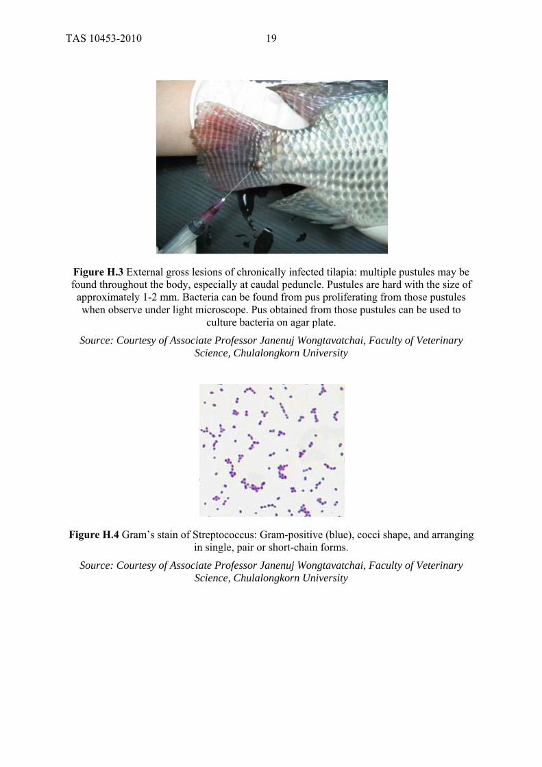
Figure H.3 External gross lesions of chronically infected tilapia: multiple pustules may be found throughout the body, especially at caudal peduncle. Pustules are hard with the size of
approximately 1-2 mm. Bacteria can be found from pus proliferating from those pustules when observe under light microscope. Pus obtained from those pustules can be used to
culture bacteria on agar plate.
Source: Courtesy of Associate Professor Janenuj Wongtavatchai, Faculty of Veterinary Science, Chulalongkorn University
Figure H.4 Gram’s stain of Streptococcus: Gram-positive (blue), cocci shape, and arranging in single, pair or short-chain forms.
Source: Courtesy of Associate Professor Janenuj Wongtavatchai, Faculty of Veterinary Science, Chulalongkorn University
TAS 10453-2010
20
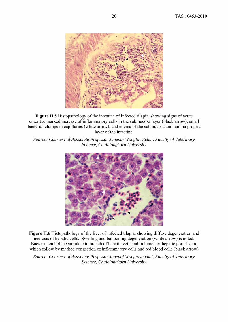
Figure H.5 Histopathology of the intestine of infected tilapia, showing signs of acute enteritis: marked increase of inflammatory cells in the submucosa layer (black arrow), small
bacterial clumps in capillaries (white arrow), and edema of the submucosa and lamina propria layer of the intestine.
Source: Courtesy of Associate Professor Janenuj Wongtavatchai, Faculty of Veterinary Science, Chulalongkorn University
Figure H.6 Histopathology of the liver of infected tilapia, showing diffuse degeneration and necrosis of hepatic cells. Swelling and ballooning degeneration (white arrow) is noted.
Bacterial emboli accumulate in branch of hepatic vein and in lumen of hepatic portal vein, which follow by marked congestion of inflammatory cells and red blood cells (black arrow)
Source: Courtesy of Associate Professor Janenuj Wongtavatchai, Faculty of Veterinary Science, Chulalongkorn University

TAS 10453-2010
21
Figure H.7 Histopathology of inflamed anterior kidney (nephritis) of infected tilapia. Marked increase of inflammatory cells is observed (black arrow) with small bacterial clumps in the
capillaries of the anterior kidney (white arrow), 100x magnification.
Source: Courtesy of Associate Professor Janenuj Wongtavatchai, Faculty of Veterinary Science, Chulalongkorn University
Figure H.8 Histopathology ofinflamed brain tissue (exudative meningitis). Marked increase of inflammatory cells, red blood cells and fibroblast are observed in the meninges with small
bacterial clumps in the capillaries of the brain (white arrow), 40x magnification.
Source: Courtesy of Associate Professor Janenuj Wongtavatchai, Faculty of Veterinary Science, Chulalongkorn University

TAS 10453-2010
22
Figure H.9 Histopathology of inflamed brain tissue (exudative meningitis). Inflammatory cells and small bacterial clumps are abundantly found in the capillaries of the brain (black arrow). In addition, diffuse bacteria infiltrationin brain tissue via the capillaries is observed
(white arrow), 100x magnification.
Source: Courtesy of Associate Professor Janenuj Wongtavatchai, Faculty of Veterinary Science, Chulalongkorn University
Figure H.10 Bacterial septicemia of infected tilapia: thin blood smear shows Streptococcus attaching itself to the red blood cell surface, 1000x magnification.
Source: Courtesy of Associate Professor Janenuj Wongtavatchai, Faculty of Veterinary Science, Chulalongkorn University
Figure H.11 Biochemistry test results of S. agalactiae ATCC 13813 using API 20 STREP test kit.
Source: Courtesy of Associate Professor Janenuj Wongtavatchai, Faculty of Veterinary Science, Chulalongkorn University.
TAS 10453-2010
23
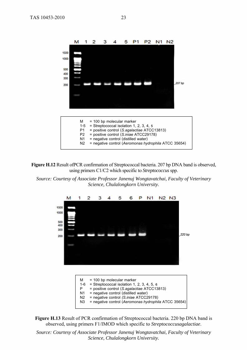
M = 100 bp molecular marker 1-5 = Streptococcal isolation 1, 2, 3, 4, 5 P1 = positive control (S.agalactiae ATCC13813) P2 = positive control (S.iniae ATCC29178) N1 = negative control (distilled water) N2 = negative control (Aeromonas hydrophila ATCC 35654)
Figure H.12 Result ofPCR confirmation of Streptococcal bacteria. 207 bp DNA band is observed, using primers C1/C2 which specific to Streptococcus spp.
Source: Courtesy of Associate Professor Janenuj Wongtavatchai, Faculty of Veterinary Science, Chulalongkorn University.
M = 100 bp molecular marker 1-6 = Streptococcal isolation 1, 2, 3, 4, 5, 6 P = positive control (S.agalactiae ATCC13813) N1 = negative control (distilled water) N2 = negative control (S.iniae ATCC29178) N3 = negative control (Aeromonas hydrophila ATCC 35654)
Figure H.13 Result of PCR confirmation of Streptococcal bacteria. 220 bp DNA band is observed, using primers F1/IMOD which specific to Streptococcusagalactiae.
Source: Courtesy of Associate Professor Janenuj Wongtavatchai, Faculty of Veterinary Science, Chulalongkorn University.

TAS 10453-2010
24
M = 100 bp molecular marker 1-2 = Streptococcal isolation 1, 2 P = positive control (S.iniae ATCC29178) N1 = negative control (distilled water) N2 = negative control (S.agalactiae ATCC13813) N3 = negative control (Aeromonas hydrophila ATCC 35654)
Figure H.14 Result of PCR confirmation of Streptococcal bacteria. 300bp DNA band is observed, using primers Sin-1/ Sin-2 which specific to Streptococcus iniae.
Source: Courtesy of Associate Professor Janenuj Wongtavatchai, Faculty of Veterinary Science, Chulalongkorn University.

TAS 10453-2010
25
ANNEX J
SAMPLE: RECORD FORMFOR DISEASE DIAGNOSIS
Sample No. .................... Date...........................
1. General information ID number ............................ 1.1 Owner’s name/address ...................House No. .......Village No. ............Sub-distric............. District......................................Province................................Tel:................................ 1.2 Species/type............................................................Amount...................... (fish/container)
1.3 Source of animal Imported Cultured Wild caught Bought from......
1.4 Rearing system Cement pond, Size................Earthen pond, Size............Glass aquaria, Size.....
Fiber tank, Size.................Other.........................Size...................
1.5 Rearing density (Fish/m2)............................................................................
2. Feed management
2.1 Feed Commercial feed Live feed.......... Supplemented feed...... Other
2.2 Feeding ratio (%)and frequency (per day).................................................................
3. Water quality management
3.1 Water source Tap Underground Surface Rain Other
3.2 Water exchange rate(per day)..................................................................................
3.3 Management for aeration Yes No
3.4 Water quality Colour. . . . . . . . . . . . . . . . . . . . . . . . . . . . . . . . . . . . . . . . . pH. . . . . . . . . . . . . . . . . . . . . . . . . . . . . . . . . . . . . . .
Hardness....................... Total alkalinity .................... Total ammonia (NH3)..................................Nitrite (NO2
-)........................................
4. Information on infected fish
4.1 General appearances Eyes............................................... Gills...................................................... Skin................................................ Pustule(s).............................................. Color.............................................. Tumor.................................................... Fin.................................................. Respiration........................................... Swimming, buoyancy ..................... Other..................................................... 4.2 Age 1-15 Day(s) 15-30 Day 1-6 month(s) 6-12 months 1-3 year(s) 3-5 years 5 years or above
4.3 Morbidity (%)...........................................................................................

TAS 10453-2010
26
4.4 General management, eg. water exchange, medicines/chemicals application................
5. Diagnostic results Parasitology........................................................................................... Bacteriology Sample No. ....................................................................................... Bacteriology result................................................................................................. Sensitivity test
oxytetracycline sulfamethoxazole + Trimethoprim Mycology Sample No. ............................................................................................
Mycology result............................................................................................................ Pathology Sample No. ............................................................................................
Pathology result............................................................................................................... Virology Sample No. .............................................................................................. Virology result................................................................................................................
PCR Sample No. .............................................................................. PCR result......................................................................................................................
6. Suggestion....................................................................................................................... 7. Treatmentfollow-up
Very Effective Moderatelyeffective Slightlyeffective Not effective Other.............................................................
8. Conclusion(s)....................................................................................................
Officer signature.............................................
(...............................)
Date..............Month.....................Year.............
TAS 10453-2010
27
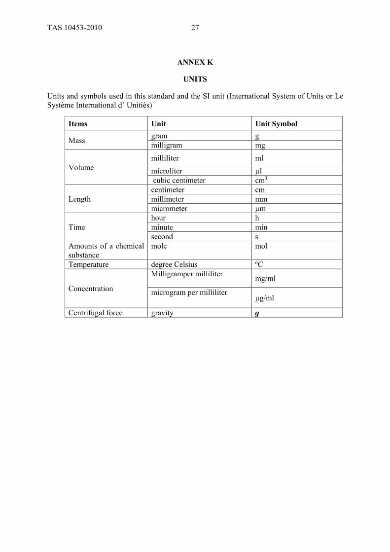
ANNEX K
UNITS
Units and symbols used in this standard and the SI unit (International System of Units or Le Système International d’ Unitiès)
Items Unit Unit Symbol
Mass gram g milligram mg
Volume milliliter ml
microliter µl cubic centimeter cm3
Length centimeter cm millimeter mm micrometer µm
Time hour h minute min second s
Amounts of a chemical substance
mole mol
Temperature degree Celsius oC
Concentration
Milligramper milliliter mg/ml
microgram per milliliter µg/ml
Centrifugal force gravity g