The Painful Adult Shoulder: evidence based history, exam and approach
Upload
zoe-zamudioCategory
view
190download
3
TUTORIAL
Diagnosis and Management of the Painful Shoulder. Part 2:Examination, Interpretation,
and Management
Phillip S. Sizer Jr., PhD, PT*; Valerie Phelps, PT**; Kerry Gilbert, MPT**Texas Tech University Health Science Center, Lubbock Texas; **International Academy of
Orthopedic Medicine-US, Tucson, Arizona
� Abstract: Diagnosis, interpretation and subsequentmanagement of shoulder pathology can be challenging toclinicians. Because of its proximal location in the schlerotomeand the extensive convergence of afferent signals from thisregion to the dorsal horn of the spinal cord, pain referencepatterns can be broadly distributed to the deltoid, trapezius,and or the posterior scapular regions. This pain behavior canmake diagnosis difficult in the shoulder region, as the loca-tion of symptoms may or may not correspond to the prox-imity of the pain generator. Therefore, a thorough historyand reliable physical examination should rest at the centerof the diagnostic process. Effective management of thepainful shoulder is closely linked to a tissue-specific clinicalexamination. Painful shoulder conditions can present with orwithout limitations in passive and or active motion. Limits in passive motion can be classified as either capsular or noncapsular patterns. Conversely, patients can present withshoulder pain that demonstrates no limitation of motion.Bursitis, tendopathy and rotator cuff tears can produceshoulder pain that is challenging to diagnose, especiallywhen they are the consequence of impingement and orinstability. Numerous nonsurgical measures can be imple-mented in treating the painful shoulder, reserving surgicalinterventions for those patients who are resistant to conser-vative care. �
Key Words: acromioclavicular, examination, gleno-humeral, scapula, shoulder, sternoclavicular, impingement,instability, labrum
INTRODUCTION
Diagnosis, interpretation and subsequent managementof shoulder pathology can be challenging to clinicians.The term “shoulder” refers to a complex of joints andsoft tissue structures in the upper quadrant of the torso.Because of its proximal location in the sclerotome andthe extensive convergence of afferent signals from thisregion to the dorsal horn of the spinal cord, pain refer-ence patterns can be broadly distributed to the deltoid,trapezius, posterior scapular regions, and or the arm allthe way to the wrist. This pain behavior can make diag-nosis difficult in the shoulder region, as the location ofsymptoms may or may not correspond to the proxim-ity of the pain generator.1 Compounding this diagnos-tic dilemma, imaging results can be misleading.2,3
Therefore, a thorough history and reliable physicalexamination should rest at the center of the diagnosticprocess.4
Afflictions in the elevation chain can be categorizedas primary or secondary in nature. For example,primary arthropathies are painful disturbances in thejoints that develop as result of trauma or disease, suchas synovitis, arthrosis, or chondropathy.5 Conversely,secondary afflictions emerge in tissues adjacent tounderlying nonpainful joint, capsuloligamentous or
Send all correspondence to: Phillip S. Sizer Jr, PhD, PT, Texas Tech Uni-versity Health Science Center, School of Allied Health, Physical TherapyProgram, 3601 4th St., Lubbock, TX 79430. Tel: (806) 743-3902.
© 2003 World Institute of Pain, 1530-7085/03/$15.00Pain Practice, Volume 3, Issue 2, 2003 152–185
Diagnosis and Management of the Painful Shoulder • 153
muscular structures. For example, shoulder impinge-ment and subsequent subacromiodeltoid bursitis oftenresults from posterior capsular limitation at the gleno-humeral joint.6
CLINICAL EXAMINATION
History
A patient’s age and sex can provide the clinician with aglimpse of the patient’s disorder, because different dis-orders are more common within specific sexes and agegroups.5 For example, traumatic synovitis of the gleno-humeral joint (GHJ) is more common in individualsolder than 45 years of age. One would expect to witnessGHJ idiopathic adhesive capsulitis (IAC or frozen shoul-der) more frequently in females between the ages of 40and 60 years.7 Primary arthrosis of the GHJ is expectedto occur more often in the shoulders of individualsgreater than 60 years, while the secondary arthrosisassociated with previous intra-articular fracture, insta-bility, or rheumatological disorder can be seen in indi-viduals less than 45 years.
Epiphyseal closure of the SCJ can occur later thanother joints (as late as 25 years). This creates a dilemmafor clinicians, who might interpret a traumatic epiphy-seal plate separation as a joint separation in post adolescent individuals. Moreover, sternoclavicular joint(SCJ) hyperostosis is more frequently observed infemales between the ages of 30 and 50 years. While thiscondition is commonly benign, age appears to be relatedto its incidence.
Increased age appears to increase the incidence ofsubacromiodeltoid bursitis, as well as the risk forrotator cuff tears associated with instability and subse-quent arthopathies. Traumatic, internal rotator cufftears are more frequently witnessed in younger athletes,while external degenerative tears are observed morecommonly among individuals older than 40 years. Fur-thermore, trauma appears to compromise the labrummore frequently in persons less than 45 years, versustears to the capsular substance with individuals olderthan 45 years. While general glenohumeral laxity ismore frequently observed in females less than 45 years,traumatic anterior glenohumeral instability is morecommonly witnessed in males in the same age group.Finally, subscapularis tendinitis is more common infemales between the ages of 40 and 60 years.
Interpreting the relevance of a patient’s shoulder painlocation can be troublesome to the clinician, due to theshoulder’s complex neuroanatomy. This neuroanatomi-
cal arrangement lends to large, vague areas of pain anddiffuse pain reference distributions.1 Distal segmentswithin an extremity are more densely innervated andsubsequently give rise to more sensory discrimination.Likewise, superficial structures are more densely inner-vated and give rise to more sensory discrimination thandeep segments. In concert with convergence of afferentpathways in the spinal cord’s dorsal horn,8,9 deep prox-imal joints will refer pain more diffusely than superfi-cial distal joints. This referred pain can be triggered by trauma, inflammation, and muscle contractions.10
Moreover, this referred pain is a deep aching11 thatoccurs distal to the pain generator and is frequentlyaccompanied by referred tenderness12–14 and possibleincreased sensitivity of surrounding tissue.15 Finally, dueto the intricacy of this symptom referral pattern and thelayers of overlying structures, early shoulder palpationduring the clinical examination is likely to have poordiagnostic relevance.
Although complicated, the location of a patient’spain can provide a clue to the clinician regarding thejoint system responsible for the patient’s pain. Pain fromstructures associated with the glenohumeral joint isexperienced distal to the acromiohumeral interval (AHI)in the C5 distribution. Additionally, acute glenohumerallesions commonly produce C5 pain in the region of thedeltoid versus chronic lesions that produce C5 pain asfar distal as the base of the thumb. Conversely, the ster-noclavicular and acromioclavicular joints produce painthat is in the C4 dermatomal distribution, proximal tothe AHI. Moreover, pain arising from the acromioclav-icular joint is localized to the joint’s proximity, due toits distal location in the C4 distribution. On the con-trary, the sternoclavicular pain is more frequently vagueand diffused over the ipsilateral cervical, retroclavicularand pectoral regions, due to the joint’s proximal loca-tion in the C4 distribution.
Consequently, when a patient presents to the clinicwith shoulder pain that is distributed distal to the AHI,one can be confident that the patient is not sufferingfrom an affliction of the acromioclavicular or stern-oclavicular joints. However, while these joints producepain that is located proximal to the AHI, one cannotassume that shoulder pain in the ipsilateral cervical ortrapezial regions is absolutely caused by a sternocla-vicular affliction. While pain in this distribution can stem from a host of cervical afflictions, a chronic glenohumeral condition could give rise to spasms anddiscomfort in this region, due to overuse and accom-panying compensatory movements, as well as central
154 • sizer et al.
sensitization in the dorsal horn of the spinal cord.Because of these complex possibilities, a tissue-specificclinical examination is merited.
Diagnostic Imaging
Imaging techniques, such as magnetic resonanceimaging (MRI) and computed tomography (CT), can besomewhat helpful in the diagnosis of shoulder afflic-tions. However, the results can frequently be mislead-ing. For example, Miniachi et al (1995), demonstratedthat 100% of supraspinatus tendons in asymptomaticshoulders demonstrated a grade-I rotator cuff lesion,described as focal linear or diffuse intermediate signalthrough the tendon.16 Needell et al discovered that 75%of asymptomatic shoulders exhibited signs characteris-tic of ACJ osteoarthrosis, and 33% demonstrated otherbony and soft tissue disturbances indicative of shoulderpathology.3 Hodge et al demonstrated that the use ofmagnetic resonance imaging for systematically gradingshoulder instability consistently underestimated thedegree of instability in symptomatic shoulders.17 Whilethis grading was improved when performed with theshoulder under stress, the investigators suggestedfurther validation was necessary prior to widespreadclinical use. Therefore, while imaging studies can serveas a helpful adjunctive diagnostic tool, they should beinterpreted in context with a thorough history and areliable functional examination.
Clinical Examination
Visual inspection of the shoulder and upper quadrantregion can be very informative and should not be over-looked. General skin integrity, color and texture areobserved. Head, shoulder, scapular position should benoted. Next, atrophy of muscles around the shouldergirdle region might suggest nerve lesions, muscle rup-tures, disuse atrophy, or possibly serious pathology inthe case of atrophy of the trapezius and sternocleido-mastoid muscles, such as a tumor. Deformity in the midclavicular region might represent a callus mass from aclavicular fracture. A prominent distal clavicle with orwithout local swelling might suggest ACJ subluxation,while a bone mass over the SCJ might indicate an idiopathic hyperostosis. Fullness in the supraclavicularregion might indicate an elevated first rib,18 while a flat-tened deltoid appearance is common with traumaticluxation of the glenohumeral joint. Postural distur-bances provide clinicians with good information aboutthe patient’s problem. Rounded shoulders are oftenlinked to impingement. Finally, holding the arm in the
sling position is an assumed posture seen with instabil-ity, tendopathy, or acute glenohumeral bursitis, whilehanging the arm at the side in effort to increase the sub-acromial space is often seen in chronic glenohumeralbursitis cases.5
Pellechia demonstrated that a clinical examinationprocess according to Cyriax demonstrates a significantdegree of construct validity and is reliable betweentesters for differential diagnosis of shoulder afflictions.4
A Cyriax-based examination model (see Appendix A)utilizes provocation testing to determine the structure ortissue involved and subsequently suggests tissue-specifictreatment strategies. Any shoulder evaluation shouldbegin with a screen of the cervical region. Here, activecervical range of motion is requested while noting quan-tity and quality of movement, as well as symptomprovocation. If cervical movement elicits the patient’spain, a more in-depth examination of the cervical andthoracic outlet regions is warranted.
Shoulder girdle testing begins with the examinationof shoulder girdle protraction, retraction, depression,and elevation. Here, the clinician should evaluate formovement quantity and quality, as well as symptomprovocation. Because of their important contribution toshould girdle function,19 these tests serve as the firstappraisal of ACJ, SCJ and scapulothoracic joint (STJ)function. Frequently, however, small limitations inmotion at the ACJ and SCJ will not become evident untilend-range arm elevation is evaluated. This subtlety isrelated to the required clavicular backward spin on thesternum with upper extremity elevation.20 Therefore,when a clinician suspects ACJ or SCJ involvement,caution must be exercised in the presence of normal iso-lated shoulder girdle movements. Consequently, a morethrough examination SCJ and ACJ function should beperformed with the arm in an elevated position.
Static and dynamic tests of the STJ help determinewhether there is a scapulothoracic junction instability.21
The first test involves observation of scapula posturewith the upper extremities in a resting position, lookingfor static scapular winging, tipping, or downward rota-tion. For the second test, the clinician observes forsimilar scapular postures while the patient places thedorsum of his or her hands on the hips. For the thirdtest, the clinician observes for similar scapular postureswhile passively positioning the patient’s arm at 90° ofabduction and again positioned at 90° flexion and inter-nal rotation.
The fourth test evaluates dynamic scapular controlduring upper extremity elevation and return to resting

Diagnosis and Management of the Painful Shoulder • 155
position. For the test, the patient is asked to elevate theupper extremity as high as possible, while the clinicianobserves for scapulothoracic movement disturbances.The clinician notes the elevation strategy selected by thepatient (flexion or abduction) and then asks the patientto repeat the procedure using the other strategy. Here,scapular winging, tipping, or downward rotation willmost commonly be observed during the eccentric returnfrom elevation at the transition between the elevationand setting phases, in the proximity of 60°.20,22 Thesepoor scapular postures may suggest weakness or fatigueof parascapular muscles, including rhomboids, trapez-ius, and/or serratus anterior.23 Such muscular dysfunc-tion may influence the onset and or severity of shoulderimpingement, instability and or elevation limita-tions24–26 and, consequently, rehabilitation of theseconditions merits re-activation of the para-scapularmuscle groups.
Scapulothoracic behavior is not the only featurenoted by the clinician during full arm elevation. Painprovocation is equally important to the clinician, espe-cially if a painful arc is observed (discussed later). Theclinician should note the location in the range, extent,and severity of the painful arc. In addition, bilateralactive arm elevation is compared to unilateral activearm elevation. Patients should demonstrate a greaterrange of unilateral elevation, due to obligatory rotationof the cervicothoracic spine. If unilateral elevation is notgreater than bilateral elevation, the limits in cervi-cothoracic mobility should be suspected.
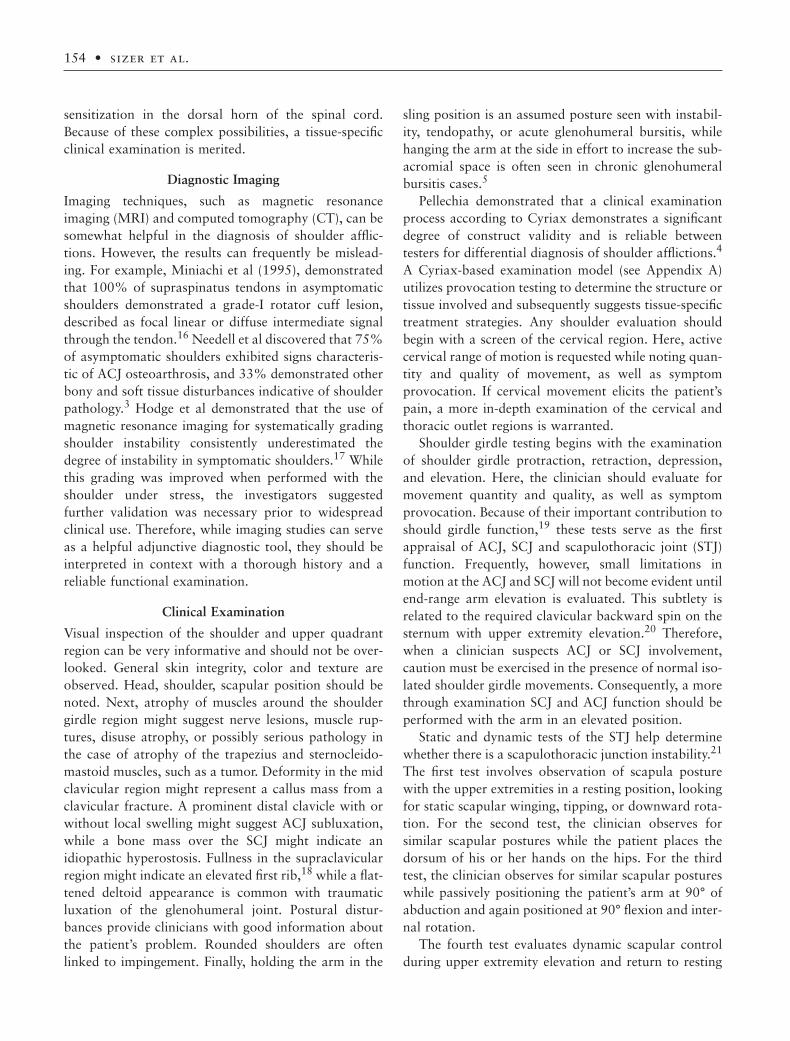
Passive arm elevation should be tested in three dif-ferent directions, so to evaluate all provocation possi-bilities (See Figure 1). In each case, the arm is passivelylifted into elevation, while the quantity of motion andquality of end feel are evaluated. In addition, provoca-tion of symptoms is considered, as differences in pro-vocation patterns between the test directions mayrepresent different afflictions. For the first direction, thecontra-lateral shoulder is stabilized while the ipsilateralupper extremity is lifted into full elevation, followed byposterior medial overpressure. If most painful of thethree tests, this direction indicates the contribution ofthe ipsilateral ACJ, SCJ, and cervicothoracic spine to thepainful affliction, meriting further tests of those struc-tures. Second, the ipsilateral scapula and acromion are fixated after passively lifting the patient’s upperextremity to 150° elevation, and end-range is assessedwith posterior medial overpressure, testing for internalimpingement of the rotator cuff. Lastly, the ipsilateralscapula and acromion are fixated after lifting the upper
extremity to 150° elevation, followed by posterior over-pressure at the endrange. If most painful, this test indi-cates subacromiodeltoid bursal involvement.
Quantity and quality of the motion, as well assymptom provocation, are evaluated during the passivetesting of GHJ abduction, internal rotation, and exter-nal rotation. Passive GHJ abduction is tested while palpating the inferior angle of the scapula, noting theextent of abduction at the first detectible scapularmotion. Internal and external rotation are tested withthe humerus at the patient’s side, as Cyriax suggestedthat testing these movements from that resting position
(a)
Figure 1. Passive Arm Elevation Testing, where: (a) the contra-lateral shoulder is stabilized while the ipsilateral upper extrem-ity is lifted into full elevation, finished with posterior medialoverpressure at end range; (b) the ipsilateral scapula andacromion are fixated after passively lifting the patient’s upperextremity to 150° elevation, finished with posterior medial over-pressure at end range; (c) the ipsilateral scapula and acromionare fixated after passively lifting the patient’s upper extremityto 150° elevation, finished with posterior overpressure at endrange.
156 • sizer et al.
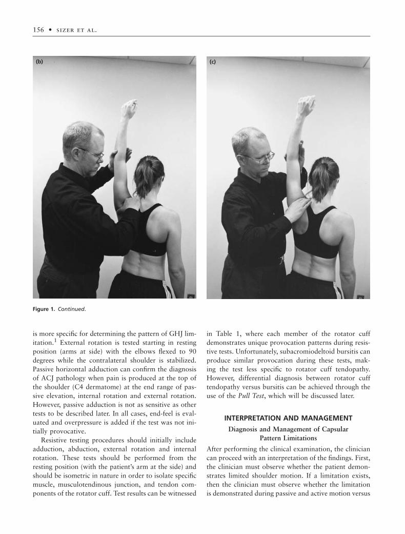
is more specific for determining the pattern of GHJ lim-itation.1 External rotation is tested starting in restingposition (arms at side) with the elbows flexed to 90degrees while the contralateral shoulder is stabilized.Passive horizontal adduction can confirm the diagnosisof ACJ pathology when pain is produced at the top ofthe shoulder (C4 dermatome) at the end range of pas-sive elevation, internal rotation and external rotation.However, passive adduction is not as sensitive as othertests to be described later. In all cases, end-feel is eval-uated and overpressure is added if the test was not ini-tially provocative.
Resistive testing procedures should initially includeadduction, abduction, external rotation and internalrotation. These tests should be performed from theresting position (with the patient’s arm at the side) andshould be isometric in nature in order to isolate specificmuscle, musculotendinous junction, and tendon com-ponents of the rotator cuff. Test results can be witnessed
in Table 1, where each member of the rotator cuffdemonstrates unique provocation patterns during resis-tive tests. Unfortunately, subacromiodeltoid bursitis canproduce similar provocation during these tests, mak-ing the test less specific to rotator cuff tendopathy.However, differential diagnosis between rotator cufftendopathy versus bursitis can be achieved through theuse of the Pull Test, which will be discussed later.
INTERPRETATION AND MANAGEMENT
Diagnosis and Management of Capsular Pattern Limitations
After performing the clinical examination, the cliniciancan proceed with an interpretation of the findings. First,the clinician must observe whether the patient demon-strates limited shoulder motion. If a limitation exists,then the clinician must observe whether the limitationis demonstrated during passive and active motion versus
(c)(b)
Figure 1. Continued.

Diagnosis and Management of the Painful Shoulder • 157
ment directions at the shoulder, without any detectablemotion limits. For example, a patient with an ACJ trau-matic synovitis would present with C4 pain at the end-range of normal arm elevation, external rotation andinternal rotation.
A clinician should pay particular attention to apatient’s historical specifics when a capsular pattern hasbeen observed. As suggested, this pattern can representeither synovitis, which can be traumatic or nontrau-matic in nature, or arthrosis associated with degenera-tive changes on the articular surfaces. Traumaticsynovitis can be related to a single macrotraumaticevent, or associated with a repetitive microtrauma, assustained while participating in a throwing sport ortennis. In addition, a patient can develop a nontrau-matic synovitis when afflicted with septic arthritis31 ora systemic disease, such as rheumatoid arthritis, psori-asis, or Reiter’s syndrome. Furthermore, a capsularpattern will slowly emerge as result of a primary arthro-sis, which is associated with the time-based degenera-tive events that accompany age. Finally, a capsularpattern can be caused by secondary arthrosis, whicharises when degeneration is accelerated in response todisease, a previous intra-articular trauma.5
Traumatic synovitis and primary arthrosis can beeffectively managed when the grade of the condition isconsidered. A minor affliction, or grade I, can presentwith a subtle capsular pattern of mildly limited (10°limit) and painful external rotation and, slightly limited(<5° limit) abduction, and painful internal rotation atend-range. The pain is mild and localized to the shoul-der, allowing continued function and sleep on either theinvolved or uninvolved side. This affliction is effectivelymanaged with joint specific mobilization, neuromuscu-lar re-education, home exercises and unloaded activities(such as pulley exercises). A grade II affliction presentswith a moderate capsular pattern, inability to sleep onthe involved side, and moderate pain often referring into the elbow. This condition can also be managed asa grade I, but may additionally require intra-articularsteroid injection. Finally, a grade III affliction presentswith severe pain that can refer distal to the elbow anda remarkable capsular pattern where the patient maynot be capable of producing any external rotation. Thegrade III lesion is best managed with rest, mild unloadedexercise and a sequential intra-articular injection series(discussed below), only allowing progressive exercisewhen the symptoms have begun to recede.
A glenohumeral capsular pattern limitation will alsobe observed when the patient suffers from idiopathic
active motion only. If the limits are during active motiononly (whereby passive motion is within normal limits),then the clinician should suspect a central nervoussystem affliction, peripheral nerve injury or rotator cufftear. Conversely, if the limitation is observed duringactive and passive motion, then the clinician mustdiscern whether the limitation is in the form of a cap-sular pattern or noncapsular pattern.5
The validity and reliability of this differential diag-nostic model has been examined within other synovialjoint systems. Investigators have demonstrated reliabil-ity when using the system at the knee,27 hip, and lumbarspine.28 Moreover, Pellechia et al found that this modelcan serve as a highly reliable schema for assessingpatients with shoulder limitation and pain.4 Therefore,identifying a joint’s limitation pattern can guide the clin-ician towards a more complete diagnostic concept of thepatient’s problem.
A capsular pattern is a predictable, repeatable andreliable pattern of passive motion limitation that is unique to each joint, is initiated by either intra-articular swelling29 or protective muscle guarding,30
and represents either synovitis (arthritis) or arthrosis (degenerative changes) within the joint.30 The capsularpattern (CP) for the glenohumeral joint is as follows:The external rotation limit is greater than the abductionlimit, which is greater than the internal rotation limit.In addition, the ratio of these limits is approximately 3 :2 :1, respectively.5 For example, a moderate capsularpattern at the glenohumeral joint could present as a 45-degree limitation in external rotation, a 30-degreelimitation in glenohumeral abduction (with the scapulastabilized), and a 15-degree limitation for internal rota-tion. On the other hand, the capsular pattern at boththe acromioclavicular and sternoclavicular joints (ACJ& SCJ) is pain produced at the end-range in all move-
Table 1. Provocation Test Patterns for Tendons of theRotator Cuff
Resisted Resisted Resisted ResistedAbd Add IR ER
Supraspinatus (++) (-) (-) (+)
Infraspinatus (-) (-) (-) (++)
Subscapularis (-) (-) (++) (-)
Teres Minor (-) (++) (-) (++)
Latissimus Dorsi (-) (++) (++) (-)Pec MinorTeres Major

158 • sizer et al.
adhesive capsulitis (IAC). While the limitations aresimilar to those observed with other synovial and chon-dropathic conditions of the glenohumeral joint, thepathology of this nontraumatic capsular transformationappears to be different from other capsular patternlesions. Although the etiology is not well understood,recent investigations have examined various features ofthis condition, lending insight into the pathologicalprocesses and development of clinical managementstrategies. For example, recent histological andimmunocytochemical findings demonstrate that theseprocesses include active fibroblastic proliferation,accompanied by limited transformation of a portion ofcapsular tissue into a smooth muscle phenotype likenedto myofibroblasts.32,33 These fibroblasts lay down col-lagen as a thick nodular band or fleshy mass that is verysimilar to that witnessed in Dupuytrens disease of thehand, with no inflammation or synovial involvement.This process appears to be most prevalent at the ante-rior-superior region of the glenohumeral capsuloliga-mentous complex, especially in the region of thecoracohumeral ligament and the rotator interval.34
Bunker et al suggested that this event cascade might betriggered by production of cytokines and growthfactors, which appear to be expressed locally in the cap-sular tissue.33 In addition, adhesive capsulitis can resultin significant humeral bone loss, which appears to betransient in nature.35
This affliction is most common in females (70%)between the ages of 40 and 60. It is more commonlyobserved in the dominant versus nondominant limb and is more prevalent in diabetic patients.7 This self-limiting affliction of unknown etiology appears only inthe glenohumeral and coxafemoral joints, transitioningthrough three distinct stages: freezing, frozen andthawing. During the freezing stage, the patient experi-ences gradually increasing painful limitations in gleno-humeral motion. With the examination of passivemotion during this stage, the examiner will provoke thepatient’s symptoms before the end-range of motion isreached, suggesting a painful, empty end-feel. Morespecifically, the patient will demonstrate a large limita-tion of elevation through flexion. In addition, externalrotation will be more limited in an adducted versusabducted testing position.
After two to nine months time, the patient’s condi-tion transitions into the frozen stage, where the limita-tions and pain plateau. During this stage, the examinerwill reach the patient’s end-range of motion, where painis immediately provoked. After an additional four to
twelve months, the condition finally reaches the thawingstage where the patient experiences a gradual resolutionof pain and variable restoration of movement. Duringthis stage the examiner is able to move the patient’sshoulder to the end range with minimal provocation ofsymptoms.
Careful consideration of the patient’s place in thevarious stages may assist the clinician with designing an effective management strategy. Clinicians haveattempted to manage this condition with a variety ofdifferent approaches, including injection, manipulationunder anesthesia (MUA), distention hydroplasty, andnonoperative rehabilitation. Vanderwindt et al found at7 weeks post-onset of IAC that 77% of the patientstreated with injections were considered to be treatmentsuccesses compared with the 46% out of those treatedwith physical therapy.36
While injection has been shown to address the seque-lae of idiopathic adhesive capsulitis, the dosage mayinfluence clinical outcomes. Dejong et al obtainedgreater symptom relief in the treatment of patients’frozen shoulders with a triamcinolone acetonide intra-articular injection of 40 versus 10mg.37 These investi-gators found that the injection produced a greater effecton the reduction of pain and sleep disturbances versusamelioration of motion deficits.
Clinicians have frequently incorporated manipula-tion under anesthesia as a treatment measure for IAC. Anderson evaluated this measure and early post-procedure continuous passive motion (CPM) on 24patients with arthroscopically verified frozen shoulders.The investigators found that 79% of the patientsreported slight or no pain after the procedure. In addi-tion, 75% returned to work at a mean of 9 weeks aftertreatment.38
Investigators have reported effective management ofIAC when incorporating a hydraulic distention of theGHJ capsule (Distention Hydroplasty) followed byearly post-procedure CPM.39–43 This procedure isaccomplished by introducing 5ml of 1% lidocaine fol-lowed by a bolus of sterile, chilled saline (up to 40ml)into the GHL capsular compartment through an 18-gauge 1.5 inch needle. Laroche et al found that the meanpain severity of patients suffering from IAC significantlydecreased after distention hydroplasty. In addition,passive abduction and external rotation each increasedsignificantly in the first five days after the procedure,whereas the change between day 5 and day 30 was notsignificant.42 Similarly, van Royen and Pavlov evaluatedthe effect of distention hydroplasty with 24 frozen

Diagnosis and Management of the Painful Shoulder • 159
joint specific mobilization to the glenohumeral joint inthe posterior translatory direction with the shoulderpositioned in adduction and submaximal internal rota-tion. These activities should be followed with a homestretching program that includes passive shoulder inter-nal rotation and horizontal adduction while the scapulais stabilized.
Patients who suffer from acute subacromial bursitiswill commonly demonstrate painfully limited passiveand active arm elevation, with internal and externalrotation relatively normal. Acute bursitis will presentwith a rapid onset of severe pain in the C5 distributionthat can potentially refer to the wrist and inability toelevate the arm in the direction of abduction or possi-bly flexion. This condition is related to rupture of apasty calcific material within a rotator cuff tendon. Thepasty, calcific material is caustic to the synovial liningof the subacromial bursa, triggering an aggressive synovial reaction. While this condition is commonlyself-limiting, the patient’s discomfort can be reducedwith rest and a steroid injection into the subacromialbursa.
Diagnosis and Management of Pain without Limitation
Many painful conditions of the shoulder complexpresent without limitation. Afflictions of the acromio-clavicular joint (ACJ) and sternoclavicular joint (SCJ)produce pain in the C4 distribution without limitation.Conversely, pain in the C5 distribution can emerge asthe result of nerve entrapment, tendinitis, tenosynovitis,bursitis, impingement, labral affliction and clinical GHJinstability. Each of these conditions presents with dis-tinctive provocation patterns during the functionalexamination. A comparison of resistive, modified resis-tive, passive and special tests can lead the clinician to a diagnostic conclusion and appropriate managementstrategy.
Acromioclavicular joint synovitis (arthritis) developsafter macro-or microtrauma, producing pain in the C4dermatome located directly over the joint. Pain is repro-duced at the end-range of all passive shoulder move-ments and resisted adduction of the arm is often painful.The most pain can be provoked with passive horizontaladduction and resisted horizontal abduction with theshoulder flexed to 90°. Synovitis at the ACJ can presentwith a hypomobility or be the consequence of jointinstability. Hypomobility at the ACJ is a consequence ofjoint effusion, as well as eventual capsular adaptations,and is best treated with intra-articular steroid injection
shoulders in 22 patients. The range of motion values at3 months after the procedure, when compared with theunaffected shoulder, can be witnessed in Table 2.43
Finally, Halverson and Maas found that 94% of theirpatients receiving distention hydroplasty for idiopathicarthritis demonstrated immediate and sustainedimprovement in shoulder pain and range of motion.44
Investigators have incorporated arthroscopic releaseinto the management of IAC.45,46 Ogilvieharris con-ducted a prospective cohort study on 40 patients suf-fering from IAC, 20 of which were treated withmanipulation and arthroscopy and 20 of which the con-tracted structures were divided through arthroscopy.The arthroscopic division was performed in 4 sequen-tial steps: (1) Resection of the inflammatory synoviumin the rotator interval; (2) Progressive division of theanterior superior glenohumeral ligament and anteriorcapsule; (3) Longitudinal separation of the subscapu-laris tendon; and (4) Longitudinal separation of the infe-rior capsule. Both groups reported similar levels ofsatisfaction during a follow up at 2–5 years post pro-cedure. However, the patients in the arthroscopic divi-sion had significantly greater pain relief and restorationof function.46
Diagnosis and Management of Non-Capsular Pattern Limitations
A noncapsular pattern (NCP) of limitation is anypattern of limitation other than the capsular pattern.5
While any combination of noncapsular pattern limi-tations can be potentially observed in the shouldercomplex, selected patterns appear to be more frequent.Patients commonly present in the clinic with shoulderinternal rotation limitation based on functional externalrotation requirements, such as throwing and reaching.This condition can be witnessed in athletes participat-ing in throwing or racket sports, and frequently perpet-uates impingement behaviors based on altered jointkinematics.47,48 This condition is best managed with
Table 2. The Effect of Distention Hydroplasty with 24Frozen Shoulders in 22 Patients at 3-Months Post-Procedure, Reporting % Return of Each MotionCompared to the Opposite Side
93% for shoulder abduction84% for glenohumeral abduction 94% for forward flexion 96% for backward extension 75% for internal rotation 73% for external rotation

160 • sizer et al.
followed in 7–10 days by mobilization and or manipu-lation. On the contrary, instability is typically the con-sequence of macrotrauma and subsequent failure of theacromioclavicular or coracoclavicular ligaments. Thiscondition presents with increased anterior-posterior orcraniocaudal ACJ movement, respectively. Coracoclav-icular ligament deficiency additionally leads to a possi-ble prominent distal clavicle associated with a droppedscapula (“Piano key sign”). Instability of the ACJ is besttreated with intra-articular steroid injection, shouldergirdle bracing and eventual shoulder girdle strengthen-ing so to utilize the investment of deltoid and trapeziusinsertions at the capsule of the ACJ.
Patients can develop posterior C5 shoulder pain inthe region of the scapula associated with suprascapularnerve entrapment. This condition develops as result oftrauma, neuritis, or slow progressive compression at thesuprascapular or spino-glenoid notch.49 Investigatorshave reported iatrogenic suprascapular nerve injuryduring Bankhart repair of rotator cuff lesions.50 Thenerve can be injured as result of macrotrauma, as witnessed during proximal humerus fractures.51
Microtrauma can lead to suprascapular nerve injury,as repeated shoulder horizontal adduction with scapularprotraction during volleyball slams or water poloreturns can traumatize the nerve. Here, the nerve isrepeatedly traction-loaded proximally through con-tralateral cervical sidebending and distally through arm and shoulder-girdle prepositioning. Additionally,pitchers can develop this condition after repeatedfollow-through maneuvers, by virtue of nerve irritationassociated with increased supra- and infraspinatus activ-ity during deceleration. This condition is provoked whenthe athlete’s shoulder is placed behind the back and pas-sively loaded in adduction, followed by contralateralcervical sidebending. Furthermore, suprascapular nervesymptoms can develop when a ganglion cyst forms onthe posterior labrum in response to a degenerative hori-zontal tear. Finally, this posterior shoulder pain shouldbe differentiated from posterior labral tear and internalimpingement of the supraspinatus against the glenoidlabrum and limbus (discussed later).
Suprascapular nerve lesions may demonstrate neuro-physiological dysfunction, but a painful injury can pre-sent with normal EMG findings. This condition is bestdetected through the suprascapular neural tension test,whereby the patient’s cervical spine is laterally flexedaway from the painful side, while the involved arm isplaced into horizontal adduction in front of the body orin adduction with the arm behind the body. This lesion
can be treated conservatively and or surgically. If thisposition reproduces the patient’s pain, a positive test isconfirmed when the pain is reduced with a return of thepatient’s neck to neutral, thus reducing tension on thenerve. Conservative management can include NSAIDs,steroid injection in the region of irritation, and eventualimplementation of a rotator cuff strengthening pro-gram. Surgical intervention includes open decompres-sion and possible possible neurolysis.49
Patients who suffer from rotator cuff tendinitis typi-cally experience the most pain provocation in the C5dermatome at the region of the deltoid during resistivetesting (See Table 1).5 Resistive testing should be per-formed isometrically and with the patient’s arm at theside, so to isolate strain within the tendon substance.1
Diagnosis of subscapularis tendinitis presents a conun-drum to clinicians. This tendinitis is provoked withresistive shoulder internal rotation, while other resistivetests are negative. This is an important distinction, dueto the contributions of pectoralis major, latissimus dorsi,and teres major to internal rotation and adduction. Ifany of these other muscles were involved in a painfulresistive internal rotation, then resisted adductionwould be painful as well. However, painless resistiveadduction (with the arm at the side) isolates the sub-scapularis as the tendon responsible for painful resistedinternal rotation.
Because of the powerful contributions of the pec-toralis major, latissimus dorsi, and teres major to inter-nal rotation, a patient suffering from a subscapularistear or rupture may demonstrate a false negative resistedinternal rotation test when the arm is positioned at theside and the elbow positioned to 90° of flexion. Thus,investigators have reported the use of additional tests toidentify subscapularis involvement, including the lift-offtest52–54 and internal rotation lag sign (IRLS).55 Thelift-off test involves placing the forearm behind the patient’s back and asking the patient to maintain theforearm off of the back’s surface. If the patient is unableto maintain the position, then the clinician shouldsuspect a complete rupture of the subscapularis (seeFigure 2). If the patient is able but the maneuver ispainful, the clinician should suspect either tendinitis ora partial tear (suspected if the patient has a history oftrauma). Finally, if the patient is limited in shouldermotion and unable to place the test arm behind theback, then the clinician can ask the patient to place the hand of the test arm against his / her stomach andthe clinician can proceed to lift the forearm and handoff of the stomach (modified lift-off test).

Diagnosis and Management of the Painful Shoulder • 161
When a patient demonstrates pain provocationduring any resistive test accompanied by findings of a painful arc during active arm elevation, the clini-cian should differentiate between tendinitis and sub-acromiodeltoid bursitis, as activation of any rotator cuffmuscle can increase acromiohumeral interval pressureand bursal pain. This is achieved through the “PullTest”,5 where the clinician repeats the same resistivetests while pulling on the humerus, thereby separatingthe acromiohumeral interval (see Figure 3). This proce-dure should resolve bursal pain produced during thepreviously positive resisted tests, due to eliminatingbursal compression. On the other hand, the “Pull Test”would increase pain associated with a tendinitis, astension loading is advanced within the tendon substancewith the pull.
Patients can suffer from tenosynovitis of the bicepslong head, where the tenosynovial sheath is inflamedaround the tendon as it courses through the intertuber-cular sulcus.56 Biceps tendon lesions are commonly asso-ciated with degenerative changes in the glenohumeraljoint and seldom occur in isolation of other lesions aboutthe joint system.57 An affected synovial sheath is mostpainful with tension, thus the patient experiences the
(a) (b)
Figure 2. The Lift-Off Test. (a) Starting position, whereby the patient places the test arm behind the back and the clinician stabilizesthe distal humerus; (b) Test movement, whereby the clinician asks the patient to lift the test arm off of the back, while stabilizingthe distal humerus and preventing shoulder extension.
greatest pain provocation when the clinician first flexesthe elbow with the patient’s arm at the side, pronates theforearm, extends the shoulder in the glenoid plane(slightly outward), and finally extends the elbow (seeFigure 4). This sequence is emphasized, so to avoid inter-nally rotating the shoulder and producing a false posi-tive test associated with a winding-up of the GHJcapsule. Additionally, tenosynovitis is differentiatedfrom biceps tendinitis at the shoulder, the latter of whichproduces the greatest shoulder pain during resistedelbow flexion and resisted forearm supination.
While the treatment of symptoms associated with thepreviously mentioned painful, nonlimited shoulder con-ditions can include transverse or longitudinal frictiontechniques, modalities and or tissue-specific injection (tobe discussed), these treatments may not completely ame-liorate the patient’s overall condition. Consequently, the clinician is encouraged to address causation, so toreduce the risk of symptom return or persistence. Ten-dinitis, tenosynovitis, and bursitis at the shoulder canemerge in response to impingement and or instability.Therefore, accurate identification of these underly-ing entities should be exercised in concert with pain-relieving management techniques.

162 • sizer et al.
Shoulder Impingement
As previously mentioned, patients can develop shoulderpain without limitation as result of numerous soft tissuelesions, including tendinitis, tenosynovitis, and bursitis.Frequently, these afflictions can emerge in response to an impingement event in the confined spaces aboutthe glenohumeral joint. Impingement is the most frequent cause of shoulder pain and has served as adiagnostic “waste-basket” for classifying shoulderpain.58 Nordt et al stated that “. . . It (impingement) has become, to some degree, a catch-all diagnosis fornumerous disorders that have diverse causes but similar presentations . . .”59
Impingement is an event whereby structures aroundthe glenohumeral joint are friction- or impact-loadedduring glenohumeral motion. Impingement has beenclassified as either external or internal, based on its loca-tion and responsible mechanisms. During externalimpingement the subacromiodeltoid bursa and or ex-ternal surface of one or more rotator cuff tendons are compressed by surrounding structures, such as the acromion, coracoacromial ligament, or coracoidprocess. Conversely, internal impingement involvescompromise to the deep fibers of the rotator cuff as aconsequence of impact loading of those fibers betweentheir insertion on the greater tubercle and the superiorand or posterior glenoid limbus and labrum.
External impingement has been comprehensivelyevaluated and investigators have attempted to associateselected anatomical factors with the incidence and sever-ity of subacromial external impingement (see Table 3).Edelson and Taitz suggested that increased acromionlength could propagate increased incidence of impinge-
Figure 3. The “Pull Test” for differentiating rotator cuff ten-dinitis from subacromiodeltoid bursitis. For this test, the clinicianrepeats the previously performed resistive abduction test whilepulling on the humerus, thereby separating the acromiohumeralinterval.
Table 3. Factors Contributing to Shoulder Impingement
Anatomical Etiologies Biomechanical Etiologies Traumatic Etiologies Degenerative Etiologies Vascular Etiologies
Acromion Morphology Scapulothoracic Instability MacroTrauma Rotator Cuff Degeneration Zone of LabilityType I Winging Partial RC Tear Fibrosis Type II Tipping Complete RC Tear Hyperplasia Ranaud’sType III Upward Elevation Wringing Out’
Downward Rotation MicroTrauma Early FatigueAcromion Direction Inc’d Eccentric LoadFlat GHJ Hypomobility Dec’d Stress Tolerance Dec’d Stress ToleranceIntermediate Diablo Effect Inc’d Injury PotentialSteep Risk for MacroTrauma
GHJ HypermobilityAcromion Length InstabilityShort Inc’d Humeral TranslationLong
Rotator Cuff ImbalanceOs Acromiale Firing Order Disturbance

Diagnosis and Management of the Painful Shoulder • 163
(a) (b)
Figure 4. Biceps Stretch Test for biceps tenosynovitis. (a) Starting position, whereby the clinician first flexes the elbow with thepatient’s arm at the side, pronates the forearm, passively extends the shoulder in the glenoid plane (slightly outward); (b) Test move-ment, whereby the clinician passively extends the elbow, thus stretching the biceps tendon through the intertubercular sulcus at theshoulder.
Figure 5. Variations in the acromionprocess, from an outlet view. (a) Type I:Straight acromion process; (b) Clavicle;(c) Scapular spine; (d) Type II: Mild inferior “hook” on the edge of theacromion; (e) Type III: Prominent“hook” on the edge of the acromion.
ment.60 In addition, a horizontally oriented acromion isat greater risk for contributing to impingement.60
Finally, investigators have reported that Os Acromiale,or nonunion and persistence of the lateral acromion past 18 years old, could serve as a probable cause ofimpingement.61–64
Historically, Bigliani classified the shape of theacromion process (Types I–III; see Figure 5) and sug-gested that differences could influence the incidence
of impingement and any subsequent rotator cufftearing.65 Later, Bigliani et al suggested that 78% of allfull thickness rotator cuff tears were associated withType III acromia.65 Investigators have suggested thatdifferences in the acromion are acquired, resulting from altered tension loads imposed by the coracoa-cromial ligament and deltoid.66,67 Getz et al observedthat Type III acromia were more common in femalepatients and discovered that Type II acromia were

164 • sizer et al.
related to posterior capsule adaptive shortening of the glenohumeral joint.68
Although external impingement has been associatedwith advances in age, the relationship between age andincidence of Type III acromia is controversial.68,69 Inaddition, other investigators have identified a poor rela-tionship between acromial classification and rotator cuff lesions. For example, Zuckerman et al observed a poor clinical agreement between impingement and clavicle type and Banas et al observed a similar outcomebetween clavicle type and MRI-determined rotator cuffdisease.70,71
Although surgeons have attempted to reduce the clin-ical sequelae of impingement by surgically removing thecoracoacromial ligament and Type II or III acromionprojections, symptoms can persist after such a proce-dure. In addition, Levy et al found that the coracoacro-mial ligament can regenerate after excision, questioningthe efficacy of these measures in managing impinge-ment.72 However, while the role of subacromial archi-tecture in impingement has remained controversial,Flatow et al identified other factors that could decreasethe acromiohumeral interval, leading to increased inter-val pressure and subsequent symptoms.73 These factorsinclude rotator cuff hypertrophy, a protracted restingscapular position, and posterior capsular adaptiveshortening.
The supraspinatus and infraspinatus demonstrate thepotential for hypertrophic changes, especially in concertwith excessive overhead activity. Excessive overheadactivity can lead to inflammation and subsequent hyper-trophic thickening of the rotator cuff, resulting in fric-tion and impingement of the tendons or bursa.74,75
These changes can be witnessed in occupational orsporting endeavors, such as swimming, throwing, andweightlifting.
Impingement can be exacerbated by scapulothoracicinstability, which can develop as result of static ordynamic dysfunction in the lower trapezius or serratusanterior. Static dysfunction results in a winged, tipped,or downwardly rotated scapular position with the armat the side or in a position of 90° to 100° abduction.Conversely, dynamic dysfunction demonstrates thesame scapular behavior during an elevation movementsequence, where the scapula wings, tips, or downwardlyrotates while the arm is elevated or returned from ele-vation to the starting position.21 In any case, malposi-tioning of the scapula produces decreased AHI space,increased intr-interval pressure, and subsequent symptoms.
Investigators have identified numerous other patho-mechanical behaviors in the shoulder complex thatcould perpetuate an impingement event. Laxity orinstability of the glenohumeral joint may produce exces-sive aphysiological translation of the humeral head,potentially increasing interval pressure in a similarfashion as previously described.74,76 Similarly, rotatorcuff imbalance could lend to poor control of humeralhead position leading to excessive superior translation,repeating the AHI pressure elevation and symptom pro-duction.77 Hypomobility in the glenohumeral jointcould perpetuate impingement, as well. A commonexample is witnessed with glenohumeral internal rota-tion limits, where the posterior capsule is tight and the humeral head is forced to migrate cranially duringelevation activities. While this affliction can be accom-panied by anterior capsular laxity, the subsequentmigration lends to increased interval pressure and pain.6
Chronic external impingement can induce rotatorcuff degeneration.78 While cuff degeneration is multi-factorial, chronic tendon injury can produce calciumphosphate accumulation and subsequent narrowing ofthe interval with accompanying elevated interval pres-sure.79 In addition, degeneration can result in decreasedstress tolerances and altered strain behaviors of atendon, thus lending to further degenerative changes.80
Progressive degeneration can lend to both glenohumeral
Figure 6. (a) Clavicle; (b) Acromioclavicular joint; (C) Acromionprocess; (d) Acromiohumeral interval; (e) Greater trochanter; (f)Lesser trochanter; (g) Humeral shaft; (h) subcoracoid interval; (j)Supraspinatus tendon approaching the insertion into the greatertubercle of the humerus; (k) External insertion of the supraspina-tus; (l) Internal insertion of the supraspinatus.

Diagnosis and Management of the Painful Shoulder • 165
instability and rotator cuff hyperplasia, resulting in an impingement event. These degenerative processesappear to be predisposed by hypovascularity in the mid-tendinous region of the cuff. This “zone of lability” hasbeen known for its contribution to compromised stresstolerance and subsequent degenerative tearing.81
External impingement can be localized to the sub-acromial or subcoracoid spaces of the shoulder complex(see Figure 6). A subacromial external impingementevent is more common with individuals greater than 30years of age and is associated with chronic rubbing ofthe supraspinatus and infraspinatus tendons, the prox-imal insertion of the biceps brachii (long head) and thesubacromiodeltoid (SAD) bursa.78 The bursa appears tobe the most frequent pain generator involved in exter-nal impingement by virtue of the unequalled sensorynerve supply.82–85 However, the rotator cuff can also
be compromised, where gradual external tearing mayensue. Clinically, the patient with external impingementpresents with pain in the C5 distribution that manifestsas a mid-range painful arc (80°–130°) during executionor return from arm elevation through flexion and orabduction.86 Although the patient may present withadditional findings of tendinitis or bursitis in the exam-ination (previously discussed), this painful arc is theclinical hallmark of external impingement and its pres-ence can be a strong diagnostic confirmation.58 Thediagnosis can be confirmed with the external impinge-ment test, whereby the clinician stabilizes the shouldergirdle, then internally rotates the humerus, passivelyflexes the shoulder to 90°, horizontally adducts the arm,and finally stresses the shoulder into end-range internalrotation (see Figure 7). In doing so, symptoms are pro-duced as the greater tubercle is forced cranially into the
(a) (b)
Figure 7. The External Impingement Test, whereby (a) the clinician stabilizes the shoulder girdle, then internally rotates the humerus,and (b) passively flexes the shoulder to 90°, horizontally adducts the arm, and finally stresses the shoulder into end-range internalrotation.

166 • sizer et al.
acromion, subsequently escalating pressure within theAHI.
External impingement can develop anterior to theglenohumeral joint in the subcoracoid interval (seeFigure 6). This subcoracoid impingement commonlyinvolves an impingement of the anterior soft tissuestructures against the coracoid process and appears tobe related to the extent and angle of coracoid processprojection.87 Clinically, the patient suffers from shoul-der pain in the C5 dermatome and exhibits a mid-rangepainful arc during flexion elevation between 80° and130° as the coracoid compresses the subcoracoid bursa,fibers of the subscapularis, biceps tendon or bicepstenosynovium.53 This affliction can best be provokedwith the anterior (or subcoracoid) impingement test,where the clinician stabilizes the shoulder girdle, flexesthe shoulder to 90°, horizontally adducts the arm andthen internally rotates the shoulder (see Figure 8).
This anterior impingement test resembles the ExternalImpingement Test, while differing in the test sequence.Whereas internal rotation is performed early in theExternal Impingement Test to ensure lesser tubercleclearance prior to elevation, it is the final step of the anterior impingement test, so to produce impinge-ment of the same tubercle against the coracoid process.
Internal impingement is more common in individualswho are less than 30 years of age. This affliction occurswhen the deep fibers of the supraspinatus or infra-spinatus impact loads against either the superior or pos-terior glenoid labrum and limbus (see Figure 9).88–90
Additionally, this impact loading can produce anaccompanying bone contusion when accompanied bymacrotrauma.91 The most common etiologies for thiscondition include throwing2 and overhead sports suchas water polo.92 Internal impingement of the deep
(a) (b)
Figure 8. The Anterior (or Subcoracoid) Impingement Test, where (a) the clinician stabilizes the shoulder girdle, flexes the shoulderto 90°, horizontally adducts the arm and then (b) internally rotates the shoulder.

Diagnosis and Management of the Painful Shoulder • 167
any associated labral cysts and axillary nerve lesions canbe elusive to the diagnostician as they can be difficult toview on imaging studies.
Finally, pain in the C4 dermatome can be provokedduring terminal elevation activities when a patientsuffers from impingement of the greater tubercle againstthe inferior surface of the acromioclavicular joint (ACJ;see Figure 6). This pain will commonly be produced at 160°–180° of flexion or abduction elevation. Theclinician must differentiate this affliction from super-ior internal impingement, as both afflictions produce pain at terminal elevation range. Thus, an aid to thisdifferentiation is identifying whether the pain is produced in a C4 or C5 distribution during terminalpassive elevation.
When managing impingement, the clinician shouldexecute measures for symptom alleviation and correc-tion of any causative factors. Symptoms that are asso-ciated with bursal irritation are best treated withinjection. Injections should be administered to the entiresubacromiodeltoid bursal compartment, as the bursacan demonstrate septally-divided compartmentalizationand the corticosteroid may not reach the inflamedregion with an injection in a single area of the space.5
Iontophoresis can also be used to reduce inflammation,alternating electrode placement to insure access tomaximum bursal regions. Placing the electrode anteriorto the acromion and positioning the shoulder in slightextension with internal rotation can allow the ion-tophoresis to access the anterior region. Conversely, theshoulder should be flexed to 60°, horizontally adductedand slightly externally rotated when the electrode isplaced posterior to the acromion.
After three to four days rest, clinicians can followinjection or iontophoresis with “bursal massage” Forthis technique, the patient is positioned supine with hisor her arm at the side. The clinician performs small arcpassive oscillations to the shoulder in the direction ofinternal-external rotation, while maintaining a longitu-dinal pull to the humerus in order to increase the sub-acromial space. This activity should be performed forseveral minutes to activate the subacromial glidingmechanism,82 to decrease inflammation and reduce painvia mechanoreceptor stimulus. The patient can producea similar activity at home by performing active small arcinternal-external rotation oscillations while gentlysqueezing a towel roll between the arm and trunk, thusactivating the adductors of the shoulder. This resistedadductor activity increases the sub-acromial space in asimilar fashion to a long axis humeral pull, thus reduc-
supra- and infraspinatus tendon fibers can occur againstthe superior glenoid labrum and limbus during over-head elevation activities.93–94 While C5 shoulder painis once again produced, the symptoms are provokedduring passive end-range elevation. The clinician canprovoke the symptoms by passively elevating the shoul-der in the abduction direction, then exerting a passiveoverpressure force to the humerus in a posterior-medialdirection while stabilizing the shoulder girdle. The clin-ician should note that in instances of internal impinge-ment, there is no painful arc during active arm elevation.
For posterior internal impingement, patients experi-ence posterior shoulder pain in the deltoid region thatis provoked in the terminal cocked position of thethrowing sequence (full GHJ Abduction / external rota-tion).92,93,95 A clinician can suspect this affliction whenthe greatest pain is produced during passive overpres-sure to glenohumeral external rotation in this position.Associated lesions can frequently develop, includinglabral cyst, axillary nerve entrapment, greater tuberclesclerosis, limbus erosions, humeral head osteochondraldefects, and Bennett’s lesion.92 A posterior labral cystcan emerge as a sequelum of a posterior labral tear orGHJ instability and can result in nonspecific posteriorshoulder pain.96 While imaging can be useful for thedetection of erosions, sclerosis, and chondral defects,
Figure 9. Location of internal impingement events at theglenoid labrum and limbus. (a) Glenoid fossa; (b) Glenoid limbus,or rim; (c) Glenoid labrum; (d) Site for internal impingement onthe posterior labrum and limbus; (e) Site for internal impinge-ment on the superior labrum and limbus.

168 • sizer et al.
ing any friction between the synovial surfaces during therepetitive motion.
Winkel et al has suggested several measures toaddress the tendopathies that develop with external orinternal impingement.5 First, both transverse and longi-tudinal friction can be applied to the supraspinatus,infraspinatus, subscapularis, or tenosynovium of thebiceps long head within the bicipital groove. It has beensuggested that this measure can promote pain reductionvia mechanoreceptor stimulation, collagen reorganiza-tion, and increased fibroblastic activity. In additionshoulder positions have been described that relocate therotator cuff tendons out from under the acromion.5
The supraspinatus can be located just anterior to theacromion when the shoulder is prepositioned in fullinternal rotation (the patient’s forearm is positionedbehind his or her back). Conversely, the infraspinatusinsertion is best located 1–1.5 inches caudal to the pos-terior acromion angle with the patient’s shoulder posi-tioned in 60° flexion, slight horizontal adduction andexternal rotation. In this position, the insertion is pal-pated on the posterior ridge of the greater tubercle onan imaginary line between the posterior acromion angleand the axilla. Friction massage can be applied to anytendon for several minutes in directions both transverseand longitudinal to the orientation of tendon fibers.
Friction techniques should be followed by gentlestretching in both the clinic and as a component of the home exercise program.97 Finally, injection using0.5–1.0mL corticosteroid and local anesthetic can beadministered to insertion tendopathy of the rotator cuff,accessing the insertions with the previously describedprepositions. The biceps tenosynovium can be injectedas well, while care must be taken to not infiltrate thetendon substance. Considering the catabolic influence ofcorticosteroids and the relative hypovascularity of longtendons, an infiltration to the midsubstance of thebiceps tendon may place that tendon at risk for ruptureor tendinosis.
Once symptom alleviation is under way, the clinicianshould incorporate measures to eradicate causativefactors. Associated muscle imbalances should beresolved (Jerosh et al, 1989), especially when the im-pingement is accompanied by laxity or instability of theglenohumeral joint or scapulothoracic junction.77 Itbehooves the clinician to begin with retraining of thescapular stabilizers, specifically the rhomboids, lowertrapezius, and serratus anterior. Serratus anterior exer-cises should emphasize eccentric control in the closedchain with the shoulder girdle positioned in retraction.
Secondly, the clinician can initiate resisted shoulderadduction, as this will decompress the subacromialspace and reduce impingement. Following this, thepatient can begin training of the shoulder internal rota-tors in the closed chain with the shoulder girdle posi-tioned in retraction, based on the importance ofsubscapularis for elevation activities.98,99 Additionally,infraspinatus appears to play a significant role in the ele-vation behaviors of the shoulder and, therefore, shouldbe activated.98,100,101 However, due to the potentialdeleterious influence supraspinatus activity can have onthe impingement event, resisted external rotation is initiated last and resisted shoulder abduction is com-pletely avoided.99,102 Finally, because neuromuscularre-education and coordination appear to supercedehypertrophic change when normalizing the shoulderfunction of the impingement patient, exercises shouldemphasize endurance versus strengthening. With this inmind, the clinician should ask the patient to ultimatelyperform the exercises for 100–200 repetitions at 25%maximum voluntary contraction (MVC). In addition,the exercise program should emphasize eccentric con-tractions, as this format appears to be more relevant tothe rotator cuff’s action during functional activities.
When an impingement patient presents with poste-rior capsular limitations, the clinician should implementjoint-specific mobilization and home exercise to stretchthe posterior capsule and restore normal glenohumeralinternal rotation.103–105 More specifically, superior pos-terior capsular limitations can be treated when the clin-ician translates the humeral head in a posterior, slightlylateral, and slightly cranial direction with the patientsupine and the shoulder positioned in internal rotationand slight abduction (see Figure 10).103,106 The patientcan follow this with a simple home exercise of placingthe hand in the rear pants pocket or behind back; fol-lowed by pulling the elbow forward while the scapulais kept retracted against the wall (see Figure 10). Conversely, a posterior-inferior capsular limitation isaddressed when the clinician performs a posterior-superior-lateral glide of the humeral head with thepatient supine and the shoulder positioned in 60°flexion, horizontal adduction, and minimal IR (seeFigure 11). For the home exercise, the patient lies oninvolved side in a semi-supine position such that thescapula is flat on mat and the shoulder elevated to 60°(see Figure 11). In this position, the patient uses theother hand to push the involved forearm toward the matwhile the elbow is kept at 90°, thus internally rotatingthe shoulder. While the clinician performs joint-specific

Diagnosis and Management of the Painful Shoulder • 169
mobilizations several times a week to coax the capsuleto adapt, the patient can perform the 20 repetitions ofthe home exercises several times per day.
Limitations in the sternoclavicular and acromioclav-icular joints (SCJ, ACJ) can influence shoulder girdlefunction and result in end-range elevation limitations.These limitations can contribute to an impingementevent, causing abnormal biomechanical behaviors in theelevation chain with resultant elevated AHI pressure.Thus, resolving limitations in the SCJ and ACJ can alleviate impingement-related symptoms. To achievenormal movement in these joints, One can use joint-specific mobilization in a position of end-range arm ele-vation. These techniques include anterior curved slideof clavicle on acromion at the ACJ, as well as caudal(slight lateral and slight ventral) slide of the clavicle onsternum at the SCJ. End-range elevation accounts for
the backward clavicular spin and subsequent twistedcapsules at each joint during elevation.20
Instability
Patients can develop instability in the shoulder thatlends to a variety of clinical sequelae, including tendopathy,107 impingement,58 degeneration,108,109
and labral afflictions.96 Prior to effectively examiningpatients for shoulder instability, one must understandthe difference between joint laxity and instability. Laxityis a nonpathological state of increased mobility in a par-ticular direction due to decreased tension or resistanceto movement that is normally provided by the bony orsoft tissue structures. Instability is a pathological con-dition that is linked to dissociation of the articular sur-faces, producing aphysiological loading and subsequentpain in from an associated soft tissue structure. Whereas
(a)(b)
Figure 10. (a) Manual treatment of a superior posterior capsularlimitation, where the clinician translates the humeral head in aposterior, slightly lateral, and slightly cranial direction, while thepatient is supine and the shoulder is positioned in internal rota-
tion and slight abduction. (b) Home exercise where the patientplaces the hand in the rear pants pocket or behind back; fol-lowed by pulling the elbow forward while the scapula is keptretracted against the wall.

170 • sizer et al.
laxity does not necessarily lead to instability, an unsta-ble shoulder can emerge from a lax state. Both laxityand instability can take place in concert with shoulderimpingement, where the impingement event occurseither in response to laxity or in concert with uni- ormultidirectional instability.58 Therefore, the clinician ischarged with the task of differentiating the patient’ssymptoms between pain related to impingement versusinstability, as each may require different, albeit inter-related, management strategies.
The stability of a joint relies on both static anddynamic supportive mechanisms. The sources of staticstability in the glenohumeral joint include the bonyarchitectural configuration and the integrity of the cap-suloligamentous structures that have been previouslyreviewed. Dynamic mechanisms not only include theactivity of the muscles around the joint, but also involvethe capsule once again, as the rotator cuff dynamizesthe capsule through collagenous investment. Failure inany of these components may result in a compromise ofglenohumeral stability, resulting in aphysiologicalloading within the joint and ensuing signs and symp-toms of instability. Whereas the glenohumeral joint nor-mally exhibits arthrokinematic rolling, spinning andtranslation, an unstable mechanism produces increasedarticular translation and potential for subluxation ordislocation. Although all instabilities do not result infrank articular perching or dislocation, these behaviorsare consistent with increased severity of the unstablecondition.
The etiological categories of glenohumeral instabilityinclude macrotraumatic, microtraumatic, nontraumatic,acquired, and congential lesions.110 Macrotrauma caninvolve an over-stretch or tearing of the capsuloliga-mentous complex, resulting in reduced motion con-straint. These lesions, which are more common inindividuals under 45 years of age, can be accompaniedby a Bankhart lesion that can disrupt the labrum andbony limbus, or Hill Sach’s Lesion that produces a dentin the cartilage and subchondral bone after the humeralhead is impact-loaded over the labrum and limbus. Con-sequently, radiographic studies should include specialviews to delineate these specific lesions.111 On the otherhand, microtraumatic lesions will result from a gradualoverstretch to the capsule, as witnessed during throw-ing or other overhead activities. Acquired nontraumaticinstability can develop as result of neuromuscular af-flictions, such as poliomyelitis, suprascapular nerveentrapment, or cerebrovascular accidient. In these cases,the locomotor system has failed to serve as a dynamic
(a)
(b)
Figure 11. Manual treatment of a superior posterior capsularlimitation, where the clinician performs a posterior-superior-lateral glide of the humeral head, while the patient is supine andthe shoulder is positioned in 60° flexion, horizontal adduction,and minimal IR. (b) Home exercise where the patient lies oninvolved side in a semi-supine position such that the scapula isflat on mat and the shoulder elevated to 60°. In this position,the patient uses the other hand to push the involved forearmtoward the mat while the elbow is kept at 90°, thus internallyrotating the shoulder.

Diagnosis and Management of the Painful Shoulder • 171
constraint to movement, resulting in laxity and painfulinstability. Moreover, the passive constraint of the cap-suloligamentous mechanism can also be compromisedby enzymatic degradation that accompanies systemicarthritides, such as rheumatoid arthritis.
Nontraumatic glenohumeral instability can emerge inresponse to congenital deformities. The capsuloliga-mentous complex can be overstretched during normalmovement when the glenoid fossa is more verticallyinclined, as well as in concert with torsional deformitiesat the scapular neck. In addition, architectural dispro-portion can contribute to instability. For example, theconstraints on the glenohumeral joint are compromisedwhen the radius of the glenoid fossa is proportionallysmall or the humeral head is either too large or small inrelation to the glenoid. Furthermore, humeral antetor-sion will excessively stretch the anterior glenohumeralcapsule over the humeral head in external rotation.112
Inherited general laxity conditions, such as Marfan’s orEhler’s Danlos, can compromise the collagen within thecapsule and ligaments, rendering it vulnerable to injuryand failure. Finally, females appear to be predisposed toglenohumeral instability, in response to increased jointlaxity.113
Instability can emerge after either greater tuberclefracture or tear of the rotator cuff and or biceps longhead. Individuals can develop partial or complete tearsin the rotator cuff in response to either macro- or micro-trauma. These tears can extend through the supraspina-tus,114 infraspinatus,115 and subscapularis 116 and can either present as full thickness or partial thick-ness,117,118 where the tear is observed on either thebursal or articular sides of the tendon.119 Rotator cufftears can be acute, where they develop in younger indi-viduals as result of abrupt macro trauma.120 Neer sug-gested that single-trauma tears are more common withindividuals under 40 years of age and can occur due toeither a traction or dislocation force.121 These tears frequently involve the anterior cuff, especially at therotator cuff interval, and can lead to multidirectionalinstability.122
A rotator cuff tear can be chronic in the 50–60 yearold male as a consequence of a repetitive microtrau-matic event, such as the recurrent external impingementthat is related to glenoid ostephytic changes123 or ex-ostotic changes in the underside of the acromionprocess.119 Degenerative rotator cuff tears appear to be more frequently initiated on the articular side nearthe insertion of the tendon,116 most likely related to the compromised blood supply at that region.119
While rotator cuff tears can be the source of significantshoulder pain,124 they can also present asymptomati-cally,16,125 lending to biomechanical changes that maymanifest as a painful condition in later years, such assubacromial bursitis.125
The onset of symptoms associated with rotator cufftear may be related to advancement of the tear and as-sociated biochemical changes within the joint.125,126
Large rotator cuff tears appear to accelerate cartilagedegradation, as evidenced by increased matrix metallo-protease markers in the synovial fluid. Considering thevital role the rotator cuff plays in the stability of theglenohumeral joint, one can recognize the relationshipthat rotator cuff failure has with instability and subse-quent degeneration.122
Pathologically, rotator cuff tears present with fattydegeneration within the tendon114 and amyloid deposi-tion within the region of the tear.127 These changes lendto irreversible structural changes, tendon retraction and subsequent joint degeneration. Degenerative jointchanges appear to be accelerated by the release of metalomatrix proteinases (MMP-1 and MMP-3) thatdegrade cartilage. Yoshihara et al observed increasedlevels of these proteinases, accompanied by increaselevels of glycosaminoglycans (GAGs), in the synovialfluid of the glenohumeral joint after cuff tear.126 In addition, they observed a correlation between the extent of the tear and the concentration of these molecules, suggesting a relationship between the extentof the tear and the potential for degenerative changes inthe joint.
Rotator cuff tears can be difficult to diagnose. Clin-ically, traumatic rotator cuff tears can present with a sig-nificant capsular pattern limitation as a consequence oftraumatic synovitis. However, a hallmark of theseinjuries is the patient’s inability to actively elevate thearm when the either the infraspinatus and or the sub-scapularis tendons are torn. As the swelling and inflam-mation subside, full thickness tears can present withnormal passive motion accompanied by the previouslymentioned resistive tests that are weak and painless.However, motions can eventually become increasinglypainful as degenerative changes progress.125 Moreover,partial tears are commonly symptomatic, potentiallyproducing painful weakness during the same resistivetests. Because rotator cuff tears can resemble ten-dopathies about the shoulder, they should be ruled outwith the appropriate imaging studies after a macro-traumatic event and or impingement symptoms persistin the aging individual.

172 • sizer et al.
Patients suffering from a degenerative tear will fre-quently present with signs of impingement, includingpainful arc and possible subacromial crepitus. In eithercase, the patients may feel weak with shoulder use andmay demonstrate a “Shrugging Sign”, where the shoul-der girdle elevates during attempts to elevate the arm.If either a tear of the infraspinatus or subscapularisaccompanies the supraspinatus tear, then the patient willdemonstrate a positive Drop Arm Test, where thepatient is unable to maintain a previously positionedshoulder in 90° abduction.128
Several imaging techniques have been suggested asconcomitants in the diagnosis of rotator cuff tears. Multidirectional plain-film imaging could be useful indetecting osseus lesions, such as glenoid osteophytes andor acromion abnormalities.123 High-resolution sono-gram has been proven to be useful in the accurate detec-tion of a cuff tear lesion.125,129 While Miniachi et alsuggested that nonenhanced MRI is of limited value forthe clinical detection of rotator cuff injury, they didsuggest its utility with full thickness rotator cuff tears.16
Similarly, Motamedi et al found that MRI could beuseful in the detection of full thickness, recurrent rotatorcuff tears in the post-surgical shoulder.117 However,these investigators suggested that the MRI tended toover-diagnose cuff tears and was limited in determiningthe size of the lesion.
Trauma can injure the patient’s glenoid labrum alongwith the capsuloligamentous complex, thus complicat-ing the clinical picture. Labrum lesions promote insta-bility, due to compromise of the negative intra-articularpressure and successive increased humeral head transla-tion. Patients can suffer from Bankhart lesions, seenespecially at the inferior anterior labral region.122 Theselesions have been classified as: (1) Type 1, or an avul-sion of the labrum from the glenoid limbus; (2) Type 2,or avulsion of the labrum and limbus; and (3) Type 3,or avulsion of the labrum limbus and fragment of theglenoid body. These lesions can be detected with appro-priate imaging and are best managed with surgicalrepair and or articular augmentation.
Another type of labral lesion from which patients candevelop instability is a lesion to the superior labrum, orSLAP lesion (Superior Labrum Anterior-to-Posterior).Investigators have reported various classification sys-tems that reflect location, involved tissues, and or causation.130–131 Type I lesions involve a fraying anddegeneration of the superior labrum, whereas Type IIlesions demonstrate a detachment of the biceps andsuperior labrum from the glenoid. Type III lesions
demonstrate a clean bucket-handle tear limited to thelabrum and Type IV lesions demonstrate an extensionof the bucket handle into the biceps tendon substance.A Type V lesion is a superior extension of a Bankhartlesion with a separation of the biceps tendon from theglenoid, while Type VI lesions demonstrate a flap tearof the labrum. Finally, the labral injury of a Type VIIlesion extends caudally into the middle glenohumeralligament.
Causes of a SLAP lesion include forced biceps ac-tivity under load, falls to the outstretched hand, trac-tion injuries, and shear loads associated withthrowing.122,132,133–134 McGough et al suggested thathigh tensile load tolerance of the proximal bicepstendon predisposes the labral-limbus interface to dis-ruption.135 Additionally, Pradhan et all found thatlabral strain is highest during the late cocking phase ofthrowing, where the shoulder is positioned in maximumexternal rotation. This strain is followed by eccentricbiceps contraction during follow through, where themuscle decelerates rapid elbow extension.136 Theseadjacent strain behaviors predispose the tendon-labruminterface to injury. Furthermore, Healey et al reportedthat the long head of the biceps (LHB) insertion into the supraglenoid tubercle provides 52% of thelinear stiffness of this tendon / cartilage / bone interface,while the labrum offers 15% of the linear stiffness.These authors reported that both the bony and thelabral insertions must be disrupted in order to create aType II SLAP lesion.137 Consequently, Patton andMcCluskey suggested that these relationships merit new surgical techniques that preserve the biceps-labralcomplex.138
Glenohumeral instability can be classified as volun-tary or involuntary. Patients suffering from voluntaryinstability possess the ability to sublux or dislocate theirafflicted shoulders on command. These individuals fre-quently gain social benefit from this capacity and rep-resent poor surgical candidates, due to the propensityfor post-operative dislocation. Conversely, the clini-cal symptoms of involuntary instability occur sponta-neously, causing sharp pain and a dead-arm feeling. Thisevent occurs when the patient is engaged in functionalactivities, especially when cognitively distracted and ormoving in a brisk fashion. The functional activities thattrigger this response are dependent on the direction ofthe instability, which includes anterior (most common),posterior, inferior, or multidirectional. An example of aninvoluntary anterior instability can be observed inpitchers during the late cocking phase of the throwing

Diagnosis and Management of the Painful Shoulder • 173
sequence. These individuals will experience the sharppain and dead arm when the arm reaches the fullycocked position, just prior to acceleration. Other ath-letes that may be predisposed to this affliction includepowerlifters, volleyball or waterpolo players, and swimmers.
Instabilities can be unidirectional or multidirectional.Shoulders can be unidirectionally unstable in either the anterior or posterior directions. Curl and Warrensuggested the ‘Circle Concept’, where the compromise inthe anterior and posterior capsulo-ligamentous struc-tures is required for instability in either the anterior orposterior directions.139 Anterior instability is frequentlyrelated to a lesion of the anterior-middle140 and or anterior-inferior glenohumeral ligaments.141 This pre-disposition may be related to increased mineralizedfibrocartilage and disorganization of ligament fibers in those regions.142 Posterior instabilities, which areuncommon and appear to be related to trauma or seizureactivity, emerge as a consequence of failure in both theanterior and posterior capsular regions. On the otherhand, multidirectional instabilities involve a sympto-matic inferior subluxation or dislocation related to aredundant inferior capsular recess and failure in theanterior and posterior capsular regions.122,143
Patients suffering from instability present withseveral distinctive clinical characteristics. Historically,younger patients can develop instability due to laxityand or trauma, whereas the middle to older age popu-lation experience instability in concert with degenera-tion or trauma. Instability produces a clinical triad of:(1) deep, sharp shoulder pain in the C5 dermatomeduring sudden or unexpected movements, followed bya “dead arm”; (2) possible noncapsular pattern of lim-itation accompanied by excessive motion on anotherdirection; and (3) a pathological end-feel during passivemotion assessment (either excessively soft or firm,depending on joint congruency and function). Thislesion can be accompanied by a painful click when thelabrum is involved in the instability. Patients are com-monly apprehensive to certain movements, especiallywith external rotation. The pain can be located in theanterior or posterior shoulder, due to either compres-sion or tension loading of the labral and or capsu-loligamentous structures. If produced while throwing,the patient may experience the pain during the cocking,acceleration or throwing phases.144
The examination of a patient suffering from gleno-humeral instability may be misleading or completelynegative, based on the elusive nature of the condition.
Therefore, several special tests have been proposed fordetection of a clinical instability at the shoulder. Thesetests can be categorized as either laxity tests or provo-cation tests, based on application and expected out-comes. Glenohumeral joint laxity can be detectedthrough joint play testing in an anterior-posterior direc-tion along the plane of the glenoid, while respecting theglenoid labrum (see Figure 12). The testing should beperformed with mild humeral proximal loading (‘loadand shift’) to increase translation.111,145 In addition, thetest can be performed in three shoulder pre-positions:(1) maximum loose packed position, with the shoulderpositioned at 55° abduction in the scapular plane; (2)with the arm at the side, so to test the superior gleno-humeral capsuloligamentous complex;146 and (3) at 90°abduction, so to test the anterior inferior capsuloliga-mentous structures.147
A laxity test is considered positive when the involvedside demonstrates increased translatory movement com-pared to the un-involved side. Hawkins has classifiedinstability as: (1) Type I: where the humeral headdemonstrates mild subluxation without perching overthe labrum; (2) Type II, where the humeral head percheson top of the glenoid labrum when the joint is fullyrotated; and (3) Type III, where the humeral headluxates, or dislocates, over the labrum.148 Type III ispotentially the easiest to diagnose, as the patient maypresent with a frank dislocation. However, the differ-ences between Type I and II may be more unclear.Although a Type II may be visualized during stressimaging, clinical laxity tests may produce glenohumeralperching, providing the patient relaxes and does notexhibit muscle guarding.
While the previous tests indicate laxity in the gleno-humeral joint, joint laxity does not necessarily equalinstability. Therefore, a diagnosis of instability needs toinclude positive provocation testing. Recent investi-gations have proposed the relocation and modified relocation tests for clinical diagnosis of shoulder instability.149,150 During the relocation test in supine, apatient suffering from anterior instability will demon-strate increased symptoms when the shoulder is posi-tioned at 90° abduction and 90° external rotation.Consequently, these symptoms will decrease when theclinician exerts a passive posterior force onto the ante-rior humeral head. Conversely, a patient suffering frominternal impingement may demonstrate increased symp-toms when the clinician exerts the posterior force,making the test a useful tool for differentiating poste-rior impingement from instability.95,151

174 • sizer et al.
As previously mentioned, patients may suffer fromlabral lesions in context with glenohumeral instability.In the case of anterior-inferior instability, the patientmay demonstrate a painful click during the previouslymentioned tests. In addition, these patients will be resis-tant to conservative management, as the painful clickwill persist and symptoms will not rescind. Selectedsuperior labral lesions can be detected through MRimaging, which is moderately sensitive and specific foras Type I, II, and III SLAP lesions.12 Additionally, whenthe clinician suspects a SLAP lesion, he or she maychoose to test the patient with the Crank test153 and oractive compression test of O’Brien.154 The crank test,demonstrating 93% specificity and 91% sensitivity forSLAP lesions, produces a painful click with passive rota-tion of the shoulder in an elevated position and thescapula stabilized. For the active compression test, pain
is produced when the clinician exerts a downward pres-sure to the patient’s shoulder that is positioned in 90°flexion, slight horizontal adduction, and full internalrotation (see Figure 13). The same test will be negativewhen repeated while the shoulder is re-positioned in fullexternal rotation. Finally, a patient with a Type II SLAPlesion and shoulder instability, will likely demonstrate apositive biceps load test. This test, which demonstrates90.9 sensitivity and 96.9 specificity for the lesion,131
produces pain during resisted elbow flexion when theforearm is fully supinated, the elbow is flexed to 90° andthe shoulder is prepositioned in 90° abduction and fullexternal rotation.155,156
Recently, clinicians have incorporated a variety ofmeasures to reduce the increased glenohumeral transla-tion associated with shoulder instability. Clinicians willfirst incorporate a nonoperative stabilization program
(a)
Figure 12. Glenohumeral joint laxity testing with the shoulder positioned in (a) maximum loose packed position, with the shoulderpositioned at 55° abduction in the scapular plane, and (b) with the arm at the side, so to test the superior glenohumeral capsu-loligamentous complex.
(b)

Diagnosis and Management of the Painful Shoulder • 175
prior to any invasive procedures. If instability is accom-panied by dislocation, the rehabilitation program maybe preceded by short-term immobilization. After reduc-tion, this problem was traditionally immobilized in aninternally rotated position, so to reduce stress on theanterior capsular structures. However, Itoi et al recentlyreported increased glenohumeral coaptation andreduced aphysiological translation when the glenhumeral joint is immobilized in external rotation (mean= 35°).157 They suggested that this reduction wasrelated to pretension in the subscapularis, thus creatinga tension-loaded restraint to anterior translation of thehumeral head.
Kibler suggested that there is no evidence indicatingthe most efficacious rehabilitation protocol for shoulderinstability.21 Regardless, a comprehensive rehabilitationprogram is aimed at improving strength in the parascapular muscles, deltoid and rotator cuff, while
reducing coordination disturbances and deficits in pro-prioception.158 The program should be initiated withNSAIDs and education in pain-reducing strategies.Patients then progress to exercises involving the scapu-lar stabilizers, especially the serratus anterior, latissimusdorsi and trapezius. The exercise program can be furtheradvanced to include activation of the rotator cuff, with particular emphasis on the subscapularis. Initially,patients should avoid end-range external rotation,abduction, and horizontal abduction. Additionally,rotator cuff activation may be enhanced when exercisesare performed in the closed chain159 and with elec-tromyographic biofeedback (EMGBF).160 Moreover,conservative rehabilitation programs appear to be moreeffective when used with patients with a nontraumaticetiology versus those with a traumatic etiology.161
Nonsurgical rehabilitation has been proven to bebeneficial to patients suffering from full thicknessrotator cuff tears.118 According to Brewster andSchwab, this program should initially focus on reduc-ing inflammation with activity modification, localmodality treatments, and posterior capsule stretch-ing.162 As inflammation begins to subside, the patientcan engage in resisted exercise to shoulder internal andexternal rotation, as well as extension and scapular ele-vation and retraction. Later, resisted exercises can beincorporated which emphasize improving both strengthand endurance of the shoulder complex. Finally, func-tional retraining is incorporated, including plyometrics,throwing, and or functional weightbearing. The successof a nonsurgical program on the rehabilitation of arotator cuff tear appears to be improved by a signifi-cantly worse post-morbid functional state and a lack ofeither litigation or worker’s comp status prior to imple-mentation, a high level of patient motivation, andinvolvement of the dominant upper extremity.163
Conservative measures are not always successful intreating the sequelae of glenohumeral instability.120
Numerous authors have reported more invasive proce-dures aimed at stabilizing the joint, including capsulor-rhaphy techniques that serve to shrink the glenohumeralcapsule. Heat-induced collagen denaturation can betriggered by thermal energy at radiofrequency tempera-tures between 65° and 80°C.164,165 Medvecky et al suggested that radiofrequency energy creates ionic agi-tation and frictional heating within the capsulartissue.110 This heat reduces heat-sensitive bonds in thecollagen, leading to collagen fibril uncoiling andrandom transformation. The coils ultimately re-contractin a disorganized fashion, producing a capsular shrink-
(a)
(b)
Figure 13. Active compression test for SLAP lesion, whereby theclinician exerts a downward pressure to the patient’s shoulderthat is positioned in (a) 90° flexion, slight horizontal adduction,and full internal rotation and (b) 90° flexion, slight horizontaladduction, and full external rotation.

176 • sizer et al.
age. This conformational change is accompanied byfibroblastic proliferation, fibrosis and connective tissuedisorganization, which all contribute to tissue matura-tion in a shrunken state. Tissue strain and yield strain change, lending to increased tissue stiffness. As a consequence, aphysiological translation within the glenohumeral joint decreases, reducing the clinicalconsequence of instability.164
This process has been incorporated to tighten anintact capsule that has stretched after microtrauma oras an adjunct to surgical repair.164 Levitz et al reportedthe use of electrothermal energy to internally shrink theglenohumeral capsules in the shoulders of baseballplayers who had previously failed conventional arthro-scopic debridement and repair.170 These authors foundthat the procedure reduced symptoms and re-injuryassociated with internal impingement. Wilk suggestedthat this procedure should be treated with two weeks ofimmobilization followed by progressive passive move-ment, limiting external rotation to 50° and abductionto 135° for four additional weeks.167 Finally, cliniciansare warned that thermal capsulorrhaphy can produceaxillary nerve injury in response to heat in close prox-imity to the nerve.168,169
Laser assisted capsular shrinkage has been proposedas an alternative to the radiofrequency thermal proce-dure. Wilk suggested that these patients should betreated with two weeks of immobilization followed byprogressive passive range, limiting external rotation to15° and abduction to 90° in the scapular plane for fouradditional weeks.167 Lyons et al reported that 96% oftheir patients’ shoulders treated with laser-assisted cap-sulorrhaphy remained persistently stable and asympto-matic at 24 months post-procedure.170 However, Levyet al reported a 36.1% failure rate in patients who weretreated with laser shrinkage, versus a 23.7% failure rate in patients treated with radiofrequency thermalshrinkage.72
Surgical re-stabilization is aimed at balancing thecapsular tension on all sides of the glenohumeral‘circle’.158 In general, the procedures involve overlap-ping and securing redundancies in the capsule, so topromote scarring and subsequent capsular tightness.While these corrections can be made from any approachto the joint, anterior inferior capsular shift proceduresappear to be more frequently implemented.158,171,172
Anterior-inferior capsular shift can reduce anterior andposterior translation, thus reflecting the ‘circle’ conceptand serving as a useful tool in the management of mul-tidirectional instability.139 Deutch et al evaluated the
biomechanical differences between glenoid-based versushumeral-based anterior-inferior capsular shift. While no differences in post-operative anterior or inferiortranslation were observed, certain mechanical featuresappeared superior with the glenoid-based shift.172
Finally, outcomes from arthroscopic restabilization withsuture anchors may equal or surpass traditional opentechniques in select cases.173
Partial thickness rotator cuff tears have beenmanaged through arthroscopic debridment with orwithout acromioplasty.115,119 Additionally, full thick-ness tears have been successfully repaired through botharthroscopic and open procedures.115,174 Longitudinalanalyses of such procedures have demonstrated persis-tent success between one and 10 years post-op.175,176
While range of motion and muscle strength deficits maypersist176 functional outcomes appear to improve overtime.175 Repair outcomes appear to improve whensuture anchors are incorporated and the superior gleno-humeral capsule and or coracohumeral ligament arereleased at the time of the repair.120,177
In the event of a Bankhart lesion to the labrum, sur-geons have incorporated a Bankhart reconstructive pro-cedure to reattach the defect to the scapular limbus.While this procedure has proven to be an effectivemeasure for restabilizing this shoulder lesion,178 differ-ences in post-surgical outcomes have been noted. First,Itoi et al. observed better post-operative external rota-tion when the surgeon performed a horizontal versusvertical incision through the joint capsule.146 Second, anarthroscopic approach to the procedure, while equal toopen procedure on most accounts, produced signifi-cantly greater external rotation in an abducted shoulderposition, which could serve functional restoration inactivities such as throwing.179
SUMMARY
Effective management of the painful shoulder is closelylinked to a tissue-specific clinical examination. Apatient’s shoulder pain presentation, while commonlyvague and referred in nature, can give the clinician cluesregarding the joint responsible for the painful affliction.Painful shoulder conditions can present with or withoutlimitations in passive and or active motion. Limits inpassive motion can be classified as either capsular ornoncapsular patterns, where capsular patterns are pre-dictable, repeatable patterns of passive motion limita-tion that are distinctive for each joint and representsynovitis or arthrosis. Common noncapsular patterns

Diagnosis and Management of the Painful Shoulder • 177
of the glenohumeral joint commonly accompany acutebursitis or impingement-related chronic bursitis and tendopathy.
Conversely, patients can present with shoulder painthat demonstrates no limitation of motion. Bursitis, ten-dopathy and rotator cuff tears can produce shoulderpain that is challenging to diagnose, especially whenthey are the consequence of underlying impingementand or instability. Furthermore, patients can developshoulder pain as a consequence of nerve entrapment,lending to a more complicated clinical presentation.Numerous nonsurgical measures can be implemented intreating the painful shoulder, reserving surgical inter-ventions for those patients who are resistant to conser-vative care.
REFERENCES1. Cyriax, J. Cyriax’s Illustrated Manual of Ortho-
paedic Medicine, Burlington, MA: Butterworth-HeinemannMedical, 1996.
2. Miniachi A, Fowler PJ. Impingement in the athlete.Clin Sports Med. 1993;12:91–110.
3. Needell SD, Zlatkin MB, Sher JS, Murphy BJ, UribeJW. MR imaging of the rotator cuff: peritendinous and boneabnormalities in an asymptomatic population. Am J Roent-gen. 1996;166:863–867.
4. Pellechia GL, Paolino J, Connell J. Intertester relia-bility of the Cyriax evaluation in assessing patients with shoul-der pain. J Orthop Sports Phys Ther. 1996;23:34–38.
5. Winkel D, Matthijs O, Phelps V. Part 1: The shoul-der. In Diagnosis and Treatment of the Upper Extremities.Gaithersburg: Aspen Publishers, Inc; 1997:1–308.
6. Tyler TF, Nicholas SJ, Roy T, Gleim GW. Quantifi-cation of posterior capsule tightness and motion loss inpatients with shoulder impingement. Am J Sports Med.2000;28:668–673.
7. Arkkila PE, Kantola IM, Viikari JS, Ronnemaa T.Shoulder capsulitis in type I and II diabetic patients: associa-tion with diabetic complications and related diseases. AnnRheum Dis. 1996;55:907–914.
8. Robinson AJ. Central nervous system pathways forpain transmission and pain control: issues relevant to the prac-ticing clinician. J Hand Ther. 1997;10:64–77.
9. Guibaud G, Bernard JF, Besson JM. Brain areasinvolved in nociception and pain. In Wall PD, Melzack R, eds.Textbook of Pain (CD ROM). Edinburgh: Churchill Living-stone; 1997, Record 1727.
10. Arendt-Nielsen L, Graven-Nielsen T, Svensson P.Assessment of muscle pain in humans—clinical and experi-mental aspects. In Vecchiet L, ed. Muscle Pain, MyofascialPain and Fibromyalgia: Recent Advances. San Antonio: Hay-worth Press; 1999:25–41.
11. Galletti R, Obletter G, Giamberardino MA, FonnicaCM, Cicchitti G, Vecchiet L. Pain in osteoarthritis of the knee.Adv Pain Res Ther. 1990;13:183–191.
12. Hoheisel U, Mense S, Simons DG, Yu XM. Appear-ance of new receptive fields in rat dorsal horn neurons fol-lowing noxious stimulation of skeletal muscle: a model forreferral of muscle pain? Neurosci Lett. 1993;153:9–12.
13. Mense S. Referral of muscle pain. Am Pain Soc J.1994;3:1–9.
14. Vecchiet L, Pizzigallo E, Iezzi S, Affaitata G, VecchietJ, Giamberardino MA. Differentiation of sensitivity in differ-ent tissues and its clinical significance. J Musculoskeletal Pain.1998;6:33–45
15. Giamberardino MA, Vecchiet L. Pathophysiology ofvisceral pain. Curr Rev Pain. 1996;1:23–33.
16. Miniachi A, Dowdy PA, Willits KR, Vellet AD. Mag-netic resonance imaging evaluation of the rotator cuff tendonsin the asymptomatic shoulder. Am J Sports Med. 1995;23:142–145.
17. Hodge DK, Beaulieu CF, Thabit GH, et al. DynamicMR imaging and stress testing in glenohumeral instability:Comparison with normal shoulders and clinical / surgical find-ings. J Mag Res Imag. 2001;13:748–756.
18. Lingren KA, Leino E, Manninen H. Cerical rotationlateral flexion test in brachialgia. Arch Phys Med Rehabil.1992;73:735–737.
19. Fung M, Kato S, Barrance PJ, Elias JJ, McFarlandEG, Nobuhara K, Chao EY. Scapular and clavicular kinemat-ics during humeral elevation: A study with cadavers. J Shoul-der Elb Surg. 2001;10:278–285.
20. van der Helm FCT. Analysis of kinematic anddynamic behavior of the shoulder mechanism. J Biomech.1994;27:527–550.
21. Kibler WB. Shoulder rehabilitation: principles andpractice. Med Sci Sports Ex. 1998;S40-S50.
22. McQuade KJ, Smidt GL. Dynamic scapulohumeralrhythm: the effects of external resistance during elevation ofthe arm in the scapular plane. J Orthop Sports Phys Ther.1998;27:125–133.
23. McQuade KJ, Hwa Wei S, Smidt GL. Effects of localmuscle fatigue on three-dimensional scapulohumeral rhythm.Clin Biomech. 1995;10:144–148.
24. McClure PW, Michener LA, Sennet BJ, Karduna AR.Direct 3-dimensional measurement of scapular kinematicsduring dynamic movements in vivo. J Shoulder Elb Surg.2001;10:269–277.
25. Eto M. Analysis of scapulohumeral rhythm for peri-arthritis scapulohumeralis. J Jap Orthop Assoc. 1991;65:693–707.
26. Warner JJ, Micheli LJ, Arslanian LE, Kennedy J,Kennedy R. Scapulothoracic motion in normal shoulders andshoulders with glenohumeral instability and impingement syn-drome. A study using Moire topographic analysis. ClinOrthop. 1992;285:191–199.

178 • sizer et al.
27. Fritz JM, Delitto A, Erhard RE, Roman M. An exam-ination of the selective tissue tension scheme, with evidencefor the concept of a capsular pattern of the knee. Phys Ther.1998;78:1046–56.
28. Greenwood MJ, Erhard RE, Jones DL. Differentialdiagnosis of the hip vs. lumbar spine: five case reports. J Orthop Sports Phys Ther. 1998;27:308–315.
29. Eyring EJ, Murray WR. The effect of joint positionon the pressure of intraarticular effusion. J Bone Joint Surg.1964;46-A:1235–1241.
30. Atkins E. Construct validity of Cyriax’s selectivetension examination: association of end-feels with pain at theknee and shoulder; Invited commentary. J Orthop Sports PhysTher. 2000;30:523–525.
31. Pommering TL, Wroble RR. Septic Arthritis of the Shoulder-Treating an Atypical Case. Phys Sportsmed.1996;24:75.
32. Hutchinson JW, Tierney GM, Parsons SL, DavisTRC. Dupuytren’s disease and frozen shoulder induced bytreatment with a matrix metalloproteinase inhibitor. J BoneJoint Surg. 1998;80-B:907–908.
33. Bunker TD, Reilly J, Baird KS, Hamblen DL. Expres-sion of growth factors, cytokines and matrix metallopro-teinases in frozen shoulder. J Bone Joint Surg. 2000;82-B:768–773.
34. Ozaki J. Pathomechanics and operative managementof chronic frozen shoulder. Ann Chir Gynaecol. 1996;85:156–158.
35. Leppala J, Kannus P, Sievanen H, Jarvinen M, VuoriI. Adhesive capsulitis of the shoulder (frozen shoulder) pro-duces bone loss in the affected humerus, but long-term bonyrecovery is good. Bone. 1998;22:691–694.
36. Vanderwindt DAWM, Koes BW, Deville W, BoekeAJP, Dejong BA, Bouter LM. Effectiveness of corticosteroidinjections versus physiotherapy for treatment of painful stiffshoulder in primary-care—Randomized trial. Br Med J.1998;317:1292–1296.
37. Dejong BA, Dahmen R, Hogeweg JA, Marti RK.Intraarticular triamcinolone acetonide injection in patientswith capsulitis of the shoulder-A comparative-study of 2 doseregimens. Clin Rehab. 1998;2:211–215.
38. Andersen NH, Sojbjerg JO, Johannsen HV, SneppenO. Frozen shoulder—Arthroscopy and manipulation undergeneral-anesthesia and early passive motion. J Shoulder ElbowSurg. 1998;7:218–222.
39. Andren L, Lundberg BJ. Treatment of rigid shouldersby joint distension during arthrography. Acta Orthp Scand.1965;36:45–53.
40. Ekelund AL, Rydell N. Combination treatment foradhesive capsulitis of the shoulder. Clin Orthop. 1992;282:105–109.
41. Fareed DO, Gallivan WR. Office management of frozen shoulder syndrome. Clin Orthop. 1989;242:177–183.
42. Laroche M, Ighilahriz O, Moulinier L, Constantin A,Cantagrel A, Mazieres B. Adhesive capsulitis of the shoulder—An open study of 40 cases treated by joint distension duringarthrography followed by an intraarticular corticosteroidinjection and immediate physical therapy. Revue du Rhuma-tisme. 1998;65:313–319.
43. van Royen BJ, Pavlov PW. Treatment of frozenshoulder by distension and manipulation under local anaes-thesia. Int Orthop. 1996;20:207–210.
44. Halverson L, Maas R. Shoulder joint capsule disten-tion (hydroplasty); A case series of patients with “frozenshoulders” treated in a primary care office. J Fam Prac.2002;51:61–64.
45. Harryman DT, Matsen FA, Sidles JA. Arthroscopicmanagement of refractory shoulder stiffness: Arthroscopy J Arthroscop Rel Surg. 1997;13:133–147.
46. Ogilvieharris DJ. The resistant frozen shoulder-manipulation versus arthroscopic release. Clin Orth.1995;319:238–248.
47. Baltaci G, Johnson R, Kohl H 3rd. Shoulder rangeof motion characteristics in collegiate baseball players. J Sports Med Phys Fit. 2001;41:236–242.
48. Safran MR, Borsa PA, Lephart SM, Fu FH, WarnerJP. Shoulder proprioception in baseball pitchers. J ShoulderElbow Surg. 2001;10:438–444.
49. Antoniou J, Tae S-K, Williams GR, Bird S, RamseyML, Iannotti JP. Suprascapular neruropathy. Variability in thediagnosis, treatment and outcome. Clin Orthop Rel Res.2001;386:131–138.
50. Shishido H, Kikuchi S. Injury of the suprascapularnerve in shoulder surgery: An anatomic study. J ShoulderElbow Surg. 2001;10:372–376.
51. Visser C, Coene N, Brand R, Tavy D. Nerve lesionsin proximal humeral fractures. J Shoulder Elbow Surg.2001;10:421–427.
52. Fabis J. Isolated traumatic rupture of the subscapu-lar tendon. Chir Narzadow Ruchu Ortop Pol. 1998;63:597–600.
53. Gerber C, Terrier F, Ganz R. The role of the cora-coid process in the chronic impingement syndrome, J BoneJoint Surg. 1985;67-B:703–708.
54. Gerber C, Krushell RJ. Isolated rupture of the tendonof the subscapularis muscle. Clinical features in 16 cases. J Bone Joint Surg. 1991;73-B:389–394.
55. Hertel R, Ballmer FT, Lombert SM, Gerber C. Lagsigns in the diagnosis of rotator cuff rupture. J ShoulderElbow Surg. 1996;5:307–313.
56. Favorito PJ, Harding WG, Heidt RS. Completearthroscopic examination of the long head of the bicepstendon. J Arthroscop Rel Surg. 2001;17:430–432.
57. Gill TJ, McIrvin E, Mair SD, Hawkins RJ. Resultsof biceps tenotomy for treatment of pathology of the longhead of the biceps brachii. J Shoulder Elbow Surg. 2001;10:247–249.

Diagnosis and Management of the Painful Shoulder • 179
58. Volpin G, Stahl S, Stein H. [Impingement syndromefollowing direct injuries of the shoulder joint]. Harefuah.1996;130:244–247; 295.
59. Nordt WE, Garrettson RB, Plotkin R. The measure-ment of subacromial contact pressure in patients withimpingement syndrome. Arthroscopy. 1999;15:121–125.
60. Edelson JG, Taitz C. Anatomy of the coraco-acromial arch. Relation to degeneration of the acromion. J Bone Joint Surg. 1992;74-B:589–594.
61. Grasso A. The incidence and role of the os acromi-ale in the acromiohumeral impingement syndrome. RadiologMed Tor. 1992;84:567–570.
62. Hutchinson MR, Veenstra MA. Arthroscopic decom-pression of shoulder impingement secondary to os acromiale.Arthroscopy. 1992;9:28–32.
63. Swain RA, Wilson FD, Harsha DM. The os acromi-ale: another cause of impingement. Med Sci Sports Ex.1996;28:1459–1462.
64. Wright RW, Heller MA, Quick DC, Buss DD.Arthroscopic decompression for impingement syndrome sec-ondary to an unstable os acromiale. Arthroscopy. 2000;16:595–599.
65. Bigliani LU, Ticker JB, Flatow EL, Soslowsky LJ,Mow VC. [Relationship of acromial architecture and diseasesof the rotator cuff]. Orthopade. 1991;20:302–309.
66. Shah NN, bayliss NC, Malcolm A. Shape of theacromion: Congenital or acquired-A macroscopic, radi-ographic, and microscopic study of acromion. J ShoulderElbow Surg. 2001;10:309–316.
67. Speer KP, Osbahr DC, Montella BJ, Apple AS, MairSD. Acromial morphotype in the young asyptomatic athleticshoulder. J Shoulder Elbow Surg. 2001;10:434–437.
68. Getz JD, Recht, MP, Piraino, DW, et al. AcromialMorphology—Relation to Sex, Age, Symmetry, and Subacro-mial Enthesophytes. Radiol. 1996;199:737–742.
69. Wang JC, MS Shapiro. Changes in Acromial Mor-phology with Age Full source, J Shoulder Elbow Surg.1997;6:55–59.
70. Zuckerman JD, FJ Kummer, F Cuomo, M Greller.Interobserver reliability of acromial morphology classifica-tion-An anatomic study. J Shoulder Elbow Surg. 1997;6:286–287.
71. Banas MP, Miller RJ, Totterman S. Relationshipbetween the lateral acromion angle and rotator cuff disease. J Shoulder Elbow Surg. 1995;4:454–461.
72. Levy O, Wilson M, Williams H, et al. Thermal capsular shrinkage for shoulder instability-Mid-term longitu-dinal outcome study. J Bone Joint Surg. 2001;83-B:640–645.
73. Flatow EL, Soslowsky LJ, Ticker JB, Pawluk RJ,Helper M, Ark J, Mow VC, Bigliani LU. Excursion of therotator cuff under the acromion. Patterns of subacromialcontact. Am J Sports Med. 1994;22:779–788.
74. Jobe CM, Kvitne RS, Giangarra CE. Shoulder painin the overhand or throwing athlete. The relationship of ante-
rior instability and rotator cuff impingement. Orthop Rev.1989;18:963–975.
75. McCann PD, Bigliani LU. Shoulder pain in tennisplayers. Sports Med. 1994;17:53–64.
76. Bigliani LU, Levine WN. Current concepts reviewsubacromial impingement syndrome. J Bone Joint Surg.1997;79-A:1854–1867.
77. Jerosh J, Castro WH, Sons HU, Moersler M. Zur atiologie des subaromialen impingement-syndroms—einebomechanische untersuchung. Bietr Othop Traumatol.1989;36:411–418.
78. Panni AS, Milano G, Lucania L, Fabbriciani C,Logroscino CA. Histological analysis of the coracoacromialarch: correlation between age-related changes and rotator cufftears. Arthroscopy. 1996;12:531–540.
79. Riley GP, Harrall RL, Constant CR, Cawston TE,Hazleman BL. Prevalence and possible pathological signifi-cance of calcium phosphate salt accumulation in tendonmatrix degeneration. Ann Rheum Dis. 1996;55:109–115.
80. Sano H, Ishii H, Yeadon A, Backman DS, Brunet JA,Uhthoff HK. Degeneration at the insertion weakens the tensilestrength of the supraspinatus tendon: A comparative mechan-ical and histologic study of the bon-tendon complex. J OrthopRes. 1997;15:719–726.
81. Determe D, Rongieres M, Kany J, Glasson JM, Bellumore Y, Mansat M, Becue J. Anatomic study of the tendinous rotator cuff of the shoulder. Surg Radiol Anat.1996;18:195–200.
82. Ide K, Shirai Y, Ito H, Ito H. Sensory nerve supplyin the human subacromial bursa. J Shoulder Elbow Surg.1996;5:371–382.
83. Soifer TB, Levy HJ, Soifer FM, Kleinbart F, VigoritaV, Bryk E. Neurohistology of the subacromial space.Arthroscopy. 1996;12:182–186.
84. Vangsness CT, Ennis M, Taylor JG, Atkinson R.Neural anatomy of the glenohumeral ligaments, labrum, andsubacromial bursa. Arthroscopy. 1995;11:180–184.
85. Aszmann OC, Dellon AL, Birely BT, McFarland EG.Innervation of the human shoulder joint and its implicationsfor surgery. Clin Orthop. 1996;330:202–207.
86. Neer CS. Impingement lesions. Clin Orthop.1983;173:70–77.
87. Anetzberger H, Putz R. [Morphometry of the sub-acromial space and its clinical relevance]. Unfallchirurg.1995;98:407–414.
88. Walch G, Boileau P, Noel E, Donell ST. J ShoulderElbow Surg. 1992;1:238–245.
89. Jobe CM, Sidles J. Evidence for a superior glenoidimpingement upon the rotator cuff. J Elbow Shoulder Surg.1993;2:S19.
90. Davidson PA, Elattrache NS, Jobe CM, Jobe FW.Rotator cuff and posterior-superior glenoid labrum injuryassociated with increased glenohumeral motion: A new site ofimpingement. J Shoulder Elbow Surg. 1995;4:384–390.

180 • sizer et al.
91. Anzilotti KF, Schweitzer ME, Oliveri M, Marone PJ.Rotator cuff strain: a post-traumatic mimicker of tendonitison MRI. Skeletal Radiol. 1996;25:555–558.
92. Giombini A, Rossi F, Pettrone FA, Dragoni S. Pos-terosuperior glenoid rim impinement as a cause of shoulderpain in top level waterpolo players. J Sports Med Phys Fit.1997;37:273–278.
93. Jobe CM. Superior glenoid impingement. Currentconcepts. Clin Orthop. 1996;330:98–107.
94. Jobe CM. Superior glenoid impingement. OrthopClin N Am. 1997;28:137–143.
95. Hamner DL, Pink MM, Jobe FW. A modification ofthe relocation test: arthroscopic findings associated with a pos-itive test. J Shoulder Elbow Surg. 2000;9:263–267.
96. Ferrick MR, Marzo JM. Ganglion cyst of the shoul-der associated with a glenoid labral tear and symptomaticglenohumeral instability. A case report. Am J Sports Med.1997;25:717–719.
97. Jerosh J, Castro WH, Drescher H, Assheuer J.Kernsinmorphologische verandeerungen an schultergelenkenvon weltklasse-wasserballspielern. Sportverl Sportsch. 1993;7:109–114.
98. Rowlands LK, Wertsch JJ, Primack SJ, Spreitzer AM,Roberts MM. Kinesiology of the empty can test. Am J PhysMed Rehabil. 1995;74:302–304.
99. Wuelker N, Roetman B, Plitz W, Knop C. [Functionof the supraspinatus muscle in a dynamic shoulder model].Unfallchirung. 1994;97:308–313.
100. Otis JC, Jiang CC, Wickiewicz TL, Peterson MG,Warren RF, Santner TJ. Changes in the moment arms of therotator cuff and deltoid muscles with abduction and rotation.J Bone Joint Surg. 1994;76-A:667–676.
101. Sharkey NA, Marder RA, Hanson PB. The entirerotator cuff contributes to elevation of the arm. J Orthop Res.1994;12:699–708.
102. Thompson WO, Debski RE, Boardman ND 3rd,Taskiran E, Warner JJ, Fu FH, Woo SL. A biomechanicalanalysis of rotator cuff deficiency in a cadaveric model. Am JSports Med. 1996;24:286–292.
103. Chandler TJ, Kibler WB, Uhl TL, Wooten B, KiserA, Stone E. Flexibility comparisons of junior elite tennisplayers to other athletes. Am J Sports Med. 1990;18:134–136.
104. Ellenbecker TS, Roetert EP, Piorkowski PA, SchulzDA. Glenohumeral joint internal and external rotation rangeof motion in elite junior tennis players. J Orthop Sports PhysTher. 1996;24:336–341.
105. Kibler WB. Evaluation of sports demands as a diag-nostic tool in shoulder disorders. In: Matsen FA, Fu FH,Hawkins RJ, eds. The Shoulder: A Balance of Mobility andStability. Rosemont: American Academy of Orthopaedic Sur-geons; 1993:379–398.
106. Pappas M, Zawacki RM, McCarthy CF. Rehabilita-tion of the pitching shoulder. Am J Sports Med. 1985;13:223–235.
107. Glousman R, Jobe F, Tibone J, Moynes D, AntonelliD, Perry J. Dynamic electromyographic analysis of the throw-ing shoulder with glenohumeral instability. J Bone Joint Surg.1988;70-A:220–226.
108. Edelson JG. Bony changes of the glenoid as a con-sequence of shoulder instability. J Shoulder Elbow Surg.1996;5:293–298.
109. Marx RG, McCarty EC, Montemurno D, AltcheckDW, Craig EV, Warren RF. Development of arthrosis follow-ing dislocation of the shoulder: a case-control study. J Shoul-der Elbow Surg. 2002;11:1–5.
110. Medvecky MJ, Ong BC, Rokito AS, Sherman OH.Thermal capsular shrinkage: Basic science and clinical appli-cations. Arthroscopy. 2001;17:624–635.
111. Mahaffey BL, Smith PA. Shoulder instability inyoung athletes. Am Fam Phys. 1999;59:2773–2787.
112. Kronberg M, Brostrom LA. Humeral head retrover-sion in patients with unstable humeroscapular joints. ClinOrthop. 1990;260:207–211.
113. Borsa PA, Sauers EL, Herling DE. Patterns of gleno-humeral joint laxity and stiffness in healthy men and women.Med Sci Sports Ex. 2000;32:1685–1690.
114. Nakagaki K, Ozaki J, Tamita Y, Tamai S. Fattydegeneration in the supraspinatus muscle after rotator cufftear. J Shoulder Elbow Surg. 1996;5:194–200.
115. Murray TF, Lajtai, Mileski RM, Snyder SJ. Arthro-scopic repair of medium to large full-thickness rotator cufftears: Outcome at 2- and 6-year follow-up. J Shoulder ElbowSurg. 2002;11:19–24.
116. Sakurai G, Ozaki J, Tomita Y, Kondo T, Tamai S.Incomplete tears of the subscapularis tendon associated withtears of the supraspinatus tendon: Cadaveric and clinicalstudies. J Shoulder Elbow Surg. 1998;7:510–515.
117. Motamedi AR, Urrea LH, Hancock RE, HawkinsRJ, Ho C. Accuracy of magnetic resonance imaging in deter-mining the presence and size of recurrent rotator cuff tears. J Shoulder Elbow Surg. 2002;11:6–10.
118. Wirth MA, Basamania C, Rockwood CA. Nonoper-ative management of full-thickness tears of the rotator cuff.Orthop Clin N Am. 1997;28:59–66.
119. Budoff JE, Nirschl RP, Guidi EJ. Debridement ofpartial-thickness tears of the rotator cuff without acromio-plasty. J Bone Joint Surg. 1998:80-A:733–748.
120. Reed SC, Glossop N, Ogilvie-Harris DJ. Full-thickness rotator cuff tears: A biomechanical comparison ofsuture versus bone anchor techniques. Am J Sports Med.1996;24:46–48.
121. Neer CS. Shoulder Reconstruction. Philadelphia, Pa:W. B. Saunders Company, 1990.
122. Levine WN, Flatow EL. The pathophysiology ofshoulder instability. Am J Sports Med. 2000;28:911–917.
123. Konno N, Itoi E, Kido T, Sano A, Urayama M, SatoK. Glenoid osteophyte and rotator cuff tears: an anatomicstudy. J Shoulder Elbow Surg. 2002;11:72–79.

Diagnosis and Management of the Painful Shoulder • 181
124. Shibata Y, Midorikawa K, Naito M. Clinical evalu-ation of sodium hyaluronate for the treatment of patients withrotator cuff tear. J Shoulder Elbow Surg. 2001;10:209–216.
125. Yamaguchi K, Tetro AM, Vlam O, Evanoff BA,Teefey SA, Middleton WD. Natural history of asymptomaticrotator cuff tears: a longitudinal analysis of asymptomatictears detected sonographically. J Shoulder Elbow Surg.2001;10:199–203.
126. Yoshihara Y, Hamada K, Nakajima T, Fujikawa K,Fukuda H. Biochemical markers in the synovial fluid of gleno-humeral joints from patients with rotator cuff tear. J OrthopRes. 2001;19:573–579.
127. Cole AS, Cordiner-Lawrie S, Carr AJ, AthanasouNA. Localised deposition of amyloid in tears of the rotatorcuff. J Bone Joint Surg. 2001;83-B:561–564.
128. Magee DJ. Orthopedic Physical Assessment.Philadelphia, Pa: W. B. Saunders Company, 2002.
129. Roberts CS, Galloway KP, Honaker JT, Hulse G,Seligson D. Sonography for the office screening of suspectedrotator cuff tears: Early experience of the orthopedic surgeon.Am J Orthop. 1998;27:503–506.
130. Snyder SJ, Banas MP, Karzel RP. AN analysis of 140injuries to the superior glenoid labrum. J Shoulder ElbowSurg. 1995;4:243–248.
131. Huijbregts PA. SLAP lesions: Structure, function, andphysical therapy diagnosis and treatment. J Man Manip Ther.2001;9:71–83.
132. Higgins LD, Warner JJP. Superior labral lesions;Anatomy, pathology and treatment. Clin Orthop Rel Res.2001;390:73–82.
133. LaBan MM, Gurin TL, Maltese JT. Slip of thelip–tears of the superior glenoid labrum-anterior to posterior(SLAP) syndrome. A report of four cases. Am J Phys MedRehabil. 1995;74:448–452.
134. Maffet MW, Gartsman GM, Molsely B. Superiorlabrum-biceps tendon complex lesions of the shoulder. Am JSports Med. 1995;23:93–98.
135. McGough RL, Debski RE, Taskiran E, Fu FH, WooSL. Mechanical properties of long head of biceps tendon. KneeSurg Sports Traum Arth. 1996;3:226–229.
136. Pradhan RL, Itoi E, Hatakeyama Y, Urayama M,Sato K. Superior labral strain during the throwing motion—A cadaveric study. Am J Sports Med. 2001;29:488–492.
137. Healey JH, Barton S, Noble P, Kohl HW, Ilahi OA.Biomechanical evaluation of the origin of the long head of thebiceps tendon. Arthroscopy. 2001;17:378–382.
138. Patton WC, McCluskey GM. Biceps tendinitis andsubluxation. Clin Sports Med. 2001;20:505–529.
139. Curl LA, Warren RF. Glenohumeral joint stability.Selective cutting studies on the static capsular restraints. ClinOrthop Rel Res. 1996;330:54–65.
140. Savoie FH, Papendik L, Field LD, Jobe C. Straightanterior instability: Lesions of the middle glenohumeral liga-ment. Arthroscopy. 2001;17:229–235.
141. Matzen FA, Arntz CT. Subacromial impingement. In:Rockwood CA, Matsen FA, eds. The Shoulder. Philadelphia,Pa: W. B. Saunders Company; 1990:623–636.
142. McMahon PJ, Dettling J, Sandusky MD, Tibone JE,Lee TQ. The anterior band of the inferior glenohumeral liga-ment. Assessment of its permanent deformation and theanatomy of its glenoid attachment. J Bone Joint Surg.1999;81-B:406–413.
143. Yoldas EA, Faber KJ, Hawkins RJ. Translation of the glenohumeral joint in patients with multidirectional and posterior instability: Awake examination versus examinationunder anesthesia. J Shoulder Elbow Surg. 2001;10:416–420.
144. Werner SL, Gill TJ, Murray TA, Cook TD, HawkinsRJ. Relationships between throwing mechanics and shoulderdistraction in professional baseball pitchers. Am J Sports Med.2001;29:354–358.
145. Halder AM, Halder CG, Zhao KD, ODriscoll SW,Morrey BF, An KN. Dynamic inferior stabilizers of the shoul-der joint. Clin Biomech. 2001;16:138–143.
146. Itoi E, Watanabe W, Yamada S, Shimizu T, Wakabayashi I. Range of motion after Bankart repair—Vertical compared with horizontal capsulotomy. Am J SportsMed. 2001;29:441–445.
147. Turkel SJ, Panio MW, Marshall JL, Girgis FG. Sta-bilizing mechanisms preventing anterior dislocation of theglenohumeral joint. J Bone Joint Surg Am. 1981;63:1208–1217.
148. Hawkins RJ, Mohtadi NGH. Controversy in ante-rior shoulder instability. Clin Orthop. 1991;272:152–161.
149. Kolbel R. A modification of the relocation test:arthroscopic findings associated with a positive test. J Shoul-der Elbow Surg. 2001;10:497–498.
150. Speer KP, Hannafin JA, Altchek DW, Warren RF. Anevaluation of the shoulder relocation test. Am J Sports Med.1994;22:177–183.
151. Paley KJ, Jobe FW, Pink MM, Kvitne RS, ElAttracheNS. Arthroscopic findings in the overhand throwing athlete:evidence for posterior internal impingement of the rotatorcuff. Arthroscopy. 2000;16:35–40.
152. Won-Hee J, McCauley TR, Katz LD, Matheny JM,Ruwe PA, Daigneault JP. Superior labral anterior posterior(SLAP) lesions of the glenoid labrum: Reliability and accuracyof MR arthrography for diagnosis. Radiol. 2001;218:127–132.
153. Liu SH, Henry MH, Nuccion SL. A prospective eval-uation of a new physical examination in predicting glenoidlabral tears. Am J Sports Med. 1996;24:721–725.
154. O’Brien SJ, Pagnani MJ, Fealy S, McGlynn SR,Wilson JB. The active compression test: a new and effectivetest for diagnosing labral tears and acromioclavicular jointabnormality. Am J Sports Med. 1998;26:610–613.
155. Kim SH, Ha KI, Han KY. Biceps load test: A clinicaltest for superior labrum anterior and posterior lesions in the

182 • sizer et al.
of the shoulder. J Shoulder Elbow Surg. 2001;10:231–235.
169. Gryler EC, Greis PE, Burks RT, West J. Axillarynerve temperatures during radiofrequency capsulorrhaphy ofthe shoulder. Arthroscopy. 2001;17:567–572.
170. Lyons TR, Griffith PL, Savoie FH, Field LD. Laser-assisted capsulorrhaphy for multidirectional instability of theshoulder. Arthroscopy. 2001;17:25–30.
171. Bak K, Spring BJ, Henderson JP. Inferior capsularshift procedure in athletes with multidirectional instabilitybased on isolated capsular and ligamentous redundancy. AmJ Sports Med. 2000;28:466–471.
172. Deutsch A, Barber JE, Davy DT, Victoroff BN. Anterior-inferior capsular shift of the shoulder: a biomechan-ical comparison of glenoid-based versus humeral-based shiftstrategies. J Shoulder Elbow Surg. 2001;10:340–352.
173. Cole BJ, Romeo AA. Arthroscopic shoulder stabi-lization with suture anchors: Technique, technology, and pit-falls. Clin Orthop Rel Res. 2001;390:17–30.
174. Iannotti JP, Bernot MP, Kuhlman JR, Kelley MJ,Williams GR. Postoperative assessment of shoulder function:a prospective study of full-thickness rotator cuff tears. J Shoul-der Elbow Surg. 1996;5:449–457.
175. Galatz LM, Griggs S, Cameron BD, Iannotti JP.Prospective longitudinal analysis of postoperative shoulderfunction: a ten-year follow-up study of full-thickness rotatorcuff tears. J Bone Joint Surg. 2001;83-A:1052–1056.
176. Pai VS, Lawson DA. Rotator cuff repair in a districthospital setting: Outcomes and analysis of prognostic factors.J Shoulder Elbow Surg. 2001;10:236–241.
177. Hatakeyama Y, Itoi E, Urayama M, Pradhan RL,Sato K. Effect of superior capsule and coracohumeral ligamentrelease on strain in the repaired rotator cuff tendon—A cadaveric study. Am J Sports Med. 2001;29:633–640.
178. Hovelius LK, Sandstrom BC, Rosmark DL, Saebo M,Sundgren KH, Malmqvist BG. Long-term results with theBankart and Bristow-Latarjet procedures: recurrent shoulderinstability and arthropathy. J Shoulder Elbow Surg. 2001;10:445–452.
179. Karlsson J, Magnusson L, Ejerhed L, Hultenheim I,Lundin O, Kartus J. Comparison of open and arthroscopicstabilization for recurrent shoulder dislocation in patients witha Bankart lesion. Am J Sports Med. 2001;29:538–542.
shoulders with recurrent anterior dislocations. Am J SportsMed. 1999;27:300–303.
156. Kim SH, Ha KI, Ahn JH, Kim SH, Choi HJ. Bicepsload test II: A clinical test for SLAP lesions of the shoulder.Arthroscopy. 2001;17:160–164.
157. Itoi E, Sashi R, Minagawa H, Shimizu T, Wakabayashi I, Sato K. Position of immobilization after dislo-cation of the glenohumeral joint. A study with use of magneticresonance imaging. J Bone Joint Surg. 2001;83-A:661–667.
158. Flatow EL, Warner JJP. Instability of the shoulder:complex problems and failed repairs. J Bone Joint Surg.1998;80-A:122–140.
159. Kibler WB. Closed kinetic chain rehabilitation forsports injuries. Phys Med Rehabil Clin N Am. 2000;11:369–384.
160. Reid DC, Saboe LA, Chepeha JC. Anterior shoulderinstability in athletes: Comparison of isokinetic resistanceexercises and an electromyographic biofeedback re-educationprogram-A pilot program. Physiother Can. 1996;48:251–256.
161. Burkhead WZ, Rockwood CA. Treatment of insta-bility of the shoulder with an exercise program. J Bone JointSurg. 1992;74-A:890–896.
162. Brewster C, Moynes Schwab DR, Rehabilitation ofthe shoulder following rotator cuff injury or surgery. J OrthopSports Phys Ther. 1993;18:422–426.
163. Goldberg BA, Nowinski RJ, Matsen FA. Outcome ofnonoperative management of full-thickness rotator cuff tears.Clin Orthop Rel Res. 2001;382:99–107.
164. Fanton GS, Khan AM. Monopolar radiofrequencyenergy for arthroscopic treatment of shoulder instability in theathlete. Orthop Clin North Am. 2001;32:511–523.
165. Hayashi K, Markel MD. Thermal capsulorrhaphytreatment of shoulder instability—Basic science. Clin OrthopRel Res. 2001;390:59–72.
166. Levitz CL, Dugas J, Andrews JR. The use of arthro-scopic thermal capsulorrhaphy to treat internal impingementin baseball players. Arthroscopy. 2001;17:573–577.
167. Wilk KE. Rehabilitation after shoulder stabilizationsurgery. In Warren RF, Craig EV, Altchek DW, eds. The Unsta-ble Shoulder. Philadelphia, Pa: Lippincott-Raven; 1999:367–402.
168. Greis PE, Burks RT, Schickendantz MS, SandmeierR. Axillary nerve injury after thermal capsular shrinkage

Diagnosis and Management of the Painful Shoulder • 183
Instructions: Please answer the following questions.Please note that the alternatives are alphabeticallyarranged for each question.
1. All of the following conditions typically producevague, diffused symptoms, except for:a. Acromioclavicular instabilityb. Glenohumeral arthritisc. Sternoclavicular synovitisd. Subacromiodeltoid bursitis
2. All of the following indicate scapulothoracic insta-bility when exhibited during the eccentric and orconcentric phases of upper extremity elevation,except for:a. Scapular downward rotationb. Scapular tippingc. Scapular upward rotationd. Scapular winging
3. If resisted shoulder external rotation is the MOSTpainful procedure during the shoulder basic func-tional examination, which tendon insertion is mostlikely the pain generator?a. Supraspinatusb. Infraspinatusc. Subscapularisd. Deltoid
4. Which of the following patterns of glenohumerallimitation can be considered a capsular pattern?
ExternalRotation Abduction Internal Rotation
Limitation Limitation Limitation a. 30 deg 80 deg 10 degb. 20 deg 40 deg 60 degc. 60 deg 40 deg 20 degd. 80 deg 20 deg 40 deg
Appendix A: Clinical Examination of the Shoulder
Chief Complaint:
Cervical Quick TestsFlexion Extension(R) Sidebending (L) Sidebending(R) Axial Rotation (L) Axial Rotation
Shoulder Girdle TestsElevation DepressionRetraction (15 sec hold) Protraction
Scapulothoracic Junction (look for wing, tip or downward rotation)Static: Arms at Side (Neutral)Static: Arms actively elevated to 90° flexion with shoulder IRStatic: Arms actively elevated to 90° abductionDynamic: Active full arm elevation in flexion and abduction
Arm ElevationTest for painful arc: Active full arm elevation in flexion, abduction and scaptionPassive elevation: With shoulder girdle fixation + posteromedial overpressurePassive elevation: With scapular fixation + posteromedial & posterior overpressure
Passive TestsInternal RotationExternal RotationGlenohumeral Abduction
Resistive TestsResisted Adduction Resisted AbductionResisted Internal Rotation Resisted External RotationLift-Off TestResisted Elbow Flexion Resisted Elbow Extension
Extra Tests“Pull Test” with Resisted Abduction, External Rotation, & Internal RotationExternal Impingement TestAnterior (Subcoracoid) Impingement TestPosterior Internal Impingement Test Suprascapular n. neural tension testModified Crank labrum test Active Compression labrum testBiceps stretch test Inferior posterior capsule test
Instability TestsAnterior Laxity tests in MLPP, arm at side, and @ 90° Ab + full ERInferior Laxity tests with arm at side and @ 90° Ab + full ERPosterior Laxity tests in MLPP, arm at side, and @ 90° Ab + full ERRelocation Test for anterior instability

184 • sizer et al.
5. You have diagnosed your patient with IdiopathicAdhesive Capsulitis (IAC). When you examinedher passive external rotation, you noted that sheexperienced her C5 pain prior to reaching the endof her range of motion. Which stage of this disor-der is she in?a. Freezing stageb. Frozen stagec. Melting staged. Thawing stage
6. All of the following management strategies are rec-ommended for a patient with Idiopathic AdhesiveCapsulitis, except for:a. Arthroscopic releaseb. Hydraulic distention of the glenohumeral
jointc. Intra-articular Injectiond. Stretching and resistive ROM exercises
7. Which of the following noncapsular patterns oflimitation most frequently accompanies subacro-mial impingement?a. Glenohumeral abduction limitsb. Glenohumeral adduction limitsc. Glenohumeral external rotation limitsd. Glenohumeral internal rotation limits
8. Your patient is a 38-year-old male who plays in aweekend volleyball league on a regular basis. Hehas developed posterior shoulder pain that isaching in nature and increases with increasedslamming of the ball over the net. You havenoticed that his symptoms are provoked when youpassively internally rotate and adduct his armbehind his back, followed by passive cervicalsidebending to the contralateral side. Which dis-order do you suspect?a. Acromioclavicular arthritisb. Bennett’s lesionc. Posterior glenohumeral labral teard. Suprascapular nerve entrapment
9. Your patient is a 42-year-old male with pain in theregion of the deltoid that began when he started tobuild a fence in his back yard 6 weeks ago. Now hispain is sharp, followed by a dull aching andincreases when he elevates and lowers his armduring activity. He demonstrates a midrangepainful arc when he elevates his arm. You are ableto provoke his symptoms with resisted shoulderabduction. However, the same test is negative whenyou perform it while pulling on his humerus alongits long axis. How will you diagnose your patient?
a. External impingement with subacromiodel-toid bursitis
b. External impingement with supraspinatus tendinitis
c. Internal impingement with infraspinatus tendinitis
d. Internal impingement with supraspinatus tendinitis
10. Your 27 y.o. female patient presents in the clinicwith glenohumeral instability. Upon examiningher imaging, you noted that she demonstrates adent in the posterior humeral head. How wouldthis dent be classified?a. Bankhart Lesionb. Bennett’s lesionc. Gray’s Lesiond. Hill Sach’s lesion
11. Your patient suffers from a detachment of thesuperior glenohumeral labrum and biceps tendonfrom the glenoid fossa after a traumatic, forcedeccentric load to his biceps while lifting. Whatlesion does he likely suffer from?a. SLAP Type 1b. SLAP Type 2c. SLAP Type 3d. SLAP Type 4
12. All of the following are clinical a part of the func-tional triad associated with clinical shoulder insta-bility, except for:a. A pathological end-feel during passive motion
assessment (either excessively soft or firm,depending on joint congruency and function)
b. Deep, sharp shoulder pain in the C5 der-matome during sudden or unexpected move-ments, followed by a “dead arm”
c. Dull aching shoulder pain at rest thatincreases without movement and decreaseswith movement.
d. Possible non-capsular pattern of limitationaccompanied by excessive motion on anotherdirection
13. According to Hawkins, how would you classify a patient’s instability if during the laxity test thehumeral head perched on top of the glenoidlabrum when the joint was in a fully rotated position?a. Type Ib. Type IIc. Type IIId. Type IV

Diagnosis and Management of the Painful Shoulder • 185
Answers:1. a2. c3. b4. c5. a6. d7. d8. d9. a10. d11. b12. c13. b14. a15. c
14. Which of the following tests positive for a SLAPlesion incorporates a downward pressure to thepatient’s shoulder that is positioned in 90° flexion,slight horizontal adduction, and full internal orexternal rotation?a. Active Compression Testb. Clunk Testc. Crank Testd. Relocation Test
15. You have applied an electrothermal capsu-lorhapphy technique to your patient’s anteriorglenohumeral instability. Please review the follow-ing physiological events associated with this procedure:I. Collagen fibril contractionII. Capsular ionic agitation and frictional
heatingIII. Collagen fibril uncoiling and transformationIV. fibroblastic proliferation and connective
tissue disorganizationV. Reduction in collagen heat-sensitive bonds
Now answer the following question: In what order dothe previously mentioned events occur in responseto this treatment?a. I, II, III, IV, Vb. II, I, IV, V, IIIc. II, V, III, I, IVd. III, V, I, V, IV


Copyright of Pain Practice is the property of Wiley-Blackwell and its content may not be copied or emailed to
multiple sites or posted to a listserv without the copyright holder's express written permission. However, users
may print, download, or email articles for individual use.