Research on a novel chitosan microsphere/scaffold system ...
HWAHAK KONGHAK Vol. 40, No. 3, June, 2002, pp. 362-370
Dextran Microsphere
†
, (2002 2 14 , 2002 3 29 )
A Study on Preparation and Properties of Dextran Microsphere
Joung-Mo Cho and Choon-Hyoung Kang†
Department of Chemical Engineering, Polymer Science and Technology Research Center, Chonnam National University, Kwangju 500-757, Korea(Received 14 February 2002; accepted 29 March 2002)
Methacryloyl(MA) dextran(dex-MA) microsphere , ,
. Dex-MA, polyethylene glycol(PEG) !" #$ % &' ! '-' ( )(
*+ dextran(Dex) microsphere, -.*/01 2 -. 34 567 8% 9: ;<=>. Dex-MA
transesterification?@ A: -.B01, 1H NMR, 13C NMR, FT-IR, GPC *+ ?@8% CD*/. MA
4E F(degree of substitution; DS)G 1H NMR HI J$KL M*/01, NO&P MQ7 RS
*/. ATPS, TUV W*+ microsphere, -.*/01, , X: YZ 25oC Dex-MA/PEG/# M
'-' ([\ ] ^_ A*+ `a*/. TU V *+ bc 7.7-38µm microsphere, d
)%e -.f & B. M DSg microsphere -." 8% 34 56 .W*/01, hi 8%G NOV7
Blue-Dex 'V, W*+ ja*/.
Abstract − Dextran-based microspheres have become growingly attractive in the fields of surface coating, printing, pharma-
ceutical and other industrial applications. In an effort to prepare the dextran-based microspheres, methacryloyl(MA) groups
were introduced into dextran(Dex-MA) molecules as a crosslinking functional group via the transesterification reaction. The
resulting Dex-MA was characterized by use of 1H NMR, 13C NMR, FT-IR, and GPC. Degree of substitution(DS) of the MAgroups in the glucopyranose ring of a dextran molecule was calculated from peak intensities of 1H NMR spectrum and com-
pared well with the calculated value. The aqueous two-phase polymer system(ATPS) emulsion, which consists of Dex-MA,
PEG, and water, was used for preparation of the microspheres. To this purpose, the phase diagrams of several Dex-MA/PEG/
water systems at 25oC were constructed. Microparticles with a volume mean diameter from 7.7 to 38µm could be prepared in
a reproducible way. The DS could be used to tailor the size, the initial water content, and the swelling property of the resulting
microspheres. In addition, the swelling properties of the microspheres were measured by means of the blue-Dex solution
method and the gravimetric method.
Key words: Dextran Hydrogels, Dextran Microspheres, Blue-Dex Solution Method, Methacryloyl, ATPS
1.
3 network
, !" # $ % &
' ( )$. *+ (,- . /0!" hydrogel1 +$[1]. Hydrogel
2 3456 7(- 89 /0 :; <"' ( )5, 9=
89> ?-89/ @A BC/ D ( )$. Hydrogel -2
C 89 89 8E, 7(F, @G /0HF I J1 K' (
)5[3], LMNO P+ QR-, "S /T+ UF, @G V
W 7X- 6 C, YW, VXW Z @ L [2 \]8^ )
_ `G a,b )$. J1_ c def , g([4], h8
G[5], GPC ij[6], Y kl mno[7, 8] I P+ [2 =!/
pqb )$.
Dextran(Dex)2 α-1,6 linked D-glucopyranose6 !- $rs tu
(,- 896v, glucopyranose G #M [2 OH @w x
Bf y- 7(-z XW !" 7X-% / )$.
Dex 6 |V + ?-% :; 7(-, ~-, VX†To whom correspondence should be addressed.E-mail: [email protected]
362
Dextran Microsphere 363
)
ed
W 7X-% 6 Y> \] I `G ,b )$.
, 89 2 82 P, Pa1 ' iF6 zc
"X 89$. Dex hydrogel Z microsphere <"
?+ 896v )5, <" )_ Dex
/0 acryloyl @w I56 Ff a, A 34$. ,
Dexc F Dex z , # , , , I x
B dextranase ; 8;D ( )_ O! Uf :+ Y
mno(DDS) 9 , $.
Dex-MA ?- 9 a,b 4 >/ methacryloyl
chloride a, A$. @*> DS "S , 4
6v MV- V$ )$. =!_ *+ %
y ¡; DS "S ,+ van Dijk-Wolthuis I + k¢
[9, 10]% £¤¥$. k¢_ 6v 4-(N,N-Dimethylamino)
pyridine(DMAP) a,f glycidyl methacrylate(GMA) methacryloyl
(MA) @w transesterification4% :; Dex FD ( )5,
Dex glucopyranose G # OH @w MA @w ¦z
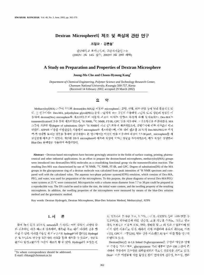
§N56M¨ p$. 4 )_ m % Fig. 1 >
©#ª$.
_6 «Xb ¬ s 89 3i ®F j56
/;¯% °, ¬ j t- $[11]. *+ ¬j 89 ,(ATPS)
2 ±E> ² ³ VW56 - ´!b 8G
> i< `G a,b )$. , Dex/PEG/ µ ATPS6 a,b
P¶z ·$ [11, 12]. PEG ?-896v ¸2 89 8E
¹ ºt 89, Dexc 3i®F j56 »?bª%
° ATPS t-+$.
Microsphere ³2 , ; b /0 2 ¼9
½, A <" / `G a,b k¢ >/ ATPS ¾
¿% ,+ k¢$. ATPS,% ÀÁ 04¥% ° ¾¿
V-b, f 9 1ÂÃ Äm< Å/f /0 mÆ% °
microsphere/ V- $. *+ k¢2 Ç "S, ¸2 Ç 8E,
È m # ¼9 ÉX, , ʼ(encapsulation) P+ , ,
- I ³2 % / )$. Microsphere <"i P+ Ä
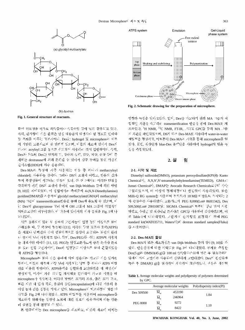
ËF Fig. 2 >©#ª$. ATPS ¾¿% ,f microsphere
<" ¡;_ jÌt F¶ P+ iÍ/ Î( j8
G ÏÐ% :; iD ( )$.
=!_ Dex microsphere <", A <" |§
ÑÒ -% ÓÍÔ$. ÕÖ, Dex /0 ¡; MA @w ¦
z §N% F transesterification 4% :; Dex-MA <
"¥$. 1H NMR, 13C NMR, FT-IR, @G GPC :; MA @w
§N% ×z¥5, DS/ $È Dex-MA a,f water-in-water
¾¿% t-, 6M¨ Dex-MA /0 :; microsphere
ª$. +Ø, ¢ blue-Dex ,¢% a,f hydrogel
-% Ùi¥$.
2.
2-1.
Dimethyl sulfoxide(DMSO), potassium peroxydisulfate(KPS) Kanto
Chemicala, N,N,N',N'-tetramethylethylenediamine(TEMED), GMA
Junsei Chemicala, DMAP Avocado Research Chemicals6M¨ ÚÚ
!¼¥5, Û j i<i> KtÜ a,¥$. 2
Milli-Q RG system% a,f FF/ 18 MΩj56 b 2
Ýs( a,¥$. 896v PEG 8,000(Lot# 86H1342), Dex
509,500(Lot# 29H1087) SIGMA Chemicala6M¨ Þß àÓ a,
¥, (Ìá Z Ìá 892 GPC a,f i¥5,
Table 1 >©#ª$. 89 89% Íi ¡; PEG
standard kit(WAT035711, Watersa)c dextran standard samples(Fluka
a,¥$.
2-2. Dex-MA
Dex-MA ¶â <"i2 van Dijk-Wolthuis I k¢[9, 10]% J
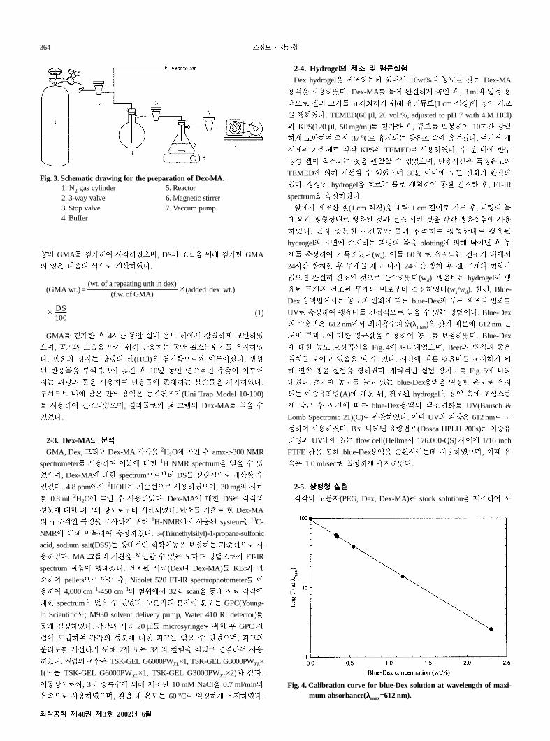
ã$. 4§ P+ ÄËF Fig. 3 >©#ª$. ~ Ùi+
Dex(5 g)c DMSO(45 g) 100 ml äÚå1nÇ æ2 ç è8¡
_ 9é 04 a,f ÀÁ 04¥$. Dex/ ê
2 ç DMAP(1 g)% Å/f µëf 04¥$. 42 µ\
Fig. 1. General structure of reactants.
Fig. 2. Schematic drawing for the preparation of microsphere.
Table 1.Average molecular weights and polydipersity of polymers determinby GPC.
Average molecular weights Polydispersity index(PI)
Dex 509500 Mw 4531901.84
Mn 246964
PEG 8000 Mw9272
1.10Mn
8422
HWAHAK KONGHAK Vol. 40, No. 3, June, 2002
364
ì GMA Å/f m¥5, DS "S% ¡; Å/+ GMA
ì2 $í 56 µ\¥$.
(GMA wt.)= î(added dex wt.)
î (1)
GMA Å/+ ç 4m ïð Ï# ñF _ ÀÁ 04¥
5, Þ òl% h ¡; 4 ïð è8¡ ¥
$. 4 i r \(HCl)% Å/&56v ¯$. V-
4% Uéóô õ ç 103 ïð =ëz öl
% a,f 4 xB ÷ø% <ù¥$.
Uéóô # ú2 ûs ,2 ïü" (Uni Trap Model 10-100)
a,f ü"bª5, 6v ý @þ Dex-MA % (
)ª$.
2-3. Dex-MA
GMA, Dex, @G Dex-MA ÚÚ% 2H2O z ç amx-r-300 NMR
spectrometer a,f P+ 1H NMR spectrum% % ( )
ª5, Dex-MA P+ spectrum56M¨ DS i56 µ\' (
)ª$. 4.8 ppm_ 2HOH âº56 a,¥5, 30 mg mC
0.8 ml 2H2O z ç a,¥$. Dex-MA P+ DS ÚÚ
-8 P+ ÿÇ ÀF6M¨ µ\bª$. è 6 + Dex-MA
!"z % "a ¡; 1H-NMR_ a, system% 13C-
NMR P; 4f Ùi¥$. 3-(Trimethylsilyl)-1-propane-sulfonic
acid, sodium salt(DSS) jPz XWï% Íi âº56 a
,¥$. MA @w §N% ×z' ( ) $È k¢56v FT-IR
spectrum ÏÐ q;¯$. ü" mC(Dex> Dex-MA) KBr 4
f pellets56 ç, Nicolet 520 FT-IR spectrophotometer
,f 4,000 cm−1-450 cm−1 ¡_ 32g scan% :; mC ÚÚ
P+ spectrum% % ( )ª$. 89 89 8E GPC(Young-
In Scientifica; M930 solvent delivery pump, Water 410 RI detector)
:; i¥$. ÚÚ mC 20µl microsyringe6 + ç GPC
F¼f ÚÚ -8 P+ ÿÇ % ( )ª5, ÿÇ
8GF ĺ ¡; 2Ä 3Ä % ¦Á6 =f a,
¥$. "?2 TSK-GEL G6000PWXL×1, TSK-GEL G3000PWXL×
1( TSK-GEL G6000PWXL×1, TSK-GEL G3000PWXL×2)c ³$.
ïj56v, 3 Ýs( ; <" 10 mM NaCl% 0.7 ml/min
ë56 a,¥5, # ñF 60oC6 3i ¥$.
2-4. Hydrogel
Dex hydrogel% <" )_ 10wt% ®F ¹ Dex-MA
,% a,¥$. Dex-MA ê z ç, 3 ml 3i ,
56 . Ç X ¡; Góô(1 cm ¦O) æ /0
q¥$. TEMED(60µl, 20 vol.%, adjusted to pH 7 with 4 M HCl)
c KPS(120µl, 50 mg/ml) Å/+ ç, óô HÊf 10 ÀÁ
04f m 37oC6 b ñ" ë õ¯$. f _ Ä
m<c /ë<6 ÚÚ KPSc TEMED a,¥$. ( 8 # 4U
- . b A% d' ( )ª5, 4m2 iñFc
TEMED ; ĺ' ( )ª5 308 # .X/ êb
ª$. V- hydrogel% 6 ±f ï ü"+ ç, FT-IR
spectrum% Ùi¥$.
_ <"+ .(1 cm ¦O)% PË 1 cm 6 9È ç,
; Ìtju6 A ü" m A% ÚÚ ÏÐ a,
¥$. ÕÖ 8+ mïð f Ìtju6
hydrogel ¶ xB % blotting ; Ó ç ~
Ùif ¥$(ws). 60oC6 b ü" #_
24m k§+ ç ~ B $m 24m k§ ç ~c KX/
Ü5 ê ü" A56 ¥$(wd). hydrogel
~c ü" ~ 6M¨ i¥$(ws/wd). +Ø, Blue-
Dex ,¢_ ®F KX JÈ blue-Dex È !" KX
UV6 Ùif 56 % ( ) k¢$. Blue-Dex
(,2 612 nm_ "P#($(λmax)% ¹ °% 612 nm &
' UF P+ Ìá(% ,f ®F Íi¥$. Blue-Dex
P+ ®F Íi)º% Fig. 4 >©#ª5, Beer ¢* +2
3§ Í )í% ( )$. m JÈ "a ¡
; =ë ÏÐ% q¥$. ÄËz ÏÐ §F Fig. 5 >©
#ª$. ®F ) blue-Dex,% 3i+ ñF6
b G,(A) £- ., ü" hydrogel% , ë "/n0
1& ç m JÈ blue-Dex, !"KX UV(Bausch &
Lomb Spectronic 21)(C)6 d¥$. ° UV $2 612 nm6
if a,¥$. B6 >© 23(Dosca HPLH 200s)
G, UV# ) flow cell(Hellmaa 176.000-QS) a 1/16 inch
PTFE d% :; blue-Dex,% øNm4 a,¥5, °
ë2 1.0 ml/sec6 3i ¥$.
2-5.
ÚÚ 89(PEG, Dex, Dex-MA) stock solution% <"f a
(wt. of a repeating unit in dex)
(f.w. of GMA)
DS100---------
Fig. 3. Schematic drawing for the preparation of Dex-MA.1. N2 gas cylinder 5. Reactor2. 3-way valve 6. Magnetic stirrer3. Stop valve 7. Vaccum pump4. Buffer
Fig. 4. Calibration curve for blue-Dex solution at wavelength of maxi-mum absorbance(λλλλmax=612 nm).
40 3 2002 6
Dextran Microsphere 365
,¥5, ° ,6 ø( <" a,f 18 MΩ j5
6 b % a,¥$. <" stock solution% ,f j8
G Ñ5 # 89 "-% ¹ »?% <"f vial, 1Ó
Á 04+ ç 25oC6 b ñ" ë 53 j k§f ¬
j56 8G¥$. 8G Ú j mC syringe ,f "
/n0 £¥$. £ mC i56 6é+ ç Ìt"-%
i ¡; microsyringe ,f 20µl GPC ¼¥$.
2-6. Microspheres
Dex-MAc PEG6 !- ¬ j Ñ5 # ATPS ,(5 ml, 0.22M potassium
chloride and 10 mM phosphate buffer, pH 8.0)% <"+ ç 108 /
è ÷ æ ,xb ) \è <ù¥$. 28 /
Á 04f ¾¿X bF f 1-108 ïð ðiXmÆ$. @
7 $í, Dex-MA /0 ¡; TEMED(100µl, 20 vol.%, adjusted
to pH 7 with 4 M HCl)c KPS(200µl, 50 mg/ml) /+ ,% 37oC
6 b ñ" 308 k§¥$. V- /0 ¼9 /
8Gc ±(Ýs() 0N 4i% :; ÷ø% <ù¥5,
8ïü" :; 9! 8½ microsphere % ( )ª$.
ê ïü" microsphere è n:; <G =56 >
?+ ç, 2 9 | SEM(JEOL, JSM-5400)56 d¥$.
+Ø, microsphere ¼9 Ç 8E 45 mm-( 100 mm-, 300 mm-)
focal length ¹ @A/ MB ¼F8é (PSA; Malvern) a
,f Ùi¥$. CO,6 2 Ýs % a,¥, 8é
3> ïð í$ af ¼9 B% k¥$.
3.
3-1. Dex-MA
=!_ Dex-MA van Dijk-Wolthuis I[9, 10] ; èÄ
Dc ³ MA @w ¦z §N% F transesterification 4
% :; <"¥$. k¢_ Dexc GMA/ y- ,z
DMSO#_ 4 pqbª5 DMAP E - 6v Dex
OH @w% y-X+ ç MA @w ¦ §N% F $.
Glucopyranose G # xB OH @w 4F )_ C-2,
C-3 ¡§ xB OH/ C-4 ¡§ OHÍ$ F$. @*> C-2 ¡
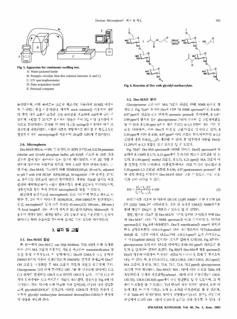
§ ¼G C-3 ¡§6 §N% F+$. 42 Fig. 6 >
©#ª$. Ö9 ; Hß Dc ³[10], 4 $È V-
6v glycidol(GDOL) V- $. *+ GDOL V-2 4
6v glycidyl methacrylate derivatized dextran(Dex-GMA)/ V-b
í% I; â$.
3-2. Dex-MA
Glucopyranose G # MA @w ×z% ¡;, NMR 8é% q
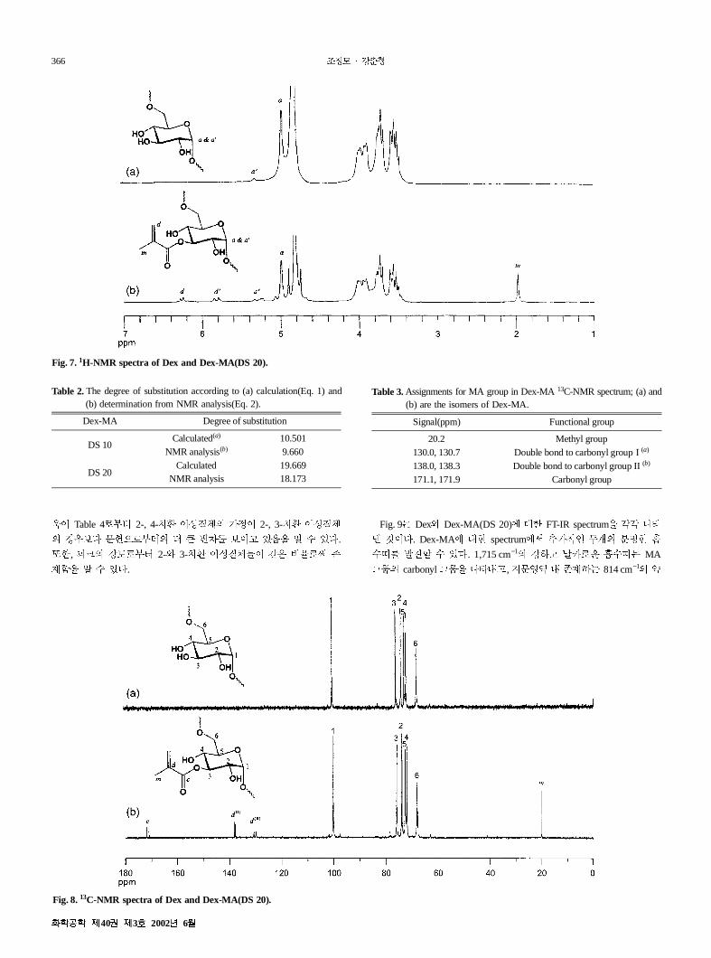
¥$. Fig. 7(a)2 Dex P+ 1H NMR spectrum$. δ 4.96-
4.97 ppm J C-1 ¡§ anomeric proton% |, δ 3.47-
3.99 ppm ¡ ¹ glucopyranose G (èc !8bK%
( )$. δ 5.30 ppm &' 2 ÿÇ α-1,3 ? P+ 2 M
8% >©#, Dex/ ºt 89L% Mm )$. δ5.30 ppm P+ δ 4.96, 4.97 ppm_ ÿÇ 6M¨ α-1,3
? P+ 8N(fα-1,3)2 µ\D ( )$. =!_ a, Dex
11.26% α-1,3 ?% ¹ )í% ( )ª$.
Fig. 7(b)2 Dex-MA spectrum% >© A$. Dex spectrum
0f δ 1.94c δ 5.75, 6.21 ppm_ ö/z ÿÇ/ OPQ% (
)$. δ 1.94 ppm2 methyl @w%, δ 5.75, 6.21 ppm2 MA @w
?% ÚÚ >©$. ?_ ÿÇ Ìá( δ5.30 ppm(α-1,3 ?)% E&+ δ 4.96, 4.97 ppm(anomeric proton) P
+ ÿÇ % 0f Dex-MA DS !' ( )ª$. $í
³ >©R ( )$.
(2)
( 6 + S DS [ (1)]c NMR + µ\ DS
[ (2)] Table 2 >©#ª$. 4 Å/+ GMA NMR8é%
:; µ\ DS( T )í% ( )ª$.
+Ø, è 6 + Dex-MA !" tu "a ¡; Dex
c Dex-MA P+ 13C NMR spectrum 0 8é¥$. ÚÚ
spectrum% Fig. 8 >©#ª$. Dex esterification2 ester MB
è Þ56M¨ +0.9-1.9 ppm U2 9 56 ï(downfield
shift)% @A V+ è6M¨ -1.8-3.3 ppm F2 9 56
ï(upfield shift)% 354 A56 )$[10]. Fig. 86M¨
glucopyranose G # è | δ 68-101 ppm ÿÇ2 %
W 3§ Í¥$. Fig. 8(b) Dex-MA P+ spectrum2
Dex Oc 0f M/z ÿÇ >©>, Ç ¬ M856
>X ( )$. , δ 171.9-171.1, 138.3-138.0, 130.7-130.0, 20.2 ppm2
MA @w, δ 97.8, 78.7, 75.8, 73.7, 72.4, 70.2 ppm2 glucopyranose
G ÚÚ ;r $. Dex-MA MA @w P+ J Table 3
I¥$. ¬Ä -(carbonyl @w P+ ?)/ 130.0-
130.7(I), 138.0-138.3(II) ppm_ ÚÚ OPQ% ( )ª5, @
N Y&% ( )ª$. G è P+ ;é )_, XW
ï P+ ¬ / /i(2-, 3- 2-, 4-§N -)% ' ( )5,
Table 4 Zù¥$. _ I[\(3-1S ]"), 4F
d_ C-3 OH @w C-4Í$ F$ A% ö' ( )$. Û
DSId Id′+( ) 2⁄
Ia fα 1 3,– Ia+--------------------------- 100×=
Fig. 6. Reaction of Dex with glycidyl methacrylate.
Fig. 5. Apparatus for continuous swelling test.A: Water-jacketed bottleB: Pump(to circulate blue-dex solution between A and C)C: UV spectrophotometerD: Data acquisition boardE: Personal computer
HWAHAK KONGHAK Vol. 40, No. 3, June, 2002
366
^ Table 46M¨ 2-, 4-§N - /i 2-, 3-§N -
OÍ$ %W56M¨ Û _ Ø Í )í% ( )$.
+, ÿÇ ÀF6M¨ 2-c 3-§N - ³2 N6v x
B&% ( )$.
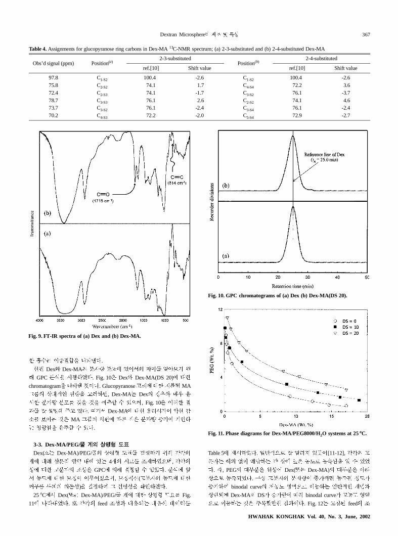
Fig. 9 Dexc Dex-MA(DS 20) P+ FT-IR spectrum% ÚÚ >©
A$. Dex-MA P+ spectrum_ ö/z ¬Ä 8+ #
(` OP' ( )$. 1,715 cm−1 À ab6- #(` MA
@w carbonyl @w% >©#, %Ñ5 # xB 814 cm−1 Y
Fig. 7.1H-NMR spectra of Dex and Dex-MA(DS 20).
Table 2.The degree of substitution according to (a) calculation(Eq. 1) and(b) determination from NMR analysis(Eq. 2).
Dex-MA Degree of substitution
DS 10Calculated(a) 10.501
NMR analysis(b) 9.660
DS 20Calculated 19.669
NMR analysis 18.173
Fig. 8.13C-NMR spectra of Dex and Dex-MA(DS 20).
Table 3.Assignments for MA group in Dex-MA 13C-NMR spectrum; (a) and(b) are the isomers of Dex-MA.
Signal(ppm) Functional group
20.2 Methyl group130.0, 130.7 Double bond to carbonyl group I (a)
138.0, 138.3 Double bond to carbonyl group II (b)
171.1, 171.9 Carbonyl group
40 3 2002 6
Dextran Microsphere 367
+ #( ?% >©$.
+Ø Dexc Dex-MA 89 8E )_ ÓÍ ¡
; GPC 8é% mq¥$. Fig. 102 Dexc Dex-MA(DS 20) P+
chromatogram% >© A$. Glucopyranose G P+ §N MA
@w jPz % , Dex-MA Dex Oc
a+ 89 8E ¹% A% ·Ù' ( )5, Fig. 102 *+
I; )$. f _ Dex-MA P+ ,Gm Y+ R
è Í A2 MA @w §N JÈ 2 89 Ý/ z
ÑÒL% ö' ( )$.
3-3. Dex-MA/PEG/
Dex( Dex-MA)/PEG/ jÌt F¶ i ¡; ÚÚ
µ P; j8G Ñ5 # ) 4Ä mC "<¥5, ÚÚ
j P+ 89 "-2 GPC ; i' ( )ª$. 8é
_ ®F P+ Íi ¯5, Íi)º(89 ®F P+
c~d Mÿ x-)% if @ ºt-% ×z¥$.
25oC_ Dex( Dex-MA)/PEG/ µ P+ jÌt F¶ Fig.
11 >©#ª$. ÚÚ feed "- Pb Pº ¨
Table 5 <m¥$. 3456 )\[11-12], ÚÚ
89 ·L Ü ;r Ú j F2 ®F6 ®eQ% ( )ª
$. , PEG PM82 ¡j Dex( Dex-MA) PM82 Óf
j56 ®ebª$. !- 89 89 Ý/ ®e iF/
Ý/f binodal curve Ö®F Ñ556 ï 34z Äg
j4b Dex-MA DS/ Ý/& J1 binodal curve/ ®F Ñ5
56 ï A2 h'+ $. Fig. 12 i feed "
Table 4.Assignments for glucopyranose ring carbons in Dex-MA 13C-NMR spectrum; (a) 2-3-substituted and (b) 2-4-substituted Dex-MA
Obs’d signal (ppm) Position(a)2-3-substituted
Position(b)2-4-substituted
ref.[10] Shift value ref.[10] Shift value
97.8 C1-S2 100.40 -2.6 C1-S2 100.40 -2.675.8 C2-S2 74.1 -1.7 C4-S4 72.2 -3.672.4 C2-S3 74.1 -1.7 C3-S2 76.1 -3.778.7 C3-S3 76.1 -2.6 C2-S2 74.1 -4.673.7 C3-S2 76.1 -2.4 C3-S4 76.1 -2.470.2 C4-S3 72.2 -2.0 C5-S4 72.9 -2.7
Fig. 9. FT-IR spectra of (a) Dex and (b) Dex-MA.
Fig. 10. GPC chromatograms of (a) Dex (b) Dex-MA(DS 20).
Fig. 11. Phase diagrams for Dex-MA/PEG8000/H2O systems at 25oC.
HWAHAK KONGHAK Vol. 40, No. 3, June, 2002
368
- P; DS JÈ Pº (tie-line length; TLL) KX >©
A$. TLL G | Ú Ìtj Lµ56M¨ ip
ùG >©$. TLL2 Óf ³ ¶j' ( )$.
TLL=
(3)
Fig. 12_ DS/ Ý/ TLL Rè, OH @w MA
@w56 §NQ J1 j8G Ñ5 eèQ% |+$.
3-4. Dex hydrogel
Dex hydrogel2 Dex-MA /0 :; % ( )ª$. MA @w
) ?2 9 1ÂÃ 4% :+ Dex /0 a,bª$.
KPSc TEMED Dex-MA, Å/f, KPS sulfate 9 1Â
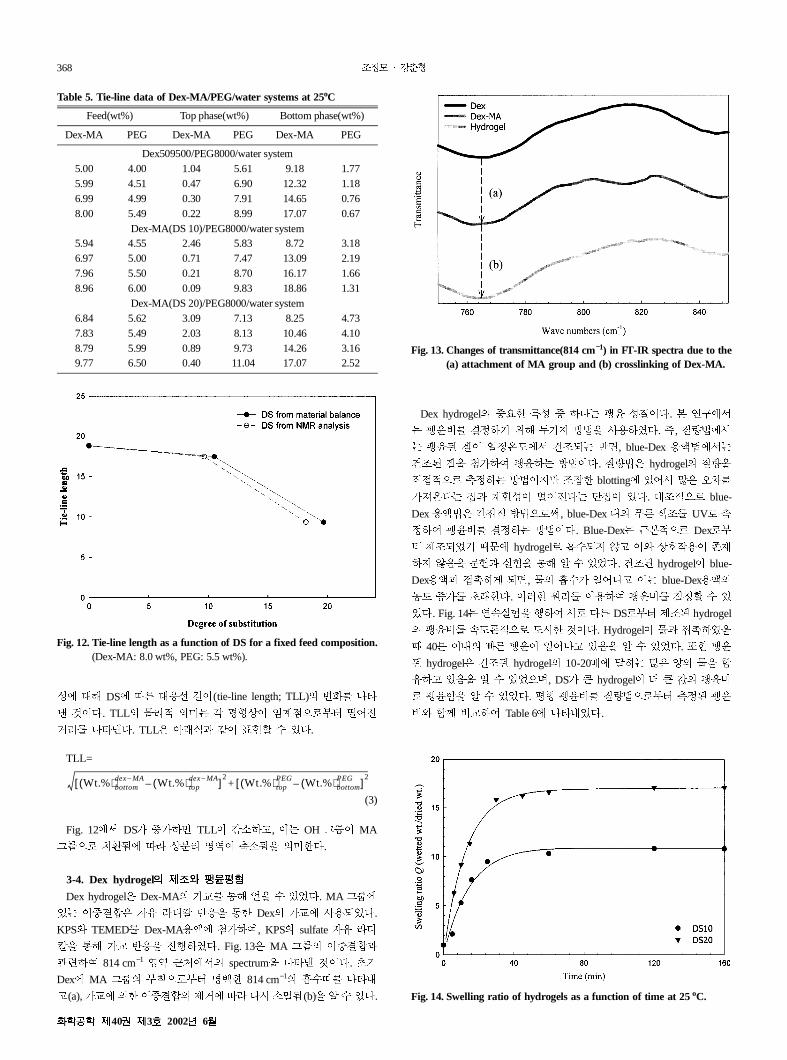
Ã% :; /0 4% pq¥$. Fig. 132 MA @w ?
def 814 cm−1 Ñ5 &'_ spectrum% >© A$.
Dex MA @w MB56M¨ ²+ 814 cm−1 #(` >©#
(a), /0 + ? <ù J1 $m èkQ(b)% ( )$.
Dex hydrogel ´+ - > -$. =!_
i ¡; ¬/ k¢% a,¥$. , ¢_
. 3iñF_ ü"b 4, blue-Dex ,¢_
ü" .% Å/f k¢$. ¢2 hydrogel %
¦56 Ùi k¢ "l+ blotting )_ [2 m
/ñ$ Bj- ip$ )$. P"56 blue-
Dex ,¢2 k¢56v, blue-Dex # È !" UV6 Ù
if i k¢$. Blue-Dex &56 Dex6M
¨ <"bª °% hydrogel6 #(b c jJ, xB
í% %W ÏÐ% :; ( )ª$. ü" hydrogel blue-
Dex, b, #(/ 3> blue-Dex,
®F Ý/ f+$. *+ G ,f i' ( )
ª$. Fig. 14 =ëÏÐ% qf _6 $È DS6M¨ <" hydrogel
ëFS56 Fm+ A$. Hydrogel ¥%
° 408 # È 3> )í% ( )ª$. +
hydrogel2 ü" hydrogel 10-20C [2 ì % &
)í% ( )ª5, DS/ _ hydrogel Û _ (
6 &% ( )ª$. Ìt ¢56M¨ Ùi
c &n 0f Table 6 >©#ª$.
Wt.%( )bottomdex MA– Wt.%( )top
dex MA––[ ]
2Wt.%( )top
PEG Wt.%( )bottomPEG–[ ]
2+
Table 5. Tie-line data of Dex-MA/PEG/water systems at 25oC
Feed(wt%) Top phase(wt%) Bottom phase(wt%)
Dex-MA PEG Dex-MA PEG Dex-MA PEG
Dex509500/PEG8000/water system5.00 4.00 1.04 5.61 9.18 1.775.99 4.51 0.47 6.90 12.32 1.186.99 4.99 0.30 7.91 14.65 0.768.00 5.49 0.22 8.99 17.07 0.67
Dex-MA(DS 10)/PEG8000/water system5.94 4.55 2.46 5.83 8.72 3.186.97 5.00 0.71 7.47 13.09 2.197.96 5.50 0.21 8.70 16.17 1.668.96 6.00 0.09 9.83 18.86 1.31
Dex-MA(DS 20)/PEG8000/water system6.84 5.62 3.09 7.13 8.25 4.737.83 5.49 2.03 8.13 10.46 4.108.79 5.99 0.89 9.73 14.26 3.169.77 6.50 0.40 11.04 17.07 2.52
Fig. 12. Tie-line length as a function of DS for a fixed feed composition.(Dex-MA: 8.0 wt%, PEG: 5.5 wt%).
Fig. 13. Changes of transmittance(814 cm−1) in FT-IR spectra due to the(a) attachment of MA group and (b) crosslinking of Dex-MA.
Fig. 14. Swelling ratio of hydrogels as a function of time at 25oC.
40 3 2002 6
Dextran Microsphere 369
3-5. Microspheres
=!_ ATPS ¾¿56M @ , # xB Dex-MA
/0 :; microsphere% <"¥$. Dex-MA ®F/ microsphere
<" |§ ÑÒ P; ÓÍ ¡; ï3 Pºj xB
feed "- $È ATPS ¾¿% a,f microsphere <"
¥$. /04 ç ATPS# emulsifier6 a, PEG $ ±
% :; C<f dextran microsphere <"' ( )ª$.
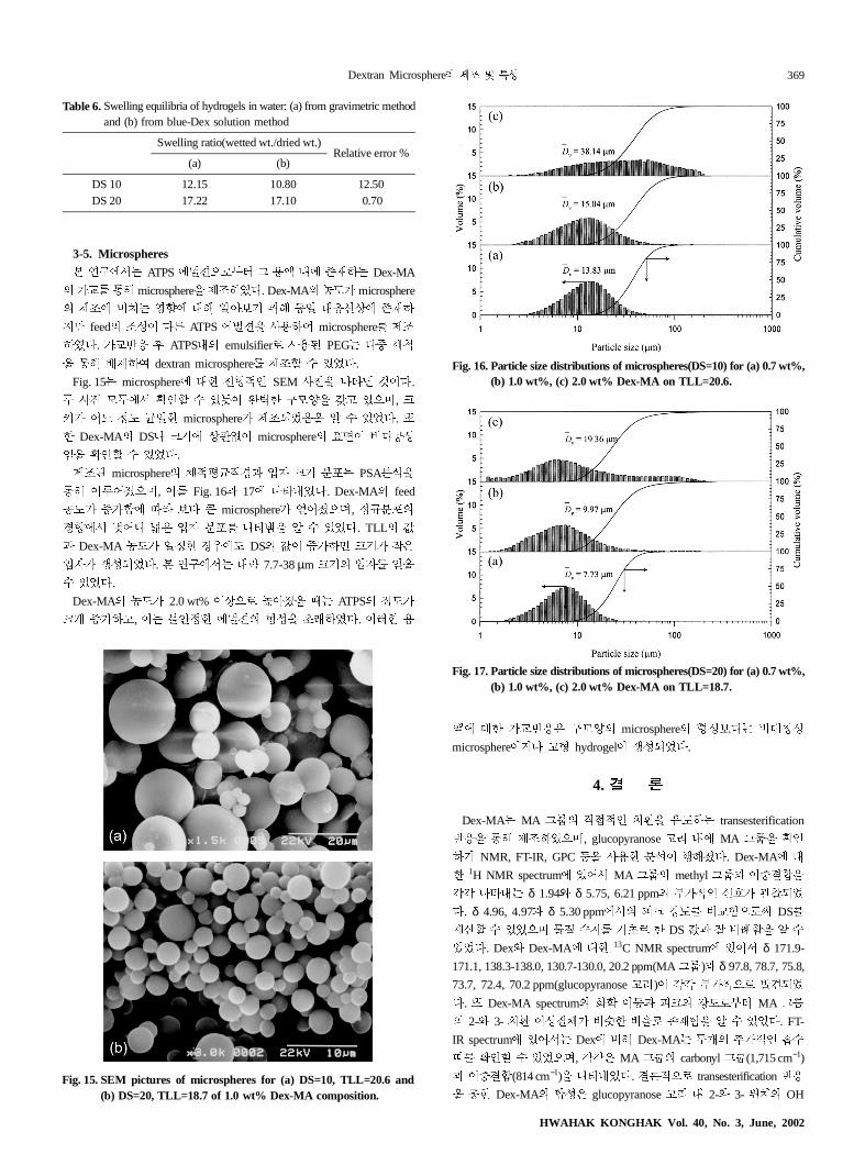
Fig. 15 microsphere P+ tz SEM ap% >© A$.
¬ ap ¬_ ×z' ( )\ êo+ !ì% ¹ )5, Ç
4/ p iF á3+ microsphere/ <"bªí% ( )ª$.
+ Dex-MA DS> Ç jdÜ microsphere ¶ $Þ-
L% ×z' ( )ª$.
<" microsphere Ìá¦O ¼9 Ç 8E PSA8é%
:; ¯5, Fig. 16 17 >©#ª$. Dex-MA feed
®F/ Ý/& J1 Í$ _ microsphere/ ¯5, i 8E
OÒ_ q> r2 ¼9 8E >©s% ( )ª$. TLL (
Dex-MA ®F/ 3i+ OF DS ( Ý/ Ç / 2
¼9/ V-bª$. =!_ PË 7.7-38µm Ç ¼9 %
( )ª$.
Dex-MA ®F/ 2.0 wt% j56 FÓ¯% ° ATPS F/
Ç Ý/, ÷ði+ ¾¿ t-% f¥$. *+ ,
P+ /042 !ì microsphere t-Í$ Pt-
microsphereù> t hydrogel V-bª$.
4.
Dex-MA MA @w ¦z §N% F transesterification
4% :; <"¥5, glucopyranose G # MA @w% ×z
NMR, FT-IR, GPC I% a,+ 8é q;¯$. Dex-MA P
+ 1H NMR spectrum )_ MA @w methyl @w ?%
ÚÚ >©# δ 1.94c δ 5.75, 6.21 ppm M/z J/ dbª
$. δ 4.96, 4.97 δ 5.30 ppm_ ÿÇ ÀF 0&56v DS
µ\' ( )ª5 ( 6 + DS ( T&% (
)ª$. Dexc Dex-MA P+ 13C NMR spectrum )_ δ 171.9-
171.1, 138.3-138.0, 130.7-130.0, 20.2 ppm(MA@w) δ 97.8, 78.7, 75.8,
73.7, 72.4, 70.2 ppm(glucopyranose G) ÚÚ M/56 OPbª
$. Dex-MA spectrum XW ï ÿÇ ÀF6M¨ MA @w
2-c 3- §N -/ Y+ N6 xB&% ( )ª$. FT-
IR spectrum )_ Dex ; Dex-MA ¬Ä ö/z #(
` ×z' ( )ª5, ÚÚ2 MA @w carbonyl @w(1,715 cm−1)
?(814 cm−1)% >©#ª$. S56 transesterification 4
% :+ Dex-MA ?-2 glucopyranose G # 2-c 3- ¡§ OH
Table 6.Swelling equilibria of hydrogels in water: (a) from gravimetric methodand (b) from blue-Dex solution method
Swelling ratio(wetted wt./dried wt.)Relative error %
(a) (b)
DS 10 12.15 10.80 12.50DS 20 17.22 17.10 0.70
Fig. 15. SEM pictures of microspheres for (a) DS=10, TLL=20.6 and(b) DS=20, TLL=18.7 of 1.0 wt% Dex-MA composition.
Fig. 16. Particle size distributions of microspheres(DS=10) for (a) 0.7 wt%,(b) 1.0 wt%, (c) 2.0 wt% Dex-MA on TLL=20.6.
Fig. 17. Particle size distributions of microspheres(DS=20) for (a) 0.7 wt%,(b) 1.0 wt%, (c) 2.0 wt% Dex-MA on TLL=18.7.
HWAHAK KONGHAK Vol. 40, No. 3, June, 2002
370
ro-
,
d
itz,
er-
der
elin,
me-
@w% MA @w56 ¦§N% F¥5, 4 Dex P
+ GMA i ; DS/ uN56 "SQ% ×z' ( )
ª$.
25oC_ Dex( Dex-MA)/PEG/ µ P+ jÌt F¶ GPC
a,f i¥$. jÌt F¶6M¨ PEG PM82 ¡j,
Dex( Dex-MA) PM82 Ófj ®ebª5, DS/ Ý/&
J1 binodal curve ®FÑ556 ï¥5 TLL2 Rè
¥$.
i ¡; ¢ blue-Dex ,¢% a,¥$.
, blue-Dex ,¢ 6v hydrogel bª% °
$ #(Q% ( )ª$. Ìt
DS10 10.80, DS20 17.10 (% ÚÚ >©#ª$.
Microsphere <" ATPS ¾¿ # Dex-MA /0 :; <"
bª$. <" microsphere tu SEM% ,f d¥5,
êo+ $Þ- !ì% ¹ )í% ×z' ( )ª$. +, PSA
¨6M¨ Dex-MA ®F/ Ý/ Ç / Ç, 8E
/ r2 microsphere/ K% ( )ª$.
ATPS : aqueous two-phase polymer solution(system)
Blue-Dex : blue dextran
DDS : drug delivery system
Dex-GMA : glycidyl methacrylate derivatized dextran
Dex-MA : methacrylated dextran
Dex : dextran
DMAP : 4-(N,N-Dimethylamino) pyridine
DMSO : dimethyl sulfoxide
DS : the degree of substitution(the amount of methacryloyl groups
per 100 dextran glucopyranose residues)
DSS : 3-(Trimethylsilyl)-1-propane-sulfonic acid, sodium salt
f.w. : formula weight, [g]
FT-IR : Fourier transform infrared spectrophotometer
GDOL : glycidol
GMA : glycidyl methacrylate
GPC : gel permeation chromatography
KPS : potassium peroxydisulfate
MA : methacryloyl
NMR : nuclear magnetic resonance
PI : polydispersity index(weight-average molecular weight/number-
average molecular weight) [-]
PSA : particle size analysis
PTFE : polytetrafluoroethylene(trade name is Teflon)
SEM : scanning electron microscope
TEMED : N,N,N’ ,N’-tetramethylethylenediamine
TLL : tie-line length
UV : ultra-violet spectrophotometer
1. Park, K. N., Shalaby, S. W. and Park, H. S.: “Biodegradable Hyd
gels for Drug Delivery,” Technomic publishing Co., Inc., Lancaster
PA(1993).
2. “Proceeding of the Tailored Polymeric Materials for Controlle
Delivery Systems-sept. 7, 1997,” ACS publications, Chapter 2(1998).
3. Beltran, S., Baker, J. P., Hooper, H. H., Blanch, H. W. and Prausn
J. M.: Macromolecule, 24, 549(1991).
4. Gehrke, S. H., Vaid, N. R. and McBride, J. F.: Biotechnol. Bioeng.,
58, 416(1998).
5. Kapur, V., Charkoudian, J. C., Kessler, S. B. and Anderson, J. L.: Ind.
Eng. Chem. Res., 35, 3179(1996).
6. Motozato, Y., Ihara, H., Tomada, T. and Hirayama, C.: J. Chro-
matogr., 355, 434(1986).
7. Horgaard, L. and BrØndsted, H.: J. Control. Release, 36, 159(1995).
8. Franssen, O., Stenekes, R. J. H. and Hennink, W. E.: J. Control.
Release, 59, 219(1999).
9. van Dijk-Wolthuis, W. N. E., Franssen, O., Talsma, H., van Steenb
gen, M. J., Kettenes-van den Bosch, J. J. and Hennink, W. E.: Mac-
romolecules, 28, 6317(1995).
10. van Dijk-Wolthuis, W. N. E., Kettenes-van den Bosch, J. J., van
Kerk-van Hoof, A. and Hennink, W. E.: Macromolecules, 30, 3411
(1997).
11. Albetsson, P.-A.: Nature, 182, 709(1958).
12. Cho, J. M. and Kang, C. H.: J. Research Inst. for Catal., 21, 105
(2000).
13. Stenekes, R. J. H., Franssen, O., van Bommel, E. M. G., Cromm
D. J. A., Hennink, and Hennink, W. E.: Pharm. Res., 15, 557(1998).
14. Franssen, O. and Hennink, W. E.: Int. J. Pharm., 168, 1(1998).
15. Stenekes, R. J. H., Franssen, O., van Bommel, E. M. G., Crom
lin, D. J. A. and Hennink, W. E.: Int. J. Pharm., 183, 29(1999).
40 3 2002 6