Developmental Validation of the Quantifiler® Duo … Barbisin,1 Ph.D.; Rixun Fang,1 Ph.D.; Cristin...
15
Maura Barbisin, 1 Ph.D.; Rixun Fang, 1 Ph.D.; Cristin E. O’Shea, 1 B.S.; Lisa M. Calandro, 1 M.P.H.; Manohar R. Furtado, 1 Ph.D.; and Jaiprakash G. Shewale, 1 Ph.D. Developmental Validation of the Quantifiler Ȧ Duo DNA Quantification Kit for Simultaneous Quantification of Total Human and Human Male DNA and Detection of PCR Inhibitors in Biological Samples* ABSTRACT: The Quantifiler Ȑ Duo DNA Quantification kit enables simultaneous quantification of human DNA and human male DNA as well as detection of inhibitors of PCR in a single real-time PCR well. Pooled human male genomic DNA is used to generate standard curves for both human (ribonuclease P RNA component H1) and human male (sex determining region Y) specific targets. A shift in the cycle threshold (C T ) values for the internal positive control monitors the presence of PCR inhibitors in a sample. The assay is human specific and exhibits a high dynamic range from 0.023 to 50 ng ⁄ lL. In addition, the multiplex assay can detect as little as 25 pg ⁄ lL of human male DNA in the presence of a 1000-fold excess of human female DNA. The multiplex assay provides assessment of the DNA extract and guidance for the selection of the appropriate AmpF‘STR Ȑ Amplification Kit to obtain interpretable short tandem repeat profiles. KEYWORDS: forensic science, DNA quantification, real-time PCR, DNA analysis, ribonuclease P RNA component H1, sex determining region Y, human DNA, human male DNA, DNA typing Forensic DNA analysis is targeted to obtain a short tandem repeat (STR) profile from an evidence sample, which is then com- pared with the STR profiles from reference samples collected from the victim and the suspect to determine the contribution to the evi- dence sample (1,2). STR genotyping systems such as Identifiler Ȑ , Profiler Plus Ȑ , COfiler Ȑ , SGM Plus Ȑ , MiniFiler TM , and Yfiler Ȑ kits are commercially available. The genotyping protocol, in general, involves extraction of DNA from the biological sample, quantifica- tion of the DNA, amplification for STR loci, and fragment analysis on a Genetic Analyzer. Quantification of human DNA in a forensic sample, which often contains nonhuman DNA, is an important step during STR profiling because the STR genotyping systems, unlike detection or single nucleotide polymorphism (SNP) assays, are sen- sitive to the quantity of DNA used in the PCR reaction: too little DNA may produce partial profiles whereas too much may produce off-scale data. For the forensic analyst, it is imperative to obtain an interpretable STR profile from forensic evidence samples, which often are in limited amount, and therefore, a reliable quantification method is vital. Hybridization-based quantification methods, e.g., Quantiblot Ȑ , that are traditionally used for the quantification of DNA in forensic samples are generally considered time-consuming, labor-intensive, and not suitable for automation (3). Further, it is difficult to predict the amplitude of the STR profile because of the difference in the sensitivity of quantification methods and STR genotyping systems. Real-time PCR assays like the Quantifiler Ȑ Human DNA Quantification Kit and Quantifiler Ȑ Y Human Male DNA Quantification Kit have proved very useful (4). Real-time PCR assays for quantification of human DNA offer several advan- tages over the traditional hybridization assays such as: (i) specificity for a certain target in the genome because of the careful assay design; (ii) ability to detect few copies of target DNA; (iii) quanti- tative relationship between the amount of target template and the amount of PCR product accumulated at any given cycle prior to reaching saturation; (iv) greater dynamic range; (v) multiplexing capabilities; (vi) easy to adopt; and (vii) automatable for high throughput. A forensic evidence sample is often a mixture of human male and female DNA. Further, a forensic biological sample may be exposed to different environmental insults leading to DNA degrada- tion and contamination with compounds that inhibit the PCR. Thus, it is desirable for the forensic analyst to have useful information about the forensic evidence sample prior to amplification for STRs. Real-time quantification assays can provide: (i) mixture ratio of human and human male DNA for choosing between autosomal and Y STR profiling based on the extent of the mixture ratio; (ii) pres- ence of PCR inhibitors so that DNA extracts containing PCR inhib- itors may be repurified prior to STR profiling; and (iii) quantification of human female and human male DNA useful for determining the quantity of extract to be used for amplification for different STR multiplex systems. To obtain the quantity of human and human male DNA in a sample using Quantifiler Ȑ Human and Y kits, it is necessary to run two separate quantification assays. This approach may consume a considerable amount of sample, which is often available in limited quantities, as well as time and reagents. To overcome these hurdles, 1 Applied Biosystems, 850 Lincoln Centre Drive, Foster City, CA 94404. *Part of the work was presented at the 59th Annual Meeting of the Amer- ican Academy of Forensic Sciences, in San Antonio, TX, February 19–24, 2007, and at the 18th International Symposium on Human Identification, Hollywood, CA, October 2007. Some of the data is also presented in the Quantifiler Ȑ Duo DNA Quantification Kit User Manual. Received 19 Jan. 2008; and in revised form 3 April 2008; accepted 20 April 2008. J Forensic Sci, March 2009, Vol. 54, No. 2 doi: 10.1111/j.1556-4029.2008.00951.x Available online at: www.blackwell-synergy.com ȑ 2009 Applied Biosystems Journal compilation ȑ 2009 American Academy of Forensic Sciences 305
-
Upload
duongxuyen -
Category
Documents
-
view
216 -
download
4
Transcript of Developmental Validation of the Quantifiler® Duo … Barbisin,1 Ph.D.; Rixun Fang,1 Ph.D.; Cristin...

Maura Barbisin,1 Ph.D.; Rixun Fang,1 Ph.D.; Cristin E. O’Shea,1 B.S.; Lisa M. Calandro,1 M.P.H.;Manohar R. Furtado,1 Ph.D.; and Jaiprakash G. Shewale,1 Ph.D.
Developmental Validation of the Quantifiler�
Duo DNA Quantification Kit for SimultaneousQuantification of Total Human and HumanMale DNA and Detection of PCR Inhibitors inBiological Samples*
ABSTRACT: The Quantifiler� Duo DNA Quantification kit enables simultaneous quantification of human DNA and human male DNA as wellas detection of inhibitors of PCR in a single real-time PCR well. Pooled human male genomic DNA is used to generate standard curves for bothhuman (ribonuclease P RNA component H1) and human male (sex determining region Y) specific targets. A shift in the cycle threshold (CT) valuesfor the internal positive control monitors the presence of PCR inhibitors in a sample. The assay is human specific and exhibits a high dynamic rangefrom 0.023 to 50 ng ⁄lL. In addition, the multiplex assay can detect as little as 25 pg ⁄lL of human male DNA in the presence of a 1000-fold excessof human female DNA. The multiplex assay provides assessment of the DNA extract and guidance for the selection of the appropriate AmpF‘STR�
Amplification Kit to obtain interpretable short tandem repeat profiles.
KEYWORDS: forensic science, DNA quantification, real-time PCR, DNA analysis, ribonuclease P RNA component H1, sex determiningregion Y, human DNA, human male DNA, DNA typing
Forensic DNA analysis is targeted to obtain a short tandemrepeat (STR) profile from an evidence sample, which is then com-pared with the STR profiles from reference samples collected fromthe victim and the suspect to determine the contribution to the evi-dence sample (1,2). STR genotyping systems such as Identifiler�,Profiler Plus�, COfiler�, SGM Plus�, MiniFilerTM, and Yfiler� kitsare commercially available. The genotyping protocol, in general,involves extraction of DNA from the biological sample, quantifica-tion of the DNA, amplification for STR loci, and fragment analysison a Genetic Analyzer. Quantification of human DNA in a forensicsample, which often contains nonhuman DNA, is an important stepduring STR profiling because the STR genotyping systems, unlikedetection or single nucleotide polymorphism (SNP) assays, are sen-sitive to the quantity of DNA used in the PCR reaction: too littleDNA may produce partial profiles whereas too much may produceoff-scale data. For the forensic analyst, it is imperative to obtain aninterpretable STR profile from forensic evidence samples, whichoften are in limited amount, and therefore, a reliable quantificationmethod is vital. Hybridization-based quantification methods, e.g.,Quantiblot�, that are traditionally used for the quantification ofDNA in forensic samples are generally considered time-consuming,labor-intensive, and not suitable for automation (3). Further, it isdifficult to predict the amplitude of the STR profile because of thedifference in the sensitivity of quantification methods and STR
genotyping systems. Real-time PCR assays like the Quantifiler�
Human DNA Quantification Kit and Quantifiler� Y Human MaleDNA Quantification Kit have proved very useful (4). Real-timePCR assays for quantification of human DNA offer several advan-tages over the traditional hybridization assays such as: (i) specificityfor a certain target in the genome because of the careful assaydesign; (ii) ability to detect few copies of target DNA; (iii) quanti-tative relationship between the amount of target template and theamount of PCR product accumulated at any given cycle prior toreaching saturation; (iv) greater dynamic range; (v) multiplexingcapabilities; (vi) easy to adopt; and (vii) automatable for highthroughput.
A forensic evidence sample is often a mixture of human maleand female DNA. Further, a forensic biological sample may beexposed to different environmental insults leading to DNA degrada-tion and contamination with compounds that inhibit the PCR. Thus,it is desirable for the forensic analyst to have useful informationabout the forensic evidence sample prior to amplification for STRs.Real-time quantification assays can provide: (i) mixture ratio ofhuman and human male DNA for choosing between autosomal andY STR profiling based on the extent of the mixture ratio; (ii) pres-ence of PCR inhibitors so that DNA extracts containing PCR inhib-itors may be repurified prior to STR profiling; and (iii)quantification of human female and human male DNA useful fordetermining the quantity of extract to be used for amplification fordifferent STR multiplex systems.
To obtain the quantity of human and human male DNA in asample using Quantifiler� Human and Y kits, it is necessary to runtwo separate quantification assays. This approach may consume aconsiderable amount of sample, which is often available in limitedquantities, as well as time and reagents. To overcome these hurdles,
1Applied Biosystems, 850 Lincoln Centre Drive, Foster City, CA 94404.*Part of the work was presented at the 59th Annual Meeting of the Amer-
ican Academy of Forensic Sciences, in San Antonio, TX, February 19–24,2007, and at the 18th International Symposium on Human Identification,Hollywood, CA, October 2007. Some of the data is also presented in theQuantifiler� Duo DNA Quantification Kit User Manual.
Received 19 Jan. 2008; and in revised form 3 April 2008; accepted 20April 2008.
J Forensic Sci, March 2009, Vol. 54, No. 2doi: 10.1111/j.1556-4029.2008.00951.x
Available online at: www.blackwell-synergy.com
� 2009 Applied BiosystemsJournal compilation � 2009 American Academy of Forensic Sciences 305
multiplex real-time quantification assays have been described inrecent years (5–8). The assays described by Walker et al. (5), Hors-man et al. (6), and Nicklas and Buel (7) enable an estimation ofthe mixture ratio of human male and female DNA but were notdesigned to detect the inhibitors of PCR in the sample. The assaydescribed by Swango et al. (8) enables forensic analysts to obtainthe mixture ratio as well as the detection of the inhibitors of PCR.However, the human nuclear DNA amplification target THO1(human tyrosine hydroxylase gene on chromosome 11) spans thepolymorphic STR region (8). THO1 is a commonly used locus forhuman identification in forensic laboratories (2). First, the incorpo-ration of such polymorphic STR target in the quantification assayis discouraged to avoid possible contamination incidences in thelaboratory. Second, the length of the amplicon would vary by 44nucleotides since alleles ranging from 3 to 14 repeat units havebeen characterized in different human population groups (for refer-ences see Short Tandem Repeat DNA Internet DataBase compiledby NIST available at http://www.cstl.nist.gov/biotech/strbase/str_TH01.htm). Though the detection probe is designed outside thepolymorphic region, the possibility of variation in the efficiency ofthe assay due to the variation in the length of the amplicon can notbe ruled out.
We describe a multiplex TaqMan� real-time PCR assay, theQuantifiler� Duo DNA Quantification Kit, for simultaneous quanti-fication of human nuclear and human male DNA as well as detec-tion of the presence of PCR inhibitors in a biological sample. Thedeveloped assay enables the assessment of the biological samplesfor downstream STR profiling.
Materials and Methods
Pooled human male genomic DNA used for generation of stan-dard curves was obtained from EMD Biosciences Inc. (San Diego,CA). Genomic DNA from unknown individuals was obtained fromBiochain (Hayward, CA), Sigma Chemical Company (St. Louis,MO), Promega (Madison, WI), or Serological Research Institute(Richmond, CA). Nonhuman samples were obtained as purifiedDNA from BIOS Laboratories, Inc. (New Haven, CT), Pel-FreezBiologicals (Rogers, AR), and American Type Culture Collection(Manassas, VA). Oligonucleotides, TaqMan� probes, Quantifiler�
human DNA quantification kit, Quantifiler� Y male DNA quantifi-cation kit, AmpF‘STR kits, 7500 Real-time PCR System, 3130Genetic Analyzers and associated software were from Applied Bio-systems (Foster City, CA). All other chemicals used in this studywere of analytical grade.
Extraction and Quantitation of DNA
The DNA from anonymous donor samples (blood, saliva andsemen, either liquid or stains, and buccal swabs) was extracted byusing standard phenol–chloroform (9), or BloodPrep� DNA Chem-istry and the ABI PRISM� 6100 Nucleic Acid PrepStation(Applied Biosystems) procedures. The quantity of DNA was deter-mined by Quantifiler� Duo, Quantifiler� Human, and Quantifiler�
Y Human Male DNA Quantification Kits (Applied Biosystems).
Real-Time PCR Amplification
Real-time PCR amplification reactions contained 10.5 lL of Pri-mer-Probe Mix, 12.5 lL of Master Mix, and 2.0 lL of DNA sam-ple. The Primer-Probe Mix contained forward and reverse primersand TaqMan� probes for ribonuclease P RNA component H1(RPPH1), sex determining region Y (SRY), and internal PCR
control (IPC) targets. The IPC template, a synthetic polynucleotide,was cloned into a plasmid. The Master Mix contained Referencedye, dNTPs, dUTP, MgCl2, AmpliTaq� Gold DNA polymeraseand preservatives in Tris-HCl, pH 8.0. Pooled human male geno-mic DNA at eight different concentrations (50, 16.7, 5.56, 1.85,0.62, 0.21, 0.068, and 0.023 ng ⁄lL) was amplified on each quanti-fication run plate for generation of standard curves for RPPH1 andSRY targets. Amplification reactions were performed in a 7500Real-Time PCR System (Applied Biosystems) following the manu-facturer’s instruction with conditions as follows: 50�C, 2 min;95�C, 10 min; 40 cycles of 95�C, 15 sec and 60�C, 1.0 min. Thedata were analyzed using 7500 System sds Software v1.2.3(Applied Biosystems) with a threshold value of 0.2.
STR Analysis
The samples were amplified with Identifiler�, Yfiler�, and Mini-FilerTM kits using the procedure described in the User’s Manual forthe respective kit. The amplified products were analyzed on a3130xl Genetic Analyzer (Applied Biosystems) with GeneMapper�
ID Software v3.2.1 (Applied Biosystems).
Sensitivity Study
Two human male genomic DNA samples, one pooled and theother single source, obtained from commercial sources were dilutedto obtain concentrations of 20.0, 5.0, 1.0, 0.1, 0.05, 0.04, 0.03,0.023, 0.0115, 0.00575, 0.002875, and 0.00144 ng ⁄lL in 10 mMTris buffer, pH 8.0 containing 0.1 mM ethylene diamine tetraaceticacid (EDTA). Each dilution was quantified in triplicate using theQuantifiler� Duo DNA Quantification Kit.
Species Specificity
The DNA from nonhuman biological species was either obtainedcommercially or purified in the laboratory. For some of these DNAsamples, the sex of the donor animal was unknown. For some spe-cies, multiple donor animals were tested. Most of the reactions uti-lized 5.0 ng of input DNA. For a few reactions, 10 ng of inputDNA was used.
Precision and Accuracy
One set of eight serial dilutions was prepared containing 50,16.7, 5.56, 1.85, 0.62, 0.21, 0.068, and 0.023 ng ⁄lL of the humanmale DNA standard present in the Quantifiler� Duo DNA Quantifi-cation Kit. Six reaction plates were set up and each of them con-tained 10 replicates of the eight dilutions. Two plates perinstrument were run on three different 7500 Real-time PCR Systeminstruments. The two runs were performed on two different days,using the same three 7500 Real-time PCR System instruments. Foreach dilution, the CT values for RPPH1, SRY, and IPC signalswere recorded for all 60 reactions.
Reproducibility
Four male and one female genomic DNA samples were dilutedfrom initial estimated concentrations to 20.0, 10.0, 1.0, 0.1, and0.05 ng ⁄lL. All dilutions were made in 10 mM Tris buffer, pH 8.0containing 0.1 mM EDTA. All samples and dilutions were run intriplicate using the Quantifiler� Duo Kit. Three runs were per-formed on different days. For each sample reaction, the CT valueswere obtained and the DNA quantities calculated.
306 JOURNAL OF FORENSIC SCIENCES

Mixture Study
Mixture samples containing 0.2 ng ⁄lL of human male DNA andvarying amounts of female DNA were prepared. The ratio of maleto female DNA in these samples was 1:0, 1:1, 1:5, 1:10, 1:20, and0:1. The mixture samples were processed in triplicate using theQuantifiler� Duo DNA Quantification Kit to determine the concen-tration of total human DNA (RPPH1 target) and male DNA (SRYtarget). Using the quantification results from the RPPH1 human tar-get, c. 1.0 ng of human genomic DNA from each sample wasadded to an Identifiler� kit reaction. Similarly, using the resultsfrom the SRY male target, c. 1.0 ng of human genomic DNA fromeach sample was added to a Yfiler� kit reaction.
Another set of mixture samples containing 25 pg ⁄lL of maleDNA and increasing quantities of female DNA was prepared toobtain male to female DNA ratios of 1:0, 1:50, 1:100, 1: 200,1:500, 1:800, 1:1000, and 0:1. The samples were processed in trip-licate using the Quantifiler� Duo Kit to determine the concentrationof total human genomic DNA (RPPH1 target) and male DNA(SRY target). In addition, based on the results from the SRY maletarget, c. 1.0 ng of human genomic DNA from each sample wasadded to the Yfiler� reaction.
Calculation of Male to Female DNA Ratio
The Quantifiler� Duo kit provides the quantity of human andhuman male DNA in biological samples. From these values, onecan calculate the ratio of male and female DNA using the follow-ing equation:
Male DNA:Female DNA Ratio
¼ Male DNAMale DNA
:ðHuman DNA�Male DNA)
Male DNA
or
Male DNA:Female DNA Ratio
¼ 1 : ðHuman DNA�Male DNAÞ=Male DNA
All quantities in the above equations are ng ⁄lL. This ratio deter-mines the extent of the mixture, which is useful for making thechoice of STR analysis method: autosomal STRs or Y-STRs.
Inhibited Samples
Human male genomic DNA was mixed with hematin to obtainfinal concentrations of 0, 2.5, 5.0, 7.5, 10, 12.5, 15, 17.5, 20, and40 lM in the 25-lL quantification PCR. A second set of inhibitedsamples was prepared by addition of humic acid to obtainfinal concentrations of 0, 1.0, 2.0, 3.0, 3.75, 7.5, 11.25, 15, and30 ng ⁄lL in the 25-lL quantification PCR. The concentrationsdescribed here were final concentrations of respective inhibitor in25-lL PCR when 2 lL of sample is added. The concentration ofthe inhibitor in the sample was, therefore, 12.5 times higher. Sincethe final concentration in the PCR was the contributing factor forthe inhibition, the samples were named accordingly and the samenomenclature was used for both the quantification and theSTR reactions for simplicity. Two microliters of each sample,containing c. 1.0 ng of DNA, was quantified in triplicate using theQuantifiler� Duo DNA Quantification Kit. Results obtained usingthe RPPH1 human target of the Quantifiler� Duo kit were used tocalculate DNA input for STR analysis using Identifiler� andMiniFilerTM kits.
Degraded DNA
One microgram of DNA (100 lL reaction at 10 ng ⁄lL concentra-tion) was treated for 20 min using varying quantities of the DNase Ienzyme; 0.002, 0.01, 0.02, 0.05, 0.1, and 0.2 units. Samples were runon a 4% agarose gel for 25 min and visualized by staining with ethi-dium bromide to monitor the extent of degradation. The degradedDNA samples were processed with the Quantifiler� Duo kit to deter-mine the quantity of amplifiable DNA at each level of degradation.Results obtained using the RPPH1 human target of the Quantifiler�
Duo kit were used to calculate DNA input for STR analysis usingIdentifiler� and MiniFilerTM kits.
Case-Type Samples
Saliva and blood samples were collected from four human maledonors (donors a and b for saliva, donors c and d for blood).Semen was from one human male donor (donor e). Forensic-typesamples were prepared by loading 50 lL of saliva on cotton swab,5 lL of blood on fabric, 5 lL of blood on denim, 5 lL of bloodon filter paper, 5 lL of blood spiked with inhibitors on fabric and1 lL of semen on fabric (samples 1–8). The DNA was isolated bya phenol:chloroform extraction method. Extracted DNA was quanti-fied in triplicate using the Quantifiler� Duo Kit.
Results and Discussion
Availability of STR profiling kits with targeted capabilities such asthe Yfiler� kit for Y-STRs (10) and the MiniFilerTM kit for compro-mised samples (11) made it essential to assess the quality of a DNAextract in addition to the DNA quantification. The developed triplexassay comprises coamplification of the ribonuclease P RNA compo-nent H1 or RPPH1 gene (Gene ID 85495) for quantification of totalhuman DNA, the sex determining region Y or SRY gene (Gene ID6736) for quantification of human male DNA, and a synthetic nucleo-tide template sequence as an IPC in a single reaction. The humantarget RPPH1 is also known as H1 RNA or H1RNA and is locatedon chromosome 14 (location 14q11.2). The male target SRY is alsoreferred as TDF or TDY and is located on chromosome Y (locationYp11.3). The genes are present at one copy per chromosome. Thus,two copies of RPPH1 and one copy of SRY are amplified during thequantification assay. The TaqMan� probes for the measurement ofthe human male, human, and IPC targets were labeled with FAMTM,VIC�, and NEDTM dyes, respectively. The PCR mixture comprisestwo oligonucleotide primers and a TaqMan� probe specific for eachtarget. The principle and mechanism of the real-time PCR quantifica-tion assay employing TaqMan� probes is described earlier (4).Briefly, the TaqMan� probes are labeled with a fluorescent reporterdye at the 5¢ end and a nonfluorescent quencher along with a minorgroove binder (MGB) moiety at the 3¢ end. The extent of amplifica-tion for each target is determined by measuring the respective fluores-cent dye released from the probe by 5¢ nuclease activity of the DNApolymerase during the extension phase of the PCR (12). A thresholdfor the fluorescence is set at the beginning of the exponential phasebased on the initial cycles when little change in the fluorescenceoccurs. Cycle threshold (CT) value is the cycle at which the fluores-cence signal crosses the threshold value. Thus, the lower the CT
value, the higher the quantity of DNA. Real-time PCR assays canquantify the DNA present in a given well by measuring the CT valueand comparing it with the standard curve CT values. The multiplexwas optimized in silico to avoid interactions between the oligonucleo-tides and minimize the formation of primer-dimers. This was con-firmed by laboratory testing. The primer and probe concentrations
BARBISIN ET AL. • QUANTIFILER� DUO KIT VALIDATION 307

were optimized to ensure that the human male DNA was detectedand quantified accurately in the presence of a large quantity of femaleDNA.
The primers were selected and designed based on the publishedsequences to obtain 140, 130, and 130-bp fragments for the RPPH1,SRY, and IPC targets, respectively. The size of the amplicons in theQuantifiler� Duo kit is greater than the length of the hTERT(62 bp) and SRY (64 bp) targets in the Quantifiler� Human andQuantifiler� Y Human Male Quantification Kits, respectively (4).The size of the amplicons was increased in the Quantifiler� Duo kitto avoid incidences of over estimation of the quantity of DNA andto obtain a better correlation between the DNA quantification valuesand predictability of the STR profiles as described in this paper.
Although the Quantifiler� Duo DNA Quantification Kit is not aDNA genotyping assay, it is intended for use before performing geno-typing assays using AmpF‘STR� PCR Amplification kits. The devel-opmental validation studies were performed following the revisedvalidation guidelines provided by the Scientific Working Group onDNA Analysis Methods (SWGDAM) Guidelines (13). By testing theprocedure with samples commonly encountered in forensic andparentage laboratories, the validation process clarifies attributes andlimitations that are critical for sound data interpretation in casework.
Standard Curves
Linearity of quantification of the triplex assay was determinedfrom the standard curves for human and human male targetsgenerated by amplification of pooled human male genomic DNAat concentrations ranging from 0.023 to 50.0 ng ⁄lL (Fig. 1). Asexpected, the CT value increased progressively with a decrease inthe amount of human DNA. The CT values for the IPC increasedby about 0.5–1.0 at higher concentrations of human DNA becauseof a slight PCR competition. A linear relationship between the CT
values and the quantity of DNA template investigated was observedfor both human and human male DNA. In general, the CT valuesfor the SRY target were higher than those for the RPPH1 target.This observation is in concordance with the haploid nature of theSRY target and the diploid nature of the RPPH1 target in thehuman male sample. The CT values for the human target are higherthan those for the male target in the multiplex assay reported byHorsman et al. (6); this could be attributed to lower amplificationefficiency of the human target.
About 45 standard curves were generated on multiple instru-ments. The slope values for both standard curves ranged between)3.0 and )3.6 indicating amplification efficiency of 100 € 10% forboth the targets. Similarly, the R2 values remained >98.0% allow-ing accurate quantification of each target simultaneously.
Sensitivity Study
Sensitivity studies were performed to determine the range of DNAconcentrations that are able to produce reliable quantification resultsand to determine the limit of detection. The quantities of human andhuman male DNA obtained from the Quantifiler� Duo kit for thetwo samples were very similar to the expected quantities across arange of concentrations from 20 ng ⁄lL to 23 pg ⁄lL (Table 1). Fur-thermore, quantities as low as 11.5 pg ⁄lL of human DNA werereproducibly detected across all replicates. At concentrations of5.75 pg ⁄lL and below, human DNA was not reproducibly detectedacross all replicates because of the stochastic variation in the amplifi-cation efficiency at low DNA input amounts. As expected, samplescontaining lower quantities of DNA exhibited greater variation in thequantification results because of stochastic effects. The limit of detec-tion of human and human male DNA by the Quantifiler� Duo kit issimilar to other real-time PCR-based assays for quantification ofhuman and ⁄or human DNA reported in the literature (4–8,14,15).Some of these reported assays have demonstrated a lower limit ofdetection than the Quantifiler� Duo kit. However, the ultimate goalof human and human male DNA quantification is to determine thevolume of extract to be used as template for amplification using STRgenotyping kits. The amount of DNA recommended for STR typingranges from 0.5 to 2.0 ng for different kits as described in the User’sManuals for AmpF‘STR� kits. In general, for samples containingDNA at concentrations of 0.1 ng ⁄lL or less, it is necessary to addthe maximum volume of DNA extract to the AmpF‘STR� kit ampli-fication reaction. Therefore, quantification values of <0.1 ng ⁄lL donot affect the downstream STR reaction set-up. The limit of quantifi-cation and detection of human and human male DNA may be deter-mined in each laboratory.
Species Specificity
The Quantifiler� Duo Kit measures the quantity of human andhuman male DNA in forensic-type samples. Since the forensic-type
FIG. 1—Example of a typical standard curve: cycle threshold (CT) valuesfor ribonuclease P RNA component H1 (RPPH1), sex determining region Y(SRY), and internal positive control (IPC) targets across the eight standardDNA concentrations (50, 16.7, 5.56, 1.85, 0.62, 0.21, 0.068, and 0.023ng ⁄ lL).
TABLE 1—Sensitivity study: measured and expected quantities for twosamples.
ExpectedQuantity (ng ⁄ lL)
Measured Quantity (ng ⁄ lL)
Sample 1 Sample 2
RPPH1 SRY RPPH1 SRY
20 18.500 19.540 20.910 20.3835 4.000 4.330 4.943 4.8001 0.832 0.909 0.802 0.7510.1 0.099 0.111 0.096 0.1080.05 0.050 0.048 0.056 0.0580.04 0.039 0.053 0.038 0.0390.03 0.026 0.033 0.038 0.0310.023 0.020 0.022 0.022 0.0330.01150 0.014 0.009 0.015 0.0160.00575 0.010 0.007 0.010 –0.00288 – 0.000 0.006 0.0070.00144 – 0.006 – –
SRY, sex determining region Y; RPPH1, ribonuclease P RNA componentH1.
308 JOURNAL OF FORENSIC SCIENCES

samples may be contaminated with nonhuman DNA, specificitymeasurements of primers and probes in the Quantifiler� Duo DNAQuantification Kit are crucial. Cross-reactivity of primers andprobes in the Quantifiler� Duo Kit was examined by testing DNAfrom various nonhuman species. Species specificity of the Quanti-filer� Duo kit was investigated by amplification of DNA fromorangutan, chimpanzee, gorilla, macaque, dog, cow, pig, cat, horse,sheep, chicken, fish (salmon), rabbit, mouse, rat, hamster, Escheri-chia coli, Pseudomonas aeruginosa, Neisseria gonorrhoeae, Staphy-lococcus aureus, Saccharomyces cerevisiae, and Candida albicans.None of the species investigated exhibited amplification signal forRPPH1, SRY, and IPC targets except for chimpanzee. DNA fromtwo chimp samples exhibited CT values of 32.3 and 31.1 for theSRY target compared with 27.9 CT for a human male sample.Thus, the primers and probes in the triplex assay are specific forhuman DNA and some higher primates. Cross-reactivity of theprimers and ⁄ or probes utilized in the PCR-based assays for quanti-fication of human DNA towards higher primates is reported byother investigators: primers and ⁄or probes for AluYb8 target exhib-ited cross-reactivity for orangutan and gorilla in the H-Quant (14)and some nonhuman species (5); male Y assay for primate species(5); and hTERT target for higher primates (4).
Precision and Accuracy
During a real-time PCR quantification, the CT values arerecorded. The CT value for a sample is then translated into thequantity of DNA by comparing it with a linear plot generated fromthe CT values of a series of standards containing known quantitiesof DNA. The precision of a real-time PCR assay is thus determinedby the variation of CT values. The precision of the Quantifiler�
Duo kit was examined by performing two runs on different dayson three different instruments using eight different concentrations
of the standard DNA sample provided in the kit. Each run platecontained 10 replicates of the eight different concentrations. Themean CT values and standard deviations (SD) (n = 60 for each con-centration of DNA) for the RPPH1, SRY, and IPC targets are sum-marized in Table 2. The SD values ranged between 0.23 and 0.58,0.15 and 0.63, and 0.17 and 0.35 for the RPPH1, SRY, and IPCtargets, respectively. In general, the SD values for the RPPH1 andSRY targets were higher at lower concentrations of DNA.
Reproducibility
Four male and one female genomic DNA samples at five con-centrations were tested on three different days to assess the repro-ducibility of the quantification results. The mean quantity, SD, and95% confidence interval (95% CI) values are summarized in
TABLE 2—Precision and accuracy study: mean and standard deviations(SD) of the CT values calculated for each dilution across all six plates.
QuantificationStandard Dilution(ng ⁄ lL)
CT
RPPH1 SRY IPC
Mean SD Mean SD Mean SD
50 23.36 0.26 23.92 0.19 29.80 0.3516.7 24.98 0.23 25.55 0.16 29.61 0.185.56 26.62 0.28 27.22 0.15 29.56 0.171.85 28.26 0.23 28.88 0.18 29.57 0.190.62 29.79 0.29 30.44 0.19 29.64 0.190.21 31.32 0.34 32.01 0.28 29.66 0.210.068 32.83 0.32 33.61 0.40 29.62 0.190.023 34.48 0.58 35.33 0.63 29.55 0.18
IPC, internal positive control; SRY, sex determining region Y; RPPH1,ribonuclease P RNA component H1.
TABLE 3—Reproducibility study: measured and expected quantities, SD and 95% CI for four male and one female DNA samples across threeconsecutive runs.
SampleSample
Dilution (ng ⁄ lL)
SRY RPPH1 IPC
MeanQuantity (ng ⁄ lL) SD
95% CI(€percent)
Mean Quantity(ng ⁄ lL) SD
95% CI(€percent) CT SD
95% CI(€percent)
A 20 20.10 1.051 10.46 21.15 0.804 7.60 29.70 0.037 0.2510 8.98 0.400 13.36 9.11 0.341 11.49 29.73 0.034 0.231 0.85 0.109 17.84 0.87 0.051 8.67 29.92 0.034 0.230.10 0.08 0.007 25.83 0.09 0.014 17.86 29.97 0.039 0.260.05 0.05 0.028 34.00 0.04 0.002 63.43 30.00 0.032 0.21
B 20 23.09 2.219 19.23 24.36 1.656 13.59 29.79 0.038 0.2510 11.22 0.485 1.45 11.49 0.529 15.11 29.77 0.046 0.311 1.15 0.142 20.76 1.14 0.083 29.23 29.89 0.030 0.200.10 0.11 0.013 42.93 0.10 0.008 42.32 29.98 0.040 0.270.05 0.05 0.015 16.92 0.06 0.007 43.10 29.98 0.012 0.08
C 20 23.11 0.821 7.10 22.51 0.294 2.61 29.62 0.055 0.3710 9.25 0.601 35.03 8.72 0.562 32.37 29.67 0.053 0.361 0.89 0.039 10.40 0.82 0.027 5.40 29.81 0.057 0.380.10 0.11 0.027 10.00 0.10 0.008 23.96 29.89 0.053 0.360.05 0.04 0.020 68.07 0.04 0.008 90.52 29.81 0.047 0.32
D 20 26.49 2.116 15.98 27.28 1.835 13.45 29.90 0.106 0.7110 13.09 0.596 12.20 13.26 0.261 9.44 29.87 0.028 0.191 1.26 0.136 8.54 1.22 0.081 16.18 29.76 0.055 0.370.10 0.12 0.032 43.59 0.12 0.008 28.35 30.02 0.034 0.220.05 0.07 0.021 14.12 0.06 0.005 22.12 29.97 0.042 0.28
E 20 Female – – 24.91 0.586 4.70 29.97 0.023 0.1610 Female – – 12.11 0.486 8.94 29.87 0.007 0.041 Female – – 1.14 0.049 6.55 29.77 0.018 0.120.10 Female – – 0.12 0.016 21.07 29.73 0.089 0.600.05 Female – – 0.06 0.008 67.71 29.71 0.047 0.32
IPC, internal positive control; SRY, sex determining region Y; RPPH1, ribonuclease P RNA component H1.
BARBISIN ET AL. • QUANTIFILER� DUO KIT VALIDATION 309

Table 3. The 95% CI values were calculated as the mean of theDNA quantity, €2 SD units for each sample, and expressed as apercentage of the mean quantification result. In general, the humanand human male DNA quantities determined for all samples at allconcentrations were close to the expected values. The quantificationresults obtained by the Quantifiler� Duo kit were highly reproduc-ible; the SD values for the quantification of the SRY and RPPH1targets ranged from 0.007 to 2.219 and 0.002 to 1.835, respectively.Similarly, the SD values for the CT of the IPC target ranged from0.04 to 0.60. The average 95% CI is €24.2% and €21.4% for thehuman and the male target, respectively.
Mixture Study
The ability of the Quantifiler� Duo kit to quantify human maleDNA in the presence of human female DNA was investigated by
using male-female mixture samples prepared by combining humanmale DNA with human female DNA preparations at varying ratios.The mixture studies were designed to simulate circumstances inwhich a small component of male DNA must be discerned from ahigh background of female DNA. Quantification results for themixture samples are summarized in Table 4. It is evident that themixture ratio measured using the Quantifiler� Duo kit was veryclose, within the variations of PCR, to the expected ratio in themixture samples containing 0.2 ng ⁄lL of male DNA (up to 1:20).In a separate experiment, the limit of detection of male DNA inthe presence of large excesses of female DNA was investigated bycombining 0.025 ng ⁄lL of male DNA and corresponding quantitiesof female DNA to obtain mixture ratios up to 1:1000. The Quanti-filer� Duo kit successfully detected male DNA in mixture samplescontaining as high as 1000-fold excess female DNA (Table 4).These results demonstrate the robustness and specificity of theQuantifiler� Duo kit. The measured mixture ratio for samples con-taining 0.025 ng ⁄lL of male DNA and excess quantity of femaleDNA was between 10% and 40% higher than the expected mixtureratio. This variation could be due to stochastic effects during thequantitation of male DNA at such low concentration.
STR profiles for the mixture samples up to a 1:20 ratio were gener-ated using the Identifiler� kit and 1.0 ng of human DNA as deter-mined by the RPPH1 target (Fig. 2). The amplitude (rfus) of malealleles decreased with increasing ratios of male and female DNA.Interpretation of the minor male profile in such mixture samples waschallenging because of the occurrence of shared alleles, minor-malealleles at stutter positions of female alleles, and dropout of minoralleles. Alleles from the minor male contributor were interpretable inthe mixture samples having 1:1, 1:5, and 1:10 ratios of male:femaleDNA (one allele is indicated by the rectangles in Fig. 2 as an exam-ple). Y-STR profiles for male DNA in all mixture samples generatedusing the Yfiler� kit and 1.0 ng of human male DNA as determinedby the SRY target were complete, conclusive, and consistent (datanot shown). Figure 3 represents profiles from mixture samples at highmixture ratios and analyzed with the Yfiler� kit. The results demon-strate the utility of the Quantifiler� Duo kit in the analysis of mixture
TABLE 4—Mixture study: measured and expected male:female ratios andquantities for mock mixture samples.
Experiment*Expected male:
female DNA ratio
Quantity (ng ⁄ lL)Measured male:
female DNA ratioSRY RPPH1
Mixturestudy I
1:0 0.228 0.236 1:0.041:1 0.229 0.507 1:1.211:5 0.240 1.410 1:4.881:10 0.280 3.030 1:9.821:20 0.235 4.070 1:16.320:1 Female 0.217 –
Mixturestudy II
1:0 0.027 0.026 1:0.041:50 0.029 1.260 1:42.451:100 0.029 2.460 1:83.251:200 0.022 6.405 1:288.161:500 0.025 13.770 1:545.431:800 0.027 24.410 1:896.431:1000 0.020 28.210 1:1388.660:1 Female 0.016 –
*Mixture samples contained 0.2 ng ⁄ lL (Mixture study I) and0.025 ng ⁄ lL (Mixture study II) of human male DNA and varying amountsof human female DNA.
FIG. 2—Mixture study I: short tandem repeat (STR) analysis using the Identifiler� kit of mixtures containing 0.2 ng ⁄ lL of male DNA and increasingamounts of female DNA according to the following male to female DNA ratios: 1:0, 1:1, 1:5, 1:10, 1:20, and 0:1. The rectangles indicate a peak belongingto the male minor component of the mixtures.
310 JOURNAL OF FORENSIC SCIENCES

samples. Thus, using the results generated from the Quantifiler� Duokit, it is possible to estimate which AmpF‘STR� kit will be likelymore successful and therefore make an educated decision to choosebetween autosomal STR and Y-STR analysis for genotyping maleDNA in a mixture sample.
Population Study
Genomic DNA samples from 534 individual donors of Cauca-sian (130 male and 60 female), African-American (116 male and24 female), and Hispanic (129 male and 75 female) populationgroups were analyzed using the Quantifiler� Duo DNA Quantifi-cation Kit. First, the kit detected and quantified DNA in all 534
human DNA samples (data not shown). All male samples exhib-ited the SRY signal. Second, the SRY signal was not detectedfor any of the female samples tested. Of the 375 male samples,for 370 the quantity values for human male DNA were within€25% of the total human quantity. This range is normal and it isdetermined by the inherent variations in the PCR and liquid han-dling (pipetting). For the five remaining male samples, the kitprovided male DNA quantity values that deviated from thehuman DNA quantity more than €25%. The results are
FIG. 3—Mixture study II: STR analysis using the Yfiler� kit of heavy mixtures containing 0.025 ng ⁄ lL of male DNA and increasing amounts of femaleDNA according to the following male to female DNA ratios: 1:0, 1:50, 1:100, 1:200, 1:500, 1:800, 1:1000, and 0:1.
TABLE 5—Population study: measured quantities of human and humanmale DNA in samples that exhibit >25% difference in the quantities of
human and human male DNA.
SampleNo. Population
Duo Human(ng ⁄ lL)
Duo Male(ng ⁄ lL)
IPCCT
Male-human,% difference
417 Caucasian 0.688 1.300 29.837 88.95334 African-American 0.244 0.475 29.957 94.67264 African-American 0.230 0.458 29.733 99.130129 African-American 0.253 0.444 29.847 75.494183 African-American 0.329 0.542 29.803 64.742
IPC, internal positive control.
FIG. 4—Inhibitor study: CT values for RPPH1, SRY, and IPC targets forinhibited samples containing 0.5 ng ⁄ lL DNA and humic acid at final con-centrations of 0, 1.0, 2.0, 3.0, 3.75, 7.5, 11.25, 15, and 30 ng ⁄ lL in theqPCR.
BARBISIN ET AL. • QUANTIFILER� DUO KIT VALIDATION 311

summarized in Table 5. Higher measured quantities of humanmale DNA for these samples were probably because of duplica-tion of the SRY gene. Similar findings were previously described
and the ratio of human male DNA:human DNA (IDYZ5:AluYa5)measured for 54 males from different population groups rangedbetween 0.53 and 1.23 (7).
TABLE 6—Stability study: measured quantities of human and human male DNA in samples spiked with the inhibitors.
InhibitorSample Name
(concentration in qPCR)
SRY RPPH1 IPC
Quantity (ng ⁄ lL) SD Quantity (ng ⁄ lL) SD CT SD
Hematin 0 lM 0.411 0.033 0.397 0.045 29.647 0.1122.5 lM 0.406 0.099 0.399 0.029 29.833 0.0785 lM 0.393 0.041 0.358 0.045 29.677 0.1237.5 lM 0.371 0.035 0.253 0.030 30.050 0.12210 lM 0.092 0.013 0.024 0.010 31.753 0.37712.5 lM 0.007 0.004 0.000 – 35.347 1.04815 lM 0.000 0.000 0.000 – 40.000 –17.5 lM 0.000 0.000 0.000 – 40.000 –20 lM 0.000 0.000 0.000 – 40.000 –40 lM 0.000 0.000 0.000 – 40.000 –
Humic acid 0 ng ⁄ lL 0.411 0.033 0.397 0.045 29.647 0.1121 ng ⁄ lL 0.381 0.077 0.359 0.034 29.783 0.0542 ng ⁄ lL 0.370 0.060 0.244 0.029 29.813 0.0713 ng ⁄ lL 0.275 0.058 0.105 0.023 30.263 0.1423.75 ng ⁄ lL 0.155 0.041 0.055 0.016 30.807 0.4117.5 ng ⁄ lL 0.006 0.005 0.000 – 35.207 1.54011.25 ng ⁄ lL 0.000 0.000 0.000 – 40.000 –15 ng ⁄ lL 0.000 0.000 0.000 – 40.000 –30 ng ⁄ lL 0.000 0.000 0.000 – 40.000 –
IPC, internal positive control; SRY, sex determining region Y; RPPH1, ribonuclease P RNA component H1.
FIG. 5—Inhibitor study: STR analysis of humic acid-inhibited samples using the Identifiler� kit. The sample name in the panel corresponds to the nomen-clature in Table 6. The final concentration of the inhibitor in the PCR for STR varied and was determined by the volume of extract used. One nanogram ofhuman DNA template (based on the quantification results summarized in Table 6) was used for amplification.
312 JOURNAL OF FORENSIC SCIENCES

Inhibited Samples
Forensic DNA extracts may contain compounds which inhibitthe amplification of nucleic acids if not removed during theextraction procedures. These PCR inhibitors can interfere with thereaction and cause varying levels of reduced PCR efficiency,including complete inhibition of PCR. An IPC template is co-amplified with the RPPH1 and SRY targets in the Quantifiler�
Duo kit to monitor the presence of PCR inhibitors. Increased CT
values for the IPC target indicate the extent of the presence ofPCR inhibitor(s). The ability of the Quantifiler� Duo assay tomonitor the presence of PCR inhibitors in a given sample wasinvestigated by adding to a DNA solution hematin and humicacid, inhibitors commonly found in blood and soiled samples,respectively. The studies were conducted to determine the limit ofPCR inhibitors concentration in a forensic sample that wouldallow relatively accurate quantification results for obtaining aninterpretable STR profile. Figure 4 summarizes the CT values atdifferent concentrations of humic acid. It is evident that the CT
values were relatively stable up to 3.0 ng ⁄lL humic acid. ThePCR efficiency in the Quantifiler� Duo kit decreased as the con-centrations of humic acid increased. Complete inhibition of theamplification occurred at 11.25 ng ⁄lL and higher concentration ofhumic acid. In general, the CT values for all targets (human,
human male, and IPC) were affected similarly at a given concen-tration of inhibitor (Fig. 4). Similar results were obtained forhematin; CT values were relatively stable up to 7.5 lM hematinand increased with increasing concentrations of the inhibitor.Complete inhibition was observed at concentrations higher than15.0 lM hematin (data not shown).
The quantification results obtained from samples spiked withthe inhibitors are shown in Table 6. The presence of hematinand humic acid resulted in the inhibition of PCR and adverselyaffected the quantification of DNA in a sample. Thus, the quan-tity of DNA was underestimated when the concentrations ofhematin and humic acid were increased. Hematin and humicacid at concentrations higher than 12.5 lM and 7.5 ng ⁄lL,respectively, completely inhibited the PCR so that no DNA wasdetectable in these samples. Using the quantification results fromthe RPPH1 target (Table 6), 1.0 ng of template DNA was intro-duced in the AmpFlSTR� Identifiler� kit PCR amplificationreaction; the results for samples spiked with humic acid are pre-sented in Fig. 5. An interpretable, complete profile was obtainedfor the control sample and the samples labeled as 1 and2 ng ⁄lL; the sample labeled as 3 ng ⁄lL exhibited partial pro-files, and all other samples did not provide any STR profiles.The results demonstrate that an upward shift in the IPC CT
value with the Quantifiler� Duo kit can be used to predict the
FIG. 6—Inhibitor study: STR analysis of humic acid-inhibited samples before dilution of the highly inhibited samples using the MiniFilerTM kit. 0.25 ng ofhuman DNA is used for amplification of the sample in panel (A); 0.1 ng in panels (B) and (C); 0.05 ng in panel (D); 0.03 ng in panel (E) (based on thequantification results summarized in Table 6). Ten microliter of the extract is used for amplification of the samples in panels (F–I). Few alleles in panels (F)and (G) provided off-scale data as indicated by the off-scale Peak Indicator. Sample names in the panels correspond to the nomenclature in Table 6. The finalconcentration of the inhibitor in the MiniFilerTM reaction varied and was determined by the volume of the extract used.
BARBISIN ET AL. • QUANTIFILER� DUO KIT VALIDATION 313

nature of the STR profile that will be generated (Figs. 4 and 5).Similar results were obtained for the samples spiked with hema-tin: full STR profiles were obtained for the control sample andsamples labeled as 2.5 lM and 5 lM, a partial profile wasobtained for the sample labeled as 7.5 lM, and no profile wasobtained for all the other samples (data not shown).
The MiniFilerTM kit enables the recovery of STR profiles fromcompromised samples such as those which may be inhibited and ⁄ or
degraded because of its design characterized by smaller ampliconlengths and improved PCR conditions (11). Figure 6 summarizes theresults obtained with the MiniFilerTM kit for the samples spiked withhumic acid. The control sample that did not contain any inhibitorgenerated a complete profiles when using 0.25 ng of template DNAfor amplification. The samples labeled as 1 and 2 ng ⁄lL providedcomplete profiles using 0.1 ng of DNA template. The sampleslabeled as 3 and 3.75 ng ⁄lL provided complete profiles using 0.05and 0.03 ng of DNA template, respectively. All of the other samplesprovided either partial or uninterpretable profiles using 10 lL of sam-ples (low or no quantification results were obtained because of inhibi-tion, Table 6). These results are consistent with observations of anIPC CT shift during quantification (Fig. 4). Every PCR system, e.g.,Quantifiler� Duo, Identifiler�, and MiniFilerTM kits, has a uniquereagent formulation that provides a different response to samples con-taining inhibitors. When samples containing higher concentrations ofhumic acid were diluted (see dilution factor in Fig. 7) and 10 lL wasamplified, conclusive interpretable profiles were obtained using theMiniFilerTM kit (Fig. 7). Similar results were obtained for the samplesspiked with different concentrations of hematin (data not shown).Swango et al. (8) have calculated the extent of the reduction in thequantification of DNA based on the shift in the IPC CT value. Thistype of prediction is applicable provided only one inhibitor is presentand correlation of the IPC CT values to the concentration of the
FIG. 7—Inhibitor study: STR analysis of humic acid-inhibited samples after dilution of the highly inhibited samples using the MiniFilerTM kit. A total of0.25 ng of human DNA is used for amplification of the sample in panel (A); 0.1 ng in panels (B) and (C) (based on the quantification results summarized inTable 6). Ten microliters of the diluted extract, as indicated, is used for amplification of the samples in panels (D–I). The final concentration of the inhibitorin the MiniFilerTM reaction varied and was determined by the volume of the extract used and the dilution.
TABLE 7—Stability study: measured quantities of human and human maleDNA in degraded samples.
SampleName
DNase I(units)
SRY RPPH1 IPC
Quantity(ng ⁄ lL) SD
Quantity(ng ⁄ lL) SD CT SD
1 0 7.07 0.08 7.69 0.64 29.61 0.242 0.002 5.92 0.20 6.51 0.39 29.60 0.133 0.01 4.77 0.06 5.11 0.13 29.67 0.114 0.02 3.23 0.10 3.43 0.33 29.75 0.085 0.05 0.50 0.04 0.57 0.13 29.77 0.076 0.1 0.10 0.01 0.08 0.01 29.90 0.117 0.2 0.02 0.01 0.03 0.01 29.83 0.05
IPC, internal positive control; SRY, sex determining region Y; RPPH1,ribonuclease P RNA component H1.
314 JOURNAL OF FORENSIC SCIENCES

inhibitor is established, as elaborated in the study (8). In our opinion,the shift in the IPC CT provides an indication of the presence of PCRinhibitors and its correlation with the extent of underestimation ofDNA quantification in forensic-type samples can become very diffi-cult because of one or more of the following reasons: (i) it is difficultto identify the inhibitor present in the forensic sample; (ii) multipleinhibitors may be present in the sample; (iii) the inhibitor may becomplexed with other compounds; and (iv) the extent of inhibition of
human, human male, and IPC targets can be affected to differentextents by an inhibitor.
Degraded DNA
A common observation in forensic evidence samples is the frag-mentation of full-length DNA molecules and the reduction of over-all concentration of amplifiable DNA because of exposure to
FIG. 8—Degraded samples study: STR analysis of artificially degraded DNA samples using the Identifiler� kit. The DNA was treated with increasing con-centrations of DNase I: (1) 0; (2) 0.002; (3) 0.01; (4) 0.02; (5) 0.05; (6) 0.1; and (7) 0.2 DNase I Units. The rfu scale varies per panel and ranges from 900to 4000 rfu.
FIG. 9—Degraded samples study: STR analysis of artificially degraded DNA samples using the MiniFilerTM kit. The DNA was treated with increasing con-centrations of DNase I. (1) 0; (2) 0.002; (3) 0.01; (4) 0.02; (5) 0.05; (6) 0.1; and (7) 0.2 DNase I Units. A total of 0.25 ng of human DNA was amplified forsamples 1–6; 0.1 ng for sample 7.
BARBISIN ET AL. • QUANTIFILER� DUO KIT VALIDATION 315

environmental insults. A sample of high-molecular weight human-genomic DNA was used to generate a series of samples with vary-ing levels of degradation. The quantity of DNA obtained by usingthe Quantifiler� Duo kit for the control and the samples at varyingdegrees of degradation is summarized in Table 7. Lower amountsof amplifiable DNA were obtained for the samples degraded withhigher amounts of DNase I; the amount of human DNA decreasedfrom about 7.69 to 3.43 ng ⁄lL when 0.02 units of DNase I wereused, and to 0.03 ng ⁄lL when 0.2 units of DNase I were usedaccording to the results provided by the human target RPPH1. Thevalues obtained from the SRY target assay were very similar.
The correlation between the DNA quantity in the degraded sam-ples and STR profiles generated for Identifiler� and MiniFilerTM
was investigated. Using the RPPH1 target quantification results,1.0 ng of DNA or 10 lL of the extract (for samples that exhibited<0.1 ng ⁄lL of human DNA) were amplified using the Identifiler�
kit. The peak heights (rfu values) of the alleles for STR loci withlonger amplicons decreased for those degraded samples generated
with 0.01 and higher units of DNase I (Fig. 8, samples 3–7). How-ever, complete interpretable STR profiles were obtained for sam-ples generated with up to 0.02 units of DNase I (samples 1–4) andpartial STR profiles were obtained for those samples generated with0.05 and higher units of DNase I (Fig. 8, samples 5–7).
Amplification for MiniFilerTM was performed using 0.25 ng ofDNA (based on the RPPH1 target quantification results) for eachsample. Conclusive and complete STR profiles were obtained forsamples that were generated using up to 0.02 units of DNase I(Fig. 9, samples 1–4). The samples that were generated using up to0.05 and 0.1 units of DNase I provided interpretable profiles (sam-ples 5 and 6); however, the amplitude (rfus) of the alleles for STRloci with longer amplicons decreased (Fig. 9). Samples that weregenerated using 0.2 units of DNase I provided interpretable profileswith low amplitude (rfus) for all loci when amplified using 0.1 ngof template DNA (Fig. 9, sample 7). The results indicate that inter-pretable profiles can be recovered from all the degraded samplesgenerated in this study when using the MinifilerTM kit. The size
TABLE 8—Correlation study: measured and expected quantities for four male and one female DNA samples using Quantifiler� Duo, Human, and Y kits.
SampleName
ExpectedQuantity (ng ⁄ lL)
Mean Quantity Human Mean Quantity Male
Quantifiler�
Duo (ng ⁄ lL)Quantifiler�
Human (ng ⁄ lL)Mean %
difference*Quantifiler�
Duo (ng ⁄ lL)Quantifiler�
Y (ng ⁄ lL)Mean %
difference*
A 20.000 21.153 21.160 )0.033 20.103 16.910 18.88210.000 9.110 10.440 )12.739 8.983 9.020 )0.4101.000 0.869 0.831 4.573 0.854 1.120 )23.7500.100 0.091 0.071 28.169 0.083 0.113 )26.5490.050 0.043 0.044 )2.273 0.046 0.060 )23.333
B 20.000 24.363 27.690 )12.015 23.087 20.380 13.28310.000 11.493 12.290 )6.485 11.220 10.650 5.3521.000 1.143 1.080 5.833 1.147 1.310 )12.4430.100 0.104 0.098 6.122 0.110 0.160 )31.2500.050 0.061 0.044 38.636 0.049 0.082 )40.244
C 20.000 22.513 24.740 )9.002 23.112 20.270 14.02110.000 8.720 11.110 )21.512 9.250 9.220 0.3251.000 0.822 1.010 )18.614 0.894 1.110 )19.4590.100 0.098 0.129 )24.031 0.108 0.099 9.0910.050 0.044 0.056 )21.429 0.044 0.046 )4.348
D 20.000 27.283 16.730 63.078 26.487 22.800 16.17110.000 13.263 10.610 25.005 13.090 11.740 11.4991.000 1.217 1.470 )17.211 1.263 1.500 )15.8000.100 0.118 0.100 18.000 0.121 0.145 )16.5520.050 0.059 0.066 )10.606 0.073 0.074 )1.351
E 20.000 24.910 27.880 )10.653 Female female female10.000 12.107 13.270 )8.764 Female female female1.000 1.137 1.300 )12.538 Female female female0.100 0.116 0.162 )28.395 Female female female0.050 0.056 0.060 )6.667 Female female female
*Mean % difference was calculated as described in the text.
TABLE 9—Case-type sample study: measured quantities in case-type samples.
Sample No. Sample Description
Quantity (ng ⁄ lL)IPCCT SRY–RPPH1 (% difference)SRY RPPH1
1 Saliva on cotton swab 2.06 2.01 29.74 2.52 Saliva on cotton swab 11.00 11.35 29.77 )3.13 Blood stain on fabric 0.82 0.91 29.84 )9.74 Blood stain on fabric 2.09 2.07 29.68 1.05 Blood stain on denim 1.37 0.76 32.54 81.06 Blood stain on filter paper 1.35 1.34 29.48 0.77 Blood spiked with inhibitors and stained on fabric 1.84 1.72 29.69 7.08 Semen stain on fabric 1.78 1.82 29.47 )2.2
IPC, internal positive control; SRY, sex determining region Y; RPPH1, ribonuclease P RNA component H1.
316 JOURNAL OF FORENSIC SCIENCES

range of the amplicons generated using the MiniFilerTM kit is rela-tively smaller (70–283 bases) than those generated using the Identi-filer� kit (102–359 bases) and therefore the success rate forobtaining STR profiles was higher.
Correlation Studies
Four male genomic DNA samples and one female genomicDNA sample at 20.0, 10.0, 1.0, 0.1, and 0.05 ng ⁄lL were quanti-fied in triplicate using the Quantifiler� Duo, Quantifiler� Humanand Quantifiler� Y Human Male Quantification Kits. The quantifi-cation results from these three kits are summarized in Table 8. Themean percentage difference was calculated using the formulas:
%Difference Human
¼ðMean Quantity Q-Duo RPPH1 - Mean Quantity Q-HÞ�100Mean Quantity Q-H
%Difference Male
¼ðMean Quantity Q-Duo SRY - Mean Quantity Q-YÞ�100Mean Quantity Q-Y
where Q-Duo is Quantifiler� Duo DNA Quantification Kit, Q-H isQuantifiler� Human DNA Quantification Kit, and Q-Y is Quantifil-er� Y Human Male DNA Quantification Kit.
All male samples provided quantification results for bothRPPH1 and SRY targets using these kits. No detectable SRY tar-get signal was obtained for the female DNA sample, at any con-centration, using the Quantifiler� Duo (male target) andQuantifiler� Y Human Male DNA Quantification Kits. The quan-tities of human and male DNA obtained from the Quantifiler�
Duo DNA Quantification Kit were similar to the quantitiesobtained using either the Quantifiler� Human or Quantifiler� YHuman Male DNA Quantification Kit, respectively. The differ-ences in the quantities of DNA obtained by the three kits in thepresent study may be due to one or more of the following: (i)the difference in the amplification targets used for quantificationof human DNA: RPPH1 in the Quantifiler� Duo DNA Quantifi-cation Kit and hTERT in the Quantifiler� Human DNA Quantifi-cation Kit; (ii) the difference in the sizes of the human DNAtargets: 140 bases in the Quantifiler� Duo DNA QuantificationKit and 62 bases in the Quantifiler� Human DNA QuantificationKit; (iii) the difference in the sizes of the male DNA targets:130 bases in the Quantifiler� Duo kit and 64 bases in the Quanti-filer� Y Human Male DNA Quantification Kit though in bothkits the male target is SRY gene; (iv) the differences in assaycomplexity: the Quantifiler� Duo DNA Quantification Kit is atriplex PCR assay and the Quantifiler� Human and Quantifiler�
Y Human Male DNA Quantification Kits are duplex PCR assays;(v) differences in the quantification standards used in the respec-tive kits: human male genomic DNA in the Quantifiler� DuoDNA Quantification Kit and a cell line DNA in the Quantifiler�
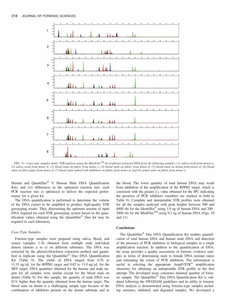
FIG. 10—Case-type samples study: STR analysis using the Identifiler� kit of phenol-extracted DNA from the following samples: (1) saliva swab from donor a;(2) saliva swab from donor b; (3) blood stain on fabric from donor c; (4) blood stain on fabric from donor d; (5) blood stain on denim from donor d; (6) bloodstain on filter paper from donor d; (7) blood stain spiked with inhibitors on fabric from donor d; and (8) semen stain on fabric from donor e.
BARBISIN ET AL. • QUANTIFILER� DUO KIT VALIDATION 317
Human and Quantifiler� Y Human Male DNA QuantificationKits; and (vi) differences in the optimized reaction mix: eachPCR reaction mix is optimized to deliver the expected perfor-mance for a given kit.
The DNA quantification is performed to determine the volumeof the DNA extract to be amplified to produce high-quality STRgenotyping results. Thus, determining the optimum amount of inputDNA required for each STR genotyping system based on the quan-tification values obtained using the Quantifiler� Duo kit may berequired in each laboratory.
Case-Type Samples
Forensic-type samples were prepared using saliva, blood, andsemen (samples 1–8) obtained from multiple male individualdonors (donors a to e) on different substrates. The DNA wasextracted by the phenol:chloroform extraction method and quanti-fied in triplicate using the Quantifiler� Duo DNA QuantificationKit (Table 9). The yields of DNA ranged from 0.76 to11.35 ng ⁄lL for the RPPH1 target and 0.82 to 11.0 ng ⁄lL for theSRY target. DNA quantities obtained for the human and male tar-gets for all samples were similar except for the blood stain ondenim (Table 9). For this sample, the quantity of male DNA was81% higher than the quantity obtained from the human target. Theblood stain on denim is a challenging sample type because of thecombination of inhibitors present on the denim substrate and in
the blood. The lower quantity of total human DNA may resultfrom inhibition of the amplification of the RPPH1 target, which isconsistent with the greater CT value obtained for the IPC indicatingthe presence of PCR inhibitors (numbers are marked in bold inTable 9). Complete and interpretable STR profiles were obtainedfor all the samples analyzed with peak heights between 500 and4000 rfu for the Identifiler� using 1.0 ng of human DNA and 200–2000 rfu for the MiniFilerTM using 0.1 ng of human DNA (Figs. 10and 11).
Conclusions
The Quantifiler� Duo DNA Quantification Kit enables quantifi-cation of total human DNA and human male DNA and detectionof the presence of PCR inhibitors in biological samples in a singleamplification reaction. In addition to the quantification of DNA,the assay provides a quality assessment of forensic evidence sam-ples in terms of determining male to female DNA mixture ratiosand estimating the extent of PCR inhibition. The information isuseful in selecting the appropriate AmpF‘STR� amplificationchemistry for obtaining an interpretable STR profile in the firstattempt. The developed assay consumes minimal quantity of foren-sic sample. The Quantifiler� Duo DNA Quantification Kit is vali-dated following the SWGDAM guidelines and its utility in forensicDNA analysis is demonstrated using forensic-type samples includ-ing mixtures, inhibited, and degraded samples. We developed a
FIG. 11—Case-type samples study: STR analysis using the MiniFilerTM kit of phenol-extracted DNA from the following samples: (1) saliva swab from donor a;(2) saliva swab from donor b; (3) blood stain on fabric from donor c; (4) blood stain on fabric from donor d; (5) blood stain on denim from donor d; (6) bloodstain on filter paper from donor d; (7) blood stain spiked with inhibitors on fabric from donor d; and (8) semen stain on fabric from donor e.
318 JOURNAL OF FORENSIC SCIENCES

reliable and robust assay for obtaining quantification results andassessment of the DNA extract for subsequent STR profiling.Based on the mixture ratio of human and human male DNA andthe extent of PCR inhibition, it is possible to choose between auto-somal and Y-STR profiling or to select a robust STR profiling kitsuch as the MiniFilerTM kit rather than repurifying a certain sam-ple. The quantification results can then be utilized to determine thevolume of DNA extracted to be amplified to obtain an interpretableSTR profile.
In conclusion, the results obtained using the Quantifiler� Duo kitcan aid in determining (i) if the sample contains sufficient humanDNA and ⁄or human male DNA to proceed with STR analysis, (ii)the optimal amount of sample to use in the various STR analysisapplications, (iii) the relative quantities of human male and femaleDNA in a sample that can assist in the selection of the applicableSTR chemistry, and (iv) if PCR inhibitors are present in a samplethat may require additional purification before proceeding to STRanalysis. The Quantifiler� Duo kit, therefore, is a useful tool thatprovides guidance for the selection of the appropriate AmpF‘STR�
amplification kit in order to increase the success of STR profilingin the first attempt. This approach will reduce the number of sam-ples that need reprocessing thereby decreasing the turnaround timein a forensic laboratory.
Acknowledgments
The authors thank Heidi Kijenski, Pius Brzoska, HemantPawar, and Linlin Chou for useful discussions and technicalsupport.
References
1. Moretti TR, Baumstark AL, Defenbaugh DA, Keys KM, Smerick JB,Budowle B. Validation of short tandem repeats (STRs) for forensicusage: performance testing of fluorescent multiplex STR systems andanalysis of authentic and simulated forensic samples. J Forensic Sci2001;46:647–60.
2. Butler JM. Forensic DNA typing, 2nd edn. Burlington: Elsevier Aca-demic Press, 2005.
3. Walsh PS, Varlaro J, Reynolds R. A rapid chemiluminescent method forquantitation of human DNA. Nucleic Acid Res 1992;20:5061–5.
4. Green RL, Roinestad IC, Boland C, Hennessy LK. Developmental vali-dation of the QuantifilerTM real-time PCR kits for the quantitation ofhuman nuclear DNA samples. J Forensic Sci 2005;50:809–25.
5. Walker JA, Hedges DJ, Perodeau BP, Landry KE, Stoilova N, LabordeME, et al. Multiplex polymerase chain reaction for simultaneous quanti-tation of human nuclear, mitochondrial, and male Y-chromosome DNA;application in human identification. Anal Biochem 2005;337:89–97.
6. Horsman KM, Hickey JA, Cotton RW, Landers JP, Maddox LO. Devel-opment of a human specific real-time PCR assay for the simultaneousquantitation of total genomic and male DNA. J Forensic Sci2006;51:758–65.
7. Nicklas JA, Buel E. Simultaneous determination of total human andmale DNA using a duplex real-time PCR assay. J Forensic Sci2006;51:1005–15.
8. Swango KL, Hudlow WR, Timken MD, Buoncristiani MR. Develop-mental validation of a multiplex qPCR assay for assessing the quantityand quality of nuclear DNA in forensic samples. Forensic Sci Int2007;170:35–45.
9. Budowle B, Smith J, Moretti T, DiZinno J. DNA typing protocols:molecular biology and forensic analysis. Natik: Eaton Publishing,2000;41–2.
10. Mulero JJ, Chang CW, Calandra LM, Green RL, Li Y, Johnson CL,et al. Development and validation of the AmpFlSTR� YfilerTM PCRamplification kit: a male specific, single amplification 17 Y-STR multi-plex system. J Forensic Sci 2006;51:64–75.
11. Mulero JJ, Chang CW, Lagac� RE, Wang DY, Bas JL, McMahon TP,et al. Development and validation of the AmpFlSTR� MiniFilerTM PCRamplification kit: a MiniSTR multiplex for the analysis of degradedand ⁄ or PCR inhibited DNA. J Forensic Sci 2008;53(4):838–52.
12. Holland PM, Abramson RD, Watson R, Gelfand DH. Detection of spe-cific polymerase chain reaction product by utilizing the 5¢ to 3¢ exonu-clease activity of Thermus aquaticus DNA polymerase. Proc Natl AcadSci USA 1991;88(16):7276–80.
13. Scientific Working Group on DNA Analysis Methods (SWGDAM).Revised Validation Guidelines issued by the Scientific Working Groupon DNA Analysis Methods (SWGDAM). Forensic Sci Commun2004;6(3). Available at http://www.fbi.gov/hq/lab/fsc/backissu/july2004/standards/2004_03_standards02.htm#perfcheck.
14. Shewale JG, Schneida E, Wilson J, Walker JA, Batzer MA, Sinha SK.Human genomic DNA quantitation system, H-Quant: development andvalidation for use in forensic casework. J Forensic Sci 2007;52:364–70.
15. Timken MD, Swango KL, Orrego C, Buoncristiani MR. A duplex real-time qPCR assay for the quantification of human nuclear and mitochon-drial DNA in forensic samples: implications for quantifying DNA indegraded samples. J Forensic Sci 2005;50:1044–60.
Additional information and reprint requests:Jaiprakash G. Shewale, Ph.D.Applied Biosystems850 Lincoln Centre Drive, Mail Stop 402Foster City, CA 94404E-mail: [email protected]
BARBISIN ET AL. • QUANTIFILER� DUO KIT VALIDATION 319