DEVELOPMENT OF IN VITRO CULTURE AND GENE TRANSFER ...
163
- i - DEVELOPMENT OF IN VITRO CULTURE AND GENE TRANSFER TECHNIQUES IN SUGARCANE (SACCHARUM SPECIES HYBRIDS) by Sandra Jane Snyman Submitted in partial fulfilment of the requirements for the degree of Master of Science, in the Department of Biology, University of Natal 1992 Durban 1992
Transcript of DEVELOPMENT OF IN VITRO CULTURE AND GENE TRANSFER ...
- i -
DEVELOPMENT OF IN VITRO CULTURE AND GENE TRANSFER TECHNIQUES IN
SUGARCANE (SACCHARUM SPECIES HYBRIDS)
by
Sandra Jane Snyman
Submitted in partial fulfilment of the
requirements for the degree of
Master of Science,
in the
Department of Biology,
University of Natal
1992
Durban
1992
- ii -
PREFACE
The experimental work described in this thesis was carried out in
the Department of Biology, University of Natal, Durban, from
January 1990 to December 1991, under the supervision of Drs B.
Huckett and M.P. Watt.
These studies represent original work by the author and have not
been submitted in any form to another University. Where use was
.made of the work of others it has been duly acknowledged in the
text.
- iii -
ACKNOWLEDGEMENTS
sincere thanks to Dr Peter Hewitt, Director of the South African
Sugar Association Experiment station for allowing me to present
research results ·carried out while in his employ, in this thesis.
A heartfelt thank-you to my supervisors Drs Barbara Huckett and
PauIa Watt for their unflagging encouragement and support over the
past two years.
I would like to extend a special word of thanks to the following
people:
Kevin Black of the SASA Experiment station for his excellent
technical assistance,
Poovie Govender of the SASA Experiment station, and Felicity
Blakeway of the Biology Department, University of Natal, Durban,
for their assistance with photography,
Deanne Zeeman of the SASA Experiment station for her assistance in
typing sections of the manuscript,
Staff of the Biometry Department, SASA Experiment Station, for
their advice on statistical analyses and use of computer software,
Genetics Department, University of Natal, Pietermaritzburg, for
their gift of a sample of pBI221, and
Dr Thomas Pinfold and Jennifer Jacobs for proof-reading themanuscript.

- iv -
A DEDICATION
scientific research is like housework;
however resourceful or resolute the
approach, the task is unending ..
My thesis is dedicated to the above concept, and to my husband and
family who, in the final months, assisted in its completion by
doing my housework.
- v -
ABSTRACT
In vitro cell and tissue culture systems were developed for
sugarcane in order to utilise current transformation techniques to
introduce genes to South African sugarcane varieties, which would
be difficult, if not impossible to achieve in conventional
breeding programmes. Embryogenic calli were initiated in the dark
from stem explants of sugarcane varieties NCo376 and N13, on a MS
medium containing sucrose (20-50 g/l), 2,4-D (2-4 mg/l), casein (1
g/l), inositol (100 mg/l) and agar (9g/l). After 2 months the
somatic embryos were cultured in a light/dark photoperiod for a
further 2 months. The best combination of sucrose and 2,4-D for
callus initiation, and subsequent plant regeneration, was 20 g/l
and 2 mg/l, respectively. Plant yields ranged from 16 to 36
plants per gram fresh weight callus, and the yields were not
significantly increased by the addition of activated charcoal to
the regeneration medium. When plantlets reached a height of 10
cm, they were transferred to autoclaved soil in pots, hardened-off
and placed in the glasshouse.
<:" ('l::" . 1 'r J L 0 J
Suspension cultures were initiated from friable NCo376 calli in
liquid MS medium shaken at 100 rev/min in the dark at 27°C, and
were subcultured every 3-7 days. Protoplasts from variou sources
(leaf, calli and suspension cultures) were obtained after
enzymatic digestion in cellulase (20-30 g/l), macerozyme (0,2
g/l), hemicellulase (5 g/l), and sorbitol (0,55 M) in a calcium
and magnesium salt solution. Protoplasts cultured for 48 h
resulted in a loss in viability of 84%.
- vi -
The potential of the seed as a recipient for direct gene uptake
was investigated, as this eliminated the need for in vitro culture
and plant regeneration. Uptake of [3 HJ pBR322 DNA by seeds was
demonstrated, and seeds with the testa removed exhibited higher
initial uptake rates than those with intact seed coats. However,
transient expression, using the GUS reporter gene (coding for
bacterial B-glucuronidase) carried on plasmid pBI221, could not be
conclusively shown using the histochemical GUS assay, due to GUS
activity generated by either microbial contamination or endogenous
plant GUS activity. Neither microwaving to eradicate contaminants
nor the addition of methanol (20%) to the GUS incubation buffer
were successful in overcoming positive results observed in control
seeds. An alternative approach to sugarcane transformation, using
PEG-mediated DNA uptake and subsequent transient expression of GUS
by protoplasts was investigated, but microbial contamination was a
persistant problem and no positive results were observed. Further
examination and elimination of endogenous contamination is
required before transformation studies can be continued.
- vii -
TABLE OF CONTENTSPage
Title pagePrefaceAcknowledgementsA DedicationAbstractTable of ContentsList of TablesList of FiguresList of PlatesList of Abbreviations and Symbols
iii
iiiiv
vviixii
xiiixivxv
CHAPTER ONE :GENERAL INTRODUCTION 1
South African sugarcane varieties and their characteristics 1Sugarcane breeding and its problems 4The introduction of genetic variation by in vitro culture 5Aims of this study 7
CHAPTER TWO :DEVELOPMENT OF IN VITRO CULTURE SYSTEMS FOR 8SUGARCANE (SACCHARUM species hybrids)
2.2.1
2 . 2 . 2
2.2.3
INTRODUCTORY REMARKS
LITERATURE REVIEW
GENERAL ASPECTS OF IN VITRO CULTURE
FACTORS AFFECTING CELL AND TISSUE CULTURE SYSTEMS
Selection of explantssterilisation procedures and maintenance ofaseptic cultures
Composition of the culture mediumLight regimes in culture
REGENERATION OF PLANTS FROM SOMATIC EMBRYOS
8
8
8
12
1213
1517
18
Formation and identification of embryogenic cells 18and structures
Embryo germination 19
2 . 2 . 4
2.2.5
CELL SUSPENSION CULTURES
Suspension culture initiation and maintenancePlant regeneration from suspension culture cells
PROTOPLAST ISOLATION AND CULTURE
Protoplast isolation and purificationProtoplast culture
21
2123
23
2325
- viii -Page
2.2.6
2.2.7
2.2.8
2.3.1
2.3.2
2.3.3
HARDENING-OFF AND PLANTING OUT OF REGENERATED PLANTS
GENETIC STABILITY VERSUS GENETIC VARIATION IN VITRO
POTENTIAL PRACTICAL APPLICATIONS OF TISSUE CULTURESYSTEMS
Large scale micropropagationCrop improvement by somaclonal variationStorage of germplasmThe production of haploid plantsRegeneration of transformed tissues and/or cells
MATERIALS AND METHODS
IN VITRO CULTURE OF SEEDS/ZYGOTIC EMBRYOS
PREPARATION OF EXPLANTS FOR IN VITRO CALLUS CULTURE
GROWTH CONDITIONS FOR CALLUS CULTURE
Production of aseptic culturesCallus induction
26
27
28
2930303132
33
33
33
34
3436
2.3.4 PRODUCTION OF SOMATIC EMBRYOGENIC CULTURES 36
2.3.5 HARDENING-OFF AND ACCLIMITISATION OF REGENERATED 37PLANTS
2.3.6 INITIATION AND MAINTENANCE OF SUSPENSION CULTURES 37
2.3.7 ISOLATION AND CULTURE OF PROTOPLASTS 38
2.3.8 MICROSCOPY 39
2.3.9 STATISTICAL ANALYSES 40
2.3.10 PHOTOGRAPHY 40
2.4 RESULTS 41
2.4.1 PROPAGATION BY MEANS OF ZYGOTIC EMBRYOS/SEEDS 41
2.4.2 CALLUS CULTURE AND PLANT REGENERATION VIA SOMATIC 41EMBRYOGENESIS IN SOUTH AFRICAN SUGARCANE VARIETIES
Photographic account of plant regeneration via 41somatic embryogenesis in variety N13
Antimicrobial treatment of field-derived explants 42The effect of sucrose and 2,4-D concentrations on 47callus induction and plant regeneration
The effect of activated charcoal on plant 50regeneration
Hardening-off of regenerated plants 51
- ix -Page
2 .4. 3
2.4.4
2.5.1
2.5.2
2.5.3
2.5.4
INITIATION AND ESTABLISHMENT OF SUSPENSION CULTURES
PROTOPLAST ISOLATION AND SHORT-TERM CULTURE
DISCUSSION
ADULT-PLANT SOURCE TISSUE
PLANT REGENERATION VIA SOMATIC EMBRYOGENESIS
ESTABLISHMENT OF SUSPENSION CULTURES
PROTOPLAST ISOLATION AND SHORT-TERM MAINTENANCE
52
55
61
62
62
65
67
CHAPTER THREE :DEVELOPMENT OF GENE TRANSFER SYSTEMS FORSUGARCANE (SACCHARUM SPECIES HYBRIDS)
INTRODUCTORY REMARKS
LITERATURE REVIEW
70
70
70
3.2.1 FACTORS INFLUENCING TRANSFORMATION EVENTS 70
3.2.2
Factors affecting introduction of DNA into plant cells 70
Methods used to introduce foreign DNA to plants 70The difference in response to transformation by 71
monocotyledonous and dicotyledonous plants
Factors affecting integration of DNA into the plant 72genome
Factors influencing expression of transforming DNA 72
Choice of promoter 73
COMMONLY USED METHODS FOR TRANSFERRING GENES TO PLANTS 74
Agrobacterium-mediated gene transfer
Direct gene transfer to protoplasts
ElectroporationPolyethylene glycol-induced DNA uptakeMicroinjection of DNA into protoplastsTechniques less frequently used for genetransfer to protoplasts
Direct gene transfer to intact cells
Microprojectile bombardment of plant cellsand tissues
Viral-mediated DNA deliveryPollen-mediated transformation of cells
74
77
78798080
81
81
8385
- x -Page
3 • 2 • 3
3.2.4
3.2.5
3.2.6
THE POTENTIAL OF THE SEED FOR USE IN GENE TRANSFERTO PLANTS
VECTORS
SELECTION OF TRANSFORMANTS BY MEANS OF GENETICMARKERS
Detection of transient gene expressionThe main features of the CAT and luciferase
systemsThe GUS system
Detection of stable integrationThe NPTII system
APPLICATION OF GENETIC ENGINEERING TO PLANTIMPROVEMENT
85
88
89
9090
919393
95
Crop improvement by genetic engineering 95
Weed control 95Insect resistance 97Disease resistance 98
Application of genetic engineering to basic research 100
MATERIALS AND METHODS 101
Bacterial strains and plasmids used 101Growth and maintenance of bacterial strains 101Transformation of E. coli 101Generation of expression vector 101Agarose gel electrophoresis of DNA 102Restriction enzyme digestion 102Labelling of plasmid DNA 102Spun-column purification of labelled DNA 103Measurement of specific activity of labelled DNA 103sterilisation of seeds 103Germination of seeds 103Protoplast isolation 104Uptake of labelled DNA by seeds 104Uptake of expression vector by seeds 104PEG-mediated DNA uptake by protoplasts 104Assay for transient expression of GUS 105Photography 105
3.4.1
3.4.2
RESULTS
UPTAKE OF LABELLED DNA BY SEEDS
UPTAKE OF EXPRESSION VECTOR pBI221 BY SEEDS
106
106
106
Confirmation of pBI221 structure 109.,Detectlon of GUS expression in seeds 109Treatment of seeds in order to overcome positive 111controls
- xi -
Testa as possible barrier to DNA entry into embryo 111Elimination of bacterial contamination 111Suppression of endogenous plant GUS activity 113
3.4.3 UPTAKE OF pBI221 BY PROTOPLASTS 114
Viability studies on protoplasts treated withGUS assay following transient expression inprotoplasts
Elimination of contamination in protoplasts
3.5 DISCUSSION
The use of the seed in transformation studiesTransformation via the protoplastTransformation alternatives
/
CONCLUDING REMARKS
REFERENCES
PEG 114116
116
117
117120121
123
124
Table 1.1
Table 2.1
Table 2.2
: Table 2.3
Table 2.4
Table 2.5
Table 2.6
Table 3.1
Table 3.2
- xii -
LIST OF TABLES
Agronomic characteristics of South Africansugarcane varieties.
A comparison of the components of four differentmedia recommended for the initiation ofsuspension cultures of sugarcane.
The effect of a cocktail of antimicrobialsubstances on contamination levels of NCo376and N13 explants, and callus formation after1 month in culture.
The effect of a range of 2,4-D and sucroseconcentrations on percentage N13 callusproduction on MS medium after 2 months inculture.
The effect of different 2,4-D and sucroseconcentrations on fresh weight (g) of N13calli after 2 months of culture on MS medium.
The effect of sucrose levels in the MSregeneration medium on the percentage of N13calli which yield plants.
Plantlet yield per gram fresh weight N13 callusgrown on regeneration medium for 2 months.
Germination and bacterial contamination levelsof sugarcane seeds exposed to microwavetreatment for various time intervals.
Effect of seed pretreatment and assay medium onsugarcane seeds as determined by the GUShistochemical assay.
Page
3
38
47
48
48
49
50
112
113
Fig. 1.1
Fig. 2.1
Fig. 2.2
Fig. 2.3
Fig 2.4
Fig. 2.5
Fig. 2.6
Fig. 2.7
Fig. 2.8
Fig. 2.9
Fig. 3.1
Fig. 3.2
Fig. 3.3
Fig. 3.4
Fig. 3.5
Fig. 3.6
Fig. 3.7
Fig. 3.8
Fig. 3.9
- xiii -
LIST OF FIGURESPage
Map showing South African sugarcane-growing areas. 2
A diagrammatic representation of the principal 10pathways of plant regeneration.
Diagrammatic representation of the stages in 11zygotic and somatic embryogenesis in carrot.
The early stages of somatic pro-embryo 20development.
The effect of the addition of activated charcoal 51to the regeneration medium on the percentage ofN13 calli producing plants.
Growth rate of NCo376 suspension culture cells as 53determined by an increase in packed cell volumeover time.
The effect of the concentration of cell wall 57degrading enzymes on yield of totaland viable mesophyll protoplasts.
The effect of sorbitol concentration on yields 58of total and viable mesophyll protoplasts.
The effect of cellulase on the yields of total 59and viable protoplasts isolated from sugarcanecallus.
Summary of relationship between types of in vitro 61cultures established for South African sugarcanevarieties N13 and NCo376.
Schematic diagram of generalised cointegrative- 75and binary-vector systems.
Schematic diagram of the microprojectile 82delivery system.
Electrophoretic transfection chamber. 87
Restriction map of pBR322. 89
Structure of pBI221. 92
Uptake of [3 HJ pBR322 DNA by sugarcane seeds. 107
Rate of uptake of [3H] pBR322 DNA by sugarcane 108seeds with testa removed and testa intact.
Agarose gell electrophoresis of restriction 110fragments of pBI221.
Total and viable numbers of protoplasts isolated 115from calli after exposure to PEG for 30 min.
- xiv -
LIST OF PLATESPage
Plate 2.1
Plate 2.2
Plate 2.3
Plate 2.4
Plate 2.5
Plate 2.6
: Plate 2.7
In vitro culture of sugarcane seeds.
Source of explants for callus induction.
Embryogenic and non-embryogenic regions oncallus.
Microscopic examination of callus.
Development of pro-embryogenic structures insugarcane callus.
Germination of somatic embryos.
Sugarcane plants which have been regeneratedfrom callus via somatic embryogenesis.
35
35
43
43
43
44
44
Plate 2.8 Regenerated plants with well-developed shoot 45and root systems in tubes.
Plate 2.9 Hardened-off sugarcane plants. 45
Plate 2.10 The appearance of morphologically abnormal 46somatic embryos.
Plate 2.11 Precocious root germination in one month-old 46calli.
Plate 2.12 Necrosis of calli on regeneration medium. 46
Plate 2.13 Suspension cultures initiated from NCo376 54callus.
Plate 2.14 Microscopic examination of suspension culture 54cells.
Plate 2.15 Microscopic examination of various sugarcane 60protoplasts isolated from leaves, suspensioncultures and callus pieces.
Plate 3.1 Sugarcane seeds following uptake of expression 110vector pBI221 and histochemical GUS assay.
- xv -
LIST OF ABBREVIATIONS AND SYMBOLS
Cauliflower mosaic virus
centimetre
chloramphenicol acetyltransferase
copy deoxyribose nucleic acid
counts per minute
degrees celcius
deoxyribose nucleic acid
2,4-dichlorophenoxyacetic acid
figure
B-glucuronidase
grams per litre
hour
kilobases
messenger ribose nucleic acid
micro Einsteins per metre2 per second
microgram
micrometre
microseconds
milligram
milligrams per litre
millilitre
millimetre
millimolar
minute (time)
molecular weight
molar (concentration)
MS medium (Murashige & Skoog, 1962)
2-[N-morpholinoJethanesulphonic acid
neomycin phosphotransferase 11
number
number of observations
packed cell volume
percent/percentage
picograms
polyethylene glycol
revolutions per minute
ribose nucleic acid
CaMV
cm
CAT
cDNA
cpm
°C
DNA
2,4-D
Fig.
GUS
g/l
h
kb
mRNA
p,E/m2/s
p,g
p,m
p,s
mg
mg/l
ml
mm
mM
min
MW
M
MS
MES
NPT11
no.
n
p.c.v.
pg
PEG
rev/min
RNA
second (time)
species
species (plural)
standard deviation
standard error
transferred-DNA
trichloroacetic acid
Tumour-inducing
ultra violet
unit
volts
volume by volume
weight by volume
- xvi -
s
sp.
spp.
s.d.
s.e.
T-DNA
TeA
Ti
u.v.
uvv/v
w/v
CHAPTER ONE
GENERAL INTRODUCTION
Sugarcane (Saccharum species hybrids), a member of the
Gramineae/Poaceae, is a crop of major importance, as it provides
about 65% of the sugar produced in the world (Liu, 1984). The
zone of sugarcane production characteristically occurs in tropical
and sUbtropical latitudes and the crop is grown in soils and
climates displaying great variations (Barnes, 1974). South Africa
is the eighth largest producer of cane sugar, after countries such
I as Brazil; Cuba, India, Australia, united States, Philippines and
I China, with a production of about 2 million tons per annum (Liu,
1984) .
south African sugarcane varieties and their characteristics
Sugarcane varieties bred at the South African Sugar Association
Experiment station at Mount Edgecombe for growth in climatic
regions of South" Africa, are grown in rainfed conditions on the
east coast and also in irrigated plantations in Northern Natal and
Eastern Transvaal (Fig. 1.1). The local variety NCo376, which was
used in this study, was released for commercial propagation in
1955, and in 1976 it represented more than 60% of the sugarcane
harvested in south Africa (Anonymous, 1977). This level decreased
to 45% in 1991 due to the release of new varieties that are more
resistant to drought and diseases, and also have higher yields
(Table 1.1). variety NCo376 has the distinction of being the most
adaptable variety in the S~ Afri~an sugar industry, but other
varieties may be more suited to specific conditions (Anonymous,
1991) . The most suitable variety for a particular location is
normally tne-one which yield~the highest return over a whole crop
cycle. Return depends not only on yield of sucrose, but also on
the costs of managing and handling the crop (Barnes, 1974). In
addition, the problem of disease and soil type vary from area to
area and may be the deciding factor in the final choice of
variety.
2
TRANSVAAL
27
28
29
30
31
32
I
o CANE GROWING AREAS
31
,IIII\ ......,
\\
\ ...\,' ......
.......................
NATAL
'-,,\\\\\
\I
",,/
,'"
TRANSKEI
-........,\,,, ~-
" ----;'......... _-,--
IIII
III,
II
II
IIII
",,,,/,,,,,,,,,1
",/
IJ
Fig. 1.1 Map showing South African sugarcane growing areas (reproduced from
Anonymous, 1977)
Table 1.1
- 3 -
Agronomic characteristics of South African sugarcane varieties.
Year of Proportion SucroseVariety release of 1990/91 yield (t suc
crop (%) /ha/yr)Best features Worst features
High sucrose yields Slow germinationDrought tolerance
--i NCo376
NCo293
\II
Y. N12
N13
N14
N16
N17
1955
1952
1979
1980
1980
1982
1984
45
3,3
12,2
0,6
15,1
1,9
1,7
12
12
12-15
>12
>13
12-22
5-22
Ratooning abilityAdaptability
Hardiness athigh altitudes
Rapid growthAnnual harvest
High yield inirrigated areas
High yieldRapid growth
High sucrose yieldSmut resistant
Drought susceptibleDisease susceptibleLow sucrose
Poor ratooningMosaic virussusceptible
Eldana susceptiblesmut, RSD susceptiblE
Drought susceptible
Eldana and smutsusceptible
Slow germination
f '
Y-- N19
N21
CP66/1043
1986
1989
1987
1,3
Data notaVfiilable
Data notavailable
12-20
11-13
12-18
High sucrose Susceptible to eldanaResistance to smut
Eldana resistance
High yields, smut Requires excellent& mosaic resistance growing conditions •
eldana Most prevalentsugarcane mosaic virussmutRSD (Ratoon stunting disease)
insect pest (Eldana saccharina Walker)}
} = Most prevalent diseases}
(modified from Anonymous, 1991)
- 4 -
sugarcane breeding and its problems
Most commercial sugarcane varieties now in use world-wide
are descendants of inter-specific hybrids between S. spontaneum,
S. sinense, S. robustum and S. officinarum (Arcenaux, 1965).
Saccharum species hybrids are highly polyploid and have chromosome
numbers ranging from 40 to 120 (Price, 1962). Sugarcane is
propagated vegetatively by means of setts, which are segments of
stalk which comprise one to five nodes, from which the shoots and
roots germinate. Once sugarcane stalks have been harvested,
;normally twelve to eighteen months after planting, ratooning
occurs (the root material which remains in the ground after
harvesting, forms new shoot material). The production of fertile
seed under normal growth conditions is rare, due to sub-optimal
night-time temperatures and day-length during the flower
initiation period (Stevenson, 1965).
Priorities in breeding programmes in the major sugar-growing areas
world-wide include selection of high sucrose-yielding cane
varieties which are also insect and disease resistant. However,
sugarcane breeders are faced with formidable characteristics such
as the narrow genetic base of most commercial cane varieties, the- -
high ploidy of Saccharum spp. hybrids, the regular occurrence of'-------
aneuploids and the inability to control the outcome of crosses
(Nuss, pers. comm.). Conventional plant breeders have to
manipulate environmental conditions to achieve cross-hybridisation
between varieties, and in South Africa, specially constructed
glasshouses with adjustable photoperiods are necessary to control
flowering, collect pollen, and to ensure successful pollination of
female plants. Once seed has been produc~d, a screening and
selection programme is initiated, which can take up to 20 years
before a new variety is released to growers (Nuss, pers. comm.).
Therefore the emergence of new techniques by which plants can be
genetically manipulated (reviewed by Vasil, 1987, 1990) may
provide an alternative to conventional and somewhat problematic
sugarcane breeding.
- 5 -
The introduction of genetic variation by in vitro culture
Routes for the introduction of genetic variation include
conventional breeding as discussed above, and the more recent
techniques of in vitro culture of tissue and cells, and gene
transfer (reviewed by Ammirato, 1989; Lindsey and Jones, 1989).
Sugarcane improvement via cell line selection or gene transfer
techniques of necessity includes in vitro culture techniques,
which have been successfully established in sugarcane varieties
grown in other sugar-growing areas in the world. In vitro
techniques are essential in order to allow researchers to work
with single cells or small selected groups of cells in a
controlled environment, that can be manipulated.
Research on sugarcane tissue and cell culture began in Hawaii
where callus cultures were established (Nickell, 1964). When it
was discovered that shoot differentiation occurred from callus
(Heinz and Mee, 1969), other researchers began similar studies
(Liu, 1971; Nadar et al., 1978; Liu et al., 1982; Ho and Vasil,
1983a). Callus culture is mutagenic in the broadest sense (Liu,
1984), as calli maintained in vitro over long time periods are
usually cytologically unstable and regenerate into plants that are
often characterised by genetic variability (Irvine, 1984).
Although this method has given rise to agronomically-useful
sugarcane variants in Taiwan (Liu, 1971; Liu and Chen, 1976; Tsay,
1987; Liu, 1990), Brazil (Evans et al., 1980) and Australia
(Larkin and Scowcroft, 1981), it has not replaced conventional
breeding programmes. The induction of genetic variability in
callus cultures can also be achieved by the use of mutagenic
agents, such as colchicine and this has been attempted in
sugarcane, although no agronomic improvements were evident (Liu,
1990; Irvine et al., 1991).
However in vitro callus culture does not only have application for
producing somaclonal variants, but also for micropropagation
purposes where the resultant plants must be genetically identical
to the parent plant, and for the regeneration of transformed cells
- 6 -
or tissues. It has been suggested that the regeneration of
sugarcane plants from callus via somatic embryos, rather than via
shoot differentiation (Heinz and Mee, 1969; Liu and Chen, 1974;
Nadar and Heinz, 1977; Nadar et al., 1978; Ho and Vasil, 1983a;
Chen et al., 1988a; Guiderdoni and Demarley, 1988), may limit the
genetic variation observed in callus cultures because each embryo
arises from a single cell and the route of development follows
that of zygotic embryo germination (Ammirato, 1983; Evans et al.,
1984a). It is however, essential that callus is not maintained in
culture for an extensive time period. The non-chimeric nature of
somatic embryogenic clones has been described for other
monocotyledonous plants (Botti and Vasil, 1983; Karp and Maddock,
1984; Abe and Futsuhara, 1985; Armstrong and Green, 1985;
Bretzinger et al., 1989; Bhaskaran and smith, 1990).
Another type of in vitro culture that has been established for
sugarcane is suspension cultures, which may be used as a source of
cells for the isolation of protoplasts and the study of plant cell
physiology and biochemistry (Ho and Vasil, 1983b; Liu and Shih,
1986; Chen et al., 1988b).
One advantage of using a single cell for transformation purposes
is that when it divides, ·genetic information is passed onto
daughter cells and eventually the whole plant. Protoplasts are
cells which have had their cell wall enzymatically removed and
this facilitates interaction with other organisms or
macromolecules. The ability to regenerate plants from single
cells has application for genetic engineering techniques, and also
for the production of normally incompatible inter- and/or intra
specific hybrids (Ozias-Akins et al., 1986; Tabaeizadeh et al.,
1986) . Maretzki and Nickell (1973) were the first to isolate
sugarcane protoplasts and induce the formation of callus clusters.
Protoplasts were isolated from leaves (Chen and Liu, 1974; Evans
et al., 1980), but no repeatable regeneration protocols could be
-established. Later Chen and Shih (1983) were able to obtain
callus from protoplasts derived from suspension cells.
Regeneration of sugarcane plants from protoplasts was achieved

- 7 -
(Srinivasan and Vasil, 1986; Chen et al., 1988b) but has not yet
been repeated.
Plant regeneration from monocotyledonous protoplasts is difficult
(Vasil, 1987; Bhaskaran and smith, 1990). Despite this, protocols
for plant regeneration have been established and transgenic rice
(Toriyama et al., 1988; Zhang et al., 1988; Datta et al., 1990;
Hayashimoto et al., 1990; Raineri et al., 1990) and maize (Rhodes
et al., 1988; Fromm et al., 1990; Gordon-Kamm et al., 1990) plants
have been produced from transformed protoplasts. A common factor
\ in the regeneration of both the above species is that embryogenicj
protoplasts (isolated from embryogenic suspension cultures) were
the cells targeted for DNA uptake. Although Chen et al. (1987)
were able to transform sugarcane protoplasts isolated from
embryogenic sugarcane suspension cultures, only callus colonies
could be recovered. Reports of transformed monocotyledonous
protoplasts appear in the literature regularly, but aside from
rice and maize, there are not many which can be regenerated to
plants (Lorz ~t al., 1985; Vasil, 1990).
Aims of this study
The aims of this study were to investigate options to supplement
the current sugarcane-breeding programme at the Experiment station
at Mount Edgecombe. In vitro cell and tissue culture techniques
for two South African sugarcane varieties were established, with a
view to ultimately being able to use them in conjunction with gene
transfer techniques to introduce specific genes into the sugarcane
genome. Preliminary work on techiques used for the introduction
of DNA into sugarcane was also undertaken.
\
- 8 -
CHAPTER TWO
DEVELOPMENT OF IN VITRO CULTURE SYSTEMS FOR SUGARCANE (SACCHARUM
species hybrids)
2.1. INTRODUCTORY REMARKS
The overall objectives of the study were to establish protocols
for the induction and establishment of callus cultures, suspension
culture initiation and maintenance, and the isolation of
:protoplasts. These in vitro systems offer the potential for; -proliferation of genetically-transformed cells and tissues, and
ultimately the regeneration of plants. The establishment of such
techniques in sugarcane is one of the steps necessary in any
breeding programme which aims to use genetic engineering
techniques to insert agronomically useful genes into South"African
sugarcane varieties.
The varieties used in this study were NCo376, which is widely
grown and occupies 45% of the land under sugarcane cultivation,
and N13 which only occupies about 1% of land under sugarcane
cUltivation. The latter variety is a potentially good candidate
for improvement by genetic engineering techniques, because it has
adequate sucrose yields, but is susceptible to many diseases.
2.2. LITERATURE REVIEW
2.2.1. GENERAL ASPECTS OF IN VITRO CULTURE
The cereals and grasses, which constitute the most important group
of crop plants, have until recently been recalcitrant to cell
culture techniques (Vasil, 1982, 1987; Bhaskaran and Smith, 1990).
However, important advances in cell culture have been made, the
highlights of which include:
1) the establishment of totipotent cell lines in Lolium grass
(Jones and Dale, 1982), Panicum grass (Lu and Vasil, 1982),
sugarcane (Ho and Vasil, 1983a, 1983b) and wheat (Vasil et al.,
- 9 -
1990a, 1990b);2) cell suspension cultures in wheat (Shimada and Yamada, 1979),
sugarcane (Ho and Vasil, 1983b) and Panicum grass (Karlsson and
Vasil, 1986);3) the development of protoplast culture systems yielding somatic
embryos and plants in Panicum (Lu et al., 1981), rice (Fujimura et
al., 1985) and sugarcane (Srinivasan and Vasil, 1986); and
4) more recently the recovery of somatic hybrid cell lines from
Pennisetum x Panicum (ozias-Akins et al., 1986) and sugarcane x
Panicum (Tabaeizadeh et al., 1986), and genetically transformed
cell lines in maize and rice (Lorz et al., 1985; Potrykus et al.,
1985; Fromm et al., 1986).
As sugarcane is a member of the GramineaejPaoceae, this literature
review will focus largely on the reported developments regarding
in vitro systems in members of this group.
Plant regeneration from the above-mentioned in vitro culture
systems can occur via one of two pathways, organogenesis or
somatic embryogenesis and either directly from explants with a
minimum or absence of callusing, or indirectly via a callus stage
first (Fig. 2.1). Callus consists of a mass of tissue, with a low
level of organisation, obtained by transferring cut pieces of
plant organs to a suitable nutrient medium (Skoog and Miller,
1957) . Generally, callus can be maintained indefinitely by
regular transfer to fresh culture medium, and plants develop from
specific regions of the callus either via somatic embryogenesis or
by shoot morphogenesis followed by root development
(organogenesis) (Fig. 2.1).
Direct organogenesis (or shoot morphogenesis) involves the
development of axillary buds following the inhibition of apical
dominance, and indirect organogenesis involves the de novo
organisation of shoot meristems in callus cultures (Shimada and
Yamada, 1979; Evans et al., 1984ai Bhaskaran and Smith, 1990).
Organogenic plant regeneration from axillary meristems, which
forms the basis of successful large-sale micropropagation in many
dicotyledonous herbaceous species (Fig. 2.1), is not common in
- 10 -
cereals and grasses but has been observed in sugarcane (Grisham
and Bourg, 1989) and some forage grasses (Ahloowalia, 1984).
organogenic plant regeneration via shoot morphogenesis in callus
culture has been observed in graminaceous plants such as maize
(Lowe et al., 1985; Woodward, 1989), sorghum (Wernicke et al.,
1982), (Liu and Chen, 1974; Ch~n et al.,_~988a) and some
of the forage grasses (Ahloowalia, 1984). It is important to note
that shoot meristems in in vitro callus culture have been shown to
be multicellular in origin (steward et al., 1958; Norris et al.,
1983; Skene and Barlass, 1983), and may give rise to chimeras
(Sacristan and Melchers, 1969; Irvine, 1984), which are unsuitable
for clonal propagation, mutation research, genetic analysis and
breeding.
-----';e Somatic seedlings
J+- Shoot tip culture -+l
~ ~Axillary Multiple ~$
branching axillary shoots ~
Rooted~ PlantlAtl'
~ Rooled
Various
El) .
~---~-_-.a........ fIT-I7TIDSE r
Somaticembryos
It!2:1... Merlstem ~te.m cultureVOR DO.~
~ j~~ot 8.e1,~O:!:lh
- ~ ~f>SI~ ~
TPropagation from
axillary buds
Propagation fromadventitious shoots
or embryos
INDIRECT Callus ~ shootMORPHOGENESI9 ESJP-O l.Q. formation
Callus growth ( 1~ .~ · P1antletson explant \ AdventItIouS
~--:; S_. . ~fromca/lu4)ISE •
SuspenSIOncultures OR
D SE*(from single cens)
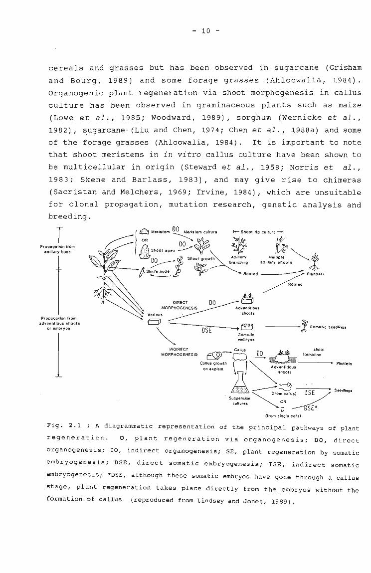
Fig. 2.1 : A diagrammatic representation of the principal pathways of plant
regeneration. 0, plant regeneration via organogenesis; DO, direct
organogenesis; IO, indirect organogenesis; SE, plant regeneration by somatic
embryogenesis; DSE, direct somatic embryogenesis; lSE, indirect somatic
embryogenesis; *DSE, although these somatic embryos have gone through a callus
stage, plant regeneration takes place directly from the embryos without the
formation of callus (reproduced from Lindsey and Jones, 1989).
- 11 -
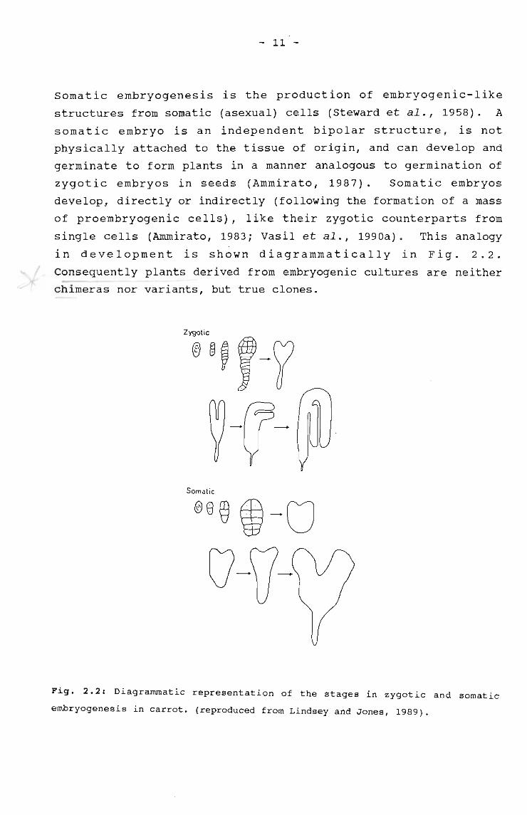
somatic embryogenesis is the production of embryogenic-like
structures from somatic (asexual) cells (Steward et al., 1958). A
somatic embryo is an independent bipolar structure, is not
physically attached to the tissue of origin, and can develop and
germinate to form plants in a manner analogous to germination of
zygotic embryos in seeds (Ammirato, 1987). Somatic embryos
develop, directly or indirectly (following the formation of a mass
of proembryogenic cells), like their zygotic counterparts from
single cells (Ammirato, 1983; Vasil et al., 1990a). This analogy
in development is shown diagrammatically in Fig. 2.2.
Consequently plants derived from embryogenic cultures are neither
chimeras nor variants, but true clones.
-
Somatic
®8@ i-Ov-fj-
Fig. 2.2: Diagrammatic representation of the stages in zygotic and somatic
embryogenesis in carrot. (reproduced from Lindsey and Jones, 1989).
- 12 -
Plant regeneration from callus cultures of sugarcane via somatic
embryogenesis was achieved over a decade ago (Liu and Chen, 1974;
Nadar and Heinz, 1977; Nadar et al., 1978; Ho and Vasil, 1983a;
Chen et al., 1988a; Guiderdoni and Demarly, 1988). Other-grasses
and cereals have also been regenerated via somatic embryogenesis,
for example maize (Lu et al., 1982, 1983; Armstrong and Green,
1985; Woodward, 1989; Ray and Ghosh, 1990), rice (Abe and
Futsuhara, 1985; Ling et al., 1983; Abdullah et al., 1986), wheat
(Karp and Maddock, 1984; Vasil et al., 1984; Rashid and Quraishi,
1989), oats (Bregi tzer et al. I 1989) I rye (Lu et al., 1984),
,sorghum (MacKinnon et al., 1986), digitaria grass (Gonzales and
Franks, 1987; watt et al., 1989) and Pennisetum spp. (Wang and
Vasil, 1982; Botti and Vasil, 1983; Chandler and Vasil, 1984).
Although most graminaceous monocotyledons regenerate via the
process of somatic embryogenesis, there are some monocotyledon
tissue culture systems where plant regeneration via both somatic
embryogenesis and organogenesis has been observed. For example,
both embryogenic callus and shoot buds were produced from callus
cultures of maize (Lowe et al., 1985; Wooodward, 1989) and sorghum
(Wernicke et al., 1982).
2.2.2. FACTORS AFFECTING CELL AND TISSUE CULTURE SYSTEMS
Selection of explants
Callus cultures of graminaceous species have been obtained, from a
variety of explants including immature embryos, young
inflorescences, young leaves, roots and anthers (Vasil and Vasil,
1984; Rout and Sarma, 1991). In embryos', callus originates from
peripheral cells in the scutellum, in inflorescences from the
floral meristems or from peripperal tissue around vascula~
bundles, and in leaves from cells of the lower epidermis and
mesophyll near vascular bundles (Botti and Vasil, 1983; 'Vasi,l and
Vasil, 1984; Vasil, 1987). The developmental and physiological
stage of graminaceous explants is critical in the establishment of
totipotent cultures, and there seems to be a brief period of t~me
- 13 -
during which embryos, inflorencences and leaves have the
competence to form embryogenic cultures (Lu and Vasil, 1982; Botti
and Vasil, 1983; Ho and Vasil, 1983a). At this stage selected
cells in the explants are meristematic, only partially
differentiated and not fully committed to specialised functions.
Hence, explants obtained before or after this stage form only non
morphogenic or non-embryogenic cells (Guiderdoni and Demarly,
1988) .
In sugarcane there appears to be a close relationship between the
\ state of differentiation of the excised region and the subsequentI
'production of callus, with nodular embryogenic calli being
obtained from the bases of fast-growing leaves, while more mature
parts of older leaves produced only friable calli (Guiderdoni and
Demarly, 1988). The age of the plant does not seem to be as
important, as young leaves obtained from mature sugarcane plants
have been used successfully for the initiation of callus cultures
(Ho and Vasil, 1983a), and this appears to occur in most of the
Gramineae (Vasil, 1987; Bhaskaran and Smith, 1990).
The two main explant-sources for the isolation of protoplasts,
leaves and suspension cultures, both provide yields of viable
protoplasts, but with the former explant source, the search for a
subsequent suitable culture method suitable for plant
regeneration, has been elusive in the Gramineae (section 2.2.5).
sterilisation procedures and maintenance of aseptic cultures
Onceat i s sue, 0 r par t. -·0 f -. a - p I ant, has been se I e c t e d for
explantation, it has to be e~cisedi disinfected and transferred to
a nutrient medium under aseptic conditions. In most cases, a
surface-sterilisation procedure is sufficient to remove all
surface contaminants. sterilising agents include sodium
hypochlorite, mercuric chloride, silver nitrate, hydrogen peroxide
and calcium hypochlorite (reviewed by Constable, 1984). Generally
standard surface-sterilisation procedures are followed, for
example, plant material is dipped into 70% ethanol, then immersed
- 14 -
in a solution containing a sterilising agent such as sodium
hypochlorite, followed by thorough rinsing.
Despite surface-sterilisation procedures, contamination of media
on which explants were cultured has been observed, possibly due to
'latent' or endogenous contaminants in the plant material (Leifert
and waites, 1990). The use of antimicrobial agents may overcome
in vitro contamination problems, especially those contaminants
which are difficult to eradicate using sterile techniques only.
To be effective, the ideal antimicrobial agent should eradicate
contaminants on plant-tissue culture media, have a non-toxic
effect on plant cells and have a broad spectrum of activity
(Falkiner, 1990).
Pollock et al. (1983) investigated the effect of a range of 20
antibiotics against protoplast-derived cells from Nicotiana~
plumbaginifolia. They found that the least toxic antibiotics were
the betalactams, 'such as ampicillin or carbenicillin, which.. .-
provide a broad spectrum of bacteriocidal activity, and
cephalosporins suc~ as cefoxitin and cefotaxime were also
recommended. Doses of up to 100Mg/ml of each of these antibiotics
can be used without apparent ill effect. A combination of the
antibiotics cefotaxime, rifampicin, tetracycline and polymixin B
in a 'cocktail' mixture has been successful in eradication of
bacterial contamination of shoot cultures in apple, walnut, plum,
rhododendron and pear (Young et al., 1984).
Yeasts have also been described as contaminants in some plant
tissue cultures, and the most frequently isolated yeast species
were Candida and Rhodotorula (pink yeasts) species (Boxus and
Terzi, 1988; Enjalric et al., 1988; Leggatt et al., 1988). These
belong to the group of osmophilic yeasts which show high sugar and
salt tolerance, and hence are well adapted to grow in plant-growth
media and in some plant material such as sugarcane stalks which
contain approximately 14% (w/v) sucrose (Alexander, 1985). This,
together with their eukaryotic nature, makes yeast contamination
very difficult or impossible to eradicate. Successful elimination
- 15 -
of yeast cells from in vitro culture was achieved by Shields et
al. (1984), who tested a number of fungicides and found
clotrimazole, miconazole, griseofulvin and fenbendazole to be the
most effective against yeast cells, without harming the tissue.
The activity of antimicrobial agents lasts for approximately 48
hours, and so replacement with fresh solutions after this time -is
recommended (Falkiner, 1990). Antimicrobial substances can be
incorporated into liquid or solid culture media. It is necessary
to determine conditions which best suit the particular explant and
type of media being used.
composition of the culture medium
Tissues of the Gramineae have been cultured in vitro on a range of
media, including B5 (Gamborg et al., 1968), SH (Schenk and
Hildebrandt, 1972), MS (Murashige and Skoog, 1962) and that of
Skoog and Miller (1957). These media contain high levels of macro
and mico-elements, vitamins, carbon and nitrogen sources, to
promote cell proliferation. White's medium (White, 1963) which
contains less salts, may be better for suspension cultures where
no differentiation of cells is required. A key element of the MS
medium is the presence of high levels of nitrogen in the form of
ammonium nitrate which is thought to be important in embryo
formation (Sharp et al., 1980).
The synthetic herbicide 2,4-dichloro~henoxyaceticacid (2,4-D) is
the most com~only-used growth regulator for the induction and
maintenance of embryogenic callus in graminaceous plants, and
concentrations of 0,5-3mgjl have been found to be satisfactory
(Abe and Futsuhara, 1985; Bregitzer et al.,1989; Yao and
Krikorian, 1981). A few other auxins have been used to obtain
embryogenesis with greater than or equal success to 2,4-D, for
example, Dicamba (2-methoxy-3,6-dichlorobenzoic acid) (Duncan et
al., 1985) and CPA (4-chlorophenoxyacetic acid) have been used in
maize cultures (Green and Phillips, 1975) and picloram in
sugarcane cultures (Fitch and Moore, 1990). 2,4-D is the only
- 16 -
hormone required for somatic embryogenic cultures of sugarcane
(Heinz and Mee, 1969; Ho and Vasil, 1983a; Srinivasan and Vasil,
1986). A two-stage production of somatic embryos is common among
systems using 2,4-D as the auxin (Sharp et al., 1980). This
involves transferring the somatic embryos which were produced on
media containing 2,4-D, to media without 2,4-D (Nadar et al.,
1978), to promote germination of the embryoids.
The role of cytokinins in' conferring competence to regenerate the
Gramineae is not clear. Initiation of embryogenic callus from
; shoot meristem cultures of sorghum required 2,4-D and low levels
of kinetin (Bhaskaran and smith, 1989), but cytokinin alone
prevented embryo formation in sorghum callus cultures (Wernicke
and Brettell, 1980). In rice, callus cultures initiated on 2,4-D
regenerated plants when transferred to a hormone-free medium,
while other workers found the addition of a cytokinin in the
regeneration medium necessary for shoot formation (Ling et
al.,1983; Abe and Futsuhara, 1985).
Hormones used in the establishment of in vitro systems other than
somatic embryogenic cultures, are giberellins for shoot-tip
culture and rooting, kinetin and benzylaminopurine for shoot
proliferation, indolebutyric acid for root formation, and
napthaleneacetic acid for shoot production (Grisham and Bourg,
1989) . In liquid culture media employed for suspension cultures,
2,4-D is the hormone most commonly used for graminaceous plant
species, and the concentrations used are critical since at low
concentrations (0,5-1,5mgjl) root proliferation occurs, and at
high concentrations (3-4mgjl) calli do not break up and
proliferate (Wernicke and Brettell, 1982; Vasil and Vasil, 1984;
Abe and Futsuhara, 1985).
Many different types and concentrations of sugars have been used
as carbohydrate sources for tissue culture of the Gramineae.
Sucrose appears to be the most effective carbon source for callus
growth and for the production of somatic embryos (Sheridan, 1975).
Sucrose requirements in sugarcane vary from 2-6% (Ho and Vasil,
- 17 -
1983a) . The inclusion of coconut milk (5-10%) and casein
hydrolysate (100-500mgjl) have often been found to be helpful, but
not essential, during the initial phase of callus induction (Ho
and Vasil, 1983a; Chandler and Vasil, 1984; Lu et al., 1984).
Similar media are employed for suspension cultures, and
manipulation of auxin concentrations can alter the synchrony of
the culture, which is important in micropropagation operations
(Lindsey and Jones, 1989).
The addition of activated charcoal to the medium has proven useful
i for somatic embryo development in many cultures, including theII
date palm, Phoenix dactylifera (Tisserat, 1985). Activated
charcoal has been shown to adsorb substantial amounts of auxins
and cytokinins (Ebert and Taylor, 1990), as well as 5
hydroxymethylfurfural, an inhibitor formed by sucrose degradation
during autoclaving (Weatherhead et al., 1978). The beneficial
effects of activated charcoal are thought to be due to its
adsorption of inhibitors that could prevent growth as well as
reducing the level of growth promotors that inhibit embryo
germination (Ammirato, 1983).
Media used for the isolation and culture of protoplasts include
those of Kao and Michayluk (1975), B5 (Gamborg et al., 1968), and
the MS medium. Generally the same osmoticum and high-calcium
conditions used in the protoplast isolation medium are retained in
the culture medium (Evans and Bravo, 1983). Glucose may be
included as a carbon source and as an osmotic stabiliser, until
after the cell wall regenerates (Fitter and Krikorian, 1983).
Culture of protoplasts occurs in either liquid media or in agar,
and cell colonies can be transferred to a variety of culture media
for the development and differentiation of plantlets (Lu et al.,
1981; Chen and Shih, 1983; Vasil, 1983).
Light regimes in culture
Light regimes in culture vary, with maize callus growing equally
well under conditions of alternating light and dark or in
- 18 -
continuous light (Sheridan, 1975), whereas rice callus was better
grown in the dark, and sUbsequent plant regeneration occurred in
the light (Abe and Futsuhara, 1985). In sugarcane, regeneration
via somatic embryogenesis occurred when callus was incubated in
the dark, as opposed to regeneration via organogenesis, when
cultures were incubated in continuous light (Liu et al., 1984;
Chen et al., 1988a).
Light also appears to influence protoplast culture, and diffuse
light seems to be the most popular choice for the culture of
;protoplasts in the Gramineae (Vasil and Vasil, 1984). suspension
cultures have commonly been incubated in the dark for sugarcane
(Ho and Vasil, 1983b), rice (Abdullah et al., 1986) and wheat
(Vasil et al., 1990a, 1990b).
2.2.3. REGENERATION OF PLANTS FROM SOMATIC EMBRYOS
Formation and identification of embryogenic cells and structures
Regardless of the explant used (embryo, leaf or inflorescence) to
initiate a culture, the first cell divisions often start near
developing procambial or vascular tissues (Lu et al., 1982; Vasil,
1982; Guiderdoni and Demarly, 1988). This may be related to the
presence of meristematic cells as well as high levels of plant
growth regulators and nutrients in such regions.
During the initial period of culture of the explant in the
presence of 2,4-D, embryogenic competence appeared to be conferred
on or expressed by only a few cells at specific sites (Vasil,
1987). Thereafter, maintenance of adequate levels of 2,4-D seemed
to help perpetuate the embryogenic nature of the cultures, and
organisation of somatic embryoid results when 2,4-D levels are
lowered (Vasil, 1987; Guiderdoni and Demarly, 1988). Embryogenic
calli are characteristically compact with nodular regions and
white to pale yellow in colour (Ammirato, 1983; Ho and Vasil,
1983a) . Embryogenic calli are often surrounded by a yellowish,
friable and translucent non-embryogenic callus and sometimes
- 19 -
pockets of embryogenic cells are randomly distributed within the
friable non-embryogenic callus (Bartkowick, 1981; Vasil and Vasil,
1982; Botti and Vasil, 1983). However, soft, friable, but highly
embryogenic callus cultures have recently been described in wheat
(Vasil et al., 1990a; Vasil et al., 1990b).
Embryogenic cells are easily recognised by their small, thin
walled, tightly-packed, round and densely-cytoplasmic nature,
usually with large nuclei which divide frequently (Vasil et al.,
1990a; Vasil et al., 1990b). In contrast, the cells of non-
,embryogenic calli are large, tubular, elongated and vacuolate
(Vasil and Vasil, 1982; Nabors et al., 1983). Identification,
visual selection and preferential culture of the embryogenic
callus at an early stage, are critical in retaining the long term
morphogenetic potential of such cultures, and to ensure that plant
regeneration is from somatic embryos only (Lu and Vasil, 1982;
Vasil and Vasil, 1982; Wang and Vasil, 1982; MacKinnon et al.,
1986) . The importance of this can be illustrated in maize where
regeneration via embryogenesis and organogenesis can occur from
the same piece of callus (Woodward, 1989), which may raise
questions about the genetic variability of regenerated plants.
Embryo germination
Embryo maturation and germination occurs in the Gramineae only
when 2,4-0 levels are decreased. Calli can either be placed onto
media containing lower 2,4-0 levels,or left on media containing
2,4-0 for a long time-period, so that there is no remaining 2,4-0
in the medium (Nadar et al., 1978; Bhaskaran and Smith, 1989).
within a population of somatic embryos that appear identical to
zygotic embryos in all of the traditional globular, heart and
torpedo stages (Hammatt and Dacey, 1987), many abnormal forms have
been found (Fig. 2.3) (Ammirato, 1987). The abnormal somatic
embryos may bear additional and smaller embryos along the axis and
this could possibly be a major cause of lack of synchrony in
culture. In addition to single embryos, twins, triplets and
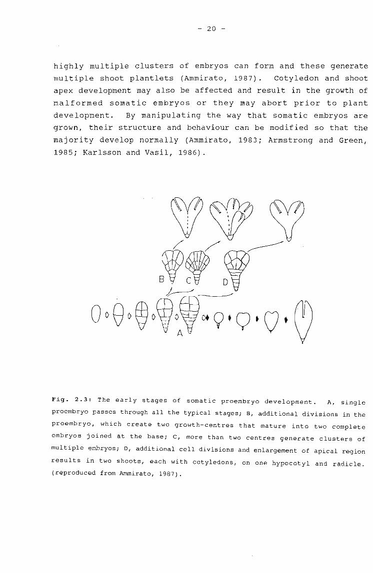
- 20 -
highly multiple clusters of embryos can form and these generate
mUltiple shoot plantlets (Ammirato, 1987). Cotyledon and shoot
apex development may also be affected and result in the growth of
malformed somatic embryos or they may abort prior to plant
development. By manipulating the way that somatic embryos are
grown, their structure and behaviour can be modified so that the
majority develop normally (Ammirato, 1983; Armstrong and Green,
1985; Karlsson and Vasil, 1986).
Fig. 2.3: The early stages of somatic proembryo development. A, single
proembryo passes through all the typical stages; B, additional divisions in the
proembryo, which create two growth-centres that mature into two complete
embryos joined at the base; C, more than two centres generate clusters of
multiple embryos; D, additional cell divisions and enlargement of apical region
results in two shoots, each with cotyledons, on one hypocotyl and radicle.
(reproduced from Ammirato, 1987).
- 21 -
Liquid suspension cultures also provide a source of somatic
embryos, which germinate to form plants under appropriate
conditions (Fig. 2.1) and this is potentially an ideal system for
micropropagation purposes (Conger et al. 1988). Cell suspension
cultures are discussed in more detail in section 2.2.4.
Examples of plants successfully produced via somatic embryogenesis
from embryogenic callus in the Gramineae include sugarcane
(Nickell, 1964; Heinz and Mee, 1969; Liu and Chen, 1974; Liu et
al., 1982; Ho and Vasil, 1983a; Chen et al., 1988a), maize (Lu et
;al., 1982; Lu et al., 1983; Kamo et al., 1985; Kamo and Hodges,
1986), rice (Abe and Futsuhara, 1985), wheat (Shimada and Yamada,
1979; Karp and Maddock, 1984; Vasil et al., 1990a, 1990b) , and
napier grass (Vasil and Vasil, 1982; Wang and Vasil, 1982; Botti
and Vasil, 1983).
2.2.4. CELL SUSPENSION CULTURES
Suspension culture initiation and maintenance
Cell suspension cultures are most commonly obtained by dispersing
friable callus in liquid culture medium, and agitating on a rotary
shaker, although they can also be initiated directly from explants
(Collin and Dix, 1990). These cultures provide a means of rapid
large-scale clonal propagation, they can be used for the isolation
of variant or mutant lines, and as a source for the isolation of
protoplasts (Lindsey and Jones, 1989).
Although morphogenically-competent cell suspension cultures of
Gramineae are difficult to obtain, they have~een established in
sugarcane Maretzki and~ickell, 1973; Chen and Shih, 1983; Ho and
Vasil, 1983b; Chen et al., 1988b; Liu and Shih, 1986; Srinivasan
and Vasil, 1986), wheat (Vasil et al., 1990a, 1990b), maize
(Bartowick, 1981; Armstrong and Green, 1985; Kamo and Hodges,
1986), rice (Abe and Futsuhara, 1985), guinea grass (Karlsson and
Vasil, 1986), Digitaria (Gonzales and Franks, 1987), and napier
grass (Lu et al., 1981). In most of these cases embryogenic calli
.-
- 22 -
have been used to establish long-term suspension cultures, from
which cells were capable of forming somatic embryos and
regenerating into plants.
Suspension cultures consist of small, richly-cytoplasmic, and
starch-filled embryogenic cells, present in small groups (Vasil et
al., 1990a). As with callus cultures (section 2.2.3), early
identification of embryogenic cells, and manipulation of the
cultures, ensures the predominance of totipotent embryogenic cells
(Vasil and Vasil, 1984; Vasil et al., 1990b). Determination of
the growth-rate of suspension cultures has been achieved in a
number of ways, for example packed cell volume, fresh and dry cell
weights, protein content and cell counts (Ho and Vasil, 1983b; Ryu
et al., 1990). Such determinations have been found necessary to
determine optimal intervals between subculture and to monitor the
state of synchrony and homogeneity of the cells (Ryu et al.,
1990). Sieving or filtration of the suspension cultures have been
suggested as means to aid selection of the smallest (and
presumably single, normal) pro-embryos for transfer to allow for a
greater degree of synchrony of maturation of somatic embryo"-- -
populations (Vasil and Vasil, 1982; Ammirato, 1987).
According to Lindsey and Yeoman (1983), the division rates of
suspension culture cells at the exponential phase are typically
higher than those of callus cells, but doubling times are slow in
comparison with those of microbial cells, and are usually in the
range of 24 to 72 hours. There appears to be a critical initial
cell density, below which cells transferred to a new medium
(liquid or solid) may fail to divide (Ryu et al., 1990). Factors
which have been found to influence the minimum size of the
inoculum include the cUlture's physiological characters, the
length of time and conditions under which the culture was
maintained, and the composition of the fresh medium (Collin and
Dix, 1990). Once established, the suspension cell culture
proceeds as a series of growth cycles characterised by a sigmoidal
nature comprising a lag, cell division, and stationary phase (Ryu
et al., 1990).
- 23 -
somatic embryos in sugarcane up to the globular or early scutellar
stage may form in suspension (Liu and Shih, 1986) or the
embryogenic cells may be removed from the suspension culture and
plated onto solid media for regeneration purposes (Vasil and
Vasil, 1982; Ho and Vasil, 1983b). Alternatively, fully mature
somatic embryos form in liquid media without plating onto solid
media, and although this occurs routinely in a number of
herbaceous dicotyledonous plants, Dactylis glomerata is the only
Graminaceous species in which embryo development has progressed
Ibeyond the proembryo stage in liquid culture (Conger et al.,i
1988) .
Plant regeneration from suspension culture cells
Regeneration from suspension cultures of Gramineae has been
difficult to achieve and in the first such report only albino
plants of Bromus inermis were recovered (Gamborg et al., 1970).
More recently, embryogenic cell suspension cultures capable of
plant regeneration via embryoid formation have been described in
some graminaceous plants such as pennesetum (Vasil and Vasil
1982), sugarcane (Ho and Vasil 1983b), maize (Kamo and Hodges
1986; Mitchell and Petolino, 1991), and crabgrass (Gonzales and
Franks, 1987).
2.2.5. PROTOPLAST ISOLATION AND CULTURE
Protoplast isolation and purification
Cocking (1960) first used enzymes to release plant protoplasts by
applying an extract of hydrolytic enzymes to tomato root tips.
since then the principle of enzymatic cell wall digestion has been
applied to plants from a wide taxonomic range, to degrade the
three primary components of cell walls, cellulose, hemicellulose
and pectin (Evans and Bravo, 1983). The enzymes used are
commercially prepared fungal cellulases, pectinases and
hemicellulases (Power and Cocking, 1970; Schenk and Hildebrandt,
- 24 -
1971; Pelcher et al., 1974; Vasil and Vasil, 1984).
It is essential that protoplasts are released into an osmotically
balanced medium after the removal of the cell wall, otherwise they
will burst (Kao and Michayluk, 1975). Appropriate osmotica
consisting of various sugars and sugar alcohols have been added to
the isolation solutions to create a hypotonic medium, so that
protoplasts do not burst when released into the isolation medium
(Evans and Bravo, 1983). Commonly used osmotica in sugarcane
include mannitol and sorbitol (Srinivasan and Vasil, 1986; Chen et
al., 1988b).
In addition to osmotic strength, other conditions during
protoplast isolation can be varied to optimise viability. Calcium
and magnesium, in the form of chloride salts may be added to the
isolation solution to increase membrane stability of protoplasts
(Gamborg et al., 1981). Protoplasts may be isolated in the dark
or at low light intensity to avoid starch accumulation, and the pH
of the isolation medium is usually between pH 5 and 6, to allow
for both enzyme activity and stability of isolated protoplasts
(Gill et al., 1981). Gentle shaking can facilitate protoplast
isolation by bringing fresh enzyme in contact with cell walls and
also by providing a physical force which might cause protoplasts
to be discharged from digested tissue, although excessive shaking
can result in protoplast destruction (Kao and Michayluk, 1975;
Fritter and Krikorian, 1983).
Following enzyme treatment, a mixture of undigested cells,
components of broken or burst cells and protoplasts is obtained,
which should be purified to remove protoplastsfrom the enzyme
solution (Power and Cocking, 1970; Schenk and Hildebrandt, 1971).
Reported purification methods include flotation on dense sucrose
solutions (Power and Cocking, 1970), flotation on Ficoll solutions
(Schenk and Hildebrandt, 1971), repeated centrifugation and
resuspension, and repeated sedimentation without centrifugation
(Pelcher et al., 1974).
- 25 -
A number of different methods for determining protoplast viability
have been reported and they include the observation of cyclosis as
an indication of active metabolism (Pelcher et al., 1974), the
exclusion of Evans-blue dye by intact membranes (Kanai and
Edwards, 1973), the use of fluorescein diacetate (Larkin, 1976),
and photosynthetic activity (Kanai and Edwards, 1973).
Both leaves and suspension cultures have been used as sources for
the isolation of protoplasts, though the latter has been more
popular in graminaceous species, such as in sugarcane (Maretzki
:and Nickell, 1973; Larkin, 1982; Chen and Shih, 1983), wheat
(Schenk and Hildebrandt, 1971; Okuno and Furusawa, 1977), maize
(Rhodes et al., 1988; Shillito et al., 1989), napier grass (Lu et
al., 1981; Vasil et al., 1986) and rice (Yamada et al., 1986). The
reason for this preference is that protoplasts isolated from
embryogenic suspension cultures appear to maintain their
totipotency and are capable of regenerating plants (see section
below) .
Protoplast culture
The osmoticum used for isolation of protoplasts is normally
maintained in the culture media, but carbon sources and growth
regulators may have to be varied to obtain optimal cUlturing
conditions (Evans and Bravo, 1983). After protoplasts have been
purified, the factors that influence cUlturing are the culture
medium (discussed in section 2.2.~) and the plating density
(Fitter and Krikorian, 1983; Vasil, 1983) (protoplast density at
1-4X10 5 jml appears to yield maximum cell wall regeneration).
Subsequent cUlturing methods include liquid droplet culture (Kao
et al., 1971) and agar culture, with (Cella and Galun, 1980;
Shaffler and Koop, 1990) or without (Vasil et al., 1986) a feeder
layer. The resulting microcalli colonies are placed on a
proliferation medium, and then a regeneration medium to ensure
production of callus or plantlets (Vasil et al., 1986; Schafflerand Koop, 1990).
- 26 -
As mentioned previously, the use of mesophyll~rotoplasts for
subsequent plant regeneration has been unsuccessful in the
Gramineae. Although Evans et al. (1980) and Chen and Liu (1974)
reported on colony formation from sugarcane mesophyll protoplasts
and Potrykus et al. (1977) achieved callus from maize mesophyll
protoplasts, plantlet regeneration did not occur. Furthermore,
to-date none of these studies have been repeated.
In contrast, the isolation of graminaceous protoplasts from
embryogenic suspension cultures and the successful regeneration of
plants has been reported for wheat (Shimada and Yamada, 1979;
Vasil et al., 1990a, 1990b), rice (Abdullah et al., 1986; Yamada
et al., 1986), maize (Shillito et al., 1989; Mitchell and
Petolino, 1991), Napier grass (Vasil et al., 1986) and Guinea
grass (Lu et al., 1981). However, some graminaceous plant species
did not regenerate plants even when embryogenic suspension
cultures were used as the source of protoplasts (Lorz et al.,
1985; Vasil, 1990). Attempts to recover sugarcane plants from
suspension culture protoplasts initially met with little success.
Protoplasts isolated from non-morphogenic suspension cultures were
found to divide and form either a few small colonies (Larkin,
1982) or calli (Maretzki and Nickell, 1973; Chen and Shih, 1983),
but not mature plants. Srinivasan and Vasil (1986) and Chen et
al. (1988b) isolated sugarcane protoplasts from embryogenic
suspension cultures and regenerated plants, but this has not yet
been repeated.
2.2.6. HARDENING-OFF AND PLANTING OUT OF REGENERATED PLANTS
Most plantlets derived in vitro by organogenesis or somatic
embryogenesis, survive and can be grown to maturity after transfer
to soil (Vasil and Vasil, 1984; Ziv, 1986). However, never having
been exposed to normal environmental conditions, plants produced
by tissue culture are accustomed to high humidity levels and
aseptic conditions, so they need to be hardened-off with gradual
exposure to the environment (Ziv, 1986).
- 27 -
According to reports in the literature, sugar cane and other
graminaceous plants are normally transferred to pots containing
vermiculite or a mixture of vermiculite and sand (4:1), watered
with a nutrient solution of choice, and maintained under a high
humidity for about 2 weeks before being transferred to the
greenhouse (Liu, 1971; Lu et al., 1982; Lu et al., 1983).
2.2.7. GENETIC STABILITY VERSUS GENETIC VARIATION IN VITRO
When plant cells are cultured via some form of unorganised callus
phase in vitro, the plants that are subsequently regenerated may
exhibit various genetic, phenotypic or biochemical charateristics
that differ from the parent material. The process whereby such
culture-induced variation is generated has been called somaclonal
variation (Evans et al., 1984b; Reisch, 1988; Lorz, 1989). The
main causes of somaclonal variation are polyploidy, aneuploidy and
chromosomal rearrangements (Larkin and Scowcroft, 1981). The
major factors affecting the extent of somaclonal variation include
the type of explant, medium composition, the time taken to culture
the explant and the regeneration pathway (Vasil, 1987; Ammirato,
1989) .
Somaclonal variation can serve as a source of breeding for new
varieties, but is undesirable if clonal fidelity is required, for
example where selected genotypes are being propagated (Ammirato,
1989) . Various safe-guards exist to try and minimise unwanted
somaclonal variation and these include:
1) selection of an explant which contains meristematic cells and
as few differentiated cells as possible, as the latter source of
cells could already contain genetic abnormalities and these would
be carried over to daughter cells (Vasil, 1987, Ammirato, 1989;
Bhaskaran and Smith, 1990);
2) 2,4-D concentrations used for callus induction in the majority
of the Gramineae should not be excessively high, so that the rate
of cell division is not too rapid for proper repair to be
maintained (Evans and Bravo, 1983);
3) exposure to potential mutagenic agents such as colchicine and
- 28 -
ultra-violet or gamma radiation should be avoided (Liu, 1990;
Irvine et al., 1991);
4) callus should be kept in culture for as short a time-period as
possible to minimise the occurrence of genetic variation (Reisch,
1988) ;
5) regeneration via indirect somatic embryogenesis, rather than
via indirect organogenesis should be encouraged, because somatic
embryos arise from single cells which are identical to parent
material (Ammirato, 1983; Evans et al., 1984b).
Somaclonal variation has been used to generate agronomically
improved plants in a number of species, and this will be discussed
in more detail in section 2.2.8.
2.2.8. POTENTIAL PRACTICAL APPLICATIONS OF TISSUE CULTURE SYSTEMS
In vitro culture has had an impact on conventional plant breeding,
particularly in the field of clonal propagation. Further
influences on breeding programmes are envisaged using genetic
engineering techniques to manipulate plant cells and tissues which
subsequently have to be regenerated in vitro to obtain transgenic
plants. other potential applications of in vitro culture systems
in the Gramineae, which will not be discussed below in any detail,
include meristem culture for the elimination of plant viruses
(reviewed by Evans et al., 1984a; Vasil, 1990), the production of
secondary metabolites from plant cells in culture (reviewed by
Lindsey, 1986), and the fusion of protoplasts to produce somatic
hybrids (Rao, 1977; Ozias-Akins et al., 1986; Power et al., 1986;
Tabaeizadeh et al., 1986; Ramill and Cocking, 1988). In the
latter instance, very few attempts have been made to obtain
somatic hybrids in the Gramineae, because of the difficulties
faced in culture and regeneration of grass and cereal protoplasts.
- 29 -
Large scale micropropagation
As discussed previously in section 2.2.7, in vitro culture systems
that involve an unorganised callus phase may produce variant
plants. Thus, in practice, most clonal propagation programmes
have made use of: direct organogenesis where axillary buds are
mUltiplied and subsequently rooted; or, direct embryogenesis,
where no callus is formed (Fig. 2.1). However, in contrast to
herbaceous dicotyledonous plants, many of the Gramineae are
propagated vegetatively without any in vitro culture being
necessary, and this is practically and economically advantageous.
Nevertheless, successful micropropagation by direct organogenesis
has been reported for a number of forage grasses (Ahloowalia,
1984) and maize (Raman et al., 1980). Sugarcane has been
propagated by shoot-tip culture and apical meristem culture
(direct organogenesis and direct embryogenesis, respectively)
(Grisham and Bourg, 1989). Successful micropropagation via
indirect embryogenesis has been reviewed for maize (King and
Shimamoto, 1984), oats (Kaur-Sawhney and Galston, 1984) and wheat
(Schaeffer et al., 1984).
The synchronous production of somatic embryos in suspension
cultures could be used for rapid regeneration, at any time of the
year, of a large number of selected genotypes in the Gramineae,
especially where somatic embryos are plated out for regeneration
purposes; for example bromegrass (Gamborg et al., 1970) and
sugarcane (Liu, 1990). Alternativel~, entire embryos may form in
suspension without the need to plate out, as in Dactylis glomerata
(orchardgrass) (Conger et al., 1988). Somatic embryos, if
protected in some way, could serve as artificial seeds, and be
used directly in the field or stored for future use (Ammirato,1989) .
- 30 -
Crop improvement by somaclonal variation
The general principles involved in somaclonal variation have been
discussed previously (section 2.2.7). Larkin and Scowcroft (1981)
reviewed a number of graminaceous plants (sugarcane, rice, oats,
maize and barley) which had been produced by somaclonal variation
via callus and protoplast culture. The characteristics displayed
by these plants included resistance to diseases, and increases in
tiller yield, plant height, fertility and phenotypic variation.
Significant improvements among sugarcane variants have been
reported, with improved sugar yield (Liu et al., 1984), and
increases in stalk number, stalk length, density and weight,
percent fibre and improvements in the attitude of the top leaf
(Liu and Chen, 1976; Liu, 1990) Anther culture of rice, wheat and
maize (Tsay, 1987), resulted in chromosomal changes in plants
which are currently being field-tested to determine whether they
are superior to plants in commercial production.
storage of germplasm
The most economical form of storing germplasm for sexually
propagated species is as seeds, "but some crops do not produce
viable seeds, and some seeds have a limited storage life, as in
certain Gramineae. Plant material may be conserved in vitro as
protoplasts, isolated cells grown in suspension culture, meristem
cultures at various stages of development, somatic embryos or
organised plantlets (Kartha, 1985; ~ithers, 1985; Grout, 1990).
Plants may be regenerated as required, without risks such as
losses due to diseases and pests, associated with maintaining
stock plants in the field. The two main approaches for in vitro
germplasm storage have been slow-growth techniques and
cryopreservation.
Various methods have been developed to slow down growth in
culture, and these include storage at low temperatures (4-8°C),
with a reduced photoperiod, and changing the culture conditions by
inducing osmotic stress or adding growth retardants (reviewed by
- 31 -
Kartha, 1985 and withers, 1985). Somatic embryos of D. glomerata
(Conger et al., 1988) and wheat have been desiccated, the latter
requiring the presence of ABA and sucrose (Carman, 1988), and this
has potential for somatic embryogenic storage and perhaps the
production of artificial seed.
cryopreservation is the only approach that completely arrests
tissue growth. This occurs at temperatures of -196°C and ensures
the maintenance of maximum genetic stability. However most
biological material is not resistant to freezing, and it is
,therefore necessary to develop specific conditions in preparation;
for freezing, during the freezing and thawing process, and during
recovery (Finkle et al., 1985). Cryopreservation has been used to
successfully store a wide range of tissue types, including embryos
of rice (Bajaj, 1981), wheat and barley (Bajaj, 1985), suspension
culture cells in sugarcane (Finkle and Ulrich, 1979), and even
protoplasts (reviewed by Bajaj, 1985).
The production of haploid plants
Haploid plants, with gametic chromosome numbers, are particularly
useful in plant breeding, both for the rapid production of
homozygous lines following chromosome doubling to the original
ploidy level (Liu, 1984), for the detection and selection of
recessive mutants (Dunwell, 1985) and for genetic transformation
purposes (Creissen et al., 1990). In vitro techniques can be used
for the routine production of haploids and involves the
regeneration of plants from cultured anthers, immature pollen or
isolated ovules (Vasil, 1990).
Anther-derived haploids in sugarcane were first produced in China
(Chen et al., 1979), with only certain genotypes responding to
this type of culture. The generation of haploid sugarcane plants
has become more streamlined since the discovery that anthers need
to be cold pre-treated (Fitch and Moore, 1983; Hinchee et al.,
1984). Sugarcane is highly polyploid, so haploids obtained from a
single sporophytic development are not likely to carry the basic

- 32 -
haploid number of chromosomes for Saccharum spp. Nevertheless,
some extent of homozygosity is likely to be achieved by doubling
the haploid, and thus plants may become more valuable for
conventional plant breeding and for in vitro manipulations
involving gene cloning.
The majority of the Graminaceous species undergo callus-mediated,
haploid plant regeneration. Species capable of haploid plant
regeneration, via callus, from cultured anthers include rice
(Tsay, 1987), barley (Kasha and Reinbergs, 1980; Creissen et al.,
1990), Datura sp. (Guha and Maheshwari, 1964) and maize (Sun et
al., 1989; Mitchell and Petolino, 1991). Barley anthers in callus
culture were transformed by genetic engineering techniques and
regenerated plants have produced fertile seed (Creissen et al.,
1990). Th e success of this research may encourage the use of
haploid culture for future transformation studies in the grasses
and cereals.
Regeneration of transformed tissues and/or cells
The potential exists to improve graminaceous monocotyledonous crop
plants by the insertion of certain agronomically useful genes
which include resistance to herbicides (Comai et al., 1985; Shah
et al., 1986; DeBlock et al., 1987; Hinchee et al., 1988; Stalker
et al., 1988), insect pests (Fischhoff et al., 1987; Hilder et
al., 1989; Vaeck et al., 1987; Herrera et al., 1991; Murrayet
al., 1991; Vail, 1991), and viruses (Abel et al., 1986; Harrison
et al., 1987; Van Dun et al., 1987; Cuozzo et al., 1988; Mayo and
Barker, 1990). The availability of an in vitro system to allow
recovery of plants from transformed cells, tissues and protoplasts
is an essential prerequisite to the sucess of any breeding
programme that aims to utilise the potential offered by
biotechnological advances to crop improvement.
- 33 -
2.3. MATERIALS AND METHODS
2.3.1. IN VITRO CULTURE OF SEEDS/ZYGOTIC EMBRYOS
Sugarcane seeds (reference number Y511) were removed from florets,
the outer husk and hairs removed (Plate 2.1 A), and the seeds were
then surface-sterilised in a sodium hypochlorite solution
(0,15%(w/v) for 2mins), followed by three rinses in sterile water.
Seeds were aseptically placed into petri-dishes containing a
germination medium (Plate 2.1 B), consisting of MS salts and
:vitamins (Murashige and Skoog, 1962), casein hydrolysate (19/1)
(Sigma, USA), sucrose (49/1) (Unilab, South Africa), benlate
(0,05g/1) (Bayer, SA), and agar (99/1) (Noble agar-Difco, UK) at
pH 5,8, and grown under a 16h light/8h dark photoperiod at 27°C,
for 4 days, after which germination and contamination levels were
monitored.
2.3.2. PREPARATION OF EXPLANTS FOR IN VITRO CALLUS CULTURE
Sugarcane varieties NCo376 and N13 were field grown (Plate 2.2 A)
and obtained from the South African Sugar Association Experiment
station, Mount Edgecombe. Stalks (Plate 2.2 B) were cut into
single-budded setts, dipped in benlate (0,75g/1) and planted into
trays containing autoclaved vermiculite. The setts were grown in
the dark for two weeks -at 28°C, until the shoots were
approximately 15cm long.
The outer leaf sheaths were peeled off the two-week old shoots and
the exposed stalk was cut into 1cm- long sections (Plate 2.2 C).
Each stalk was separately surface-sterilized in ethanol (70% for
Imin) , followed by a wash in sodium hypochlorite (1,5% (w/v» plus
Tween 80 (0,01% (v/v» for 20mins, after which it was rinsed three
times in sterile distilled water.
Another explant source used was sterile in vitro plants which had
regenerated from variety N13 via somatic embryogenesis. The stems
from these regenerated plants were aseptically removed and were
- 34 -
placed under the same culturing conditions as the surface
sterilised field explants.
2.3.3. GROWTH CONDITIONS FOR CALLUS CULTURE
Production of aseptic cultures
Microbial contamination levels from field-grown material placed in
culture were observed to be very high (approximately 65% of the
explants were contaminated). Colonies observed to be
contaminating the culture media were picked off, Gram stained
(Harrigan and McCance, 1966) and observed by light microscopy
(Axioskop, Zeiss, Germany). The main contaminant identified was a
budding yeast, but there were also Gram positive cocci and rods.
The sensitivity of the contaminants to several antimiciobial
agents was tested by means of a zonal inhibition test (Harrigan
and McCance,1966). Overnight cultures of the contaminants were
spread onto nutrient agar (pH7) (Biolab, South Africa) and MS
(pH5,8) plates. Plugs of agar were removed using a number 3 plug-
borer, and O,5ml of each of the antibiotics (at concentrations
given below) was filter-sterilised into the wells. Plates were
incubated for 24h at 27°C and inhibition zones were measured, from
the rim of the well to the outer edge of the inhibition zone, and
recorded. From these results, the antimicrobial substances used
in an attempt to reduce contamination of field-grown explants were
combined in a 'cocktail' mixture. The cocktail, made up in liquid
MS medium, contained the antibiotics ampicillin and streptomycin
(each at 100mg/l) and rifampicin (30mg/l) and the fungicides
clotrimazole and miconazole (each 100mg/l) and nystatin (30mg/l)
(Sigma, USA).
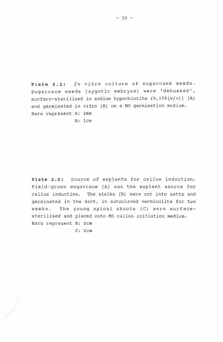
/
/
- 35 -
Plate 2.1: In vitro culture of sugarcane seeds.
Sugarcane seeds (zygotic embryos) were 'dehusked',
surface-sterilised in sodium hypochlorite (O,15%(wjv)) (A)
and germinated in vitro (B) on a MS germination medium.
,Bars represent A: 2mm
B: lcm
Plate 2.2: Source of explants for callus induction.
Field-grown sugarcane (A) was the explant source for
callus induction. The stalks (B) were cut into setts and
germinated in the dark, in autoclaved vermiculite for two
weeks. The young apical shoots (C) were surface
sterilised and placed onto MS callus initiation medium.
Bars represent B: 5cm
c: 2cm
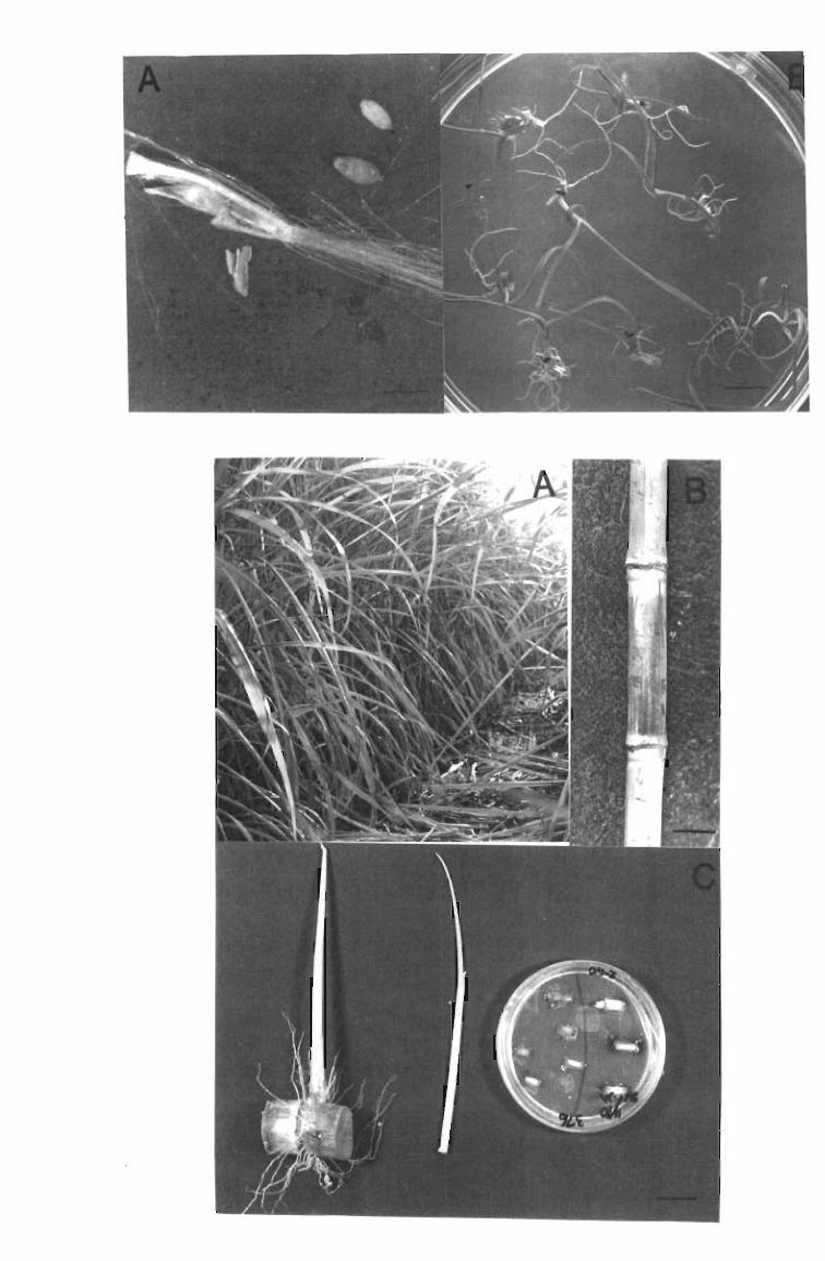
- 36 -
Once the field-grown explants had been surface-sterilised (section
2.3.2), they were placed into the MS antibiotic medium (Murashige
and Skoog, 1962) for 48h, with shaking. The explants were then
aseptically recut, removing 2mm from each end of the lcm explant,
which had been exposed to the surface~sterilants. The explants
were aseptically placed on to callus induction medium, and
contamination levels and callus production were recorded over a 1
month period. Explants not exposed to antibiotics after surface
sterilisation served as controls.
Callus induction
stalk explants were aseptically placed on to callus induction
medium containing MS salts and vitamins (Murashige and Skoog,
1962), sucrose (20-S0g/I), 2,4-D (2-4mg/l) (Sigma, USA), inositol
(100mg/l) (Sigrna, USA), casein hydrolysate (lg/l) and agar (9g/I).
Explants were placed at 27°C in the dark.
2.3.4. PRODUCTION OF SOMATIC EMBRYOGENIC CULTURES
Embryogenic calli were sub-cultured every four weeks on to MS
callus induction medium containing 2,4-D (2-4mg/l) and "sucrose
(20-S0g/I). The non-embryogenic areas of the" calli were dissected
out and discarded.
After 2 months of culture in the dark, most of the calli contained
immature embryos, and were sub-cultured onto a regeneration medium
of MS salts and vitamins, with sucrose (20-S0g/l) and agar (9g/l)
at pHS,8, without 2,4-D. In all cases, the regeneration medium
contained the same concentration of sucrose as the medium on which
the calli were initiated. Cultures were incubated in a 16h light
(200~E/m2/s)/8h dark photoperiod at 27°C.
Activated charcoal (4g/l) (Unilab, South Africa) was added to theregeneration medium.
- 37 -
2.3.5. HARDENING-OFF AND ACCLIMITISATION OF REGENERATED PLANTS
After 1-2 months of culture on the MS regeneration medium,
plantlets (3cm in height) exhibited well-developed root systems,
and were transferred to glass tubes containing fresh MS
regeneration medium with sucrose at a lower concentration (lOg/I).
After approximately 2 months (or when they had reached a height of
10cm) , the plants were planted out into sterile plastic containers
(10x15cm), in autoclaved potting soil. A plastic bag was placed
over the plant in the pot to maintain humidity. A gradual
: decrease in humidity was achieved by opening the bags daily, for
increasingly longer periods of time, over a 2 week period.
2.3.6. INITIATION AND MAINTENANCE OF SUSPENSION CULTURES
Four different media were tested to initiate cell suspension
cultures (Table 2.1). Cell suspension cultures were initiated by
transfer of approximately 19 fresh weight, 2 month-old, friable
NCo376 callus (produced as described in section 2.3.3) to 50ml
aliquots of medium in 125ml Erlenmyer flasks. The cultures were
maintained at 27°C, in the dark, with rotary shaking at
120rev/min. The medium was replaced weekly and aliquots were
observed microscopically and monitored for presence of small
clumps of embryogenic cells.
After 1 month in CUlture, the larger pieces of callus were allowed
to settle to the base of the container and the lighter, more
dispersed cells were transferred to 250ml Erlenmyer flasks,
containing a total volume of 100ml of cells and medium.
Suspension culture growth-rate was monitored daily over a 2 week
period. Samples of 5ml were removed aseptically,. and packed cell
volume (after centrifugation at 2000g for 5min) was determined.
- 38 -
Table 2.1 : A comparison of the components of four different media recommended
for the initiation of suspension cultures of sugarcane.
Media
Composition Ho & Vasil Liu & Shih Chen et al. Thom et al.
(1983a) (1986) (1988b) (1981)
Macroelements MS MS MS White's basalsalts (Nickell
and
Microelements MS MS MS Maretzki, 1969)
Vitamins MS MS MS thiamine 100
(/.1g/1 ) nicotinamide800
pyridoxine800
sucrose(g/l) 30 20 30 18,8
2,4-D(mg/1) 3 3 3 2
Other(mg/1) casein 500 casein 400 casein 50 arginine 57,5thiamine 900 thiamine 900 yeast extract
1000
2.3.7. ISOLATION AND CULTURE OF PROTOPLASTS
Protoplasts were isolated from leaves, suspension cultures and
calli. Mesophyll protoplasts were isolated from the youngest
fully-expanded leaves of N13 hardened-off plants, which were
placed in the dark for 24h prior to protoplast isolation. The
leaves were cut into squares (O,Scm) and the mid-rib discarded
before leaf tissue (lg fresh weight/lOml enzyme solution) was
incubated in filter-sterilised enzyme solution. Similarly, 2
month old NCo376 suspension culture cells (2ml settled cell
volume/lOml enzyme solution) and 2 month old friable NCo376 or N13
calli (lg/lOml enzyme solution) were used as source material for
protoplast isolation.
- 39 -
The protoplast osmoticum contained sorbitol (0,6M), KH2P04 (lmM),
CaC1 2 (lmM) and MgC1 2 (lmM) at pHS,6. Enzymes used for protoplast
isolation were cellulase R-10 (10-S0g/1) (Onozuka, Japan),
macerozyme R10 (1-4g/1) (Onozuka, Japan) and hemicellulase (2,S
109 /1) (S igma, USA). In order to obtain the optimum enzyme
combination for isolation of protoplasts, one enzyme concentration
was changed at a time. The enzyme solution containing the
protoplast source material was incubated at 26°C in the dark. The
protoplast solution was filtered through a series of filters (pore
sizes of 1000 and 200J..Lm; Nybond, switzerland) after 3-6h
; incubation. The protoplasts were washed in the above-mentioned
medium (minus the enzymes; pH7) by centrifugation for 10mins at
100rev/min (MSE Minor, England).
Protoplasts isolated from calli were cultured for 48h at a density
of 1x10 5 protoplasts/ml in MS medium containing 2,4-D (l,Smg/l)
and mannitol (O,SM), in the dark, in petri-dishes (3cm in
diameter). These were placed on moist filter in larger petri
dishes which in turn, were placed in a plastic container which
contained a layer of water at the base.
Protoplast yield was determined by counting protoplasts on a
counting chamber (Fuchs-Rosenthal; O,200mm x O,062Smm). Viability
was assessed by the ability of intact protoplasts to exclude
Evan's Blue dye (159/1) (Larkin, 1976), made up in the osmoticum.
2.3.8. MICROSCOPY
Periodically, small pieces of calli were squashed onto a
microscope slide, stained with dilute safranin (Harrigan and
McCance, 19.66) and viewed by light microscopy to assess the
development of embryogenic structures.
- 40 -
2.3.9. STATISTICAL ANALYSES
Results obtained for callus initiation and plant regeneration
initiated on media containing a variety of sucrose and 2,4-D
concentrations were analysed using an F test, in order to see
whether there was an interaction between sucrose and 2,4-D. An F
test (Mulder, 1988) was carried out and a regression surface was
fitted to the data (y=a+ blx1+b2x12. +b3x2+b4x22 +b5xlx2)' and
values were weighted to take into account the different precision
of points.
The s.e. were calculated by dividing the s.d. by the Vn. The
s.e. obtained were mUltiplied by values in t-tables, at the 95%
confidence level.
2.3.10. PHOTOGRAPHY
Photomicroscopy was undertaken using dissecting and compound
light-microscopes.
- 41 -
2.4. RESULTS
2.4.1. PROPAGATION BY MEANS OF ZYGOTIC EMBRYOS/SEEDS
Although sugarcane is propagated vegetatively for commercial
purposes, it can also be propagated by true se~~. A preliminary
investigation involving the uptake of DNA by seeds was undertaken
and is described in Chapter 3. To achieve this it was necessary
to have seed material that was free of microbial contaminants and
which could be cultured in vitro in order to monitor DNA
: expression. Surface-sterilised seeds, placed onto a germination
medium, showed 100% germination and no contamination was observed.
2.4.2. CALLUS CULTURE AND PLANT REGENERATION VIA SOMATIC
EMBRYOGENESIS IN SOUTH AFRICAN SUGARCANE VARIETIES
photographic account of plant regeneration via somatic
embryogenesis in variety N13
The developmental stages involved in the process of somatic
embryogenesis in variety N13, from callus production to
regeneration of pantlets from somatic embryos, are shown in Plates
2.3-2.9. It took 11-14 days from the time that the explants were
placed on the callus induction medium to the time that callus was
first observed. Examination of calli cultured in the dark, showed
two distinct regions: the embryogenic regions were compact,
nodular and white in colour as opposed to the non-embryogenic ones
which were friable and yellowish in colour (Plate 2.3).
Microscopic examination of squashed callus revealed that non
embryogenic cells were irregular in shape, larger and less compact
than embryogenic cells which occurred in clusters, and appeared to
have dense cytoplasmic contents (Plate 2.4). Typical embryogenic
heart- (Plate 2.5 A) and torpedo-shaped structures (Plate 2.5 B)
were visible toward the end of the second month of incubation in
the dark. Embryo germination occurred approximately 14 days to 1
month after dark grown calli were exposed to light (Plate 2.6 A
and B). Simultaneous production of roots and shoots occurred on
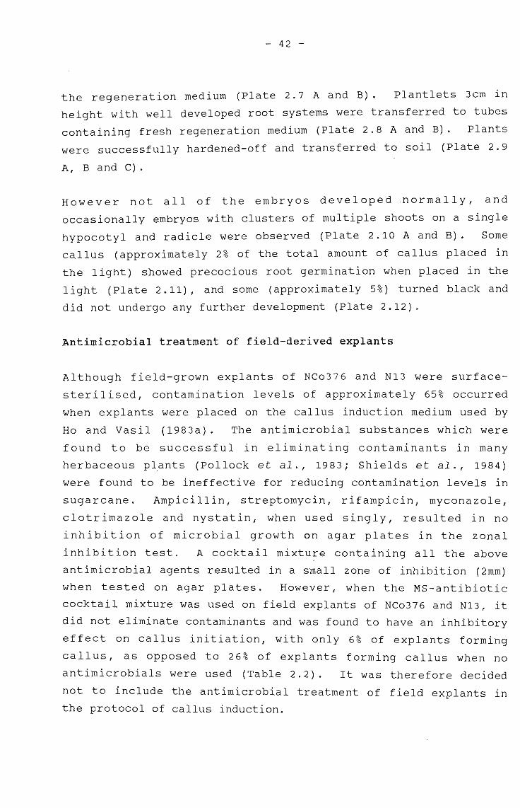
- 42 -
the regeneration medium (Plate 2.7 A and B). Plantlets 3cm in
height with well developed root systems were transferred to tubes
containing fresh regeneration medium (Plate 2.8 A and B). Plants
were successfully hardened-off and transferred to soil (Plate 2.9
A, B and C).
However not all of the embryos developed .normally, and
occasionally embryos with clusters of mUltiple shoots on a single
hypocotyl and radicle were observed (Plate 2.10 A and B). Some
callus (approximately 2% of the total amount of callus placed in
the light) showed precocious root germination when placed in the
light (Plate 2.11), and some (approximately 5%) turned black and
did not undergo any further development (Plate 2.12).
Antimicrobial treatment of field-derived explants
Although field-grown explants of NCo376 and N13 were surface
sterilised, contamination levels of approximately 65% occurred
when explants were placed on the callus induction medium used by
Ho and Vasil (1983a). The antimicrobial substances which were
found to be successful in eliminating contaminants in many
herbaceous plants (Pollock et al., 1983; Shields et al., 1984)
were found to be ineffective for reducing contamination levels in
sugarcane. Ampicillin, streptomycin, rifampicin, myconazole,
clotrimazole and nystatin, when used singly, resulted in no
inhibition of microbial growth on agar plates in the zonal
inhibition test. A cocktail mixtu~e containing all the above
antimicrobial agents resulted in a small zone of inhibition (2mm)
when tested on agar plates. However, when the MS-antibiotic
cocktail mixture was used on field explants of NCo376 and N13, it
did not eliminate contaminants and was found to have an inhibitory
effect on callus initiation, with only 6% of explants forming
callus, as opposed to 26% of explants forming callus when no
antimicrobials were used (Table 2.2). It was therefore decided
not to include the antimicrobial treatment of field explants in
the protocol of callus induction.
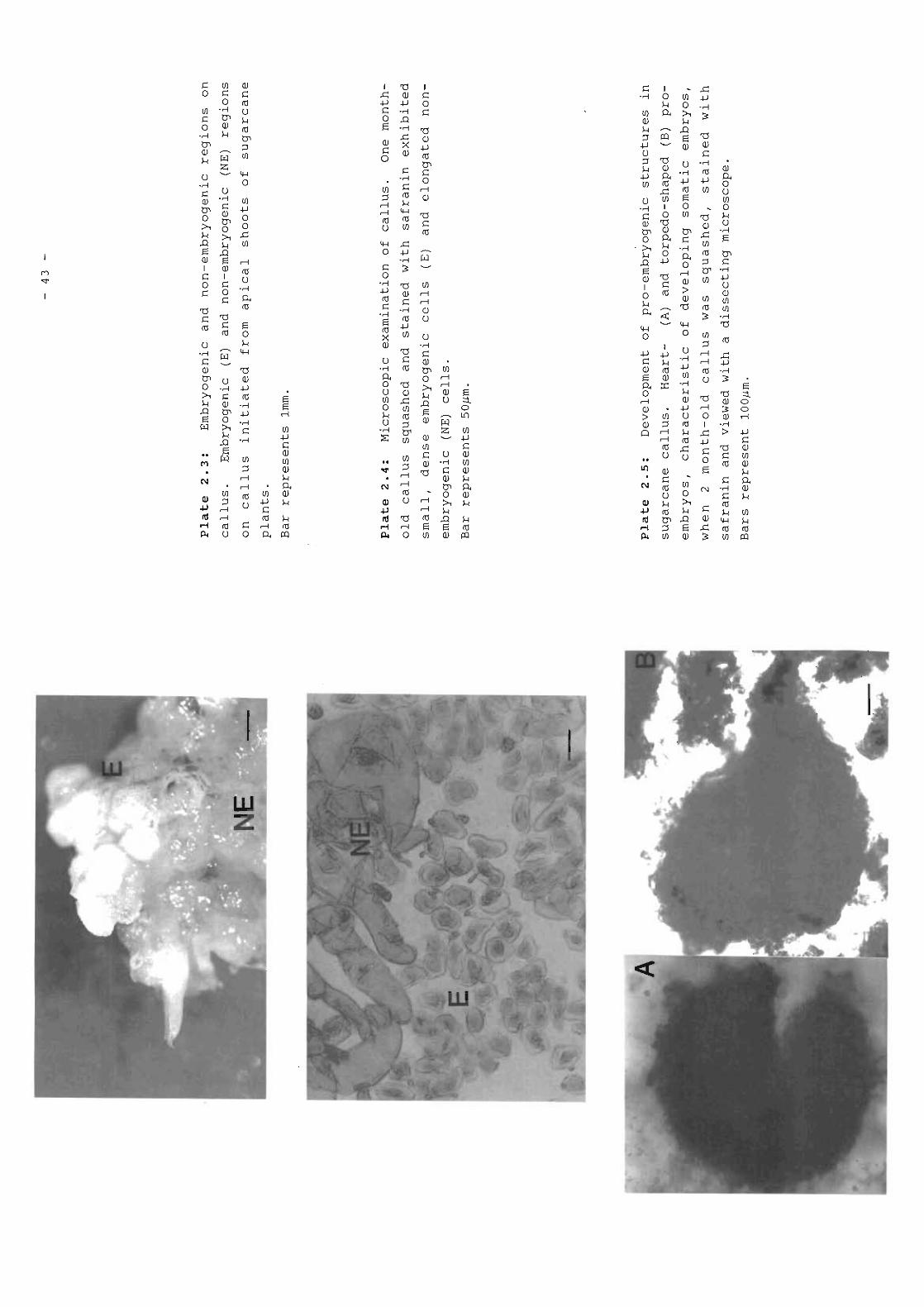
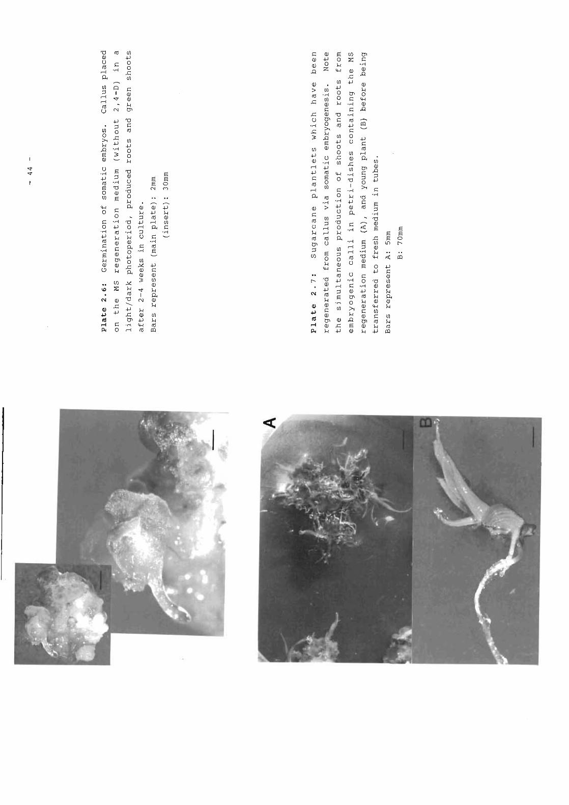
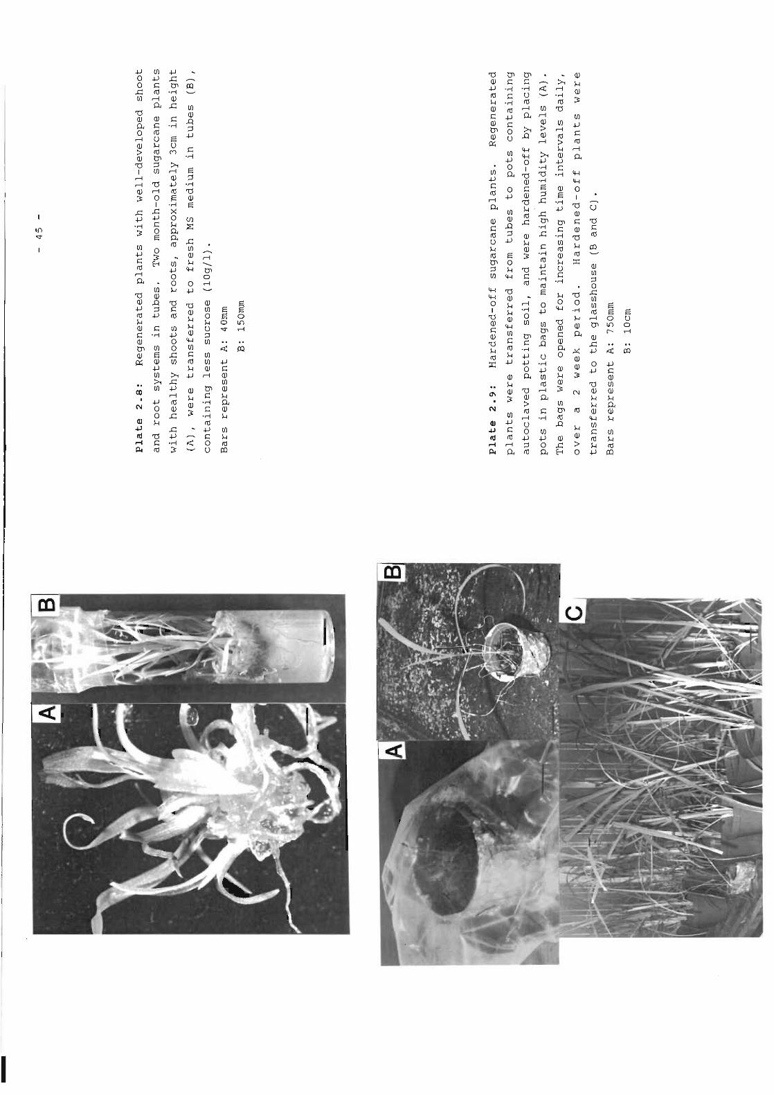
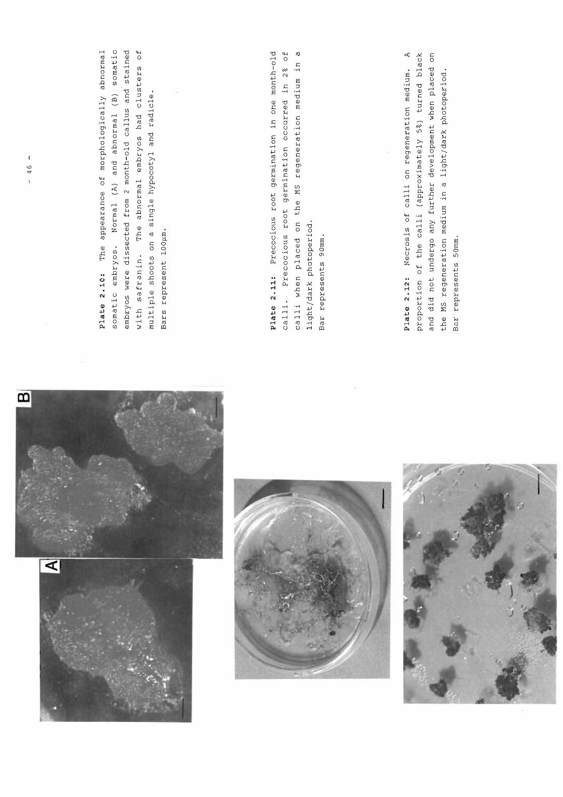
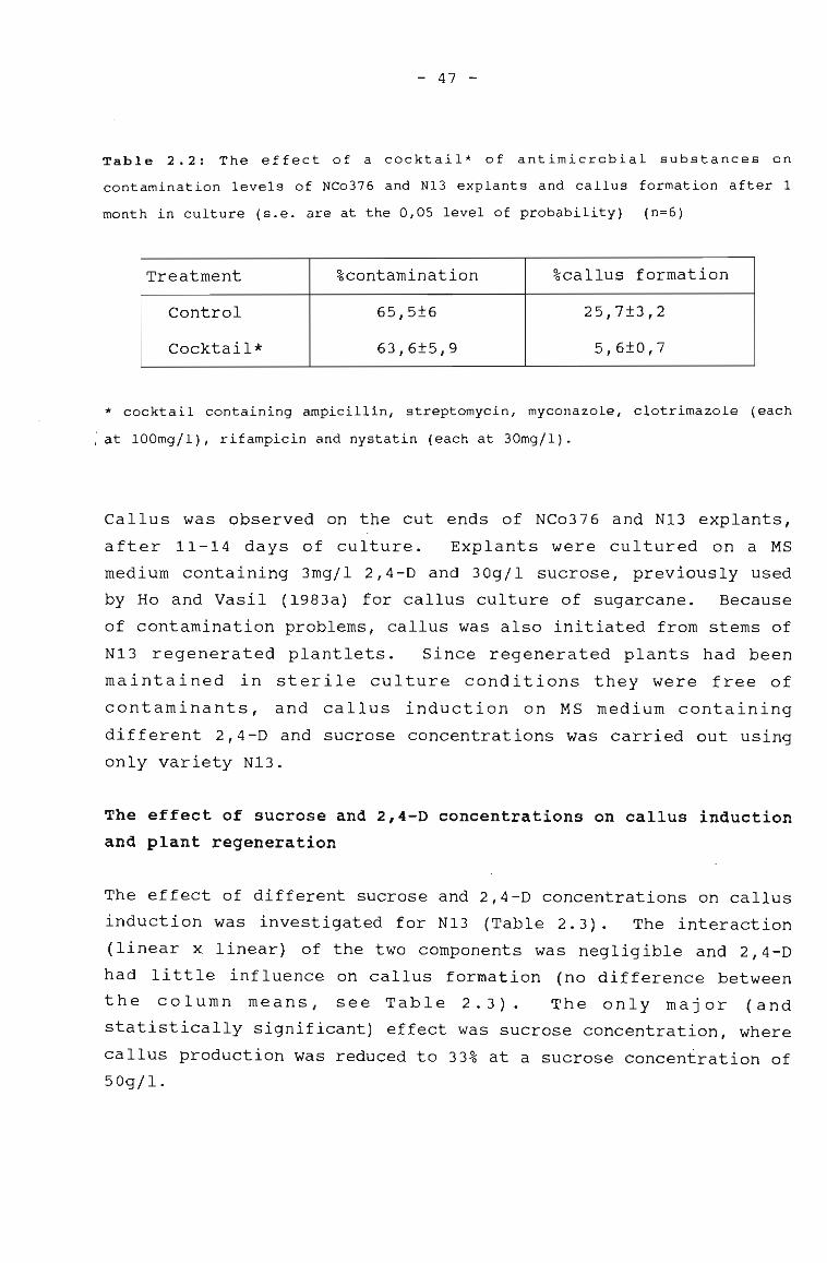
- 47 -
Table 2.2: The effect of a cocktail* of antimicrobial substances on
contamination levels of NCo376 and NI3 explants and callus formation after 1
month in culture (s.e. are at the 0,05 level of probability) (n=6)
Treatment %contamination %callus formation
Control 65,5±6 25,7±3,2
Cocktail* 63,6±5,9 5,6±0,7
, * cocktail containing ampicillin, streptomycin, myconazole, clotrimazole (each
) at 100mg/l), rifampicin and nystatin (each at 30mg/l).
Callus was observed on the cut ends of NCo376 and N13 explants,
after 11-14 days of culture. Explants were cultured on a MS
medium containing 3mg/l 2,4-D and 30g/1 sucrose, previously used
by Ho and Vasil (1983a) for callus culture of sugarcane. Because
of contamination problems, callus was also initiated from stems of
N13 regenerated plantlets. Since regenerated plants had been
maintained in sterile culture conditions they were free of
contaminants, and callus induction on MS medium containing
different 2,4-D and sucrose concentrations was carried out using
only variety N13.
The effect of sucrose and 2,4-D concentrations on callus inductionand plant regeneration
The effect of different sucrose and 2,4-D concentrations on callus
induction was investigated for NI3 (Table 2.3). The interaction
(linear x linear) of the two components was negligible and 2,4-D
had little influence on callus formation (no difference between
the column means, see Table 2.3). The only major (and
statistically significant) effect was sucrose concentration, where
callus production was reduced to 33% at a sucrose concentration of50g/l.
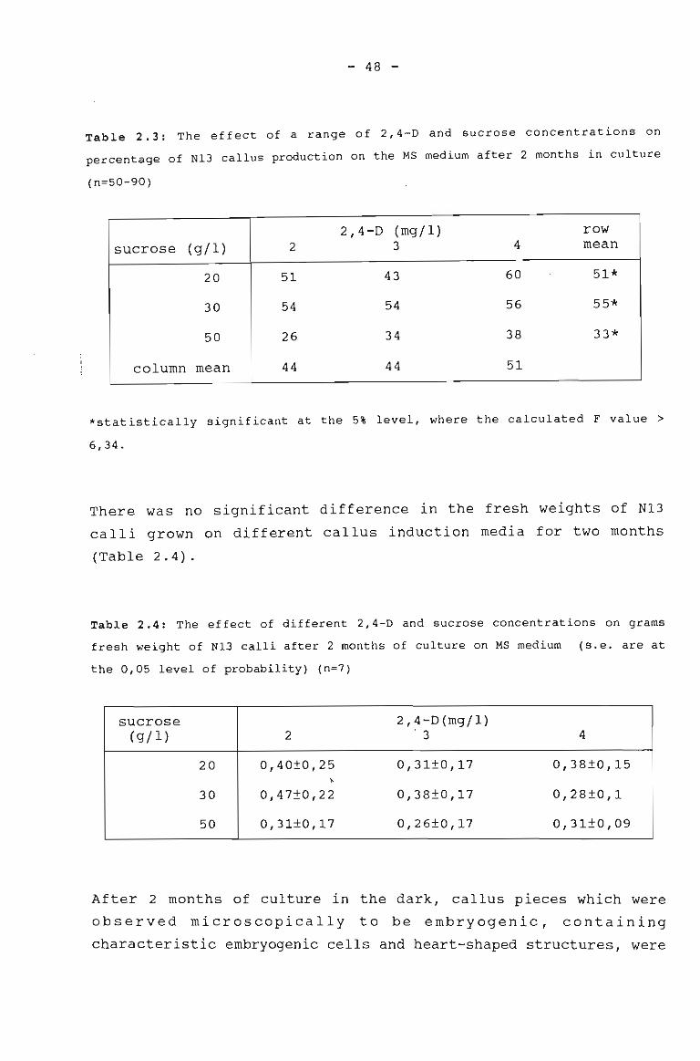
- 48 -
Table 2.3: The effect of a range of 2,4-D and sucrose concentrations on
percentage of N13 callus production on the MS medium after 2 months in culture
(n=50-90)
2,4-D (mgj 1) rowsucrose (gj 1) 2 3 4 mean
20 51 43 60 51*
30 54 54 56 55*
50 26 34 38 33*
column mean 44 44 51
*statistically significant at the 5% level, where the calculated F value >
6,34.
There was no significant difference in the fresh weights of N13
calli grown on different callus induction media for two months
(Table 2.4).
Table 2.4: The effect of different 2,4-D and sucrose concentrations on grams
fresh weight of N13 calli after 2 months of culture on MS medium (s.e. are at
the 0,05 level of probability) (n=7)
sucrose 2,4-D(mgjl)(gjl) 2 '3 4
20 0,40±0,25 0,31±0,17 0,38±0,15"-
3O 0,47±0,22 0,38±0,17 0,28±0,1
50 0,31±0,17 0,26±0,17 0,31±0,09
After 2 months of culture in the dark, callus pieces which were
observed microscopically to be embryogenic, containing
characteristic embryogenic cells and heart-shaped structures, were
- 49 -
placed in the light on a regeneration medium. The regeneration
media contained identical sugar concentrations to those used in
the induction media. The percentage of N13 calli giving rise to
plants is presented in Table 2.5. The greatest number of calli
giving rise to plantlets were observed on regeneration media
containing 20g/1 sucrose (especially from callus initiated on
medium containing 2mg/l 2,4-D). There was no statistically
significant interaction between sucrose and 2,4-D on the
regeneration of plants from calli. However, a significant
reduction in the percentage calli producing plantlets occurred at
,30 and 50g/1 sucrose, when compared to percentage calli producing
plants at a sucrose concentration of 20g/1.
Table 2.5: The effect of sucrose levels in the regeneration medium on the
percentage of N13 calli which yielded plants (n=9-33)
sucrose 2,4-D(mg/l)# row(g/l) 2 3 4 mean
20 60 47 33 47*
30 33 28 6 22*
50 11 44 19 25*
column mean 35 40 19
# for the purposes of comparison, 2,4-0 concentrations on which calli were
initiated are included, even though the regeneration medium contained no 2,4-0
* significant effect at the 5% level, where the calculated F value> 5,14
The number of plants produced per gram fresh weight callus was
compared, to see whether any particular concentration of sucrose
and 2,4-D yielded a greater number of plants (Table 2.6).
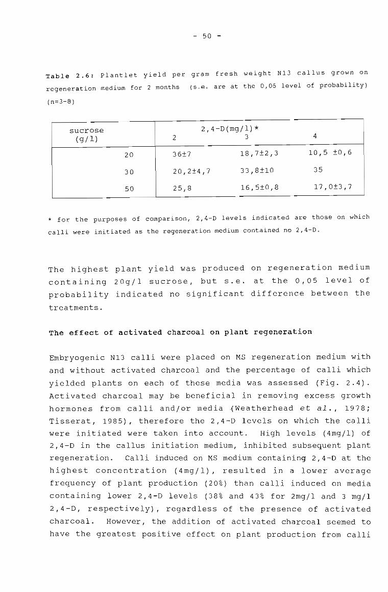
- 50 -
Table 2.6: Plantlet yield per gram fresh weight Nl3 callus grown on
regeneration medium for 2 months (s.e. are at the 0,05 level of probability)
(n=3-8)
sucrose 2,4-0(mgjl)*(gjl) 2 3 4
20 36±7 18,7±2,3 10,5 ±0,6
30 20,2±4,7 33,8±10 35
50 25,8 16,5±0,8 17,0±3,7
* for the purposes of comparison, 2,4-D levels indicated are those on which
calli were initiated as the regeneration medium contained no 2,4-D.
The highest plant yield was produced on regeneration medium
containing 20gjl sucrose, but s.e. at the 0,05 level of
probability indicated no significant difference between the
treatments.
The effect of activated charcoal on plant regeneration
Embryogenic N13 calli were placed on MS regeneration medium with
and without activated charcoal and the percentage of calli which
yielded plants on each of these media was assessed (Fig. 2.4).
Activated charcoal may be beneficial in removing excess growth
hormones from calli andjor media .(Weatherhead et al., 1978;
Tisserat, 1985), therefore the 2,4-0 levels on which the calli
were initiated were taken into account. High levels (4mgjl) of
2,4-D in the callus initiation medium, inhibited subsequent plant
regeneration. Calli induced on MS medium containing 2,4-D at the
highest concentration (4mgjl), resulted in a lower average
frequency of plant production (20%) than calli induced on media
containing lower 2,4-0 levels (38% and 43% for 2mgjl and 3 mgjl
2,4-D, respectively), regardless of the presence of activated
charcoal. However, the addition of activated charcoal seemed to
have the greatest positive effect on plant production from calli
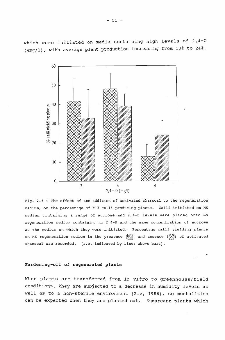
- 51 -
which were initiated on media containing high levels of 2,4-D
(4mg/l), with average plant production increasing from 13% to 24%.
60
50
1~40c::co~bOc::;a 30
(1)
->.
~C)
~ 20
10
o2 3
2,4-D (mgfl)4
Fig. 2.4 g The effect of the addition of activated charcoal to the regeneration
medium, on the percentage of N13 calli producing plants. Calli initiated on MS
medium containing a range of sucrose and 2,4-D levels were placed onto MS
regeneration medium containing no 2,4-D and the same concentration of sucrose
as the medium on which they were initiated. Percentage calli yielding plants
on MS regeneration medium in the presence (~) and absence (~) of activated
charcoal was recorded. (s.e. indicated by lines above bars).
Hardening-off of regenerated plants
When plants are transferred from in vitro to greenhouse/field
conditions, they are sUbjected to a decrease in humidity levels as
well as to a non-sterile environment (Ziv, 1986), so mortalities
can be expected when they are planted out. Sugarcane plants which
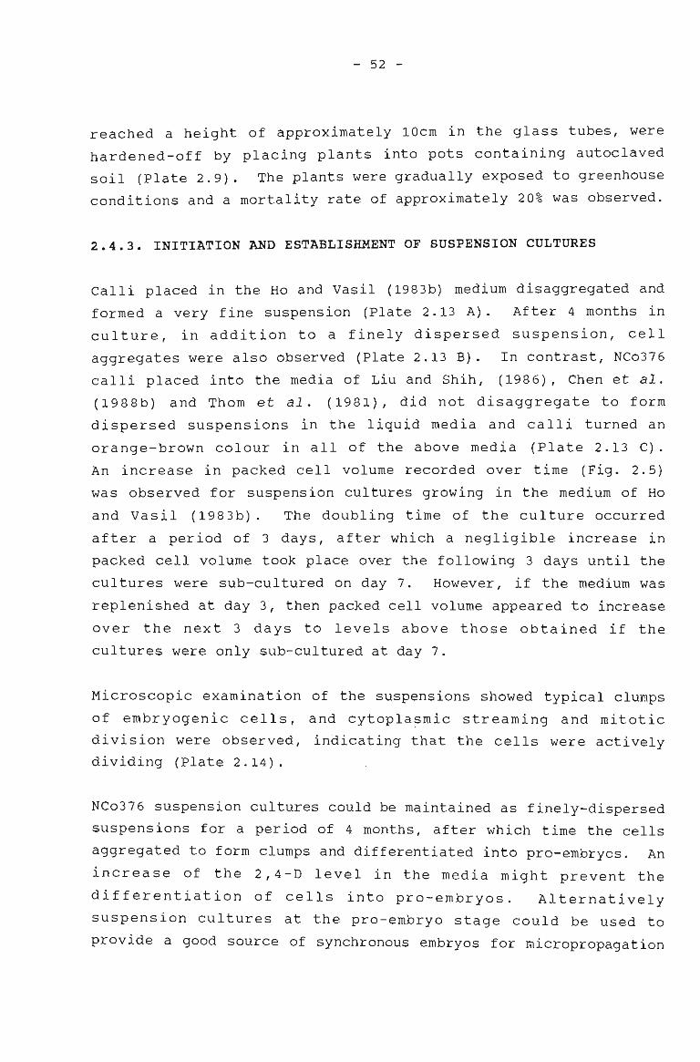
- 52 -
reached a height of approximately 10cm in the glass tubes, were
hardened-off by placing plants into pots containing autoclaved
soil (Plate 2.9). The plants were gradually exposed to greenhouse
conditions and a mortality rate of approximately 20% was observed.
2.4.3. INITIATION AND ESTABLISHMENT OF SUSPENSION CULTURES
Calli placed in the Ho and Vasil (1983b) medium disaggregated and
formed a very fine suspension (Plate 2.13 A). After 4 months in
culture, in addition to a finely dispersed suspension, cell
aggregates were also observed (Plate 2.13 B). In contrast, NCo376
calli placed into the media of Liu and Shih, (1986), Chen et al.
(1988b) and Thom et al. (1981), did not disaggregate to form
dispersed suspensions in the liquid media and calli turned an
orange-brown colour in all of the above media (Plate 2.13 C).
An increase in packed cell volume recorded over time (Fig. 2.5)
was observed for suspension cultures growing in the medium of Ho
and Vasil (1983b). The doubling time of the culture occurred
after a period of 3 days, after which a negligible increase in
packed cell volume took place over the following 3 days until the
cultures were sub-cultured on day 7. However, if the medium was
replenished at day 3, then packed cell volume appeared to increase
over the next 3 days to levels above those obtained if the
cultures were only sub-cultured at day 7.
Microscopic examination of the suspensions showed typical clumps
of embryogenic cells, and cytopla~mic streaming and mitotic
division were observed, indicating that the cells were actively
dividing (Plate 2.14).
NCo376 suspension cultures could be maintained as finely-dispersed
suspensions for a period of 4 months, after which time the cells
aggregated to form clumps and differentiated into pro-embryos. An
increase of the 2,4-D level in the media might prevent the
differentiation of cells into pro-embryos. Alternatively
suspension cultures at the pro-embryo stage could be used to
provide a good source of synchronous embryos for micropropagation
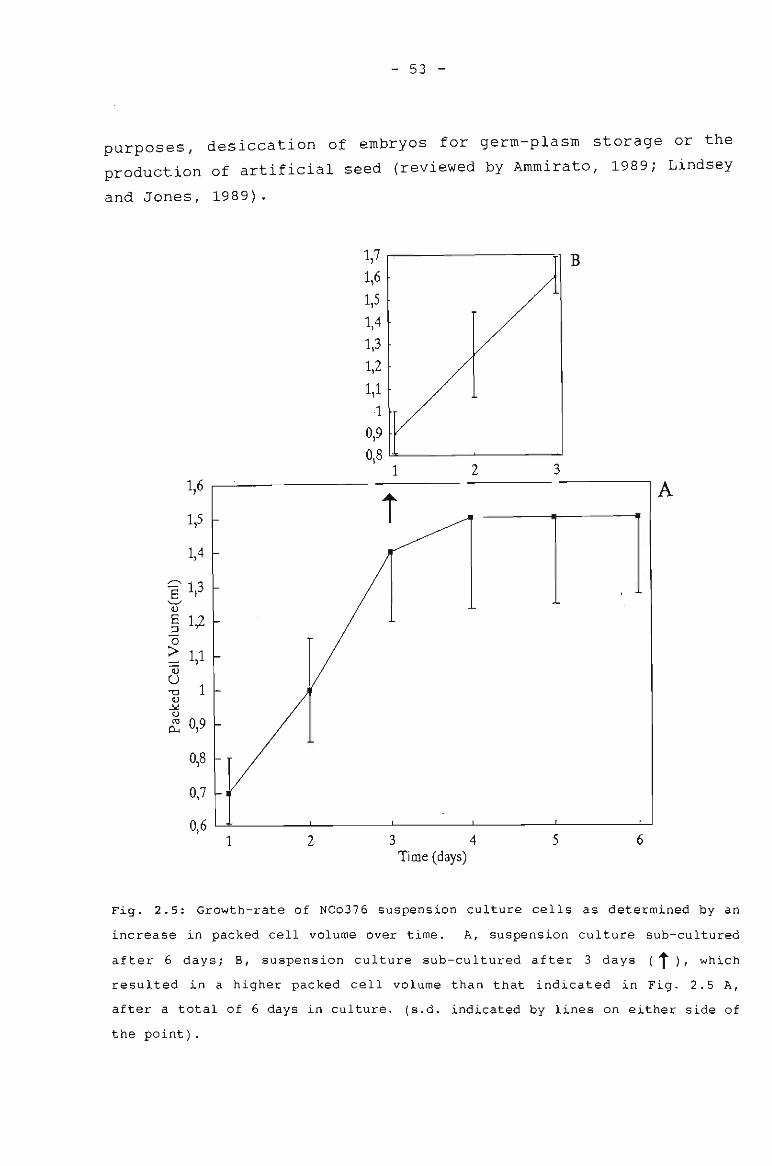
- 53 -
purposes, desiccation of embryos for germ-plasm storage or the
production of artificial seed (reviewed by Ammirato, 1989; Lindsey
and Jones, 1989).
1,7 B1,61,51,41,31,21,1
10,90,8
2 311,6
iA
1.,5
1,4
8 1,3"--'"v8 1,2:J(5
> 11= ,a1"'0
V~0ca 0,9~
0,8
0,7
0,61 2 3 4 5 6
Time (days)
Fig. 2.5: Growth-rate of NCo376 suspension culture cells as determined by an
increase in packed cell volume over time. A, suspension culture sub-cultured
after 6 days; E, suspension culture sub-cultured after 3 days (t), which
resulted in a higher packed cell volume than that indicated in Fig. 2.5 A,
after a total of 6 days in culture. (s.d. indicated by lines on either side of
the point).
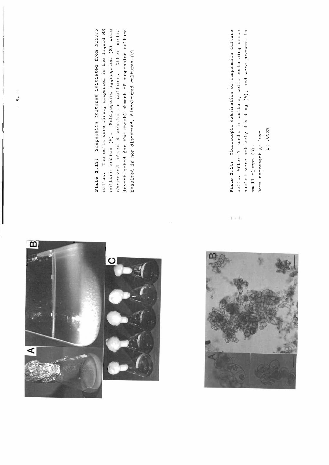
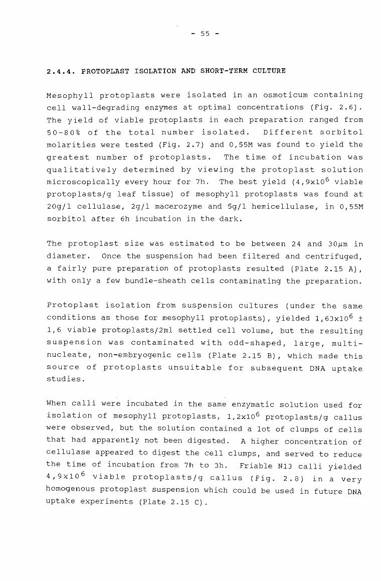
- 55 -
2.4.4. PROTOPLAST ISOLATION AND SHORT-TERM CULTURE
Mesophyll protoplasts were isolated in an osmoticum containing
cell wall-degrading enzymes at optimal concentrations (Fig. 2.6).
The yield of viable protoplasts in each preparation ranged from
50-80% of the total number isolated. Different sorbitol
molarities were tested (Fig. 2.7) and 0,55M was found to yield the
greatest number of protoplasts. The time of incubation was
qualitatively determined by viewing the protoplast solution
microscopically every hour for 7h. The best yield (4,9Xl0 6 viable
protoplastsjg leaf tissue) of mesophyll protoplasts was found at
20gjl cellulase, 2gjl macerozyme and 5gjl hemicellulase, in 0,55M
sorbitol after 6h incubation in the dark.
The protoplast size was estimated to be between 24 and 30~m in
diameter. Once the suspension had been filtered and centrifuged,
a fairly pure preparation of protoplasts resulted (Plate 2.15 A),
with only a few bundle-sheath cells contaminating the preparation.
Protoplast isolation from suspension cultures (under the same
conditions as those for mesophyll protoplasts), yielded 1,63x10⁶ ±
1,6 viable protoplastsj2ml settled cell volume, but the resulting
suspension was contaminated with odd-shaped, large, multi
nucleate, non-embryogenic cells (Plate 2.15 B), which made this
source of protoplasts unsuitable for subsequent DNA uptake
studies.
When calli were incubated in the same enzymatic solution used for
isolation of mesophyll protoplasts, 1,2xl06 protoplastsjg callus
were observed, but the solution contained a lot of clumps of cells
that had apparently not been digested. A higher concentration of
cellulase appeared to digest the cell clumps, and served to reduce
the time of incubation from 7h to 3h. Friable N13 calli yielded
4,9xl0 6 viable protoplastsjg callus (Fig. 2.8) in a very
homogenous protoplast suspension which could be used in future DNA
uptake experiments (Plate 2.15 C).
- 56 -
Protoplasts were cultured for 48h and although 84% mortality
occurred after this time period, 1,3X10 6 ± 0,57 protoplastsjg of
callus were still viable, and could be used for future transient
gene expression studies as discusssed in chapter 3.
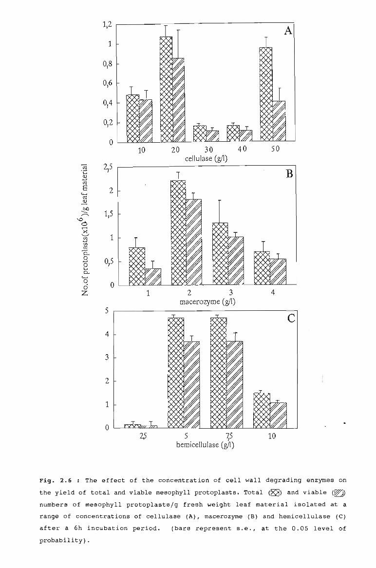
c
50
4
5 15bemiceIlulase (gIl)
2 3macerozyme (gIl)
20 30 40cellulase (gIl)
1
10
2
o
o
2
1
4
3
o
5
1
0,5
2,5
1
1,5
0,2
0,4
0,6
1,2
0,8
Fig. 2.6 : The effect of the concentration of cell wall degrading enzymes on
the yield of total and viable mesophyll protoplasts. Total (~) and viable (~)
numbers of mesophyll protoplastsjg fresh weight leaf material isolated at a
range of concentrations of cellulase (A), macerozYrne (B) and hemicellulase (G)
after a 6h incubation period.
probability) •
(bars represent s.e., at the 0.05 level of
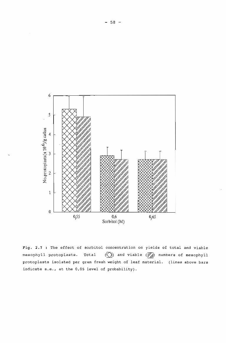
- 58 -
6
5
1
o0,55 0,6
Sorbitol CM)
Fig. 2.7 : The effect of sorbitol concentration on yields of total and viable
rnesophyll protoplasts. Total (~) and viable (~) numbers of mesophyll
protoplasts isolated per gram fresh weight of leaf material. (lines above bars
indicate s.e., at the 0,05 level of probability).
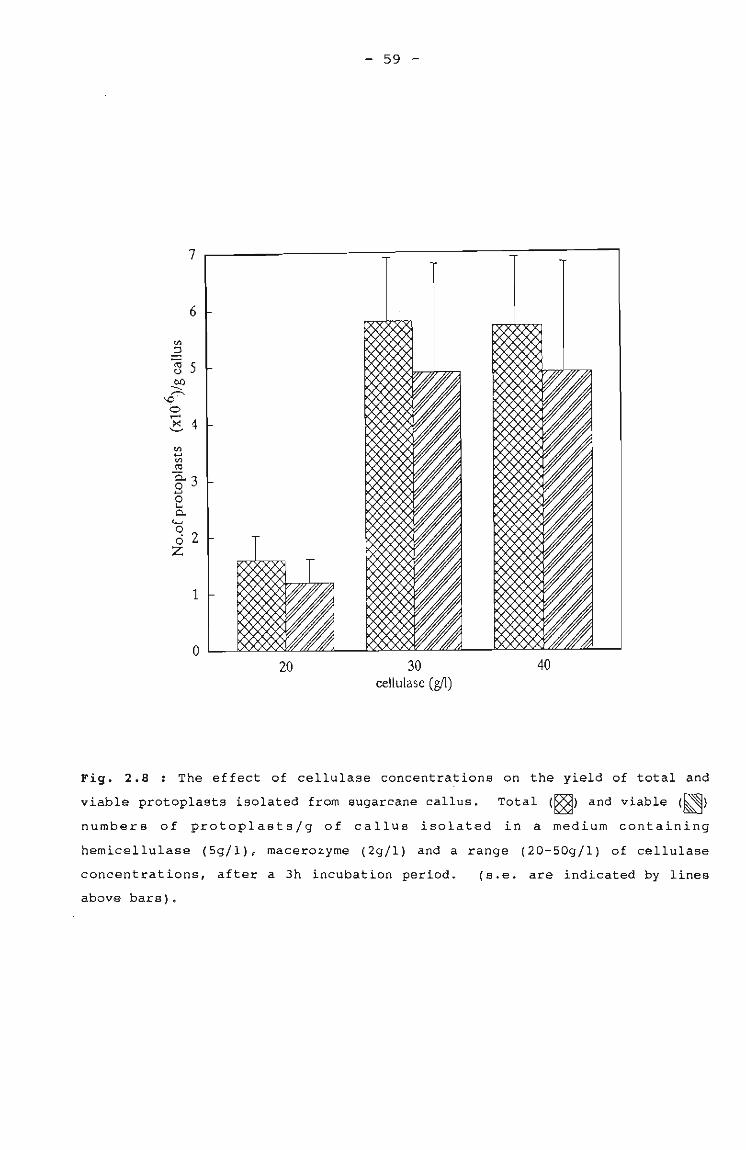
- 59 -
20 30cellulase (gIl)
40
Fig. 2.8 : The effect of cellulase concentra~ions on the yield of total and
viable protoplasts isolated from sugarcane callus. Total (S8) and viable (~)
numbers of protoplasts/g of callus isolated in a medium containing
hemicellulase (Sg/l), macerozyme (2g/1) and a range (20-S0g/l) of cellulase
concentrations, after a 3h incubation period.
above bars).
(s.e. are indicated by lines
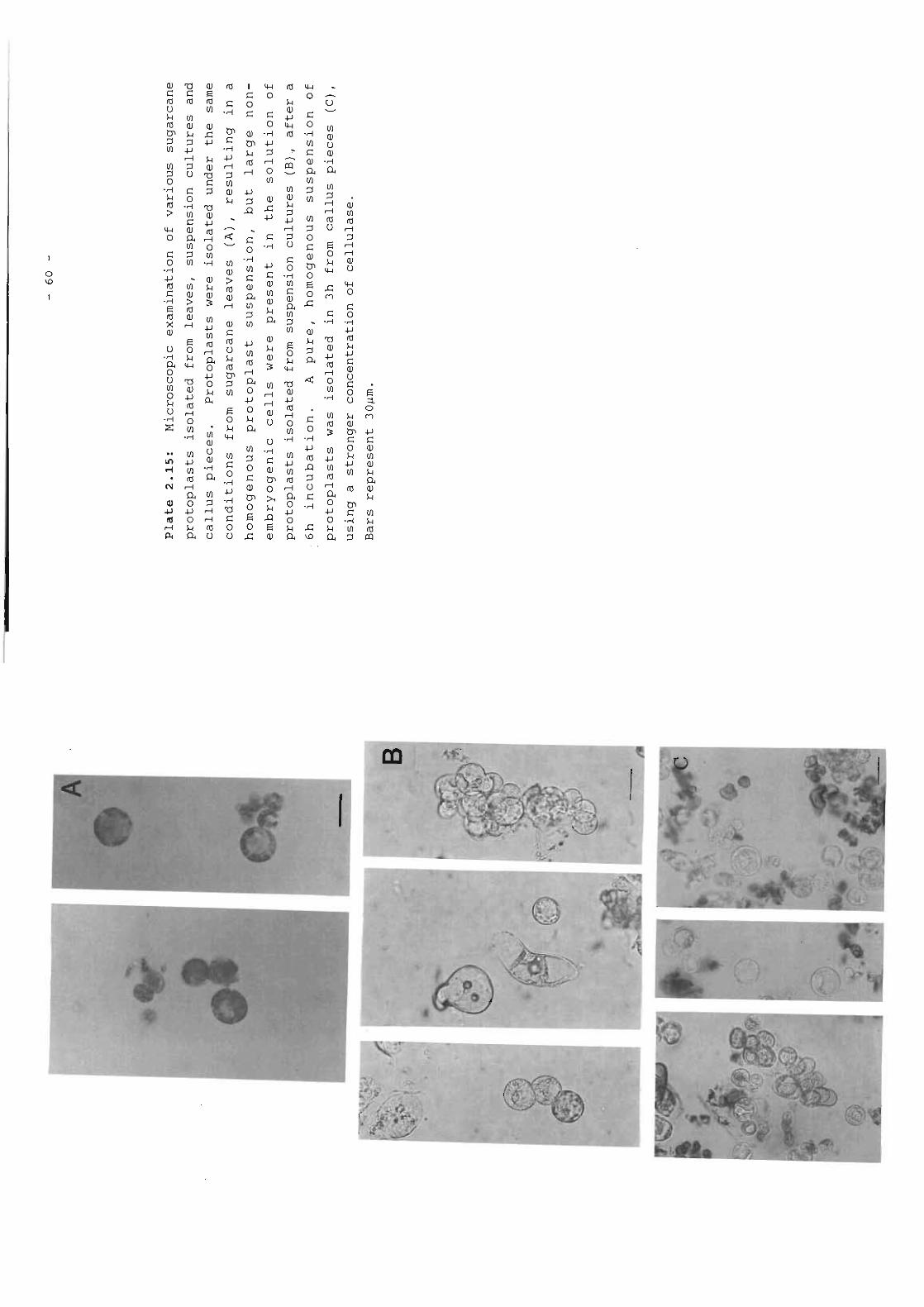
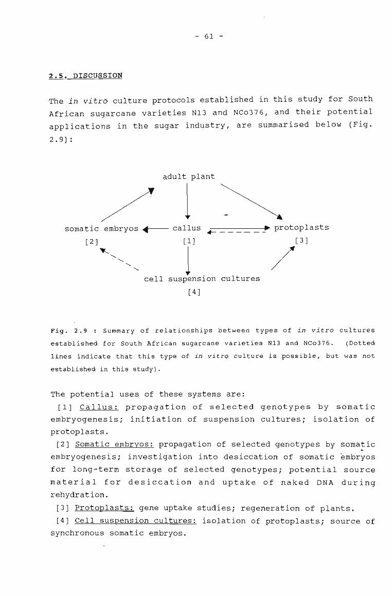
- 61 -
2.5. DISCUSSION
The in vitro culture protocols established in this study for South
African sugarcane varieties N13 and NCo376, and their potential
applications in the sugar industry, are summarised below (Fig.
2 .9) :
adult plant
1somatic embryos ~~-- callus .- ~ protoplasts
[2]~
"-.........
.........
[1]
1cell suspension cultures
[ 4 ]
[3 ]
/
Fig. 2.9 Summary of relationships between types of in vitro cultures
established for South African sugarcane varieties N13 and NCo376. (Dotted
lines indicate that this type of in vitro culture is possible, but was not
established in this study).
The potential uses of these systems are:
[1] Callus: propagation of selected genotypes by somatic
embryogenesis; initiation of suspension cultures; isolation of
protoplasts.
[2] Somatic embryos: propagation of selected genotypes by somatic
embryogenesis; investigation into desiccation of somatic embryos
for long-term storage of selected genotypes; potential source
material for desiccation and uptake of naked DNA during
rehydration.
[3] Protoplasts: gene uptake studies; regeneration of plants.
[4] Cell suspension cultures: isolation of protoplasts; source of
synchronous somatic embryos.
- 62 -
2.5.1. ADULT-PLANT SOURCE TISSUE
The starting material for some of the in vitro culture protocols
in this study, were explants from field-derived sugarcane, which
were severely contaminated with microorganisms. The use of
antimicrobial agents to control contaminants in tissue culture has
been well documented (Pollock et al., 1983; Shields et al., 1984;
Young et al., 1984; Falkiner, 1990; Leifert and waites, ~990).
However, problems encountered with microbial contamination of
sugarcane varieties NCo376 and N13, which resulted in 65% of the
explants being discarded due to contamination, were not overcome
by the use of any of the tested antimicrobial agents. This was
due to an inhibitory effect on callus induction when explants were
treated with these compounds (Table 2.2). Consequently, the
majority of the investigation into the establishment of in vitro
cultures was carried out using N13 regenerated plantlets as the
major explant source, as these were produced in vitro and were
therefore free of contaminants. However the need to continually
place fresh plant material into culture was recognised, because of
the danger of propagating genomic changes. Despite high
contamination levels, this was carried out routinely. Because
such a high proportion of the explants are discarded, the process
of initiating cultures is laborious and time-consuming and could
delay the progress of a research programme. One possibility may
be to have parent plants in the greenhouse, where they can be
treated for contaminants, before bei~g used as explant material.
However, such studies were not carried out in this project.
2.5.2. PLANT REGENERATION VIA SOMATIC EMBRYOGENESIS
Microscopic examination of callus which showed the presence of
typical embryogenic structures (Plates 2.4 and 2.5), and the
observation that simultaneous production of shoots and roots
occurred in sugarcane variety N13 on MS regeneration medium (Plate
2.7), suggested that plant regeneration occurred by the process of
somatic embryogenesis. The formation of embryogenic callus and
- 63 -
the regeneration of plants via somatic embryogenesis reported here
for the South African sugarcane variety N13, corresponds to
observations made in other plant regeneration studies, carried out
on sugarcane and other grasses. Similar requirements for growth
regulators and carbon sources were observed, and the rate of
regeneration and yields obtained were also similar. These various
aspects are discussed below.
The regeneration of graminaceous monocotyledon plants by the
process of somatic embryogenesis does not appear to require
I extensive manipulation of medium in terms of hormones; 2,4-D is
the auxin most commonly used for callus induction and
proliferation both in previous work carried out on sugarcane
(Heinz and Mee, 1969; Ho and Vasil, 1983a; Chen et al., 1988a;
Guiderdoni and Demarley, 1988) and other graminaceous species such
as maize (Lu et al., 1982, 1983; Vasil et al., 1984), rye (Lu et
al., 1984), Pennisetum sp. (Chandler and Vasil, 1984), Digitaria
sp. (Gonzales and Franks, 1987) and sorghum (MacKinnon et al.,
1986). The 2,4-D concentration of 4mg/l which yielded the
greatest percentage of calli in sugarcane variety N13 (Table 2.3),
gave rise to the lowest number of calli yielding plants (Table
2.5), indicating that although high concentrations of 2,4-D are
favourable for callus induction, subsequent regeneration occurs at
a higher frequency on media containing low levels of 2,4-D (Ho and
Vasil, 1983a; Ahloowalia, 1984; Bhaskaran and Smith, 1989). The
best 2,4-D concentration for callus induction, and subsequent
plant regeneration in variety N13 ~as 2mg/l (Table 2.3). The
observation in this study that germination of sugarcane embryoids
occurred on the removal of 2,4-D from the medium is consistent
with the observations in other graminaceous plants (Nadar et al.,
1978; Yao and Krikorian, 1981; Abe and Futsuhara, 1985; Bregitzer
et al., 1989).
Sucrose concentrations also appear to have an effect on the
process of somatic embryogenesis. In this study, the highest
concentration of sucrose (50g/l) used resulted in significantly
less callus induction (Table 2.3) and sUbsequent plant
- 64 -
regeneration (Table 2.5), than sucrose concentratiqns of 20 and
30g/l sucrose. Similarly, Chandler and vasil (1984) found that
high levels of sucrose (60-120gjl) had an inhibitory effect on
both callus growth and yield of somatic embryos of Pennisetum.
However researchers using other material have found that higher
concentrations of sucrose were necessary for callus induction, for
example in Digitaria, 90gjl sucrose was used and this also
resulted in the best regeneration yields (Watt et al., 1989). The
best sucrose concentration for sugarcane variety N13 callus
induction and subsequent plant regeneration was 20g/l (Table 2.5).
The time taken for plant regeneration via somatic embryogenesis
varies between species, but generally occurs 1 month after
embryogenic calli have been placed on regeneration media in the
light (Lu et al., 1984i Bhakasran and Smith, 1988). This is
supported by work carried out previously in sugarcane (Chen et
al., 1988ai Guiderdoni and Demarley, 1988) and by observations
made in this study (Plate 2.7).
When comparing yields of plants produced by somatic embryogenesis,
it is difficult to relate the yields of 16-36 sugarcane plants/g
fresh weight callus obtained in this study to reports of 25 000
Pennisetum sp. plants from a single leaf explant in 7 months
(Chandler and Vasil, 1984). In terms of percentage regeneration
levels (32%) and the number of Pennisetum plants produced per gram
fresh weight of callus (1-39), the results compare favourably to
those obtained for sugarcane variety N13, in this study (Table
2.6). However, much higher plant yields of 200 plantsjg callus
for sugarcane (Guiderdonui and Demarley, 1988) and orchardgrass
(Hanning and Conger, 1986) have been reported, so the potential
does exist for higher plant yields. Since the culture conditions
and media used in this study were similar to those used by
Guiderdoni and Demarley (1988), variation in plant yield can be
attributed to the use of different sugarcane varieties, namely Q75
and C06415.
In order to determine whether plant regeneration percentages and
- 65 -
yield of plants could be increased, activated charcoal was
incorporated into the regeneration medium. Observations by other
researchers (Weatherhead et al., 1978; Tisserat, 1985; Ebert and
Taylor, 1990) that activated charcoal seemed to remove the
inhibitory effect of 2,4-D on embryo germination, were matched in
sugarcane variety N13 at the highest 2,4-D concentration, where
calli producing plants increased from 13% to 24% in the presence
of activated charcoal (Fig. 2.4). However higher regeneration
percentages than these were observed from calli initiated on low
2,4-D levels (42% and 48% at 2mg/1 and 3mg/1 2,4-D, respectively)
'without the addition of activated charcoal. Activated charcoal
was therefore not routinely added to the regeneration medium.
Somatic embryogenesis has no direct practical application in the
field of micropropagation, because sugarcane is propagated
vegetatively for commercial purposes. However, the potential for
mass propagation of transformed genotypes in a short period of
time by the process of somatic embryogenesis is a most important
consideration. Other advantages of somatic embryos which may have
application in sugarcane-breeding programmes are the desiccation,
storage, rehydration and germination of embryos in vitro, and
their use in germ-plasm storage and artificial seed production
(Ammirato, 1989). Of vital significance in all of these
applications is the fact that somatic embryos arise from single
cells, ensuring the genetic stability and uniformity of the clone
(Ammirato, 1983; Evans and Bravo, 1983) provided that callus
produced by the indirect somatic embryogenic regeneration pathway
is not kept in culture for too long (Larkin and Scowcroft, 1981;
Tsay, 1987; Irvine et al., 1991).
2.5.3. ESTABLISHMENT OF SUSPENSION CULTURES
Sugarcane suspension cultures have been established since the
1970's for use in protoplast isolation (Maretzki and Nickell,
1973; Larkin, 1982; Liu, 1988), studies in nutrient transport
(Thorn et al., 1981) and salt tolerance (Liu and Shih, 1986).
Totipotent suspension cultures have been established in genera
- 66 -
within the Gramineae such as wheat (Vasil et al., 1990a, 1990b),
maize (Bartowick, 1981; Armstrong and Green, 1985; Kamo and
Hodges, 1986) and rice (Abe and Futsuhara, 1985).
Successful establishment of suspensions using friable callus from
the South African sugarcane variety NCo376 was achieved in this
study (Plate 2.13 A). Packed cell volume measurements indicated
that the doubling time for the culture was 3 days (Fig. 2.5).
Suspension cultures established in sugarcane previously (Ho and
Vasil, 1983b; Srinivasan and Vasil, 1986) had similar doubling
times (approximately 4 days), but appeared to reach much higher
packed cell volume levels than did the NCo376 culture in this
study. Such differences might be attributed to different
sugarcane varieties, as the medium used in this study did not
differ from the media used in the previous studies (with the
exception that coconut water was omitted in this study) or the
coconut derivatives might allow greater cell numbers to develop.
Coconut milk was omitted from media used in this study because it
is difficult to sterilise and could cause increased contamination
problems, and results are difficult to reproduce and compare,
because the concentration of proteins and growth regulators varies
between coconuts.
The NCo376 suspension cultures appeared to be embryogenic in
nature after a 4 month period; at this stage the cells aggregated
and formed embryogenic structures (Plate 2.13 B). Although the
NCo376 suspension cultures were no~ plated out, they exhibited
totipotent tendencies, and would probably be capable of
regenerating plants. Organised structures resembling the early
stages of embryogeny were reported in previous sugarcane
suspension cultures (Ho and Vasil, 1983b; Srinivasan and Vasil,
1986) when 2,4-D levels were lowered to O,l-lmg/l and sucrose
levels increased to 60-100g/l. However the medium used for the
establishment and embryogenic development of suspension cultures
in this study contained 3mg/l 2,4-D and 30g/l sucrose. Depending
on the intended use of the cUlture, manipulation of 2,4-D and
sucrose levels might be used to encourage cells to either disperse
- 67 -
in liquid culture or become organised into embryogenic structures.
In the present study, embryogenic suspension cultures of South
African sugarcane varieties were required for the isolation of
protoplasts for gene transfer investigation. The embryogenic
nature of the cultures gives them the potential to regenerate
plants from transformed cells. The cultures could also be used
for quick micropropagation of selected genotypes (particularly of
transformed cell lines) on a large scale by the production of
somatic embryos, which could be plated out subsequently and
,regenerated into plants.
2.5.4. PROTOPLAST ISOLATION AND SHORT-TERM MAINTENANCE
Protoplasts were isolated from leaves of sugarcane variety N13 in
order to establish a general protoplast isolation protocol with
readily available material. Mesophyll protoplasts have been
isolated from sugarcane leaves by other workers (Chen and Liu,
1974; Evans et al., 1980). Once a general isolation protocol was
available protoplasts were isolated from suspension cultures of
sugarcane variety NCo376. Yields of 1,6xl0 6 protoplastsj2ml
settled cell volume suspension culture were obtained, which
compared unfavourably to yields of 4,9Xl0 6 mesophyll protoplastsjg
leaf material (Figs. 2.6 and 2.7). However, the protoplast
preparation obtained from suspension cultures contained
protoplasts and undigested cells that varied in size and were
difficult to purify. Bartkowiak (1981) encountered similar
problems with heterogenous suspension cultures in maize.
Callus provided a good source of protoplasts after slight
modification of enzyme concentration and incubation times used for
mesophyll protoplast isolation, and yields of 4,9Xl0 6 viable
protoplastsjg callus were obtained (Fig. 2.8). The protoplasts
were homogenous and uniform in size. There is little evidence in
the literature to suggest that calli are used routinely as source
material for protoplast isolation, but Vasil et al. (1990a) found
it impractical to obtain a sufficient number of protoplasts from
- 68 -
wheat callus, although no figures for yields were presented.
In most graminaceous monocotyledons, regeneration of plants has
not been achieved from mesophyll protoplasts (Vasil, 1987). No
reports have substantiated the regeneration of sugarcane plants
from mesophyll protoplasts claimed possible by Chen and Liu (1974)
and Evans et al. (1980). Embryogenic suspension cultures have
served as the major source material for protoplast isolation and
subsequent regeneration in sugarcane (Srinivasan and Vasil, 1986;
Chen et al., 1988b) and the Gramineae in general, for example
maize, (Shillito et al., 1989; Mitchell and Petolino, 1991), rice
(Abdullah et al., 1986; Yamada et al., 1986; Toriyama et al.,
1988; Zhang et al., 1988)), wheat (Shimada and Yamada, 1979) and
Panicum sp. (Lu et al., 1981).
In order to select the best source of protoplasts for gene uptake
work, factors such as the yield, purity and potential regeneration
ability of the protoplasts were taken into consideration.
Although the NCo376 suspension cultures might provide protoplasts
most likely to have totipotent potential, the protoplast
suspension consisted of a heterogenous popUlation of cells in
terms of cell size, and yields were lower than those obtained from
callus and leaves. Therefore no further gene uptake studies were
carried out on protoplasts from this source. Mesophyll
protoplasts isolated from leaf material provided a homogenous
protoplast suspension with good yields of 4,9xI06 protoplasts per
gram fresh weight leaf, but regeneration of plants from this
source was unlikely. Callus provided a homogenous protoplast
suspension with yields equal to those from leaves, and although
the regeneration potential of protoplasts from this source has not
been documented, the chances of obtaining cells with regenerative
potential is good because the callus is embryogenic in nature. On
this basis, callus was chosen as the source of protoplasts for
gene uptake studies.
It was the intention that protoplasts isolated from South African
sugarcane callus would be used in DNA uptake work and subsequently
- 69 -
cultured in order to monitor transient expression of introduced
genes (Chapter 3). Transient expression peaks at approximately
48h, so it is critical to maintain the protoplasts in a viable
state for this time period (Draper et al., 1988). There appears
to be a critical protoplast density of at least 105 protoplasts/ml
for gene uptake and subsequent monitoring of expression (Lorz et
al., 1985; Potrykus et al., 1985; Larkin et al., 1990). A viable
number of 1,3X10 6 sugarcane protoplasts/g of callus was observed
after culture for 48h in this study .
. Genetic manipulations of plants at the protoplast stage has been
investigated for graminaceous monocotyledons such as maize (Fromm
et al., 1986; Rhodes et al., 1988; Huang and Dennis, 1989; Gordon
Kamm et al., 1990), rice (Toriyama et al., 1988; Yang et al.,
1988; Zhang et al., 1988; Datta et al., 1990; Hayashimoto et al.,
1990; Izawa et al., 1991), wheat (Lorz et al., 1985; Larkin et
al., 1990) and sugarcane (Chen et al., 1987). A preliminary
investigation into DNA uptake by sugarcane protoplasts isolated
from South African varieties is reported in the following chapter.
- 70 -
CHAPTER THREE
DEVELOPMENT OF GENE TRANSFER TECHNIQUES IN SUGARCANE (SACCHARUM
SPECIES HYBRIDS).
3.1 INTRODUCTORY REMARKS
There is a range of techniques available for gene transfer to
plants, and these are reviewed in section 3.2.2. Of all the
transformation techniques available, it was decided to use two
approaches for the genetic engineering of South African sugarcane
varieties, and seeds and protoplasts were used as recipients for
transforming DNA. The seed is an attractive option for
introducing foreign genes to plants, because no in vitro plant
regeneration is necesary. Protoplasts provide a simple system for
investigating transformation, and despite problems with
regeneration to plants, protoplasts have been widely used in
transformation studies.
3.2 LITERATURE REVIEW
3.2.1 FACTORS INFLUENCING TRANSFORMATION EVENTS
The successful genetic transformation of a cell or tissue
comprises three essential elements: the introduction of foreign
DNA into the cell, the integration of the DNA into the host
genetic material (nuclear, chloroplastic or mitochondrial) and the
controlled expression of the introduced DNA (Lindsey and Jones,
1990) . The factors that influence each of these steps in the
transformation process will be discussed below.
Factors affecting introduction of DNA into plant cells
Methods used to introduce foreign DNA to plants
Several techniques for the introduction of specific gene sequences
into plant cells or tissues exist, and these are reviewed more
- 71 -
extensively in section 3.2.2. The most common method used is
mediated by the bacterium Agrobacterium tumefaciens , which is the
causal agent of crown-gall disease and can insert part of its DNA
(transferred or T-DNA) into the plant genome (Chilton et al.,
1977; Braun, 1978; Nester et al., 1984). This phenomenon has been
exploited and foreign gene sequences have been inserted into
Agrobacterium T-DNA, which has facilitated their introduction to
susceptible plants (Barker et al., 1983; Simpson et al., 1986;
Hamill et al., 1987).
,Not all plant species are amenable to infection by Agrobacterium,
but genes can be directly transferred to protoplasts using
techniques such as electroporation (Fromm et al., 1985; Shillito
et al., 1985; Larkin et al., 1990), microinjection (Lawrence and
Davies, 1985; Crossway et al., 1986; Reich et al., 1986) and
chemical means (Krens et al., 1982; Lorz et al., 1985; Werr and
Lorz, 1986; Meijer et al., 1991). However direct DNA uptake by
protoplasts as a means of producing transgenic plants is limited
to those plant species that can regenerate from the protoplast
stage.
The difference in response to transformation by monocotyledonous
and dicotyledonous plants
The majority of monocotyledonous species are not susceptible to
infection by Agrobacterium, and it has been suggested that
monocotyledonous cells may not posse~s the appropriate cell wall
binding sites, which are necessary for Agrobacterium attachment
(Lippincott and Lippincott, 1978). It is also possible that
monocots respond differently at a molecular level to wounding,
which is a prerequisite for infection of dicots (Chilton et al.,
1977; Nester et al., 1984). However, a recent study carried out
by Mooney and Goodwin (1991) indicate that Agrobacterium can
attach to immature wheat embryos, and that attachment was not
wound-dependent (attachment no different between sites
enzymatically wounded and those untreated). There is some
evidence of Agrobacterium T-DNA transfer, if not stable
- 72 -
integration, in some monocots (Hernalsteens et al., 1984;
Hooykaas-van Slogteren et al., 1984; Graves and Goldman, 1986;
Mooneyet al., 1991).
Alternative systems for the introduction of foreign genes to
monocots include those direct DNA-uptake methods mentioned earlier
and some which make use of regenerable plant material, eg.
microprojectile bombardment of tissue and callus (Klein et al.,
1987; McCabe et al., 1988; Wang et al., 1988; Mendel et al., 1989;
Twell et al., 1989; Fromm et al., 1990; Gordon-Kamm et al., 1990,
DNA uptake by pollen of flowering plants (De la Pena et al., 1987;
Luo and Wu, 1988; Hess et al., 1990) and uptake of naked DNA into
seeds (Ledoux and Huart, 1968, 1969; Ledoux et al., 1971, 1974;
Topfer et al., 1989), all of which are discussed further in
sections 3.2.2 and 3.2.3.
Factors affecting integration of DNA in the plant genome
There are a number of methods available for transforming plants,
and the methods used to insert DNA into cells will be discussed in
section 3.2.2. The bacteria of the genus Agrobacterium, have a
natural ability to insert a particular segment of DNA (and
consequently any passenger DNA) into plant genomes, and this type
of insertion is under the control of genetic elements within the
bacterium (Chilton et al., 1977; Nester et al., 1984). However,
Agrobacterium does not have the capacity to insert into all plant
species, and other transformation ~echniques rely upon random
insertion of DNA into the plant genome (Potrykus et al., 1985).
Here, factors such as DNA concentration, the conformation of the
DNA (linear or circular), the actual sequence of the transforming
DNA and type of vector used (section 3.2.4), are important in
determining successful integration.
Factors influencing expression of transforming DNA
Much remains unknown about the factors influencing the expression
of foreign genes in plants. Factors such as the position of
- 73 -
insertion of the DNA in the host genome, the copy number, whether
DNA methylation occurs, and the transcriptional levels directed by
a 'foreign' promoter, can all affect the expression of an
introduced gene (Walden, 1988).
Choice of promoter
A promoter can be defined as the DNA region, usually upstream to
the coding sequence of a gene or operon, which binds RNA
polymerase and directs the enzyme to the correct transcriptional
Isite so that RNA synthesis can be initiated (Oliver and Ward,
1985). Bacterial genes are not transcribed after integration into
the plant genome unless they are placed under the control of
suitable promoter elements (with the exception of Agrobacterium T
DNA, which contains its own promoter elements) (Fraley et al.,
1983). Plant genes, however, are usually transcribed and spliced
correctly if transferred to a homologous/heterologous host genome
(Murai et al., 1983). Different promoters work with different
efficiencies, with strong promoters causing mRNA synthesis to be
initiated at a high frequency, which enables selection of
transformed cells whereas weak promoters direct synthesis of rarer
transcripts (Maniatis et al., 1982).
Promoters studied in the most detail are the nopaline synthase
(nos) promoter from Agrobacterium and the 358 transcript promoter
of the cauliflower mosaic virus (CaMV) (Cocking and Davey, 1987).
By combining such promoters upstrea~ of the coding sequence of
bacterial antibiotic resistance genes, it is possible to produce
chimeric genes that function in plants (reviewed by Weising et
al., 1988) ~ The bacterial nptII gene from.the transposon Tn5
under the control of the CaMV 358 promoter, is a chimeric gene
(Paszkowski et al., 1984; Jenson et al., 1986) which has been used
to transform many plants (reviewed in more detail in section3.2.5) .
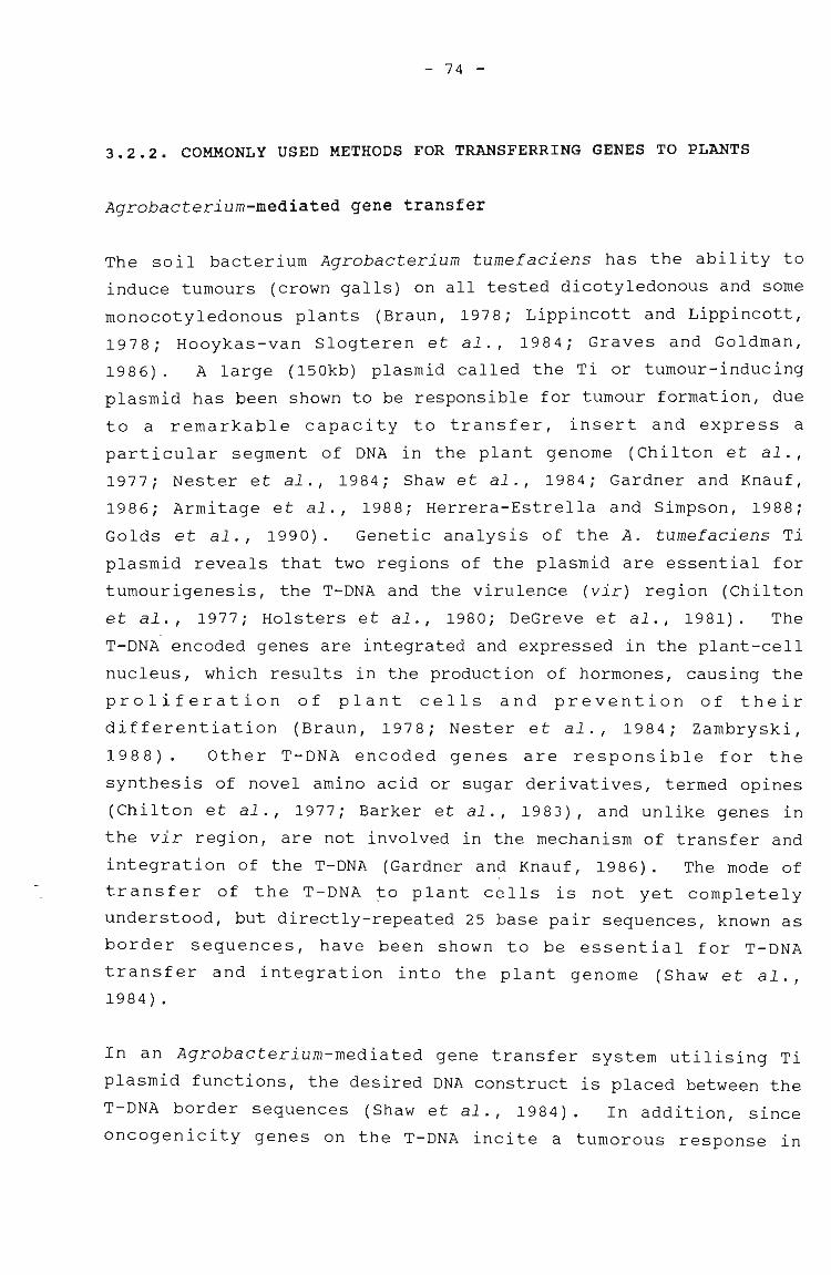
- 74 -
3.2.2. COMMONLY USED METHODS FOR TRANSFERRING GENES TO PLANTS
Agrobacterium-mediated gene transfer
The soil bacterium Agrobacterium tumefaciens has the ability to
induce tumours (crown galls) on all tested dicotyledonous and some
monocotyledonous plants (Braun, 1978; Lippincott and Lippincott,
1978; Hooykas-van Slogteren et al., 1984; Graves and Goldman,
1986) . A large (150kb) plasmid called the Ti or tumour-inducing
plasmid has been shown to be responsible for tumour formation, due
to a remarkable capacity to transfer, insert and express a
particular segment of DNA in the plant genome (Chilton et al.,
1977; Nester et al., 1984; Shaw et al., 1984; Gardner and Knauf,
1986; Armitage et al., 1988; Herrera-Estrella and Simpson, 1988;
Golds et al., 1990). Genetic analysis of the A. tumefaciens Ti
plasmid reveals that two regions of the plasmid are essential for
tumourigenesis, the T-DNA and the virulence (vir) region (Chilton
et al., 1977; Holsters et al., 1980; DeGreve et al., 1981). The
T-DNA encoded genes are integrated and expressed in the plant-cell
nucleus, which results in the production of hormones, causing the
proliferation of plant cells and prevention of their
differentiation (Braun, 1978; Nester et al., 1984; Zambryski,
1988). Other T-DNA encoded genes are responsible for the
synthesis of novel amino acid or sugar derivatives, termed opines
(Chilton et al., 1977; Barker et al., 1983), and unlike genes in
the vir region, are not involved in the mechanism of transfer and
integration of the T-DNA (Gardner and Knauf, 1986). The mode of
transfer of the T-DNA ~o plant cells is not yet completely
understood, but directly-repeated 25 base pair sequences, known as
border sequences, have been shown to be essential for T-DNA
transfer and integration into the plant genome (Shaw et al.,
1984) .
In an Agrobacterium-mediated gene transfer system utilising Ti
plasmid functions, the desired DNA construct is placed between the
T-DNA border sequences (Shaw et al., 1984). In addition, since
oncogenicity genes on the T-DNA incite a tumorous response in
- 75 -
plant cells, these genes are generally deleted and replaced by an
antibiotic resistance gene that can be used as a marker to select
transformed cells (Hernalsteens et al., 1984; Zambryski, 1988).
The Ti plasmid is too large to manipulate in vitro (Zambryski,
1988), so one of two different vector systems is used for plant
transformation studies (Fig. 3.1). Cointegrative-type Ti vectors
have been constructed in which a gene of interest, cloned on an E.
coli plasmid, is inserted into the T-DNA by a single recombination
event, at a region of common homology (Chilton et al., 1977;
Gardner and Knauf, 1986). Binary vectors can also be used, in
which the Agrobacterium carries two plasmids, one of which has
selectable markers and cloning sites between T-DNA borders, which
can replicate in E. coli, and the other is a Ti plasmid which acts
in trans, supplying virulence functions to enable the transfer of
the desired gene to plant cells (Simpson et al., 1986; Hamill et
al., 1987).
A VIR
~mnuflII1J7:~
B
MCS
Col El
RES
+
Fig 3.1: Schematic diagram of generalised cointegrative (A) and binary (B)
vector systems. VIR, virulence region; HOM, homologous region; LB and RB, left
and right borders; MCS, multicloning site; PTM, plant transformation marker;
RES, antibiotic resistance marker. (reproduced from Draper et al., 1988)
- 76 -
Plant tissues and cells may be exposed to the Agrobacterium vector
system in a number of different ways. In vivo inoculation of
seedlings with Agrobacterium cells (Graves and Goldman, 1986;
Hinchee et al., 1988; Chee et al., 1989; Raineri et al., 1990) or
well-established plants (Murai et al., 1983; Hernalsteens et al.,
1984; Hookaas-Van Slogteren et al., 1984; Shaw et al., 1984;
Gardner and Knauf, 1986) that have been propagated in vitro or in
vivo, is the traditional way to obtain transformed cells.
Selection of the transformed tissues or plants is based on
expression of selective marker genes such as antibiotic resistance
(Hinchee et al., 19.88; Raineri et al., 1990). The leaf-disc
technique, where leaf-discs are co-cultivated with a suspension of
Agrobacterium (De Block et al., 1987), is a simple way to
transform cells and these are cultured directly thereafter, going
either through a callus stage first or in some cases, by direct
regeneration from somatic cells in the leaf discs. This technique
is limited to those species that respond to Agrobacterium
infection and that also produce plants readily from leaf explants.
Protoplasts may also be co-cultivated with Agrobacterium (Marton
et al., 1979; Van Lijsebettens et al., 1986), and the advantage of
this is that large numbers of independent transformation events
can occur. However this technique is only useful if the
transformed protoplasts can be regenerated to whole plants. A
novel approach used by Mooney et al. (1991), involved the transfer
of Agrobacterium to wheat tissues by co-cultivation with the
bacterium, following partial enzymatic digestion of the scutellum
of immature wheat embryos.
The majority of Agrobacterium-mediated gene transfer work has been
performed with A. tumefaciens, but some work has been done with A.
rhizogenes (Comai et al., 1985; Simpson et al.., 1986; Hamill et
al., 1987). Both species transform plant cells in a similar way in
terms of DNA transfer and production of novel amino acids in
target tissue, but infection by A. rhizogenes results in so-called
hairy roots as opposed to crown galls, and the T-DNA responsible
for transformation is located on the Ri (root-inducing) plasmid
(Jensen et al., 1986; McKnight et al., 1987; Stougaard et al.,
- 77 -
1987) . In view of the possible host-range extension, A.
rhizogenes-mediated gene transfer should not be overlooked as a
means of producing transgenic plants, but will not be discussed in
this review.
Most successful Agrobacterium-mediated transformations have been
achieved in dicotyledonous plants but there have been some reports
of monocotyledonous plants being transformed by Agrobacterium.
Hooykaas-van Slogteren et al. (1984) found that stem inoculation
of plants from the Lilliaceae and Amarillidaceae groups resulted
.in swelling at the site of inoculation, where octopine and
nopaline activity was observed, indicating T-DNA transfer to
cells. Similarly, asparagus inoculated with A. tumefaciens
developed tumourous proliferations on the stem, and opine
synthesis was detected (Hernalsteens et al. 1984). Expression of
T-DNA linked markers has been demonstrated in rice cultures
(Raineri et al.,1990) and in maize seedlings (Graves and Goldman,
1986) transformed by Agrobacterium.
The Ti plasmid of A. tumefaciens has been used in conjunction with
plant-virus DNA to inoculate plants and to monitor the transfer of
DNA by the expression of disease symptoms, in a process termed
I agroinfection I which will be discussed later in the review.
Direct gene transfer to protoplasts
The failure to achieve Agrobacterium-induced transformation in the
majority of monocotyledonous plants, the cereals in particular,
has led to increased interest in assessing transformation
techniques for direct gene transfer. This field has been
extensively reviewed (Cocking and Davey, 1987; Davey et al., 1989;
Lorz, 1989; Walden, 1988; Golds et al., 1990). Direct uptake of
foreign DNA by protoplasts using techniques such as
electroporation, microinjection and chemical methods has resul~ed
in some transformation success (Potrykus et al., 1985; Rhodes et
al., 1988; Larkin et al., 1990). However, not many transgenic
monocotyledonous plants have been produced using these techniques
- 78 -
because of the difficulties involved in regenerating plants from
the protoplast stage (section 2.2.5).
Electroporation
The transformation frequency of cultured mammalia~ cells exposed
to exogenous DNA was found to be significantly increased by
electric pulses (Neumann et al., 1982; Potter et al., 1984).
Electroporation, as the process has been named, produces transient
pores in the plasma membrane when cells are exposed to a suitable
'electric field, and macromolecules such as DNA can pass into the
cell (Shillito et al., 1985). The pores created in the membrane
appear to be about 30nm in diameter and persist for several
minutes after the pulse, after which the membrane can 're-seal'
(Okada et al., 1986a). Field strength (voltage gradient) and
pulse duration (decay time) are the two main variables affecting
the permeabilisation of the cell membrane by electroporation
(reviewed by Draper et al., 1988).
The electroporation pulse is generated by discharging a capacitor
across an electroporation chamber, which consists of two flat
metal electrodes placed within a sterile plastic cuvette. The
protoplasts are suspended between the electrodes in an ionic
solution (Larkin et al., 1990; Potter et al., 1984). There are
two different approaches to electroporation for the introduction
of DNA into plant protoplasts: a) using a high voltage pUlse of
short duration, for example 1,5kV for 10~s (Shillito et al., ,1985), or b) using a lower voltage pulse of a longer duration, for
example 350V over 54ms (Fromm et al., 1985).
The advantages of the electroporation system are that it is
efficient and convenient, there is a low cell toxicity and it can
be used on a wide range of plant protoplasts. These include
monocotyledonous plants such as rice (Junker et al., 1987;
Toriyama et al., 1988; Yang et al., 1988; Zhang et al., 1988;
Izawa et al., 1991), maize (Fromm et al., 1986; Huang and Dennis,
1989) which was successfully regenerated from the protoplast stage
- 79 -
(Rhodes et al., 1988), and dicotyledonous plants such as carrot,
tobacco and lucerne (Fromm et al., 1985; Larkin et al., 1990). In
a slightly different approach, designed to bypass the requirement
for protoplast regeneration, Dekeyser et al. (1990) electroporated
leaf bases on rice seedlings with a pulse duration ten times
higher than that used for protoplasts. It was found that DNA
could be introduced into six layers of cells. Using a similar
strategy, tobacco-pollen grains were successfully electroporated
(Abdul-Baki et al., 1990) and these could be used for in vivo
fertilisation which could ultimately yield transformed seed.
Polyethylene glycol-induced DNA uptake
Polyethylene glycol (PEG)-mediated protoplast fusion is a method
used to generate interspecific plant hybrids (Power et al., 1986),
but PEG has also been used to facilitate the entry of DNA
molecules into plant protoplasts (Lorz et al., 1985; Werr and
Lorz, 1986; Meijer et al., 1991). PEG is a hydrophilic molecule
and at high concentrations (30-40%) it appears to minimise charge
repulsion effects between negatively-charged membranes and DNA
(negatively charged by virtue of its phosphate groups). It
stimulates endocytosis in protoplasts, enabling the uptake of
large particulate matter such as whole chloroplasts, liposomes and
bacteria (Power et al., 1986; Larkin et al., 1990). In DNAI •
experlments, the total effect of PEG is somewhat unclear, but
probably involves the precipitation of plasmid DNA onto the plasma
membrane and" then the stimulation ~f its uptake by endocytosis
(Freeman et al., 1984; Potrykus et al., 1985).
Krens et al. (1982) produced a reproducible method for the stable
transformation of tobacco protoplasts using PEG-mediated DNA
uptake. SUbsequently tobacco protoplasts have been transformed by
the PEG-induced DNA technique by several groups of workers (Hein
et al., 1983; Kartzke et al., 1990; Larkin et al., 1990).
Attempts to transform monocotyledonous protoplasts using PEG
mediated DNA uptake have met with equal success, for example
ryegrass (Potrykus et al., 1985), rice (Datta et al., 1990;
- 80 -
Hayashimoto et al., 1990), sugarcane (Chen et al., 1987) and wheat
(Lorz et al., 1985; Larkin et al., 1990), but in most of these
cases only transformed callus cultures have been recovered.
However transgenic rice plants have been produced as a result of
PEG-mediated DNA delivery (Datta et al., 1990; Hayashimoto et al.,
1990; Meijer et al., 1991).
Microinjection of DNA into protoplasts
Microinjection was originally developed for animal cells and can
\be used for the introduction of small molecules, macromoleculesi
(DNA, RNA and protein), organelles and virus particles into a wide
range of animal cells (Stacey, 1981; Celis, 1984; Hammer et al.,
1985). Development of comparable methods for transforming plant
protoplasts was attempted by Griesbach (1983) and steinbiss and
Stable (1983), but it is only recently that the feasibility of
plant cell transformation by direct microinjection of foreign DNA
into protoplasts has been shown (Crossway et al., 19$6; Reich et
al., 1986). The success of the technique depends on the ability
to orientate protoplasts and locate nuclei so that DNA can be
injected into the nucleus without damaging it (Lawrence and
Davies, 1985). Advantages of microinjection are that removal of
the cell wall is not required, and the amount of DNA injected into
the cell can be controlled. However it is expensive to set up,
requires skilled personnel to carry out the injection, which is a
slow process, and the cells are easily damaged (Crossway et al.,
1986) •
Techniques less frequently-used for gene transfer to protoplasts
Direct gene transfer to protoplasts has been achieved by mild
sonication of sugar beet and tobacco protoplasts (Joersbo and
Brunstedt, 1990), and transient expression was observed in a large
proportion of the cells.
The delivery of DNA and other macromolecules to protoplasts by
different kinds of liposomes was the sUbject of early experiments
- 81 -
by Fraley and Papahadjopoulos (1982), but integration of foreign
DNA into plant genomes following liposome-mediated transformation,
has only recently been demonstrated (Deshayes et al., 1985).
Liposomes are artificial vesicles encapsulating DNA or other
macromolecules, and the advantages of using this technique is that
the membrane encapsidation provides some protection against
nucleases and no carrier DNA is required (Hain et al., 1984).
Recently, a study to determine whether silicon carbide fibres
could be used to deliver DNA to plant cells was carried out
(Kaeppler et al., 1990). This method involves vortexing
suspension culture cells with a mixture of plasmid DNA, encoding a
selectable or screenable marker gene, and the silicon carbide
fibres. Both maize and tobacco cells expressed transient gene
activity after the treatment.
Direct gene transfer to intact cells
Microprojectile bombardment of plant cells and tissues
Klein et al. (1987) first reported the delivery of nucleic acids
to plant cells using high-velocity microprojectiles. The
mechanisms involved in the process have been described by Klein
and Fromm (1989) and the main points can be summarised as follows:
a particle gun is used to accelerate tungsten microprojectiles
(4~m in size), under the charge of a .22 calibre blank cartridge.
The force of the exploding gun-pow~er sends a plastic cylinder
with DNA-coated particles down the barrel of the particle gun and
into a steel plate. The plate stops the cylinder but accelerates
the tungsten beads through a small hole in the plate so that they
penetrate into cells of leaf, stem or even callus tissue contained
in a petri-dish below (Fig. 3.2). The microprojectiles pierce
cell walls and membranes and enter intact plant cells withoutkilling them.
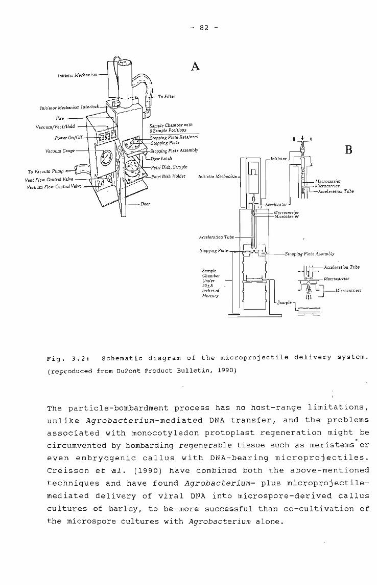
- 82 -
B
MBcrocarrier},fjcrocarrier
.4.cceleration Tube
-UiFAccelerati~n Tube
~ Macrocarner
~1~~MjCrOCarriersIH
SBlDplel--:::=J L-
Initiator
II~--+-MacrocarrjerI ~-:+-1vfjcrocarrjer
SampleCb BlDberUnder20+5inChes ofMercury
Initiator Mechanism
A
To Filter
Petri Dish Holder
Stopping Plate -==+-::~;>Q -+-t--.....'itopping Plate Assembly
Acceleration Tube -+---;+11
Door
S8IIlple Chamber with5 S8IIlple Positions
..........:~:+-....,topping Plate Retainers~~-Stopping Plate
Stopping Plate Assembl.v
Door Latch
Petri Dish, Sample
Initator Mechanism
VBCU WD Gauge ---+--7----v::~
Fire .---k--~
VacuWDfVen t/Hold -+-".----,
To VacuWD PWDp
Vent Flow Control Valve --t!-t~-T~
VBCUWD Flow Control Valve
Fig. 3.2: Schematic diagram of the microprojectile delivery system.
(reproduced from DuPont Product Bulletin, 1990)
The particle-bombardment process has no host-range limitations,
unlike Agrobacterium-mediated DNA transfer, and the problems
associated with monocotyledon protoplast regeneration might be..circumvented by bombarding regenerable tissue such as meristems or
even embryogenic callus with DNA-bearing microprojectiles.
Creisson et ~l. (1990) have combined both the above-mentioned
techniques and have found Agrobacterium- plus microprojectile
mediated delivery of viral DNA into microspore-derived callus
cultures of barley, to be more successful than co-cultivation of
the microspore cultures with Agrobacterium alone.
- 83 -
The microprojectile technique has been used to achieve stable
transformation of soybean plants, which expressed resistance to
the antibiotic kanamycin (McCabe et al., 1988), and wheat which
has produced stably-transformed callus lines resistant to
kanamycin (Vasil et al., 1991). Fertile transgenic maize plants,
expressing luciferase (Fromm et al., 1990) and resistance to the
herbicide bialaphos (Gordon-Kamm et al., 1990) were produced by
microprojectile bombardment of calli and suspension culture cells,
respectively. Transient GUS expression was monitored in
suspension culture cells of rice (Wang et al., 1988) and barley
(Mendel et al., 1989), as well as tobacco leaves (Tomes et al.,
1990), pollen grains (Twell et al., 1989) and maize coleoptiles
(Reggiardo et al., 1991) after being sUbjected to microprojectile
bombardment. Sugarcane callus and regenerated plantlets have
shown transient GUS expression after being bombarded using a
modification of the particle gun, which instead of using the DNA
coated tungsten particles, employs an airless spray-gun to deliver
the DNA (Irvine and Almeida, 1991).
Viral-mediated DNA delivery
Plant viruses have received attention as potential vectors for the
introduction of foreign genes into plants because their host range
includes many of the cereals (Walden, 1988). In view of the
difficulties encountered with in vitro culture of this group of
plants and the fact that they ar~ often not susceptible to
transformation using vectors based on the Ti plasmid, viral
mediated DNA delivery is an alternative worth considering.
Factors such as the ease of insertion into the plant cell (by
mechanical wounding of tissue), ease of manipulation (small genome
size: 8kb or smaller), and the fact that viruses replicate in
plant cells to achieve a high copy number, make them attractive
agents for plant transformation studies (reviewed by Mantell etal., 1985).
The two groups of plant viruses whose genomes comprise DNA are the
- 84 -
caulimovirus family and the gemini viruses. Cauliflower mosaic
virus (CaMV), containing double-stranded DNA, has been used to
insert foreign genes into whole plants and their correct
expression has been obtained (Brisson et al., 1984; Grimsleyet
al., 1986). Problems with using infectious agents such as viruses
are that 1) a systemic infection may result, causing the death of
the plant and 2) the genome (particularly of CaMV) is tightly
packed, so it is difficult to insert foreign DNA (Walden, 1988).
One advantage of using CaMV is that foreign DNA could be
transferred to seed of transgenic plants because viral infection
is systemic and is passed on to progeny (Grimsley et al., 1986).
Another group of plant viruses, the gemini viruses, which contain
single-stranded DNA enclosed in a capsid, may be useful for future
gene transfer in monocotyledonous plants. Viruses such as maize
streak virus (MSV) and wheat dwarf virus (WDV) are normally
transmitted to monocotyledonous plants by a species of leafhopper,
but transfer has also been achieved by 'agroinfection' (see
below) .
viral DNA integrated between border sequences of the Agrobacterium
Ti plasmid has been transferred to inoculated plants in a process
termed 'agroinfection' (Agrobacterium-mediated viral infection)
(Hohn et al., 1987; Woolston et al., 1988). As naked viral DNA is
itself not infectious to plants, successful DNA transfer is
confirmed by the development of viral symptoms in the plant
(Grimsley et al., 1986, 1987). Inoculation of a plant,
susceptible to Agrobacterium and sensitive to the introduced
virus, leads to the escape of infectious viral nucleic acid from
the transforming T-DNA (Grimsley et al., 1986). One or a few
copies of successfully transforming viral DNA can spread
systemically and cause development of disease symptoms on the
whole plant. Agroinfection has been studied and described for
CaMV in turnip (Grimsley et al., 1986), maize streak virus in
maize ~Grimsley et al., 1987; Hohn et al., 1987) and wheat dwarf
virus in wheat seedlings (Woolston et al., 1988). It should be
emphasised that agroinfection is not an assay for the integration
of T-DNA into the plant genome as little is yet known about the
- 85 -
role of the T-DNA transfer process in the initiation of viral
infection or the mechanism by which the viral DNA escapes from the
T-DNA.
other mechanisms of viral-mediated DNA delivery include using cDNA
from single-stranded RNA plant viruses (French et al., 1986) but
this will not be discussed here. Viral cross protection of
plants, whereby the introduction of a mild form of the virus
protects plants from a more serious infection (Van den Elzen et
al., 1985; Van Dun et al., 1987), is discussed later in section
13.2.6.
Pollen-mediated transformation of cells
The 'pollen-tube pathway' involves incubating pollen with
transforming DNA and then applying the mixture to the stigma of
the plant in order to produce transgenic seeds without involving
any tissue culture (Hess et al., 1990). Rice has been transformed
using pollen-mediated DNA transfer, and DNA applied to the styles
of pollinated rice flowers, after the excision of the stigma, was
thought to reach the ovules by flowing down the pollen tUbes (Luo
and Wu, 1988). De la Pena et al. (1987) achieved transformation
of rye by injecting DNA into young floral tillers, resulting in
seeds and seedlings in which the foreign gene is expressed. Hess
et al. (1990) pipetted Agrobacterium onto wheat spikelets before
or during pollination, resulting in the production of transformed
seed.
3.2.3. THE POTENTIAL OF THE SEED FOR USE IN GENE TRANSFER TO
PLANTS
In a biochemical study of the changes that occur in cell membranes
during processes such as desiccation and imbibition, Simon (1974)
illustrated the uptake of DNA by dry plant tissues. This
phenomenon could offer an alternate and simple way of gene
transfer to seeds, particularly of monocotyledonous plants,
thereby eliminating tissue culture techniques. It would also
- 86 -
serve to introduce DNA into the germ line of plants (Heberle-Bors,
1991) .
Attempts had been made to transfer naked DNA to plant seeds during
imbibition as long ago as the 1960's (Ledoux and Huart, 1968,
1969; Ledoux et al., 1971, 1974). However the results obtained in
these studies were criticised (Kleinhofs et al., 1975; Kleinhofs
and Behki, 1977) because bacterial contamination could not be
ruled out as being the source of DNA, and there were no effective
molecular markers for monitoring of uptake and expression.
More recently, Feldmann and Marks (1987) used the seed imbibition
technique to generate a high frequency of A. thaliana T-DNA
insertional mutants. The seeds were first imbibed in water and
then incubated with Agrobacterium containing a disarmed plasmid
vector (with kanamycin resistance linked to a Ti plasmid-derived
marker). A. thaliana plants were selfed, and the resulting seeds
grown on kanamycin-containing medium to select transformants.
Further evidence for the uptake of naked DNA by seeds was provided
by Topfer et al. (1989) who demonstrated uptake in seeds of
certain cereals (wheat, maize, rice, oat, rye and barley) and
legumes (bean and pea). Embryos were removed from seeds either
chemically or mechanically, and imbibed in the DNA solution for 2h
before being placed onto a MS culture medium. An assay for
transient NPTII activity confirmed the presence of the introduced
NPTII gene in all the embryos tested. Transient gene expression
resulting from contaminating microorganisms was discounted in
these experiments, because NPTII signals were only observed when
embryos were incubated in a DNA solution, and not in untreated
embryos. Embryos which were preimbibed in water yielded no NPTII
activity, indicating that it is essential for the membranes to be
in a desiccated state for DNA uptake to take place. No NPTII
activity was observed in endosperm tissue surrounding the embryo,
illustrating that uptake occurs in the actively dividing
embryonic cells. The question of DNA penetration through the cell
wall and plasma membrane was addressed by Topfer et al. (1989),
and it was postulated that substances leak out of the embryo
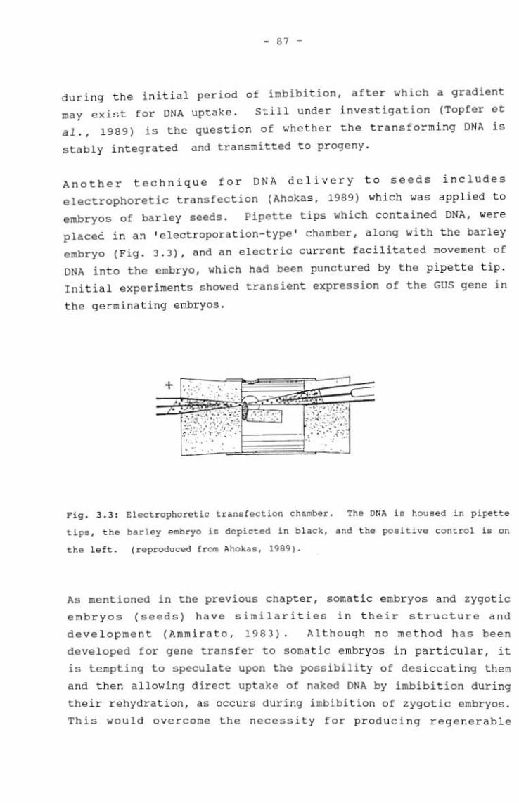
- 87 -
during the initial period of imbibition, after which a gradient
may exist for DNA uptake. still under investigation (Topfer et
al., 1989) is the question of whether the transforming DNA is
stably integrated and transmitted to progeny.
Another technique for DNA delivery to seeds includes
electrophoretic transfection (Ahokas, 1989) which was applied to
embryos of barley seeds. Pipette tips which contained DNA, were
placed in an 'electroporation-type' chamber, along with the barley
embryo (Fig. 3.3), and an electric current facilitated movement of
DNA into the embryo, which had been punctured by the pipette tip.
Initial experiments showed transient expression of the GUS gene in
the germinating embryos.
Fig. 3.3: Electrophoretic transfection chamber. The DNA is housed in pipette
tips, the barley embryo is depicted in black, and the positive control is on
the left. (reproduced from Ahokas, 1989).
As mentioned in the previous chapter, somatic embryos and zygotic
embryos (seeds) have similarities in their structure and
development (Ammirato, 1983). Although no method has been
developed for gene transfer to somatic embryos in particular, it
is tempting to speculate upon the possibility of desiccating them
and then allowing direct uptake of naked DNA by imbibition during
their rehydration, as occurs during imbibition of zygotic embryos.
This would overcome the necessity for producing regenerable
- 88 -
protoplasts, and would be particularly advantageous in the
graminaceous monocotyledons, many of which regenerate by somatic
embryogenesis (Vasil, 1987; Bhaskaran and Smith, 1990).
3.2.4 VECTORS
An important step in any DNA recombination procedure is the
selection of a suitable plasmid vector (carrier) into which one or
more DNA sequences can be inserted (Maniatis et al., 1982). The
vector should contain one or more restriction sites for cloning,
~and be capable of autonomous replication in a defined host or;
vehicle organism so that the sequence is reproduced· (Oliver and
Ward, 1985). There are different types of vectors, for example
cloning and expression vectors, and their use is dictated by the
nature and size of the gene to be transferred, the host to which
it is transferred, and the type of marker genes that are required.
Cloning vectors contain certain DNA sequences which are inserted
into complimentary unique (that is there are no
duplicate/triplicate sites) restriction sites in the vectors in
vitro (Maniatis et al., 1982). pBR322 is the most versatile
plasmid-cloning vector (derived from E. coli), and contains
ampicillin and tetracycline resistance and a number of convenient
restriction sites (Fig. 3.4). There are several restriction
enzymes which have a single target site within one or the other
resistance genes, so that insertion of foreign DNA at one site can
be detected by insertional acti~ation of that antibiotic
resistance, while recombinants can be selected using the other
antibiotic (Bolivaret al., 1977; Sutcliffe, 1979). The plasmid
is under relaxed control, that is the replication is not coupled
to host genome replication, so copy numbers of 10-200 plasmids per
cell can occur, and when host protein synthesis is halted by the
addition of chloramphenicol, copy numbers of several thousand
plasmids can result (Maniatis et al., 1982).
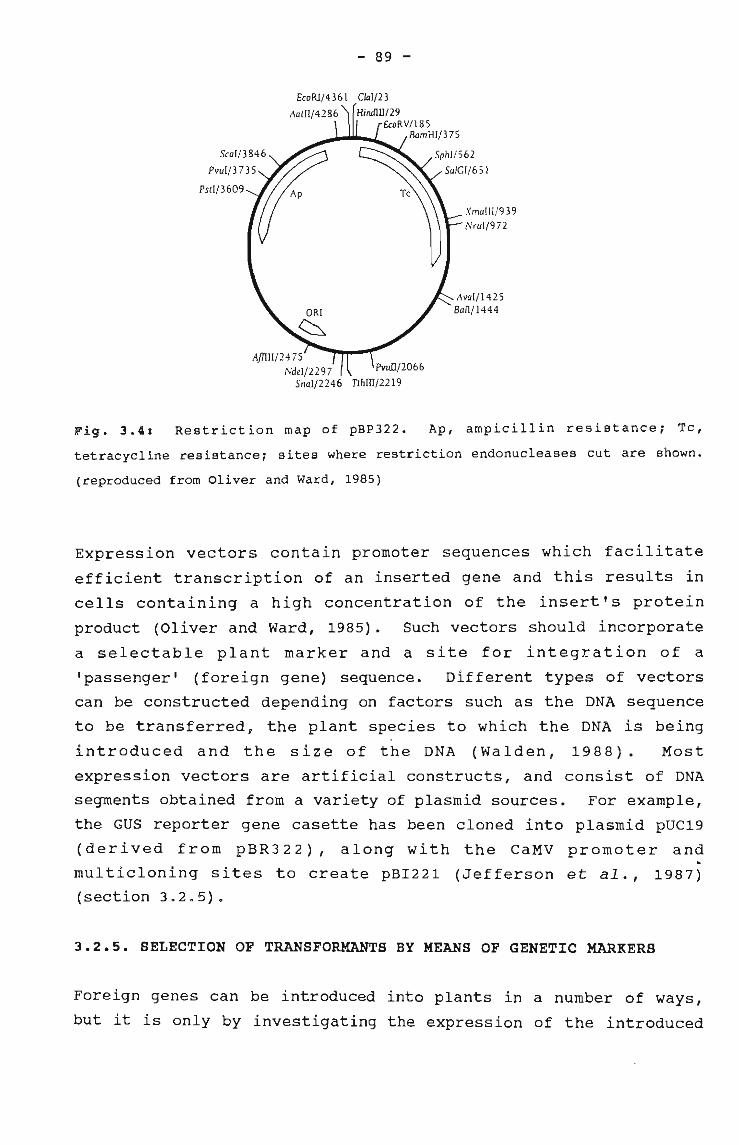
- 89 -
Clal/23
Hindlll/29£CoRV1185
BamHI/375
XmaIII/939NruI/972
Fig. 3.4: Restriction map of pBP322. Ap, ampicillin resistance; Tc,
tetracycline resistance; sites where restriction endonucleases cut are shown.
(reproduced from Oliver and Ward, 1985)
Expression vectors contain promoter sequences which facilitate
efficient transcription of an inserted gene and this results in
cells containing a high concentration of the insert's protein
product (Oliver and Ward, 1985). Such vectors should incorporate
a selectable plant marker and a site for integration of a
'passenger' (foreign gene) sequence. Different types of vectors
can be constructed depending on factors such as the DNA sequence
to be transferred, the plant species to which the DNA is being
introduced and the size of the DNA (Walden, 1988). Most
expression vectors are artificial constructs, and consist of DNA
segments obtained from a variety of plasmid sources. For example,
the GUS reporter gene casette has been cloned into plasmid pUC19
(derived from pBR322), along with the CaMV promoter and
multicloning sites to create pBI221 (Jefferson et al., 1987)
(section 3.2.5).
3.2.5. SELECTION OF TRANSFORMANTS BY MEANS OF GENETIC MARKERS
Foreign genes can be introduced into plants in a number of ways,
but it is only by investigating the expression of the introduced
- 90 -
DNA that the success of the transformation and the efficiency of
particular promoter constructs can be jUdged. To avoid the
lengthy process of selection and bulking up of the tissue for
analysis of integrated constructs, it is possible to study
transient expression of reporter/screenable genes by immediate
analysis (reviewed by Cocking and Davey, 1987; Walden, 1988;
weising et al., 1988).
Detection of transient gene expression
·If the methods described previously (section 3.2.2) deliver
foreign DNA to plant cells effectively, and a large proportion of
the cells take up several copies of the DNA sequence, then
transient expression of the DNA may be detectable. Transient
expression of the inserted gene occurs cytoplasmically,
chromosomal integration is unlikely to occur and is not a
prerequisite for expression to take place. Expression normally
peaks after 48 hours, after which time the DNA is degraded, or
limited integration of concatenated DNA might take place (Walden,
1988) .
Transient expression systems require a sensitive method for
detection of transformed cells/tissue (Hauptmann et al., 1987).
sensitive assays are available to monitor the expression of
reporter genes such as GUS (Jefferson et al., al., 1987), CAT
(Gorman et al., 1982) and luciferase (De Wet et al., 1987; Fromm
et al., 1990). In the above examp~es, the gene product is an
enzyme, the activity of which can be assayed either in tissue
extracts or immunologically.
The main features of the CAT and luciferase systems
The CAT gene, derived from the transposon Tn9, encodes the
chloramphenicol acetyltransferase enzyme which catalyses the
acetylation of chloramphenicol (Gorman et al., 1982). [14 C]_
chloramphenicol and acetyl-CoA are supplied as substrates in the
assay, and the radioactive acetylated products are separated by
- 91 -
thin-layer chromatography and detected by autoradiography.
Transient expression of the CAT gene was first studied in cultured
mammalian cells to study promoter function (Gorman et al., 1982)
and has subsequently been observed in protoplasts of maize (Fromm
et al., 1985), sugarbeet, tobacco (Joersbo and Brunstedt, 1990)
and in onion epidermal tissue (Klein et al., 1987).
Luciferases are enzymes which confer upon the host cell the
ability to bioluminesce, and two types of genes encoding these
enzymes have been cloned in mammalian cells (De Wet et al., 1987),
\ namely bacterial and firefly luciferase. Light emission can beidetected by photographic film or by a photometer connected to a
recording apparatus. Transient gene expression of firefly
luciferase has been monitored in maize (Fromm et al., 1990) and
bacterial luciferase in tobacco and carrot (Koncz et al., 1987).
The GU8 system
The B-glucuronidase (GU8) system .can be used for the purposes of
studying plant promoters (Werr and Lorz, 1986), the activity of
chimeric gene constructs (Jefferson, 1987; Jefferson et al., 1987;
Dekeyser et al., 1990), evaluating the efficiency of the technique
used to introduce foreign DNA (Abdul-Baki et al., 1990; Creisson
et al., 1990; Larkin et al., 1990) and, to some extent to
establish in which cells or tissues the gene is being expressed
(Irvine and Almeida, 1991; Meijer et al., 1991; Reggiardo et al.,
1991) .
The E. coli B-glucuroni~ase gene bas been cloned ·and sequenced,
and encodes a stable enzyme (Jefferson, 1987; Jefferson et al.,
1987): B-Glucuronidase is a hydrolase, calalysing the cleavage of
a wide variety of B-glucuronides of which many are available
commercially as spectrophotometric, fluorimetric and histochemical
substrates (Lindsey and Jones, 1990). A general purpose vector
for constructing gene fusions has been made (Jefferson et al.,
1987), and a derivative of this containing the B-glucuronidase
gene, the CaMV 358 or the nopaline synthase promoter, terminator
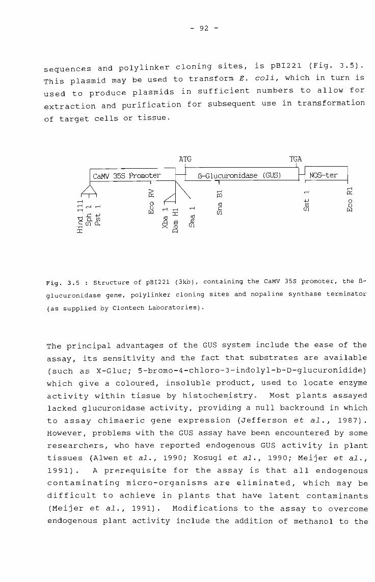
- 92 -
sequences and polylinker cloning sites, is pBI221 (Fig. 3.5).
This plasmid may be used to transform E. coli, which in turn is
used to produce plasmids in sufficient numbers to allow for
extraction and purification for sUbsequent use in transformation
of target cells or tissue.
ATG TGA
(GUS) NOS-ter
~~
IIIrl 0::
rl
~4J 0
rlrlrl rlrn 0
rl ~ rl en w'E'.c: 4J
~ito
eno.rn
~'M en ~::r::
Fig. 3.5 : Structure of pBI221 (3kb), containing the CaMV 35S promoter, the B
glucuronidase gene, polylinker cloning sites and nopaline synthase terminator
(as supplied by Clontech Laboratories).
The principal advantages of the GUS system include the ease of the
assay, its sensitivity and the fact that substrates are available
(such as X-Gluc; 5-bromo-4-chloro-3-indolyl-b-D-glucuronidide)
which give a coloured, insoluble product, used to locate enzyme
activity within tissue by histochemistry. Most plants assayed
lacked glucuronidase activity, providing a null backround in which
to assay chimaeric gene expression (Jefferson et al., 1987).
However, problems with the GUS assay have been encountered by some
researchers, who have reported endogenous GUS activity in plant
tissues (Alwen et al., 1990; Kosugi et al., 1990; Meijer et al.,
1991). A prerequisite for the assay is that all endogenous
contaminating micro-organisms are eliminated, which may be
difficult to achieve in plants that have latent contaminants
(Meijer et al., 1991). Modifications to the assay to overcome
endogenous plant activity include the addition of methanol to the
- 93 -
incubation buffer (Kosugi et al., 1990) and pH alterations (Alwen
et al., 1990).
Despite the abovementioned problems, the GUS assay has been used
to test transient expression in a variety of tissues and cells,
such as protoplasts (Werr and Lorz, 1986; Larkin et al., 1990;
Meijer et al., 1991), pollen grains (Twell et al., 1989),
suspension culture cells (Abdul-Baki et al., 1990), callus (Fromm
et al., 1990) and intact tissues (Jefferson et al., 1987; Wang et
al., 1988; Ahokas, 1989; Creisson et al., 1990; Irvine and
Almeida, 1991; Reggiardo et al., 1991).
Detection of stable integration
The development of dominant selectable markers functional in plant
tissue, allows the direct selection of transgenic cells by their
ability to grow and proliferate under selective conditions, which
kill (or prevent the division of) all untransformed cells (Lindsey
and Jones, 1990). This ensures the recovery of transgenic
material which might only be produced at very low frequencies.
The most widely-adopted, stable-integration marker is the neomycin
phosphotransferase (NPTII) gene (Beck et al., 1982), which is
discussed below.
The NPTII system
Incorporated into vectors with a range of promoter and" termination
sequences, the NPTII gene has been used with great success for
stable transformation of a number of monocotyledons an~
dicotyledons (reviewed by Weissing et al., 1988; Davey et al.,
1989). It is possible to introduce expression vectors which carry
both NPTII and GUS encoding genes, for example pBI121 (Jefferson
et al., 1987). Using this type of vector, it is possible to
rapidly screen all plants which come through the primary selection
system, before determining stable transformants by the more time
consuming Southern blots (Tomes et al., 1990).
- 94 -
The NPTII gene from the bacterial transposon Tn5 encodes a small
enzyme which confers resistance to a group of aminoglycoside
antibiotics which include neomycin, kanamycin and G418, by
catalysing their inactivation by phosphorylation (Beck et al.,
1982) . The reaction involves the transfer of the 8-phosphate
group of ATP to the antibiotic molecule, and this addition
detoxifies the antibiotic by preventing its interaction with the
target site of the ribosome (Beck et al., 1982). Kanamycin is
the antibiotic most commonly used, and transformation to
resistance can be determined by the phenotypic trait, by Western
,blotting of the NPTII enzyme or by assaying for NPTII activity in
tissue extracts (Reiss et al., 1984).
Although some plants, particularly the graminaceous
monocotyledons, exhibit natural resistance to kanamycin (even at
concentrations of 800mg/l) (Baribaultet al., 1990), gene
expression in protoplasts transformed by NPTII and selected on
kanamycin-containing medium has been studied for rice (Toriyama et
al., 1988; Yang et al., 1988; Zhang et al., 1988), maize (Rhodes
et al., 1988; Huang and Dennis, 1989), ryegrass (Potrykus et al.,
1985), sugarcane (Chen et al., 1987) and barley (Lorz et al.,
1985). In rice, Yang et al. (1988) reported regenerated plants
which contained the NPTII gene, but only 30% expressed NPTII
activity, of which 50% were fertile and produced seed. Other
tissues to which NPTII has been successfully delivered, and which
expressed resistance to kanamycin, are callus, derived from
transformed suspension culture cel~s of wheat, (Vasil et al.,
1991), soybean seed meristems (McCabe et al., 1988), tobacco
leaves (Tomes et al., 1990) and wheat pollen grains (Hess et al.,1990) .
Other antibiotic agents available for plant selection are
hygromycin (resistance is encoded by a hygromycin
phosphotransferase gene; van den Elzen et al., 1985; Datta et al.,
1990; Hayashimoto et al., 1990) and bialaphos which is produced by
the bacterium streptomyces hygroscopicus and has been found to be
an effective herbicide (De Block et al., 1987). The BAR gene
- 95 -
which encodes phosphinothricin acetyltransferase and enables the
bacteria to resist the effects of their own bialaphos, has been
transferred to potato, tomato (De Block et al., 1987), and maize
plants (Gordon-Kamm et al., 1990), resulting in the expression of
resistance to high levels of the antibiotic.
Herbicides such as the sUlphonylureas (Guerineau et al., 1990),
imidazoles, and glyphosate are potentially useful as selective
agents for transformation studies because they act by inhibiting
the activity of a single essential enzyme, and are discussed
below.
3.2.6. APPLICATION OF GENETIC ENGINEERING TO PLANT IMPROVEMENT
As the techniques of gene transfer have developed, attention has
been focused on their potential application to crop improvement,
with the aim of engineering specific traits into a wide variety of
plants (reviewed by Walden, 1988, Weissing et al., 1988; Lindsey
and Jones, 1989). Unfortunately gene technology has advanced
faster than our understanding of plant biochemistry, but it is
hoped that in due course these techniques will provide a better
comprehension of basic mechanisms involved in plant-gene
regulation.
crop improvement by genetic engineering
Weed control
Resistance to herbicides was among the first traits to whic~
genetic engineering techniques were applied, possibly because it
was shown to be a dominant trait exhibiting a simple Mendelian
inheritance pattern in a single nuclear genome (reviewed by Mazur
and Falco, 1989). The potential utility of herbicide resistant
genes as selectable genetic markers for research in plants, in a
manner analagous to that for antibiotic resistance genes, has
provided an incentive for research (Weising et al., 1988).
- 96 -
Engineering herbicide resistance/tolerance into plants presents a
new alternative for conferring selectivity and enhancing crop
safety of herbicides. Research has concentrated on those
herbicides with properties such as high unit activity, low
mammalian toxicity, low soil mobility, rapid biodegradation and a
broad spectrum of activity against various weeds (Gasser and
Fraley, 1989). The development of crop plants that are resistant
to such herbicides would provide more effective, less costly and
more environmentally-attractive weed control (reviewed by Walden,
1988; Lindsey and Jones, 1989). In order to engineer resistance
to herbicides into a plant, the molecular basis of its mode of
action needs to be well understood. Where the herbicide is known
to act on a single biochemical step, several stategies can be
adopted in attempting to engineer resistance:
1) selection for resistance in tissue culture, which involves
culturing the cells in the presence of the herbicide in order to
select resistant cells (reviewed by Lindsey and Jones, 1989;
Collin and Dix, 1990),
2) altering the level and sensitivity of the target enzyme for the
herbicide (Comai et al., 1985; Shah et al., 1986; Hinchee et al.,
1988) and
3) incorporating a gene that will detoxify the herbicide (De Block
et al., 1987; Stalker et al., 1988).
The procedure mentioned in the first approach involves either a
direct or stepwise selection :using callus, suspension culture
cells or protoplasts produced in -vit~o (Lindsey and Jones; 1989).
Most of the selections for resistance to herbicides involve the
direct approach, where callus is exposed to one concentration of
the herbicide and the surviving cells are isolated (Collin and
Dix, 1990). The alternative is to use the stepwise approach where
the surviving callus or suspension culture cells are sequentially
transferred to increased levels of the herbicide. Herbicide
resistant mutants could be induced by exposure to physical (gamma
radiation) or chemical (colchicine) agents (Collin and Dix, 1990).
As an example of the second approach, glyphosate (the active
.--
- 97 -
ingredient of the Roundup herbicide) acts by specifically
inhibiting the enzyme 5-enolpyruvylshikimate-3-phosphate synthase
(EPSPS) (Shah et al., 1986). Glyphosate is active against annual
and perennial broadleaf and grassy weeds, has a low animal
toxicity, and is rapidly inactivated and degraded in all soils
(Mousdale and coggins, 1984). Resistance to glyphosate has been
engineered in various crops by introducing genetic constructs for
the overproduction of EPSPS (Shah et al., 1986) or for the
glyphosate-tolerant variant EPSPS enzymes (Comai et al.,1985;
Hinchee et al., 1988).
The third approach is aimed at modifying the herbicide within the
cell before it can act on the target. This has been achieved by
screening plants that are resistant to the herbicide or micro
organisms that are found in herbicide-contaminated soils, and
which degrade the active ingredient of the herbicide (Stalker et
al., 1988). If the active ingredient of the herbicide is
synthesised by a micro-organism it is likely that the micro
organism will produce a detoxifying enzyme to protect itself (De
Block et al., 1987). Resistance to gluphosinate (De Block et al.,
1987) and bromoxynil (Stalker et al., 1988) has been achieved by
genes coding for enzymes, which inactivate the herbicides by
acetylation or nitryl hydrolysis, respectively.
Insect resistance
The production of insect resistant plants is another application
of genetic engineering with potential crop improvement envisaged
by both the seed and -agricultural companies. As well as causing
crop damage, insects _a~so act as vectors for viral diseases. Two
approaches have been used to engineer resistance to insects in
plants. One capitalises on the discovery that the bacterium
Bacillus thuringiensis (Bt) synthesises a polypeptide lethal to
lepidopterous insects (Fischhoff et al., 1987; Vaeck et al., 1987;
Murray et al., 1991; Vail, 1991), and the other approach utilises
natural defence mechanisms of plants (Hilder et al., 1987).
- 98 -
The Bt endotoxin resides in a parasporal inclusion body which
forms during sporulation of the bacterium, and different strains
of the bacterium synthesise toxins with different specificities
(Dulmage, 1970). Insecticidal proteins ingested by the target
insect are solubilised and cleaved by proteases active in the gut,
to produce the active toxin. Truncated Bt toxin genes have been
introduced into tomato (Vaeck et al., 1987), carrot (Murrayet
al., 1991), walnut (Vail, 1991), cotton and tobacco (Fischhoff et
al., 1987), resulting in transgenic plants exhibiting tolerance to
insect larvae in laboratory tests.
Using a slightly different approach, the Bt toxin gene was
transferred to epiphytic bacteria which form associations with the
roots/leaves of crop plants, thereby ensuring continuous toxin
production. The advantage of this is the ease with which bacteria
can be transformed, compared to plants. In South Africa, the
toxin gene isolated from a Bt causing mortality in a popUlation of
stalk-borer pests of sugarcane, was inserted into Pseudomonas
fluorescens, an epiphyic coloniser (Herrera and Thomson, 1989;
Herrera et al., 1991). Laboratory assays indicate successful
transfer and expression of the toxin gene, but this has yet to be
tested in the field.
A further approach is to enhance the expression of plant genes
which are thought to be a naturally-evolved defence response to
insect attack, and transfer -those genes to plants where they are
not normally found; for example proteinase inhibitors, which have
a broader spectrum of activity than the B. thuringiensis toxin
(Hilder et al., 1987). The cowpea trypsin inhibitor has been
cloned and inserted into tobacco, where it was found to enhance
plant resistance to predatory insects (Hilder et al., 1987).
Disease resistance
Three strategies have been used to engineer plants so that
symptoms of a viral infection are reduced. One is an adaptation
of viral cross protection (Abel et al., 1986; Van Dun et al.,
- 99 -
1987; Cuozzo et al., 1988; Mayo and Barker, 1990); the second
utilises symptom modification by viral satellite RNA (Harrison et
al., 1987); and the third makes use of anti-sense RNA (reviewed by
Lindsey and Jones, 1989).
Cross protection is a phenomenon whereby infection of a plant with
one viral strain protects it against superinfection by another,
related strain (reviewed by Walden, 1988; Lindsey and Jones,
1989). with the advent of new technologies, it has become
possible to transfer specific viral genes to plants in an attempt
to confer resistance in a similar way to that of cross protection
(Cuozzo et al., 1988). Significant resistance to tobacco mosaic
virus (TMV) infection has been achieved by expression of the coat
protein gene of TMV in transgenic plants (Abel et al., 1986),
where progeny of transgenic plants either developed delayed viral
symptoms upon TMV infection or failed to develop symptoms at all.
This approach of coat protein protection produced similar results
in transgenic tomato, tobacco and potato plants against a broad
spectrum of plant viruses, including alfalfa mosaic virus,
cucumber mosaic virus, potato virus X and potato virus Y (Cuozzo
et al., 1988; Mayo and Barker, 1990; van Dun et al., 1987).
The second approach involving the suppression of viral infection
has been described by Harrison et al. (1987), and makes use of the
observation that in some plant RNA viruses, certain RNA sequences
may ameliorate disease symptoms in infected plants. These
sequences, termed satellite RNA, bear no homology to the genomic
RNA, and are not required for virus replication and spread.
Transformed tobacco plants supplied with a cDNA copy of satellite
RNA of cucumber mosaic virus (CMV) developed less severe symptoms
than non-transformed plants. It was found that transformed, but
non-infected plants, produced only low levels of satellite RNA,
but on infection with satellite-free CMV, the synthesis of large
amounts of the RNA was induced. The accumulation of satellite RNA
was correlated with a reduction in symptom formation.
The third approach to inhibit viral infection involves the

- 100 -
possibility of using anti-sense RNA (minus-strand RNA which binds
to plus-strand or mRNA to prevent its translation)., which can
potentially inhibit the virus replication, packaging and systemic
spread in transgenic plants. There is no conclusive evidence yet
to suggest that anti-sense RNA can reduce viral symptoms (Lindsey
and Jones, 1989).
Application of genetic engineering to basic research
The limitations of genetic engineering systems include the
polygenic nature of traits (not knowing the number of genes
involved and their precise function), the potential genetic
instability of in vitro cultured cells and plants, the lack of
information on plant metabolism and its regulation (particularly
in stress responses), and more specific problems such as random
DNA integration and how it influences the expression of other
genes (Lindsey and Jones, 1989).
An important contribution of plant-genetic transformation is that
it provides a basis for new experimental strategies in plant
molecular biology research. These include analysis of viral gene
functions by integrating viral DNA (via T-DNA borders) into the
plant genome (Grimsley et al., 1986, 1987), inactivating specific
genes in the plant genome by the introduction of genes coding for
antisense RNA (which interferes by binding to specific mRNA) , and
the isolation and study of plant genes and promoters (Werr and
Lorz, 1986). Applications also lie in the development of
strategies such as cDNA cloning, in vitro translation and gene
library screening (Walden,' 1988). Genes responsible for a
specific trait may be isolated by the use of ±ransposable
elements, which can be used~as probes for the isolation of
specific genes (transposable elements can modify the regulation of
specific genes by inserting into sequences 5' to the coding
region) (Haring et al., 1991).
- 101 -
3.3. MATERIALS AND METHODS
Bacterial strains and plasmids used
E. coli HB101 is ampicillin-sensitive (Bolivar and Backman, 1979),
and is used routinely for the purpose of plasmid amplification.
pBR322 (Bolivar et al., 1977; Sutcliffe, 1979) is a plasmid used
as a multipurpose cloning vector, and in this study was used for
radiolabelling purposes.
pBI221 (Clontech, USA) is a plasmid expression vector coding for
the B-glucuronidase enzyme.
Growth and maintenance of bacterial strains
E. coli HB101 was grown on Luria agar (10g/1 bacto-tryptone, 5g/1
bacto yeast extract, 10g/1 NaCl, pH 7,5). Long-term storage of
the bacterium was in Luria agar in stab-inoculated bijou bottles,
stored at room temperature. Overnight bacterial cultures were
grown in Luria broth (5ml), inoculated with a single bacterial
colony (Armitage et al., 1988). The overnight cultures were
shaken for 12-16h at 37°C on an orbital shaker.
Transformation of E. coli
The expression vector plasmid ~BI221 (Clontech, USA), was
introduced into E. coli HB101 (ampicillin-sensitive) by the
transformation technique of Hanaha~ (1985). Transformed cells
were selected on Luria agar containing ampicillin (100mg/l).
Generation of expression vector
A technique for large scale plasmid preparation (Armitage et al.,
1988) was used to extract pBI221 from transformed E. coli. The
concentration of purified DNA (determined spectrophotometrically
from the absorption reading at 260nm) was 380~g/ml, and the ratio
of 260nm to 280nm was 1.8.
- 102 -
Agarose gel electrophoresis of DNA
Plasmids and restriction digests of plasmids were analysed on
agarose mini-gels (1% DNA-grade agarosei BioRad, USA), run in TBE
buffer (45mM Tris-borate, 1mM EDTA, pH 8) and stained with
ethidium bromide (Sigma, USAi 0,5Mgjml). The voltage applied
across the horizontal gel apparatus (Hoeffer-HE 33, 7x10cm in
size) was 90-100V.
Loading buffer (50% (wjv) sucrose, 4M urea, 0,1% (wjv) bromophenol
blue, 50mM EDTA, pH 8) was added to the DNA (in a 1:2 loading
buffer:DNA ratio). The DNA plus loading buffer was pretreated at
60°C for 10min after the restriction enzyme digestion to prevent
the reannealment of linearised 'sticky' fragments.
Restriction enzyme digestions
Restriction enzyme digestion was performed on Lambda DNA or
plasmid DNA with the endonucleases ECoRI, HindIII, BamHI (used
singly or in combination) (Boehringer Mannheim, Germany). Each
reaction mixture contained DNA (0,2-1Mg), 1U of appropriate
restriction enzyme, digestion buffer (Boeringer Mannheim,
Germany), made up to a final volume of 20Ml (Maniatis et al.,
1982) .
Labelling of plasmid DNA
Labelling of plasmid pBR322 was carried out by nick translation ifi
the presence of [methyl, -1 1 2_ , _ 3HJthymidine 5'-triphosphate
([3HJTTP) according to Rigby~et al. (1977). A reaction mixture
(40MI in total), containing 40MCi [3HJTTP (Amersham, England),
unlabelled nucleotides (0, 4J..Lg) , DNase 1 (121pg) (Sigma, USA), DNA
polymerase 1 (16U) (Boehringer Mannheim, Germany) and plasmid DNA
(1,25J..Lg) in Tris-HCI (50mM), B-Mercaptoethanol (lmM) and MgCl 2(10mM) at pH 7,6, was incubated at 15°C for 80 min, after which
the DNA sample was applied to a Sephadex G50 spun-column.
- 103 -
spun-column purification of labelled DNA
Spun-columns were prepared according to Maniatis et al. (1982).
Syringes (lml) were plugged at the base with glass wool and packed
with Sephadex G50 (Pharmacia, England) which had been hydrated
overnight in STE buffer (O,lM NaCI, 10mM Tris-HCI, 1rnM EDTA, pH
8) . The columns were suspended in glass test-tubes over open
Eppendorf tubes (1,5ml). Eluted DNA was collected by
centrifugation (3000rev/min for 5min) after equilibration of the
columns with STE under the same conditions. The product was
stored at -20°C.
Measurement of specific activity of labelled DNA
Labelled DNA samples were precipitated with trichloroacetic acid
(TCA) (Maniatis et al., 1982) in the presence of calf-thymus
carrier DNA (30~g carrier DNA/~l labelled DNA) to determine
specific activity of high molecular weight fragments. Samples
were counted in a Beckman LS 7500 Liquid scintillation Counter in
Beckman 'Ready Value' scintillation cocktail (5ml). Specific
activities of 0,9-2,7 x 10⁶ cpm/~g DNA were obtained.
sterilisation of seeds
Seeds were surface-sterilised as described previously (section
1.3.1). The testa of the seed was scraped prior to surface
sterilisation, using a sharp scalpe~ blade, care being taken to
avoid damaging the embryo.~icrowave treatments were carried out
on seeds placed on filter paper for 5-60s in an Aim Pentamicrowave, set on high.
Germination of seeds
Seeds were placed aseptically on germination medium (section2.3.1.).
- 104 -
Protoplast isolation
Protoplasts were isolated from friable achlorophyllous calli, as
described in section 2.3.7.
Uptake of labelled DNA by seeds
Seeds were imbibed for various time intervals in solutions of
radiolabelled pBR322 plasmid DNA (30 seeds per 25~1; O,0138~g
DNA/~l). After imbibition, seeds were placed in wire mesh (pore
size 100~m) baskets suspended above a glass beaker, and were
washed extensively under running tap water for 15min. Seeds were
crushed in cold TCA (100Ml of a 10% solution) in Eppendorf tubes
using a tapered glass rod. The seed homogenate was filtered onto
a glass fibre filter (Millipore, USA filter unit), and washed with
cold TCA (5x1ml aliqouts) and ethanol (3x1ml of a 95% solution)
prior to scintillation counting.
Uptake of expression vector by seeds
Seeds were incubated in a solution of pBI221 DNA (6-10 seeds per
25Ml; 126M9 DNA/ml) for 24h in Eppendorf tubes placed in a
sterile, moist container. After imbibition, seeds were
transferred to the germination medium and kept in the dark for
48h, before the GUS assay was carried out.
PEG-mediated DNA uptake by protoplasts
Protoplasts (3x10 6 /ml) w~re w~shed and suspended in 2ml·of MS
culture medium (1,5mg/l 2,4-D,· 30g/1 sucrose, 0,5M mannitol)
containing PEG (10-30% (w/v); MW 6000; Merck, Germany) and pBI221
(20M9/10 5 protoplasts) for 30min. Thereafter, they were washed
and suspended in the above MS culture medium for 48h. In some
cases, the broad-spectrum antibiotics penicillin and polymixin B
(Sigma, USA; 50Mg/ml) were incorporated into the culture medium.
Protoplasts were fixed (0,3% formaldehyde, O,5M mannitol, 9mM MES
at pH 5,8) and washed with sodium phosphate buffer (0,2 M) prior
- 105 -
to carrying out the GUS assay.
Assay for transient expression of GUS
Transient expression of B-glucuronidase (encoded on pBI221) was
tested by means of a histochemical assay (Jefferson et al., 1987).
Seeds or protoplasts were immersed in the GUS substrate (2mM 5
bromo-4-chloro-3-indolyl-B-D-glucuronic acid (X-Gluc, Sigma),
K3 [Fe(CN)6J (O,IM), K4Fe(CN)6 (O,IM), Na2EDTA (O,IM), mannitol
(1,23mM) and sodium phosphate buffer (0,2Mi pH7) at 37°C
overnight. In some cases methanol (20%) was added to the
incubation buffer (Kosugi et al., 1990). Presence of the enzyme
was visualised by the appearance of a blue stain in the plant
material.
Photography
Seeds and protoplasts were photographed as described in section
2.2.9. The agarose gel was photographed under u.v.
transillumination using black and white T Max 400 ASA Kodak film
and a 1xR red filter.
- 106 -
3.4. RESULTS
3.4.1.UPTAKE OF LABELLED DNA BY SEEDS
In order to establish whether or not DNA could be taken up by
sugarcane seeds during imbibition, surface sterilised, dehusked,
dry seeds were incubated in [3 H] pBR322 plasmid DNA for various
time intervals and the radioactivity measured. Seeds took up DNA
steadily over a 120min time period (Fig. 3.6A), after which uptake
: levels decreased (Fig. 3.6B). The sharp decrease in levels of
radioactive DNA in the latter seed-batch could indicate that DNA
is degraded to some extent and lost by leakage from disaggregated
membranes which exist during the process of imbibition.
A comparison was made of DNA uptake by seeds with the testa/seed
coat intact and those where the testa had been removed by scraping
with a sharp scalpel blade. Fig. 3.7 shows a typical result. It
was observed that seeds which had their testa removed, imbibed DNA
at a faster rate initially than seeds with the testa intact, but
after a 120min time period, the uptake levels were similar.-
3.4.2 UPTAKE OF EXPRESSION VECTOR pBI221 BY SEEDS
Having established that DNA could be taken up by seeds, the
expression of introduced DNA was investigated. The GUS assay
system was chosen to investigate expression by seeds because the
assay is simple to perform and has been used to detect transient
expression in a number of plants (section 3.2.5; Jefferson et al.,
1987). Most plants assayed exhibit no endogenous GUS activity
(Jefferson, 1987). The DNA that was introduced into seeds to
monitor expression was that of the plasmid expression vector
pBI221, which encodes the bacterial enzyme a-glucuronidase (Fig.3 • 5) •
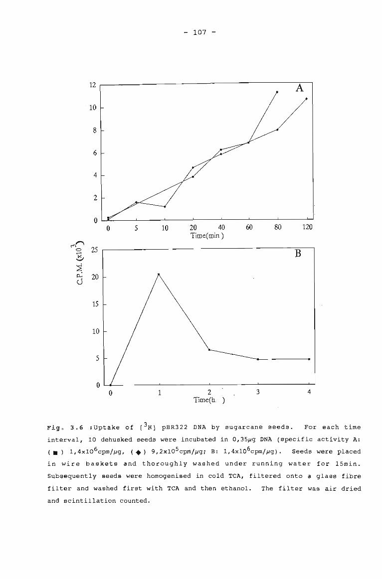
- 107 -
12
·10
8
6
4
2
00 5 10 20 40 60 80 120
Time(min)~~
250 B~
~
~~ 20cJ
15
5
10
OL.6.-----L-------'--------L------........o 1 2
Time(h )3 4
Figo 3.6 ~Uptake of [3 H] pBR322 DNA by sugarcane seeds. For each time
interval, 10 dehusked seeds were incubated in O,3S~g DNA (specific activity A:
( .) 1,4xl06cpm//-Jg, (.) 9,2x10Scpm//-Jgj B: 1,4x10⁶cpm/~g). Seeds were placed
in wire baskets and thoroughly washed under running water for lSmin.
Subsequently seeds were homogenised in cold TCA, filtered onto a glass fibre
filter and washed first with TCA and then ethanol. The filter was air dried
and scintillation counted.
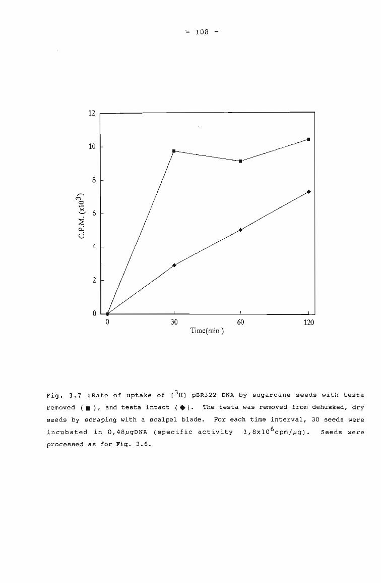
... 108 -
12
10
8
,.-...M
0~
~6'-"
~~
c..54
2
oo 30
Time(min)60 120
Fig. 3.7 :Rate of uptake of [3H] pBR322 DNA, by sugarcane seeds with testa
removed (.), and testa intact (.). The testa was removed from dehusked, dry
seeds by scraping with a scalpel blade. For each time interval, 30 seeds were
incubated in 0,48pgDNA (specific activity 1,8xl0 6 cpm/pg).
processed as for Fig. 3.6.
Seeds were
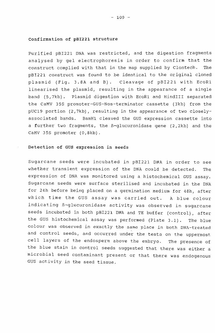
- 109 -
Confirmation of pBI221 structure
Purified pBI221 DNA was restricted, and the digestion fragments
analysed by gel electrophoresis in order to confirm that the
construct complied with that in the map supplied by Clontech. The
pBI221 construct was found to be identical to the original cloned
plasmid (Fig. 3.8A and B). Cleavage of pBI221 with EcoRl
linearised the plasmid, resulting in the appearance of a single
band (5,7kb). Plasmid digestion with EcoRl and HindIII separated
the CaMV 35S promoter-GUS-Nos-terminator cassette (3kb) from the
pUC19 portion (2,7kb), resulting in the appearance of two closely
associated bands. BamHl cleaved the GUS expression cassette into
a further two fragments, the B-glucuronidase gene (2,2kb) and the
CaMV 35S promoter (0,8kb).
Detection of GUS expression in seeds
Sugarcane seeds were incubated in pBI221 DNA in order to see
whether transient expression of the DNA could be detected. The
expression of DNA was monitored using a histochemical GUS assay.
Sugarcane seeds were surface sterilised and incubated in the DNA
for 24h before being placed on a germination medium for 48h, after
which time the GUS assay was carried out. A blue colour
indicating B-glucuronidase activity was observed in sugarcane
seeds incubated in both pBI221 DNA and TE buffer (control), after
the GUS histochemical assay was performed (Plate 3.1). The blue
colour was observed in exactly the same place in both DNA-treated
and control seeds, and occurred under the testa on the uppermost
cell layers of the endosperm above the embryo. The presence of
the blue stain in control seeds suggested that there was either a
microbial seed contaminant present or that there was endogenous
GUS activity in the seed tissue.
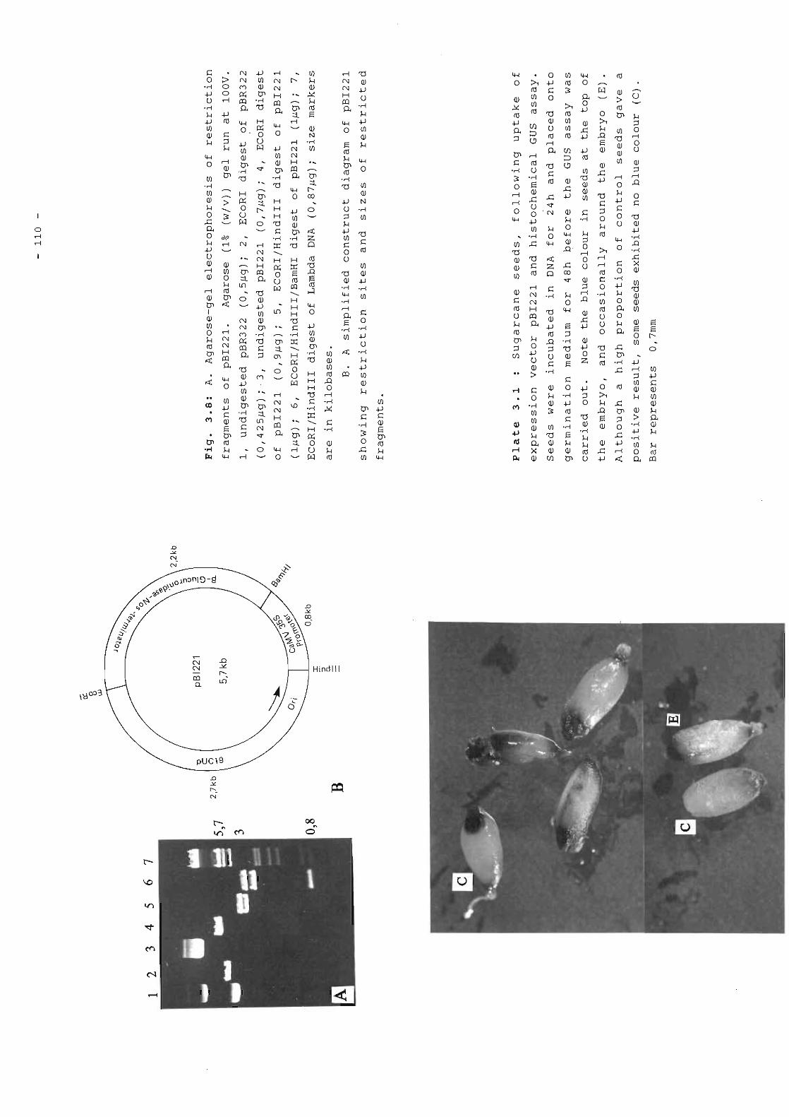
- 111 -
Treatment of seeds in order to overcome positive controls
In an attempt to investigate the two possibilities of microbial
seed contamination or endogenous GUS activity in the seed tissue
causing positive results in control seeds, a number of variations
to seed pretreatment and medium composition of the GUS assay were
carried out: the testa was removed in order to see whether this
served as a barrier to DNA uptake by the embryo of the seed, more
extensive sterilisation treatments were undertaken in the form of
microwaving, the addition of methanol to the GUS incubation buffer
was investigated, and lastly, a combination of all the above
treatments was carried out.
Testa as a possible barrier to DNA entry into embryo
Uptake and expression of pBI221 by seeds which had their testa
removed were compared to seeds which had the testa intact, because
it was necessary to establish whether or not the testa was a
barrier to DNA uptake by the embryo. In addition, previous
results with radiolabelled DNA (Figs. 3.6 and 3.7) indicated that
seeds with testa removed showed faster initial rates of DNA uptake
than seeds with the testa intact. The testa was removed from
seeds by scraping it with a scalpel blade, prior to surface
sterilisation and incubation in DNA. Seeds with the testa removed
resulted in 40% and 30% of seeds expressing B-glucuronidase in theI
DNA treated and control seeds, respectively. Seeds where the
testa was left intact resulted in 60% of seeds in both the control
"._~ and DNA-treated seeds expressing B-glucuronidase activity (summary
of treatments in Table 3.2). The blue colour was more visible in
seeds which had the testa removed, so this was carried out
routinely for subsequent assays.
Elimination of bacterial contamination
In order to try and eliminate microbial contamination from
sugarcane seeds, more extensive sterilisation treatments were
undertaken. A series of microwave treatments were carried out and
- 112 -
germination and bacterial contamination levels were recorded after
microwaving the seeds for various time intervals. There was 100%
germinability in seeds not microwaved, but a 50% loss in
germinability was recorded when seeds were microwaved for 60s
(Table 3.1). All of the microwave treatments reduced
contamination levels from 20%, observed in seeds not microwaved,
to 0%, except at 15s where 16% contamination was observed (Table
3.1). For further GUS assays, seeds were microwaved for 60s, as
this treatment maintained 50% germinability of seeds and seemed to
eradicate bacterial contamination.
Table 3.1 : Germination and bacterial contamination levels of sugarcane seeds
exposed to microwave treatment for various time intervals. After microwaving,
seeds were surface sterilised, squashed in a sterile petri-dish and placed onto
nutrient agar for 2 days in order to monitor bacterial contamination.
Germination levels were determined by placing whole seeds onto germination
medium for 5 days (section 2.3.1). (n=5-6)
Time (s) ~ germination ~ seeds contaminated0 0
0 100 205 100 0
10 80 015 100 1620 66 040 40 060 50 0
A GUS assay was carried out on seeds that had been microwaved for
60s, and although 90% of seeds. incubated in pBI221 were positive
for B-glucuronidase, 60%·of seeds incubated in TE buffer also
exhibited a positive result (Table 3.2). Seeds which had.the
testa removed and which were microwaved prior to carrying out the
GUS assay, exhibited blue colour in 60% of the seeds incubated in
pBI221 and 40% of the seeds incubated in TE buffer (Table 3.2).
Although the suspected bacterial contaminant seemed to have been
reduced by microwave treatments, positive results were still
- 113 -
observed in control seeds. Therefore, if the positive control
results are due to a contamination problem, it is likely that the
contaminant is endogenous and is symbiotic in sugarcane seeds.
Table 3.2 : Effect of seed pretreatment and assay medium on sugarcane seeds, as
determined by the GUS histochemical assay. Seed treatments included
microwaving for up to 60s prior to surface sterilisation, removal of testa by
scraping with a scalpel blade, the addition of 20% methanol to the GUS
incubation buffer and a combination of the above treatments in order to
, overcome positive results in the controls. (n=S-lO)
Seed treatment
Seeds demonstratingGUS expression (%)
DNA Control(pBI22l) (TE buffer)
Comments
microwaved for 60s
testa removed by scrapingwith scalpel blade vstesta intact
testa removed, microwavedfor 60s
testa removed, microwavedfor 60s, 20% methanol addedto GUS incubation buffer
90 60 positive controls
40 30 blue colour morevisible in
60 60 scraped seeds
60 40 positive controls
100 80 positive controls
testa removed, 20% methanoladded to GUS incubation buffer
100 100 positive controls
Suppression of endogenous plant GUS activity
Kosugi et al. (1990) found that the addition of methanol to the
GUS incubation buffer was effective at lowering endogenous plant
GUS activity in leaves, calli and suspension culture- cells of
rice, tobacco and bean. Therefore, in an attempt to investigate
endogenous GUS activity in seed tissue, 20% methanol was added to
the GUS assay incubation buffer. However, in sugarcane seeds, the
- 114 -
problem of a positive response in control seeds was not eliminated
by this treatment. All of the microwaved, DNA-treated seeds and
80% of microwaved control seeds, exhibited blue colouration (Table
3.2). Seeds incubated in DNA and TE buffer, and which were not
microwaved also exhibited blue colouration. This suggests that
sugarcane seeds contain an endogenous microbial contaminant as
endogenous plant GUS activity is likely to have been suppresed by
the addition of methanol to the incubation buffer.
3.4.3 UPTAKE OF pBI221 DNA BY PROTOPLASTS
There are numerous methods available for protoplast
transformation, and the options available for sugarcane include
electroporation, microinjection, liposome-mediated DNA delivery
and chemical-mediated DNA uptake (reviewed in section 3.2.2).
Direct uptake of naked DNA via the last-mentioned method provides
the most simple approach, without the need for specialised
equipment. Chemical-induced DNA uptake by PEG has been used by
numerous workers with considerable success for transformation of
protoplasts (reviewed in section 3.2.2). However, PEG at high
concentrations has a deleterious effect on protoplasts, and it is
necessary to determine the optimum concentration which allows DNA
uptake without excessive loss in protoplast viability. The only
drawback with this technique is that protoplasts have to be
regenerated to plants, and this is a problem in other graminaceous
monocotyledons, as well as sugarcane.
Viability studies on protoplasts treated with PEG
The viability of callus protoplasts after exposure to PEG was
monitored by the exclusion of Evan's blue dye as described in
section 2.3.7. Viability studies using PEG at a concentration
range of 10-30% were carried out as this is the range which has
had success in previous work on transformation of protoplasts
(Krens et al., 1982; Lorz et al., 1985; Werr and Lorz, 1986; Chen
et al., 1987). Protoplasts were exposed to PEG for 30min, before
the PEG was diluted with culture medium, following the time
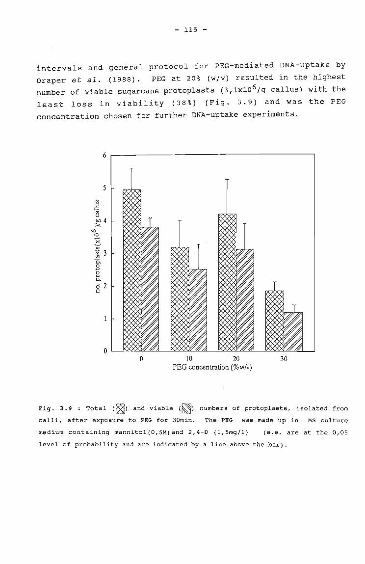
- 115 -
intervals and general protocol for PEG-mediated DNA-uptake by
Draper et al. (1988). PEG at 20% (w/v) resulted in the highest
number of viable sugarcane protoplasts (3,lX106 /g callus) with the
least loss in viabili ty (38 %) (F ig. 3.9) and was the PEG
concentration chosen for further DNA-uptake experiments.
o 10 . 20PEG concentration (%w/v)
30
Fig. 3.9 : Total (~) and viable (~) numbers of protoplasts, isolated from
calli, after exposure to PEG for 30min. The PEG was made up in MS culture
medium containing mannitol (O,SM) and 2,4-D (l,Smg/l) (s.e. are at the 0,05
level of probability and are indicated by a line above the bar).
- 116 -
GUS assay for transient expression in protoplasts
Once protoplast viability in different PEG concentrations had been
determined, transient expression of transforming pBI221 DNA in
protoplasts was investigated. Protoplasts were prepared as
discussed in section 2.3.7 and exposed to pBI221 DNA suspended in
20% PEG for 30min, after which time the protoplasts were incubated
in MS protoplast culture medium for 48h before the histochemical
GUS assay was carried out. Microscopic examination of the
protoplasts revealed that the suspensions were contaminated and no
blue protoplasts were observed.
Elimination of contamination in protoplasts
In an attempt to overcome the contamination problem, calli were
screened for endogenous microbial contamination prior to
protoplast isolation. A small piece of the callus to be used was
placed into nutrient broth, and turbidity was monitored. Once an
uncontaminated source of callus had been obtained, protoplasts
were isolated and incubated in a PEG solution containing DNA,
after which the GUS assay was carried out. Contamination problems
persisted and no blue protoplasts were observed.
In a subsequent step, the antibiotics penicillin and polymixin B
were incorporated into the protoplast culture medium after
protoplast isolation and DNA incubation. Contamination levels
were reduced but not entirely eliminated, and no blue protoplasts
were observed after carrying out the GUS assay.
Perhaps the frequency of transformation is too low to pick up
transformed cells, and it is possible that even low levels of
bacterial contamination could interfere with DNA uptake and
subsequent culture of the protoplasts.
- 117 -
3.5. DISCUSSION
The use of the seed in transformation studies
Using radiolabelled pBR322 DNA, it was demonstrated in this study
that sugarcane seeds were able to take up naked DNA during
imbibition. It was established that DNA was taken up during the
first 1-2h of imbibition (Fig. 3.6), and that uptake was more
rapid initially in those seeds which had the testa removed (Fig.
3.7). Other researchers have found benefit in doing a
preliminary investigation into DNA uptake using radiolabelled DNA.
Ledoux and Huart (1968, 1969) imbibed bar ley embryos in
radiolabelled DNA and observed that the DNA was taken up by the
seed endosperm and later by cells in the root of the germinating
seedling. In another study, radiolabelled DNA was
electrophoretically transferred to barley embryos and the results
detected by microscopic autoradiography (Ahokas, 1989), and the
initial set of results used to improve the uptake procedure.
Abdul-Baki et al. (1990) investigated uptake of radiolabelled DNA
by tobacco pollen grains and subsequently established protocols
for the transformation of pollen.
In the present study, the uptake of the GUS expression vector,
pBI221, and subsequent transient expression of the B-glucuronidase
gene encoded on the vector, could not be conclusively demonstrated
in sugarcane seeds. Blue colouration which is indicative of B
glucuronidase activity was _observed in control seeds incubated in
TE buffer which confounded the -interpretation of blue 'colour in
the seeds incubated in pBI221 DNA preparations.
Possible reasons for the blue colouration in the control seeds are
that there was a microbial contaminant in sugarcane seeds, as has
been postulated for causing positive results in early work carried
out on barley embryos (Kleinhofs et al., 1975; Kleinhofs and
Behki, 1977) or that there was endogenous plant GUS activity,
which has been found previously in rice (Kosugi et al., 1990;
Meijer et al., 1991) and tobacco (Alwen et al., 1990).
- 118 -
Attempts to eliminate the blue colour in control seeds caused by
either of the above two factors included removing testa,
microwaving seeds for 60s, the addition of methanol to GUS assay
incubation buffer and a combination of the above treatments.
Although the microwave treatments appeared to eradicate bacterial
contamination detectable by plating onto nutrient agar in most
cases (Table 3.1), this did not overcome the problem of positive
controls. Bearing in mind that the blue spot was localised (Plate
3.1), and found in both DNA-treated and control seeds (Table 3.2),
this brings to light the possibility that the contaminant is an
endogenous symbiont, which would be difficult to eradicate.
Leifert and waites (1990) have encountered latent bacteria in
plants from a number of species produced in vitro, so this seems
to support the fact that these types of contaminants are not
uncommon, as well as difficult to eradicate. Future verification
of this could include microscopic studies, but this is beyond the
scope of the present study.
Another possibility for blue colour persisting in the controls is
that there was intrinsic plant GUS activity present in sugarcane
seed tissue. Similar problems have been encountered in rice
(Kosugi et al., 1990; Meijer et al., 1991), and tobacco (Alwen et
al., 1990). Alwen et al. (1990) found that plant B-glucuronidase
has an optimal activity at pH5, whereas the bacterial B
glucuronidase is active at pH7, so under appropriate experimental
conditions, it might be possible for future GUS assays for
bacterial enzyme a~tivity to be carried out without the
interference of endogenous plant enzyme activity. Kosugi et al.
(1990) were able to overcome the endogeous plant GUS activity in
rice callus, suspension cells and shoots on partially regenerated
plants; tobacco and bean calli and leaf tissue; and maize
suspension culture cells, by the addition of methanol to the GUS
incubation buffer. This modification also enhanced the blue
colour in the histochemical assay. However, in sugarcane seeds,
the blue cOlouration in control seeds was not nullified by the
addition of methanol to the incubation buffer (Table 3.2).
- 119 -
Different tissue types could harbour varying levels of endogenous
plant GUS activity, and Kosugi et al. (1990) found that endogenous
GUS activities in calli and suspension culture cells were higher
than that of leaves, but a reduction of endogenous activity was
observed in all the above tissues when methanol was added to the
GUS incubation buffer. This suggests that in sugarcane seeds the
endogenous activity may indeed be due to endogenous contaminating
microorganisms.
The use of the seed as a recipient for DNA provided a novel and
convenient approach for DNA uptake, but it has not been widely
used, even though it eliminates the need for in vitro regeneration
of plants. Reports of successful transformation of seeds which
have imbibed naked DNA, include Arabidopsis (Feldmann and Marks,
1987), barley (Ledoux and Huart, 1968, 1969; Ledoux et al., 1971,
1974; Topfer et al., 1989) and maize, rice, rye, oats, wheat, bean
and pea (Topfer et al., 1989). The recent revival of the seed
imbibition approach by Topfer et al. (1989) seemed encouraging,
particularly for use with members of the graminaceous
monocotyledonous plant group. However taking into account the
problems encountered in this study, and the recent observations
that so-called transgenic plants produced by Topfer et al. (1989)
exhibit non-Mendelian inheritance patterns, and microbial
contaminants are thought to have affected the interpretation of
results (Schell, pers. comm.), the seed is not a likely candidate
for DNA uptake at the present time.
Future work in this area-may include looking at desiccating
sugarcane somatic embryos and-investigating naked DNA uptake
during the rehydration process. The analogy in development and
germination of zygotic and somatic embryos has been discussed
(section 2.i.2), and this approach also offers the potential for
easy regeneration of plants in vitro and provides a novel
alternative to using the seed.
- 120 -
Transformation via the protoplast
In this study PEG-mediated DNA uptake and transient expression in
sugarcane protoplasts was investigated. No positive results were
observed in DNA-treated protoplasts after the histochemical GUS
assay was carried out. Problems with contamination of the
protoplast suspension were encountered, but the protoplast
contaminants did not express any B-glucuronidase activity, and
were therefore different from those observed in the seeds.
contaminants in the protoplast suspension could have bound to the
plasma membrane and prevented uptake of DNA. Even if DNA was
taken up successfully, contaminants in the culture medium could
have altered the medium composition and pH, and caused
unfavourable conditions for protoplast growth. Attempts to
eliminate contamination by screening calli prior to protoplast
isolation and incorporating antibiotics into the culture medium,
were only partially successful. Once again the possibility of the
presence of an endogenous-contaminating symbiont in sugarcane
tissue/cells is raised. The processes necessary for screening and
identification of such a symbiont are beyond the scope of this
study, but would form an important preliminary study if the work
was to be developed further.
Another possible reason for the lack of positive response in
protoplasts is that the frequency of transformation was so low
that any positive results went undetected. Werr and Lorz (1986)
reported successful transformation of wheat protoplasts, but at a
low frequency of 1 transformed cell in 10 5 . Perhaps a more
effective method for screening a low proportion of transformed
cells should be investigated. This could include introducing a
dominant selectable marker gene like NPTII (section 3.2.5), which
would confer the ability to grow in an antibiotic-containing
medium on transformed cells and, in addition, may help to overcome
contamination problems. Despite problems experienced in this
study, protoplasts of a Taiwanese sugarcane variety have been
successfully transformed by a research group in England using PEG
(Chen et al., 1987). Transformed sugarcane colonies resulted when
- 121 -
these protoplasts were transformed with the selective marker
NPTII. Protoplasts were cultured on media containing kanamycin
for 8 weeks, but the transformation frequency was still low,
approximately 8 in 10 7 cells. Chen et al. (1987) reported that
glucose was an essential component of the PEG solution, and that
it minimised protoplast damage. Perhaps this could be included in
future experiments using South African varieties.
Although protoplasts from the grasses and cereals are not
regenerated into plants in vitro on a regular basis, they are
commonly used as recipients for the introduction of foreign genes
into plants. Recent success in the regeneration of rice and maize
plants from protoplasts (Rhodes et al., 1988; Datta et al., 1990)
may encourage continued research on members of the Gramineae.
PEG-mediated DNA uptake by protoplasts has been established as a
reliable and simple method for the transformation of protoplasts
of tobacco (Krens et al., 1982; Hein et al., 1983; Kartzke et al.,
1990; Larkin et al., 1990), rice (Yang et al., 1988; Datta et al.,
1990; Hayashimoto et al., 1990), ryegrass (Potrykus et al., 1985),
maize and barley (Junker et al., 1987), and sugarcane (Chen et
al., 1987), so it would seem beneficial to persevere with the same
approach in the South African sugar industry.
Transformation alternatives
In the light of results obtained in this preliminary investigation
into gene uptake and transient expre~sion in sugarcane plants, it
is necessary to consider other transformation methods. Options
which are available for protoplast transformation include
electroporation but this method-does not appear to be superior to
PEG-induced uptake for transformation of sugarcane protoplasts
(Chen et al., 1987). Microinjection and liposome -mediated DNA
delivery are further possibilities (section 3.2.2). However, all
of the abovementioned techniques require the regeneration of
plants from protoplasts, and so far this has only been achieved on
a repeatable basis for rice and maize.
- 122 -
The microprojectile bombardment of tissue/cells has been used with
some success in a number of plants, and taking into account the
difficulties in regeneration from protoplasts, is an appealing
choice (section 3.2.2). A modification of the machine used to
carry out microprojectile bombardment has been developed for use
in sugarcane (Irvine and Almeida, 1991), and as well as reducing
the cost of the equipment, gives promising results (Irvine, pers.
comm. ) .
CONCLUDING REMARKS
- 123 -
•
Throughout this study contamination problems have persisted. The
adult-plant source material for callus induction was severely
contaminated and this was not overcome with the addition of
antibiotics, which had an adverse effect on callus initiation.
Transient expression studies using the seed as a recipient for DNA
uptake were unsuccessful because of a suspected endogenous,
microbial symbiont. The use of protoplasts isolated from calli
for PEG-mediated DNA uptake provided no conclusive evidence to
; suggest that this system could be used in the transformation of
sugarcane, because of contamination problems. Even the use of
alternative transformation procedures, such as microprojectile
bombardment, may be unsuccessful because of interference from
endogenous contaminants. This problem would have to be addressed
before any further transformation procedures are investigated.
- 124 -
REFERENCES
Abdul-Baki, A., Saunders, J.A., Matthews, B.F. and Pitarelli, G.W.uptake during electroporation of germinating pollen grains.70: 181-190.
(1990). DNAPlant Sci.
Abdullah, R., Cocking, E.C. and Thompson, J.A. (1986). Efficient plantregeneration from rice protoplasts through somatic embryogenesis.Bio/Technology 4: 1087-1090.
Abe, T. and Futsuhara, Y. (1985). Efficient plant regeneration by somaticembryogenesis from root callus cultures of rice (Oryza sativa L.). J.Plant Physiol. 121: 111-118.
Abel, P.P., Nelson, R.S., De, B., Hoffman, N., Rogers, S.G., Fraley, R.T. andBeachy, R.N. (1986). Delay of disease development in transgenic plantsthat express the tobacco mosaic virus coat-protein gene. Science 232:738.
Ahloowalia, B.S. (1984). Forage grasses. In: Handbook of Plant cell culture.Vol. 3. Eds: P.V. Ammirato, D.A. Evans, W.R. Sharp and Y. Yamada.MacMillan Publishing Co., New York. pp 91-125.
Ahokas, H. (1989). Transfection of germinating barley seed electrophoreticallywith exogenous DNA. Theor. Appl. Genet. 77: 469-472.
Alexander, A.G.Publishers,
(1985). The energy cane alternative. Elsevier ScienceThe Netherlands. pp 31-33, 233.
iI
Alwen, A., Vicente, O. and Heberle-Bors, E. (1990). Use of E. coli GUS as areporter gene in plants: possible interference of endogenous Bglucuronidase. Book of Abatracts: VII Int. Congress of plant tissue andcell culture, Amsterdam, June.
Ammirato, P.V. (1983). Embrogenesis. In: Handbook of plant cell culture:Techniques for propagation and breeding. Ed: D.A. Evans, W.R. Sharp, P.V.Ammirato, Y. Yamada. Macmillan Publishing Co., New York. pp 82-122.
Ammirato, P.V. (1987). Organizational events during somatic embrogenesis. In:Plant. Tissue and Cell Cult.ure. Ed: C.E. Green, D.A. Sorners, W.P.Hackett, 0.0. Biesboer. Alcu R. Liss, New York. pp 57-81.
Ammirato, P.V. (1989). Recent pr0.9ress in somatic embrogenesis. IAPTCNewsletter 57: 2-16.
Anonymous. (1977). Sugarcane production in South Africa. Bulletin No. 1.Published by Experiment Station of the Sugar Association of South Africa.44pp.
Anonymous. (1991). History of varieties. Notes from Certificate Course insugarcane agriculture. Published by South African Sugar AssociationExperiment Station. 31pp.
Arcenaux, G. (1965). Cultivated sugarcanes of the world and their botanicalderivation. Proc. 12th Congr. Int. Soc. Sugar Cane Technol. Puerto Rico.844-854.
- 125 -
Armitage, P., Walden,R. and Draper, J. (1988). Vectors for the transformationof plant cells using Agrobacterium. In : Plant genetic transformation andand gene expression: A laboratory manual. Eds.: J. Draper, R. Scott andR. Walden. Blackwell Scientific publications, UK. pp 1-68.
Armstrong, C.L. and Green, L.E. (1985). Establishment and maintenance offriable, embrogenic maize callus and the involvement of proline. Planta
164: 207-214.
Bajaj, Y.P.S. (1981). Growth and morphogenesis in frozen (-196°C) endospermand embryos of rice. Curr. Sci. 50: 947-950.
Bajaj, Y.P.S. (1985). Cryopreservation of embryos. In: Cryopreservation ofplant cells and organs. Ed: K.K. Kartha. CRC Press, Boca Raton, U.S.A.pp 227-240.
Baribault, T.J., Skene, K.G.M., Cain, P.A. and Steele Scott, N. (1990).Transgenic grapevines: regeneration of shoots expressing Bglucuronidases. J. EXp, Bot. 41: 1045-1049.
Barker, R.F., Idler, K.B., Thompson, D.V. and Kemp, J.D. (1983). Nucleotidesequence of the T-DNA region from the A. tumefaciens octopine Ti plasmidpTi 15955. Plant Mol. Bioi. 2: 335-350.
Barnes, A.C. (1974). The Sugar Cane. 2nd Ed. Leonora Hill Books, U.K. pp 2033.
Bartkowiak, E. (1981). Tissue culture of maize: Selection of friable calluslines. Plant Cell Rep. 1: 52-55.
Beck, E., Ludwig, G., Auerswald, E.A., Reiss, B. and Schaller, H. (1982).Nucleotide sequence and exact localisation of the neomycinphosphotransferase gene from transposon Tn5. Gene. 19: 327-336.
Bhaskaran, S. and Smith, R.H. (1988). Enhanced somatic embryogenesis inSorghum bicolor from shoot tip culture. In vitro Cell and Dev. Bioi. 24:65-70.
Bhaskaran, S. and Smith,2,4-D & cytokinins.
R.H. (1989). Control of morphogenesis inAnn. Bot. 64: 217~224.
sorghum by
Bhaskaran, S. and Smith, R.H. (1990). Regeneration in cereal tissue culture: Areview. Crop Sci. 30: 1328-1336.
Bolivar, F. and Backrnan, K. (1979). Plasmids of Escherichia coli as cloningvectors. Methods in Enzymol. 68: 245-248.
Bolivar, F., Rodriquez, R.L., Green, P.J., Betlack, M.C., Heynecker, H.L. andBoyer, H. W. (1977). Construction and characterisation of new cloningvehicles. 11. A multi-purpose cloning system. Gene 2: 95-113.
Botti, C. and Vasil, I.K. (1983). Plant regeneration by somatic embrogenesisfrom parts of cultured embryos of Pennisetum americanum (L). K. Schurn. Z.Pflanzenphysiol. 111: 319-325.
- 126 -
Boxus, P. and Terzi, J.M. (1988). Control of accidental contamination duringmass propagation. Acta Hort. 225: 189-193.
Braun, A.C. (1978). Plant tumors. Biochim. Biophys. Acta. 516: 167-191.
Bregitzer, P., Somers, D.A. and Rines, H.W. (1989) Development andcharacterization of friable, embryogenic oat callus. Crop Sci. 29: 798
803.
Brisson, N., Paszkowski, J., Penswick, J.R., Gronenborn, B., Potrykus, I. andHohn, T. (1984). Expression of a bacterial gene in plants by using aviral vector. Nature 310: 511-514.
Carman, J.G. (1988). Improved somatic embryogenesis in wheat by partialsimulation of the in-ovulo oxygen, growth-regulator and desiccationenvironments. Planta. 175: 417-424.
Cella, R. and Galun, E. (1980). Utilization of irradiated carrot cellsuspensions as feeder layers for cultured Nicotiana cells andprotoplasts. Plant Sci. Lett. 19: 243-252.
Celis, J.E. (1984). Microinjection of somatic cells with micropipettesComparison with other transfer techniques. Biochem. J. 223: 281-291.
Chandler, S.F. and Vasil, I.K. (1984). Optimization of plant regeneration fromlong term embryogenic callus cultures of Pennisetum purpureum Schum.(Napier Grass). J. Plant Physiol. 117: 147-156.
Chee, P.P., Fober, K.A. and Slightom, J.L. (1989).(Glycine max) by infecting germinatingtumefaciens. Plant Physiol. 91: 1212-1218.
Transformation of soybeanseeds with Agrobacterium
Chen, W.H., Davey, M.R., Power, J.B., and Cocking, E.C. (1988a). Control andmaintenance of plant regeneration in sugarcane callus cultures. J. ExpBot. 39: 251-261.
Chen, W.H., Davey, M.R., Power, J.B. and Cocking, E.C. (1988b). Sugarcaneprotoplasts: factors affecting division and plant regeneration. PlantCell Rep. 7: 344-347.
Chen, W.H., Gartland, K.M.A., Davey, M.R., S6tak, R., Gartland, J.S., Mulligan,B.J., Power, J.B. and Cocking, E.C. (1987). Transformation of sugarcaneprotoplasts by direct uptake of a selectable chimaeric gene. Plant CellRep. 6: 297-301.
Chen, W.H. and Liu, M.C. (1974). Isolation of protoplasts from young sugarcaneleaves. Rep. Taiwan Sugar Res. Inst. 64: 1-10.
Chen, W.H. and Shih, S.C. (1983). Isolation of protoplasts from cell cultureand subsequent callus regeneration in sugarcane. Int. Sug. Technol. Ass.(18th Congress): 93-109.
Chen, Z.X.Q., Wang, C., Su, I.C.O., Chen, F and Deng, Z. (1979). The inductionof pollen plants in sugarcane. Acad. Sin. Inst. Genet. Annu. Rep. pp 9193.
- 127 -
Chilton, D., Drummond, M.H., Merlo, D.J., Sciaky, D., Montoya, A.L., Gordon,M.P. and Nester, G.W. (1977). Stable incorporation of plasmid DNA intohigher plant cells : the molecular basis of crown gall tumorigenesis.
Cell 11: 263.
Cocking, E. C. (1960). A method for the isolat ion of plant protoplasts andvacuoles. Nature 187: 962-963.
Cocking, E.C. and Davey, M.R. (1987). Gene transfer in cereals. Science 236:
1259-1262.
Collin, H.A. and Dix, P.J. (1990). Culture systems and selection procedures.In: Plant cell line selection: procedures and applications. Ed: P.J. Dix.VCH Publishers, Germany. pp 1-17.
Comai, L., Facclotti, D., Hiatt, W.R., Thompson, G., Rose, R.E. and Stalker,D.M. (1985). Expression in plants of a mutant aroA gene from Salmonellatyphimurium confers tolerance to glyphosate. Nature 317: 714-744.
Conger, B.V., Triglano, R.N. and Gray, D.J. (1988). Cell culture of thePoaceae (Gramineae). In: Plant cell biotechnology. Eds: M.S.S. Pais, F.Mavituna and J.M. Novais. Springer-Verlag, Berlin, Germany. pp 49-61.
Constable, F. (1984). Callus culture: induction and maintenance. In: CellCulture and somatic cell genetics of plants. vol 1: Laboratory proceduresand their applications. Ed: I.K. Vasil. Academic Press, London. pp27-35.
Creisson, G., Smith, C., Francis, R., Reynolds, H. and Mullineaux, P. (1990).Agrobacterium- and microprojectile - mediated viral DNA delivery intobarley microspore-derived cultures. Plant Cell Rep. 8: 680-683.
Crossway, A., Hauptli, H., Houck, C.M., Irvine, J.M., Oakes, J.V. and Perani,L. A. (1986). Micromanipulation techniques in plant biotechnology.Biotechniques 4: 320-324.
Cuozzo, M., O'Connell, K.M., Kaniewski, W., Fang, R.-X., Chua, N.H. and Turner,N.E. (1988). Viral protection in transgenic tobac~o plants expressingthe cucumber mosaic virus coat protein or its antisense RNA.Bio/Technology 6: 549-557.
Datta, S.K., Peterhans, A., Datta, K. and 'Potrykus, I. (1990). Geneticallyengineered fertile India-rice recovered from protoplasts. Bio/Technology8: 736-740.
Davey, M.R., Rech, E.L. and Mu11igan, B.J. (1989).plant cells. Plant Mol. Bioi. 13: 273-285.
Direct DNA transfer to
De Block, M., Botterman, J., Vandeweile, M., Dockx, J., Thoen, C., Gossele, V.,Rao Movva, N., Thompson, C., Van Montagu, M. and Leemans, J. (1987).Engineering herbicide resistance in plants by expression of a detoxifyingenzyme. EMBO J. 6: 2513-2518.
De Greve, H., Decraemer, H., Seurinck, J., Van Montagu, M. and Schell, J.(1981) . The funct ional organisation of the octopine A. tumefaciensplasmid pTi B653. Plasmid 6: 235-248.
- 128 -
Dekeyser, R.A., Claes, B., De Rycke, R.M.U., Habets, M.E., Van Montagu, M.C.and Caplan, A. B. (1990). Transient gene expression in intact andorganised rice tissues. The Plant Cell 2: 591-602.
De la Pena, A., Lorz, H. and Schell, J. (1987).by injecting DNA into young floral tillers.
Transgenic rye plants obtainedNature 325: 274-276.
Deshayes, A., Herrera-Estrella, L. and Caboche, M. (1985). Liposome-mediatedtransformation of tobacco mesophyll protoplasts by an Escherichia coliplasmid. EMBO J. 4: 2731-2737.
De Wet, J.R., Wood, K.V., DeLuce, M., Helsinki, D.R. and Subramani, S. (1987).Firefly luciferase: structure and expression in mammalian cells. Mol.Cell Biol. 7: 725-737.
Draper, J., Scott, R., Kumar, A. and Dury, G. (1988). Transformation of plantcells by DNA-mediated gene transfer. In: Plant genetic transformationand gene expression: A laboratory manual. Eds: J. Draper, R. Scott, P.Armitage and R. Walden. Blackwell Scientific Publications, Oxford. pp69-160.
Dulmage, H.T. (1970). Production of spore-cS-endotoxin variants of Bacillusthuringiensis in two fermentation media. J. Invertebr. Pathol. 16: 385389.
Duncan, P.R., Williams, M.E., Zehr, B.E. and Wiaholm, J.M. (1985). Theproduction of callus capable of plant regeneration from immature embryosof numerous Zea mays genotypes. Planta 165: 322-332.
Dunwell, J.M. (1985). Haploid cell cultures. In: Plant cell culture: apractical approach. Ed: R.A. Dixon. IRL Press, Oxford, England. pp 2036.
Ebert, A. and Taylor, H.F. (1990). Assessment of the changes of 2,4-Dconcentrations in plant tissue culture media in the presence of activatedcharcoal. Plant Cell Tissue and Organ Cult. 20: 165-172.
Enjalric, F., Carron, M.P. and Lardet, L. (1988). Contamination of primarycultures in tropical areas: The case of Hevea brasilfiensis. ActaHorticulturae 225: 57-65.
Evans, D.A. and Bravo, J.E. (1983). Protoplast isolation and culture. In:Handbook of plant cell culture: Techniques for propagation and breeding.Ed: D.A. Evans, W.R. Sharp, P.V. Ammirato, Y. Yamada. MacmillanPublishing Co, New York. pp 124-176.
Evans, D.A., Crocomo, O.J. and de Carvalho, M.T.V. (1980). Protoplast isolationand subsequent callus regeneration in sugarcane. Z. Pflanzenphysiol. 98:355-358.
Evans, D.A., Sharp, W.R. and Bravo, J.E. (1984a). Cell culture methods for cropimprovement. In: Handbook of Plant Cell Culture. Vol 2: Crop Species. Ed:W.R. Sharp, D.A. Evans, P.V. Ammirato and Y. Yamada. MacmillanPublishing Co, New York. pp 47-68.
- 129 -
Evans, D.A., Sharp, W.R. and Medina-Filho, H.P. (1984b).gametaclonal variation. Am J. Bot. 7: 759-774.
Somaclonal and
Falkiner, F.R. (1990). The criteria for choosing an antibiotic for control ofbacteria in plant tissue culture. IAPTC Newsletter 60: 13-23.
Feldmann, K.A. and Marks, M.D. (1987). Agrobacterium-mediated transformationof germinating seeds of Arabidopsis thaliana: A non-tissue cultureapproach. Mol. Gen. Genet. 208: 1-9.
Finkle, B.J. and Ulrich, J.M. (1979). Effects of cryoprotectants incombination on the survival of frozen sugarcane cells. Plant Physiol.
63: 598-601.
Finkle, B.J., Zavala, M.E. and Ulrich, J.M. (1985).in the viable freezing of plant tissues. In:cells and organs. Ed: K.K. Kartha. CRC Press,107.
Cryoprotective compoundsCryopreservation of plantBoca Raton, U.S.A. pp 75-
Fischhoff, D.A., Bowdish, K.S., Perlak, F.J., Marrone, P.G., McCormick, S.M.,Niedermeyer, J.G., Dean, D.A., Kusano-Kretzmer, K., Meyer, E.M.,Rochester, D.E., Rogers, S.C. and Fraley, R.T. (1987). Insect toleranttomato plants. Bio/Technology 5: 807.
Fitch, M.M. and Moore, P.H. (1983). Haploid production from anther culture ofSaccharum spontaneum L. z. Pflanzenphysiol. 109: 197-206.
Fitch, M.M. and Moore, P.H. (1990). Comparison of 2,4-D and picloram forselection of long-term totipotent green callus cultures of sugarcane.Plant Cell Tissue and Organ Cult. 20: 157-163.
Fitter, M.S. and Krikorian, A.D. (1983). Plant protoplasts: Some guidelines fortheir preparation and manipulation in culture. Calbiochem-Behring,Hoescht Corp, New York. 28pp.
Fraley, R.T. and Papahadjopoulos, D. (1982). Liposomes: the development of anew carrier system for introducing nucleic acids into plant and animalcells. Curr. Tech. Microbiol. 96: 171-191.
Fraley, R.T., Rogers, s.G., Horsch, R.D., Sanders, P.R., Flick, J.S. et al.(1983). Expression of bacterial genes 'in plant cells. Proc. Natl. Acad.Sci. USA 80: 4803-4807.
Freeman, J.P., Draper, J., Davey, M.R., Cocking, E.C., Gartland, K.M.A.,Harding, K. and Pental, D. (1984). A comparison of methods for plasmiddelivery into plant protoplasts. Plant Cell Physiol. 25: 1353-1365.
French, R., Janda, M. and Ahlquist, P. (1986). Bacterial gene inserted in anengineered RNA virus : efficient expression in monocotyledonous plantcells. Science 231: 1294-1296.
Fromm, M.E., Morrish, F., Armstrong, C., Williams, R., Thomas, J. and Klein,T.M. (1990). Inheritance and expression of chimeric genes in the progenyof transgenic maize plants. Bio/Technology 8: 833-839.
- 130 -
Fromm, M.E., Taylor, L.P. and Walbot, V. (1985). Expression of genestransferred into monocot and dicot plant cells by electroporation. Proc.
Natl. Acad. Sci. USA 82: 5824-5828.
Fromm, M., Taylor, L.P. and Walbot, V. (1986). Stable transformation of maizeafter gene transfer by electroporation. Nature 319: 791-793.
Fujimura, T., Sakurai, M., Akagi, H., Negishi, T. and Hirose, A. (1985).Regeneration of rice plants from protoplasts. Plant Tissue Culture Lett.
2: 74-75.
Gamborg, O.L., Constabel, F. and Miller, R.A. "(1970). Embryogenesis andproduction of albino plants from cell cultures of Bromous inermis. Planta
95: 355-358.
Gamborg, O.L., Miller, R.A. and Ojima, K. (1968). Nutrient requirements ofsuspension cultures of soybean root cells. Exp. Cell Res. 50: 151-158.
Gamborg, O.L., Shyluk, J.P. and Shahin, E.A. (1981). In: Plant tissue culturemethods and applications in agriculture. Ed: T.A. Thorpe, Academic Press,New York. pp 115-154.
Gardner, R.C. and Knauf, V. (1986). Transfer of Agrobacterium DNA to plantsrequires a T-DNA border but not the vir E locus. Science 231: 725-727.
Gasser, C.S. and Fraley, R.T. (1989). Genetically engineering plants for cropimprovement. Science 244: 1293-1299.
Gill, R., Rashid, A., and Maheshwari, S.C. (1981). Dark requirement for cellregeneration and colony formation by mesophyll protop1asts of Nicotianaplumbaginifolia Vivo Protoplasma 106: 351-354.
Golds, T.J., Davey, M.R., Rech, E.L. and Power, J.B. (1990). Methods 0 genetransfer and analysis in higher plants. In: methods in molecular biologyplant cell and tissue culture. Vol 16. Ed: J.W. Pollard and J.M. Walker.Havana Press, England. pp 341-371.
Gonzales, R.A. and Franks, E. (1987). Induction of morphogenically competentcallus and suspension cell cultures of Digitaria sanguinalis. In vitroCell. Dev. Biol. 23: 581-584.
Gordon-Kamm, W., et al. (1990). Transformation of maize cells and regenerationof fertile transgenic plants. The Plant Cell 2: 603-618.
Gorman, C.M., Moffat, L.F. and Howard, B.H. (1982). Recombinant genomes whichexpress chloramphenicol acetyltransferase in mammalian cells. Mol. Cell.Biol. 2: 1044-1051.
Graves, A.C.F. and Goldman, S.L. (1986). The transformation of Zea maysseedlings with Agrobacterium tumefaciens. Plant Mol. Biol. 7: 43-50.
Green, C.E. and Philips, R.L. (1975). Plant regeneration from tissue culturesof maize. Crop Sci. 15: 417-421.
Griesbach, R.J. (1983). Protoplast microinjection. Plant Mol. BioI. Rep.1: 3237.
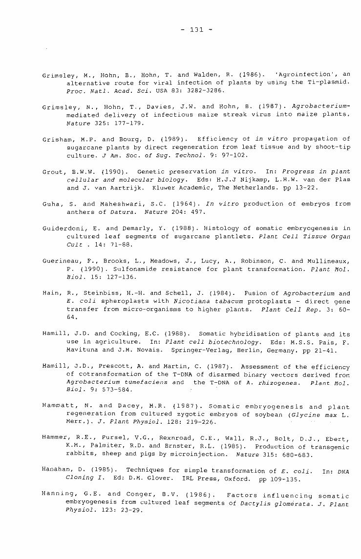
- 131 -
Grimsley, M., Hohn, B., Hohn, T. and Walden, R. (1986). 'Agroinfection', analternative route for viral infection of plants by using the Ti-p1asmid.Proc. Natl. Acad. Sci. USA 83: 3282-3286.
Grimsley, N., Hohn, T., Davies, J.W. and Hohn, B. (1987). Agrobacteriummediated delivery of infectious maize streak virus into maize plants.Nature 325: 177-179.
Grisham, M.P. and Bourg, D. (1989). Efficiency of in vitro propagation ofsugarcane plants by direct regeneration from leaf tissue and by shoot-tipculture. J Am. Soc. of Sug. Technol. 9: 97-102.
Grout, B.W.W. (1990). Genetic preservation in vitro. In: Progress in plantcellular and molecular biology. Eds: H.J.J Nijkamp, L.H.W. van der Plasand J. van Aartrijk. Kluwer Academic, The Netherlands. pp 13-22.
Guha, S. and Maheshwari, S.C. (1964). In vitro production of embryos fromanthers of Datura. Nature 204: 497.
Guiderdoni, E. and Demarly, Y. (1988). Histology of somatic embryogenesis incultured leaf segments of sugarcane plantlets. Plant Cell Tissue OrganCult . 14: 71-88.
Guerineau, F., Brooks, L., Meadows, J., Lucy, A., Robinson, C. and Mullineaux,P. (1990). Sulfonamide resistance for plant transformation. Plant Mol.Bioi. 15: 127-136.
Hain, R., Steinbiss, H.-H. and Schell, J. (1984). Fusion of Agrobacterium andE. coli spheroplasts with Nicotiana tabacum protoplasts - direct genetransfer from micro-organisms to higher plants. Plant Cell Rep. 3: 6064.
Hamill, J.D. and Cocking, E.C. (1988). somatic hybridisation of plants and itsuse in agriculture. In: Plant cell biotechnology. Eds: M.S.S. Pais, F.Mavituna and J.M. Novais. Springer-Verlag, Berlin, Germany. pp 21-41.
Hamill, J.D., Prescott, A. and Martin, C. (1987). Assessment of the efficiencyof cotransformation of the T-DNA of disarmed binary vectors derived fromAgrobacterium tumefaciens and the T-DNA of A. rhizogenes. Plant Mol.Bioi. 9: 573-584.
Hammatt, N. and Dacey, M.R. (1987). Somatic embryogenesis and plantregeneration from cultured zygotic embryos of soybean (Glycine max L.Merr.). J. Plant Physiol. 128: 219-226.
Hammer, R.E., Pursel, V.G., Rexnroad, C.E., Wall, R.J., Bolt, D.J., Ebert,K.M., Palmiter, R.D. and Brnster, R.L. (1985). Production of transgenicrabbits, sheep and pigs by microinjection. Nature 315: 680-683.
Hanahan, D. (1985). Techniques for simple transformation of E. coli.Cloning I. Ed: D.M. Glover. IRL Press, Oxford. pp 109-135.
In: DNA
Hanning, G.E. and Conger, B.V. (1986). Factors influencing somaticembryogenesis from cultured leaf segments of Dactylis glomerata. J. PlantPhysiol. 123: 23-29.
- 132 -
Haring, M.A., Rommens, C.M.T., John, H., Nijkamp, J. and Hille, J. (1991). Theuse of transgenic plants to understand transposition mechanisms and todevelop transposon-tagging strategies. Plant Mol. Biol. 16: 449-461.
Harrigan, W.F. and McCance, M.E. (1966). Laboratory Methods in Microbiology.
Academic Press, London. pp 8-10, 154.
Harrison, B.D., Mayo, M.A. and Baulcombe, D.C. (1987). Virus resistance intransgenic plants that express cucumber mosaic virus satellite RNA.
Nature 328: 799-802.
Hauptmann, R.M., Ozias-Akins, P., Vasil, V., Tabaeizadeh, Z., Rogers, S.C.,Horsch, R.B., Vasil, I.K. and Fraley, R.T. (1987). Transient expressionof electroporated DNA in monocotyledonous and dicotyledonous species.
Plant Cell Rep. 6: 265-270.
Hayashimoto, A., Li, Z. and Murai, N. (1990). A polyethylene glycol-mediatedprotoplast transformation system for production of fertile transgenicrice plants. Plant Physiol. 93: 857-863.
Heberle-Bors, E. (1991). Germ line transformation in higher plants.Newsletter 64: 2-10.
IAPTC
Hein, T., Prezewozny, T. and Schieder, o. (1983).somatic hybrids using an auxotrophic cell line.119-122.
Culture and selection ofTheor. Appl. Genet. 64:
Heinz, D.J. and Mee, G.W.P. (1969). Plant differentiation from callus tissue ofSaccharum sp. Crop Sci. 9: 346-348.
Hernalsteens, J.P., Thiu-Toong, L., Schell, J. and Van Montagu, M. (1984). AnAgrobacterium-transformed cell culture from the mono cot Asparagusofficinalis. EMBO J. 3: 3039-3041.
Herrera, G., Snyman, S.J. and Thomson, J.A. (1991). Development of a potentialbiological agent for the sugarcane borer Eldana saccharina. Frontiers ofbiotechnology in Agriculture. Satellite Meeting of the 15thInternational Congress of Biochemistry. Israel, August 1991, p19.
Herrera, G. and Thomson, J.A. (1989). Biological control of Eldana saccharinausing cloned Bacillus thuringiensis genes. Cloning of delta-endotoxinDNA sequences from B.thuringiensis 234. Proc. S. Afr. Sug. Technol. Ass.63: 189-191.
Herrera-Estrella, L. and Simpson, J. (1988). Foreign gene expression inplants. In: Plant molecular biology : A practical approach. Ed. C.H.Shaw. IRL Press, Oxford. pp 131-160.
Hess, D., DressIer, K. and Nirnmrichter, R. (1990). Transformation experimentsby pipetting Agrobacterium into the spikelets of wheat (Triticum aestivumL.). Plant Sci. 72: 233-244.
- 133 -
Hilder, V.A., Gatehouse, A.M.R. and Boulter, D. (1989). Potential forexploiting plant genes to genetically engineer insect resistance,exemplified by the cowpea trypsin inhibitor gene. Pestic. Sci. 27: 165-171.
Hinchee, M.A., Connor-Ward, D.V., Newell, C.A., McDonnell, R.E., Sato, S.J.,Gasser, C.S., Fischhoff, D.A., Re, D.B., Fraley, R.T. and Horsch, R.B.(1988). Production of transgenic soybean plants using Agrobacteriummediated DNA transfer. Bio/Technology 6: 915-922.
Hinchee, M.A., Dela Cruz, A. and Maretzki, A. (1984). Developmental andbiochemical characteristics of cold-treated anthers of Saccharumsponteneum. J. Plant Physiol. 115: 271-284.
/- Ho, W. and Vasil, I.K. (1983a). Somatic embryogenesis in sugarcane (Saccharumofficinarum L.) I. The morphology and physiology of callus formation andthe ontogeny of somatic embryos. Protoplasma 118: 169-180.
Ho, W. and Vasil, I.K. (1983b) Somatic embryogenesises in sugarcane (Saccharumofficinarum L): Growth and plant regeneration from embryogenic cellsuspension cultures. Ann. Bot. 51: 719-726.
Hohn, B., Hohn, T., Boulton, M.I., Davies, J.W. and Grimsley, N. (1987).Agroinfection of Zea mays with maize streak virus DNA. Plant Mol. Bioi.10: 459-468.
Holsters, M., Silva, B., Van vliet, F., Genetello, C. and De Block, M. (1980).The functional organization of the nopaline A. tumefaciens plasmidpTiC58. Plasmid 3: 212-230.
Hookyaas-Van Slogteren, G.M.S., -Hooykaas, P.J.J. and Schilperoot, R.A. (1984).Expression of Ti-plasmid genes in monocotyledonous plants infected withAgrobacterium tumefacines. Nature 311: 763-764.
Huang, Y. and Dennis, E. (1989). Factors influencing stable transformation ofmaize protoplasts by electroporation. Plant Cell Tissue and Organ Cult.18: 281-296.
Irvine, J.E. (1991). Texas Agricultural Experiment Station, Texas, U.S.A.
Irvine, J.E. (1984). The frequency of marker changes in sugarcane plantsregenerated from callus tissue. Plant Cell Tissue Organ Cult. 3: 201-209.
Irvine, J.E. and Almeida, C.G. (1991). - Delivery of plasmid DNA throughparticle bombardment with an airless sprayer. Texas AgriculturalExperiment Station. Leaflet No. MP-1726. 5pp.
Irvine, J.E., Benda, C.T.A., Legendre, B.L. and Machado, G.R. (1991). Thefrequency of marker changes in sugarcane plant regenerated from callusculture. II. Evidence for vegetative and genetic transmission,epigenetic effects and chimeral disruption. Plant Cell Tissue and OrganCult. 26: 115-125.
Izawa, T., Miyazaki, C., Yamamoto, M., Terada, R., Iida, S. and Shimamoto, K.(1991). Introduction and transposition of the maize transposable elementAc in rice (Oryza sativa L.) Mol. Gen. Genet. 227: 391-396.
- 134 -
Jefferson, R.A. (1987). Assaying chimeric genes in plantsfusion system. Plant Mol. Bioi. Rep. 5: 387-405.
The GUS gene
Jefferson, R.A., Kavanagh, T.A. and Bevan, M.W. (1987). GUS fusions : Bglucuronidase as a sensitive and versatile gene fusion marker in higher
plants. EMBO J. 6: 3901-3907.
Jensen, J.S., Marcker, K.A., Otten, L. and Schell, J. (1986). Nodule-specificexpression of a chimaeric soybean leghemaglobin gene in transgenic Lotuscorniculatus. Nature 321: 669-674.
Joersbo, M. and Brunstedt, J. (1990). Direct gene transfer to plant protoplastsby mild sonication. Plant Cell Rep. 9: 207-210.
Jones, M.G.K. and Dale, P.J. (1982). Reproducible regeneration of callus fromsuspension culture protoplasts of the grass Lolium multiflorum. Z.Pflanzenphysiol. 105: 267-274.
Junker, B., Zimrny, J., Luhrs, R. and Lorz, H. (1987). Transient expression ofchimaeric genes in dividing and non-dividing cereal protoplasts afterPEG-induced DNA uptake. Plant Cell Rep. 6: 329-332.
Kaeppler, H.F., Gu, W., Somers, D.A., Rines, H.W. and Cockburn, A.F.Silicon carbide filter-mediated DNA delivery into plant cells.Cell Rep. 9: 415-418.
(1990).Plant
Kamo, K.K., Becwar, M.R. and Hodges, T.K. (1985). Regeneration of Zea mays L.from embryogenic callus. Bot. Gaz. 146: 327-334.
Kamo, K.K. and Hodges, T.K. (1986). Establishment and characterisation of longterm embryogenic maize callus and cell suspension cultures. Plant Sci.45: 111-117.
Kanai, R. and Edwards, G.E. (1973). Purification of enzymatically- isolatedmesophyll protoplasts from C3, C4 and crassulacean acid metabolism plantsusing an aqueous dextran-polyethylene glycol 2-phase system. PlantPhysiol. 52: 484-490.
Kao, K.N. (1977). Chromosomal behaviour in somatic hybrids of soybean Nicotiana glauca. Molec. Gen. Genet. 150: 225-230.
Kao, K.N., Gamborg, O.L., Miller, R.A. and Keller, W.A. (1971). Cell divisionsin cells regenerated from protoplasts of soybean and Haplopappusgracilis. Nature 232: 124.
Kao, K.N. and Michayluk, M.R. (1975). Nutritional requirements for growth ofVicia hajastona cells and protoplasts at a very low population density inliquid media. Planta 126: 105-110.
Karlsson, S.B. and Vasil, I.K. (1986). Morphology and ultrastructure ofembryogenic cell-suspension cultures of Panicum maximum Jacq. (Guineagrass) and Pennisetum purpureum Schum. (Napier grass). Am. J. Bot. 73:894-901.
- 135 -
Karp, A. and Maddock, S.C. (1984). Chromosomal variation in wheat plantsregenerated from cultured immature embryos. Theor. Appl. Genet. 67: 249255.
Kartha, K.K. (1985). Cryopreservation of plant cells and organs. CRC Press,Boca Raton, USA. 276pp.
Kartzke, S., Saedler, H. and Meyer, P. (1990). Molecular analysis oftransgenic plants derived from transformants of protoplasts at variousstages of the cell cycle. Plant Science 67: 63-72.
Kasha, K.J. and Reinbergs, E. (1980). Achievements with haploids in barleyresearch and breeding. In: The Plant genome. Eds: D.R. Davies and D.A.Hapwood. The John Innes Charity, Norwich. pp 215-230.
Kaur-Sawhney, R. and Galston, A.W. (1984). Oats. In: Handbook of plant cellculture. Vcl 2. Crop species. Eds: W.R. Sharp, D.A. Evans, R.V. Amrniratoand Y. Yamada. MacMillan Publishing Company, New York. pp92-107.
King, P.J. and Shimamoto, K. (1984). Maize. In: Handbook of Plant cellculture. Vcl 2. Crop species. Eds: W.R. Sharp, D.A. Evans, R.V. Amrniratoand Y. Yamada. MacMillan Publishing Company, New York. pp 69-91.
Klein, T.M. and Fromm, M.E. (1989). Blast those genes! Agricultural Res. 37:14-15.
Klein, T.M., Wolf, E.D., Wu, R. and Sanford, J.C. (1987). High-velocitymicroprojectiles for delivering nucleic acids into living cells. Nature327: 70-73.
Kleinhofs, A. and Behki, R. (1977). Prospects for plant genome modification bynonconventional methods. Ann. Rev. Genet. 11: 79-101.
Kleinhofs, A., Eden, F.C., Chilton, M.D. and Bendich, A.J. (1975). On thequestion of the integration of exogenous bacterial DNA into plant DNA.Proc. Natl. Acad. Sci. USA 72: 2748-2752.
Koncz, C., Olsson, 0., Langridge, W.H.R., Schell, J. and Szalay, A.A. (1987).Expression and assembly of functional bacterial luciferase in plants.Proc. Natl. Acad. Sci. USA 84: 313-315.
Kosugi, S., Ohashi, Y., Nakajima, K. and Arai, Y. (1990). An improved assayfor B-glucuronidase in transformed cells methanol almost completelysuppresses a putative endogenous B-glucuronidase activity. Plant Science70: 133-140.
Krens, F.A., Molendijk, L., Wullems, G.J. and Schilperoort, R~A. (1982). Invitro transformation of plant protoplasts with Ti-plasmid DNA. Nature296: 72-74.
Larkin, P.J. (1976). Purification and viability determinations of plantprotoplasts. Planta 128: 213-216.
Larkin, P.J. (1982). Sugarcane tissue and protoplast culture. Plant Cell TissueOrgan Cult. 1: 149-164.
- 136 -
Larkin, P.J. and Scowcroft, W.R. (1981). Somaclonal variation - a novel sourceof variability from cell cultures for plant improvement. Theor. Appl.
Genet. 60: 197-214.
Lark in P J Taylor B.H., Gersmann, M. and Brettell, R.I.S. (1990). Direct., .. , ,gene transfer to protoplasts. Aust. J. Plant Physiol. 17: 291-302.
Lawrence, W.A. and Davies, R.D. (1985). A simple method for the microinjectionand culture of protoplasts at very low densities. Plant Cell Rep. 4: 33
35.
Ledoux, L. and Huart, R. (1968). Integration and replication of DNA of M.lysodeikticus in DNA of germinating barley. Nature 218: 1256-1259.
Ledoux, L. and Huart, R. (1969).acids in barley seedlings.
Fate of exogenous bacterial deoxyribonucleicJ. Mol. Biol. 43: 243-262.
Ledoux, L., Huart, R. and Jacobs, M. (1971). Fate of exogenous DNA inArabidopsis thaliana. Eur. J. Biochem. 23: 96-108.
Ledoux, L.' Huart, J. and Jacobs, M. (1974). DNA-mediated genetic correctionof thiaminless Arabidopsis thaliana. Nature 249: 17-21.
Leggatt, I., Waites, W.M., Leifert, C. and Nicholas, J.R. (1988).Characterisation of microorganisms isolated from plants duringmicropropagation. Acta Hort. 225: 93-102.
Leifert, C and Waites, W.M. (1990). Contaminants of plant tissue cultures.IAPTC Newsletter 60: 2-13.
Lindsey, K. (1986). The production of secondary metabolites by immobilisedplant cells. In: Secondary metabolism in plant cell cultures. Eds: P.Morris, A.H. Scragg, A. Stafford and M.W. Fowler. Cambridge UniversityPress, England. pp143-155.
Lindsey, K. and Jones, M.G.K. (1989). Plant Biotechnology in Agriculture. OpenUniversity Press, Milton Keynes. pp 57-85.
Lindsey, K. and Jones, M.G.K. (1990). Selection of transformed cells. In:Plant cell line selection Procedures and applications. Ed: P.J.Dix. VCHPublishers, Germany. 241pp.
Lindsey, K. and Yeoman, M.M. (1983). The relationship between growth rate,differentiation and alkaloid accumulation in cell cultures. J. Exp. Bot.34: 1055-1065.
Ling, O.H., Chen, W.Y., Chen., M.F. and Ma, Z.R. (1983). Somatic embrogenesisand plant regeneration in an interspecific hybrid of Oryza L. Plant CellRep. 2: 169-171.
Lippincott, B.B. and Lippincott, J.A. (1978). Cell walls of crown-gall tumorsand embryogenic plant tissues lack Agrobacterium adherence sites.Science 199: 1075-1078.
Liu, M. (1971). A new method for sugarcane breeding - tissue culture technique.Taiwan Sugar 18: 8-10.
- 137 -
Liu, M.C. (1984). Sugarcane. In: Handbook of plant cell culture. Vol 2. Ed.W.H. Sharp, D.A. Evans, P.V. Ammirato and Y. Yamada. MacMillan Press, NewYork. pp 572-605.
Liu, M.C. (1988). High frequency callus formation from protoplasts of sugarcanesuspension cultures. Taiwan Sugar (Sept.-Oct.): 9-14.
Liu, M.C. (1990). Selection of agronomically useful mutants through plant cellculture systems. Taiwan Sugar 37: 8-13.
Liu, M.C. and Chen, W.H. (1974). Histological studies on the origin and processof plantlet differentiation in sugarcane callus mass. Proc. 15th Int.Soc. Sug. Technol. Durban. 118-121.
Liu, M.C. and Chen, W.H. (1976). Tissue and cell culture as aids to sugarcanebreeding 1. Creation of genetic variation through callus culture.Euphytica 25: 393-403.
Liu, M.C., Chen, W.C. and Shih, S.C. (1982). Histogenesis of sugarcane callusoriginating from young leaf and stem tip explants. Can. J. Bot. 60: 28892895.
Liu, M.C. and Shih, S.C. (1986). Salt stress-induced increases in nitratereductase activity and protein content in sugarcane cell culture. Proc.19th Int. Soc. Sug. Technol. Jakarta, Indonesia. 263-273.
Liu, M.C., Yeh, H.S. and Chen, W.H. (1984). A high-sucrose and vigorouslygrowing calliclone 71-4829. Rep. Taiwan Sugar Res. Inst. 102: 1-11.
Lorz, H. (1989). In vitro manipulation of cereal plants. In: 2ndInternational symposium on genetic manipulation in crops. Lisbon. Eds:A. Mujeeb-Kazi and L.A. Sitch. pp 93-101.
Lorz, H., Baker, B. and Schell, J. (1985). Gene transfer to cereal cellsmediated by protoplast transformation. Mol. Gen. Genet. 199: 178-182.
Lowe, K., Taylor, D.B., Ryan, P. and Paterson, K.E. (1985). Plant regenerationvia organogenesis and embryogenesis in the maize inbred line B73. PlantSci. 41: 125-132.
Lu, C., Chandler, S.F and Vasil, I.K. (1984). Somatic embryogenesis and plantregeneration from cultured immature embryos of rye (Secale cereale L.) J.Plant Physiol. 115: 237-244.
Lu, C. and Vasil, I.K. (1982). Somatic embryogenesis and plant regenerationfrom tissue cultures of Panicum maximum. Am. J. Bot. 69: 77-81.
Lu, C., Vasil, I.K. and Ozias-Akins, P. (1982). Somatic embryogenesis in ZeaMays L. Theoret. Appl. Genet. 62: 109-112.
Lu, C., Vasil, V and Vasil, I.K. (1981). Isolation and culture of protoplastsof Panicum maximum Jacq. (Guinea grass): somatic embryogenesis andplantlet formation. Z. Pflanzenphysiol. 104: 311-318.
- 138 -
Lu, c., Vasil, v., and Vasil I.K. (1983). Improved efficiency of somaticembryogenesis and plant regeneration in tissue culture of maize (Zea MaysL.) Theoret. Appl. Genet. 66: 285-289.
Luo, z. and Wu, R. (1988). A simple method for the transformation of rice viathe pollen-tube pathway. Plant Mol. Biol. Rep. 6: 165-174.
MacKinnon, C., Gunderson, G. and Nabors, M.W. (1986). Plant regeneration bysomatic embryogenesis from callus cultures of sweet sorghum. Plant CellRep. 5: 349-351.
Maniatis, T., Fritsch,laboratory manual.
E.F. and Sambrook, J. (1982). Molecular Cloning: ACold Spring Harbor Laboratory Press, New York. 545pp.
Mante11, S.H., Mathews, J.A. and McKee, R.A. (1985). Principles of plantbiotechnology: An introduction to genetic engineering in plants.Blackwell Scientific Publications, Oxford. pp 89-158.
Maretzki, A. and Nickell, L.G. (1973). Formation of protoplasts from sugarcanecell suspensions and the regeneration of cell cultures from protoplasts.Collog. Int. C.N.R.S. 212: 51-63.
Marton, L., Wullems, G.J., Molendijk, L. and Schilperoot, R.A. (1979). Invitro transformation of cultured cells from Nicotiana tabacum byAgrobacterium tumefaciens. Nature 277: 129-130.
Mayo, M.A. and Barker, H. (1990). Induction of resistance to plant viruses bytransformation with virus genes: the poacher turned gamekeeper. In:Aspects of Applied Biology 24. Association of Applied Biologists,England. pp 65-78.
Mazur, B.J.crops.
and Falco, S.C. (1989). The development of herbicide resistantAnnu. Rev. Plant Physiol. and Plant Mol. Biol. 40: 441-470.
McCabe, D.E., Swain, W.F. and Christou, P. (1988). Stable transformation ofsoybean (Glycine max) by particle acceleration. Bio/Technology 6: 923926.
McKnight, T.D., Lillis, M.T. and Simpson, R.B. (1987). Segregation of genestransferred to one plant cell from two separate Agrobacterium strains.Plant Mol. Biol. 4: 439-445.
Meijer, E.G.M., Schilperoot, R.A., Rueb, S., Van Os-Ruygrok, P.E. and Hensgens,L.A.M. (1991). Transgenic rice cell lines: expression of transferredchimeric genes. Plant Mol. Biol. 16: 807-820.
Mendel, R.R., Muller, B., Schulze, J., Kolesnikov, V. and Zelenin, A. (1988).Delivery of foreign genes to intact barley cells by high velocitymicroprojectiles. Theor. Appl. Genet. 78: 31-34.
Mitchell, J.C. and Petolino, J.F. (1991). Plant regeneration from haploidsuspension and protoplast cultures from isolated microspores of maize.J. Plant Physiol. 137: 530-536.
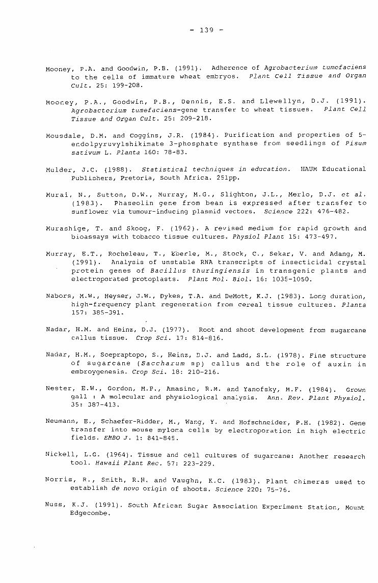
- 139 -
Mooney, P.A. and Goodwin, P.B. (1991). Adherence of Agrobacterium tumefaciensto the cells of immature wheat embryos. Plant Cell Tissue and Organ
Cult. 25: 199-208.
Mooney, P.A., Goodwin, P.B., Dennis, E.S. and Llewellyn, D.J. (1991).Agrobacterium tumefaciens-gene transfer to wheat tissues. Plant CellTissue and Organ Cult. 25: 209-218.
Mousdale, D.M. and Coggins, J.R. (1984). Purification and properties of 5endolpyruvylshikimate 3-phosphate synthase from seedlings of Pisumsativum L. Planta 160: 78-83.
Mulder, J.C. (1988). Statistical techniques in education.Publishers, Pretoria, South Africa. 251pp.
HAUM Educational
Murai, N., Sutton, D.W., Murray, M.G., Slighton, J.L., Merlo, D.J. et al.(1983). Phaseolin gene from bean is expressed after transfer tosunflower via tumour-inducing plasmid vectors. Science 222: 476-482.
Murashige, T. and Skoog, F. (1962). A revised medium for rapid growth andbioassays with tobacco tissue cultures. Physiol Plant 15: 473-497.
Murray, E.T., Rocheleau, T., Eberle, M., Stock, C., Sekar, V. and Adang, M.(1991). Analysis of unstable RNA transcripts of insecticidal crystalprotein genes of Bacillus thuringiensis in transgenic plants andelectroporated protoplasts. Plant Mol. Bioi. 16: 1035-1050.
Nabors, M.W., Heyser, J.W., Dykes, T.A. and DeMott, K.J. (1983). Long duration,high-frequency plant regeneration from cereal tissue cultures. Planta157: 385-391.
Nadar, H.M. and Heinz, D.J. (1977). Root and shoot development from sugarcanecallus tissue. Crop Sci. 17: 814-816.
Nadar, H.M., Soepraptopo, S., Heinz, D.J. and Ladd, S.L. (1978). Fine structureof sugarcane (Saccharum sp) callus and the role of auxin inembroygenesis. Crop Sci. 18: 210-216.
Nester, E.W., Gordon, M.P., Amasino, R.M. and Yanofsky, M.F. (1984). Growngall : A molecular and physiological analysis. Ann. Rev. Plant Physiol.35: 387-413.
Neumann, E., Schaefer-Ridder, M., Wang, Y. and Hofschneider, P.H. (1982). Genetransfer into mouse myloma cells by electroporation in high electricfields. EMBO J. 1: 841-845.
Nickell, L.G. (1964). Tissue and cell cultures of sugarcane: Another researchtool. Hawaii Plant Rec. 57: 223-229.
Norris, R., Smith, R.H. and Vaughn, K.C. (1983). Plant chimeras used toestablish de novo origin of shoots. Science 220: 75-76.
Nuss, K.J. (1991). South African Sugar Association Experiment Station, MountEdgecombe.
Propagation of Zea mays L.Ann. Bot. 45: 183-189.
- 140 -
Okada, K., Nagata, T. and Takebe, I. (1986). Introduction of functional RNAinto plant protoplasts by electroporation. Plant Cell Physiol. 27: 619-
626
Okuno, T. and Furusawa, I. (1977). A simple method for the isolation of intactmesophyll protoplasts from cereal plants. Plant Cell Physiol. 18: 1357-
1362.
Oliver, S.G. and Ward, J.M. (1985). A dictionary of genetic engineering.Cambridge University Press, England. 153pp.
Ozias-Akins, P., Ferl, R.J. and Vasil, I.K. (1986). Somatic hybridization inthe Gramineae: Pennisetum americanum (L). K. Schum. and Panicum maximumJcq. (Guinea grass). Molec. Gen. Genet. 203: 365-370.
Paszkowski, J., Shillito, R.D., Saul, M., Mandak, V., Hohn, T., Hohn, B. andPotrykus, I. (1984). Direct gene transfer to plants. EMBO J. 3: 27172722.
Pelcher, L.E., Garnborg, O.L. and Kao, K.N. (1974). Bean mesophyll protoplasts:production, culture and callus formation. Plant Sci. Lett. 3: 107-111.
Pollock, K., Barfield, D.G. and Shields, R. (1983). The toxicity of antibioticsto plant cell cultures. Plant Cell Rep. 2: 34-39.
Potrykus, I., Harms, C.T., Lorz, H. and Thomas, E. (1977). Callus formationfrom stern protoplasts of corn (Zea mays L.). Molee. Gen. Genet. 156: 347350.
Potrykus, I., Saul, M., Petruska, J., Paszowski, J. and Shillito, R.D. (1985).Direct gene transfer to cells of a graminaceous monocot. Molec. Gen.Genet. 199: 183-188.
Potter, H., Weir, L. and Leder, P. (1984). Enhancer-dependent expression ofhuman immunoglobulin genes introduced into mouse pre-B lymphocytes byelectroporation. Proc. Natl. Acad. Sci. USA 81: 7161-7165.
Power, J.B. and Cocking, E.C. (1970). Isolation of leaf protoplasts: macromolecule uptake and growth substance response. J. Exp. Bot. 21: 64-70.
Power, J.B., Davey, M.R., Freeman, J.P., 'Mulligan, B.J. and Cocking, E.C.(1986) . Fusion and transformation of plant protoplasts. MethodsEnzymol. 118: 578-594.
Price, S. (1962). A modified leaf-squash technique for counting chromosomes insomatic cells of Saccharum and related grasses. Proe. 11th Congr. Int.Soc. Sugar Cane Technol. Reduit, Mauritius. 583-585.
Raineri, D.M., Bottino, P., Gordon, M.P. and Nester, E.W. (1990).Agrobacterium-mediated transformation of rice (Oryza sativa L.).Bio/Technology 8: 33-38.
Raman, K., Walden, D.B. and Greyson, R.I. (1980).by shoot tip culture: a feasibility study.
- 141 -
Rashid, H. and Quraishi, A. (1989). High frequency embryogenic callus inductionand its regeneration in three wheat cultivars. In: Second InternationalSymposium on Genetic Manipulation in crops. Lisbon. Eds: A. Mujeeb-Kaziand L.A. Sitch. pp 205-215.
Ray, D.S. and Ghosh, P.D. (1990). Somatic embryogenesis and plant regenerationfrom cultured leaf explants of Zea mays. Ann. Bot. 66: 497-500.
Reggiardo, M.I., Arana, J.L., Orsaria, L.M., Permingeat, H.R., Spitteler, M.A.and Vallejos, R.H. (1991). Transient transformation of maize tissues bymicroprojectile bombardment. Plant Sci. 75: 237-243.
Reich, T.J., Iver, V.N. and Miki, B.L. (1986). Efficient transformation ofalfalfa protoplasts by the intranuclear microinjection of Ti-plasmids.Biotechnology 4: 1001-1004.
Reisch, B.I. (1988). Genetic instability in plant cell cultures: utilisationin plant breeding and genetic studies. In: Plant Cell Biotechnology.Eds: M.S.S. Pais, F. Mavituna and J.M. Novais. Springer-Verlag, Berlin,Germany. pp 87-96.
Reiss, B., Sprengel, R., Will, H. and Schaller, H. (1984). A sensitive methodfor qualitative and quantitative assay of neomycin phosphotransferase incrude cell extracts. Gene 30: 211-218.
Rhodes, C.A., Pierce, D.A., Mettler, I.J., Mascarenhas, D. and Detmer, J.J.(1988). Genetically transformed maize plants from protoplasts. Science240: 204-207.
Rigby, P.W.J., Dieckmann, M., Rhodes, C. and Berg, P. (1977). Labellingdeoxyribonucleic acid to a high specific activity in vitro by nicktranslation with DNA polymerase I. J. Mol. Biol. 113: 237-251.
Rout, J.R. and Sarma, N.P. (1991). Anther callus induction and green plantregeneration at high frequencies from an interspecific rice hybrid Oryzasativa. L x o. rufipogon. Grieff. Euphytica 54: 155-159.
Ryu, D.D., Lee, S.D. and Romani, R.J. (1990). Determination of growth rates ofplant cell cultures: comparative studies. Biotechnol. and Bioeng. 35:305-311.
Sacristan, M.D. and Melchers, G. (1969). The karyologica1 analysis of plantsregenerated from tumorous and other callus cultures of tobacco. Molec.Gen. Genet. 105: 317-333.
Schaeffer, G.W., Lazar, M.D. and Baenzinger, P.S. (1984). Wheat. In: Handbookof Plant cell culture. Vol 2. Crop species. Eds: W.R. Sharp, D.A. Evans,R.V. Ammirato and Y. Yamada. MacMillan Publishing Company, New York. pp108-136.
Schaffler, G. and Koop, H, (1990). Single cell nurse culture of tobaccoprotoplasts : physiological analysis of conditioning factors. J. PlantPhysiol. 137: 95-101.
Schell, J. (1991). Max-Planck Institute, Germany.
- 142 -
Schenk, R.U. and Hildebrandt, A.C. (1971). Production, manipulation and fusionof plant cell protoplasts as steps towards somatic hybridisation. Coll.
Int. CNRS 193: 319-333.
Schenk, R.U. and Hildebrandt, A.C. (1972). Medium and techniques for inductionof growth of monocotyledonous and dicotyledonous plant cell cultures.Can. J. Bot. 50: 166-204.
Shah, D.M., Horsch, R.B., Klee, H.J., Kishore, G.M., Winter, J.A., Turner, N.E.,Hironaka, C.M., Sanders, P.R., Gasser, C.S., Aykent, S., Stiegel, N.R.,Rogers, S.G. and Fraley, R.T. (1986). Engineering herbicide tolerance intransgenic plants. Science 233: 478.
Sharp, W.R., Sondahl, M.R., Caldas, L.S. and Maraffa, S.B. (1980). Thephysiology of in vitro asexual embryognesis. Hort. Rev. 2: 268-310.
Shaw, C.H., Watson, M.D., Carter, G.H. and Shaw, C.H. (1984). The right handcopy of the nopaline Ti-plasmid 25 bp repeat is required for tumourformation. Nucleic Acids Res. 12:6031-6041.
Sheridan, W.F. (1975). Tissue culture of maize I. Callus induction and growth.Physiol. Plant 33: 151-156.
Shields, R., Robinson, S.J. and Anslow, P.A. (1984). Use of fungicides in planttissue culture. Plant Cell Rep. 3: 33-36.
Shillito, R.D., Carswill, G.K., Johnson, C.M., DiMaio, J.J. and Harms, C.T.(1989). Regeneration of fertile plants from protoplasts of elite inbredmaize. Bio/Technology 7: 581-587.
Shillito, R.D., Saul, M.W., Paszkowski, J., Muller, M. and Potrykus, I. (1985).High efficiency direct gene transfer to plants. Biotechnology 3: 10991103.
Shimada, T. and Yamada, Y. (1979). Wheat plants regenerated from embryo cellcultures. Jpn. J. Genet. 54: 379-385.
Simon, E.W. (1974). Phospholipids and plant membrane permeability. New Phytol.73: 377-420.
Simpson, R.B., Spielmann, A., Margossian, 'L. and McKnight, T.D. (1986). Adisarmed binary vector from Agrobacterium tumefaciens functions in A.rhizogenes. Frequent cotransformation of two distinct T-DNA's. PlantMol. Biol. 6: 403-415.
Skene, K.G.M. and Barlass, M. (1983). Studies on the fragmented shoot apex ofgrapevine. J. Exp. Bot. 34: 1271-1280.
Skoog, F, and Miller, C.o. (1957). Chemical regulation of growth and organformation in plant tissue~ cultured in vitro. Symp. Soc. Exp. Biol. 11:118-130.
Srinivasan, C. and Vasil, I.K. (1986). Plant regeneration from protoplasts ofsugarcane (Saccharum officinarum L.). J. Plant Physiol. 126: 41-48.
- 143 -
Stacey, D.W. (1981). Microinjection of mRNA and other macromolecules intoliving cells. Methods in Enzymol. 79: 76-88.
stalker, D.M., McBride, K.E. and Malyj, L.D. (1988). Herbicide resistance intransgenic plants expressing a bacterial detoxification gene. Science242: 419-423.
Steinbiss, H.H. and Stable, P. (1983). Protoplast derived tobacco cells cansurvive capillary microinjection of the fluorescent dye Lucifer Yellow.Protoplasma 116: 223-227.
Stevenson, G.C. (1965). Genetics and breeding of sugarcane. Longmans, London.284pp.
Steward, F.C., Mapes, M.O. and Mears, K. (1958). Growth and organiseddevelopment of cultured cells. 11. Organisation in cultures grown fromfreely suspended cells. Am. J. Bot. 445: 705-708.
Stougaard, J., Abildsten, D. and Marcker, K.A. (1987). The Agrobacteriumrhizogenes pRi TL-DNA segment as a gene vector system for transformationof plants. Mol. Gen. Genet. 207: 251-255.
Sun, C.S., Prioli, L.M. and Sondahl, M.R. (1989). Regeneration of haploid anddihaploid plants from protoplasts of supersweet (sh2 sh2) corn. PlantCell Rep. 8: 313-316.
Sutcliffe, J.G. (1979). Complete nucleotide sequence on the E. coli plasmidpBR322. Cold Spring Harbor Symp. Quant. Biol. 43: 77-90.
Tabaeizadeh, Z., Ferl, R.J. and Vasil, I.K. (1986). Somatic hybridization inthe Gramineae: Saccharum officinarum L. (sugarcane) + Pennisetumamericanum (L). K. Schum. (pearl millet). Proc. Natl. Acad. Sci. USA 83:5616-5619.
Thorn, M., Maretzki, A., Konor, E. and Sakai, W.S. (1981). Nutrient uptake andaccumulation of sugarcane cell cultures in relation to the growth cycle.Plant Cell Tissue Organ Cult. 1: 3-14.
Tisserat, B. (1985). Embryogenesis, organogenesis and plant regeneration. In:Plant Cell Culture. Ed: R.A. Dixon. ~RL Press, Oxford. pp 79-105.
Tomes, D.T., Weissinger, A.K., Ross, M., Higgins, R., Drummond, B.J., Schaaf,S., Malone-Schoneberg, J., Staebell, M., Flynn, M., Anderson, J. andHoward, J. (1990). Transgenic tobacco plants and their progeny derivedby rnicroprojectile bombardment of tobacco leaves. Plant Mol. Biol. 14:261-268.
Topfer, R., Gronenborn, B., Schell, J. and Steinbiss, H. (1989). Uptake andtransient expression of chimeric genes in seed-derived embryos. The PlantCell 1: 133-139.
Toriyama, K., Arimoto, Y., Uchimiya, H. and Hinata, K. (1988). Transgenic riceplants after direct gene transfer into protoplasts. Bio/Technology 6:1072-1074.
I
- 144 -
Tsay, H.S. (1987). Crop improvement through somaclonal variation. ROC-USAAgricultural Biotechnology Workshop. Tapei, Taiwan. pp 1-15.
Twell, D., Klein, T.M. Fromm, M.E. and McCormick, S. (1989). Transientexpression of chimeric genes delivered into pollen by microprojectilebombardment. Plant Physiol. 91: 1270-1274.
Vaeck, M., Reynaerts, A., Hofte, H., Jansens, S., De Beuckeleer, M., Dean, C.,Zabeau, M., Van Montegu, M. and Leemans, J. (1987). Transgenic plantsprotected from insect attack. Nature 328: 33-37.
Vail, P.V. (1991).1991: 10-11.
Mothproofing walnut trees of the future. Ag. Res. June
Van den Elzen, P.J.M., Townsend, J., Lee, K.Y. and Bedbrook, J.R. (1985). Achimaeric hygromycin resistance gene as a selectable marker in plantcells. Plant Mol. Bioi. 5: 299-302.
Van Dun, C.M.P., Bol, J.F. and Van Vloten-Doting, L. (1987). Expression ofalfalfa mosaic virus and tobacco rattle virus coat protein genes intransgenic tobacco plants. Virology 159: 299-305.
Van Lijsebettens, M., Inze, D., Van Montagu, M. and Schell, J. (1986).Transformed cell clones as a tool to study T-DNA integration mediated byAgrobacterium tumefaciens. J. Mol. Bioi. 188:129-145.
Vasil, I.K. (1982). Plant cell culture and somatic cell genetics of cereals andgrasses. In: Plant improvement and somatic cell genetics. Ed: I.K. Vasil,W.R. Scowcroft and K.J. Frey. Academic Press, Orlando USA. pp 179-204.
Vasil, I.K. (1983). Isolation and culture of protoplasts of grasses. In: PlantProtoplasts. Ed: K.G. Giles. Academic Press, London pp 79-90.
Vasil, I.K. (1987). Developing cell and tissue culture systems for theimprovement of cereal and grass crops. J. Plant Physiol. 128: 193-218.
Vasil, I.K. (1990). The contributions of biotechnology and its challenges.IAPTC Newsletter 62: 2-11.
Vasi1, V., Brown, S.M., Re, D., Fromm, M.E. and Vasil, I.K. (1991). Stablytransformed callus lines from mic~oprojectile bombardment of cellsuspension cultures of wheat. Bio/Technology 9: 743-747.
Vasil, V., Redway, F. and Vasil, I.K. (1990a). Regeneration of plants fromembroygenic suspension culture protoplasts of wheat (Triticum aestivumL.). Bio/Technology 8: 429-432.
Vasil, V. and Vasil, I.K. (1982). The ontogeny of somatic embryos of Pennisetumamericanum (L). K. Schum. Bot. Gaz. 143: 454-465.
Vasil, V. and Vasil, I.K. (1984). Induction and maintenance of embryogeniccallus cultures of Gramineae. In: Cell culture and somatic cell geneticsof plants Vol 1: Procedures and their applications. Ed: I.K. Vasil.Academic Press, London. pp 37-40.
- 145 -
Vasil, v., Vasil, I.K. and Lu, C.H. (1984). Somatic embryogenesis in long-termcallus cultures of Zea mays L. (Gramineae). Am. J. Bot. 71: 158-161.
Vasil, I.K., Vasil, V. and Redway, F. (1990b). Plant regeneration fromembryogenic calli, cell suspension cultures and protoplasts of Triticumaestivum L. (Wheat). In: Progress in plant cellular and molecularbiology. Eds: H.J.J. Nijkamp, L.H.W. van der Plas and J. von Aartrijk.Kluwer Academic, The Netherlands. pp 33-37.
Vasil, V., Wang, D. and Vasil, I.K., (1986). Plant regeneration fromprotoplasts of Pennisetum purpureum. Schumm (Napier grass). Z.Pflanzenphysiol. Ill: 233-239.
Walden, R. (1988). Genetic transformation in plants. Institute Biology :Biotechnology series. Open University Press, Milton-Keynes, U.K. pp 7081.
Wang, Y.C., Klein, T.M., Fromm, M., Cao, J., Sanford, J.C. and Wu, R. (1988).Transformation of rice, wheat and soybean by the particle bombardmentmethod. Plant Mol. Biol. 11: 433-439.
Wang, D. and Vasil, I.K. (1982). Somatic embryogenesis and plant regenerationfrom inflorescence segments of Pennisetum purpurea Schum. (Napier orElephant grass). Plant Sci. Lett. 25: 147-154.
Watt, M.P., Mycock, D.J. and Cresswell, C.F. (1989). Plant regeneration bysomatic embryogenesis from leaf explants of Digitaria eriantha (Stead)subsp. eriantha. XVI Int. Grasslands Conference Nice, France.pp 421-422.
Weatherhead, M.A., Bardon, J. and Henshaw, G.G. (1978). Some effects ofactivated charcoal as an additive to plant tissue culture media. Z.Pflanzenphysiol. 89: 141-147.
Weising, K., Schell, J. and Kahl, G. (1988). Foreign genes in plants:Transfer, structure, expression and applications. Annu. Rev. Genet. 22:421-477.
Wernicke, W. and Brettell, R.I.S. (1980). Somatic embroygenesis from Sorghumbicolor leaves. Nature 138: 138-139.
Wernicke, W., Potrykus, I. and Thomas, E. (1982). Morphogenesis from culturedleaf tissue of Sorghum biocolor (L.). The morphogenetic pathways.Protoplasma 111: 53-62.
Werr, W. and Lorz, H. (1986). Transient gene expression in a Gramineae cellline: A rapid procedure for studying plant promoters. Mol. Gen. Genet.202:471-475.
White, P.R. (1963). A handbook of plant and animal tissue culture. JaquesCattel Press, USA. 214pp.
Withers, L.A. (1985). Cryopreservation and storage of germplasm. In: Plantcell culture: A practical approach. Ed: R.A. Dixon. IRL Press, Oxford,England. pp 169-191.
- 146 -
Woodward, B.R. (1989). Somatic embryogenesis in maize (Zea mays L). MSc thesis.
195pp.
Woo1ston, C.J., Barker, R., Gunn, H., Boulton, M.I. and Mullineaux, P.M.(1988). Agroinfection and nucleotide sequence of cloned wheat dwarfvirus DNA. Plant Mol. Biol. 11: 35-43.
Yamada, Y., Yang, Z. and Tang, D. (1986). Plant regeneration from protoplastderived callus of rice (Oryza sativa L.) Plant Cell Rep. 5: 85-88.
Yang, H., Zhang, H.M., Davey, M.R., Mulligan, B.J. and Cocking, E.C. (1988).Production of kanamycin resistant rice tissues following DNA uptake intoprotoplasts. Plant Cell Rep. 7: 421-425.
Yao, D.Y. and Krikorian, A.D. (1981). Manipulation of rice (Oryza sativa L.)from aseptically cultured nodes. Ann. Bot. 48: 255-259.
Young, P.M., Hutchins, A.S. and Canfield, M.L. (1984). Use of antibiotics tocontrol bacteria in shoot cultures of woody plants. Plant Sci. Lett. 34:203-209.
Zambryski, P. (1988). Basic processes underlying Agrobacterium-mediated DNAtransfer to plant cells. Annu. Rev. Genet. 22: 1-30.
Zhang, H.M., Yang, H., Rech, E.L., Golds, T.J., Davis, A.S., Mulligan, B,J.,Cocking, E.C. and Davey, M.R. (1988). Transgenic rice plants produced byelectroporation-mediated plasmid uptake into protoplasts. Plant Cell Rep.7: 379-384.
Ziv, M. (1986). In vitro hardening and acclimitization of tissue cultureplants. In: Plant tissue culture and its agricultural applications. Ed:L.A. Withers and P.G. Aldeson. University Press, Cambridge. pp 187-196.