Gene ofthe structural hepatitis C in vitro
5
Proc. Nati. Acad. Sci. USA Vol. 88, pp. 5547-5551, July 1991 Biochemistry Gene mapping of the putative structural region of the hepatitis C virus genome by in vitro processing analysis (RNA virus/structural proteins/glycoproteins/signal peptidase) MAKOTO HuIIKATA, NOBUYUKI KATO, YUKO OOTSUYAMA, MASANORI NAKAGAWA, AND KUNITADA SHIMOTOHNO Virology Division, National Cancer Center Research Institute, 5-1-1, Tsukiji, Chuoku, Tokyo 104, Japan Communicated by Motoo Kimura, March 26, 1991 ABSTRACT Processing of the putative structural proteins of hepatitis C virus was examined by using an in vitro expres- sion system. An RNA transcript for cell-free translation was prepared from a cDNA construct that encompasses the region encoding the 980 amino-terminal residues of the viral polypro- tein precursor. Processing of the in vitro translation product proceeded cotranslationally in the presence of microsomal membranes and generated four major membrane-associated products. Two of these four major products, named gp35 and gp7O, were shown to be transported into microsomes and heavily glycosylated, suggesting that the processing events are partly mediated by the signal peptidase of the endoplasmic reticulum. The other two products, p19 and p21, were prob- ably associated with the outer surface of the microsomal membrane. Analysis of processed proteins translated from a series of truncated forms of the cDNA construct as well as determination of amino-terminal amino acid sequences of gp35 and gp7O indicated that these four products are arranged from the amino-terminal end of the polyprotein precursor in the order: NH-p22-gp35-gp7O-pl9. Both gp35 and gp7O could be candidates of initially processed forms of envelope proteins of the hepatitis C virus. Hepatitis C virus (HCV) is considered to be a causative agent of post-transfusion non-A, non-B hepatitis (1, 2). The envel- oped virions consist of unknown species of structural pro- teins encoded by positive-stranded RNA genomes. This virus is probably related to pestiviruses and to flaviviruses judging from the similarities of the deduced amino acid sequences of the putative viral proteins (refs. 3 and 4 and unpublished results). Our recent study showed that genomic RNA of HCV from Japanese patients with non-A, non-B hepatitis is more than 9413 nucleotides long and includes a single open reading frame (ORF) encoding a precursor polyprotein of 3010 amino acids (4). This precursor polyprotein has many basic amino acid residues clustered in its 120 amino-terminal residues deduced from the sequence of the ORF, as in the amino- terminal regions of nucleocapsid (C) proteins of flaviviruses (4, 5). It also has 15 potential aspargine-linked glycosylation (N-glycosylation) sites clustered in the region of amino acid residues 196-645 like those found in putative envelope (E) glycoproteins of pestiviruses (4, 6). These data suggest that the genetic organization of HCV is almost identical to those of pestiviruses or flaviviruses and that the 5' portion of the genome encodes viral structural proteins. The gene order in the genome of flaviviruses has been determined to be C-premembrane (preM)-E-NS1-NS2A- NS2B-NS3-NS4A-NS4B-NS5 (5), where NS is nonstruc- tural protein. The viral gene products of flaviviruses have been postulated to be released from a polyprotein precursor co- and post-translationally by both cellular and viral protein- ases (5, 7-9). Recent in vitro studies on processing of pre- cursor proteins of flaviviruses using fractionated microsomal membranes have indicated that the initial processing events to generate the viral structural proteins are mediated by a host signal peptidase located in the lumen of the endoplasmic reticulum (10-12). In this work, to characterize the putative structural pro- teins encoded by the HCV genome, we studied the process- ing of a truncated polyprotein precursor of HCV using in vitro transcription/translation system in the presence of a mi- crosomal membrane fraction. MATERIALS AND METHODS Construction of Expression Plasmids. An expression plas- mid, pC980, containing an 81-base-pair 5' noncoding region and a 2940-base-pair coding region of the HCV cDNA sequence,* which, therefore, encodes the 980 amino-terminal residues of HCV ORF, and its serial deletion mutants were constructed as follows (see Fig. 1). The EcoRI fragment of HCV-J clone 2 (4) was subcloned into the EcoRI site of the pTZ18U vector, which contains T7 phage promoter just upstream of the multicloning region, to construct expression plasmid pC163. The Nar 1-HindIl fragment of pC163 was replaced by the Nar 1-HindIll fragment of HCV-J clone 3 (4) to yield pC511. pC740 was obtained by replacing the Mlu 1-HindIII fragment of pC511 by the Mlu I-HindIII fragment of clone 33, which overlapped the cDNA clone for HCV-J (4). pC980 was then constructed by replacing the BssHII-HindIII fragment of pC740 by the BssHII-HindIII fragment of HCV-J clone 6 (4). pN124 and pN340 were constructed from pC980 by deleting the Nru I-Cla I fragment and Sma I-BamHI fragment, respectively, and ligating using Sph I linker (Takara Shuzo, Kyoto) after filling-in the cohesive ends of the restriction sites by using the Klenow fragment of DNA polymerase I. pN124 was constructed by deleting 187 base pairs of the 3' terminus by Sph I digestion. In Vitro Transcription. All purified plasmids were linear- ized by cutting them at either the unique HindIII site of multicloning region downstream of the insert or at appropri- ate sites within the insert. They were then used as templates for in vitro transcription. RNA transcripts were synthesized in vitro using T7 RNA polymerase (United States Biochem- ical) as described (13). C281, C381, N340B, and N340N were obtained as transcripts after in vitro transcription by using pC511 truncated with BamHI or HincII and pN340 truncated with BssHII or Not I, respectively, as templates (Fig. 1). In Vitro Processing Assay and Protein Analyses. The tran- scripts were translated using a rabbit reticulocyte lysate Abbreviations: HCV, hepatitis C virus; NS, nonstructural protein; ORF, open reading frame; BVDV, bovine viral diarrhea virus. *The sequences reported in this paper have been deposited in the GenBank data base (accession nos. D90208 and D00757). 5547 The publication costs of this article were defrayed in part by page charge payment. This article must therefore be hereby marked "advertisement" in accordance with 18 U.S.C. §1734 solely to indicate this fact.
Transcript of Gene ofthe structural hepatitis C in vitro

Proc. Nati. Acad. Sci. USAVol. 88, pp. 5547-5551, July 1991Biochemistry
Gene mapping of the putative structural region of the hepatitis Cvirus genome by in vitro processing analysis
(RNA virus/structural proteins/glycoproteins/signal peptidase)
MAKOTO HuIIKATA, NOBUYUKI KATO, YUKO OOTSUYAMA, MASANORI NAKAGAWA,AND KUNITADA SHIMOTOHNOVirology Division, National Cancer Center Research Institute, 5-1-1, Tsukiji, Chuoku, Tokyo 104, Japan
Communicated by Motoo Kimura, March 26, 1991
ABSTRACT Processing of the putative structural proteinsof hepatitis C virus was examined by using an in vitro expres-sion system. An RNA transcript for cell-free translation wasprepared from a cDNA construct that encompasses the regionencoding the 980 amino-terminal residues of the viral polypro-tein precursor. Processing of the in vitro translation productproceeded cotranslationally in the presence of microsomalmembranes and generated four major membrane-associatedproducts. Two of these four major products, named gp35 andgp7O, were shown to be transported into microsomes andheavily glycosylated, suggesting that the processing events arepartly mediated by the signal peptidase of the endoplasmicreticulum. The other two products, p19 and p21, were prob-ably associated with the outer surface of the microsomalmembrane. Analysis of processed proteins translated from aseries of truncated forms of the cDNA construct as well asdetermination of amino-terminal amino acid sequences of gp35and gp7O indicated that these four products are arranged fromthe amino-terminal end of the polyprotein precursor in theorder: NH-p22-gp35-gp7O-pl9. Both gp35 and gp7O could becandidates of initially processed forms of envelope proteins ofthe hepatitis C virus.
Hepatitis C virus (HCV) is considered to be a causative agentof post-transfusion non-A, non-B hepatitis (1, 2). The envel-oped virions consist of unknown species of structural pro-teins encoded by positive-stranded RNA genomes. This virusis probably related to pestiviruses and to flaviviruses judgingfrom the similarities of the deduced amino acid sequences ofthe putative viral proteins (refs. 3 and 4 and unpublishedresults). Our recent study showed that genomic RNA ofHCVfrom Japanese patients with non-A, non-B hepatitis is morethan 9413 nucleotides long and includes a single open readingframe (ORF) encoding a precursor polyprotein of 3010 aminoacids (4). This precursor polyprotein has many basic aminoacid residues clustered in its 120 amino-terminal residuesdeduced from the sequence of the ORF, as in the amino-terminal regions of nucleocapsid (C) proteins of flaviviruses(4, 5). It also has 15 potential aspargine-linked glycosylation(N-glycosylation) sites clustered in the region of amino acidresidues 196-645 like those found in putative envelope (E)glycoproteins of pestiviruses (4, 6). These data suggest thatthe genetic organization of HCV is almost identical to thoseof pestiviruses or flaviviruses and that the 5' portion of thegenome encodes viral structural proteins.The gene order in the genome of flaviviruses has been
determined to be C-premembrane (preM)-E-NS1-NS2A-NS2B-NS3-NS4A-NS4B-NS5 (5), where NS is nonstruc-tural protein. The viral gene products of flaviviruses havebeen postulated to be released from a polyprotein precursor
co- and post-translationally by both cellular and viral protein-ases (5, 7-9). Recent in vitro studies on processing of pre-cursor proteins of flaviviruses using fractionated microsomalmembranes have indicated that the initial processing eventsto generate the viral structural proteins are mediated by ahost signal peptidase located in the lumen of the endoplasmicreticulum (10-12).
In this work, to characterize the putative structural pro-teins encoded by the HCV genome, we studied the process-ing ofa truncated polyprotein precursor ofHCV using in vitrotranscription/translation system in the presence of a mi-crosomal membrane fraction.
MATERIALS AND METHODS
Construction of Expression Plasmids. An expression plas-mid, pC980, containing an 81-base-pair 5' noncoding regionand a 2940-base-pair coding region of the HCV cDNAsequence,* which, therefore, encodes the 980 amino-terminalresidues of HCV ORF, and its serial deletion mutants wereconstructed as follows (see Fig. 1). The EcoRI fragment ofHCV-J clone 2 (4) was subcloned into the EcoRI site of thepTZ18U vector, which contains T7 phage promoter justupstream of the multicloning region, to construct expressionplasmid pC163. The Nar 1-HindIl fragment of pC163 wasreplaced by the Nar 1-HindIll fragment of HCV-J clone 3 (4)to yield pC511. pC740 was obtained by replacing the Mlu1-HindIII fragment of pC511 by the Mlu I-HindIII fragmentofclone 33, which overlapped the cDNA clone for HCV-J (4).pC980 was then constructed by replacing the BssHII-HindIIIfragment ofpC740 by the BssHII-HindIII fragment ofHCV-Jclone 6 (4). pN124 and pN340 were constructed from pC980by deleting the Nru I-Cla I fragment and Sma I-BamHIfragment, respectively, and ligating using Sph I linker(Takara Shuzo, Kyoto) after filling-in the cohesive ends ofthe restriction sites by using the Klenow fragment of DNApolymerase I. pN124 was constructed by deleting 187 basepairs of the 3' terminus by Sph I digestion.In Vitro Transcription. All purified plasmids were linear-
ized by cutting them at either the unique HindIII site ofmulticloning region downstream of the insert or at appropri-ate sites within the insert. They were then used as templatesfor in vitro transcription. RNA transcripts were synthesizedin vitro using T7 RNA polymerase (United States Biochem-ical) as described (13). C281, C381, N340B, and N340N wereobtained as transcripts after in vitro transcription by usingpC511 truncated with BamHI or HincII and pN340 truncatedwith BssHII or Not I, respectively, as templates (Fig. 1).In Vitro Processing Assay and Protein Analyses. The tran-
scripts were translated using a rabbit reticulocyte lysate
Abbreviations: HCV, hepatitis C virus; NS, nonstructural protein;ORF, open reading frame; BVDV, bovine viral diarrhea virus.*The sequences reported in this paper have been deposited in theGenBank data base (accession nos. D90208 and D00757).
5547
The publication costs of this article were defrayed in part by page chargepayment. This article must therefore be hereby marked "advertisement"in accordance with 18 U.S.C. §1734 solely to indicate this fact.

Proc. Natl. Acad. Sci. USA 88 (1991)
0.5 1.0 1.5 2.0 2.5 3;0 kb(R)NrS CNaS B BHcM NoHcHc Bs Sp (H)
11/ lO I i IVI I I
ATGclone2clone3clone33clone6
1 2 3 4 5 6 7 8 9 x 100a.a.C980C740C511C381C281C163N124N340N340BN340N
FIG. 1. Diagram of the strategy for construction of expressionplasmids and a map of deletion constructs used in in vitro processinganalysis. Positions of the initiator methionine and the ORF areindicated by "ATG" and a box, respectively. B, BamHI; Bs,BssHII; C, Cla I; H, HindI1; Hc, Hincil; M, Mlu I; Na, Nar I; No,Not I; Nr, Nru I; R, EcoRI; and S, Sma I; aa, amino acid(s); kb,kilobase(s). Restriction sites originating from the vectors are inparentheses.
(Amersham) in the presence or absence of canine pancreaticmicrosomal membrane (Promega) with [35S]methionine (Am-ersham) for labeling. Samples of translation mixtures werethen treated with proteinase K (200,g/ml) in the presence orabsence of detergents (1% sodium deoxycholate/1% TritonX-100) at 0°C for 30 min. The mixtures were centrifuged at10,000 x g for 20 min, and samples of the pellets andsupernatants were analyzed by SDS/PAGE and fluorogra-phy (13, 14). Treatment with endoglycosidase H (BoehringerMannheim) was performed as described by Ruiz-Linares et
al. (12). Partial amino-terminal amino acid sequences of thetranslation products labeled with [35SJmethionine plus [3H]-valine, [35SJmethionine plus [3H]tyrosine, or [35S]methionineplus [3H]histidine (New England Nuclear) were determinedmanually essentially by the procedure ofPreugschat et al. (8).
RESULTSProcessing of Truncated Polyprotein Precursor of HCV in
Vitro. The results of an in vitro processing assay using pC980are shown in Fig. 2A. The translation product (C980) frompC980 was found to be processed and to generate four majorcleavage products, a (maximum 70 kDa), b (35 kDa), c (22kDa), and d (19 kDa) (Fig. 2A, lane 5), only when microsomalmembranes were present during translation in vitro. Theunprocessed form synthesized in the absence of microsomeswas not detected except as a possible stacked form at theorigin of the gel (Fig. 2A, lane 3). The bands seen betweenbands b and c would be immature products, or products ofdegradation ofC980, because similar bands are also observedin Fig. 2A, lane 3. Two smaller products (17 and 15 kDa) wereoccasionally detected. These seemed to originate from theamino-terminal region of this ORF, because both productswere detectable among the translation products ofC163, evenin the absence of microsomal membranes, on long exposure(data not shown). These four major products, a, b, c, and d,were all found in the pelleted microsomal membrane fraction.Products a and b were resistant to the proteinase K treat-ment, whereas products c and d were degraded by thisenzyme (Fig. 2A, lane 7). The resistance to proteinasetreatment was abolished by treatment with detergents (Fig.2A, lane 9), indicating that products a and b were translocatedinto the microsomes. The cleavage of the polyprotein pre-cursors probably occurred cotranslationally because suchprocessing was not observed after addition of cycloheximide
A noRNA
RC980
MM + + +PK - - +DT - -
S P S P
200-115-98-66 -
45-36
29 -24
20 -
1 4-
S P
1s:
I2 3 4 5 6 7 8 9
L
C)"I ,/ \ /-\
MM - + + - + +PK - - + -
200-
C--
N
C)C
- + +
GC- \Q t~/---\ " N N-
..1 15-98-
66 -~ la{
+b 45-36-
29-24-
20- -* a* %W -Cd+
14
1 2 3 4 5 6 7 8 9 10 1112 1314 15 16 17 18 19 20 21 22 23 24 25 26 27
FIG. 2. Analysis of the in vitro translation products by SDS/PAGE. (A) The in vitro transcripts of pC980 were translated with a rabbitreticulocyte lysate in the presence (+, lanes 4-9) or absence (-, lane 3) of microsomal membranes (MM), followed by proteinase K (PK)treatment in the presence (+, lanes 8 and 9) or absence (-, lanes 6 and 7) ofdetergents (DT). S (lanes 4, 6, and 8) and P (lanes 5, 7, and 9) indicatesupernatant and particle fractions, respectively, obtained by centrifugation after treatment with proteinase K. The products translated withoutaddition of RNA in the presence (+, lane 2) or absence (-, lane 1) of microsomal membranes are shown as controls. The four major productsdetected are indicated as a, b, c, and d on the right. (B) In vitro-processed products of deletion mutants of C980. Transcripts synthesized in vitrowere translated in vitro in the presence (lanes 2, 3, 5, 6, 8, 9, 11, 12, 14, 15, 17, 18, 20, 21, 23, 24, 26, and 27) or absence (lanes 1, 4, 7, 10, 13,16, 19, 22, and 25) of microsomal membranes (MM), followed by proteinase K (PK) treatment (lanes 3, 6, 9, 12, 15, 18, 21, 24, and 27). Onlyparticle fractions of translation mixtures in the presence of microsomal membranes are shown. (A and B) Positions of molecular mass markersare indicated at the left in kDa.
S
5548 Biochemistry: Hijikata et al.
+G-*d
Proc. Natl. Acad. Sci. USA 88 (1991) 5549
to reaction mixture in which the precursor protein was stillpresent (data not shown).Gene Mapping of the Processed Products on the HCV Ge-
nome. To determine the order of the four processed productsencoded by the HCV ORF, we examined the in vitro proc-essing of the precursor protein using various deletion mutantsofC980 (Fig. 2B). The in vitro translation products C740, C511,C381, C281, and C163 had serial carboxyl-terminal deletions ofC980 and included 740, 511, 381, 281, and 163 residues,respectively, ofthe amino-terminal sequences. In the productsof pN124 and pN340, respectively, 123 and 339 residues of theamino-terminal sequence of the product of pC980-ORF werereplaced with the amino acid sequence Met-Pro. pN124-ORFlacks the sequence for the 59 carboxyl-terminal residues ofpC980. N340B and N340N lacked 252 and 438 residues,respectively, of the carboxyl-terminal sequence of N340.C740, C511, and C163 included 18 extra residues (Met-Glu-Phe-Glu-Leu-Gly-Thr-Arg-Gly-Ser-Ser-Arg-Val-Asp-Leu-Gln-Ala-Cys, Arg-Asn-Ser-Ser-Ser-Val-Pro-Gly-Asp-Pro-Leu-Glu-Ser-Thr-Cys-Arg-His-Ala, and Trp-Asn-Ser-Ser-Ser-Val-Pro-Gly-Asp-Pro-Leu-Glu-Ser-Thr-Cys-Arg-His-Ala, respectively) at their carboxyl termini. C980 and N340included 4 extra residues (Pro-Ala-Gly-Met) at their carboxyltermini. All these extra peptides originated from the multi-cloning sites of pTZ18U. Each transcript was translated invitro in the presence or absence ofmicrosomal membranes andthe reaction products were tested for sensitivity to proteinaseK (Fig. 2B). Since proteinase-resistant product a (70 kDa)could be detected only in the processed forms of C740, N124,N340, and N340B (Fig. 2B, lanes 3,18,21, and 24), the locationofthis protein in theHCV ORF should be roughly from residue340 to residue 740, which includes a possible signal sequencefor this protein. The proteinase-resistant product of the pro-cessed form of N340N (34-40 kDa) was found to be smallerthan that of N340B, also supporting this estimation (Fig. 2B,lane 27). The other proteinase-resistant product b (35 kDa) wasnot detected in the processed forms of C281, C163, N340,N340B, and N340N (Fig. 2B, lanes 12, 15, 21, 24, and 27),although a smaller proteinase-resistant product (-30 kDa) wasfound in processed forms of C281 (Fig. 2B, lane 12). Thisindicates that product b with the signal sequence is located inthe region from about residue 163 to residue 381 in the HCVORF. Product c (22 kDa) should originate from the amino-terminal end of HCV ORF, because the band for this proteinwas missing in the processed reaction products from theamino-terminal deletion mutants pN124, pN340, pN340B, andpN340N (Fig. 2B, lanes 17, 20, 23, and 26, respectively). Thefact that band ofthe proteinase-sensitive translation product ofC163 migrated slightly slower than product c may be due to thepresence ofthe extra peptide at its carboxyl terminus (Fig. 2B,lane 14). Similar abnormal migration of the product in SDS/PAGE was observed in our previous study (15). Product d (19kDa) was detected only in the processed product of C980 andof N340, which has the same carboxyl-terminal sequence asC980 (Fig. 2 A, lane 5, and B, lane 20), indicating that thisprotein is encoded by the carboxyl-terminal region of theC980-ORF. From these results, we concluded that the order ofthe processed proteins in the HCV ORF was NH2-c(22kDa)-b(35 kDa)-a(70 kDa)-d(19 kDa)-COOH.
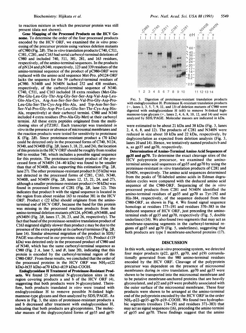
Endoglycosidase H Treatment of Proteinase-Resistant Prod-ucts. We found 15 potential N-glycosylation sites in theregion covering products a and b of the HCV ORF (4),suggesting that both products were N-glycosylated. There-fore, both products translated in vitro were treated withendoglycosidase H to remove possible N-linked high-mannose-type glycans and then analyzed by SDS/PAGE. Asshown in Fig. 3, the sizes of proteinase-resistant products aand b decreased after treatment with endoglycosidase H,indicating that both products are glycoproteins. The molec-ular masses of the deglycosylated forms of gp35 and gp7O
- + - + - + - + - + eH - + - +I l ll
\ 66-
00 4~~~~~~%.4-t" \ 36-_"- 29-
- 24-NI.,ff ,20-
- 17-0_- 14
si--8.1 -
-6.2--2.5-_
1 2 3 4 5 6 7 8 9 10 11 12 13 14
FIG. 3. Digestion of proteinase-resistant translation productswith endoglycosidase H. Proteinase K-resistant translation products(-, lanes 1, 3, 5, 7, 9, 11, and 13) of deletion mutants of C980 weredigested with endoglycosidase H (eH) to remove N-linked high-mannose-type glycans (+, lanes 2, 4, 6, 8, 10, 12, and 14) and wereanalyzed by SDS/PAGE. Molecular masses are indicated in kDa.
were estimated to be about 21 kDa and 38 kDa (Fig. 3, lanes2, 4, 6, 8, and 12). The products of C281 and N340N werereduced in size about 10 kDa and 22 kDa, respectively, bydeglycosylation as expected from deletion analysis (Fig. 3,lanes 10 and 14). Hence, we tentatively named products b anda, as gp35 and gp7O, respectively.
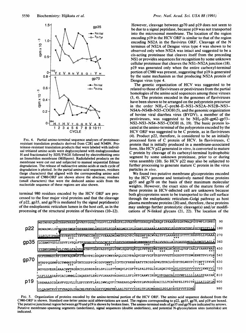
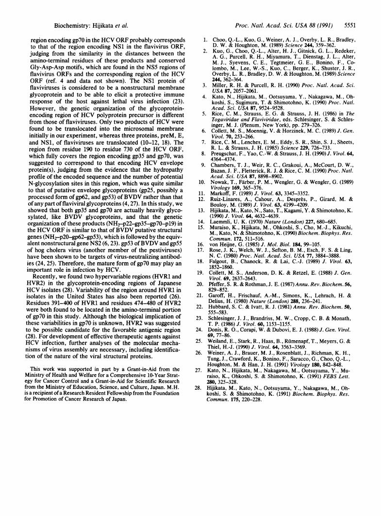
Determination of Amino-Terminal Amino Acid Sequences ofgp35 and gp7O. To determine the exact cleavage sites of theHCV polyprotein precursor, we examined the amino-terminal amino acid sequences of gp35 and gp7O by using theproteinase-resistant in vitro translation products of C281 andN340N, respectively. The amino acid sequences determinedfrom the peaks of 3H-labeled amino acids in Edman degra-dation cycles were compared with the deduced amino acidsequence of the C980-ORF. Sequencing of the in vitroprocessed products from C281 and N340N identified theamino-terminal residues of gp35 and gp7O as Tyr-192 andHis-384, respectively, of the sequence deduced from theC980-ORF, as shown in Fig. 4. We found signal sequencehomologs at residues 175-191 and residues 370-383 in thededuced sequence of HCV ORF, just preceding the amino-terminal ends of gp35 and gp7O, respectively (Fig. 5, doubleunderlines) (16). We also found two segments that may act asmembrane-spanning segments in the carboxyl-terminal re-gions of gp35 and gp7O (Fig. 5, underlines), suggesting thatboth products are type I membrane-anchored proteins (17).
DISCUSSIONIn this work, using an in vitro processing system, we detectedfour major products (p22, gp35, gp7O, and p19) cotransla-tionally generated from the 980 amino-terminal residuesencoded by the HCV ORF. Cleavage of the polyproteinprecursor was dependent on the presence of microsomalmembranes during in vitro translation. gp7O and gp35 wereshown to be transported into the microsomal membrane andto be putative membrane-anchored proteins that are heavilyglycosylated, and p22 and p19 were probably associated withthe outer surface of the microsomal membrane. These fourproducts were shown to be arranged at the amino-terminalend of the polyprotein encoded by the HCV ORF in the orderNH2-p22-gp35-gp70-p19-C00H. We found two hydropho-bic segments (residues 174-191 and residues 371-383) thatmay act as signal sequences (16), preceding the amino terminiof gp35 and gp7O. These findings suggest that the amino
Biochemistry: Hijikata et al.
5550 Biochemistry: Hijikata et al.
C,
EQ.C.
cnC.,
x
EC.)
1 2 3 4 5 6 7 8 9 1-011CYCLE
FIG. 4. Partial amino-terminal sequence analyses of proteinase-resistant translation products derived from C281 and N340N. Pro-teinase-resistant translation products that were labeled with individ-ual tritiated amino acids were deglycosylated with endoglycosidaseH and fractionated by SDS/PAGE followed by electroblotting ontoan Immobilon membrane (Millipore).'Radiolabeled products on themembrane were cut out and subjected to manual sequential Edmandegradation. The release of radioactive amino acids at each cycle ofdegradation is plotted. In the partial amino acid sequences, residues(large characters) that aligned with the corresponding amino acidsequences of C980-ORF are shown above the abscissa; residues(small characters) that were the deduced amino acids'from thenucleotide sequence of these regions are also shown.
terminal 980 residues encoded by the HCV ORF are pro-cessed to the four major viral proteins and that the cleavageof p22, gp35, and gp7O is mediated by the signal peptidase(s)of the endoplasmic reticulum lumen in the host cell, as in theprocessing of the structural proteins of flaviviruses (10-12).
p22
gp35
gp7O
p19
Proc. Natl. Acad. Sci. USA 88 (1991)
However, cleavage between gp7O and p19 does not seem tobe due to a signal peptidase, because p19 was not transportedinto the microsomal membrane. The location of the regionencoding p19 in the HCV ORF is similar to that of the regionencoding NS2A in the flavivirus ORF. Cleavage of the Nterminus of NS2A of Dengue virus type 4 was shown to beobserved only when NS2A was intact and suggested to be acis-acting proteinase that cleaves itself from the precedingNS1 or provides sequences for recognition by some unknowncellular proteinase that cleaves the NS1-NS2Ajunction (18).p19 was generated only when the entire carboxyl-terminalportion of C980 was present, suggesting that p19 is generatedby the same mechanism as that producing NS2A protein ofDengue virus type 4.The genetic organization of HCV was suggested to be
related to those offlaviviruses or pestiviruses from the partialhomologies of the amino acid sequences among these viruses(3, 4). The proteins encoded in the genomes of flaviviruseshave been shown to be arranged on the polyprotein precursorin the order NH2-C-preM-E-NS1-NS2A-NS2B-NS3-NS4A-NS4B-NS5-COOH (5), and the genomic organizationof bovine viral diarrhea virus (BVDV), a member' of thepestiviruses, was suggested to be NH2-p20-gp62-gp53-NS2-NS3-NS4-NS5-COOH (6, 19). The basic protein lo-cated at the amino-terminal ofthe polyprotein encoded by theHCV ORF was suggested to be C protein, as in flaviviruses(4). Product p22, therefore, is considered to be an initiallyprocessed form of C protein of HCV. In flaviviruse's, Cprotein that is initially produced in a membrane-associatedform, like HCV p22 generated in vitro, is converted to matureC protein by cleavage of its carboxyl-terminal hydrophobicsegment by some unknown proteinase, prior to or duringvirus assembly (10). So HCV p22 may also be subjected tofurther processing to generate mature C protein in the virusparticles in vivo.We found two putative membrane glycoproteins encoded
by the HCV genome and tentatively named these proteinsgp35 and gp7O on the basis of their maximum molecularweights. However, the exact sizes of the mature forms ofthese proteins in HCV-infected cell are unknown becauseboth glycoproteins seem to be transported to the cell surfacethrough the endoplasmic reticulum-Golgi pathway as hostplasma membrane proteins (20) and, therefore, these proteinsmay undergo further proteolytic cleavage(s) and/or modifi-cations of N-linked glycans (21, 22). The location of the
MSTNPKPQRKTKRNTNRRPQDVKFPGGGQIVGGVYLLPRRGPRLGVRATRKTSERSQPRGRRQPIPKARRPEGRTWAQPGYPWPLYGNEG 90
MGWAGWLLSP] SR~q~r,'- IqRNT-r.KV~nTL~r-rF~n YTPLV ARAT-A~r-.VRVLE YA~rNT.Prr4F.SlE~j~a180
LLSCLTIPA EVRNVSGIYHVTNDCSNSSIVYEAADMIMHTPGCVPCVRESNFSRCWVALTPTLAARNSSIPTTTIRRHVDLLVGAAA 270
IT.r.AvrDT.rGvVSOLFTFSP360J% lSVJlAALFYAHRVFNJA$VOW'UL
YYMVNWKLIMLF' _VGRVST*** .GS ***TGWHNTLNNSLTFAAFAHFA 450
~~CP~RMASCRP IDK~AQ~WGF IDVPKISDQRPYCWHYAPQLCGIVPASQVCGPVYCFTPSPVVVGTTDGSGVPTYSWGENETDVLVLN 540
NTRPPQGNWFGCTWMNSTGFTKTCGGPPCNIGGVGNNTLICPTDCFRKHPEATYTKCGSGPWLTPRCLVDYPYRLWHYPCTINFTIFKVR 630** **** .i* * igi**,***MYVGGVEHRLNAACNWTRGERCDLEDRDRSELSPLLLSTTEWQVLPCSFTTLPALSTGLIHLHQNIVDVQYLYGVGSVVVSVVIKWEYVL 720
ILLFLLLADARVCACLWMMLLIAQAEATLENLVVLNAASVAGAHGLLSFLVFFCAAWYIKGRLVPGAAYALYGVWPLLLLLLALPPRAYAM,810
DREMAASCGGAVFVGLVLLTLSPYYKVFLARLIWWLQYFITRAEAHLQVWVPPLNVRGGRDAIILLTCAVHPELIFDITKLLLAILGPLM 900
VLQAGMTRVPYFVRAQGLIRACMLVRKVAGGHYVQMAFMKLAALTGTYVYDHLTPLRDWAHAGLRDLAVAVEPVVFSDME 980
FIG. 5. Organization of proteins encoded by the amino-terminal portion of the HCV ORF. The amino acid sequence deduced from theC980-ORF is shown. Standard one-letter amino acid abbreviations are used. The regions corresponding to p22, gp35, gp7O, and p19 are boxed.The putativejunctional region between gp7O and p19 is shown by broken lines. The amino-terminal ends ofgp35 and gp7O are indicated by arrows.Putative membrane-spanning segments (underlines), signal sequences (double underlines), and-potential N-glycosylation sites (asterisks) areindicated.
a I I a I I I
I
Proc. Natl. Acad. Sci. USA 88 (1991) 5551
region encoding gp70 in the HCV ORF probably correspondsto that of the region encoding NS1 in the flavivirus ORF,judging from the similarity in the distances between theamino-terminal residues of these products and conservedGly-Asp-Asp motifs, which are found in the NS5 regions offlavivirus ORFs and the corresponding region of the HCVORF (ref. 4 and data not shown). The NS1 protein offlaviviruses is considered to be a nonstructural membraneglycoprotein and to be able to elicit a protective immuneresponse of the host against lethal virus infection (23).However, the genetic organization of the glycoprotein-encoding region of HCV polyprotein precursor is differentfrom those of flaviviruses. Only two products of HCV werefound to be translocated into the microsomal membraneinitially in our experiment, whereas three proteins, preM, E,and NS1, of flaviviruses are translocated (10-12, 18). Theregion from residue 190 to residue 730 of the HCV ORF,which fully covers the region encoding gp35 and gp7O, wassuggested to correspond to that encoding HCV envelopeprotein(s), judging from the evidence that the hydropathyprofile of the encoded sequence and the number of potentialN-glycosylation sites in this region, which was quite similarto that of putative envelope glycoproteins (gp25, possibly aprocessed form of gp62, and gp53) ofBVDV rather than thatofany part offlaviviral glycoproteins (4, 27). In this study, weshowed that both gp35 and gp7O are actually heavily glyco-sylated, like BVDV glycoproteins, and that the geneticorganization of these products (NH2-p22-gp35-gp70-p19) inthe HCV ORF is similar to that ofBVDV putative structuralgenes (NH2_p20-gp62-gp53), which is followed by the equiv-alent nonstructural gene NS2 (6, 23). gp53 ofBVDV and gp55of hog cholera virus (another member of the pestiviruses)have been shown to be targets of virus-neutralizing antibod-ies (24, 25). Therefore, the mature form of gp7O may play animportant role in infection by HCV.
Recently, we found two hypervariable regions (HVR1 andHVR2) in the glycoprotein-encoding regions of JapaneseHCV isolates (28). Variability of the region around HVR1 inisolates in the United States has also been reported (26).Residues 391-400 of HVR1 and residues 474-480 of HVR2were both found to be located in the amino-terminal portionof gp7O in this study. Although the biological implication ofthese variabilities in gp70 is unknown, HVR2 was suggestedto be possible candidate for the favorable antigenic region(28). For development of effective therapeutic agents againstHCV infection, further analyses of the molecular mecha-nisms of virus assembly are necessary, including identifica-tion of the nature of the viral structural proteins.
This work was supported in part by a Grant-in-Aid from theMinistry of Health and Welfare for a Comprehensive 10-Year Strat-egy for Cancer Control and a Grant-in-Aid for Scientific Researchfrom the Ministry of Education, Science, and Culture, Japan. M.H.is a recipient of a Research Resident Fellowship from the Foundationfor Promotion of Cancer Research of Japan.
1. Choo, Q.-L., Kuo, G., Weiner, A. J., Overby, L. R., Bradley,D. W. & Houghton, M. (1989) Science 244, 359-362.
2. Kuo, G., Choo, Q.-L., Alter, H. J., Gitnick, G. L., Redeker,A. G., Purcell, R. H., Miyamura, T., Dienstag, J. L., Alter,M. J., Syevens, C. E., Tegtmeier, G. E., Bonino, F., Co-lombo, M., Lee, W.-S., Kuo, C., Berger, K., Shuster, J. R.,Overby, L. R., Bradley, D. W. & Houghton, M. (1989) Science244, 362-364.
3. Miller, R. H. & Purcell, R. H. (1990) Proc. Nat!. Acad. Sci.USA 87, 2057-2061.
4. Kato, N., Hijikata, M., Ootsuyama, Y., Nakagawa, M., Oh-koshi, S., Sugimura, T. & Shimotohno, K. (1990) Proc. Natl.Acad. Sci. USA 87, 9524-9528.
5. Rice, C. M., Strauss, E. G. & Strauss, J. H. (1986) in TheTogaviridae and Flaviviridae, eds. Schlesinger, S. & Schles-inger, M. J. (Plenum, New York), pp. 279-326.
6. Collett, M. S., Moennig, V. & Horzinek, M. C. (1989) J. Gen.Virol. 70, 253-266.
7. Rice, C. M., Lenches, E. M., Eddy, S. R., Shin, S. J., Sheets,R. L. & Strauss, J. H. (1985) Science 229, 726-733.
8. Preugschat, F., Yao, C.-W. & Strauss, J. H. (1990) J. Virol. 64,4364-4374.
9. Chambers, T. J., Weir, R. C., Grakoui, A., McCourt, D. W.,Bazan, J. F., Fletterick, R. J. & Rice, C. M. (1990) Proc. Natl.Acad. Sci. USA 87, 8898-8902.
10. Nowak, T., Farber, P. M., Wengler, G. & Wengler, G. (1989)Virology 169, 365-376.
11. Markoff, F. (1989) J. Virol. 63, 3345-3352.12. Ruiz-Linares, A., Cahour, A., Despres, P., Girard, M. &
Bouloy, M. (1989) J. Virol. 63, 4199-4209.13. Hijikata, M., Kato, N., Sato, T., Kagami, Y. & Shimotohno, K.
(1990) J. Virol. 64, 4632-4639.14. Laemmli, U. K. (1970) Nature (London) 227, 680-685.15. Muraiso, K., Hijikata, M., Ohkoshi, S., Cho, M.-J., Kikuchi,
M., Kato, N. & Shimotohno, K. (1990) Biochem. Biophys. Res.Commun. 172, 511-516.
16. von Heijne, G. (1985) J. Mol. Biol. 184, 99-105.17. Rose, J. K., Welch, W. J., Sefton, B. M., Esch, F. S. & Ling,
N. C. (1980) Proc. Natl. Acad. Sci. USA 77, 3884-3888.18. Falgout, B., Chanock, R. & Lai, C.-J. (1989) J. Virol. 63,
1852-1860.19. Collett, M. S., Anderson, D. K. & Retzel, E. (1988) J. Gen.
Virol. 69, 2637-2643.20. Pfeffer, S. R. & Rothman, J. E. (1987) Annu. Rev. Biochem. 56,
829-852.21. Garoff, H., Frischauf, A.-M., Simons, K., Lehrach, H. &
Delius, H. (1980) Nature (London) 288, 236-241.22. Hubbard, S. C. & Ivatt, R. J. (1981) Annu. Rev. Biochem. 50,
555-583.23. Schlesinger, J. J., Brandriss, M. W., Cropp, C. B. & Monath,
T. P. (1986) J. Virol. 60, 1153-1155.24. Donis, R. O., Corapi, W. & Dubovi, E. J. (1988) J. Gen. Virol.
69, 77-86.25. Weiland, E., Stark, R., Haas, B., Rumenapf, T., Meyers, G. &
Thiel, H.-J. (1990) J. Virol. 64, 3563-3569.26. Weiner, A. J., Brauer, M. J., Rosenblatt, J., Richman, K. H.,
Tung, J., Crawford, K., Bonino, F., Saracco, G., Choo, Q.-L.,Houghton, M. & Han, J. H. (1991) Virology 180, 842-848.
27. Kato, N., Hijikata, M., Nakagawa, M., Ootsuyama, Y., Mu-raiso, K., Ohkoshi, S. & Shimotohno, K. (1991) FEBS Lett.280, 325-328.
28. Hijikata, M., Kato, N., Ootsuyama, Y., Nakagawa, M., Oh-koshi, S. & Shimotohno, K. (1991) Biochem. Biophys. Res.Commun. 175, 220-228.
Biochemistry: Hijikata et al.