Development of affordable molecular techniques for the...
84
Development of affordable molecular techniques for the diagnosis of leishmaniasis in Yemen Inauguraldissertation zur Erlangung der Doktorwürde der Naturwissenschaftlichen Fachbereiche Biologie, Chemie und Geowissenschaften im Fachbereich Biologie der Justus-Liebig-Universität Giessen vorgelegt von Abdullatif Ali Master of science Biochemisches Institut der Medizinischen Fakultät der Justus-Liebig-Universität Giessen Giessen, Juli 2009
Transcript of Development of affordable molecular techniques for the...

Development of affordable molecular techniques for the diagnosis of leishmaniasis in Yemen
Inauguraldissertation zur Erlangung der Doktorwürde
der Naturwissenschaftlichen Fachbereiche
Biologie, Chemie und Geowissenschaften
im Fachbereich Biologie
der Justus-Liebig-Universität Giessen
vorgelegt von
Abdullatif Ali
Master of science
Biochemisches Institut
der Medizinischen Fakultät der Justus-Liebig-Universität Giessen
Giessen, Juli 2009

I
ZUSAMMENFASSUNG
Die Leishmaniose ist eine durch einzellige Parasiten der Gattung Leishmania
verursachte Infektionskrankheit, die durch den Stich von Sandmücken übertragen
wird. Als erstes klinisches Symptom bildet sich eine nicht abheilende ulceröse
Wunde von bis zu mehreren cm Durchmesser um die Einstichstelle herum. Nach
mehreren Monaten kann die Wunde unter Hinterlassung einer Narbe spontan
abheilen. Falls es sich um eine Infektion mit einer weniger virulenten Form des
Parasiten gehandelt hat, ist der Patient geheilt und für den Rest des Lebens resistent
gegen diese Form. Andere Stämme des Parasiten lösen aber die mucocutane Form
der Krankheit aus, die zu schmerzhaften destruktiven Läsionen der Lippen- und
Nasenschleimhäute führt, oder die viszerale Form der Leishmaniose, die innere
Organe angreift und unbehandelt tödlich verläuft. In der Republik Jemen sind die
cutane und die viszerale Leishmaniose endemisch, werden aber aus Mangel an Geld
in der Bevölkerung weder hinreichend diagnostiziert noch behandelt.
Aufzeichnungen über epidemiologische Daten gibt es nur für wenige staatliche
Krankenhäuser, die Zahl der unregistrierten Fälle ist wahrscheinlich sehr hoch. Um in
Zukunft zu Kontrollmaßnahmen gegen die Krankheit beitragen zu können, wurde der
Versuch unternommen, neue molekulare Diagnoseverfahren zu entwickeln, die
einerseits sehr empfindlich und spezifisch sind, andererseits aber nicht zu kompliziert
und teuer, so dass sie unter den limitierten Voraussetzungen des Landes realisiert
werden können.
Um eine Einblick in das wahre Ausmaß der Krankheit im Jemen zu erhalten,
wurden die in den staatlichen Krankenhäusern in den letzten Jahren gesammelten
Daten ausgewertet. Außerdem wurden in verschiedenen Teilen des Landes mehr als
200 Proben von Patienten mit dem Verdacht auf cutane oder viscerale Leishmaniose
gesammelt. Dies Proben wurden zunächst im Jemen mikroskopisch auf cutane
Leishmaniose und mit Hilfe einer sehr unspezifischen Formol-Präzipitation-Methode
auf viscerale Leishmaniose untersucht. Für einige Proben stand ein kommerzieller
ELISA zur Verfügung. Von allen gesammelten Blutproben wurde die Plasmafraktion
und aus angereicherten Leukozyten die DNA gewonnen und für nähere Analysen zur
Universität Giessen gebracht.
Von den mittlerweile verfügbaren Sequenzdaten für die Leischmania-Stämme
L. infantum (viszerale Leishmaniose) und L. major (cutane Leishmaniose) wurden
II
spezifische PCR-Primer für die Entwicklung neuer Diagnoseverfahren abgeleitet. Die
neuen Verfahren wurden anhand von gereinigter DNA dieser beiden im Labor
kultivierten Stämme getestet. Zur Entwicklung neuer PCR-Verfahren wurden fünf
verschiedene Zielsequenzen von hoch-repetitiven Regionen der Genome
ausgewählt. Zwei Genus-spezifische Tests und ein Test, der die Unterscheidung von
cutaner und viszeraler Leishmaniose zulässt, wurden erfolgreich entwickelt.
Die Anwendung dieser Tests für die im Jemen gesammelten Patientenproben
ergab, dass die PCR-Diagnose wesentlich empfindlicher und spezifischer ist, als die
traditionell eingesetzten Methoden. Freilich konnte im Rahmen dieser Arbeit weder
ein vollständiges Bild über die Epidemiologie der Leishmaniose im Jemen noch ein
endgültig perfektes Diagnoseprotokoll entwickelt werden. Dies wird Aufgabe für die
folgenden Jahre sein. Immerhin weisen die ermittelten Daten einerseits darauf hin,
dass die Lage der Leishmaniose im Jemen sehr viel schwerwiegender ist, als offiziell
zugegeben. Andererseits wurden diagnostische Verfahren entwickelt, die
ausreichend einfach und so kostengünstig sind, dass sie im Land selbst eingeführt
werden können. In optimierter Form könnten diese Verfahren zu einem besseren
Stand der Kenntnis über diese Krankheit im Land führen und auf Dauer zu einer
Verbesserung der gesundheitlichen Situation der Bevölkerung beitragen

III
SUMMARY
Leishmaniasis is an infectious disease caused by different species of
unicellular Leishmania parasites which are transmitted by the bite of sandflies. As
first clinical symptome, a non-healing ulcerous wound of up to several centimeters
diameter forms around the byte of the insect. After several months, the wound may
heal spontaneously leaving a scar. If the infection was due to a less virulent species
of the parasite, the patient is healed and resistant against this form for the rest of life.
However, other strains of the parasite lead to mucocutaneous leishmaniasis which
lead to painfull destructions of the mucosa of lipps and nose, or to visceral
leishmaniasis which affects inner organs and is lethal without treatment. Cutaneous
as well as visceral leishmaniasis are endemic in the Republic of Yemen, but due to
limiting financial resources, the disease is neither appropriatelytly diagnosed nor
treated in the population.
Recorded epidemiological data exist only for some governmental hospitals,
but the number of unregisterd cases may be huge. In order to shed more light in the
real extent of the disease in Yemen and to contribute to future control measures, it
was attempted to design new molecular procedures for the diagnosis, which are, at
the one hand, highly sensitive and specific, at the other hand not too complicated and
inexpensive, so that they could be performed under the limiting conditions of the
country.
To obtain some insight in the sitation of leishmaniasis, the data registered in
governmental hospitals during the past years were collected and evaluated. In
addition, samples from more than 200 patients with suspected cutaneous and
visceral leishmaniasis were collected in different parts of the country. These samples
were analysed first in Yemen by microscopy for cutaneous leishmaniasis and by a
highly unspecific procedure called formol gel precipitation for visceral leishmaniasis
Some of the samples were also analysed by a commercial ELISA. From all collected
blood samples the plasma fraction and DNA from enriched leucocytes were prepared
and transported to the University of Giessen for further analysis.
By using the available sequence data of the Leishmania strains L. infantum
(visceral leishmaniasis) and L. major (cutaneous leishmaniasis), specific PCR
primers were developed for the design of new diagnostic procedures. The assays
were tested by means of purifed DNA from these two strains which had been

IV
cultivated in the laboratory. Five different target sites from highly repeated sequences
of the genomes were selected to develop new diagnostic PCR assays. Two assays
specific for the genus Leishmania, and one assay allowing differentiation of the two
strains were constucted allowing to discriminate between cutaneous and visceral
leishmaniasis.
Applying these tests on the patient samples collected in Yemen revealed that
PCR diagnosis is more sensitive and specific than the traditional methods used. Of
course, neither a comprehensive epidemiology of Yemen nor a ultimative perfect
diagnostic protocol could be obtained during this work. This has to be established
during the following years. However, the results reveal at the one hand that the
situation of leishmaniasis in Yemen is more severe than officially stated. At the other
hand, diagnostic procedures were developed which are simple and inexpensive
enough to be introduced in the country. When optimized, these techniques may lead
to more awareness of this disease in the country and to an improvement of the health
condition of the suffering population.

V
CONTENT
ZUSAMMENFASSUNG ..................................................................................................... I
Summary ................................................................................................................. III
CONTENT ................................................................................................................... V
1. INTRODUCTION ........................................................................................................ 1
1.1 Epidemiology of leishmaniasis .......................................................................... 1
1.2 The Parasite ...................................................................................................... 2
1.3 Immunology of Leishmania infections ............................................................... 6
1.4 Leishmaniasis and HIV-coinfection ................................................................... 8
1.5 Treatment of leishmaniasis ................................................................................ 9
1.6 Transmission of leishmaniasis ......................................................................... 10
1.7 Introduction to Yemen ...................................................................................... 11
1.8 Leishmaniasis in Yemen .................................................................................. 15
1.9 Diagnosis of leishmaniasis .............................................................................. 15
1.10 Objective of the study ..................................................................................... 18
2. MATERIALS AND METHODS .......................................................................... 20
2.1 Instruments ........................................................................................................20
2.2 Chemicals ................................................................................................. ...... 20
2.3 Leishmania parasites ....................................................................................... 21
2.4 Patient samples .................................................................................................22
2.5 DNA purification .................................................................................................23
2.6 Purification of recombinant Taq DNA polymerase .............................................24
2.7 Polymerase chain reaction (PCR) ................................................................... 26
2.8 DNA gel electrophoresis .................................................................................. 28
3. RESULTS ............................................................................................................ 31
3.1 Evaluation of epidemiological data on leishmaniasis in Yemen ...................... 31
3.2 Collecting samples of leishmaniasis patients in Yemen ...................... ........... 36
3.3 Laboratory diagnosis of patient samples in Yemen ......................................... 38
3.4 Establishing new procedures for the diagnosis of leishmaniasis ..................... 38
3.5 Establishing new diagnostic PCR procedures ................................................. 39
3.5.1 Kinetoplast minicircle(kDNA)-specific PCR .................................................. 41
3.5.2 18S rRNA specific PCR ................................................,................................ 43
3.5.3 45-basepair repeat-specific PCR .............................,.................................... 47
3.5.4 64-basepair repeat-specific PCR .................................................................. 48

VI
3.5.5 39-basepair repeat-specific PCR .................................................................. 50
3.5.6 Compilation of the PCR results .................................................................... 53
4. DISCUSSION ........................................................................................................ 57
4.1 Epidemiology of leishmaniasis ........................................................................ 58
4.2 Diagnosis of leishmaniasis .............................................................................. 61
4.3 Collection and processing of patient samples ................................................. 61
4.4 Kinetoplast DNA (kDNA)-specific PCR ............................................................ 62
4.5 18S ribosomal RNA-specific PCR ................................................................... 63
4.6 PCR assays with tandem repeat sequences ................................................... 65
4.7 Concluding remarks ........................................................................................ 66
5. REFERENCES ....................................................................................................... 68
ACKNOWLEDGEMENTS ............................................................................................... 77
1
1. INTRODUCTION
1.1 Epidemiology of leishmaniasis
Leishmaniasis is a protozoan parasitic disease which is prevalent in many
parts of the tropics, subtropics, and Southern Europe. The disease is caused by
infection with different species of Leishmania, which are transmitted by the bite of
phlebotomine sand flies. The most common forms of leishmaniasis in the Old World
are cutaneous leishmaniasis (CL), causing skin sores (review: Reithinger et al., 2007),
and visceral leishmaniasis (VL) , affecting internal organs of the body such as spleen,
liver, and bone marrow (review: Berman, 2006). A third form of the disease,
mucocutaneous leishmaniasis (MCL) leading to severe destruction of mucosal areas
of the mouth, nose and pharynx occurs exclusively in Latin America (reviewed by
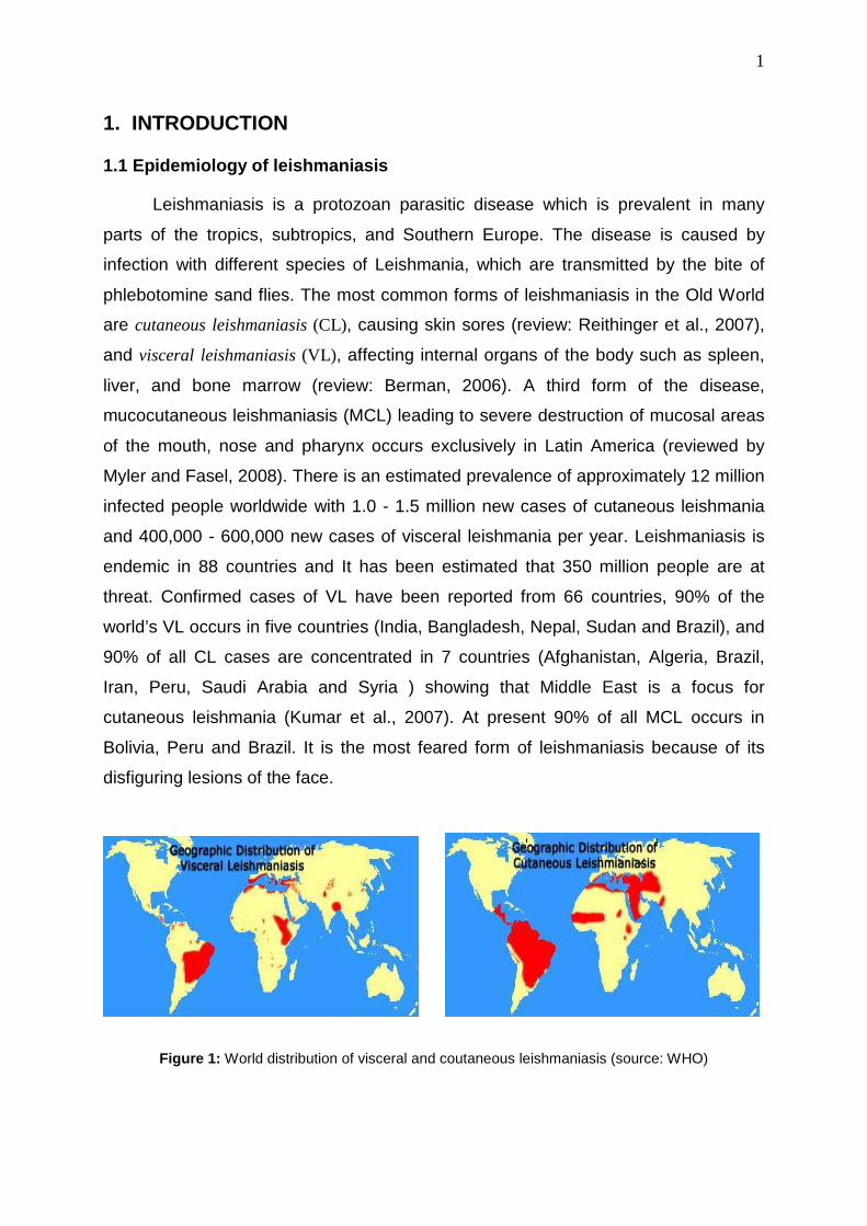
Myler and Fasel, 2008). There is an estimated prevalence of approximately 12 million
infected people worldwide with 1.0 - 1.5 million new cases of cutaneous leishmania
and 400,000 - 600,000 new cases of visceral leishmania per year. Leishmaniasis is
endemic in 88 countries and It has been estimated that 350 million people are at
threat. Confirmed cases of VL have been reported from 66 countries, 90% of the
world’s VL occurs in five countries (India, Bangladesh, Nepal, Sudan and Brazil), and
90% of all CL cases are concentrated in 7 countries (Afghanistan, Algeria, Brazil,
Iran, Peru, Saudi Arabia and Syria ) showing that Middle East is a focus for
cutaneous leishmania (Kumar et al., 2007). At present 90% of all MCL occurs in
Bolivia, Peru and Brazil. It is the most feared form of leishmaniasis because of its
disfiguring lesions of the face.
Figure 1: World distribution of visceral and coutaneous leishmaniasis (source: WHO)

2
1.2 The Parasite
Leishmania is a protozoon belonging to the order of Kinetoplastida and to the
family of Trypanosomatidae. The genus Leishmania includes more than 20 different
species. The parasites exist in two morphological forms: the non-motile intracellular
amastigote (3-5 micrometer in diameter) living in macrophages of the mammalian
host, and the motile extracellular promastigote (15-30 micrometer in length, plus the
flagellum) living in the intestinal tract of the sandfly vector. The amastigotes are able
to survive inside the macrophages and to multiply within the acidic phagolysosomes
of these host cells (reviewed by Alexander et al., 1999).
After infection by a bite of a sandfly, promastigotes enter macrophages,
transform into amastigotes within 12-24 h and continue multiplication until the host
cell dies. The released amastigotes infect other macrophages and the infection
spreads. The parasite contains two striking organelles, the nucleus and the
kinetoplast. The kinetoplast is found in all protozoa of the order kinetoplastidae
(Leishmania, Trypanosoma, Plastocrithidia, Crithidia, Endotrypanum, Herpetomonas,
Leptomonas, Phytomonas, and Wallaceina). It is a rod-shaped mitochondrial
structure consisting of a DNA network with two types of DNA: about 10,000
minicircles of approximately 2 kilobasepairs (kb) and 25-250 maxicircles of
approximately 30 kb each. These together constitute the mitochondrial genome
(reviewed by Singh 2006 ).
Figure 2: Intracellular and extracellular leishmanial amastigotes in a Giemsa-stained smear made
from scrapings of cutaneous lesions (bright-field microscopy, x 1000; pictures from Al-Jawabreh,
2005).

3
The life cycle of Leishmania:
During its life cycle, Leishmania is alternating between two major forms: as
extracellular promastigotes in the gut of the sand fly and as amastigotes inside the
macrophages of the mammalian host. When the sand fly feeds, promastigotes are
injected into the skin and are engulfed by host mononuclear phagocytes, where they
transform into amastigotes and multiply within the phagolysosomal compartment until
the phagocytic cells are destroyed, releasing the parasites to enter further cells and
repeat the cycle.The life cycle is shown in Figure 3.
Figure 3: Scheme of the life cycle of Leishmania (picture from Rey, 2007)
Clinical symptoms of leishmaniasis
The three different clinical forms of leishmaniasis, cutaneous leishmaniasis
(CL), visceral leishmaniasis (VL) and mucocutaneous leishmaniasis (MCL) and their
4
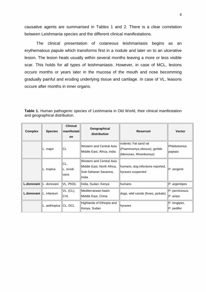
causative agents are summarised in Tables 1 and 2. There is a clear correlation
between Leishmania species and the different clinical manifestations.
The clinical presentation of cutaneous leishmaniasis begins as an
erythematous papule which transforms first in a nodule and later on to an ulcerative
lesion. The lesion heals usually within several months leaving a more or less visible
scar. This holds for all types of leishmaniasis. However, in case of MCL, lesions
occure months or years later in the mucosa of the mouth and nose becomming
gradually painful and eroding underlying tissue and cartilage. In case of VL, leasons
occure after months in inner organs.
Table 1. Human pathogenic species of Leishmania in Old World, their clinical manifestation and geographical distribution.
Complex Species
Clinical
manifestati
on
Geographical
distribution Reservoir Vector
L. major CL Western and Central Asia
Middle East, Africa, India
rodents: Fat sand rat
(Psammomys.obesus), gerbils
(Meriones, Rhombomys)
Phlebotomus
paptasi
L. tropica
CL,
L. recidi-
vans
Western and Central Asia
Middle East, North Africa,
Sub-Saharan Savanna,
India
humans, dog infections reported,
hyraxes suspected P. sergenti
L.donovani L. donovani VL, PKDL India, Sudan, Kenya humans P. argentipes
L.donovani L. infantum VL, (CL),
CVL
Mediterranean basin
Middle East, China dogs, wild canids (foxes, jackals)
P. perniciosus,
P. ariasi
L. aethiopica CL, DCL Highlands of Ethiopia and
Kenya, Sudan hyraxes
P. longipes,
P. pedifer

5
Table 2. Human pathogenic species of Leishmania in New World , their clinical manifestation and geographical distribution.
Mucocutaneous leishmaniasis is manifested solely in South America,
especially in Brazil, Paraguay, Ecaudor, Bolivia, Peru, Colombia, and Venezuela. The
highest percentage of the cases occur in Brazil, Bolivia, and Peru. Twenty percent of
all leishmaniasis patients in Brazil develop MCL. It is a chronic and very serious
condition, developing years after self-cure of cutaneous lesions. Mucosal lesions can
progress to involve the entire nasal mucosa and the hard and soft palates. Without
treatment, the entire nasal mucosa and palates become deformed with ulceration and
erosion of the nasal septum, lips, and palate. The disease attacks cartilaginous
areas, but usually spares bony structures, and it can leave extreme disfigurement. It
can be lethal, mostly by aspiration pneumonia (Myler and Fasel, 2008).
Visceral leishmaniasis has many different symptoms as the disease
developes. At the beginning, headache and fever occurre. Lateron, sweating with
cough, diarrhea, vomiting, bleeding of the gums and weight loss are observed. In the
late stage of disease, the clinical symptoms transforme to hepatomegally,
splenomegally, anemia with leucopenia and lymphadenopathy. The mortality ranges
from one year in acute cases up to 2-3 years in chronic cases. (Garg and Dube
2006).
Complex Species Clinical
manifestation Geographical distribution Reservoir Vector
L. braziliensis L. braziliensis CL, MCL Central and South America forest rodents Lutzomyia sp.
L. braziliensis L. panamanensis CL Central America, Columbia sloths Lutzomyia sp.
L. braziliensis L. guyanensis CL Guyana, Brazil sloths Lutzomyia sp.
L. braziliensis L. peruviana CL Peru, Argentine dogs, humans? Lutzomyia sp.
L. mexicana L. mexicana CL, Chiclero's
ulcer
Central and South America,
Texas forest rodents Lutzomyia sp.
L. mexicana L. amazonensis CL, DCL Brazil, Venezuela,
mostly north of the Amazon
forest rodents,
Opossums Lutzomyia sp.
L. donovani L. chagasi VL, atypical CL,
CVL
Central and South America
CL in Honduras, Nicaragua dogs, wild canids
Lutzomyia
longipalpis

6
1.3 Immunology of Leishmania infections
Within the mammalian host, Leishmania is able to survive as an amastigote in
phagocytic cells such as macrophages, dendritic cells and neutrophils. The entering
promastigote forms of the parasites are attacked by the complement system,
however, some of them survive and enter the macrophages via the C3 receptor
(Figure 4).
Figure 4: Life cycle of L. major. After inoculation of metacyclic promastigotes of L. major into the skin by the bite of a sand fly, parasites activate complement and are partially lysed. Surviving parasites utilize C3 to invade macrophage (MΦ) host cells via CR3, and – within these cells – they transform into obligate intracellular amastigotes. MΦ infection does not lead to cell activation (e.g. IL-12 release). Inside of MΦ, parasites replicate and more amastigotes are released into the tissue. The life cycle of L. major is completed upon uptake of parasites by another bite of a sand fly. (Reproduced from von Stebut, 2007)

7
Because the organisms are located intracellularly, an effective immune
response includes mainly cell-mediated mechanisms for resistance to the parasite.
Cytokines produced by T cells, natural killer (NK) cells, and antigen-presenting cells
(APC) play an essential role in this process. (Roberts, 2006). The outcome of
infection is determined by the parasite species and the host’s immunological
response. The CD4+ T helper cell is critical with animal models demonstrating that
cure is associated with strong IFN-gamma, interleukin (IL)-2 and IL-12 responses in
the absence of classical Th2 cytokines or IL-10.
The outcome of the infection appears also to be influenced by the saliva of the
transmitting sand fly. The saliva contains a repertoire of pharmacologically active
molecules that hampers the host's haemostatic, inflammatory and immune
responses. This interferes with the early interactions between Leishmania and the
host's immune response. There are concrete indications that the host response
against sand fly saliva influences disease outcome in leishmaniasis (Andrade et al.,
2007, Figure 5).
Figure 5: Effects of Lutzomyia longipalpis saliva on vertebrate host. When the sand fly saliva is injected into host skin, it induces an inflammatory cell infiltration (1), and antibody production (2). In this scenario, immune complexes are formed [20] at early phases of exposure. Moreover, sand fly saliva also modulates co-stimulatory molecules and cytokine release by antigen presenting cells. (Reproduced from Andrade et al., 2007)

8
Cutaneous leishmaniasis (CL) is worldwide by far the most frequent
consequence of the disease. In the Old World, CL is also known as Oriental Sore,
Delhi boil, Baghdad boil and Quetta Sore (Myint et al., 2008) and is usually caused
by L. major, L, tropica and L. aethiopica. In Latin America, CL is caused by other
Leishmania strains (see Table 2). This form of disease heals spontaneously after few
months leading to life-long immunity against re-infection and is normally not treated
with drugs. Healing is characterized by induction of IFN-gamma releasing T-cells
(Kurtzhals et al., 1994), whereas failure to cure is associated with elevated levels of
IL-4 with low IFN-gamma responses from Leishmania-specific T-cells (Ajdary e al.,
2000). As all forms of leishmaniasis begin with a cutaneous lesion at the site of entry
of the parasites, clear diagnostic differentiation at this stage would be needed to
discriminate CL from the severe forms MCL and VL.
MCL is mostly caused by Leishmania species belonging to the L. braziliensis
complex. However, it can also be caused by L. panamanensis (Barral et al., 1992).
The serious hazard of developing MCL after cure of CL is estimated to be up to 40%.
The immunology of MCL has not yet been studied in detail.
Visceral Leishmaniasis (VL) or Kala azar is usually caused by members of the
L. donovani complex (Singh 2006). L. donovani is found to be the causative agent in
endemic areas of Bangladesh, India and Sudan, L. d. infantum is widly distributed in
Middel East and in Mediterranean countries and L. d. chagasi is the only causative
agent in the New World. VL is characterized by suppression of T cell activity in acute
disease due to increased expression of IL-10 (Ghalib et al., 1993). Increased
expression of classical Th2 cytokines has been reported in VL with elevated levels of
IL-4 particularly associated with treatment faillure (Sundar et al., 1997). Elevated
levels of IL-13 have been observed in active disease that returned to normal after
successful treatment (Babaloo et al., 2001).
1.4 Leishmaniasis and HIV-coinfection
Leishmaniasis has been identified as a frequent opportunistic infection in
patients infected with human immunodeficiency virus type-1 (HIV-1) which amplifies
in cells of the immune system thus reducing the ability to resist other infections. It has

9
been observed that the visceral form of leishmaniasis accelerates the course of HIV-
1 disease progression and shortens the life expectancy of persons in areas where
both diseases are endemic (Pourahmad 2009).
The greatest prevalence of co-infection with HIV and Leishmania have been
observed in the Mediterranean basin with more than 2,000 cases reported from 35
countries to the World Health Organization. About 90 per cent of all cases come from
Spain, Italy, France and Portugal. In Africa the increase of co-infection with HIV has
been reported in Ethiopia and Sudan which is mainly caused by social troubles such
as war or increase of population in urban and suburban areas in regions where both
vector and reservoir are abundant. In South America, the majority of co-infections
has been reported in Brazil.(Cruz et al., 2005)
1.5 Treatment of leishmaniasis
Pentavalent antimony-containing compounds are the main drugs used to treat
leishmaniasis. These drugs need to be delivered by intramuscular or intravenous
routes daily for 4-6 weeks and are quite toxic leading to severe side effects including
serious heart arrhythmias, pancreatitis and and hepatic dysfunction. They cannot be
used for pregnant women. In rich countries the fungizide amphotericin B in liposomal
form in has become the treatment of choice. Since short time, a new oral agent,
Miltefosine, has been licensed in India, Germany and Colombia for use in adults and
children. Both drugs have lower side effects and show high cure rates. However, the
cost of these agents restrict a more widespread use (Roberts, 2006).
By these reasons, less virulent cutaneous leishmaniasis is normally left to self-
healing. Treatment is only important for the severe forms MCL and VL and to reduce
scarring when cosmetically sites are involved. Plastic surgery may be needed to
correct disfigurement by destructive facial lesions caused by cutaneous
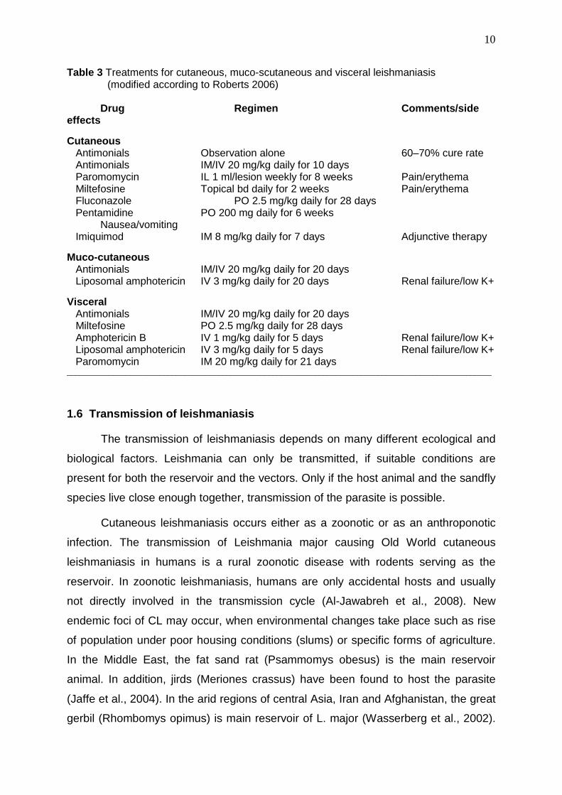
leishmaniasis. The most common drugs used for treatment of leishmaniasis, their
application and side effects are listed in Table 3.
10
Table 3 Treatments for cutaneous, muco-scutaneous and visceral leishmaniasis (modified according to Roberts 2006) Drug Regimen Comments/side effects
Cutaneous Antimonials Observation alone 60–70% cure rate Antimonials IM/IV 20 mg/kg daily for 10 days Paromomycin IL 1 ml/lesion weekly for 8 weeks Pain/erythema Miltefosine Topical bd daily for 2 weeks Pain/erythema Fluconazole PO 2.5 mg/kg daily for 28 days Pentamidine PO 200 mg daily for 6 weeks Nausea/vomiting Imiquimod IM 8 mg/kg daily for 7 days Adjunctive therapy
Muco-cutaneous Antimonials IM/IV 20 mg/kg daily for 20 days Liposomal amphotericin IV 3 mg/kg daily for 20 days Renal failure/low K+
Visceral Antimonials IM/IV 20 mg/kg daily for 20 days Miltefosine PO 2.5 mg/kg daily for 28 days Amphotericin B IV 1 mg/kg daily for 5 days Renal failure/low K+ Liposomal amphotericin IV 3 mg/kg daily for 5 days Renal failure/low K+ Paromomycin IM 20 mg/kg daily for 21 days _____________________________________________________________________________________________________
1.6 Transmission of leishmaniasis
The transmission of leishmaniasis depends on many different ecological and
biological factors. Leishmania can only be transmitted, if suitable conditions are
present for both the reservoir and the vectors. Only if the host animal and the sandfly
species live close enough together, transmission of the parasite is possible.
Cutaneous leishmaniasis occurs either as a zoonotic or as an anthroponotic
infection. The transmission of Leishmania major causing Old World cutaneous
leishmaniasis in humans is a rural zoonotic disease with rodents serving as the
reservoir. In zoonotic leishmaniasis, humans are only accidental hosts and usually
not directly involved in the transmission cycle (Al-Jawabreh et al., 2008). New
endemic foci of CL may occur, when environmental changes take place such as rise
of population under poor housing conditions (slums) or specific forms of agriculture.
In the Middle East, the fat sand rat (Psammomys obesus) is the main reservoir
animal. In addition, jirds (Meriones crassus) have been found to host the parasite
(Jaffe et al., 2004). In the arid regions of central Asia, Iran and Afghanistan, the great
gerbil (Rhombomys opimus) is main reservoir of L. major (Wasserberg et al., 2002).

11
The main insect vector for transmission of L. major is the sand fly species
Phlebotomus papatasi (EL- Badry et al., 2008). In Kenya, L. tropica has been
isolated from hyraxes (procavia ssp. German: Klippschliefer) (Sang et al., 1993).
Hyraxes are also the main reservoir of L. aethiopica in Ethiopia (Ashford et al., 1973).
In some urban centers of Middle East and Asia exist completely anthroponotic
life cycles of the parasites, i.e. human beings are the main or only reservoir host. In
such places cutaneous leishmaniasis caused by L. tropica can be highly endemic,
but no animal reservoir is to be recognized. In Central and South Western Morocco,
the transmission of L. tropica is anthroponotically as well. The parasite is mainly
transmitted by sandflies of the species Phlebotomus sergenti (Ramaoui et al., 2008).
Prevention of infection
The best way to prevent leishmaniasis is by personal protection from sandfly
bites, which includes for example long sleeve clothing in evening hours. The use of
insecticides for house and space spraying reduce sandfly populations, and fine-
weave pyrethroid-impregnated bednets have successfully been used in Burkina
Faso, Sudan, and Columbia (Markle and Makhoul, 2004).
1.7 Introduction to Yemen
The Republic of Yemen – once ruled by the Queen of Sheba, and still in our
days full of historical tresures from ancient times - is situated in the southwestern
area of the Arabian peninsula. The country which covers about 460,000 km2 is
bordered by Saudi Arabia in the north, by the Arabian Sea and Indian Ocean the
south, by the Red Sea in the west and by the Sultanate of Oman in the east (Figure
6)
Yemen is characterized by varied landscape, diversified terrain and climate.
Overlooking the coastal plains are ranges of low and high altitude mountains, many
of which are terraced. Throughout, Yemen maintains plateaus, hills and plains. These
can be wide in some areas or narrow in others. Green wadis (or valleys) are

12
riverbeds for the rainwater runoff during the two rainy seasons in December to
February and May to June. Desert extends eastward and northward. This natural
terrain is broken down into the coastal region, the plateaus, the mountainous
highlands and desert of the Empty Quarter.
Figure 6: Map of the Republic of Yemen
(source: http://www.maparchive.org/details.php?image_id=415

13
The most populated coastal area includes the lower plains overlooking the
Red Sea, the Gulf of Aden and the Arabian Sea. The plains are interconnected to
form a coastal strip starting with the Tehama (meaning hot land), which extends from
Gizan in the North to Bad Al-Mandab Strait southwards. This region also includes a
range of mountains, with an altitude ranging between 200 to 900 meters above sea
level. It also encompasses the coastal plains tapering into the Gulf of Aden. The total
length of this coastline reaches approximately 2,000 km and covers approximately
3000 km2. Administratively, this area is divided in the governates Aden, parts of
Hadhramaut and Al-Mahara.
The Plateaus Region in the East is reaching elevations as hihg as 1,000
meters is weakly populated and extends towards the Empty Quarter desert, with
water flowing down from the mountains to wadis like famous Wadi Hadhramaut,
which has been an essential centre of Arabian culture since the second millennium
b.C. The peripheries of the Plateaus Region intertwine from the east with the rough
terrain of Ramlat Al-Sabein and desert of Saada, Al-jawf, Shabwa, Hadhramaut and
Al-Mahara.
The Mountain Highlands Region was formed as a result of the African split
from Asia, which led to the cleavage forming the Red Sea and the Gulf of Aden. The
altitude of Yemen's mountain range reaches between 1,000 to 3,600 meters above
sea level. The summit of Jabal (Mount) Al-Nabi Shu'aib with an altitude of 3,666
meters and a snowy cap during winter time is the highest elevation in the Arabian
Peninsula. At the foot of the highland are different flat basins and many wadis with
land for agriculture and water reservoirs. Wadis flowing into the Gulf of Aden and the
Arabian Sea include Wadi Tiban, Wadi Bana, Wadi Ahwar and Wadi Hadhramaut.
Other wadis slope eastward towards the desert (Wadi Khabb, Wadi Al-Jawf and Wadi
Adhanah) or towards the North and Northeast (Wadi Harib, Wadi Marakhah, Wadi
Jirdan, Wadi Aiwah Al-Sai'ar, Wadi Rammah and Wadi Sha'at). Administratively, the
Mountain Highlands Region comprises the Governorates of Raimah, Sana'a, Hajjah,
Al-Mahweet, Sa'ada, Dhamar, Ibb, Ta'ez, AI-Beidha and Al-Dhali'a.
The almost unpopulated Empty Quarter represents the desert region of
Yemen. It is intertwined with wild vegetation, especially in the peripheries that run into
the Plateau Region through wadis and sand beds. Vegetative life and water get
scarce inward of the Empty Quarter, whereas shifting sand dunes increase. These

14
dunes bury archeological sites and ruins. Former names of the Empty Quarter region
were Rijraj Sea, the Safi Sea, the Great Desert and Al-Ahqaf Desert.
Climate
Yemen is a country with a high level of solar radiation all around the year.
Diversity in topography provides varied climate. In the coastal areas, the climate is
characterized by high temperatures and humidity during summer and moderate
temperatures during winter. Moderate climate prevails in the western slopes,
plateaus and flat land. Temperatures reach 10-30° C entigrade and may fall below 0°
C in winter. Humidity can reach 80%. The annual average rainfall is 300 to 1,000
mm. The climate in the eastern parts of Yemen differs considerably with
temperatures exceeding 40° C during the summer fall ing to 10°-15° C in the winter.
Annual rainfall does not exceed 50-100 mm, especially in the periphery of the Empty
Quarter. The transitory area is found between these two climatic regions. extending
from north and east of Sana'a to the western parts of Marib. The entire area of
Yemen is affected by monsoon winds blowing from the east due to low air pressures
in the west. The monsoons are usually accompanied by rain in the summer and
lesser rain in winter.
Population
The population of Yemen is 19.7 million people according to the population
census of 2004. The annual population growth rate has declined from 3.7 % in 1994
to 3.0 %. This is seen as a major success in curbing population growth.
About 27 % of the population lives in major cities and 73 % live in rural areas.
The movement of people is a decisive factor in the re-distribution of population
among the different regions of Yemen. The rate of urban growth reaches 9 % in the
Capital Secretariat (City of Sana'a). Sana’a includes about 28 % of the total urban
population of the country followed by Hodeida and Aden with 16 % and 14 %.
With a per capita-income of only 450 Euro per year (in 2008), Yemen is by far
the poorest country in Near and Middle East. More than two thirds of the inhabitants
live from less than two US-Dollars per day. The health system of the country is
underdeveloped, children and mother mortality are high, and life expectancy is low.

15
Medical installations and qualified personell are limited and the population, especially
women and children, suffer from deficiencies of medical care. Under these
circumstances, there exist no much information on specific diseases such as
leishmaniasis.
1.8 Leishmaniasis in Yemen
There are very few reports on leishmaniasis in Yemen in the international
literature. Even though it is not well documented, the disease seems to be endemic in
the country, and is primarily widespread in arid and semiarid areas. It is also endemic
in in the plateau and mountainous areas of Hajjah and Amran governorates.
Cutaneous leishmaniasis has first been reported in Sana’a as early as 1933 (Sarnelli
1933). In 1986, Leishmania tropica was identified in 18 patients in Naghdi Dhamran
and Wadi Dhamran (Rioux 1986). Another study revealed 42 cases of cutaneous
leishmaniasis in the Hajjah governorate and neighboring regions (reviewed by Khatri
et al., 2006). Leishmania major as well as Leishmania tropica account for the majority
of infections in Yemen. Pratlong et al. (1995) reported from a patient who developed
localized cutaneous lesions due to Leishmania donovani sensu stricto, following a
stay in Yemen. The patient lived in southern France and visited Yemen for 2 weeks
during November 1992 when he was bitten by sandflies. He had not traveled
elsewhere in the previous 3 years. Studies of Leishmania strains in Yemen and in
countries close to Yemen suggest that coutaneous leishmaniasis is mainly caused by
L. major and L. tropica, whereas the agent of visceral leishmaniasis is L. infantum
(Boyer, 1993; Piarroux et al., 1995; Khatri et al., 2006; Nasereddin et al., 2009)
1.9 Diagnosis of Leishmaniasis
Parasitological diagnosis is considered to be golden standard in diagnosis of
leishmaniasis because of its high specificity. It includes microscopic examination of
Gimsa- stained smears derived from biopsies of cutaneous lesions, or aspirates from
lymph nodes, bone marrow, or spleen. Histopathological examination of fixed lesion
biopsies or culture of biopsies is also performed. Microscopic examination is still a
widespread method in endemic countries, because it is quite inexpensive and does
not require sophisticated laboratory equipment. In contrast, culture in combination

16
with multilocus enzyme electrophoresis allowing identification of the parasite species
and leading to definite diagnosis requires a wealth of technical training and takes
long time to obtain results. Disadvantages of microscopy and culture are the low
sensitivity, especially in the chronic stage of the disease when the number of
parasites decreases (Reithinger, 2007).
Serological methods
Indirect diagnostical methods include the relatively sensitive, but not highly
specific diagnostic procedure is the Leishmanin skin test (LST) also known as the
Montenegro reaction (Montenegro, 1926). Leishmanial antigen (killed promastigotes),
or disrupted promastigotes in pyrogen – free phenol saline - is applied intradermally
and a delayed hypersensitivity reaction can be observed. A cross-reaction has been
reported with tuberculosis. Leishmanin skin test is used as an indicator of the
distribution of cutaneous and mucocutaneous leishmaniasis in both humans and
animals, in visceral leishmaniasis usually only after treatment and cure (Sadeghian et
al., 2006).
Most sensitive and specific test especially for the diagnosis of visceral
leishmaniasis include serological methods such as the enzyme linked
immunosorbent assay (ELISA). Since few years, a highly specific recombinant
antigen, K39, which is part of a kinesin-related gene and contains a repetition of 39
amino acid residues has been developed and is being widely used for diagnosis
meanwhile. K39-ELISA was found to be more sensitive and specific than
immunoassays including crude perasite extracts such as the direct agglutination test
(DAT) and soluble antigen (SA) ELISA used before. In India, recombinant K39 has
been widely used for detecting visceral leishmaniasis even in immunocompromised
patients with VL-HIV co-infection (Singh, 2006).
Western blots have been used as a diagnostic tool to identify by anti-
leishmanial antibodies by Leishmania antigens. In the western blot technique several
antigenic products can be distinguish by different antibodies in one assay. Before
introduction of recombinant K39, the western blot technique proved to be the most
sensitive and specific serological methods for diagnosis of leishmaniasis. The
development of the rk39 dipstick test it is successfully employed in the diagnosis of
17
VL as will as PKDL in India. It was found to be 95-100 % sensitive when tested in VL
Patients. (Salotra et al., 2003)
The direct agglutination test (DAT) is a comparatively simple and inexpensive
test suitable for field work and for screening patient sera in the laboratory (Harith et
al., 1986). The method is based on the agglutination of stained promastigotes
available either as a suspension or in a freeze- dried form with Leishmania-specific
antibodies. The freeze-dried form is more stable and improves the usefulness of the
DAT in the field. The sensitivity of the test is almost 100 %, and the specificity for
leishmaniasis is equally high except of countries with Chagas disease, where
crossreactivity with Trypanosoma cruzi is being observed (El Harith et al., 1988).
Molecular methods
The polymerase chain reaction (PCR) has offered a new dimension in the
diagnosis and characterization of various infectious agents, enabling to perform tests
in few hours. It proved to be very useful for the diagnosis of leishmaniasis. In recent
studies many different PCR assays have been developed for the diagnosis of
Leishmania DNA in a variety of clinical samples such as peripheral blood, skin
biopsies, bone marrow and lymph node aspirates. In several studies comparison of
PCR was found to be more sensitive than microscopy and culture for diagnosis of VL,
especially in immune- compromised patients. Sensitivities between 80 and 90 %
have been reported which is markedly better than microscopy and culture
(approximately 55 % sensitivity each). Different targets have been chosen for the
PCR, multicopy sequence repeats such as genes for ribosomal RNAs and kinetoplast
DNA are usually prefered (Schallig and Oskam, 2002). The genes of the small
subunit ribosomal RNA (SSUrRNA) consist of both conserved and variable regions
which are almost species-specific and can be used for the development of specific
diagnostic approaches. The kinetoplast DNA (kDNA) contains the most highly
repetitive sequences in Leishmania. It consists of approximately 10.000 copies of
plasmid-like minicircles of different size, but all of them share a conserved region of
120-140 bp. This region contains two short highly conserved sequences which are
perfectly homologous in all Leishmania species. This fact has been utilized for a
genus-specific diagnostic PCR approach (Review: Henk et al., 2002).

18
1.10 Objective of the study
The objective of this study was to shed some light in the epidemiology of
leishmaniasis in the Republic of Yemen and to develop simple molecular approaches
for the detection of the disease under the limited economical conditions of the
country. The official data on leishmaniasis provided by the government are
incomplete and may only indicate the peak of an iceberg of under-reported cases.
Diagnosis of cutaneous leishmaniasis in Yemen is based solely on
microscopic examination, which yields a good sensitivity in acute infection but is of
limited use in the chronic phase of the disesase when the number of parasites in
biopsy samples decreases. Diagnosis of visceral leishmaniasis by microscopic
examination of spleen aspirates is rarely used because it is technically demanding
and needs trained personell. Instead, a formol gel test is wildly used for the diagnosis
of visceral leishmaniasis because it is very cheap, but the specificity reaches hardly
50 %. In order to get a more detailed aspect on the epidemiology of leishmaniasis in
Yemen and to contribute to a more specific and sensitive diagnosis, the following
issues were addressed:
• To collect information of incidence of leishmaniasis in different parts of Yemen.
• To collect information on control measures and treatment of leishmaniasis in
Yemen.
• To take blood and tissue samples from patients suspected to have cutaneous
or visceral leishmaniasis.
• To analyse the tissue samples of CL patients (Giemsa-stained smears from
the wound) by microscopy in Yemen.
• To harvest lymphozytes from the blood samples and extract DNA for later
PCR analysis.
• To prepare serum (or plasma) from the blood samples for later immunological
analysis.
• To establish affordable procedures for molecular diagnosis of human
leishmaniasis in the laboratory in Giessen by designing primers which lead to
high specificity and sensitivity in the PCR assays. These procedures had to

19
bo be simple, easy to perform and inexpensive in order to make their use
realistic in a poor country.
• To increase sensitivity and specificty of diagnostic tests by developping nested
PCR assays.
• To transfer the new molecular technology to Yemen in order to improve
diagnosis of the disease in the country.

20
2 MATERIALS and METHODS
2.1 Instruments
Gel electrophoresis systems
Horizontal minigel system (8 x 8 cm) AGS, Heidelberg, Germany
Vertical minigel chamber (8 x 10 cm) Keutz, Reiskirchen, Germany
Power supply EPS 500/400 Pharmacia, Freiburg, Germany
Shakers
Certomat R Braun, Melsungen, Germany
Vortex Genie 2 Scientific Industries, Bohemia, NY, USA
Centrifuges
Cooling centrifuge Beckman J2-21, Beckman Instruments, Summerset, USA
Microfuge: Biofuge Pico Heraeus Instruments, Hanau, Germany
Multifuge 3 Heraeus Instruments, Hanau, Germany
Waterbath
GFL Wasserbad 1013 Gesellschaft für Labortechnik, Burgwedel,
Germany
Thermocyclers
T1 Thermocycler 96 Biometra GmbH, Göttingen, Germany
Primus 96 MWG Biotech AG, Ebersberg, Germany
Photographic equipment
UV Transilluminator Herolab GmbH, Wiesloch, Germany
Polaroid MP-4 Land Camera Polaroid Corporation, Cambridge, MA, USA
Film: Polaroid 667 Professional Polaroid Corporation, Cambridge, MA, USA
2.2 Chemicals
Acrylamid Serva, Heidelberg, Germany
Agarose for gel electrophoresis Sigma-Aldrich, Munich, Germany
Ammoniumpersulfate (APS) Serva, Heidelberg, Germany
Ammoniumsulfate Carl Roth GmbH, Karlsruhe, Germany
Ampicillin Sigma-Aldrich, Munich, Germany

21
Adenil Triphosphate (ATP) Carl Roth GmbH, Karlsruhe, Germany
Biorex 70 resin BIO-RAD Laboratories, Hercules, USA
Coomassie Brilliant Blue R250 Serva, Heidelberg, Germany
Dithiothreitol (DTT) Biomol, Heidelberg, Germany
dNTP-Set (100 mM per dNTP) Carl Roth, Karlsruhe, Germany
Ethidium bromide Serva, Heidelberg, Germany
Ethylenediaminetetraacetic acid (EDTA) Serva, Heidelberg, Germany
Fetal calf serum Sigma-Aldrich, München, Germany
Glutamine Carl Roth, Karlsruhe, Germany
Guanidiniumhydrochlorid (GuHCl) ICN Biomedicals, Germany
Heparin sepharose Pharmacia-LKB, Upsala, Sweden
Medium 199 Sigma-Aldrich, München, Germany
ß-Mercaptoethanol Serva, Heidelberg, Germany
MEM Earle’s Medium Gibco BRL, Eggenstein
N,N,N‘,N‘-Tetramethylethylendiamine (TEMED) Serva, Heidelberg, Germany
Oligonucleotides MWG-Biotech, Ebersberg, Germany
Phenylmethylsulphonylfluoride (PMSF) Carl Roth GmbH, Karlsruhe, Germany
Penicillin/Streptomycin Carl Roth GmbH, Karlsruhe, Germany
Polyethyleneimine Sigma-Aldrich, München, Germany
Tween-20 Serva, Heidelberg, Germany
Triton X-100 Merck, Darmstadt, Germany
Tris (hydroxymethyl) aminomethane Carl Roth GmbH, Karlsruhe, Germany
2.3 Leishmania parasites
L. major and L. infantum were obtained from Prof. Bernhardt Fleischer, Bernhard-
Nocht-Institut, Hamburg. They were cultivated in vitro using either medium 199 plus
10 % heat-inactivated fetal calf serum, or Earle’s medium supplemented with

22
glutamine and 10 % heat-inactivated fetal calf serum at 30°C in the presence of 5 %
CO2. A mixture of penicilline and streptomycin (from 100 x penicillin/streptomycin
stock solution) was added to prevent bacterial contaminations. After 3-4 days,
parasites were transfered in 3 volumes of fresh medium and further incubated. The
parasites were harvested by centrifugation for 20 min at 2.000 x g and frozen.
2.4 Patient samples
Blood samples were collected from approximately 150 patients visiting
hospitals in six different regions in north Yemen and the central blood bank in Sana’a
with lesions suspected to be cutaneous leishmaniasis over a period of more than two
years. Skin scrapings were taken from the borders of the wound and analysed
microscopically. As this kind of diagnosis has a limited sensitivity (approximately 70
%), all blood samples were kept and processed as described below for further
analysis by molecular methods lateron. Most of the examined patients came from
endemic areas (urban as well as rural), had been travelling inside the country, or had
been stationed as soldiers in the desert area. Blood samples from 26 patients with
suspected visceral leishmaniasis were collected in several locations in the western
part of Yemen.
3-5 ml of collected blood were processed immediately by sedimenting the
blood cells using a manual centrifuge (see Figure 10 in the Result section). Plasma
and buffy coat were collected as described below. Plasma was conserved by the
addition of 50 % glycerol, and the DNA of the purified buffy coat was stabilized in 500
µl of guanidinium-hydrochloride lysis buffer. Whenever possible, the samples were
kept at 4°C, however, they were transported for sev eral days at ambient
temperatures without loss of biological activity.
Microscopical analysis of skin scrapings
Small quantities of tissue obtained by skin scrapings were smeared on glass
slides, air dried and fixed with methanol for a few seconds. Giemsa stain was filtered
and diluted 1:20 with phosphate buffer (pH 7.2). After 20 minutes of staining the
slides were washed with tap water and air dried. The stained smears were examined
under the microscope with a 40 x lens and with a 100 x oil immersion lens. If at least
one intra- or extra-cellular amastigote with a distinctive kinetoplast was found the
23
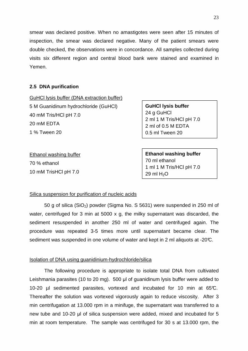
GuHCl lysis buffer 24 g GuHCl 2 ml 1 M Tris/HCl pH 7.0 2 ml of 0.5 M EDTA 0.5 ml Tween 20
smear was declared positive. When no amastigotes were seen after 15 minutes of
inspection, the smear was declared negative. Many of the patient smears were
double checked, the observations were in concordance. All samples collected during
visits six different region and central blood bank were stained and examined in
Yemen.
2.5 DNA purification
GuHCl lysis buffer (DNA extraction buffer)
5 M Guanidinum hydrochloride (GuHCl)
40 mM Tris/HCl pH 7.0
20 mM EDTA
1 % Tween 20
Ethanol washing buffer
70 % ethanol
10 mM TrisHCl pH 7.0
Silica suspension for purification of nucleic acids
50 g of silica (SiO2) powder (Sigma No. S 5631) were suspended in 250 ml of
water, centrifuged for 3 min at 5000 x g, the milky supernatant was discarded, the
sediment resuspended in another 250 ml of water and centrifuged again. The
procedure was repeated 3-5 times more until supernatant became clear. The
sediment was suspended in one volume of water and kept in 2 ml aliquots at -20°C.
Isolation of DNA using guanidinium-hydrochloride/silica
The following procedure is appropriate to isolate total DNA from cultivated
Leishmania parasites (10 to 20 mg). 500 µl of guanidinum lysis buffer were added to
10-20 µl sedimented parasites, vortexed and incubated for 10 min at 65°C.
Thereafter the solution was vortexed vigorously again to reduce viscosity. After 3
min centrifugation at 13.000 rpm in a minifuge, the supernatant was transferred to a
new tube and 10-20 µl of silica suspension were added, mixed and incubated for 5
min at room temperature. The sample was centrifuged for 30 s at 13.000 rpm, the
Ethanol washing buffer 70 ml ethanol 1 ml 1 M Tris/HCl pH 7.0 29 ml H2O

24
pellet resuspended in 500 µl 70 % ethanol washing buffer, centrifuged for 30 s at
13.000 rpm and the supernatant removed. The pellet was washed in the same way
three more times with ethanol washing buffer. The last pellet was centrifuged again
and residual ethanol was removed. 10-20 µl of TE buffer (10 mM Tris/HCl pH 7.5, 0.1
mM EDTA) were added, incubated for 5 min at 55°C an d centrifuged for 1 min at
13.000 rpm. The supernatant was collected and the pellet eluted with another 10-20
µl TE buffer. The supernatants were combined.
DNA of Trypanosoma cruzi was a kind gift of Prof. Bernhardt Fleischer,
Bernhard-Nocht-Institut, Hamburg.
DNA extraction from buffy coat
2-3 ml of citrate or EDTA-blood were centrifuged for 15 minutes at 1000 x g.
Then, supernatant plasma was collected and kept in the refrigerator. The yellow thin
“buffy coat” layer on top of the sedimented erythrocytes was collected with a pipette
(200 to 300 µl) and transferred to an Eppendorf tube. 1.2 ml of RBC lysis buffer (300
mM NH4Cl, 30 mM NH4HCO3, 30 mM KCl. 0.1 mM EDTA) were added, mixed shortly
by vortexing, incubated 10 minutes at room temperatures, inverting the tube several
times. Then, the tube was centrifuged 2 minutes at 2000 x g, and the supernatant
removed. A pellet of 10-30 µl was obtained and mixed with DNA extraction buffer.
DNA was extracted as described above.
2.6 Purification of recombinant Taq DNA polymerase
Taq DNA polymerase was purified from a recombinant strain of E. coli (E. coli
XL-1 Blue/pQE-Taq) expressing an exonuclease-free mutant of the enzyme. The
strain was constructed previously in the laboratory of Prof. E. Beck, University of
Giessen (unpublished results). The thermostable enzyme can also be purified without
the cooling steps described in the following protocol.
1 litre of LB medium containing 25 µg/ml kanamycin and 50 µg/ml ampicillin
was inoculated with 100 ml of an over night culture of XL-1 Blue pQE-Taq cells. The
cells were grown at 37°C with optimal aeration for 1-2 hours. At an OD600 of 1.8, 1 ml
of 1 M IPTG was added and then the culture vigorously shaked for another 4 hours.
Cells were harvested by centrifugation (yield ~5 g) and frozen at -20°C. Freezing of

25
the cells can be omitted, but is recommended if the enzyme preparation is not
performed immediately. The cells were resuspended in 15 ml TMN buffer (50 mM
Tris-HCl pH 8.5, 1 mM EDTA, 20 mM (NH4)2SO4) together with 8 mg of lysozyme
(from 10 mg/ml stock solution) and incubated for 15 min at 20-25°C. After adding
200 µl of 10 % Triton X-100 and 200 µl of 10 % Tween 20, the culture was mixed and
incubated at 80°C for 20 min in a 50 ml screw-cap F alcon tube. After the heating
step, 250 µl of 100 mM PMSF were added to prevent proteolytic degradation.
DNA was sheared by 1 min sonification (it is also possible to do it by repeated
pressing through a syringe first with a 1 mm diameter needle and then with smaller
needles) and centrifuged 15 min at 20.000 x g at 4°C. The supernatant (~15 ml) was
collected and 0.6 ml of 5 M NaCl (0.2 M final concentration) were added.
The DNA was precipitated with polyethyleneimine (PEI) by adding dropwise
500 µl of 5 % PEI solution, mixing and incubating in ice for 10 min. The sample was
centrifuged at 10.000 x g for 5 min. 4 aliquots of 500 µl of supernatant were
transferred to Eppendorf tubes each and mixed with increasing amounts (2-8 µl) of 5
% PEI solution. They were incubated 5 min on ice, centrifuged and the amount of
pellet compared. The minimal amount of PEI to precipitate DNA quantitatively was
determinated and added to the bulk extract (usually 100-200 µl; use 1/100 volume of
5 % PEI solution in excess). The suspension was left 20 min on ice and centrifuged
at 20.000 x g for 20 min. The supernatant was collected and diluted 6 fold with KTA
buffer (20 mM Tris/HCl pH 8.5, 10 mM beta-mercaptoethanol, 10 % (w/v) glycerol,
0.1 mM EDTA, 0.05 % Triton X 100, 0.05 % Tween 20).
The excess of PEI was removed by passing the extract through a 2 ml BioRex
70 column equilibrated in KTA buffer + 30 mM ammonium sulphate. A disposable 4
ml plastic column (International Sorbent Technology, Hengoed, Mid Glamorgan, UK)
was used. The column was rinsed with 2 ml KTA buffer and the flow-through loaded
on another plastic column containing 2 ml heparin sepharose equilibrated with KTA
buffer, 30 mM ammonium sulphate. The column was washed first with 50 ml KTA
buffer, 40 mM ammonium sulphate. Thereafter, the column was washed with 20 ml
KTA buffer, 40 mM ammonium sulphate, 50 % glycerol. Taq DNA polymerase was
eluted with KTA buffer, 150 mM ammonium sulphate, 50 % glycerol. 0.5 ml fractions
were collected and 3 µl aliquots analysed on a 12.5 % SDS polyacrylamide gel. The
enzyme was found in fractions 4 to 7. The enzyme was stored at -20°C. For long

26
term storage, Tween 20 to 1 % final concentration was added. The yield was
approximately 30.000 units of enzyme at a concentration of 80 units/µl in the peak
fraction.
2.7 Polymerase chain reaction (PCR)
For all PCR assays, the same Taq DNA polymerase buffer was used. It is
prepared as a 10 x buffer and has the following composition:
1 x Taq polymerase buffer
40 mM Tricine-KOH (pH 8.7 at 25°C)
15 mM K acetate
3.5 mM acetate
0.1 % gelatine
0.05 % Tween 20
Standard PCR reaction assay
Using efficient standard thermocyclers such as T1 thermocycler 96 from
Biometra, or Primus 96 from MWG Biotech which are appropriate for 200 µl reaction
tubes, have a heated lid and heating/cooling rates of at least 2-3°C/s, highly
reproducible results were obtained with assays as small as 10 µl. A standard PCR
reaction mixture had the following composition:
1 µl 10 x Taq buffer
1 µl mixture of 4 dNTPs (2.5 mM each)
0.5 µl 10 µM forward primer
0.5 µl 10 µM reverse primer
1 µl template DNA
5.8 µl H2O
0.2 µl Taq DNA polymerase (5 units/µl)
10.0 µl
10 x Taq polymerase buffer (10 ml) 4 ml 1 M Tricine-KOH 1.5 ml 1 M K acetate 350 µl 1 M acetate 2.5 ml 4 % heated gelatine solution 500 µl Tween 20 1.15 ml H2O
27
The standard program of the thermocycler was set as follows:
step 1: 2 min at 94°C (denaturing of DNA templat e)
step 2: 35 cycles of [94°C for 20 s, 60°C for 1 min, 72°C for 1 min]
step 3: 5 min at 72°C (optional, to fill-in the DNA ends completely)
The same conditions were applied for nested PCR. 1 µl of a 1:10 dilution (with
H2O) of the result of first PCR assay was used as DNA template.
Avoiding contamination
The sensitivity of PCR diagnosis is very high. Theoretically DNA from less
than one parasite can be detected. This implied a high risk for contamination, which
had to be strictly avoided. As a general rule, the extraction of DNA, the preparation of
the PCR and electrophoresis of the PCR products should be strictly separated. It is
also recommended to use different sets of pipettes for the different parts of the work.
Due to limitations of space and technical equipment this standard could not always
be met. Instead, extra precautions had to be taken.
• DNA extraction from clinical material and preparation of the PCR was
performed in a hood, located in a small room separated from the rest of
the laboratory. PCR-cycling and gel electrophoresis were performed on
an extra bench.
• All solutions used for DNA extractions were divided in small aliquots
from a stock solution and discarded in case of a suspected
contamination
• An extra pipette (“dirty pipette”) was used exclusively for loading of the
gels with the higly amplified PCR products.
• Filter tips were used for DNA extraction and for all pipetting steps for
PCR.
• Gloves were changed during the procedures from time to time.
• The rotor of the micro-centrifuge was washed prior to DNA extractions
since the tops of the 1.5 ml tubes came in contact with the upper
margin of the cups holding the tubes. Since also highly amplified DNA

28
was centrifuged in the same centrifuge, these cups were assumed to be
a source of contamination.
• Whenever possible 200 µl tips were used instead of 10 µl tips. Due to
the length of the 200 µl tips the shaft of the pipette could not touch the
1.5 ml tubes from inside.
Negative controls
The extraction of DNA as well as the PCR had to be monitored strictly for
possible cntamination. Two negative extraction controls were routinely used for every
extraction series. The PCR itself was monitored by one reaction in every PCR, which
contained only the reagents and no template.
2.8 DNA gel electrophoresis
Agarose gel electrophoresis buffer (E-buffer)
40 mM Tris/acetate (pH 8,0)
40 mM Na acetate
2.0 mM EDTA
Acrylamide gel electrophoresis buffer (TBE-buffer)
90 mM Tris/borate pH 8.3
2.5 mM EDTA
20 x E-buffer 193.8 g Tris-base 131.2 g Na acetate 160 ml 0.5 M EDTA adjust pH 8.3 with acetic acid add H2O to 2 litre
10 x TBE-buffer 108 g Tris-OH
55 g boric acid
40 ml 0.5M EDTA
adjust pH 8.0 with acetic acid
add H2O to 1 litre
29
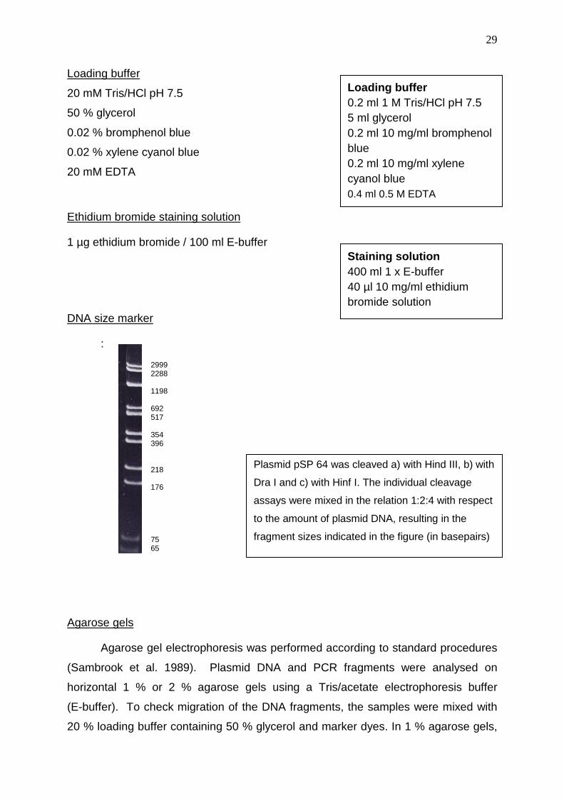
Loading buffer
20 mM Tris/HCl pH 7.5
50 % glycerol
0.02 % bromphenol blue
0.02 % xylene cyanol blue
20 mM EDTA
Ethidium bromide staining solution
1 µg ethidium bromide / 100 ml E-buffer
DNA size marker
:
Agarose gels
Agarose gel electrophoresis was performed according to standard procedures
(Sambrook et al. 1989). Plasmid DNA and PCR fragments were analysed on
horizontal 1 % or 2 % agarose gels using a Tris/acetate electrophoresis buffer
(E-buffer). To check migration of the DNA fragments, the samples were mixed with
20 % loading buffer containing 50 % glycerol and marker dyes. In 1 % agarose gels,
Loading buffer 0.2 ml 1 M Tris/HCl pH 7.5 5 ml glycerol 0.2 ml 10 mg/ml bromphenol blue 0.2 ml 10 mg/ml xylene cyanol blue 0.4 ml 0.5 M EDTA
Staining solution 400 ml 1 x E-buffer 40 µl 10 mg/ml ethidium bromide solution
2999 2288 1198 692 517 354 396 218 176 75 65
Plasmid pSP 64 was cleaved a) with Hind III, b) with
Dra I and c) with Hinf I. The individual cleavage
assays were mixed in the relation 1:2:4 with respect
to the amount of plasmid DNA, resulting in the
fragment sizes indicated in the figure (in basepairs)

30
bromphenol blue migrates at the position of 200 bp, whereas xylenecyanol blue
migrates at the position of 300 bp. The gels were stained for 20 min with ethidium
bromide staining solution. Then they were rinsed with tape water, the DNA bands
visualised on an UV transilluminator and photographed by a Polaroid camera.
Alternatively, a simple digital camera in combination with a yellow filter was used.
Polyacrylamide gels
For the analysis of proteins, discontinuous polyacrylamide gels (10 x 8 x 0.1
cm) containing SDS (Lämmli, 1970) were prepared in different concentrations
according to the following recipe:
Stacking gel Separating gel
10 ml 16 ml 16 ml
Stacking gel buffer:
125 mM Tris-HCl (pH 6.8)
0.1 % SDS
Separating gel buffer:
375 mM Tris-HCl (pH 8.8)
0.1 % SDS
6 % 12,5 % 15 %
40 % Acrylamide /
1.3 % bisacrylamide solution 1,5 ml 5,0 ml 6,0 ml
4 x buffer 2,5 ml 4,0 ml 4,0 ml
H2O 6,0 ml 7,0 ml 6,0 ml
Ammonium persulphate 10 µg 10 µg 10 µg
TEMED 15 µl 20 µl 20 µl
4 x Stacking gel buffer 12.14g Tris-OH 8 ml 10% SDS adjust pH 6.8 with HCl (~8 ml HCl 37%) add H2O to 200 ml
4 x Separating gel buffer 45.43 g Tris-OH 10 ml 10% SDS adjust pH 8.8 with HCl (~6 ml HCl 37 %) add H2O to 250 ml

31
3 RESULTS
3.1 Evaluation of epidemiological data on leishman iasis in Yemen
Official data on the prevalence rate of leishmaniasis in Yemen have been
obtained from the Ministry of Health and from the Office of Surveillance of Disease in
the capital Sana’a. However, these data have to be regarded with care, as they
include only infections reported from governmental hospitals, the two biggest ones
located in Sana’a and Taizz. Much of the medical treatment for the population is
managed in private clinics, or by non-governmental organisations which do not report
to the central administration of the country. Furthermore, in the absence of any
support for health care, poor people do not seek medical aid for small wounds such
as the ulcers of cutaneous leishmaniasis. Therefore, the real number of Leishmania
infections is most probably by far higher than officially documented. The data
obtained from the Ministry of health for the years 2005 to 2008 are shown in Table 4.
Table 4 :Leishmaniasis in the diferent governates of Yemen in 2005-2008
2005
Gov
erna
te
Rim
ah
Aby
an
Ibb
San
a’a
city
Alm
ahra
Had
arm
ot
Ald
hale
Ade
n
Am
ran
Alh
udai
dah
Alb
aida
h
Alg
awf
Mar
ib
Dha
mar
Lahj
Tai
z
Sad
ah
Sha
bwah
Alm
ahw
eet
San
a’a
Haj
jah
tota
l
number of patients 58 0 15 247 0 279 5 1 62 0 18 90 19 12 4 53 13 16 10 17 72 991
male 40 0 12 201 0 240 5 1 45 0 14 75 14 8 4 45 10 12 7 12 60 805
female 18 0 3 46 0 39 0 0 7 0 4 15 5 4 0 8 3 4 3 5 12 176
cutaneous leishmaniasis 55 0 14 227 0 249 5 1 60 0 18 90 19 11 4 51 13 16 9 17 65 924
visceral leishmaniasis
3 0 1 20 0 0 0 0 2 0 0 0 0 1 0 2 0 0 1 0 7 37
2006
Gov
erna
te
Rim
ah
Aby
an
Ibb
San
a’a
city
Alm
ahra
Had
arm
ot
Ald
hale
Ade
n
Am
ran
Alh
udai
dah
Alb
aida
h
Alg
awf
Mar
ib
Dha
mar
Lahj
Tai
z
Sad
ah
Sha
bwah
Alm
ahw
eet
San
a’a
Haj
jah
tota
l
number of patients 642 0 81 230 0 153 12 5 76 44 5 242 91 57 85 130 43 3 0 17 54 1970
male 480 0 60 160 0 96 7 0 46 27 5 220 72 45 70 110 37 3 0 12 45 1495
female 162 0 21 70 0 57 5 5 30 17 0 22 19 12 15 20 6 0 0 5 9 475
cutaneous leishmaniasis 605 0 77 220 0 153 12 5 70 44 5 242 91 57 85 125 43 3 0 12 50 1899
visceral leishmaniasis
37 0 4 10 0 0 0 0 6 0 0 0 0 0 0 5 0 0 0 0 4 66

32
Table 4 continued
2007 G
over
nate
Rim
ah
Aby
an
Ibb
San
a’a
city
Alm
ahra
Had
arm
ot
Ald
hale
Ade
n
Am
ran
Alh
udai
dah
Alb
aida
h
Alg
awf
Mar
ib
Dha
mar
Lahj
Tai
z
Sad
ah
Sha
bwah
Alm
ahw
eet
San
a’a
Haj
jah
tota
l
number of patients 65 1 51 343 0 74 15 1 44 9 2 20 89 61 37 303 31 0 4 0 29 1179
male 55 1 47 320 0 65 12 1 35 6 2 16 81 55 33 370 28 0 4 0 25 1156
female 10 0 4 23 0 9 3 0 9 3 0 4 8 6 4 33 3 0 0 0 4 123
cutaneous leishmaniasis 58 1 48 327 0 74 15 1 44 9 2 20 89 58 37 293 31 0 4 0 26 1137
visceral leishmaniasis
7 0 3 16 0 0 0 0 0 0 0 0 0 3 0 10 0 0 0 0 3 42
2008
Gov
erna
te
Rim
ah
Aby
an
Ibb
San
a’a
city
Alm
ahra
Had
arm
ot
Ald
hale
Ade
n
Am
ran
Alh
udai
dah
Alb
aida
h
Alg
awf
Mar
ib
Dha
mar
Lahj
Tai
z
Sad
ah
Sha
bwah
Alm
ahw
eet
San
a’a
Haj
jah
tota
l
number of patients 70 0 6 367 2 120 13 7 32 0 7 25 89 40 110 105 0 5 85 32 45 1160
male 62 0 4 350 2 109 10 6 24 0 5 22 81 32 98 92 0 5 78 25 35 1040
female 8 0 2 17 0 11 3 1 8 0 2 3 8 8 12 13 0 0 7 7 10 120
cutaneous leishmaniasis 60 0 5 355 2 120 13 7 32 0 7 25 89 35 110 105 0 5 82 32 36 1120
visceral leishmaniasis
10 0 1 12 0 0 0 0 0 0 0 0 0 5 0 0 0 0 3 0 9 40
__________________________________________________________________________
Figure 7: Distribution of Leishmaniasis in the different governates of Yemen
Cutaneous leishmaniasis Visceral leishmaniasis
33
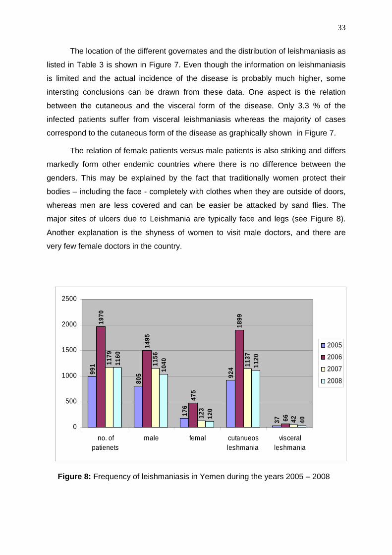
The location of the different governates and the distribution of leishmaniasis as
listed in Table 3 is shown in Figure 7. Even though the information on leishmaniasis
is limited and the actual incidence of the disease is probably much higher, some
intersting conclusions can be drawn from these data. One aspect is the relation
between the cutaneous and the visceral form of the disease. Only 3.3 % of the
infected patients suffer from visceral leishmaniasis whereas the majority of cases
correspond to the cutaneous form of the disease as graphically shown in Figure 7.
The relation of female patients versus male patients is also striking and differs
markedly form other endemic countries where there is no difference between the
genders. This may be explained by the fact that traditionally women protect their
bodies – including the face - completely with clothes when they are outside of doors,
whereas men are less covered and can be easier be attacked by sand flies. The
major sites of ulcers due to Leishmania are typically face and legs (see Figure 8).
Another explanation is the shyness of women to visit male doctors, and there are
very few female doctors in the country.
Figure 8: Frequency of leishmaniasis in Yemen during the years 2005 – 2008
991
805
176
924
37
1970
1495
475
1899
66
1179
1156
123
1137
42
1160
1040
120
1120
40
0
500
1000
1500
2000
2500
no. ofpatienets
male femal cutanueosleshmania
visceralleshmania
2005
2006
2007
2008

34
Figure 9: Cutaneous leishmaniasis. Typical sites of ulcers are face and legs which are
usually most exposed to bite of sandflies.
Another interesting aspect is the number of cases of leishmaniasis during the
different years. In 2006, there was a high increase of the disease as compared to the
other years. This may be explained by the climate, more specifically by the amount of
rainfall which is needed for breeding of the phlebotomine vectors. There are no
measures to control sandflies in Yemen.
Figure 10: Monthly distribution of leishmaniasis Rimah (Western Yemen)
In the fertile areas in the western part of Yemen, the majority of patients with
leishmaniasis are registerd in the middle of summer as exemplified by Figures 10and
11.
1 1 1
6
1516
12
10
8
0 0 00
2
4
6
8
10
12
14
16
18
Janu
ary
Febru
ary
March
April
MayJu
ne July
Augus
t
Septe
mber
Octobe
r
Novem
ber
Decem
ber
mean monthly distribution ofleishmania in Rimah

35
Figure 11: Monthly distribution of leishmaniasis Haija (Western Yemen)
In the arid region in the eastern governates, the majority of cases occurs
between January and March (Figure 12). This area is extremely hot im summer time
and there is almost no rain. The vectors can only develop during the cooler winter
season, and water exists only in so called wadis, deep canyons which carry some
water from the mountains only part of the year.
Figure 12: Monthly distribution of leishmaniasis in Hadramont (Eastern Yemen)
0 0
2
13
16
12
6
18
4
10 0
0
2
4
6
8
10
12
14
16
18
20
Janu
ary
Febru
ary
March
April
MayJu
ne July
Augus
t
Septe
mbe
r
Octobe
r
Novem
ber
Decem
ber
mean monthly distribution ofleishmania in Hajjah
14
22
44
25
35
1
69
5 4
0
5
10
15
20
25
30
35
40
45
50
Janu
ary
Febru
ary
March
April
MayJu
ne July
Augus
t
Septe
mber
Octobe
r
Novem
ber
Decem
ber
mean monthly distribution ofleishmania in Hadramoat

36
3.2 Collecting samples of leishmaniasis patients i n Yemen
In order to obtain more data on the epidemiology of leishmaniasis and to
collect material for future attempts to develop a new, inexpensive, but highly specific
diagnostical procedure, blood samples from patients with suspected leishmaniasis
were collected in several locations in the western part of Yemen. The differential
diagnosis was generally done based on history of exposure (sandfly bites) and
physical examination focussed on ulceric lesions and scar formation. Whenever
possible, scrapings from open ulcers were analysed by microscpopy upon Giemsa
staining in order to detect amastigote parasites inside of infected cells (see Figure 1).
In suspected cases of visceral leishmaniasis, involvement of internal organs was
analysed by palpating liver and spleen.
The blood samples were immediately mixed with EDTA (10 mM final
concentration) and centrifuged with a manual centrifuge (Roth, Karlsruhe, Germany)
in order to sediment the blood cells (Figure 13).
Figure 13: Manual centrifuge used to prepare plasma and buffy coat from blood
samples under field condition
The supernatant plasma was withdrawn and mixed with one volume of
glycerol. The yellow layer on top of the sedimented erythrocytes (buffy coat,
consisting mainly of white blood cells and platelets) was collected with a pipette and
mixed with one volume of 5 M guanidinium hydrochloride dissolved in 20 mM Tris
buffer pH 7.5 and 50 mM EDTA. By this way, the plasma as well as the DNA were

37
conserved and could be kept at ambient temperature for several weeks without to be
damaged. The samples were transported to Germany for detailed laboratory
diagnosis (see below). They served as a basis for developing new diagnostic assays,
the plasma for an immunoassay with recombinant antigens (doctoral thesis of
Michael Heiman in our group), and the DNA after extraction from the buffy coat for
developing new PCR assays as described below.
In addition to patients with open wounds characteristic for cutaneous
leishmaniasis, 26 samples were collected from hospitalized persons suspected to
have the visceral form of disease. Typical symptoms of visceral leishmaniasis are
general weakness, a swollen abdomen (Figure 14) and low counts of white blood
cells. Normally, such persons have no cutaneous wounds, but are hospitalized
because of their weakness. In the absence of good immunological tests, correct
diagnosis is very difficult. In Yemen, a formalin precipitation test is being used (see
Materials and Methods), but this test has a low specificity. This test is being used to
detect the greatly increased serum proteins in visceral leishmaniasis; one drop of
concentrated formalin is added to 1 ml of serum, with rapid and complete coagulation
indicating the positive reaction. The parasites could be found microscopically in
biopsies from liver or bone marrow, but this test is not performed in Yemen. Blood
samples from these persons were collected and processed as described above.
Figure 14: Visceral leishmaniasis
38
3.3 Laboratory diagnosis of the collected patient samples in Yemen
After collection, samples were taken for laboratory confirmation of
leishmaniasis. Basically, there are four major categories of diagnostic methods used
routinely: Parasitological methods (for the detection of amastigotes in Giemsa-
stained smears of scrapings from the wound, aspirates of bone marrow or lymph
nodes), in vitro cultivation, immunological tests such as the enzyme-linked
immunosorbent assay (ELISA), and molecular methods (polymerase chain reaction,
PCR). In Yemen, only microscopical analysis was performed.
Scrapings from the wounds of all 119 patients suspected of having cutaneous
leishmaniasis were analysed by microscopical examination in the International
Central Blood Bank in Sana’a, Yemen during the years 2005-2006. According to this
analysis, 47 of the patients (39 %) were found to be positive. 25 patients were male
(53%) and 22 were female (47%). The majority of the positive persons were in the
age group 15-34 years (55%). The age group less than 15 years was represented by
14 patients (30%). The highest prevalence rate of disease (74%) was seen in
patients comming from rural areas.
It is well known that microscopical analysis has a sensitivity of maximal 70 %
only (Al-Jawabreh, 2008), i.e. at least one third of the actual infections could not be
confirmed by this way. Furthermore, it is not possible to discriminate different species
of Leishmania by microscopy. Several of the cases characterized as cutaneous
leishmaniasis could have been caused by L. infantum and would correspond to
visceral leishmaniasis. The samples from the suspected visceral leishmaniasis
patients were no futher alanlysed in Yemen. The samples of all patients were
transported to the laboratory in Giessen for further analysis.
3.4 Establishing new procedures for the diagnosis of leishmaniasis
Commercially available diagnostic test kits for leishmaniasis are very
expensive and not affordable under the social and economical conditions of Yemen.
In addition, Leishmania strains differ markedly in geographical regions of the world so
that tests developed elswhere may not function sufficiently in the country (Burns et
al., 1993; Jensen et al., 1999; Kumar et al., 2001; Maalei et al., 2003: Boarino et al.,
2005; Chappuis et al., 2005). Appropriate diagnostic procedures should be highly
sensitive and specific, but at the same time inexpensive and simply to perform. Two
39
different approaches to this problem were attemptet in our group in parallel, one
included immunoassays with recombinant antigens (Heimann, 2008), and the other,
which is topic of this thesis, included molecular methods. PCR is very useful for
diagnosis, as it is highly sensitive and specific and can be performed with many
different biological samples including skin scrapings, blood or enriched leukocytes
and biopsy material such as liver or bone marrow. In addition, PCR diagnosis can be
made highly specific and more sensitive than immunological methods. In any case, it
is always recommended to use more than one diagnostic test.
To obtain sufficient DNA in pure form to establish PCR assays, two laboratory
strains of the parasite, L. major and L. infantum, were cultivated in tissue culture
medium (medium 199 or earle’s medium) supplemented with glutamine and fetal calf
serum each at 26 °C for at least two weeks as descr ibed in the Materials and
Methods section. Growth of the promastigotes was observed by microscopy. DNA
was extracted by the guanidinium silica procedure. Aliquots of the purified DNA were
kept at -20°C and used to set up PCR assays with di fferent sets of primers.
3.5 Establishing new diagnostic PCR procedures
To establish diagnosis of leishmaniasis with PCR, several genes of
Leishmania have been identified, preferentially targets with multiple copies, for
example kinetoplast DNA (kDNA), ribosomal RNA genes (rRNA), mini-exon-derived
RNAs (medRNA) and multiple repeats in protein coding genomic regions. Leishmania
is member of the order kinetoplastida, a group of organisms possessing a unique
organelle, the kinetoplast, which is a single mitochondrion containing a characteristic
DNA structure known as the kinetoplast DNA network. Kinetoplast DNA comprises
two components, maxicircle and minicircle kDNA. Maxicircles, 20.000 - 40.000
basepairs (bp) in length, are present in 30 - 50 copies and carry the genes encoding
the mitochondrial enzymes. Minicircles are approximately 1000 bp in length, they
encode guide RNA (gRNA) molecules involved in the RNA editing of maxicircle
cryptogenes and are present in 10.000 – 20.000 copies per parasite. Because of the
high copy number of the minicircles, the kDNA is a prefered target, even though
these plasmid-like elements are not fully homogenous, but contain variable sizes and
sequences. However, all minicircles contain two highly conserved sequences of 23

40
and 26 nucleotides which are localized in a distance of approximately 120 bp from
each other. Primers directed to these conserved regions can be used to detect all
Leishmania species. Because the distance between these conserved regions are
somewhat variable amongst the different minicircle species, the resulting PCR
fragment looks sometimes slightly diffuse. In addition, the small size of the resulting
fragment cannot be analysed easily by agarose gel electrophoresis. Nevertheless,
there is no comparable highly amplified sequence in the genome of the parasite and
kDNA-specific primers have been used by several groups for specific PCR diagnosis
of leishmaniasis (Singh et al., 1999; Disch et al., 2005 Reithinger and Dujardin,
Bensoussan et al., 2006; 2007; Gomes et al., 2008). A limitation of this highly
sensitive test is that it cannot be used to discriminate between different species of
Leishmania.
Another widely used target are the approximately 200 copies of ribosomal
genes distributed in the different chomosomes of Leishmania. Preferntially used by
many groups is the coding sequence of the 18S small ribosomal RNA gene (Van Eys
et al., 1992; Wortmann et al., 2001; Schönian et al., 2003). This sequence has been
determined for more than hundred isolates of different leishmania strains and is
highly conserved. At the other hand, this sequence is distant enough from closely
related species such as Trypanosoma cruzi and Trypanosoma tarantolae which both
occur in areas of South America in parallel to Leishmania and may even co-infect the
patients. As with the kDNA, discrimination between different Leishmania species is
not feasible, however, specific PCR fragments of 400 to 600 bp can obtained which
allow perfect resolution on agarose gels. The disadvantage of a lower copy number
can easily be compensated by nested PCR with a second set of primers, although
this is not needed in most cases.
To obtain PCR assays which discriminate between different Leishmania
strains, repetitive sequences specific for individual Leishmania species were
selected. As the former sequences, this information was extracted from the complete
genomic sequences of L. major and L. infantum as contained in the Sanger gene
data base (GeneDB) (http://www.genedb.org/genedb/leish/index.jsp and
http://www.genedb.org/genedb/linfantum) which is freely available in the internet.
These sequences consist of approximately 100 million bp each and can be handled
only by using appropriate computer software programs. One of these programs is the
41
“Tandem Repeats Finder” (http://tandem.bu.edu/trf/trf.html) which allows to analyse a
whole chromosome (1-2 million bp) of Leishmania at once. Large parts of the
genomes consits of tandem repeats with lengths between 15 to several hundred bp
and it was attempted to find repetitions which were firstly specific for either L.major,
or for L. infantum and secondly of a size easily to be analysed by agarose gel
electrophoresis. Two of such sites were selected for designing PCR primers. As the
sequences were arranged in mutiple copies as tandem arrays, the resulting PCR
product was not a single band, but rather a ladder of fragments corresponding to
one, two or more repeats of such sequences. In the following, the design of specific
primers for PCR amplification of the above target sites are described. Altogether, four
different PCR assays were developed and tested, two of them turned out to be highly
useful for diagnosis of Leishmaniasis.
3.5.1 Kinetoplast minicircle (kDNA) specific PCR
In order to define regions in the kDNA appropriate for PCR primer design, a
number of representative DNA sequences of relevant Leishmania strains were
selected from data bases and aligned by means of the software ClustalW
(http://www.ch.embnet.org/software/ClustalW.html). This alignment is shown in
Figure 10. Only the most conserved part of the corresponding kDNAs are depicted.
Nucleotides identical in all sequences are indicated by an asterix below the lines. As
can be seen, only two sequences represent common regions with sufficient length for
primer design. The sequence marked in blue was chosen as forward primer
(Leishmini-F, ATTTTACACCAACCCCCAGTT) and the sequence marked in blue as
reverse primer (Leishmini-R, GGG(G/T)AGGGGCGTTCTGC). The calculated
annealing temperature for both primers is the same (approximately 60°C) which is
essential for optimal PCR results. The reverse primer contains in position 4 a mixture
of the nucleotides G and T in order to match the kDNA sequence of all Leishmania
species.

42
Multiple alignment of Leishmania spp minicircle DNA genes
L.donovani CACCCCACACCGCCT ATTTTACACCAACCCCCAGTTTCCACCGCCGGAGCCCGGGATT--
L amazon.complex CCACGCACCGGG--T ATTTTACACCAACCCCCAGTTGTGACCGCCGGAG-CCGGAATTC-
L mexicana complex GAGACCCCCGCCGCT ATTTTACACCAACCCCCAGTTTCGAGCCCGGGAG-CCGATTTTCG
L infantum GGACCACCCGGCCCT ATTTTACACCAACCCCCAGTTTCCCGCCCCGGAGGCCAAAAATG-
L.tropica complex GGACCACCCCCACCT ATTTTACACCAACCCCCAGTTTGCAGCCTCCGAGCCCAAAAATG-
L.major complex GGACCCACCCGGCCT ATTTTACACCAACCCCCAGTTTGCACGCCCCGAGGCCAAAAATG-
. ..*. ***************** ***** . *** **.. ::*
L donovani ----------TTTGCCCAAAATATGAACGG- GGTTTCTGCACTCATTTTTCCCAAAATCG
L.amazon.complex ----CCCAAAATTCCCCAAAATATGAACGG- GGTTTCTGCACTCATTTTTCCCAAATTCG
L.mexicana complex AGGAATTCGAATTCCCCAAAATATGAACGG-GGTTTCTGCACTCATTTTTGCCCTCCCCG
L.infantum -----ACATTTTCGGGCCAAAAATGAACGGG AATTTCTGCACCCATTTTT-CGGTTTTCG
L tropica complex ----GCATTTTTTCAGG-ATTTTTGAACGG- GGTTTCTG-TATCAATTTTCGCGATTTTG
L.major complex ----GCCATTTTCCGTGGATTTTTGAACGG- GGTTTCTG-TATCGATTTTTCGAAATTCG
:* *::::******* ..****** :. *.:**** : *
L donovani CAGAACGCCCCTCCCCCACGGGGCAG-AAAAGTTTGGATTTCTGGCCCATTTTTTGCCAC
L.amazon.complex CAGAACGCCCCTCCCCCACGGGGCAGAAAAGTTTGGG-----------------------
L.mexicana complex CAGAACGCCCCTCCCCCACGG--CAGAAAAGTTTGGAATTCTGGCCATTTTTGACCCCAT
L.infantum CAGAACGCCCCTCCCCCACGGACCAGAAAAGTTTGGATTTTTAGGCCATTTTTTGGGATT
L.tropica complex CAGAACGCCCCTACCCGGAG--CTACTCCATGTTTGGAAATTC-CCCCATTTTTGGCCAT
L.major complex CAGAACGCCCCTACCCGGAGGGCTACTCCATGCTACGATTTCTGCCCCTTTTTTGACGAA
************.*** ..* * ..* * . Figure 15: Alignment of the conserved kDNA regions of representative Leishmania strains. Complex means a number of geographically distinct strains showing the same sequence. The sequences marked in blue and red were selected to design PCR primers.
Tests with purified DNA from the cultivated strains L. major and L. infantum led
to the expected product of approximately 120 bp by electrophoresis on 6 %
polyacrylamide gels (Figure 16). However, using this set of primers in PCR with DNA
extracted from buffy coats, no band was detectable in the majority of cases. Only
when the PCR was repeated by using 1 microliter of first PCR product as a template
and the same set of primers again, the expected band became visible.

Figure 16: Analysis of Leishmania kDNA PCR products on 6 % polyacrylamide gels. DNA
was extracted from buffy coat samples of patients number 50
leishmaniasis). m = size marker, Lm =
control, C = water instead of DNA as a negative control.
3.5.2 18S rRNA specific PCR
In principle, the results with kDNA
sensitive and specific, however, the small size of the resulting fragment demands the
use of polyacrylamide gels for resolution which makes handling of large numbers of
samples demanding. Therefore, the gene for the 18S ribosomal RNA was selected
as an alternative target for s
identical copies in the genome of
highly related form and probably in similar numbers in other
has been used as a target for PCR diagnosis by several groups already using
different sets of primers. However, these pri
of Old World Leishmania
Leishmania species from Trypanosoma cruzi, the agent of Chagas disease,
occurs in Latin America in regions endemic for both,
m Lm Lin 50 51 52 53 54 55 56 57
Analysis of Leishmania kDNA PCR products on 6 % polyacrylamide gels. DNA
was extracted from buffy coat samples of patients number 50-65 (patients with cutaneous
ze marker, Lm = L. major positive control, Lin = L. infantum
control, C = water instead of DNA as a negative control.
3.5.2 18S rRNA specific PCR
In principle, the results with kDNA-specific PCR appeared to be sufficiently
c, however, the small size of the resulting fragment demands the
use of polyacrylamide gels for resolution which makes handling of large numbers of
samples demanding. Therefore, the gene for the 18S ribosomal RNA was selected
as an alternative target for specific PCR. The gene occurs in approximately 160
identical copies in the genome of L. brasiliensis (Villalba and Ramírez,
highly related form and probably in similar numbers in other Leishmania
has been used as a target for PCR diagnosis by several groups already using
different sets of primers. However, these primers had been selected for PCR analysis
strains and were not sufficiently specific to discriminate
Trypanosoma cruzi, the agent of Chagas disease,
occurs in Latin America in regions endemic for both, Leishmania as well as
in 50 51 52 53 54 55 56 57 C m Lm Lin 58 59 60 61 62 63 64 65 C
43
Analysis of Leishmania kDNA PCR products on 6 % polyacrylamide gels. DNA
65 (patients with cutaneous
L. infantum positive
specific PCR appeared to be sufficiently
c, however, the small size of the resulting fragment demands the
use of polyacrylamide gels for resolution which makes handling of large numbers of
samples demanding. Therefore, the gene for the 18S ribosomal RNA was selected
pecific PCR. The gene occurs in approximately 160
Ramírez, 1982) and in
Leishmania strains. It
has been used as a target for PCR diagnosis by several groups already using
mers had been selected for PCR analysis
strains and were not sufficiently specific to discriminate
Trypanosoma cruzi, the agent of Chagas disease, which
as well as T. cruzi.
C m Lm Lin 58 59 60 61 62 63 64 65 C

44
I--Leish18S-out-F-->
L. tarentolae AGAAATAGAGTTG TCAGTCCATTTGGATTGTCATTT-CAATGGGGGATATTTAAACCCAT
L. donovani AGAAATAGAGTTG TCAGTCCATTTGGATTGTCATTT-CAATGGGGGATATTTAAACCCAT
L. amazonensis AGAAATAGAGTTG TCAGTCCATTTGGATTGTCATTT-CAATGGGGGATATTTAAACCCAT
L. major AGAAATAGAGTTG TCAGTCCATTTGGATTGTCATTT-CAATGGGGGATATTTAAACCCAT
T. cruzi AGAAATAGAGCCGACAGTGCTTTTGCATTGTCG TTTTCAATGGGGGATATTTAAACCCAT
********** * **** * **** ****** *** ***********************
// 100 nucleotides //
I--Leish18S-inn-F-->
L. tarentolae TAGTTGAACTGTGGGCTGTG CAGGTTTGTCCCTGGTCGTCCCGTCCATGTCGGATTTGGT
L. donovani TAGTTGAACTGTGGGCTGTG CAGGTTTGTTCCTGGTCGTCCCGTCCATGTCGGATTTGGT
L. amazonensis TAGTTGAACTGTGGGCTGTG CAGGTTTGTTCCTGGTCGTCCCGTCCATGTCGGATTTGGT
L. major TAGTTGAACTGTGGGCTGTG CAGGTTTGTTCCTGGTCGTCCCGTCCATGTCGGATTTGGT
T. cruzi TAGTTGAATTGAGGGCCTCTAAGGCGCAATGGT T-TAGTCCCATCCACTTCGGATT-GGT
******** ** **** *** * * ***** **** ******* ***
// 380 nucleotides //
<---------Leish18S-inn-R-------------I
L. tarentolae TCGTCCGG CCGTA----------------ACGCCTTT--TCAACTCACGGCCTCTAGGAA
L. donovani TCGTCCGG CCGTA----------------ACGCCTTT--TCAACTCACGGCCTCTAGGAA
L. amazonensis TCGTCCGG CCGTA----------------ACGCCTTT--TCAACTCACGGCCTCTAGGAA
L. major TCGTCCGG CCGTA----------------ACGCCTTT--TCAACTCACGGCCTCTAGGAA
T. cruzi CTTGCC-TCCGCATATTTTCACATGTGTCATGC CTTCCCTCAACTCACGGCATCCAGGAA
** ** * * ** *** ************ ** *****
<--Leish18S-out-R--I
L. tarentolae TGAAGGAGGGT AGTTCGGGGGAGAACGTACTGGGGCGTCAGAGGTGAAATTCTTAGACCG
L. donovani TGAAGGAGGGT AGTTCGGGGGAGAACGTACTGGGGCGTCAGAGGTGAAATTCTTAGACCG
L. amazonensis TGAAGGAGGGT AGTTCGGGGGAGAACGTACTGGGGCGTCAGAGGTGAAATTCTTAGACCG
L. major TGAAGGAGGGT AGTTCGGGGGAGAACGTACTGGGGCGTCAGAGGTGAAATTCTTAGACCG
T. cruzi TGAAGGAGGGT AGTTCGGGGGAGAACGTACTGGTGCGTCAGAGGTGAAATTCTTAGACCG
********************************* *************************** Figure 17: Alignment of part of 18S rRNA genes of different Leishmania strains with the corresponding sequence of T. cruzi. The complete genes consist of approximately 2200 bp each, however, only the central part (some 600 bp) for most of these sequences is known and was aligned. Regions highly conserved between Leishmania and T. cruzi have been ommitted (100 nucleotides in the first third, and 380 nucleotides in the middle of the sequence). The locations of the inner (blue) and outer PCR primers (red) are indicated.
PCR primers were selected based on the nucleotide sequence of L. major as
template. It was attempted to select primers in regions clearly different between

45
Leishmania and T. cruzi. However, only two such regions existed as shown in the
above alignment. They were used to design optimal primers with help of the primer
seach program “Primer3” (http://frodo.wi.mit.edu/). The resulting PCR fragment was
335 bp. Since it was to be expected that like in the case of kDNA a second PCR
would be needed to obtain visible bands upon gel electrophoresis, another set of
primers was selected with the same software. As the above PCR product was
relatively short, so called “outer” primers were searched in the flanking region of the
18S rRNA sequence. There was no chance to find another set of primers which
would discriminate Leishmania and T. cruzi, sequences with optimal physical
characteristics were chosen (Primer3 includes more than 20 different criteria for
optimising primers, such as the content of CG versus AT nucleotides, potential
hairpin structures, annealing temperature, stability of the hybrid at 3’ and 5’ ends
etc.). The outer primers discriminater clearly between Leishmania and other
organisms including men (not shown), but they cannot well discriminate between
Leishmania and T. cruzi. The reverse primer hybridizes to the identical sequence in
both species, for the forward primer there are five base exchanges, at least, They
would be sufficient under stringent conditions, however, under such conditions the
amplification of DNA is reduced thus reducing the sensitivity of the assay. As a
compromise, one would accept limited specificity in the first PCR using the outer
primers, and use part of the amplification product as template for the second, clearly
discriminatory second PCR. The location of inner and outer primers are indicated in
Figure 17.
These primers were used to analyse the DNA extracted from the buffy coat
frction of 63 blood samples from Yemenit patients suspected to have cutaneous
leishmaniasis. As the expected PCR fragments were 480 bp (outer primers) or 335
bp long (inner primers), 2 % agarose gels could be used simplifying markedly the
analysis of results. As can be seen from from the upper halfs of the gels shown in
Figure 15, except of the positive control with purified L. major DNA, only in rare cases
(in numbers 9 and 12) a weak band was visible at position 480 upon staining the gels
with ethidiumbromide. However, in the second PCR shown in he lower halfs of the
gels, clear-cut positive results were obtained with the majority of samples, whereas
other samples were clearly negative.

46
Figure 18. PCR amplification of buffy coat sample obtained from patients with cutaneous
leishmaniasis by 18S rRNA-specific primers. m = size marker, Lm = Leishmania major purified DNA
as positive control, and C = water as negative control.
The results with the 18S rRNA specific PCR were consistent with the results obtained
with the kDNA specific PCR except of three samples (numbers 50, 51 and 61) which
were positive in the latter assay, but negative with the 18S rRNA PCR. This could be
explained by the fact, that the kDNA-specific PCR is more sensitive (see Discussion
section).
m Lm 1 2 3 4 5 6 7 8 9 10 11 12 13 14 15 C C C m Lm 16 17 18 19 20 21 22 23 24 25 26 27 28 29 C C
m Lm 1 2 3 4 5 6 7 8 9 10 11 12 13 14 15 C C C m Lm 16 1718 19 20 21 22 23 24 25 26 27 28 29 C C
m Lm 30 31 32 33 34 35 36 37 38 39 40 41 42 43 44 45 46 C m Lm 47 48 49 50 51 52 53 54 55 56 57 58 59 60 61 62 63 C
m Lm 30 31 32 33 34 35 36 37 38 39 40 41 42 43 44 45 46 C m Lm 47 48 49 50 51 52 53 54 55 56 57 58 59 60 61 62 63 C

47
3.5.3 45 basepair repeat-specific PCR
As another approach to a sensitive molecular assay, the amplification of a
45 bp repeat was attempted. Analysis of the genome of L. major with Tandem
Repeats Finder software (http://tandem.bu.edu/trf/trf.html) revealed that this repeat
occurred in more than 1000 copies in the genome. The sequence exists in identical
form and almost the same frequency also in L. infantum, but does not occure in
Trypanosoma cruzi. It is spread in ten different arrays of 7 to more than 200 repeats
over the whole genome. A section of 4 of these repetitive sequence was used to
select optimal PCR primers with the computer program Primer3 as shown in Figure
16. The primers were named Leish45-F and Leish45-R.
OLIGO start len tm gc% any 3' seq LEFT PRIMER 9 17 61.96 70.59 6.00 1.00 CGGAGCTGCTGCTGGAC RIGHT PRIMER 181 17 61.65 70.59 3.00 1.00 GGCGTCCTCGTCGTCTG SEQUENCE SIZE: 225 INCLUDED REGION SIZE: 225 PRODUCT SIZE: 173, PAIR ANY COMPL: 4.00, PAIR 3' CO MPL: 1.00 1 CGACGGCG CGGAGCTGCTGCTGGACGGCGCAGACGACGAGGACGCCGACGGCGCGGAGCT >>>>>>>>>>>>>>>>> 61 GCTGCTGGACGGCGCAGACGACGAGGACGCCGACGGCGCGGAGCTGCTGCTGGACGGCGC 121 AGACGACGAGGACGCCGACGGCGCGGAGCTGCTGCTGGACGGCGCAGACGACGAGGACGC < <<<<<<<<<<<<<<< 181 CGACGGCGCGGAGCTGCTGCTGGACGGCGCAGACGACGAGGACGC
Figure 19: Result of the search for an optimal set of PCR primers for the 45 bp repeat of L. major
using the computer program Primer3. The program was asked for optimal primers producing
fragments in a length between 150 and 200 bp on the basis of 4 copies of the repeated sequence. For
easier visualisation, the predicted forward and reverse primers have been marked in red and blue. As
the sequence is repetitive, each primer occurs for times. The sequence of the recommended right
primer (blue) as shown in the upper part of the figure is present in complementary form in the 225
nucleotide sequence which corresponds to the positive DNA strand. Amongst others, the program also
indicates the calculated optimal annealing temperature (tm) for each primer.
Because the sequence is repetitive, hybridisation sites for the primers occur
every 45 bp in the plus strand as well as in the minus strand of DNA. This leads to
the production of not only one PCR product, but to a characteristic ladder of bands
differing by the length of 45 bp from each other. Testing these primers in a PCR with
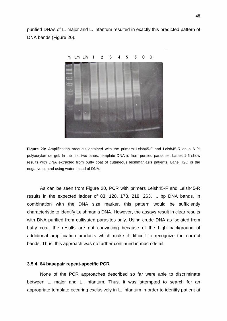
48
purified DNAs of L. major and L. infantum resulted in exactly this predicted pattern of
DNA bands (Figure 20).
Figure 20: Amplification products obtained with the primers Leish45-F and Leish45-R on a 6 %
polyacrylamide gel. In the first two lanes, template DNA is from purified parasites. Lanes 1-6 show
results with DNA extracted from buffy coat of cutaneous leishmaniasis patients. Lane H2O is the
negative control using water istead of DNA.
As can be seen from Figure 20, PCR with primers Leish45-F and Leish45-R
results in the expected ladder of 83, 128, 173, 218, 263, ... bp DNA bands. In
combination with the DNA size marker, this pattern would be sufficiently
characteristic to identify Leishmania DNA. However, the assays result in clear results
with DNA purified from cultivated parasites only. Using crude DNA as isolated from
buffy coat, the results are not convincing because of the high background of
addidional amplification products which make it difficult to recognize the correct
bands. Thus, this approach was no further continued in much detail.
3.5.4 64 basepair repeat-specific PCR
None of the PCR approaches described so far were able to discriminate
between L. major and L. infantum. Thus, it was attempted to search for an
appropriate template occuring exclusively in L. infantum in order to identify patient at
m Lm Lin 1 2 3 4 5 6 C C

49
risk to develop visceral leishmaniasis. Such a sequence was found again using the
Tandem Repeats Finder program in the genome of L. infantum in the form of a 64 bp
tandem repeat. This repeat occurs more than 400 times in different positions of
chromosome 27 of the parasite. It is present neither in L.major nor in Trypanosoma
cruzi and appeared to be an ideal target for PCR therefore.
The sequence of this repeat (6 repeats) is depicted in Figure 21. As with the
above described Leish45-repeat, Primer3 software was used to select optimal PCR
primers. OLIGO start len tm gc% any 3' seq LEFT PRIMER 167 20 59.57 45.00 4.00 2.00 AAAACGGGAATGCTCCAGTA RIGHT PRIMER 361 18 62.64 61.11 7.00 2.00 TTTCGCTGCGAGGGAGAC SEQUENCE SIZE: 384 INCLUDED REGION SIZE: 384 PRODUCT SIZE: 195, PAIR ANY COMPL: 4.00, PAIR 3' COMPL: 1.00 1 CGCGAAGCAATGCTCCCGCGAGGGTCTCCCTCGCAGCGAAAACGGGAATGCTCCAGTACT 61 GGGGCGCGAAGCAATGCTCCCGCGAGGGTCTCCCTCGCAGCGAAAACGGGAATGCTCCAG 121 TACTGGGGCGCGAAGCAATGCTCCCGCGAGGGTCTCCCTCGCAGCGAAAACGGGAATGCT >>>>>>>>>>>>>> 181 CCAGTACTGGGGCGCGAAGCAATGCTCCCGCGAGGGTCTCCCTCGCAGCGAAAACGGGAA >>>>>> 241 TGCTCCAGTACTGGGGCGCGAAGCAATGCTCCCGCGAGGGTCTCCCTCGCAGCGAAAACG 301 GGAATGCTCCAGTACTGGGGCGCGAAGCAATGCTCCCGCGAGGGTCTCCCTCGCAGCGAA << <<<<<<<<<<<<<<< 361 AACGGGAATGCTCCAGTACTGGGG < Figure 21: DNA sequence and primer selection of the Leish64 repeat. The sequences of forward
primer (red) and reverse primer (blue) are indicated. The corresponding areas on the DNA overlap as
indicated (purple and underlined) what holds only for the (hypothetical) shortest PCR fragment.
As can be seen from Figure 19, the primers Leis64-F and Leish64-R would
lead to a ladder of DNA fragments with lengths of 67, 131, 195, 259, 323, 387 ... bp.
This was tested with DNA extracted from cultivated parasites as well as with buffy
coat DNA from patients with visceral and cutaneous leishmaniasis. The results are
shown in Figure 22.

50
Figure 22: PCR products obtained with primers Leish64-F and Leish64-R with DNA purified from
cultivated L. major (Lm) and L. infantum (Lin) parasites, with DNA from buffy coat of two visceral
leishmaniasis patients (v1 and v2), with buffy coat DNA of two cutaneous leishmaniasis patients (c1
and c2) and with buffy coat DNA from two healthy German persons (D1 and D2), C = control PCR in
the absence of DNA, m = size marker.
As can be seen from Figure 22, the PCR with the primers Leish64-F and
Leish64-R are not appropriate to differentiate between L. major and L. infantum. The
reason for this is the high background arising even with DNA extracted from buffy
coat of healthy German blood donors (lanes D 1 and 2). There are obviously
sequences in the human genome which ross-react with these primers. The
background could not be reduced by using higher annealing temperatures (not
shown). Therefore, no further tests were performed with this set of primers.
3.5.5 39 basepair repeat-specific PCR
At last, a set of PCR primers was designed for a 39 bp repetition occuring
more than 450 times exclusively in chromosome 22 of L. infantum in three different
locations. No homologous sequences were detected in the genomes of L. major or
Trypanosoma cruzi. As before for the other tandem repeats, a fragment of five
Lm Lin v1 v2 c1 c2 D1 D2 C C m

51
repeats was used to design optimal PCR primers using Primer3 software. The result
of this calculation is shown in Figure 23.
OLIGO start len tm gc% any 3' seq LEFT PRIMER 78 25 51.80 32.00 6.00 2.00 CAAAGATGTATATAGACGTTAGTGT RIGHT PRIMER 163 22 56.11 36.36 2.00 0.00 CATCTTTGTTTGTGTGTTGTGT SEQUENCE SIZE: 195 INCLUDED REGION SIZE: 195 PRODUCT SIZE: 86, PAIR ANY COMPL: 8.00, PAIR 3' COM PL: 0.00 1 AAAGATGTATATAGACGTTAGTGTACACAACACACAAACAAAGATGTATATAGACGTTAG 61 TGTACACAACACACAAACAAAGATGTATATAGACGTTAGTGTACACAACACACAAACAAA >>>>>>>>>>>>>>>>>>>>>>>>> 121 GATGTATATAGACGTTAGTGTACACAACACACAAACAAAGATGTATATAGACGTTAGTGT <<<<<<<<<<<<<<<<<<<<<< 181 ACACAACACACAAAC Figure 23: DNA sequence and primer design for the 39 bp repeat of L. infantum. The sequences of
forward primer (red) and reverse primer (blue) and overlaps (purple and underlined) are indicated as in
Figure 19. The calculated size of the amplfication products is a ladder of 86, 125, 164, 203, 242, 281,
320, bp.
Test of these primers with purified Leishmania DNA as template revealed that
the primers were highly specific for L. infantum exclusively as shown in Figure 24.
There was no cross-reaction with DNA from L. major (lane Lm) or Trypanosoma cruzi
(not shown).
With all of the 26 samples from patients suspected to have visceral
leishmaniasis there is a amplification of DNA which could correspond to tri- tetra- and
pentamers of the 39 bp repeat, even though the band pattern looks diffuse. In some
of the samples the bands are better to be recognized than in the others. The L. major
positive control Is always negative, and so is buffy coat DNA from healthy German
persons (data not shown). Thus it looks like PCR using Leish39 primers is specific for
the visceral form of leishmaniasis even though PCR products leading to sharper
bands would be desirable.

Figure 24: PCR amplification of Leishmania from buffy coat sample (lanes 1
from patients with suspected visceral leishmaniasis.
positive control, L.inf = L. infantum
PCR amplification of Leishmania from buffy coat sample (lanes 1
from patients with suspected visceral leishmaniasis. M = size marker, L
L. infantum positive control, and h2o = water as a negative control.
52
PCR amplification of Leishmania from buffy coat sample (lanes 1-26) obtained
= size marker, L.major = L. major
as a negative control.

53
3.5.6 Compilation of the PCR results
Even though the PCR procedures developed during this work need to be
verified by well characterized patient samples, it is clear from the data that the
methods used in Yemen up to now are not sufficiently sensitive.
The primers used for the assays described above are listed in the following
Table 5.
Table 5. Primers for diagnsosis of Leishmaniasis
Specificity Gene Primer name Sequence Size (bp)
Tm (°C)
Leishmania all species
Minicircle (kDNA)
Leishmini-F Leishmini-R
ATTTTACACCAACCCCCAGTT GGG(G/T)AGGGGCGTTCTGC 113 59.5
61.6
Leishmania all species
18S rRNA
Leish18S-out-F Leish18S-out-R Leish18S-inn-F Leish18S-inn-R
TCAGTCCATTTGGATTGTCA AGTACGTTCTCCCCCGAACT CAGGTTTGTTCCTGGTCGTC GAGTTGAAAAGGCGTTACGG
480 335
64.3 63.2.0 60.5 59.7
Leishmania all species 45 bp repeat Leish45-F
Leish45-R CGGAGCTGCTGCTGGAC GGCGTCCTCGTCGTCTG ladder 62.0
61.6
L. infantum 64 bp repeat Linf64-F Linf64-R
AAAACGGGAATGCTCCAGTA TTTCGCTGCGAGGGAGAC ladder 59.6
62.6
L. infantum 39 bp repeat
Linf39-F Linf39-R
CAAAGATGTATATAGACGTTAGTGT CATCTTTGTTTGTGTGTTGTGT ladder
51.8 56.1
During the study, 119 samples from patients with suspected cutaneous
leishmaniasis have been collected in different locations in Yemen. These samples
have first been analysed by microscopy of smears of wound material. It is agreed in
the international literature that microscopical analysis alone has an insufficient
sensitivity. Better results could be obtained by addidinal tests such as culivation or
immunological techniques. However, these procedures are to expensive for use in
Yemen. The results for the 119 patient samples obtained obtained in Yemen by
microscopy are shown in the following Table 6.
As can been seen, only 47 cases (39%) of the 119 cases were identified to be
positive by microscopic examination. However, by the combined tests with kDNA-
and 18S rRNA-specific PCR, 76 (64 %) samples were found to be positive.

54
Table 6: Clinical and epidemiological features from 119 patients with cutaneous
leishmaniasis in Yemen.
Patients features Positive * (%) Negative *(%) n = 47(39) n = 72(61)
Sex Male
Female
25(53) 49(68) 22(47) 23(32)
Age (years) ≤15
15–34 35–54 55–64 ≥65
14(30) 19(26) 26(55) 39(54) 5(11) 10(14) 1 (2) 3(4) 1(2) 3(4)
Habitation Urban
Periurban Rural
9(19) 13(18) 12(26) 18(25) 26(55) 41(57)
Duration (in months) 1–6 7–12 ≥12
38(81) 56(78) 9(19) 12(17) 1(1) 3(4)
Place of lesions Legs and feet
Arms and hands Chest
Head and face Multiple lesions
5(11) 13(18) 8(17) 24(33)
0 0 31(66) 34(47)
3(6) 1(1)
* as determined by microscopy in Yemen
Similar results have been obtained with the samples of patients with suspected
visceral leishmaniasis. The 26 patients were diagnosis in the central blood bank in
Sana’a, Yemen, by both ELISA and formol gel examination. 8 cases have been
classified according to these criteria as positive. The data are shown in Table 7.
From the patients identified as positive 4 were male (50%) were and 4 were
female (50%). The majority of the positive patients (62%) were in the age group 15-
34 years and in the age group less than 15 years (25%). The highest prevalence of
disease (62%) was seen in the urban areas.

55
Table 7: Clinical and epidemiological features from 26 patients with suspected
visceral leishmaniasis in Yemen.
Patients features Positive *(%) Negative *
(%) n =8 (31) n = 18 (69)
Sex Male
Female
4(50) 11(61) 4(50) 7(39)
Age (years) ≤15
15–34 35–54 55–64 ≥65
2(25) 0 5(62) 13(72) 1(12) 4(22)
0 1(6)
Habitation Urban
Periurban Rural
5(62) 13(72) 3(37) 4(22)
0 1(6)
Duration (in months) 1–6 7–12 ≥12
1(12) 9(50) 3(37) 7(39) 4(50) 2(11)
Symptoms Hepatomegaly Splenomegaly
Hepatosplenomegaly
2(25) 10(56) 4(50) 3(17) 2(25) 5(28)
* as determined by the combination of a Leishmania-specific ELISA and formol precipitation assay in Yemen
Analysis of buffy coat DNA samples from the same patients in the laboratory in
Giessen by the different PCR assays described above yielded a different result. Even
if these assays need to be validated by additional tests and there were some
inconsitencies between the single PCR runs, it is obvious that the molecular
approach is much more sensitive than the traditional procedures used in Yemen.
Combination of kDNA-specific PCR and 18S rRNA-specific PCR led to 76 positive
results out of the 119 samples indicating that 64 % of the suspected cutaneous
leishmaniasis patients were actually infected. By microscopy, only 39 % of the
patients had been recognized as positive.
As with the coutaneous leishmaniasis, also the data for the patients with
suspected visceral leishmaniasis obtained in Germany were much higher. By
combining the PCR data from rDNA-, 18S rRNA- and Leish39-specific PCR, 16

56
cases (62 %) were found to be positive. Even though the results with the new PCR
techniques are preliminary and need to be confirmed, the results show clearly that
the occurence of leishmaniasis in Yemen is highly underestimated.
Table 8: Comparison of PCR and traditional methods in patients with leishmaniasis
suspected cutaneous leishmaniasis patients
PCR Microscopy
positive negative positive negative
119 (100 %) 76 (64 %) 43 (36 %) 47 (39 %) 72 (61 %)
suspected visceral leishmaniasis patients
PCR ELISA Formol gel
positive negative positive negative positive negative
26 (100 %) 16(62%) 10(38%) 2 (8 %) 3 (12%) 6 (23 %) 15(58%)

57
4. DISCUSSION
The Republic of Yemen is a developing country with very poor health
conditions. According to the Ministry of Public Health and the National Center for
Surveillance and Disease Control, deaths among children under five years of age are
mainly due to infectious diseases such as diarrhea with an estimated 45,000 and
malaria reaching about 25.000 cases of death per year. According to the U.S.A.
Census Bureau (http://www.census.gov/ipc/prod/wp02/tabA-11.pdf), the total children
mortality within the first 5 years of life in Yemen is 92 per 1000, ranging amongst the
last 20 % countries worlwide (for comparison: in Europe this number is 6 per 1000).
These data are indicatory for the poor quality of life in this country and for the realistic
possibilities to improve heath conditions.
The aim of this study was first to shed some light on the real extent of
leishmaniasis in Yemen, and second, to contribute to the development of a more
trustable diagnosis than the traditional procedures used in the country such as
microscopy and serum precipitation with formaldehyde. The limiting sensitivity and
specificity of these traditional methods are well known, but the limiting economical
conditions in Yemen do not allow the use of more sensitive and specific diagnostic
techniques such as modern immunoassays with recombinant antigens. New
diagnostic techniques to be established in the country should not only be highly
specific and sensitive, but also inexpensive and simple to be applicable under the
given limiting conditions. This latter goal was followed in our laboratory at the
University of Giessen in two different ways: one approach included the development
of recombinant antigens capable to discriminate between cutaneous and visceral
leishmaniasis for use in simple and inexpensive immunoassays (ELISA and
immunochromatographic test stripes) which was topic of another doctoral thesis
(Heimann, 2008). The second approach, which is realised in this work, was the
development of molecular diagnostic tests in the form of different PCR assays.
The basis for both approaches were the comlete genomic sequences of three
Leishmania strains (L. major, L. infantum and L. brasiliensis) together with the closely
related genome of Trypanosoma cruzi wich are available free of charge since a short
time in the internet. Many computer programmes needed to handle these huge
amounts of genetic information (more than 100 million basepairs) are equally free
accessible from the internet.
58
4.1 Epidemiology of leishmaniasis
There are no solid data on the incidence of Leishmania infections in Yemen,
but the disease is certainly under-reported, especially in women and children, and
may exceed 10.000 cases per year. Most cases correspond to cutaneous
leishmaniasis due to infections with L. major and L. tropica which may not necessarily
need treatment, but up to 10 % of the infections may be due to L. infantum leading to
visceral leishmaniasis which is associated with up to 90% mortality when untreated.
There are only few reports on leishmaniasis in Yemen in the literature (Sarnelli
1933; Rioux 1986; Pratlong et al. 1995: Khatri et al., 2006) which can be used to
predict the parasite strains circulating in the country (L. major and L. tropica for
cutaneous leishmaniasis and L. infantum for visceral leishmaniasis), but these
publications do not indicate the frequency and severity of the disease in the
population. In order to get more information on the occurence of leishmaniasis, data
collected during the years 2005-2008 by the the National Center of Surveillance of
Disease from all governmental hospitals in the 20 governorates were analysed (Table
1). However, these data can only be used in a qualitative way, but they are by no
means quantitative. Data obtained during a visit of the (private) Alolfi Hospital in the
Alhodeidah governorate indicated 105 cases during the same period of time (2005-
2008) in this single place which is twofold the number of cases reported by the
government for the whole governate (53 cases).
The closest countries with similar kinds of Leishmania strains, phlebotomine
vectors and comparable geographic conditions are Oman in the East, Saudi Arabia in
the North-West and, in a distance of some 2000 km North-West from Yemen, Jordan
and Palestine. Some usful epidemiological data exist from Saudi Arabia (al-Zahrani
et al., 1989 and Ibrahim et al., 1992), wheras most informatve data have been
published for Jordan and Israel (Nimri et al., 2002; Wasserberg et al., 2002; Al-
Jawabreh et al., 2003; Al-Jawabreh, 2005; Singer et al., 2009; Amro et al.,
2009;Hamarsheh et al., 2009). These data could form a basis for the understandig of
leishmaniasis in Yemen. As in Yemen, two forms of transmission of the disease exist:
in urban areas the parasites are transmitted from infected individuals by the bite of
phlebotomine sand flies to naïve persons (anthroponotic cutaneous leishmaniasis),
whereas In rural areas animals are thought to be the reservoir (zoonotic cutaneous
leishmaniasis). The anthroponotic form of disease is frequent in large cities where

59
many people live close together under poor hygienic conditions. The vectors can
multiply in small water resevoirs close to the dwellings as illustrated by the following
pictures (Figure 25).
Figure 25: Conditions for anthroponotic cutaneous leishmaniasis. In many cities of Yemen,
leishmaniasis is not dependent on animal hosts, but is transmitted by sand flies directly from infected individuals to naïve persons. In such loations the infection rate can be very high. The larvae of the sand flies can perfectly thrive in nearby water pools.
Hosts involved in the transmission of the zoonotic form of the disease include
primarily dogs and small rodents living in arid regions. In the neighbouring contries
Iran, Jordan and Israel host species such as Psammomys obesus, Rattus rattus,
Gerbillus dasyurus, Nesokia indica, Rhombomys opimus and Meriones spp.

60
(Wasserberg et al,. 2002; Yaghoobi-Ershadi et al., 2004; Pourmohammadi et al.,
2008) and rock hyraxes (Svobodova et al., 2006) have been found to be infected with
Leishmania species. Which of these animals occure in Yemen remains to be
determined. Among the Saudi populations in the south-west of Saudi Arabia (Asir
and Al-Baha provinces), an annual incidence of 12 and 38 per 10,000 respectively
was estimated for cutaneous leishmaniasis, mainly caused by L. tropica (al-Zahrani
et al., 1989), whereas for the Jordan Valley an average of 7.5 cases per 10,000 per
year was calculated (Mosleh et al., 2008).
Seasonality of cutaneous leishmaniasis
According to data collected from the Ministry of Health and National Centre for
Surveillance and Disease Control the prevalence of leishmaniasis in the coastal
governorates of Yemen is saisonal, ranging from October to April. This has been
confirmed in this study during collecting samples from October 2004 to March 2005.
Another study on patients in mountainous governorates showed a striking difference
in seasonality, starting in June and reching till October. The reasons for seasonality
of outbreak of the disease are the activity of sand fly vectors and the incubation
period of the infection. L. major has an incubation period that ranges from a few
weeks to a few months, but is longer for L. tropica which may take 2 -24 months
(Harrison’s Principles of Internal Medicine, Chapter 205, Leishmaniasis. 1991). Ph.
papatasi, vector of L. major, has its activity and abundance peaks in the spring (April)
and the autumn (October), and was found to be not active in mid-summer (July)
(Wasserberg et al., 2002).
Another interesting phenomenon with cutaneous leishmaniasis is the fact, that
men and children are by far more affected by the disease than women. This may be
explained by behavioural or cultural factors which make males more exposed than
females, and lack of immunity to make children more exposed than adults (Al-
Jawabreh et al., 2003).
In summary it can be concluded that the epidemiological situation is
comparable to the neighbouring countries. As the epidemiology of leishmaniasis of
these countries is best described for Palestine, these data may serve as a basis to
elucidate the situation in Yemen. However, this was not the purpose of this work, but
has to be done by future analyses.

61
4.2 Diagnosis of leishmaniasis
Health indicators for Yemen are poor, with high infant and maternal mortality
rates as stated above. The per capita health expenditures are as low as 33 US Dollar
per year, and government spending on health was only 3.3 per cent of total public
expenditure, a proportion that is the fourth smallest among World Health
Organisation (WHO) member states (CSO [Central Statistical Organization], Republic
of Yemen. Demographic and Maternal and Child Health Survey 1998).
This facts have to be kept in mind when planning to improve the health
situation in the country by introducing new methods for the diagnosis of disease.
Commercial diagnostics for infectious diseases are too expensive to be used under
such conditions, however, the progress in molecular biology in the past years allows
to produce essential diagnostic tools such as recombinant antigens, monoclonal
antibodies or molecular probes at costs which can be afforded also by poor countries.
The material costs for highly sensitive and specific tests such as ELISA with
recombinant antigens, of molecular diagnosis using PCR are few cents per assay
and the tests can be produced independent from external support inside the country
when the essential theoretical and practical knowledge has been achieved.
Purpose of the experimental work of this thesis was to develop molecular
methods for the diagnosis of Leishmania infections in Yemen in a most simple way.
This could be realized using DNA sequence data for different Leishmania species
available through the internet in combination with free available computer
programmes which are needed to process the huge amount of genetic information.
Five different PCR assays have been designed and tested, and some of them turned
out to possess a good potential for specific and sensitive diagnosis of leishmaniasis.
4.3 Collection and processing of patient samples
In planning inexpensive molecular tests for developing countries it is also
essential to have appropriate methods for collecting samples from patients, for
processing these samples and for conservation of the essential components under
field conditions. In general, there is litte chance to keep such samples in a freezer at -
20°C, and even normal refrigerators are not always available. Traditionally, srapings
from leishmanial ulcers are used for microscopical analysis. However, frequently only
few, or no parasites at all, are contained in such samples. Better sources for

62
parasites are biopsies from the border of the ulcer, from lymph nodes, from bone
marrow, or from spleen. However, biopsies can hardly be taken under field
conditions, but alternatively, few millilitres of peripheral blood can be taken with a
syringe without the risk of accidental infection or cross-contamination. The parasites
amplify mainly in macrophages, and even in the chronical stage of disease sufficient
infected white blood cells can be detected in the circulation.
A simple method for blood collection and processing under field conditions has
been developed in our laboratory in Giessen. The protocol is described in the
Materials and Methods section. In short, blood is collected in syringes containing
EDTA or citrate as anti-coagulant. Blood cells are sediment by centrifugation (a
simple hand-operated centrifuge has been used during this work, see Figure.10), the
supernatant plasma was collected and conserved in 50 % glycerol, and finally, the
yellow layer of white blood cells and platelets (buffy coat) was harvested with a
pipette. The excess of red blood cells in this crude buffy coat preparation were lysed
by a low ionic stength buffer, the cells sedimented again by short centrifugation and
mixed with a guanidinium hydrochloride solution. The guanidinium hydrochloride has
two functions: first, it lyses the cells for later DNA extraction, and second, it protects
the DNA for extended periods of time (months) from degradation even at ambient
temperatures.
The DNA from the lysed white blood cells was lateron easily purified in the
laboratory by adding small amounts of silica (pulverized SiO2 which binds exclusively
DNA under these conditions), several washing steps with diluted ethanol and finally
eluted in pure and concentrated form by a low salt buffer. This procedure proved to
be very appropriate for field conditions as no toxic chemicals such as phenol and
chloroform are needed, and the costs of consumables are minmal. Furthermore, the
whole procedure can be carried out in a single tube, which is important in terms of
avoiding cross-contamination. DNA obtained by this way was used to test new PCR
assays, and the plasma fraction was used to test new immunological assays by
another member of the group.
4.4 Kinetoplast DNA (kDNA)-specific PCR
The first test developed was a PCR with primers specific for the kinetoplast
DNA of Leishmania which consists of approximately 10.000 copies of circular DNA

63
molecules ("minicircles") containing a conserved sequence of approximately 120 bp.
This DNA has been perfered for PCR diagnosis by many groups because due to the
high copy number of target sites, less than a single parsite could be detected
theoretically. kDNA-PCR has repeatedly been found to be the most sensitive of
diagnostic assays for leishmaniasis (Lachaud et al., 2002; Nasereddin et al., 2006;
Romero et al., 2009).
The common part of the kDNA minicircles is not perfectly conserved, but
consensus sequences can be found at the two ends of a more conserved region of
approximately 120 bp. These consensus sequences have been used to develop
primers for PCR amplification by others (references see above). According to the
Prime3 software used for selection of PCR primers, the published primers appeared
to be not optimal, basically because forward and reverse primers revealed
differences in the annealing temperatures of more than 20°C. This is not appropriate
for specific PCR since the hybridization temperature has to be set according to the
primer with the lower binding strength. The other primer can bind under these
conditions also to sites which do not match perfectly, thus creating unspecific
background amplification. Therfore, it was attempted to predict primers with more
equal annealing temperatures. The performance of the resulting (shorter) primers in
kDNA-specific PCR assays was compared with the efficiency of the primers used by
other groups, but no significant difference in sensitivity and specificity were observed.
The kDNA-specific PCR assay developed by this way appeared to be most
sensitive, however, it did not discriminate between cutaneous and visceral
leishmaniasis. Furthermore, processing of the results was somewhat demanding
because the small resulting amplification product of 113 bp could only be redissolved
on 6 % polyacrylamide gels. Therefore, some alternative PCR assays were
developed in addition.
4.5 18S ribosomal RNA-specific PCR
Leishmania species contain approximately 160 copies of the genes for the
small subunit of ribosomal RNA (18S rRNA or ssu rRNA). These genes have been
used by several groups as target for diagnostic PCR, even though most of them were
using different sets of primers (Spanakos et al., 2002; Schönian et al., 2003; Chargui
et al., 2005). It is agreed within all these groups that 18S rRNA-specific PCR is highly

64
sensitive, superior to PCR assays with other target genes in the Leishmania genome.
The genes for the 18S rRNA are highly conserved between the different species of
Leishmania, thus, discrimination within the genus is not possible. However, clear
discrimination is feasible from other species such as the closely related Trypanosoma
cruzi, or the non-pathogenic Trypanosoma rangelli which occurs frequently in Latin
America. Only part of the sequence of the gene can be used for establishing
discriminatory PCR because of the high degree of homology of 18S rRNA sequences
between all organisms, However, some regions differ sufficiently to be appropriate for
this purpose.
By means of Primer3 software, two sets of PCR primers were selected to
produce a fragment size that could be easily analysed be agarose gel
electrophoresis. Due to the lower gene copy number as compared with kDNA, it
appeared to be necessary to employ nested PCR using a second set of primers. The
disadvantage of a second amplification cycle is compensated not only by higher
sensitivity, but also by a higher specificity. The second set of primers leads to
enrichment of correct amplification products only, thus avoiding false positive results.
The first PCR with the outer set of primers led only in few cases to a visible
band on agarose gels (see Figure 15). This could be due to several reasons. First,
one or both primers are not optimal and higher sensitivity could be reached with
alternative primers. However, also the set of inner primers, when used alone, did not
lead to more sensitivity. Another reason for this fact could have been the relative low
amont of DNA used for the assays. The DNA of the samples from Yemen was limited
and had to be spared for many other assays. Normally not more than 2 per cent of
the DNA isolated from one sample were used per assay. Taking 10 per cent of DNA
from a sample would probably improve the sensitivity markedly. Furthermore, in all
assays a self-made Taq DNA polymerase was used which may not have the same
quality as expensive commercial enzymes. Nevertheless, clear results were obtained
with nested PCR using aliquots of the amplification products of the first PCR as
template for the second PCR with inner primers. The sensitivity was almost as high
as obtained with the kDNA-specific PCR, and the fact that the fragments can be
easily separated on agarose gels allows to use this test in large scale. To compare
the quality of this test with other 18S rRNA-specific PCR assays, tests with PCR
primers used by other groups need to be done.
65
4.6 PCR assays with tandem repeat sequences
The three completely sequenced Leishmania strains (L. major, L. infantum and
L. brasiliensis) accessible in the Sanger Gene Database contain plenty of tandemly
repeated sequences. Using the "Tandem Repeats Finder" computer program
(http://tandem.bu.edu/trf/trf.html), the three genomes were analysed for tandem
repeats in the DNA. This analysis revealed many different repeats with different
lengths of the repetitive unit. The highest number were more than 1000 repeats of a
45 nucleotide sequence, other repeats with lengths between ten and more than 100
nucleotides occured somewhat less frequently, but several hundred repetitions were
not uncommon. Approximately 10 per cent of the genomes consist of such repetitive
sequences. The repeats were usually fragmented in several subfractions (2-10)
distributed in the same, but also in different chromosomes. Some repeats were highly
conserved, others differed up to 10 per cent in the nucleotide composition of the
single units. Most of the repeats occured in related form in all three Leishmania
strains even though in different numbers. However, there were several repeats
specific for a single strain thus representing candidates for species-specific PCR.
Some repeats were also to be observed in related form in the genome of
Trypanosoma cruzi.
The repeats occure in non-coding as well as in protein coding regions of the
genomes. In coding regions, they are translated in tandem arrays of amino acid
sequences which are in part highly immunogenic leading to a strong antibody
reaction. Such tandem arrays of amino acids have been used in recombinant form as
antigens for developing specific immunoassays such as the K39 antigen (Burns etal.,
1993).
As these tandemly repeated sequences differ in part between the three
Leishmania species, it seemed to be interesting to test if they could serve as targets
for specific PCR assays. In contrast to normal PCR, the amplification products were
not expected as a single band of defined length, but as a ladder of bands with
distances between the different steps corresponding to the length of a single repeat
unit. By means of the primer3 software appropriate primers were selected for three
such repeats: the 45 basepair repeat with approximately 1000 copies occuring in all
strains, a 64 basepair repeat with a frequency of approximately 400 copies common

66
to all strains as well, and a 39 basepair repeat present in L. infantum exclusively.
None of these repeats was present in related form in Trypanosoma cruzi.
As can be seen from the Results section, the PCR amplification of these
repeats worked out in principle as predicted, however, the results were not as
convincing as expected. Even though the 45 basepair repeat led to a characteristic
ladder with the purified DNAs of L. major and L. infantum, this ladder diaspeared
almost completely when the Leishmania DNA was mixed with buffy coat DNA.
Obviously, human DNA contains many sequences which serve as template for the
primers used in this PCR. The results were not much different under more stringent
conditions, i.e when the highest possible annealing temperature for these primers
was selected.
The results with the PCR specific for the 64 basepair repeat looked even
worse. The primers led to strong background reactions even with the purified parasite
DNAs indicating a too low selectivity of this PCR.
However, the 39 basepair specific PCR led to results which look promising for
diagnosis. The resulting ladder of bands is specific for L. infantum, that means for
visceral leishmaniasis (Figure 19). There is no background reaction in controls
without DNA and with buffy coat DNA from patients with cutaneous leishmaniasis
(not shown in the Figure). The lowest bands of the ladder are not visible in the
amplification products with buffy coat DNA from visceral leishmaniasis patients,
however, higher multimeric forms are present, at least in some of the lanes. As with
the 18S rRNA-specific PCR, the weak reaction may be due to the limiting amounts of
DNA added to the assays. In any case it appears to be worthwhile to repeat this
approach with more DNA. In addition, the stringency of the assay could probably be
improved, and alternative primers specific for the 39 basepair repeat could be tested.
The PCR may not be sufficient as only test, but it could be used as a second test
after demostration of the infection either by kDNA- or 18S rRNA-specific PCR.
4.7 Concluding remarks
Leishmaniasis is still a widely negected disease which means that even though
millions of people in developing countries suffer from the infection, there are neither
appropriate diagnostic procedures nor sufficiently effective drugs for treatment available. At
present, several groups worldwide study the variability of Leishmania strains with complex
67
techniques such as analysis of enzyme polymorphismes, restriction length polymorphism, or
determination of microsatellite patterns of the organisms. Even though these techniques
contribute to evaluate the genetics and epidemiology of the parasite in much detail, they are
by far too demanding to be used for answering the simple question: is there an infection with
Leishmania or not? To reduce suffering from this disease, prove of the infection has to be
simple and inexpensive since it affects mainly the poorest of the poor.
The first step of diagnosis has to discriminate between an infection with Leishmania
and other causes of the dermal ulcer which may have bacterial or fungal origin. Only in a
second step is needed to differentiate between cutaneous and potentially visceral
leishmaniasis in the Old World, or between cutaneous, and mucocutaneous leishmaniasis in
the New World. As up to 20 per cent of all cutaneous cases in Latin America develop to the
mucocutaneous form after some time, a proven Leishmania infection is treated anyway and
this discrimination may not be needed therefore. A detailed analysis of the specific strain
may be helpful for the prognosis of disease and for the prediction of drug-resistancy, but this
is not the first priority.
Because of the severe side effects of the drugs, cutaneous leishmaniasis in the Old
World is normally only treated if the ulcers are severe and could have disfiguring
consequences. However, since also the life-threatening visceral form of the disease begins
with a cutaneous ulcer as a first clinical symptom, it will be needed in the future to decide
already in this early stage whether the ulcer is caused by a cutaneous form (L. tropica and L.
mayor in the Arabian peninsula), or by the dangerous visceral form (L. donovani or L.
infantum). There are good immunodiagnostic assays available, mainly based on the
recombinant antigen rK39, but these assays are not sufficient because specific antibodies
can persist for many years and it is difficult to decide, wether they derive from an healed
infection, or indicate acute disease. Therefore, a simple and inexpensive molecular test
would be very helpful.
It was goal of this work to contribute to this aspect. Even though the assays
established so far could be improved to a certain degree, it becomes obvious that PCR
diagnosis is extremely useful and not as complicated and expensive that it could not be
introduced to Yemen. For this purpose it was also essential that procedures basic for PCR
diagnosis, the collection of patient samples, the conservation of the essential fractions DNA
and plasma, the isolation and purification of DNA and the preparation of Taq DNA
polymerase have been simplified in such a way that they can also be performed in a low
equiped laboratory and for little money.

68
REFERENCES
Ajdary S, Alimohammadian MH, Eslami MB, Kemp K, Kharazmi A. (2000)
Comparison of the immune profile of nonhealing cutaneous Leishmaniasis patients
with those with active lesions and those who have recovered from infection. Infect
Immun. 68:1760-1764.
Alexander J, Satoskar AR and Russell DG (1999) Leishmania species: models of
intracellular parasitism. J Cell Science 112:2993-3002
Al-Jawabreh A, Barghuthy F, Schnur LF, Jacobson RL, Schönian G, Abdeen Z.
(2003) Epidemiology of cutaneous leishmaniasis in the endemic area of Jericho,
Palestine. East Mediterr Health J. 9:805-815.
Al-Jawabreh A (2005) Molecular Epidemiology, Clinical Molecular Diagnosis and
Genetic Diversity of Cutaneous Leishmaniasis in Jericho, Palestine. Dissertation at
the Medical Faculty, Charité, University of Berlin
Al-Jawabreh A, Diezmann S, Müller M, Wirth T, Schnur LF, Strelkova MV, Kovalenko
DA, Razakov SA, Schwenkenbecher J, Kuhls K, and Schönian G (2008) Identification
of geographically distributed sub-populations of Leishmania (Leishmania) major by
microsatellite analysis. BMC Evolutionary Biology 8:183
al-Zahrani MA, Peters W, Evans DA, Smith V, Ching Chin I. (1989) Leishmania
infecting man and wild animals in Saudi Arabia. 6. Cutaneous leishmaniasis of man
in the south-west. Trans R Soc Trop Med Hyg. 83:621-628.
Amro A, Azmi K, Schönian G, Nasereddin A, Alsharabati MB, Sawalha S,
Hamarsheh O, Ereqat S, Abdeen Z. (2009) Epidemiology of paediatric visceral
leishmaniasis in Hebron district, Palestine. Trans R Soc Trop Med Hyg. 2009
Jul;103(7):731-6. Epub 2008 Nov 20.
Andrade BB, de Oliveira CI, Brodskyn CI, Barral A and Barral-Netto M, (2007) Role of
Sand Fly Saliva in Human and Experimental Leishmaniasis: Current Insights.
Scandinavian Journal of Immunology 66:122–127

Ashford RW , Bray MA, Hutchinson MP
cutaneous leishmaniasis in Ethiopia.
601.
Babaloo Z, Kaye PM, Eslami MB
visceral leishmaniasis: relationship to other Th2 and Th1 cytokines.
Trop Med Hyg. 95:85-88.
Barral A, Barral-Netto M, Almeida R, de Jesus AR, Grimaldi JG, Netto EM, Santos I,
Bacellar O, Carvalho EM. (1992)
braziliensis cutaneous infection.
Bensoussan E, Nasereddin A
PCR assays for diagnosis of cutaneous leishmaniasis.
1439.
Boarino A, Scalone A, Gradoni L
Rosati S. Development of recombinant chimeric antigen expressing immunodominant
B epitopes of Leishmania infantum for serodiagnosis of visceral leishmaniasis.
Diagn Lab Immunol. 2005 May;12(5):647
Boyer MH (1993) Visceral leishmaniasis in Desert Storm veterans.
329:1503-1504.
Burns, J. M., Jr., W. G. Shreffler, D. R. Benson, H. W. Ghalib, R. Badaro`, and S. G.
Reed (1993) Molecular characterization of a kinesin
chagasi that detects specific antibody in African and American visceral leishmaniasis.
Proc. Natl. Acad. Sci. USA
Chang KP and McGwire GS (2002)
Leishmania virulence. Kinetoplastid Biology and Disease
Chappuis F, Mueller Y, Nguimfack A. Rwakimari JB, Couffignal S, Boelaert M
Cavailler P. Loutan L and Piola P (2005) Diagnostic Accuracy of Two rK39 Antigen
Based Dipsticks and the Formol Gel Test for Rapid Diagnosis of Visceral
Leishmaniasis in Northeastern Uganda. J. Clin. Microbiol.43:5973
, Bray MA, Hutchinson MP , and BrayRS (1973) The epidemiology of
cutaneous leishmaniasis in Ethiopia. Transactions Royal Soc Trop Med Hyg 67:
Eslami MB. (2001) Interleukin-13 in Iranian patients with
visceral leishmaniasis: relationship to other Th2 and Th1 cytokines.
Netto M, Almeida R, de Jesus AR, Grimaldi JG, Netto EM, Santos I,
Bacellar O, Carvalho EM. (1992) Lymphadenopathy associated with Leishmania
braziliensis cutaneous infection. Am. J. Trop. Med. Hyg. 47:587-592
Nasereddin A, Jonas F, Schnur LF, Jaffe CL (2006) Comparison of
PCR assays for diagnosis of cutaneous leishmaniasis. J Clin Microbiol.
Gradoni L, Ferroglio E, Vitale F, Zanatta R
. Development of recombinant chimeric antigen expressing immunodominant
B epitopes of Leishmania infantum for serodiagnosis of visceral leishmaniasis.
2005 May;12(5):647-53.
(1993) Visceral leishmaniasis in Desert Storm veterans.
. G. Shreffler, D. R. Benson, H. W. Ghalib, R. Badaro`, and S. G.
(1993) Molecular characterization of a kinesin-related antigen of
that detects specific antibody in African and American visceral leishmaniasis.
. USA 90:775–779.
Chang KP and McGwire GS (2002) Molecular determinants and regulation of
Kinetoplastid Biology and Disease 1:1-7, Review
Chappuis F, Mueller Y, Nguimfack A. Rwakimari JB, Couffignal S, Boelaert M
and Piola P (2005) Diagnostic Accuracy of Two rK39 Antigen
Based Dipsticks and the Formol Gel Test for Rapid Diagnosis of Visceral
Leishmaniasis in Northeastern Uganda. J. Clin. Microbiol.43:5973–5977
69
The epidemiology of
Transactions Royal Soc Trop Med Hyg 67:568–
13 in Iranian patients with
visceral leishmaniasis: relationship to other Th2 and Th1 cytokines. Trans R Soc
Netto M, Almeida R, de Jesus AR, Grimaldi JG, Netto EM, Santos I,
Lymphadenopathy associated with Leishmania
592
(2006) Comparison of
J Clin Microbiol. 44:1435-
Zanatta R, Giuffrida MG,
. Development of recombinant chimeric antigen expressing immunodominant
B epitopes of Leishmania infantum for serodiagnosis of visceral leishmaniasis. Clin
(1993) Visceral leishmaniasis in Desert Storm veterans. N Engl J Med.
. G. Shreffler, D. R. Benson, H. W. Ghalib, R. Badaro`, and S. G.
related antigen of Leishmania
that detects specific antibody in African and American visceral leishmaniasis.
Molecular determinants and regulation of
7, Review
Chappuis F, Mueller Y, Nguimfack A. Rwakimari JB, Couffignal S, Boelaert M
and Piola P (2005) Diagnostic Accuracy of Two rK39 Antigen-
Based Dipsticks and the Formol Gel Test for Rapid Diagnosis of Visceral
5977

70
Chargui N, Bastien P, Kallel K, Haouas N, Akrout FM, Masmoudi A, Zili J, Chaker E,
Othman AD, Azaiez R, Crobu L, Mezhoud H, Babba H. (2005) Usefulness of PCR in
the diagnosis of cutaneous leishmaniasis in Tunisia. Trans R Soc Trop Med Hyg.
99:762-768.
Cruz I, Nieto J, Moreno J, Cañavate C, Desjeux P, Alvar J. (2005) Leishmania/HIV
co-infections in the second decade. Indian J Med Res. 123:357-388. Review.
Disch J, Pedras MJ, Orsini M, Pirmez C, de Oliveira MC, Castro M, Rabello A (2005)
Leishmania (Viannia) subgenus kDNA amplification for the diagnosis of mucosal
leishmaniasis. Diagn Microbiol Infect Dis. 51:185-90.
El-Badry A, Al-Juhani A, El-Kheir I and Al-Zubiany S (2008) Distribution of sand flies
in El-Nekheil province, in Al-Madinah Al-Munawwarah region, western of Saudi
Arabia. Parasitol Res (2008) 103:151–156.
El Harith A, Slappendel RJ, Reiter I, van Knapen F, de Korte P, Huigen E, and Kolk
AH (1989) Application of a direct agglutination test for detection of specific anti-
Leishmania antibodies in the canine reservoir. J Clin Microbiol. 27:2252–2257.
Garg R and Dube A (2006) Animal models for vaccine studies for visceral
leishmaniasis. Indian J Med Res 123:439-454. Review
Ghalib HW, Piuvezam MR, Skeiky YA, Siddig M, Hashim FA, el-Hassan AM, Russo
DM, Reed SG. (1993) Interleukin 10 production correlates with pathology in human
Leishmania donovani infections. J Clin Invest. 92:324-329.
Gomes AHS I, Armelin IM, Menon SZ, and Pereira-Chioccola VL (2008) Leishmania
(V.) braziliensis: Detection by PCR in biopsies from patients with cutaneous
leishmaniasis Exp Parasitol 119 319–324.
Hamarsheh O, Presber W, Yaghoobi-Ershadi MR, Amro A, Al-Jawabreh A, Sawalha
S, Al-Lahem A, Das ML, Guernaoui S, Seridi N, Dhiman RC, Hashiguchi Y, Ghrab J,
Hassan M, Schönian G. (2009) Population structure and geographical subdivision of
the Leishmania major vector Phlebotomus papatasi as revealed by microsatellite
variation. Med Vet Entomol. 23:69-77.

71
Harith AE, Kolk AH, Kager PA, Leeuwenburg J, Muigai R, Kiugu S, Kiugu S, Laarman
JJ. (1986) A simple and economical direct agglutination test for serodiagnosis and
sero-epidemiological studies of visceral leishmaniasis. Trans R Soc Trop Med Hyg.
80:583–536.
Heimann M (2008) Entwicklung eines ELISA zur Diagnose von viszeraler
Leishmaniose. Dissertation an Fachbereich Biologie der Justus-Liebig-Universität
Gessen
Henk DF, Schallig H and Oskam L. (2002) Molecular biological applications in the
diagnosis and control of leishmaniasis and parasite identification. Tropical Medicine
and International Health, 7:641–651. Review
Ibrahim EA, al-Zahrani MA, al-Tuwaigri AS, al-Shammary FJ, Evans DA (1992)
Leishmania infecting man and wild animals in Saudi Arabia. 9. The black rat (Rattus
rattus) a probable reservoir of visceral leishmaniasis in Gizan province, south-west
Saudi Arabia. Trans R Soc Trop Med Hyg. 86:513-514.
Jaffe CL, Baneth G, Abdeen ZA, Schlein Y and Warburg A (2004) Leishmaniasis in
Israel and the Palestinian Authority. Trends Parasitol 20:151-154.
Jensen, A. T., S. Gasim, T. Moller, A. Ismail, A. Gaafar, M. Kemp, A. M. el Hassan,
A. Kharazmi, T. G. Theander, and K. Andersen. 1999. Serodiagnosis of Leishmania
donovani infections: assessment of enzyme-linked immunosorbent assays using
recombinant L. donovani gene B protein (GBP) and a peptide sequence of L.
donovani GBP. Trans. R. Soc. Trop. Med. Hyg. 93:157–160.
Khatri ML, Haider N, Di Muccio T, and Gramiccia M (2006) Cutaneous leishmaniasis
in Yemen: clinicoepidemiologic features and a preliminary report on species
identification. Int J Dermatol 45:40–45.
Kumar, R., K. Pai, K. Pathak, and S. Sundar. 2001. Enzyme-linked immunosorbent
assay for recombinant K39 antigen in diagnosis and prognosis of Indian visceral
leishmaniasis. Clin. Diagn. Lab. Immunol. 8:1220–1224.
Kumar R, Bumb RA, Ansari NA, Mehta RD, and Salotra P. 2007. Cutaneous
leishmaniasis caused by Leishmania tropica in Bikaner, India: parasite identification

72
and characterization using molecular and immunologic tools. Am. J. Trop. Med. Hyg.
76:896-901.
Kurtzhals JA, Hey AS, Jardim A, Kemp M, Schaefer KU, Odera EO, Christensen CB,
Githure JI, Olafson RW, Theander TG, (1994) Dichotomy of the human T cell
response to Leishmania antigens. II. Absent or Th2-like response to gp63 and Th1-
like response to lipophosphoglycan-associated protein in cells from cured visceral
leishmaniasis patients. Clin Exp Immunol.96:416-421.
Lachaud L, Marchergui-Hammami S, Chabbert E, Dereure J, Dedet JP, Bastien P.
(2002) Comparison of six PCR methods using peripheral blood for detection of
canine visceral leishmaniasis. J Clin Microbiol. 40:210-215.
Maalej, I. A., M. Chenik, H. Louzir, S. A. Ben Salah, C. Bahloul, F. Amri, and K.
Dellagi. 2003. Comparative evaluation of ELISAs based on ten recombinant or
purified Leishmania antigens for the serodiagnosis of Mediterranean visceral
leishmaniasis. Am. J. Trop. Med. Hyg. 68:312–320.
Markle WH and Makhoul K. (2004) Cutaneous leishmaniasis: recognition and
treatment. Amercan Family Physican 69:1455-1460.
Montenegro J. (1926) Cutaneous reaction in leishmaniasis. Arch Derm Syph; 13:
187-94.
Mosleh IM, Geith E, Natsheh L, Abdul-Dayem M, Abotteen N. (2008) Cutaneous
leishmaniasis in the Jordanian side of the Jordan Valley: severe under-reporting and
consequences on public health management. Trop Med Int Health. 13:855-60.
Myint CK, Asato Y, Yamamoto Y, Kato H, Bhutto AM, Soomro FR, Memon MZ,
Matsumoto J, Marco JD, Oshiro M, Katakura K, Hashiguchi Y, and Uezato H (2008)
Polymorphisms of cytochrome b gene in Leishmania parasites and their relation to
types of cutaneous leishmaniasis lesions in Pakistan. The Journal of Dermatology
35:76-85.
Myler P and Fasel N. (2008). Leishmania: After The Genome. Caister Academic
Press. ISBN 978-1-904455-28-8

73
Naderer T and McConville MJ (2008) The Leishmania–macrophage interaction: a
metabolic perspective. Cellular Microbiology 10:301–308.
Nasereddin A, Ereqat S, Azmi K, Baneth G, Jaffe CL, Abdeen Z. (2006) Serological
survey with PCR validation for canine visceral leishmaniasis in northern Palestine. J
Parasitol. 92:178-183.
Nasereddin A, Azmi K, Jaffe CL, Ereqat S, Amro A, Sawalhah S, Baneth G, Schönian
G, Abdeen Z (2009) Kinetoplast DNA heterogeneity among Leishmania infantum
strains in central Israel and Palestine. Vet Parasitol. 161:126-130.
Nimri L, Soubani R and Marina Gramiccia M (2002) Leishmania species and
zymodemes isolated from endemic areas of cutaneous leishmaniasis in Jordan.
Kinetoplastid Biology and Disease 1:7
Piarroux R, Fontes M, Perasso R, Gambarelli F, Joblet C, Dumon H, Quilici M (1995)
Phylogenetic relationships between Old World Leishmania strains revealed by
analysis of a repetitive DNA sequence. Mol Biochem Parasitol. 73:249-252.
Pourahmad M, Farhang Hooshmand F, and Masood Rahiminejad M (2009)
Cutaneous leishmaniasis associated with visceral leishmaniasis in a case of acquired
immunodeficiency syndrome (AIDS). Intern J Dermatol 48, 59–61
Pourmohammadi B, Motazedian MH, Kalantari M (2008) Rodent infection with
Leishmania in a new focus of human cutaneous leishmaniasis, in northern Iran. Ann
Trop Med Parasitol. 102:127-133.
Pratlong F, Bastien P, Perello R, Lami P and Dedet IP (1995) Human cutaneous
leishmaniasis caused by Leishmania donovani sensu strictu in Yemen. –Trans Royal
Soc Trop Med Hyg 89:398-399
Ramaoui K, Guernaoui S and Boumezzough A (2008) Entomological and
epidemiological study of a new focus of cutaneous leishmaniasis in Morocco.
Parasitol Res 103:859–863.
Reithinger R, Dujardin JC, Louzir H, Pirmez C, Alexander B, Brooker S (2007)
Cutaneous leishmaniasis. Lancet Infect Dis 7:581–596, Review

74
Reithinger R and Dujardin JC (2007) Molecular Diagnosis of Leishmaniasis: Current
Status and Future Applications. J Clin Microbiol 45:21–25
Rey JR (2007) Document ENY-738 (IN705)of the Entomology and Nematology
Department, Florida Cooperative Extension Service, Institute of Food and Agricultural
Sciences, University of Florida. http://edis.ifas.ufl.edu/pdffiles/IN/IN70500.pdf
Rioux JA, Daoud W, Pratlong F, Rioux JA, Daoud W and Pratlong F (1986) Les
complexes Leishmania donovani sst.Leishmania tropica et Leishmania major en
Republique Arabe du Yemen. In: Rioux JA, ed. Leishmania: Taxonomie-
Phylogenese. Montpellier: Institut National de la Sante et de la Recherche Medicale,
1986: 357–363.
Roberts MTM. (2006). Current understandings on the immunology of leishmaniasis
and recent developments in prevention and treatment British Medical Bulletin 2006;
75 and 76:115–130
Romero GA, Noronha EF, Pirmez C, Pires Fdo E, Fernandes O, Nehme NS,
Cupolillo E, Firoozmand L, da Graça GC, Volpini A, Santos SL, Romanha AJ. (2008)
Sensitivity and reproducibility of a PCR assay for Leishmania detection using skin
biopsy imprints on filter paper. Acta Trop. 109:74-7.
Sadeghian G, Momeni A, Siadat AH, Usefi P (2006) Evaluation of leishmanin skin
test and its relationship with the clinical form and duration of cutaneous
leishmaniasis. Dermatology Online Journal 12:3.
Salotra P, Sreenivas G, Beena KR, Mukherjee A and Ramesh V (2003) Parasite
detection in patients with post kala-azar dermal leishmaniasis in India: a comparison
between molecular and immunological methods. J Clin Pathol 56:840–843.
Sang DK, Okelo GBA, Ndegwa CW, Ashford RW (1993) New foci of cutaneous
leishmaniasis in central Kenya and the Rift Valley. Trans R Soc Trop Med Hyg
87:629–632.
Sarnelli T (1933) Presenza di leishmaniosi muco-cutanee sugli altipiani dell’arabia
sudoccidentale. Arch Italiano Scienze Med Coloniali;14:227–228.

75
Schallig HDFH and Oskam L. (2002) Molecular biological applications in the
diagnosis and control of leishmaniasis and parasite identification. Tropical Medicine
and International Health 7:641–651.
Schönian G, Nasereddin A, Dinse N, Schweynoch C, Schallig HD, Presber W, Jaffe
CL (2003) PCR diagnosis and characterization of Leishmania in local and imported
clinical samples. Diagn Microbiol Infect Dis. 47:349-358.
Singer SR, Abramson N, Shoob H, Zaken O, Zentner G, and Stein-Zamir C (2008)
Ecoepidemiology of Cutaneous Leishmaniasis Outbreak, Israel. Emerging Infectious
Diseases 14:1424-1426.
Singh N, Curran MD, Middleton D. Rastogi AK (1999) Characterization of kinetoplast
DNA minicircles of an Indian isolate of Leishmania donovani. Acta Tropica 73:313–
319.
Singh S (2006) New developments in diagnosis of leishmaniasis. Indian J Med Res
123: 311-330, Review
Spanakos G, Patsoula E, Kremastinou T, Saroglou G, Vakalis N.(2002) Development
of a PCR-based method for diagnosis of Leishmania in blood samples. Mol Cell
Probes. 16:415-420.
Sundar S, Reed SG, Sharma S, Mehrotra A, Murray HW. (1997) Circulating T helper
1 (Th1) cell- and Th2 cell-associated cytokines in Indian patients with visceral
leishmaniasis. Am J Trop Med Hyg. 56:522-525.
Svobodova M, Votypka J, Peckova J, Dvorak V, Nasereddin A, Baneth G, Sztern J,
Kravchenko V, Orr A, Meir D, Schnur LF, Volf P, Warburg A (2006) Distinct
transmission cycles of Leishmania tropica in 2 adjacent foci, Northern Israel. Emerg
Infect Dis 12:1860-1868.
Van Eys G, Schoone GJ, Croon NCM, Ebeling SB, 1992. Sequence analysis of small
subunit ribosomal RNA genes and its use for detection and identification of
Leishmania parasites. Mol Biochem Parasitol 51: 133–142.

76
Villalba E, Ramírez JL (1982) Ribosomal DNA of Leishmania brasiliensis: number of
ribosomal copies and gene isolation. J Protozool. 29:438-441.
von Stebut E (2007) Cutaneous Leishmania infection: progress in pathogenesis
research and experimental therapy. Experimental Dermatology 16:340–346.
Wasserberg G, Abramskya Z, Anders G, El-Faric M, Schoenianc G, Schnur L,
Kotlerd BP, Kabaloe I, Warburg A (2002) The ecology of cutaneous leishmaniasis in
Nizzana, Israel: infection patterns in the reservoir host, and epidemiological
implications. Internat J Parasitol 32:133–143.
Wortmann G, Sweeney C, Houng HS, Aronson N, Stiteler J, Jackson J, Ockenhouse
C (2001) Rapid diagnosis of leishmaniasis by fluorogenic polymerase chain reaction.
Am J Trop Med Hyg. 2001 Nov;65:583-587.

77
EIDESSTATTLICHE ERKLÄRUNG
Ich habe die vorgelegte Dissertation selbständig und ohne unerlaubte fremde Hilfe
und nur mit den Hilfen angefertigt, die ich in der Dissertation angegeben habe. Alle
Textstellen, die wörtlich oder sinngemäß aus veröffentlichten Schriften entnommen
sind, und alle Angaben, die auf mündlichen Auskünften beruhen, sind als solche
kenntlich gemacht. Bei den von mir durchgeführten und in der Dissertation erwähnten
Untersuchungen habe ich die Grundsätze guter wissenschaftlicher Praxis, wie sie in
der „Satzung der Justus-Liebig-Universität Gießen zur Sicherung guter
wissenschaftlicher Praxis“ niedergelegt sind, eingehalten
Giessen im Juli 2009
Abdullatif Ali